guideline for blood grouping and red cell · pdf fileguideline for blood grouping and red cell...
TRANSCRIPT

Page 1 of 34
1
GUIDELINE FOR BLOOD GROUPING AND RED CELL ANTIBODY TESTING IN PREGNANCY
British Committee for Standards in Haematology
Address for correspondence:
BCSH Secretary
British Society for Haematology
100 White Lion Street
London
N1 9PF
e-mail [email protected]
Writing group on behalf of BCSH: White J1, Qureshi H2, Massey E3, Needs M4, Byrne G5, Daniels G6, Allard S7 1. UK National External Quality Assessment Service 2. Department of Haematology, University Hospitals of Leicester 3. NHS Blood and Transplant & University Hospitals Bristol NHS Foundation Trust 4. Institute of Biomedical Scientists and NHS Blood and Transplant 5. University Hospitals of Leicester 6. International Blood Group Reference Laboratory, NHS Blood and Transplant. 7. Barts Health NHS Trust and NHS Blood and Transplant Disclaimer
While the advice and information in these guidelines is believed to be true and accurate at the time of going to press, neither the authors, the British Society for Haematology nor the publishers accept any legal responsibility for the content of these guidelines.
guide.medlive.cn
Page 2 of 34
2
INTRODUCTION
The purpose of the guideline is to make evidence based recommendations for the application of blood grouping and red cell antibody testing in pregnancy. The aim is to predict the potential for, and where possible prevent, haemolytic disease of the fetus and newborn (HDFN). The blood group and antibody status of a pregnant woman should be tested at booking and at 28 weeks gestation to identify the ABO group and D status and to detect red cell antibodies that have the potential to be clinically significant. Some antibodies (including anti-D, anti-K and anti-c) are associated with significant fetal and neonatal risks, such as anaemia, jaundice or perinatal loss. There are many antibodies that are unlikely to significantly affect the fetus but that can cause neonatal anaemia and hyperbilirubinaemia, while others may cause problems for the screening and timely provision of appropriate blood for the woman or baby. This guideline updates the previous guidance published in 2007 [BCSH 2007, Gooch et al], and takes into account recent developments in fetal medicine, such as the widespread use of non-invasive monitoring for fetal anaemia by middle cerebral artery (MCA) Doppler ultrasound scanning, together with the facility to determine the relevant genotype of the fetus from DNA in maternal blood samples in many potential cases of HDFN as described in the recent Royal College of Obstetricians and Gynaecologists (RCOG) Green-top Guidelines (2014) for the management of women with red cell antibodies during pregnancy. Information from these investigations has changed the requirement for ongoing serological monitoring of red cell antibodies identified in pregnancy, and this has been taken into consideration in developing updated recommendations. The National Institute for Health and Care Excellence, (NICE) Guideline 62, ‘Antenatal Care’ was reviewed and re-published in 2010 but updated recommendations for blood grouping and antibody screening were however not included.
The challenges flagged up in the BCSH guideline for the use of anti-D immunoglobulin for the prevention of haemolytic disease of the fetus and newborn. (2014) of distinguishing between passive and immune anti-D are now covered in detail. The annual Serious Hazards of Transfusion (SHOT) reports continue to highlight errors in the administration of anti-D immunoglobulin (anti-D Ig) prophylaxis, some of which can be attributed to shortcomings in the clinical information provided with screening samples. The testing protocols recommended here are designed to provide clarity for practice in order to protect pregnant women, and their babies. METHODS
This guideline was developed in accordance with the British Committee for Standards in Haematology (BCSH) methodology. The guideline group was selected to be representative of medical and scientific experts. A search of published literature was undertaken using the Cochrane Library, Pubmed, MedLine, Embase and internet searches using the following key words and relevant MeSH terms: anti- D, anti-D Ig immune globulin, pregnancy, antibodies in pregnancy, antenatal prophylaxis, rhesus, RhD, RhD haemolytic disease, erythroblastosis fetalis. This search covered the period 1999 to July 2014 and was limited to the English language and humans. In addition, appropriate non-published literature, published policy documents and knowledge from experts in the field were incorporated and utilised. The papers included were subjected to critical reading by the authors using the CASP appraisal tool (CASP, 2004) and were ranked according to the hierarchy of evidence. This approach took account of the National Institute for Health and Care Excellence (NICE) systematic review (Chilcott et al, 2002), and the NICE Health Technology Assessment report published in 2007. The writing group produced the draft guideline, which was subsequently revised by
guide.medlive.cn

Page 3 of 34
3
consensus by members of the Transfusion Task Force of the British Committee for Standards in Haematology.
The guideline was reviewed by a sounding board of UK haematologists, the BCSH (British Committee for Standards in Haematology) and the BSH Committee (British Society for Haematology) as well as representatives from the Royal College of Obstetrics and Gynaecology. Reviewers’ comments were incorporated where appropriate. Criteria used to assign levels of evidence and grades of recommendations are as outlined by the GRADE (Grading of Recommendations Assessment, Development and Evaluation) working group (www.gradeworkinggroup.org), as outlined in Table 3.
SECTION 1. RECOMMENDATIONS FOR CONSENT, SAMPLES AND REQUEST FORMS. Providing information about any blood test and obtaining consent is a clinical responsibility and informed consent should be obtained and documented prior to samples being taken [NICE, Guideline 62, 2008]. 1.1 Identification of samples and completion of request forms. It is essential that samples from pregnant women are correctly identified and that request forms are accurately completed. Misidentification at the time of sampling could lead to an incorrect blood group being assigned to the transfusion record. This could result in errors in anti-D Ig prophylaxis (missed or inappropriate administration of prophylactic anti-D Ig) and errors in the selection of blood components. [BCSH 2014 and SHOT 2011]. It is essential that the request form and sample conform to the requirements described in the guidelines on the administration of blood components [BCSH, 2009] In addition, it is essential that any previous administration of prophylactic anti-D Ig in the current pregnancy, including date and dose, is recorded on the laboratory request form. A clinical history, particularly of previous children who were affected by HDFN and of previous transfusions, is essential information and should be stated on the request form. Pre-printed labels should not be used to label pre-transfusion blood sample tubes for compatibility testing or antenatal screening. Samples should be hand labelled or labels that are printed "on demand" (printed at the patient’s bedside) are acceptable as an alternative to handwritten labels. Recommendation: It is essential that the request form and sample co nform to the requirements described in the guidelines on the administration o f blood components. [BCSH 2009-a, Grade 1B]. Recommendation: Samples should be dated, labelled and signed by the person taking them, in the presence of the pregnant woman who should, whenever possible, be asked to state her full name and date of birth. Sample labels pre- printed away from the phlebotomy procedure or taken from the notes e.g. ‘addressogra ph’ labels should not be used [BCSH 2009-a, Grade 1B].
SECTION 2. LABORATORY TESTS. All laboratory testing procedures must be validated in compliance with published guidelines [BCSH 2012-b, Milkins et al]. Wherever possible, testing should be performed on automated equipment which ensures positive sample identification, and with electronic transfer of results to the Laboratory Information Management System (LIMS).
guide.medlive.cn
Page 4 of 34
4
Recommendation: All laboratory testing procedures should be validat ed in compliance with published guidelines [BCSH 2012-a, Grade 1B]. 2.1 ABO/D typing. A record of the pregnant woman’s ABO and D type performed at booking is useful as confirmation of any subsequent testing performed on another sample taken at the point of need, should the woman or her baby require blood transfusion at a later date. Maternal D typing is also undertaken to identify D negative women who require anti-D Ig prophylaxis. Recommendation: ABO and D grouping should be performed in accordanc e with the guidelines for compatibility procedures in blood transfusion labor atories [BCSH, 2012-b]. (Grade 1B). Recommendation: If clear-cut positive results are not obtained in D typing, the woman should be classified as D negative until the D status is conf irmed. [BCSH, 2012-b]. (Grade 1B) Recommendation: All pregnant women found to be D negative should be given written information about their D negative status and the importance of anti- D Ig prophylaxis. The D status should be clearly recorded in the notes to inform t hose responsible for their care of the need to offer prophylactic anti-D Ig [BCSH 2014 ] (Grade 1C). 2.2. Screening for red cell antibodies. Maternal antibody screening is undertaken to detect clinically significant antibodies, which might affect the fetus and/or newborn, and to detect antibodies that may cause problems with the provision of compatible blood components for the woman and for the fetus/newborn. Approximately 1% of pregnant women are found to have clinically significant red cell antibodies [Howard et al, 1998, Koelwyn et al 2008, Smith et al 2013]. Of these, the most common specificity is still anti-D, although the universal introduction of routine ante-natal anti-D Ig prophylaxis (RAADP) has reduced the sensitisation rate. There has been an increase in the number of positive antibody screens as a result of passive anti-D Ig. Recommendation: The screening cells and methods used for red cell a ntibody screening should comply with the guidelines for compatibility procedures in blood transfusion laboratories; BCSH 2012-b (Grade 1B). A recent metanalysis has suggested that there is a correlation between IgG anti-A / B titres and outcomes (Li et al 2015). Due to poor reproducibility in individual cases and a relatively low incidence of severe disease in a UK population however testing for a high concentration of immune anti-A and/or anti-B in pregnant women is not recommended. [Mollison et al, 1997]. There is no additional value in using an enzyme technique in routine antibody screening, because additional clinically insignificant antibodies might be detected, resulting in unnecessary follow-up testing. [Clark et al, 1999].
guide.medlive.cn
Page 5 of 34
5
2.3. Antibody identification When a positive antibody screen is obtained and red cell antibodies are detected, further testing of maternal blood should be undertaken to determine the specificity(ies) present, to exclude other clinically significant specificities, to determine the concentration / strength of antibodies (using titration or a method of quantification) and the likelihood of HDFN. Recommendation: The procedures used for identification and exclusio n of red cell antibodies should comply with the guidelines for compatibility proced ures in blood transfusion laboratories; BCSH 2012-b (Grade 1B) . Once red cell antibodies have been identified in pregnancy, the identification process should be repeated with each additional sample taken to identify or exclude any additional clinically significant maternal alloantibodies, since women who have developed an alloantibody are at greater risk of developing additional antibodies. This will ensure that all antibodies that have potential to cause HDFN are monitored, and will facilitate timely provision of compatible blood if required for the woman and/or for the baby. The frequency of repeat tests for antibody screening and identification will be determined by the specificity and strength of antibody and whether an intrauterine transfusion (IUT) has been administered. 2.4 Measurement of antibody concentration The concentration of each clinically significant red cell antibody is measured throughout pregnancy, initially to guide the need for referral to a fetal medicine specialist and subsequently to guide management of the pregnancy including investigations and intervention. [RCOG 2014]. Recommendation: The concentration of each antibody capable of causi ng HDFN should be assessed independently. For specificities where there is a n ational standard preparation, quantification should be undertaken. Other antibody specificities should be measured using titration. (Grade 1C) . 2.4.1 Antibody quantification Quantification requires specific equipment and measures antibody concentration against a national standard [National Institute for Biological Standards and Control (NIBSC)]. Anti-D and anti-c are the only antibodies that are currently quantified and they are reported as IU/mL. Where possible, each sample should be tested in parallel with the previous sample and the results compared to identify significant changes in antibody concentration. 2.4.2 Antibody titration Titration is used to assess the concentration of clinically significant red cell antibodies other than anti-D and anti-c. Doubling dilutions (1 in 2, 1 in 4, etc) of plasma prepared in phosphate buffered saline are tested by IAT using reagent red cells, where possible, showing heterozygous expression of the corresponding antigen(s). Care must be taken in selecting cells for titration where more than one antibody specificity is present (including prophylactic anti-D Ig and clinically non-significant antibodies) to ensure that the concentration of each specificity is assessed independently, e.g. where anti-K+Fya are present titrate against K-, Fy(a+b+) and K+k+, Fy(a-) cells. Careful attention to technique is necessary to minimise the variables in the methodology employed, and it is recommended that the NIBSC anti-D standard [NIBSC 2010a] be titrated
guide.medlive.cn
Page 6 of 34
6
in parallel, as an internal control, to ensure reproducibility of results in-house. The reported titre is the reciprocal of the highest dilution that gives a positive reaction, and the grade of reaction taken as the end point for this should be defined in the standard operating procedure, e.g. the last dilution giving a 1+ reaction. An increase in titre of more than one dilution (e.g. a previous titre of 2 rising to a subsequent titre of 8) is considered to be a significant rise, and the titration of the previous sample in parallel is recommended wherever possible, to verify that the change in titre is not due to variability in the method. Evidence from external quality assessment (EQA) exercises shows variability in titration results both between and within technologies in common use (e.g. tube, column agglutination (gel and bead), and solid phase), and this should be taken into consideration when assessing results from different institutions. [UK External Quality Assessment Scheme for Blood Transfusion Laboratory Practice - reports of antibody titration exercises and questionnaires distributed with exercises 07E7 July 2007 and 11E8 November 2011]. 2.5 Paternal Testing Where a clinically significant antibody capable of causing HDFN is present in a maternal sample, determining the father’s phenotype can provide useful information to predict the likelihood of the fetus expressing the relevant red cell antigen and for counselling the couple regarding future pregnancies. It should be recognised that in any pregnancy the partner may not be the biological father. Furthermore, in cases where the pregnancy has been facilitated by assisted conception with sperm donation from a donor panel, the pregnant woman’s partner will not be the biological father. It is reasonable to omit paternal testing and proceed directly to fetal genotyping using cffDNA, where available, in order to avoid issues of non-paternity where indicated. [RCOG 2014]. Recommendation If potentially clinically significant maternal anti bodies have been identified paternal testing should be considered to predict the risk to current and future pregnancies. This may be particularly relevant if non-invasive f etal genotyping is not available for the corresponding red cell antigen. (Grade 1B) 2.6 Fetal genotyping
Fetal DNA for genotyping by PCR can be obtained by amniocentesis or chorionic villus sampling. However, such invasive techniques carry a risk of miscarriage and may boost maternal antibodies, if present. It is now possible to determine fetal RHD, RHCE and KEL*01 genotypes at or after 16 weeks gestation using cell-free fetal DNA (cffDNA) from maternal blood samples, thereby avoiding the need for invasive fetal blood sampling [Daniels et al 2009]. This is useful both in predicting HDFN in individual cases where clinically significant red cell antibodies are present, and as mass screening to guide anti-D Ig prophylaxis for D negative women with no immune anti-D. 2.6.1 Fetal genotyping in alloimmunised pregnancies Fetal genotyping is a useful diagnostic tool when either a, b or both are identified in combination with c: a) A pregnant woman has a clinically significant antibody b) A pregnant woman has a history of HDFN and c) The father’s antigen status is unknown or he expresses the corresponding antigen. The false negative rate for such tests may be approximately 0.1- 0.3% based on large volume testing (Finning et al 2008 Chitty et al 2014). For low throughput testing the figure is
guide.medlive.cn
Page 7 of 34
7
less well defined. The KEL*01 assay detecting a single nucleotide polymorphism in the fetus has a particularly close cut off between positivity and negativity. It is particularly important that reference laboratories are provided with feedback on the blood group of the baby at delivery to provide additional quality assurance. Given the low rate of response to requests for feedback on the neonatal blood group there may be additional assay failures that are not known about or investigated by the reference laboratory (E Massey personal communication). It is important that samples are not sent too early in pregnancy as the levels of cffDNA rise with gestation. At the time of writing there are two laboratories in the United Kingdom providing cffDNA blood grouping. This situation may change and it is important to ensure that samples are taken and referred in accordance with the defined requirements of the laboratory performing the testing. False negative results of cffDNA typing have been reported for KEL*01 (K) at 17 weeks gestation [Finning 2007]. The false negative rate for RHCE (for c) and RHD (for D) genotyping performed at or after 16 weeks gestation is less than 1% [Finning et al 2008]. Testing even later than 16 weeks may be advised for certain assays by individual reference laboratories and it is important to ensure that laboratory specific guidance on the earliest reliable gestation for testing is followed. Similarly, repeat sampling may need to be undertaken, where this is advised by the reference laboratory (SNBTS 2013, IBGRL website). cffDNA tests should be performed in pregnant women who have a history of HDFN, or where quantification values or titres suggest that the pregnancy is at risk of HDFN. These investigations should be requested by obstetricians or fetal medicine specialists who have the expertise to evaluate and explain the implications of the test results, in the context of clinical history and the outcome of any other investigations. These test results may then be used to guide the frequency and nature of further monitoring. Recommendation: Non-invasive fetal blood grouping using cell-free f etal DNA (cffDNA) from maternal plasma in alloimmunised pregnancies can be performe d for RHD (D), RHCE (c and/or E) or KEL*01 (K) with a false negative rate of <1%. These tests should be requested at the gestation advised by the reference laboratory u sed, by obstetricians or fetal medicine specialists who can explain the implicatio ns of the test findings having undertaken testing at the appropriate gestation. (G rade 1C) 2.6.2 Fetal genotyping to guide anti-D Ig prophylaxis in non-immunised D negative women Currently approximately 40% of D negative women (40,000 in the UK/ per annum) will be given anti-D Ig prophylaxis unnecessarily as they are carrying a D negative fetus. Routine fetal RHD typing for all D negative pregnant women has been introduced in Denmark, Finland and the Netherlands to allow selective use of anti-D Ig prophylaxis. This helps reduce unnecessary exposure of young women and their fetuses to a blood product with reduction in costs related to the provision of anti-D Ig prophylaxis and tests related to fetomaternal haemorrhage (Clausen et al 2012, Chitty et al 2014, Soothill et al 2014). This service is now provided by IBGRL in England but not routinely implemented. The NICE diagnostics assessment programme will assess the clinical and cost-effectiveness of high-throughput, non-invasive prenatal diagnosis of fetal RHD status in order to make recommendations on its routine use within England (www.nice.org.uk). For mass throughput screening of all D negative pregnant women cffDNA testing for RHD is sufficiently accurate from 11 weeks gestation with false negative rates of 0.1-0.3% (Finning et al. 2008; Geifman-Holzman et al. 2006; Daniels et al. 2009, Chitty et al. 2014). It is
guide.medlive.cn
Page 8 of 34
8
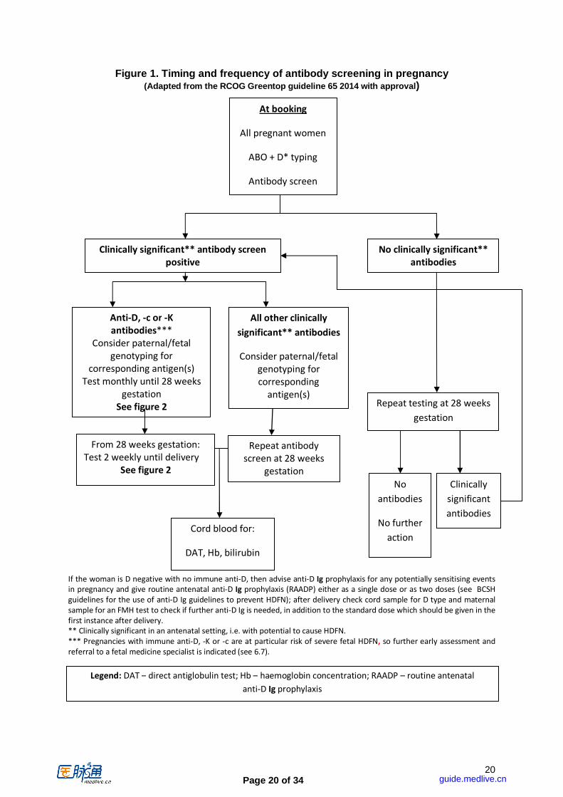
however advisable to test the fetal RHD status using a more sensitive and specific methodology at a later gestation for alloimmunised pregnant women as described above. Recommendation: Fetal RHD typing using a high-throughput methodolog y in pregnant women who have not formed anti-D, as part of a screening program t o target anti-D Ig prophylaxis, is sufficiently accurate for implementation from 11 we eks gestation. The cost effectiveness of such testing is dependent on provi sion at a point in the antenatal care pathway that does not necessitate an additiona l visit. (Grade 1C). SECTION 3. ANTENATAL TESTING PROTOCOLS (See Figures 1 & 2). 3.1 Routine antenatal testing All pregnant women should have samples taken early in pregnancy, ideally at booking, typically 8-12 weeks gestation, for ABO and D grouping and for screening for the presence of red cell alloantibodies. When an antibody screen is positive, further tests should be carried out to determine the antibody specificity and significance. [see section 2]. All pregnant women, whether D positive or D negative, should have a further blood sample taken at 28 weeks gestation for re-checking the ABO and D group and further screening for red cell alloantibodies [NICE clinical guidance 62 2008]. D positive women are just as likely as D negative women to form antibodies (other than anti-D) late in pregnancy [Koelewijn et al 2008, Thompson et al, 2003]. Local policies must ensure that D negative women who are eligible for routine antenatal anti-D Ig prophylaxis (RAADP) have the 28 week antibody screening sample taken before the first dose of RAADP anti-D Ig is administered. Samples taken after the injection could result in passive anti-D being detected, which may be mistaken for immune anti-D, and conversely, potentially dangerous immune anti-D being mistaken for passive anti-D Ig [New et al, 2001] see 4.3.1. Recommendation: Pregnant women who are D negative should be offered prophylactic anti-D immunoglobulin (anti-D Ig) for potentially sensitis ing events as defined in guidelines for the use of anti-D Ig for the prevention of HDFN [BC SH, 2014]. (Grade 1A) There is evidence that antibodies first detected after 28 weeks gestation are less likely to cause clinically significant HDFN [Rothenberg et al, 1999; Heddle et al, 1993]. Further, and significantly, the introduction of RAADP has resulted in the detection of anti-D Ig in samples taken after 28 weeks gestation from D negative women [Cambic et al 2010]. Since it is not possible to differentiate between prophylactic anti-D Ig and immune anti-D until the latter has reached a high enough concentration in reference quantification to exclude the former, there is the potential for confusion between the two [New et al, 2001]. (see section 4.1) Recommendation: All pregnant women should be ABO and D typed and sc reened for the presence of red cell antibodies early in pregnancy (at booking) and at 28 weeks gestation. In D negative women the 28-week sample should be taken b efore the administration of RAADP (Grade 1B, NICE guideline 62 2008) . Recommendation: In pregnant women with no clinically significant re d cell antibodies capable of causing HDFN present at 28 weeks gestation, no furt her routine antenatal blood grouping or antibody screening is necessary. (Grade 1B)
guide.medlive.cn
Page 9 of 34
9
SECTION 4. RED CELL ANTIBODIES DETECTED IN PREGNANCY. Anti-D, anti-c and anti-K are the antibodies most often implicated in causing haemolytic disease severe enough to warrant antenatal intervention [Koelewijn et al 2008]. Pregnancies in women with a previous history of confirmed significant HDFN should however be considered at risk, and referral to a fetal medicine specialist made regardless of the antibody specificity(ies) identified. In all other cases, follow-up testing protocols are dictated by the specificity and concentration of the antibodies identified. Regardless of antenatal follow-up regimes, antibody identification should always be performed to check for the appearance of additional specificities, prior to maternal or fetal transfusion. Recommendation: All pregnant women who have previously had a baby a ffected by HDFN should be referred before 20 weeks gestation to a fetal medic ine specialist for assessment and advice, irrespective of antibody concentration or s pecificity. (Grade 1B) 4.1 Pregnant women with detectable anti-D Prophylactic anti-D Ig has been very successful in reducing the number of sensitisations to D but it has created difficulties in determining whether anti-D detected in pregnancy is passive (due to anti-D Ig prophylaxis given as RAADP or for a potentially sensitising event) or immune. The risks associated with the misinterpretation of the nature of anti-D are clear: if passive anti-D Ig is misinterpreted as immune anti-D, then further anti-D Ig prophylaxis may be omitted leaving the women unprotected from sensitisation. If immune anti-D is misinterpreted as passive anti-D Ig, appropriate follow-up of the antibody concentration during pregnancy may be curtailed, and interventions that might be required to manage HDFN, not instigated. The SHOT annual report 2011 included a ‘learning point’ highlighting the difficulties in differentiating between immune anti-D and passive anti-D Ig, as there were seven cases reported where women with immune anti-D were not followed up as closely as they should have been because the anti-D detected was wrongly assumed to be passive anti-D Ig, and in six of these cases the babies were born with some degree of HDFN [SHOT 2012]. 4.1.1 Distinguishing between passive and immune anti-D (also see Figure 3) Passive anti-D Ig and immune anti-D cannot be qualitatively distinguished serologically. While the concentration of passive anti-D Ig will fall with time, the concentration of immune anti-D will usually remain stable, or rise if there is re-stimulation. The concentration of passive anti-D Ig in maternal samples post-prophylaxis rarely exceeds 0.4 IU/mL unless anti-D Ig totalling more than 1500 IU has been administered. The peak concentration of anti-D Ig detected after 1500 IU of intravenous anti-D Ig in a pharmacokinetic study in pregnant women was equivalent to 0.4 IU/mL, and following intramuscular anti-D Ig 0.2 IU/mL [Bichler et al 2003]. These values are approximate conversions based upon the concentrations stated in the publication. Following administration of an intramuscular injection of anti-D Ig, a serologically detectable concentration of anti-D Ig is potentially present within minutes and the peak blood concentration is reached within three to seven days. The half-life of passive anti-D Ig is approximately 3 weeks [Eklund et al, 1982]. Passive anti-D Ig can be detected by serological tests for several weeks: by an indirect antiglobulin test ( IAT) at 8 weeks or more following injection of 500 IU, and for more than 12 weeks where more sensitive techniques are used or following higher doses of anti-D Ig. Immune anti-D becomes detectable approximately 4 weeks after exposure to D positive cells, and reaches a peak concentration after 6 to 8 weeks, if there is no further exposure. [Mollison et al c, 1997]. Prediction of the nature of anti-D (immune anti-D or passive anti-D Ig) based on the strength of reaction with D positive cells is unreliable, as this will vary with the technique and Rh
guide.medlive.cn

Page 10 of 34
10
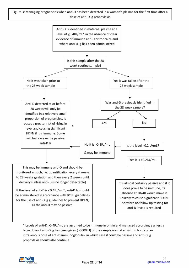
phenotype of the reagent red cells used. Babies have been severely affected by HDFN as a result of assumptions made on the basis of the strength of reactions using methods that have not been validated. [SHOT 2012]. Quantification by continuous flow analyser (CFA) gives an objective measurement of antibody concentration in IU/mL anti-D. All anti-D detected in pregnancy should be quantified by CFA with reference to the NIBSC anti-D standard [NIBSC 2010b], or tested by a method that has been extensively validated against CFA and that gives a result that is expressed in or can easily be converted to IU/mL anti-D. The only exception is where anti-D is detected for the first time immediately prior to or at the time of delivery, e.g. in a pre-delivery group and screen sample, in which case the sample need not be sent for quantification, but the baby should be monitored for signs of HDFN as treatment decisions will need to be made on the basis of the severity of any anaemia and/or jaundice. The results of quantification are unlikely to have a major influence on clinical decisions at this stage and are not available as quickly and measures of haemoglobin and bilirubin. Maternal anti-D quantification may be performed at a later stage if deemed necessary. The quantification results should be viewed in the context of the timing and dose of any anti-D Ig given previously, the reason for its administration, the route of administration and the antibody status at the time of administration. The clinical history and knowledge of the results of previous laboratory testing are paramount in clinical decision making where anti-D is detected in pregnancy, and every effort should therefore be made to obtain this information. Even if anti-D Ig has been administered for a potentially sensitising event prior to 28 weeks it must be assumed that anti-D detectable at or before 28 weeks may be immune as levels can rise dangerously between 28 weeks and term. Therefore monitoring should be undertaken as if the antibody may be immune (until anti-D is no longer detectable), while continuing to administer anti-D Ig as indicated in the BCSH guideline for the use of anti-D Ig to prevent HDFN (BCSH 2014).
4.1.2 Procedures to follow where anti-D is detected in pregnancy (figure 3) a. Determine whether anti-D Ig has been administered (not just issued) and its clinical
indication i.e., whether as RAADP or following a potential sensitising event, by asking the woman and seeking written confirmation in the notes.
b. Undertake quantification (unless immediately prior to delivery, in which case the baby should be monitored for signs of HDFN).
c. If any of the following apply, then the antibody should be monitored as for immunised
women, i.e. at 4 weekly intervals to 28 weeks gestation, and at 2 weekly intervals after 28 weeks, until delivery (or until serial monitoring by middle cerebral artery (MCA) Doppler has been instituted and serial testing deemed unnecessary, or anti-D is no longer detectable):
• Anti-D was detected at or before the 28 week gestation RAADP administration (even if anti-D Ig was administered earlier in pregnancy as the risk of falsely labelling immune anti-D as passive anti-D Ig when detected at or prior to 28 weeks gestation is significant)
• There is no definite record of prior anti-D Ig administration. • Anti-D was present before the first administration of anti-D Ig at any gestation. • The level of anti-D is greater than or equal to 0.2 IU/mL.
guide.medlive.cn
Page 11 of 34
11
d. If the anti-D level is < / = 0.4 IU/mL after up to 1500 IU of anti-D Ig have been administered, then prophylactic anti-D Ig should continue to be offered for routine prophylaxis and potentially sensitising events in accordance with the BCSH guideline for the use of anti-D Ig to prevent HDFN (BCSH 2014, Qureshi et al) unless it is established beyond doubt that the anti-D is immune, in which case anti-D Ig prophylaxis is no longer indicated.
e. If doses in excess of 1500 IU anti-D Ig have been administered then a level of >0.4 IU/mL may be achieved as a result of passive anti-D Ig. Therefore prophylactic anti-D Ig should continue to be offered for routine prophylaxis and potentially sensitising events in accordance with BCSH guideline for the use of anti-D Ig to prevent HDFN (BCSH 2014, Qureshi et al) unless it is established beyond doubt that the anti-D is immune, in which case anti-D Ig prophylaxis is no longer indicated.
f. After 28 weeks, where all of the following apply, testing should continue as for non-
sensitised women, i.e. there is no requirement for further antibody testing after 28 weeks gestation, and anti-D Ig prophylaxis should continue to be offered in accordance with the BCSH guideline for the use of anti-D Ig to prevent HDFN (BCSH 2014).
• Anti-D was not detectable in a sample taken immediately prior to RAADP administration (the timing of RAADP should be at 28 weeks gestation but will sometimes be slightly earlier or later).
• There is a written record of administration of anti-D Ig in the preceding 8 weeks • The concentration of anti-D is <0.2 IU/mL
Women with anti-D should not be issued with an antibody card documenting the finding of anti-D until it is conclusively established that the anti-D is immune. Recommendation: Blood transfusion laboratories should keep a record of anti-D Ig administration to provide a basis for distinguishing between immune a nti-D and passive anti-D Ig. (Grade 1C). Recommendation: If anti-D is detected in an antenatal maternal samp le (except for that taken immediately prior to delivery), testing should incl ude a measurement of antibody concentration by CFA, or by a technique that has be en validated using large numbers of samples of known concentration, and that gives a result that is expressed in or can easily be converted to IU/mL of anti-D. (Grade 1C) Recommendation: When there is doubt as to the passive or immune nat ure of anti-D, the level should be monitored as if it could be immune. In this situati on anti-D Ig prophylaxis should continue to be offered, where indicated, until the nature of the anti-D is established (Grade 2C). 4.1.3 Pregnant women with immune anti-D Anti-D is the most frequent cause of serious HDFN. Blood samples from pregnant women with immune anti-D should be tested at least monthly until 28 weeks gestation and every 2 weeks thereafter until delivery, to monitor the concentration of anti-D (with reference to the NIBSC anti-D standard [NIBSC 2010b]) and to identify any additional antibodies that may develop. Where anti-D has been shown to be immune (see section 4.1.2), an increase in concentration of 50% or greater, compared with the previous concentration, suggests a
guide.medlive.cn
Page 12 of 34
12
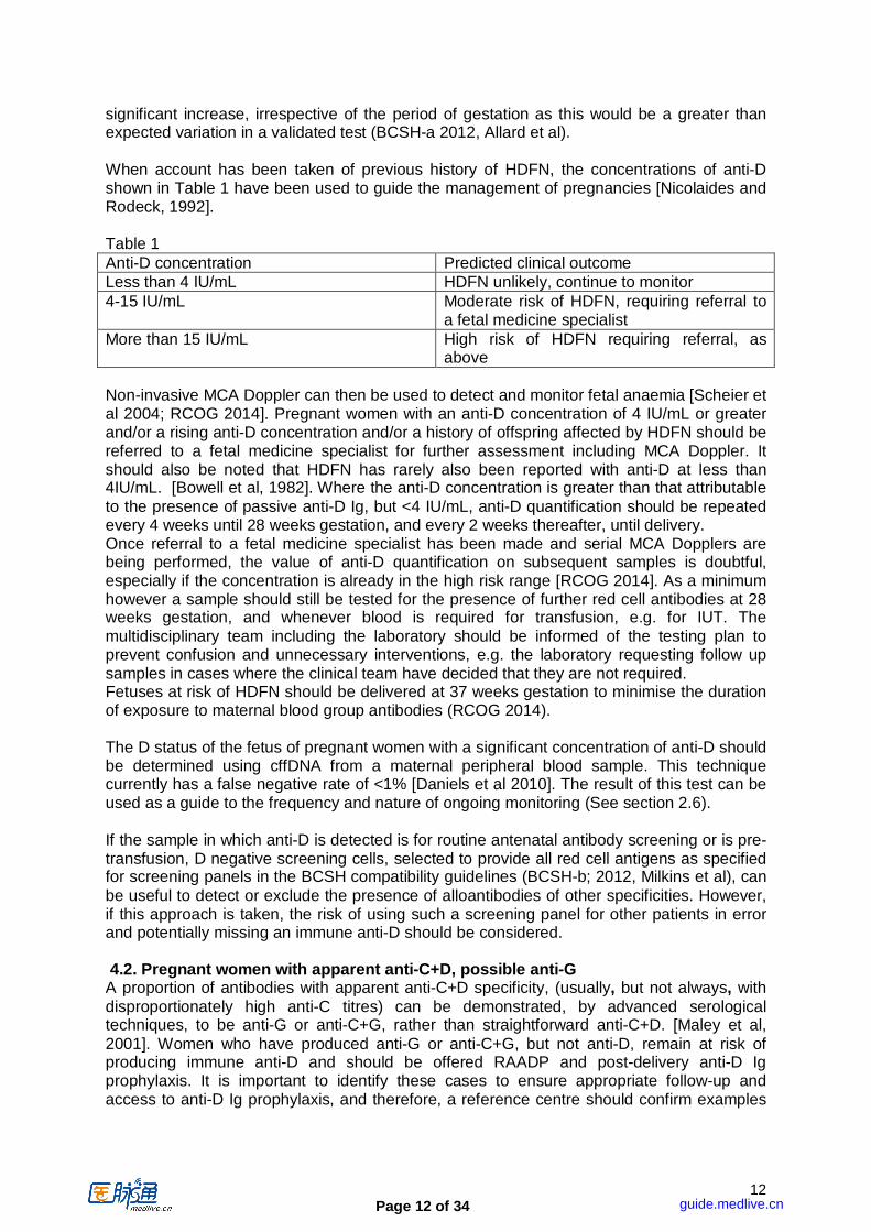
significant increase, irrespective of the period of gestation as this would be a greater than expected variation in a validated test (BCSH-a 2012, Allard et al). When account has been taken of previous history of HDFN, the concentrations of anti-D shown in Table 1 have been used to guide the management of pregnancies [Nicolaides and Rodeck, 1992]. Table 1 Anti-D concentration Predicted clinical outcome Less than 4 IU/mL HDFN unlikely, continue to monitor 4-15 IU/mL Moderate risk of HDFN, requiring referral to
a fetal medicine specialist More than 15 IU/mL High risk of HDFN requiring referral, as
above Non-invasive MCA Doppler can then be used to detect and monitor fetal anaemia [Scheier et al 2004; RCOG 2014]. Pregnant women with an anti-D concentration of 4 IU/mL or greater and/or a rising anti-D concentration and/or a history of offspring affected by HDFN should be referred to a fetal medicine specialist for further assessment including MCA Doppler. It should also be noted that HDFN has rarely also been reported with anti-D at less than 4IU/mL. [Bowell et al, 1982]. Where the anti-D concentration is greater than that attributable to the presence of passive anti-D Ig, but <4 IU/mL, anti-D quantification should be repeated every 4 weeks until 28 weeks gestation, and every 2 weeks thereafter, until delivery. Once referral to a fetal medicine specialist has been made and serial MCA Dopplers are being performed, the value of anti-D quantification on subsequent samples is doubtful, especially if the concentration is already in the high risk range [RCOG 2014]. As a minimum however a sample should still be tested for the presence of further red cell antibodies at 28 weeks gestation, and whenever blood is required for transfusion, e.g. for IUT. The multidisciplinary team including the laboratory should be informed of the testing plan to prevent confusion and unnecessary interventions, e.g. the laboratory requesting follow up samples in cases where the clinical team have decided that they are not required. Fetuses at risk of HDFN should be delivered at 37 weeks gestation to minimise the duration of exposure to maternal blood group antibodies (RCOG 2014). The D status of the fetus of pregnant women with a significant concentration of anti-D should be determined using cffDNA from a maternal peripheral blood sample. This technique currently has a false negative rate of <1% [Daniels et al 2010]. The result of this test can be used as a guide to the frequency and nature of ongoing monitoring (See section 2.6). If the sample in which anti-D is detected is for routine antenatal antibody screening or is pre-transfusion, D negative screening cells, selected to provide all red cell antigens as specified for screening panels in the BCSH compatibility guidelines (BCSH-b; 2012, Milkins et al), can be useful to detect or exclude the presence of alloantibodies of other specificities. However, if this approach is taken, the risk of using such a screening panel for other patients in error and potentially missing an immune anti-D should be considered. 4.2. Pregnant women with apparent anti-C+D, possible anti-G A proportion of antibodies with apparent anti-C+D specificity, (usually, but not always, with disproportionately high anti-C titres) can be demonstrated, by advanced serological techniques, to be anti-G or anti-C+G, rather than straightforward anti-C+D. [Maley et al, 2001]. Women who have produced anti-G or anti-C+G, but not anti-D, remain at risk of producing immune anti-D and should be offered RAADP and post-delivery anti-D Ig prophylaxis. It is important to identify these cases to ensure appropriate follow-up and access to anti-D Ig prophylaxis, and therefore, a reference centre should confirm examples
guide.medlive.cn
Page 13 of 34
13
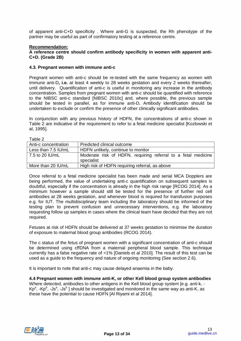
of apparent anti-C+D specificity . Where anti-G is suspected, the Rh phenotype of the partner may be useful as part of confirmatory testing at a reference centre. Recommendation: A reference centre should confirm antibody specific ity in women with apparent anti-C+D. (Grade 2B) 4.3. Pregnant women with immune anti-c Pregnant women with anti-c should be re-tested with the same frequency as women with immune anti-D, i.e. at least 4 weekly to 28 weeks gestation and every 2 weeks thereafter, until delivery. Quantification of anti-c is useful in monitoring any increase in the antibody concentration. Samples from pregnant women with anti-c should be quantified with reference to the NIBSC anti-c standard [NIBSC 2010c] and, where possible, the previous sample should be tested in parallel, as for immune anti-D. Antibody identification should be undertaken to exclude or confirm the presence of other clinically significant antibodies. In conjunction with any previous history of HDFN, the concentrations of anti-c shown in Table 2 are indicative of the requirement to refer to a fetal medicine specialist [Kozlowski et al, 1995]. Table 2 Anti-c concentration Predicted clinical outcome Less than 7.5 IU/mL HDFN unlikely, continue to monitor 7.5 to 20 IU/mL Moderate risk of HDFN, requiring referral to a fetal medicine
specialist More than 20 IU/mL High risk of HDFN requiring referral, as above Once referral to a fetal medicine specialist has been made and serial MCA Dopplers are being performed, the value of undertaking anti-c quantification on subsequent samples is doubtful, especially if the concentration is already in the high risk range [RCOG 2014]. As a minimum however a sample should still be tested for the presence of further red cell antibodies at 28 weeks gestation, and whenever blood is required for transfusion purposes e.g. for IUT. The multidisciplinary team including the laboratory should be informed of the testing plan to prevent confusion and unnecessary interventions, e.g. the laboratory requesting follow up samples in cases where the clinical team have decided that they are not required. Fetuses at risk of HDFN should be delivered at 37 weeks gestation to minimise the duration of exposure to maternal blood group antibodies (RCOG 2014). The c status of the fetus of pregnant women with a significant concentration of anti-c should be determined using cffDNA from a maternal peripheral blood sample. This technique currently has a false negative rate of <1% [Daniels et al 2010]. The result of this test can be used as a guide to the frequency and nature of ongoing monitoring (See section 2.6). It is important to note that anti-c may cause delayed anaemia in the baby. 4.4 Pregnant women with immune anti-K, or other Kell blood group system antibodies Where detected, antibodies to other antigens in the Kell blood group system [e.g. anti-k, -Kpa, -Kpb, -Jsa, -Jsb ] should be investigated and monitored in the same way as anti-K, as these have the potential to cause HDFN [Al Riyami et al 2014].
guide.medlive.cn
Page 14 of 34
14
HDFN due to anti-K is characterised by low haemoglobin concentration, but elevated amniotic and/or cord bilirubin levels are not generally reported. The fetal anaemia associated with anti-K may be due to the inhibition of K positive erythroid early progenitor cells [Vaughan et al, 1998] and/or to promotion of their immune destruction [Daniels et al b, 2003]. It has been stated in some texts that the severity of HDFN due to anti-K is not correlated with titre of the antibody and publications prior to 1995 cited occasions where severe HDFN occurred despite low titres of anti-K. More recent case series of affected pregnancies have however suggested that severe HDFN is associated with antibodies with a titre of at least 32 [McKenna et al 1999; Ahaded et al 2000]. Therefore, samples from women with anti-K should be titrated (see 2.4.2) when first identified in the pregnancy, as for any clinically significant antibody, and serial titration should then be undertaken every 4 weeks until 28 weeks gestation, and 2 weekly thereafter, until delivery. Once referral to a fetal medicine specialist has been made and serial MCA Dopplers are being performed, the value of undertaking anti-K titration on subsequent samples is doubtful, especially if the titres are already in the high risk range [RCOG 2014]. The multidisciplinary team including the laboratory should be informed of the testing plan to prevent confusion and unnecessary interventions, e.g. the laboratory requesting follow up samples in cases where the clinical team have decided that they are not required. The majority of cases of anti-K in pregnant women are the consequence of previous K positive transfusions. The incidence of anti-K can be reduced by selecting K negative units for transfusion to females with potential for childbearing, unless known to be K positive themselves [Lee & de Silva, 2004]. Therefore K negative units should be selected for females under the age of 50 years who are themselves K negative, or whose K type is unknown. However, emergency transfusions should not be delayed if suitable K negative units are not immediately available [BCSH 2012-b, Milkins et al]. Partner testing, discussed in section 2.5, may be particularly relevant for women with anti-K as approximately 80% of anti-K detected in pregnant women results from previous transfusion and only 9% of the general population are K positive (although a higher proportion of partners of women with anti-K are K positive) [Lee & de Silva, 2004, Daniels 2013]. It is important that the transfusion history of women with anti-K should be established and a sample from the father of the fetus should be K typed for counselling purposes. If the father’s K type is unknown or he is K positive the pregnant woman should be referred to a fetal medicine specialist when anti-K is first identified for counselling and monitoring. If the father is K negative and a confidential enquiry establishes paternity, no further samples are required until 28 weeks gestation when further antibodies should be excluded (as for all women and any antibodies detected at 28 weeks). Any clinically significant antibodies identified, in addition to anti-K, should be monitored according to their specificity. The K (KEL*01) status of the fetus of pregnant women with anti-K should be determined using cffDNA from a maternal peripheral blood sample if the father is heterozygous for the K antigen or if his antigen status is unknown. This technique currently has a false negative rate of <1% [Daniels et al 2010]. The result of this test can be used as a guide to the frequency and nature of ongoing monitoring (See section 2.6). Recommendation: Samples from pregnant women with immune anti-D or a nti-c should be assessed serologically at 4 weekly intervals to 28 weeks ges tation and at fortnightly intervals thereafter, until delivery. Such cases should be re ferred to a fetal medicine specialist if the antibody reaches the critical level and/or t he level is rising significantly, where assessment of the need for further monitoring will be made (Grade 1B). Recommendation:
guide.medlive.cn
Page 15 of 34
15
Pregnant women with anti-K or other Kell system ant ibodies [unless the father is confirmed to be negative for the corresponding anti gen] should be assessed serologically at monthly intervals to 28 weeks gest ation and at fortnightly intervals thereafter, until delivery. All cases should be ref erred to a fetal medicine specialist when the antibody is first identified. (Grade 1B) Recommendation: In all cases where serial Doppler assessment of fet al middle cerebral artery flow rates is being undertaken and antibody levels are in the high risk range, ongoing assessment of antibody strength is unlikely to be o f value and can be discontinued at the discretion of a fetal medicine specialist (RCOG Green-top 2014, Grade 1C). 4.5. Pregnant women with other red cell antibodies It is only IgG antibodies that are capable of entering the fetal circulation, and red cell antibodies with a significant IgG component are detectable by IAT. ‘Cold reactive’, IgM and low affinity antibodies to high prevalence antigens [e.g. CR1-related antibodies] have not been implicated in HDFN. In addition to anti-D, -c and –K, the following specificities are most commonly associated with HDFN: anti-C, -e, -E, -Fya, and -Jka [Moise 2000; Moran 2000; Goodrick et al 1997; Koelewijn 2008]. Many other specificities have however been reported as the cause of HDFN. Less common antibody specificities may be identified, some of which may be more prevalent in women of a specific ethnic origin, such as anti-Ge3 in the Hispanic population [Pate 2013], and anti-M in the Japanese population [Yasuda 2013]; the anti-M having caused a form of delayed HDFN, similar to that caused by anti-K, including suppression of erythropoiesis. In most cases, re-testing at 28 weeks gestation generally provides sufficient information to determine management of the pregnancy.
A clinical decision should be made regarding the more frequent testing of women with a previous history of children with HDFN. The nature of past history may help define the intensity of monitoring. If there has been a previous baby which suffered hydrops, significant anaemia or jaundice then monitoring to prevent this at an intensity similar to that for anti-c, -D or -K would be warranted with regular Doppler USS and consideration of delivery at 37 weeks gestation. Similar to anti-c, -D and -K if the titres are above the trigger for MCA Doppler monitoring the value of serial measurement of antibody titre in addition is unclear.
In the absence of a history of HDFN, the likelihood of developing hydrops with antibodies of other specificities is lower than that of anti-D, -c and –K. For example in a case series of pregnant women with anti-E and titres >32, 5 (15%) had Hb <100g/L at delivery, 1 (3%) had hydrops fetalis and there was 1 (3%) perinatal death attributable to the antibodies [Joy et al 2005]. The likelihood appears to be even lower for other specificities such as anti-Fya, -Fyb, -Fy3, -S, -s or -U, though some case series are selective and do not provide a denominator of a population of pregnant women with antibodies of the defined specificity [Goodrick et al 1997, Smith et all 1998, Nordvall et al 2009, Pal and Williams 2015].
It is likely that in the absence of a previous history of HDFN pregnant women with antibodies with titres >32 and specificities other than anti-D, -c and -K would not need to be monitored as frequently as those with a history of severe HDFN and / or the high risk antibody specificities anti-c, -D or -K. Rare cases of significant anaemia would be likely to be detected by MCA Doppler monitoring repeated at 28, 32 and 36 weeks gestation. The decision on the frequency of monitoring and the need for early delivery therefore will primarily be based on the history of previous pregnancies for the affected family and the reports and case series available in the world literature. A balance has to be made between intensive monitoring to the level advised for anti-c, -D and -K with a low yield in terms of intervention being required and a reduced level of monitoring with an attendant risk of missing progressive anaemia and hence delaying intervention.
guide.medlive.cn
Page 16 of 34
16
The numbers of reported cases and well defined case series for antibodies against antigens other than anti-c, -D and -K are low. Research defining the risks and benefits of taking a less intensive approach to monitoring pregnant women with such antibodies would be welcomed. For information about other antibodies and their ability to cause clinically significant HDFN, it is recommended that further advice is sought from a transfusion medicine specialist in a red cell reference department. Reference tables for antibodies that have been associated with HDFN are available in the SNBTS guidelines, RCOG guidelines and NHSBT guidelines (SNBTS 2013, RCOG 2014, NHSBT 2015)
Where an IgG antibody has been detected, testing of both first appointment and 28 week gestation samples should include titration (see section 2.4.2). In general, there is a risk that an antibody with a titre of 32 or greater will cause some degree of HDFN, although a clear-cut association between titre and HDFN has not been established. The presence of additional antibodies should be established and any clinically significant antibodies should be titrated or quantified depending on the specificity (see section 2.4). Recommendation: Clinically significant antibodies, other than anti- D, -c or -K, should be excluded or, if present, assessed by titration, at the booking appo intment and at 28 weeks gestation. If deemed necessary based on a high titre (>32) and / or a past history of HDFN, referral to a specialist in fetal medicine should b e made for further assessment. (Grade 1C). SECTION 5. REPORTS OF LABORATORY INVESTIGATIONS In addition to blood group and specificity of any red cell alloantibodies present, reports must inform the clinician[s] responsible for the pregnant woman’s antenatal care of the likely significance of the antibodies, with respect to both the development of HDFN and potential difficulties in providing compatible blood for transfusion [NICE (2008) CG62]. Reports should also, where relevant, alert the clinician to the need to refer the woman to a fetal medicine specialist. The woman should be given verbal and written information about the clinical significance of the antibody(ies) and it should be documented that she has been counselled in the patient record. Details of the timing of further samples required should also be provided. Recommendation: In addition to blood group and specificity of any r ed cell alloantibodies present, reports must inform the clinician[s] responsible fo r the pregnant woman’s antenatal care of the likely significance of the antibodies, with respect to both the development of HDFN and transfusion problems [NICE (2008) CG62. Grade 1B] Recommendation: Pregnant women with clinically significant red cell antibodies should be given verbal and written information with details of the antibod y specificity and its potential for causing HDFN and delay in providing compatible bloo d. (GPP, Grade 1D) SECTION 6. ACTION AT TIME OF BIRTH 6.1 D typing of cord samples from D negative women with no immune anti-D A maternal sample and a cord blood sample should be obtained. The cord blood sample should be used to determine the baby’s D group if it is not already known, thus identifying pregnant women who must receive post-delivery prophylactic anti-D Ig.
guide.medlive.cn
Page 17 of 34
17
There is minimal evidence that fetal red cells expressing the DVI antigen can cause maternal sensitisation. Therefore, the use of different reagents (detecting DVI as D positive) for typing cord samples is not recommended, as the risks of using the wrong reagent for routine testing, outweighs the risk of missing a DVI cord sample. Most examples of weak D antigen can be easily detected by selecting high affinity anti-D reagents. [BCSH, 2012]
6.2 Direct antiglobulin test [DAT] on cord samples 6.2.1 Routine DAT on the cord samples of D positive babies born to D negative women with no red cell alloantibodies This is not recommended as a routine investigation. It has been shown that following RAADP, anti-D Ig can cross the placenta, enter the fetal circulation and bind to fetal D antigen sites. Consequently 3-6% of D positive cord samples have been found to have a positive DAT [Dalton et al, 2003; Parker et al, 2003; Dillon et al, 2011) and this may result in unnecessary additional investigations. There is evidence that prophylactic anti-D Ig does not cause significant haemolysis of fetal/neonatal red cells [Maayan-Metzger et al, 2001]. 6.2.2. Testing on the cords and babies of women who have IAT reactive red cell antibodies When the maternal serum has been found to contain an immune, IAT reactive red cell antibody(ies), the red cells from the cord should be tested for the corresponding antigen[s], wherever possible. A DAT should be performed on the cord sample, and the haemoglobin concentration and bilirubin levels should be checked (RCOG 2014). In addition, the baby should be observed for clinical signs of jaundice [NICE guidance on neonatal jaundice, CG98]. A positive DAT is not, in itself, diagnostic of HDFN. Where the DAT is positive and the baby shows signs of HDFN, a red cell eluate may be helpful to confirm the red cell antibody specificity. IgG ABO antibodies occasionally cause severe HDFN, and so, if the baby has a major ABO mismatch with the woman, the eluate should also be tested with A1 and/or B cells, negative for any other antigen against which the woman has made IgG alloantibodies. Regular assessment of bilirubin and haemoglobin concentrations is necessary and hence early discharge is not advisable [RCOG 2014]. Babies who have been transfused in utero with D negative units for the prevention of HDFN due to anti-D may type as D negative for several months after birth. [BCSH, 2014, Qureshi et al]. In this case, the baby’s haemoglobin and bilirubin concentrations should still be checked to diagnose and/or exclude further evidence of clinically significant red cell destruction when D positive cells are released into the circulation. Recommendation: All babies born to women who have clinically signif icant antibodies should be closely observed for evidence of HDFN. A DAT should be perf ormed on a cord blood sample and haemoglobin and bilirubin concentrations should be measured. (Grade 1C). 6.3. Pre- and Post-delivery testing of maternal samples For women with no red cell antibodies at 28 weeks gestation, routine blood grouping and antibody screening of maternal samples, other than confirmatory D typing, is not necessary unless pre-transfusion compatibility testing is required. Detection of new antibodies immediately pre-delivery in previously non-sensitised pregnancies will not influence management of the current pregnancy, although if testing is undertaken and antibodies detected the woman should be informed as this there may be implications for future pregnancies. In alloimmunised pregnancies a pre or post-delivery maternal sample is required to predict neonatal transfusion requirements (as additional red cell antibodies
guide.medlive.cn

Page 18 of 34
18
potentially formed since the last screen will need to be matched for) and for crossmatching should neonatal transfusion be necessary. A test for fetomaternal haemorrhage should be performed on the maternal blood sample from D negative women with no immune anti-D, to detect fetal cells in the maternal circulation, and, if necessary, to measure the volume of FMH, in order to determine any requirement for additional anti-D Ig. [BCSH 2009-b, Austin et al]. Recommendation: D negative women with no immune anti-D should have an initial FMH test at delivery and follow up testing if required, in accordance wi th BCSH guidelines for estimation of FMH . [BCSH 2009-b; GRADE 1B). 7. AUDIT Audits of clinical and laboratory practice should be undertaken on a continuing basis to ensure compliance with these guidelines and, where identified, variance or concerns in relation to compliance, should be addressed [Department of Health, DH- a 1997; DH-b, 1998]. Examples of audits relevant to these guidelines (See appendix 1, audit tool):
• Sample labelling • Laboratory audit of testing and procedures vs. BCSH guidelines and performance in
External Quality Assessment Schemes. • Appropriate utilisation of non-invasive fetal typing. • Compliance with the care pathway for blood grouping, antibody screening and
administration of anti-D Ig. • Ongoing monitoring and referral for specialist advice. • That all women of childbearing potential receive K antigen-negative blood. (this could
be an audit of the reason for alloimmunisation in women identified to have anti-K, or a laboratory practice audit),
A detailed gap analysis for choosing audit topics is included as appendix 1. Acknowledgements and declarations of interest HQ reviewed the literature and wrote the initial draft of the manuscript. JW and EM led on subsequent redrafting of the manuscript prior to and following peer review GD, MN reviewed the literature and manuscript. GB drafted the audit tool SA represented the BCSH transfusion task force, reviewed the literature and revised the manuscript. The BCSH task force membership at the time of writing this guidance was Allard S (chair), Bolton–Maggs P (SHOT), Cho G, Jones J, Massey E, Zahra S, Needs M, Robinson S, White J. The BCSH paid the expenses incurred during the writing of this guidance. See: http://www.bcshguidelines.com/BCSH_PROCESS/DOCUMENTS_FOR_TASK_FORCES_AND_WRITING_
GROUPS/203_Expense_forms_and_policy.html All authors have made a declaration of interests to the BCSH and Task Force Chairs which may be viewed on request. The members of the writing group have no conflicts of interest to declare.
guide.medlive.cn
Page 19 of 34
19
Grading of Recommendations Assessment, Development and Evaluation (GRADE)
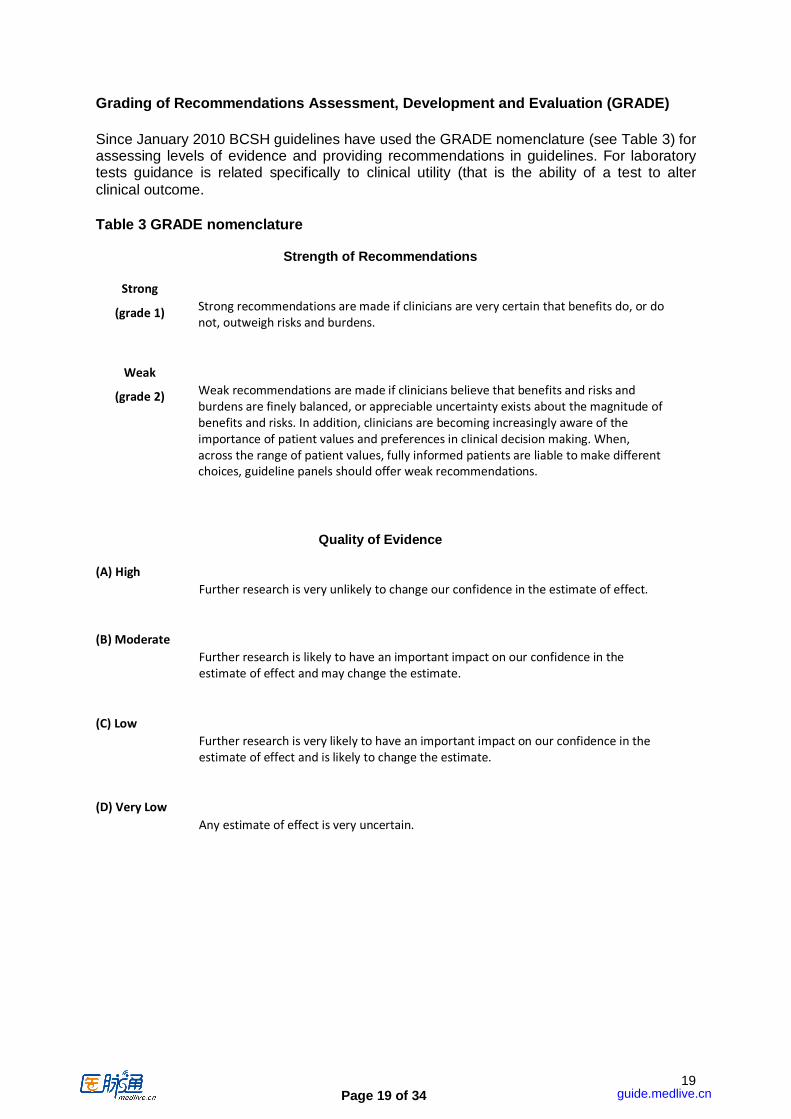
Since January 2010 BCSH guidelines have used the GRADE nomenclature (see Table 3) for assessing levels of evidence and providing recommendations in guidelines. For laboratory tests guidance is related specifically to clinical utility (that is the ability of a test to alter clinical outcome.
Table 3 GRADE nomenclature
Strength of Recommendations
Strong
(grade 1)
Strong recommendations are made if clinicians are very certain that benefits do, or do
not, outweigh risks and burdens.
Weak
(grade 2)
Weak recommendations are made if clinicians believe that benefits and risks and
burdens are finely balanced, or appreciable uncertainty exists about the magnitude of
benefits and risks. In addition, clinicians are becoming increasingly aware of the
importance of patient values and preferences in clinical decision making. When,
across the range of patient values, fully informed patients are liable to make different
choices, guideline panels should offer weak recommendations.
Quality of Evidence
(A) High
Further research is very unlikely to change our confidence in the estimate of effect.
(B) Moderate
Further research is likely to have an important impact on our confidence in the
estimate of effect and may change the estimate.
(C) Low
Further research is very likely to have an important impact on our confidence in the
estimate of effect and is likely to change the estimate.
(D) Very Low
Any estimate of effect is very uncertain.
guide.medlive.cn
Page 20 of 34
20
All other clinically
significant** antibodies
Consider paternal/fetal
genotyping for
corresponding
antigen(s)
Clinically significant** antibody screen
positive
Anti-D, -c or -K
antibodies***
Consider paternal/fetal
genotyping for
corresponding antigen(s)
Test monthly until 28 weeks
gestation
See figure 2
At booking
All pregnant women
ABO + D* typing
Antibody screen
Cord blood for:
DAT, Hb, bilirubin
Repeat testing at 28 weeks
gestation
From 28 weeks gestation:
Test 2 weekly until delivery
See figure 2
Repeat antibody
screen at 28 weeks
gestation
No
antibodies
No further
action
Clinically
significant
antibodies
No clinically significant**
antibodies
Figure 1. Timing and frequency of antibody screening in pregnancy
(Adapted from the RCOG Greentop guideline 65 2014 with approval)
If the woman is D negative with no immune anti-D, then advise anti-D Ig prophylaxis for any potentially sensitising events
in pregnancy and give routine antenatal anti-D Ig prophylaxis (RAADP) either as a single dose or as two doses (see BCSH
guidelines for the use of anti-D Ig guidelines to prevent HDFN); after delivery check cord sample for D type and maternal
sample for an FMH test to check if further anti-D Ig is needed, in addition to the standard dose which should be given in the
first instance after delivery. ** Clinically significant in an antenatal setting, i.e. with potential to cause HDFN.
*** Pregnancies with immune anti-D, -K or -c are at particular risk of severe fetal HDFN, so further early assessment and
referral to a fetal medicine specialist is indicated (see 6.7).
Legend: DAT – direct antiglobulin test; Hb – haemoglobin concentration; RAADP – routine antenatal
anti-D Ig prophylaxis
guide.medlive.cn
Page 21 of 34
21
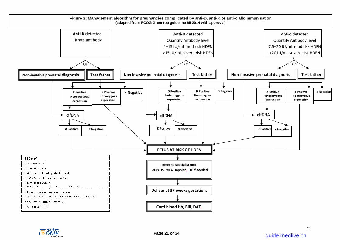
Or Or Or
Test father
K Positive
Heterozygous expression
K Positive
Homozygous
expression
K Negative
cffDNA
K Positive K Negative
Refer to specialist unit
Fetus US, MCA Doppler, IUT if needed
Deliver at 37 weeks gestation.
Cord blood Hb, Bili, DAT.
FETUS AT RISK OF HDFN
Anti-c detected
Quantify Antibody level
7.5–20 IU/mL mod risk HDFN
>20 IU/mL severe risk HDFN
Anti-D detected
Quantify Antibody level
4–15 IU/mL mod risk HDFN
>15 IU/mL severe risk HDFN
Anti-K detected
Titrate antibody
Non-invasive pre-natal diagnosis Test father
D Positive
Heterozygous
expression
D Positive
Homozygous
expression
D Negative
cffDNA
D Positive D Negative
Non-invasive pre-natal diagnosis Test father
c Positive
Heterozygous
expression
c Positive
Homozygous
expression
c-Negative
cffDNA
c Positive c Negative
Non-invasive prenatal diagnosis
Figure 2: Management algorithm for pregnancies complicated by anti-D, anti-K or anti-c alloimmunisation (adapted from RCOG Greentop guideline 65 2014 with approval)
guide.medlive.cn
Page 22 of 34
22
* Levels of anti-D >0.4IU/mL are assumed to be immune in origin and managed accordingly unless a
large dose of anti-D Ig has been given (>3000IU) or the sample was taken within hours of an
intravenous dose of anti-D immunoglobulin, in which case it could be passive and anti-D Ig
prophylaxis should also continue.
Yes
No it is >0.2IU/mL
& may be immune
Anti-D is identified in maternal plasma at a
level of <0.4IU/mL* in the absence of clear
evidence of immune anti-D historically, and
where anti-D Ig has been administered
Is this sample after the 28
week routine sample?
Yes it was taken after the
28 week sample
No it was taken prior to
the 28 week sample
Was anti-D previously identified in
the 28 week sample?
No
Is the level <0.2IU/mL?
Anti-D detected at or before
28 weeks will only be
identified in a relatively small
proportion of pregnancies. It
poses a greater risk of rising in
level and causing significant
HDFN if it is immune. Some
will be however be passive
anti-D Ig
Yes it is <0.2IU/mL
It is almost certainly passive and if it
does prove to be immune, its
absence at 28/40 would make it
unlikely to cause significant HDFN.
Therefore no follow up testing for
anti-D levels is required
This may be immune anti-D and should be
monitored as such, i.e. quantification every 4 weeks
to 28 weeks gestation and then every 2 weeks until
delivery (unless anti- D is no longer detectable)
If the level of anti-D is <0.4IU/mL*, anti-D Ig should
be administered in accordance with BCSH guidelines
for the use of anti-D Ig guidelines to prevent HDFN,
as the anti-D may be passive.
Figure 3: Managing pregnancies when anti-D has been detected in a woman’s plasma for the first time after a
dose of anti-D Ig prophylaxis
guide.medlive.cn

Page 23 of 34
23
References: Ahaded A, Brossard Y, Debbia M, Lambin P. (2000) Quantitative determination of anti-K [KEL1] IgG and IgG subclasses in the serum of severely alloimmunized pregnant women by ELISA. Transfusion; 40: 1239-1245. Al Riyami AZ, Al Salmani M, Al Hashami S et al. Successful management of severe hemolytic disease of the fetus due to anti-Jsb using intrauterine transfusions with serial maternal blood donations: a case report and a review of the literature. Transfusion 2014; 54: 238-243. Bichler J, Schondorfer G, Pabst G, Andresen I. (2003) Pharmacokinetics of anti-D IgG in pregnant RhD-negative women. British Journal of Obstetrics and Gynaecology; 110: 39-45. Bowell P, Wainscoat JS, Peto TEA, Gunson HH (1982) Maternal anti-D concentrations and outcome in rhesus haemolytic disease of the newborn. British Medical Journal; 285: 327-329. British Committee for Standards in Haematology (BCSH 2007): Gooch A, Parker J, Wray J, Qureshi H. Guidelines for blood grouping and antibody testing in pregnancy. Transfusion Medicine; 17: 252-262. British Committee for Standards in Haematology (BCSH 2009-a): Harris AM, Atterbury CLJ, Chaffe B, Elliott C, Hawkins T, Hennem SJ, Howell C, Jones J, Murray S, New HV, Norfolk D, Pirie L, Russell J, Taylor C. Guideline on the Administration of Blood Components, http://www.bcshguidelines.com (accessed 5/4/15) British Committee for Standards in Haematology (BCSH 2009-b): E Austin, S Bates, M de Silva, D Howarth, A Lubenko, M Rowley, M Scott, E Thomas, J White, M Williams. Guidelines for the Estimation of Fetomaternal Haemorrhage. http://www.bcshguidelines.com/documents/BCSH_FMH_bcsh_sept2009.pdf
British Committee for Standards in Haematology (BCSH-a, 2012): S. Allard, G. Burgess, B. Cuthbertson, C. Elliott, R. Haggas, J. Jones, B. Robertson, D. Sadani and K. Smith. Guidelines for Validation and Qualification, including Change Control, for Hospital Transfusion Laboratories. Transfusion Medicine; 22(1): 5-43.
British Committee for Standards in Haematology (BCSH-b 2012): C. Milkins, J. Berryman, C. Cantwell, C. Elliott, R. Haggas, J. Jones, M. Rowley, M. Williams and N. Win. Guidelines for pre-transfusion compatibility procedures in blood transfusion laboratories, Transfusion Medicine; 23: 3-35.
British Committee for Standards in Haematology (BCSH, 2014): H. Qureshi, E. Massey, D. Kirwan, T. Davies, S. Robson, J. White, J. Jones & S. Allard; Guideline for the use of anti-D immunoglobulin for the prevention of haemolytic disease of the fetus and newborn. Transfusion Medicine 24(1): 8-20. Bruce DG, Tinegate HN, Williams M, Babb R, Wells AW. (2013) Antenatal monitoring of anti-D and anti-c: could titre scores determined by column agglutination technology replace continuous flow analyser quantification? Transfusion Medicine; 23(1): 36–41. Cambic CR, Scavone BM, McCarthy RJ, Eisenberg P, Sanchez EM, Sullivan JT, Wong CA. (2010) A retrospective study of positive antibody screens at delivery in Rh-negative parturients. Can J Anaesth; 57(9): 811-6. Epub 2010 Jul 27.
guide.medlive.cn
Page 24 of 34
24
Chilcott J, Lloyd-Jones M, Wight J, Forman K, Wray J, Beverley C, Tappenden P. (2003) A review of the clinical effectiveness and cost-effectiveness of routine anti-D prophylaxis for pregnant women who are rhesus-negative. Health Technology Assessment: 7(4):iii-62.
Chitty LS, Finning, K, Wade, A, Soothill P, Martin B, Oxenford K, Daniels G, Massey E. (2014) Diagnostic accuracy of routine antenatal determination of fetal RHD status across gestation: population based cohort study. BMJ , 349 , Article g5243 . 10.1136/bmj.g5243.
Clark D, Greiss MA, Urbaniak SJ (1999) A prospective study of routine antenatal enzyme antibody screening demonstrates lack of clinical value in predicting haemolytic disease of the newborn. Br J Haem; 106: 824-826.
Clausen FB, Christiansen M, Steffensen R, Jørgensen S, Nielsen C, Jakobsen MA, Madsen RD, Jensen K, Krog GR, Rieneck K, Sprogøe U, Homburg KM, Grunnet N, Dziegiel MH. (2012), Report of the first nationally implemented clinical routine screening for fetal RHD in D− pregnant women to ascertain the requirement for antenatal RhD prophylaxis. Transfusion; 52: 752–758. Dalton J. Personal communication June 2003. Daniels G, Poole J, de Silva M, Callaghan T, MacLennan S, Smith N. (2002) The clinical significance of blood group antibodies. Transfusion Medicine; 12: 287-295. Daniels G, Hadley A, Green CA. (2003) Causes of fetal anemia in haemolytic disease due to anti-K. Transfusion [letter]; 43: 115-116. Daniels G, Finning K, Martin P, Soothill P. (2004) Fetal blood group genotyping from DNA from maternal plasma: an important advance in the management and prevention of haemolytic disease of the fetus and newborn. Vox Sanguinis; 87: 225-232. Daniels G, Finning K, Martin P, Massey E. (2009) Noninvasive prenatal diagnosis of fetal blood group phenotypes: current practice and future prospects. Prenat Diagn.; 29(2): 101-7. Daniels G, Finning K, Martin P. (2010) Noninvasive fetal blood grouping: present and future. Clin Lab Med ; 30: 431-442. Daniels G. (2013). Human Blood Groups, 3rd Ed. Wiley Blackwell ISBN: 9781444333244
Dillon A, Chaudhari T, Crispin P, Shadbolt B, Kent A. (2011) Has anti-D prophylaxis increased the rate of positive direct antiglobulin test results and can the direct antiglobulin test predict need for phototherapy in Rh/ABO incompatibility? J Paediatr Child Health.; 47(1-2): 40-3. Eklund J, Hermann M, Kjellman H, Pohja P. (1982) Turnover rate of anti-D IgG injected during pregnancy. British Medical Journal; 284: 854-855. Finning K, Martin P, Summers J, Massey E, Poole G, Daniels G. (2008) Effect of high throughput RHD typing of fetal DNA in maternal plasma on use of anti-RhD immunoglobulin in RhD negative pregnant women: prospective feasibility study. BMJ; 336(7648): 816-8. Finning K, Martin P, Summers J, Daniels G. (2007) Fetal genotyping for the K (Kell) and Rh C, c, and E blood groups on cell-free fetal DNA in maternal plasma Transfusion; 47:2126-2133.
guide.medlive.cn
Page 25 of 34
25
Garratty G. (2003) How concerned should we be about missing antibodies to low frequency antigens? Transfusion; 43: 844-847. Goodrick MJ, Hadley AG, Poole G. (1997) Haemolytic disease of the fetus and newborn due to anti-Fya and the potential clinical value of Duffy genotyping in pregnancies at risk. Transfusion Medicine; 7: 301-304.
Heddle NM, Klama L, Frassetto R, O’Hoski P, Leman B. (1993) A retrospective study to determine the risk of red cell alloimmunization and transfusion during pregnancy. Transfusion; 33: 217-220.
Howard H, Martlew V, McFaden I, Clarke C, Duguid J, Bromilow I, Eggington J. (1998) Consequences for fetus and neonate of maternal red cell allo-immunisation. Arch Dis Child Fetal Neonatal Ed.; 78: F62-F66. The International Blood Group Reference Laboratory (IBGRL) website: http://ibgrl.blood.co.uk/ReferenceServices/ReferenceServhome2015.htm (accessed on 18/01/16). Joy SD, Rossi KQ, Krugh D, O’Shaughnessy RW (2005). Management of Pregnancies Complicated by Anti-E Alloimmunization. Obstetrics and Gynecology 2005;105:24–8. Knowles S and Cohen H on behalf of the Serious Hazards of Transfusion (SHOT) Steering Group. The 2010 Annual SHOT Report (2011). ISBN 978-0-9558648-3-4. Koelewijn JM, Vrijkotte TG, van der Schoot CE, Bonsel GJ, de Haas M. (2008) Effect of screening for red cell antibodies, other than anti-D, to detect hemolytic disease of the fetus and newborn: a population study in the Netherlands. Transfusion; 48 (5): 941-952.
Kozlowski CL, Lee D, Shwe KH, Love EM. (1995) Quantification of anti-c in haemolytic disease of the newborn. Transfusion Medicine; 5: 37-42.
Lee E, de Silva M. (2004) Unlike anti-c, anti-K in pregnancy is more likely to have been induced by previous transfusion; this can be prevented. Transfusion; 44 9S: 104A.
Maayan-Metzger A, Schwartz T, Sulkes J, Merlob P. (2001) Maternal anti-D prophylaxis during pregnancy does not cause neonatal haemolysis. Arch Dis in Child Fetal Neonatal, edition 84 F60-F62.
Maley M, Babb R, Chapman CE, Fitzgerald J, Cavanagh G. (2001) Identification and quantification of anti-D, -C and –G in alloimmunized pregnant women. Transfusion Medicine; 11: 443-446.
MacKenzie IZ, Bowell P, Gregory H, Pratt G, Guest C, Entwistle CC. (1999) Routine antenatal Rhesus D immunoglobulin prophylaxis: the results of a prospective 10 year study. British Journal Obstetrics and Gynaecology; 106: 492-497.
Mari G. (2000) Non-invasive diagnosis by Doppler ultrasonography of fetal anemia due to maternal red cell alloimmunization. NEJM; 342: 9-14.
Mayne S, Parker JH, Harden TA, Dodds SD, Beale JA. (1997) Rate of RhD sensitisation before and after implementation of community based prophylactic programme. BMJ 315: 1588.
guide.medlive.cn
Page 26 of 34
26
McKenna DS, Nagaraja HN, O’Shaughnessy R. (1999) Management of pregnancies complicated by anti-Kell isoimmunisation. Obstetrics and Gynecology; 93: 667-673. Moise KJ. (2000) Non-anti-D antibodies in red cell alloimmunisation. European Journal of Obstetrics and Gynecology and Reproductive Biology; 92: 75-81.
Mollinson PL, Engelfreit CP, Contreras M. (b1997) Dispersal of fetal cells after delivery. Blood Transfusion in Clinical Medicine 10th edition p318 Blackwell Oxford. Mollison PL, Engelfreit CP, Contreras M. (a1997) No value in immune anti-A or anti-B in predicting HDFN. Blood Transfusion in Clinical Medicine 10th edition Blackwell Oxford, p418-24. Mollinson PL, Engelfreit CP, Contreras M. (c1997). Development of immune anti-D Blood Transfusion in Clinical Medicine 10th edition p82.
Moran P, Robson SC, Reid MM. (2000) Anti-E in pregnancy. British Journal of Obstetrics and Gynecology; 107: 1436-1438. National Institute for Biological Standards and Control (NIBSC 2010a) ‘ Anti-D for assuring operator and test performance NIBSC code: 07/304 Instructions for use (Version 2.0, Dated 21/06/2010). http://www.nibsc.org/documents/ifu/07-304.pdf (accessed on 22/02/16). National Institute for Biological Standards and Control (NIBSC 2010b) ‘ Anti-D (Rho) Antibodies, Human British Standard NIBSC code: 73/515 Instructions for use (Version 7.0, Dated 20/11/15). http://www.nibsc.org/documents/ifu/73-515.pdf (accessed on 22/02/16). National Institute for Biological Standards and Control (NIBSC 2010c) Anti-c Serum, Human NIBSC code: 84/628 Instructions for use (Version 8.0, Dated 19/11/2015) http://www.nibsc.org/documents/ifu/84-628.pdf (accessed on 22/02/16).
National Institute for Health and Care Excellence. (NICE 2002) The clinical effectiveness and cost effectiveness of routine anti-D prophylaxis for RhD negative pregnant women. Technical Appraisal No 41 July 2002.) No longer accessible, Replaced by TA156. National Institute for Health and Care Excellence (NICE), Clinical Guidance 62, 2008) Antenatal care. http://www.nice.org.uk/guidance/cg62/resources/guidance-antenatal-care-pdf (accessed 30/10/2014). National Institute for Health and Care Excellence (NICE), Health Technology Appraisal Guidance 156. (2008) Routine antenatal anti-D prophylaxis for women who are rhesus D negative Review of NICE technology appraisal guidance 41 http://www.nice.org.uk/guidance/ta156/resources/guidance-routine-antenatal-antid-prophylaxis-for-mothers-who-are-rhesus-d-negative-pdf (accessed on 30/10/2014). National Institute for Health and Care Excellence (NICE), Clinical Guidance 98 (2010) Jaundice in Newborn Babies http://www.nice.org.uk/guidance/cg98/resources/guidance-neonatal-jaundice-pdf (accessed on 30/10/2014).
guide.medlive.cn

Page 27 of 34
27
New HV, Harrison CN, Rodeck CH, de Silva M, Cohen H. (2001) Antenatal antibody screening. Lancet [letter]; 357: 1295. NHS Blood and Transplant (2015): SPN214/3. The Clinical Significance of Blood Group Alloantibodies and the Supply of Blood for Transfusion. http://hospital.blood.co.uk/media/27446/spn2143-the-clinical-significance-of-blood-group-alloantibodies-and-the-supply-of-blood-for-transfusion.pdf (accessed 5/5/2015)
Nicolaides KH, Rodeck CH. (1992) Maternal serum anti-D antibody concentration and assessment of rhesus isoimmunisation. British Medical Journal; 304: 1155-1156. Nordvall M, Dziegiel M, Hegaard HK, Bidstrup M, Jonsbo F, Christensen B, Hedegaard M (2009). Red blood cell antibodies in pregnancy and their clinical consequences: synergistic effects of multiple specificities. Transfusion; 49(10):2070-5.
Li P, Pang LH, Liang HF, Chen HY, Fan XJ (2015). Maternal IgG Anti-A and Anti-B Titer Levels Screening in Predicting ABO Hemolytic Disease of the Newborn: A Meta-Analysis. Fetal Pediatr Pathol. 34(6):341-50.
Oxman AD, Guyatt GH. (1994) Users Guides to The Medical Literature, VI How to use an overview. JAMA; 272 (17): 1367-1371.
Pal M, Williams B (2015). Prevalence of maternal red cell alloimmunisation: a population study from Queensland, Australia. Pathology;47(2):151-5
Parker J. Personal communication June 2003. Pate LL, Myers JC, Palma JP, Viele M, Galel SA, Ferrer Z, Gonzalez CL, Benitz WE, Garratty G, Fontaine MJ. (2013) Anti-Ge3 causes late-onset hemolytic disease of the newborn: the fourth case in three Hispanic families. Transfusion 53: 2152-2157. Poole G (1996) Evaluation of six systems for the detection of red cell antibodies. Medical Devices Agency Report, HMSO.
Royal College of Obstetricians and Gynaecologists (RCOG) The management of women with red cell antibodies during pregnancy. Green-top Guidelines no. 65 May 2014. https://www.rcog.org.uk/globalassets/documents/guidelines/rbc_gtg65.pdf (accessed on 30/10/2014). Rothenberg JM, Weirermiller B, Dirig K, Hurd WW, Schilder J, Golichowski A. (1999) Is a third-trimester antibody screen for Rh+ women necessary?. American Journal of Managed Care; 5: 1145-50.
Scheier M, Hernandez-Andrade E, Carmo A, Dezerega V, Nicolaides KH. (2004) Prediction of fetal anemia in rhesus disease by measurement of fetal middle cerebral artery peak systolic velocity. Ultrasound Obstet Gynecol; 23 [5]: 432-436. Scottish National Blood Transfusion Service (SNBTS) (2013): Pregnant Women with Red Cell Antibodies: Scottish National Clinical Guidance. Version 2 July 2013. http://www.scotblood.co.uk/media/101700/pregnant_women_with_red_cell_antibodies_scottish_national_clinical_guidance_august13.pdf (accessed 05/05/15)
guide.medlive.cn
Page 28 of 34
28
Serious Hazards of Transfusion (SHOT UK 2006), 2005 Annual SHOT report. www.shot-uk.org (accessed on 30/10/14). Serious Hazards of Transfusion (SHOT UK 2012), 2011 Annual SHOT report. www.shot-uk.org (accessed on 30/10/14). Smith G, Knott P, Rissik J, de la Fuente, J, Win N (1998). Anti-U and haemolytic disease of the fetus and newborn. British Journal of Obstetrics and Gynaecology 105, 1318-1321 Smith HM, Shirey RS, Thoman SK, Jackson JB. (2013) Prevalence of clinically significant red blood cell alloantibodies in pregnant women at a large tertiary-care facility. Immunohematology; 29(4):127-30.
Soothill P, Finning K, Latham T, Wreford-Bush T, Ford J, Daniels G. (2014) Use of cffDNA to avoid administration of anti-D to pregnant women when the fetus is RhD-negative: implementation in the NHS. British Journal of Obstetrics & Gynaecology DOI: 10.1111/1471-0528.13055. Thompson S, Eggington J, Dodd A, Qureshi R, Turner E. (2003) Late developing red cell antibodies in pregnancy. Transfusion Medicine; 13 (supp): 8-9. Vaughan JI, Manning M, Warwick RM, Letsky EA, Murray NA, Roberts IAG. (1998) Inhibition of erythroid progenitor cells by anti-Kell antibodies in fetal alloimmune anemia. New England Journal of Medicine; 338: 798-803. Yasuda H, Ohto H, Nollet KE, Kawabata K, Saito S, Yagi Y, Yutaka N, Ishida A. Hemolytic disease of the fetus and newborn with late-onset anemia due to anti-M: a case report and review of the Japanese literature. Transfusion Medicine Reviews 2013, doi: 10.1016/j.tmrv.2013.10.002.
guide.medlive.cn
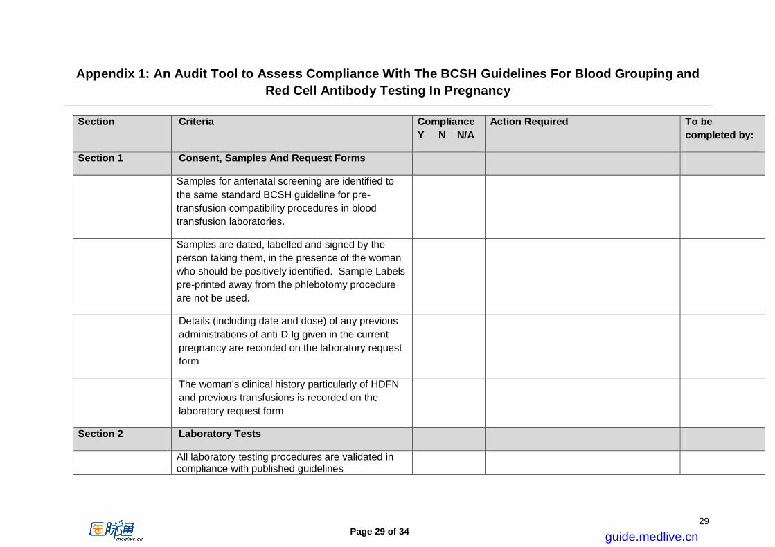
Page 29 of 34
29
Appendix 1: An Audit Tool to Assess Compliance With The BCSH Guidelines For Blood Grouping and Red Cell Antibody Testing In Pregnancy
Section Criteria Compliance Y N N/A
Action Required To be completed by:
Section 1 Consent, Samples And Request Forms
Samples for antenatal screening are identified to the same standard BCSH guideline for pre-transfusion compatibility procedures in blood transfusion laboratories.
Samples are dated, labelled and signed by the person taking them, in the presence of the woman who should be positively identified. Sample Labels pre-printed away from the phlebotomy procedure are not be used.
Details (including date and dose) of any previous administrations of anti-D Ig given in the current pregnancy are recorded on the laboratory request form
The woman’s clinical history particularly of HDFN and previous transfusions is recorded on the laboratory request form
Section 2 Laboratory Tests
All laboratory testing procedures are validated in compliance with published guidelines
guide.medlive.cn
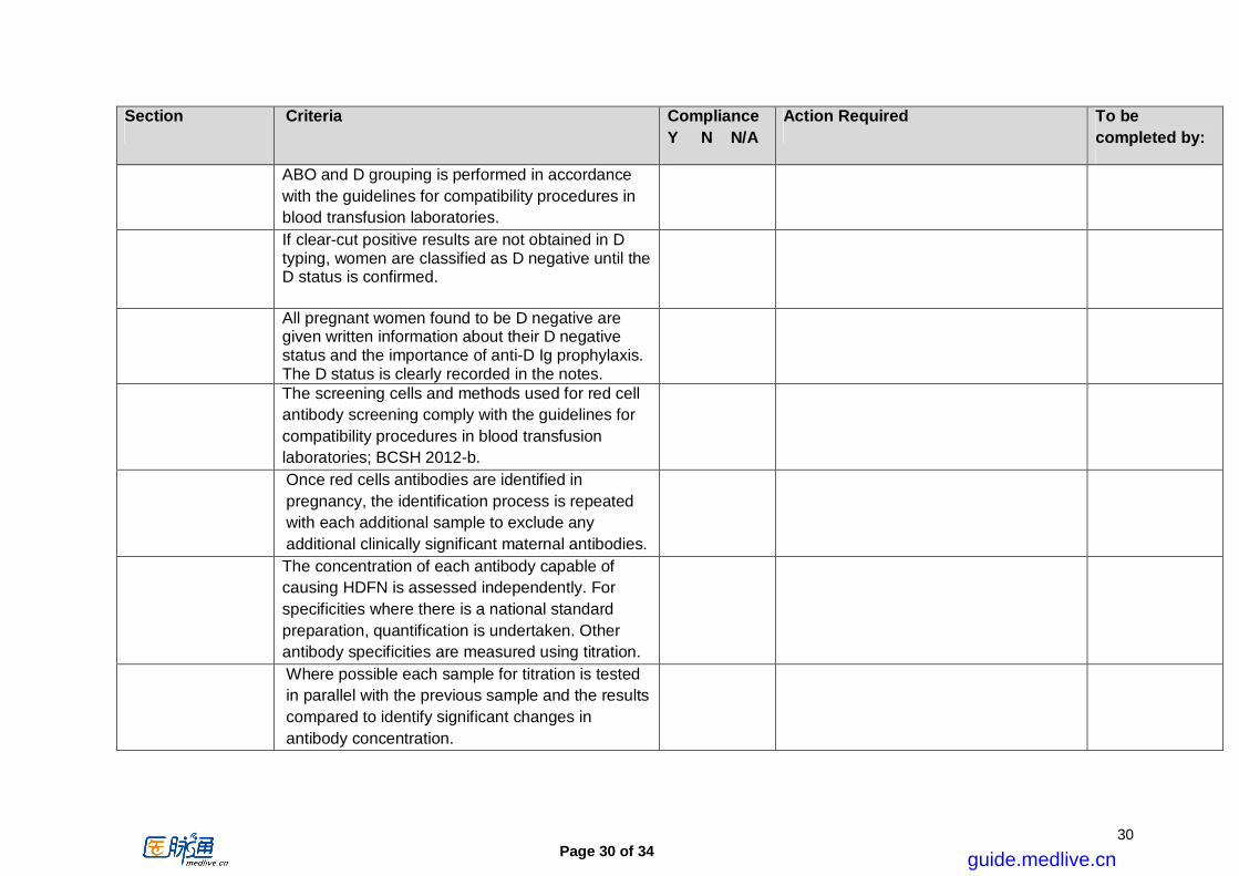
Page 30 of 34
30
Section Criteria Compliance Y N N/A
Action Required To be completed by:
ABO and D grouping is performed in accordance with the guidelines for compatibility procedures in blood transfusion laboratories.
If clear-cut positive results are not obtained in D typing, women are classified as D negative until the D status is confirmed.
All pregnant women found to be D negative are given written information about their D negative status and the importance of anti-D Ig prophylaxis. The D status is clearly recorded in the notes.
The screening cells and methods used for red cell antibody screening comply with the guidelines for compatibility procedures in blood transfusion laboratories; BCSH 2012-b.
Once red cells antibodies are identified in pregnancy, the identification process is repeated with each additional sample to exclude any additional clinically significant maternal antibodies.
The concentration of each antibody capable of causing HDFN is assessed independently. For specificities where there is a national standard preparation, quantification is undertaken. Other antibody specificities are measured using titration.
Where possible each sample for titration is tested in parallel with the previous sample and the results compared to identify significant changes in antibody concentration.
guide.medlive.cn
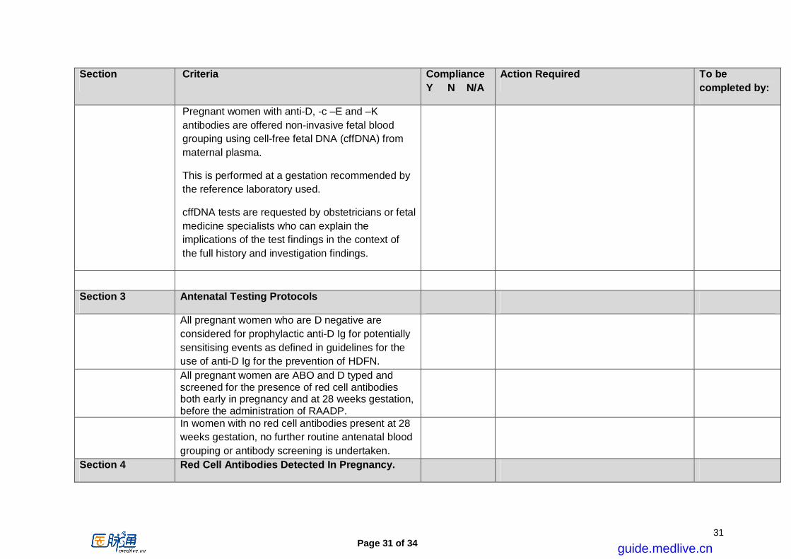
Page 31 of 34
31
Section Criteria Compliance Y N N/A
Action Required To be completed by:
Pregnant women with anti-D, -c –E and –K antibodies are offered non-invasive fetal blood grouping using cell-free fetal DNA (cffDNA) from maternal plasma.
This is performed at a gestation recommended by the reference laboratory used.
cffDNA tests are requested by obstetricians or fetal medicine specialists who can explain the implications of the test findings in the context of the full history and investigation findings.
Section 3 Antenatal Testing Protocols
All pregnant women who are D negative are considered for prophylactic anti-D Ig for potentially sensitising events as defined in guidelines for the use of anti-D Ig for the prevention of HDFN.
All pregnant women are ABO and D typed and screened for the presence of red cell antibodies both early in pregnancy and at 28 weeks gestation, before the administration of RAADP.
In women with no red cell antibodies present at 28 weeks gestation, no further routine antenatal blood grouping or antibody screening is undertaken.
Section 4 Red Cell Antibodies Detected In Pregnancy.
guide.medlive.cn

Page 32 of 34
32
Section Criteria Compliance Y N N/A
Action Required To be completed by:
All women who have previously had a baby affected by HDFN are referred before 20 weeks gestation to a fetal medicine specialist for advice and for assessment of fetal haemolysis, irrespective of antibody concentration or specificity.
The laboratory keeps clear records of anti-D Ig administration.
All anti-D detected in pregnancy should be quantified by CFA, or tested by a method that has been extensively validated against quantification, and there is a clear procedure to distinguish between immune and passive anti-D Ig, including review of clinical history and previous laboratory results.
When there is doubt as to the passive or immune nature of anti-D, concentration is monitored and prophylactic anti-D Ig offered, where indicated, until the nature of anti-D is established.
A reference centre confirms antibody specificity in women with apparent anti-C+D.
Cases of anti-D and anti-c are referred to a fetal medicine specialist if the antibody concentration reaches the critical level and/or the level is rising significantly. The antibody concentration is assessed serologically at 4 weekly intervals to 28 weeks gestation and at fortnightly intervals thereafter until delivery, unless advised otherwise by clinicians undertaking serial MCA Doppler.
guide.medlive.cn

Page 33 of 34
33
Section Criteria Compliance Y N N/A
Action Required To be completed by:
Cases of anti-K or other Kell system antibody [unless the father is confirmed to be antigen negative] are referred to a fetal medicine specialist when the antibody is first identified. The antibody strength is assessed serologically at monthly intervals to 28 weeks gestation and at fortnightly intervals thereafter until delivery, unless advised otherwise by clinicians undertaking serial MCA Doppler. All cases
Clinically significant antibodies, other than anti-D, -c or -K, are excluded or, if present, assessed by titration, at the booking appointment and at 28 weeks gestation. Where the titre is>32 and/or there is a past history of HDFN, referral to a specialist in fetal medicine is made for further assessment.
Section 5 Reports Of Laboratory Investigations
Reports inform the clinician[s] of the significance of the antibodies, with respect to the development of HDFN and transfusion problems.
Section 6 Action At Time Of Birth
All babies born to women who have clinically significant antibodies are closely observed for evidence of HDFN. A DAT is performed, and haemoglobin and bilirubin concentrations are measured.
guide.medlive.cn

Page 34 of 34
34
Section Criteria Compliance Y N N/A
Action Required To be completed by:
A DAT is not routinely performed on cord samples of D positive babies born to D negative women leading to unnecessary additional investigations.
D negative women with no immune anti-D have an initial FMH test at delivery and follow up testing if required, in accordance with BCSH guidelines for estimation of FMH.
Section 7 Audit
Audits of compliance with the clinical and laboratory aspects of these guidelines are undertaken on a continuing basis and all variations or concerns addressed in a timely manner.
See text of guidelines for references and the grades of evidence.
guide.medlive.cn