green route synthesis of silver nano particles … · green route synthesis of silver nano...
TRANSCRIPT

DOI: 10.22623/IJAPSA.2017.3007.IDZBN Page 48
GREEN ROUTE SYNTHESIS OF SILVER NANO PARTICLES USING LEAF
EXTRACTS OF MELALEUCALEUCADENDRA
Mita R. Patel1*, Dr. Rasmikant A. Patel
2, Dr. Dharmesh Varade
3 and Dr. Kespi A. Pithawala
4
1Department of Chemistry, Gujarat Arts and Science College, Ahmedabad, Gujarat, India, 380006 2Department of Chemistry, Municipal Arts and Urban Science College, Mehsana, Gujarat, India
3School of Engineering & Applied Sciences, Ahmedabad University, Gujarat, India, 380009
4Department of Biology, Gujarat Arts and Science College, Ahmedabad, Gujarat, India, 380006
*correspondences Author- Mita R. Patel1
Abstract
The ebb and flow explore in nanotechnology is primarily gone for condition benevolent and savvy
techniques for green amalgamation of nanoparticles. Here watery leaf concentrates of Melaleuca
leucadendra which go about as reducing and in addition topping operators were utilized for the
development of silver nanoparticles (SNP) from 0.1 mM AgNO3 arrangement. The SNP were
framed inside hour and the development was steady for a considerable length of time. The
arrangement, qualities, size and compliance of silver nanoparticles framed were completed
utilizing UV - VIS spectroscopy, FTIR, Dynamic Light Scattering (DLS), SEM, TEM and EDX.
Likewise the SNP framed along these lines demonstrated reasonable antimicrobial movement
against Gram positive and Gram negative microorganisms. Additionally SNP showed great
antioxidant (cancer prevention) agent properties. On the premise of result acquired one might say
that the simple generation of silver nanoparticle utilizing green science can be successfully used
in different fields in biomedical-nanotechnology and in addition decreasing the pathogenic
response of microbial flora.
Keywords: Silver nanoparticles, Melaleuca leucadendra, Antibacterial Activity, FTIR, DLS, SEM,
TEM, EDX, antioxidant activity.
I. INTRODUCTION
Nano particles of different metals and metal salts have been shaped utilizing different
synthetic, physical and green synthesis [1-5]. Of the previously mentioned strategies the most simple
and eco-friendly technique is through green union and today it is the most well-known strategy for
nanoparticle combination [6-10]. Of the metals and the salts the most regularly utilized are the
coinage metals as Au, Ag and Cu however Fe, Zn and Pt has likewise been used [11-14]. Ag NPs are
for the most part put to use in pharmaceutical and biomedical items and in this way should be
incorporated by green course utilizing characteristic plant materials which is naturally savvy as well
as expands amalgamation that does not have to utilize high weight, vitality, temperature and harmful
chemicals making it ecofriendly and biocompatible [15]. Bio-motivated union of nanoparticles gives
headway over concoction and physical strategies as it is quick and particular in their objective
towards the applications where they are evaluated for their antimicrobial action [16]. Medicinal
plants are utilized as a part of extensive extents nowadays in view of the enduring change against
maladies after natural treatment [17]. In this exploration silver nano particles have been combined
utilizing the green course by utilizing the fluid leaf concentrates of Melaleuca leucadendra regularly
called paper bark tree [18].
International Journal of Applied and Pure Science and Agriculture (IJAPSA)
Volume 03, Issue 3, [March- 2017] e-ISSN: 2394-5532, p-ISSN: 2394-823X
@IJAPSA-2017, All rights Reserved Page 49
II. MATERIALS AND METHOD
Fresh new leaves of Melaleuca leucadendra were gathered from the tree grownin botanical
gardenof Gujarat College, Ahmedabad [19]. Silver nitrate was bought from Hi-Media and chemicals
for cell reinforcement movement including 2,2-azino-bis(3-ethylbenzothiazoline-6-sulphonic
corrosive) diammonium salt (ABTS+), 2,2-diphenyl-1-picrylhydrazyl (DPPH), were acquired from
Sigma-Aldrich (St. Louis, MO, USA). BHA, 2-thiobarbituric corrosive (TBARS), trichloroacetic
corrosive (TCA), Ammonium thiocyanate, Nitroblue tetrazolium (NBT), phosphate cradle (pH 7.4)
and nonenzymatic phenazine methosulfate (PMS) were purchased from SRL chemicals, and n-
butanol from Burgoyn, Methanol, ethanol, (CH3)2CO, chloroform, hydrochloric corrosive from
Rankem, Nicotinamide adenine dinucleotide (PMS-NADH) from Spectrochem, Potassium persulfate
from HPLC, Acetic corrosive, vitamin C, vitamin E and BHA from Merk, K3Fe(CN)6, FeCl3 and
FeSO4 from Nice, Bradford Reagent was prepared freshly, All chemicals were of logical review with
98-100% immaculateness measure as analytical reagents and chemical grade. Deionized and twofold
distilled water was utilized wherever required.
2.1 Preparation of Extracts Freshleaves of Melaleucaleucadendra leaves were initially washed with deionized water and
after that with refined water to free them of any earth material these were then subjected to dry in
space for around 5 days andlastly pounded into fine powder utilizing a clean sanitized local
mechanized processor (stainless steel cutting edges).This powder was put away in water/air proof
compartment and was utilized for planning fluid concentrates. These were set up by including 1 g of
leaf powder to 50 ml of twofold distilled water and left overnight. The arrangement accordingly
framed was sifted through Whatmann filter paper No. 1 and the concentrate was utilized for planning
of nanoparticles.
2.2 Synthesis of Ag nanoparticles To 30 ml of 0.1mM AgNO3 10 ml of leaf extract was added and at interval of every 5
minutes colorimetric readings were taken at 410nm to find the presence of formation of nano
particles the color change was an indication of formation of nano particles.These were formed within
15 minutes and these particles were stable for about one month.
2.3 Characterization techniques
The biosynthesized silver nanoparticles were characterized by the following methods:
2.3.1 Visual Observation A change of colour from pale yellow to reddish brown was observed in the solution after
visible irradiation.
2.3.2 UV Spectrophotometric analysis The characterization technique involves ultra-violet and visible spectroscopy. UV-Vis
absorption spectra were measured using Systronic UV-117 spectrometer from 300nm to 700nm
continuously and the leaf powder extract was used as the reference for the baseline correction.
2.3.3 Fourier Transform Infrared Spectroscopy Analysis: FTIR analysis was carried out to determine the functional groups present in leaf extract and
their possible involvement in the synthesis of silver nanoparticles. FTIR analysis were carried using
a FTIR SHIMADZU 8300 instrument with a wavelength range of 4000 to 400 nm where the
samples were incorporated with KBr pellets to acquire the spectra. The results were compared for
shift in functional peaks. A FTIR graph can be useful for preliminary investigation of surface
chemistry of biogenic nanoparticles (i.e. those chemicals that contain carbon). This technique is
widely used for identification of chemical residues such as amine, carbonyl and hydroxyl functional
groups in a molecule [20]. The FTIR analysis was performed with reduced silver nanoparticles. The
synthesized AgNPs sample was mixed with KBr to make a pellet in the ratio of 1:100. The FTIR
instrument with diffuse reflectance mode attachment. All measurements were carried out in the range

International Journal of Applied and Pure Science and Agriculture (IJAPSA)
Volume 03, Issue 3, [March- 2017] e-ISSN: 2394-5532, p-ISSN: 2394-823X
@IJAPSA-2017, All rights Reserved Page 50
of 400-4,000 cm-1 at a resolution of 4 cm-1 [21]. For this fresh sample were sent for FTIR Analysis
at Gujarat Laboratory, Ahmedabad. Samples with total of volume 1-2 ml were given in aqueous form
formed by producing SNPs using the reduction reaction of 9 ml of 0.1 mM Silver Nitrate solution
through 3 ml of plant extract.
2.3.4 Dynamic Light Scattering
These studies were carried out to get to know the particle size distribution in the solution. The
particle size comes out to be 100nm and hence this can be further verified from SEM analysis.
2.3.5 SEM & EDX Analysis The surface morphology of silver nanoparticles was examined using a scanning electron
microscopy (6010 LA, Jeol).The elemental composition of the synthesized silver nanoparticles was
analyzed using Energy Dispersive X-Ray Spectrometer.
2.3.6. TEM Analysis
Since the particle sizes were small TEM studies were carried out to get to know the exact
shape of the particles this was done using transmission electron microscope (JEM1400 Plus, Jeol).
The particle sizes of about 30nm to 60nm can be seen clearly.
2.3.7Determination of antimicrobial activity
A. Microorganisms Human pathogens were obtained from the Department of Microbiology, Gujarat Arts and
Science College, Ahmedabad. The bacterial pathogens namely Staphylococcus aureus, Streptococcus
pneumonia, Escherchia coli, Salmonella typhi, were used to study the antibacterial activity. The
fungal pathogens such as Aspergillus niger, Penicillium sp, Rhizopus stolonifer were used to study
the antifungal activity. The nutrient broth, potato dextrose broth, nutrient agar and potato dextrose
agar were used growing the test bacterial and fungal strains and were maintained on corresponding
agar slants at 4°C.
B. Preparation of inoculums
The bacterial pathogens were inoculated into sterile nutrient broth and incubated at 37°C for
24 hours until the culture attained a turbidity of 0.5 McFarland units. The final inoculum was
standardized to 105 CFU/ml by diluting fresh cultures with sterile distilled water. For fungal
pathogens, inoculums was prepared in potato dextrose broth by choosing five distinct colonies
approximately 1 mm from 24 h old culture grown on Potato dextrose agar (PDA) and incubated at
28±2ºC.
Colonies were suspended in 5 ml of sterile 0.85% saline. The resulting suspension was vortex
and the turbidity was adjusted to yield 2×10⁶ cells/ml (≅0.5 McFarland standards).
C. Antibacterial activity
Antibacterial activity of AgNPs was determined by the agar disc diffusion method [22].
Plates of Nutrient agar were evenly streaked across the complete surface throughout the petri plate so
as to get a loan growth of the inoculums with the help of spread plate technique with a known
volume of 0.01 ml of active young culture with approx. Microbial count as 105 CFU/ml. Sterile filter
paper discs (5 mm diameter) were immersed in the 50 µl of synthesized AgNPs (10, 20, 30, 40, 50
µg/ml) and allowed to dry at room temperature and it was placed over the Nutrient agar plates.
Streptomycin 10 mcg/disc was used as positive control and the disc immersed in distilled water was
used as negative control. The plates were incubated overnight at 37 ºC and the zone of inhibition
around each disc was measured. Experiments were done in triplicate and mean values of zone
diameter were taken.
D. Antifungal activity The growth inhibition of fungal pathogens by AgNPs was determined by the agar disc
diffusion method [23]. Sterile swabs were dipped in the microbial suspensions which contains fungal
International Journal of Applied and Pure Science and Agriculture (IJAPSA)
Volume 03, Issue 3, [March- 2017] e-ISSN: 2394-5532, p-ISSN: 2394-823X
@IJAPSA-2017, All rights Reserved Page 51
strains were uniformly applied to petridishes containing PDA. Sterile filter paper discs (5 mm
diameter) were immersed in the 50 µl of synthesized AgNPs (10, 20, 30, 40, 50 µg/ml) and allowed
to dry at room temperature and it was placed over the petridishes. Nystatin was used as positive
control and plates were incubated at 28 ± 2 ºC for 48-72 h and the zone of inhibition around each
disc was measured.
2.3.5 Determination of antioxidant activity
A. DPPH Radical-Scavenging Activity DPPH radical-scavenging activity was estimated according to the method of Ravikumar et al.
(2008) [24]. Briefly, 1.0 ml of synthesized AgNPs (50 µg/ml) was added to 1.0 ml of 0.16mM DPPH
methanolic solution. Similiarly simple 50 µg/ml plant extract and 0.1 M of AgNO3was also tested for
antioxidant properties. The mixture was vortexed for 1 min and left to stand at room temperature for
30 min in dark, and the absorbance was read at 517 nm. Lower absorbance indicated higher radical-
scavenging activity. The ability to scavenge the DPPH radical was calculated using the following
equation:
Scavenging activity (%) = [(A control – A sample) /A control] × 100
Where, A control is the absorbance of the control (DPPH solution without AgNPs), A sample
is the absorbance of the test sample (DPPH solution plus AgNPs) BHT was used as the standard.
Each samples were performed for triplicate to minimize error.
B. Superoxide Radical Scavenging Activity The superoxide radical scavenging activity was assayed according to the method mentioned
by Nishimiki M. et al. (1972)[25]. Superoxide anions were generated in a phenazine methosulfate-
nicotinamide adenine dinucleotide (PMS-NADH) system through the reaction of PMS-NADH and
oxygen. It was assayed by the reduction of nitroblue tetrazolium (NBT). All the solutions used in this
experiment were prepared in phosphate buffer (pH 7.4). 1ml of NBT (156µM), 1ml of NADH
(468µM) and each 1ml of plant extract (10-50 µg/ml) and each AgNPs (10-50 µg/ml) were mixed.
The reaction was started by adding 1ml of PMS (60µM) and the mixture was incubated at 25°C for
5min followed by measurement of absorbance at 560nm spectrophotometrically. Decreased
absorbance of the reaction mixture indicated increased superoxide anion scavenging activity. The
percentage inhibition was calculated using the formula,
Inhibition (%) = [(A0 – AS) / A0] × 100……………. (Nishimiki M. et al. 1972)
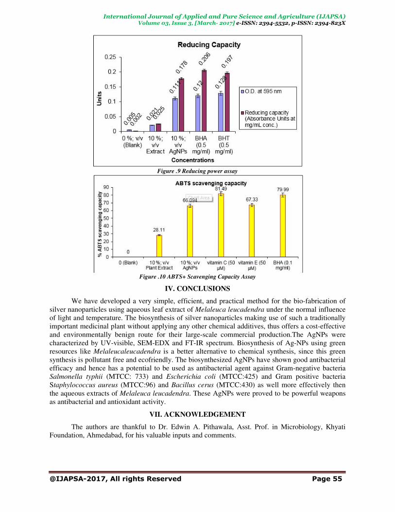
C.Reducing power assay The reducing power of the AgNPs was measured according to the method of Oyaizu, (1986)
[26].Briefly, 500 µl of synthesized AgNPs (100, 200, 300, 400, 500 µg/ml) was mixed with 2.5ml of
phosphate buffer (0.2 M, pH 6.6) and 2.5ml of one percent potassium ferricyanide. Reaction mixture
was incubated at 50°C for 20 min. After incubation, 2.5ml of trichloroaceticacid (10%) was added
and centrifuged at 650×g for 10 min. 2.5ml of upper layer was mixed with 2.5ml of distilled water
and 0.5ml ferric chloride (0.1%). Absorbance of the samples was read at 700 nm. Increased
absorbance of the reaction mixture indicated increased reducing power. BHT was used as the
standard.
D. ABTS+ Scavenging Capacity Assay
The ABTS decolonization assays were carried out involving the generation of ABTS+
chromophore by oxidation of ABTS with potassium persulfate. The ABTS radical cation (ABTS+)
was produced by reacting 7 mM stock solution of ABTS with 2.45 mM potassium persulfate and
allowing the mixture to incubate in the dark condition at room temperature for at least 6 hour before
use. Absorbance at 734 nm was measured after 10 minutes of incubation after mixing different
concentrations of the Melaleuca leucadendra extracted (final concentration as 10, 15, 20 %; v/v)
with 1 ml of ABTS+ solution. The ABTS
+ scavenging capacity of the filtrate was compared with that
of vitamin C (50 µM), vitamin E (50 µM) and BHA (0.1 mg/ml).

International Journal of Applied and Pure Science and Agriculture (IJAPSA) Volume 03, Issue 3, [March
@IJAPSA-2017, All rights Reserved
III. RESULT
A robust practical way for eco
extract of Melaleuca leucadendra (White paper bark tree) as both reducing and capping agent, under
the prescribed condition of room temperature has been adopted without applying any other
precursors, inducers or hazardous chemical additives or rigorous reactions.
The formation of AgNP was predicted usi
confirmed by SEM,Figure-2, and also EDX
characteristics of AgNP was confirmed,
and 3328.189 wave numbers confirming the formation of nano particles of silver.
determined c.a. as 60 nm using DLS,
the shape of the particles was known using transmission
The prepared silver nanoparticles exhibited considerable antioxidant as
activity,Table-1 and 2. The effects were more pronounced on Gram
typhii (MTCC: 733) and Escherichia coli
activity on Gram-positive bacteria S
(MTCC:430). A bactericidal mode of action was observed more for both Gram
negative bacteria by the nanoparticles as compared to Plant extracts. Similarly antifungal activity
also showed more pronouncing activity through nanoparticles formed using extracts of
Melaleucaleucadendra as compared to simple extracts of
property reveals dose dependent relationship
Figure
International Journal of Applied and Pure Science and Agriculture (IJAPSA)Volume 03, Issue 3, [March- 2017] e-ISSN: 2394-5532, p
2017, All rights Reserved
RESULTS AND DISCUSSION
A robust practical way for eco-friendly synthesis of silver nanoparticles using aqueous leaf
(White paper bark tree) as both reducing and capping agent, under
the prescribed condition of room temperature has been adopted without applying any other
precursors, inducers or hazardous chemical additives or rigorous reactions.
NP was predicted using UV-visible spectroscopy (410 nm),
and also EDX, Figure-3, of AgNP.Also using FT-IR Spectroscopy the
NP was confirmed, Figure-4,FTIR studies show peaks at 1634.550, 2113.342
and 3328.189 wave numbers confirming the formation of nano particles of silver.
DLS,Figure-5, this was further affirmed from the TEM studies also
s known using transmission electron microscopy, Figure
The prepared silver nanoparticles exhibited considerable antioxidant as well as antibacterial
The effects were more pronounced on Gram-negative bacteria
Escherichia coli (MTCC:425). The nanoparticles also showed prominent
positive bacteria Staphylococcus aureus (MTCC:96) and
(MTCC:430). A bactericidal mode of action was observed more for both Gram-positive a
negative bacteria by the nanoparticles as compared to Plant extracts. Similarly antifungal activity
also showed more pronouncing activity through nanoparticles formed using extracts of
as compared to simple extracts of Melaleucaleucadendra
property reveals dose dependent relationship, Figure-7, 8, 9 & 10.
Figure .1 UV-VIS scan of AgNP
Figure .2 SEM of AgNP
Figure .3EDX of AgNP
International Journal of Applied and Pure Science and Agriculture (IJAPSA)
5532, p-ISSN: 2394-823X
Page 52
friendly synthesis of silver nanoparticles using aqueous leaf
(White paper bark tree) as both reducing and capping agent, under
the prescribed condition of room temperature has been adopted without applying any other
nm),Figure-1,further
IR Spectroscopy the
studies show peaks at 1634.550, 2113.342
and 3328.189 wave numbers confirming the formation of nano particles of silver. Size was
this was further affirmed from the TEM studies also
Figure-6.
well as antibacterial
negative bacteria Salmonella
(MTCC:425). The nanoparticles also showed prominent
(MTCC:96) and Bacillus cerus
positive and Gram-
negative bacteria by the nanoparticles as compared to Plant extracts. Similarly antifungal activity
also showed more pronouncing activity through nanoparticles formed using extracts of
aleucadendra. Antioxidant

International Journal of Applied and Pure Science and Agriculture (IJAPSA) Volume 03, Issue 3, [March
@IJAPSA-2017, All rights Reserved
Table.1.: Anti-Bacterial and Anti
Test Culture Plant
Extract
(50
µg/ml)
AgNPs Concentrations
10
µg/ml
International Journal of Applied and Pure Science and Agriculture (IJAPSA)Volume 03, Issue 3, [March- 2017] e-ISSN: 2394-5532, p
2017, All rights Reserved
Figure .4 FTIR of AgNP
Figure .5 DLS of AgNP
Figure .6 TEM of AgNP
Bacterial and Anti-fungal Activity of formed AgNPs using Melaleuca
AgNPs Concentrations Positive
Control
(Strepto
mycin)
10
µg/ml
20
µg/ml
30
µg/ml
40
µg/ml
50
µg/ml
Antibacterial Property
International Journal of Applied and Pure Science and Agriculture (IJAPSA)
5532, p-ISSN: 2394-823X
Page 53
extracts
Positive
Control
(Strepto-
mycin)
Negative
Control
(Sterile
D/W)

International Journal of Applied and Pure Science and Agriculture (IJAPSA)
Volume 03, Issue 3, [March- 2017] e-ISSN: 2394-5532, p-ISSN: 2394-823X
@IJAPSA-2017, All rights Reserved Page 54
Staphylococcus aureus 18 16 18 21 17 12 24 5
Bacillus cerus 17 15 14 14 19 11 24 5
Escherchia coli 16 19 21 18 22 24 24 5
Salmonella typhi 15 17 19 22 24 24 24 5
Antifungal Property
Aspergillus niger 22 21 27 24 28 29 32 5
Penicillium sp 24 24 24 23 34 30 32 5
Rhizopus stolonifer 24 26 24 26 30 31 32 5
Table.2. : Activity Index of the AgNPs at various concentrations
Test Culture Plant Extract
(50 µg/ml)
AgNPs Concentrations
10 µg/ml 20 µg/ml 30 µg/ml 40 µg/ml 50 µg/ml
Antibacterial Property
Staphylococcus aureus 0.75 0.66 0.75 0.87 0.70 0.50
Bacillus cerus 0.70 0.625 0.58 0.58 0.79 0.45
Escherchia coli 0.66 0.79 0.87 0.75 0.91 1.00
Salmonella typhi 0.625 0.70 0.79 0.91 1.00 1.00
Antifungal Property
Aspergillus niger 0.68 0.65 0.84 0.75 0.87 0.90
Penicillium sp 0.75 0.75 0.75 0.71 1.06 0.93
Rhizopus stolonifer 0.75 0.81 0.75 0.81 0.93 0.96
Note: Zone of inhibition by streptomycin as a standard drug = 24 mm (Mean Value) and for antifungal Nistatin as a
standard drug = 32 mm (Mean Value)
�����������(�. �. ) =MeanofZoneofInbitionbyAgNPs
ZoneofInhibitionobtainedforstandardAntibioticDrug
Figure .7 DPPH Radical-Scavenging Activity
Figure .8Superoxide Radical Scavenging Activity UV-VIS scan of AgNP
International Journal of Applied and Pure Science and Agriculture (IJAPSA)
Volume 03, Issue 3, [March- 2017] e-ISSN: 2394-5532, p-ISSN: 2394-823X
@IJAPSA-2017, All rights Reserved Page 55
Figure .9 Reducing power assay
Figure .10 ABTS+ Scavenging Capacity Assay
IV. CONCLUSIONS
We have developed a very simple, efficient, and practical method for the bio-fabrication of
silver nanoparticles using aqueous leaf extract of Melaleuca leucadendra under the normal influence
of light and temperature. The biosynthesis of silver nanoparticles making use of such a traditionally
important medicinal plant without applying any other chemical additives, thus offers a cost-effective
and environmentally benign route for their large-scale commercial production.The AgNPs were
characterized by UV-visible, SEM-EDX and FT-IR spectrum. Biosynthesis of Ag-NPs using green
resources like Melaleucaleucadendra is a better alternative to chemical synthesis, since this green
synthesis is pollutant free and ecofriendly. The biosynthesized AgNPs have shown good antibacterial
efficacy and hence has a potential to be used as antibacterial agent against Gram-negative bacteria
Salmonella typhii (MTCC: 733) and Escherichia coli (MTCC:425) and Gram positive bacteria
Staphylococcus aureus (MTCC:96) and Bacillus cerus (MTCC:430) as well more effectively then
the aqueous extracts of Melaleuca leucadendra. These AgNPs were proved to be powerful weapons
as antibacterial and antioxidant activity.
VII. ACKNOWLEDGEMENT
The authors are thankful to Dr. Edwin A. Pithawala, Asst. Prof. in Microbiology, Khyati
Foundation, Ahmedabad, for his valuable inputs and comments.
International Journal of Applied and Pure Science and Agriculture (IJAPSA)
Volume 03, Issue 3, [March- 2017] e-ISSN: 2394-5532, p-ISSN: 2394-823X
@IJAPSA-2017, All rights Reserved Page 56
BIBLIOGRAPHY
[1] Shakeel Ahmed, Mudasir Ahmad, Babu Lal Swami, Saiqa Ikram. 2016. A review on plants extract mediated
synthesis of silver nanoparticles for antimicrobial applications: A green expertise, Journal of Advanced Research..,
7[1]: 17–28
[2] H. Korbekandi, S. Iravani, S. Abbasi.2009.Production of nanoparticles using organisms, Critical Reviews in
Biotechnology., 29: 279–306
[3] D.A. Kumar, V. Palanichamy, S.M. Roopan.2014.Green synthesis of silver nanoparticles using Alternanthera
dentata leaf extract at room temperature and their antimicrobial activity, Spectrochimica Acta Part A-Molecular and
Biomolecular Spectroscopy., 127:168–171
[4] V. Kumar, S.K. Yadav. 2009.Plant-mediated synthesis of silver and gold nanoparticles and their applications,
Journal of Chemical Technology and Biotechnology., 84: 151–157
[5] P. Mohanpuria, N.K. Rana, S.K. Yadav. 2008. Biosynthesis of nanoparticles: technological concepts and future
applications Journal of Nanoparticle Research., 10:507–517
[6] D. MubarakAli, N. Thajuddin, K. Jeganathan, M. Gunasekaran.2011.Plant extract mediated synthesis of silver and
gold nanoparticles and its antibacterial activity against clinically isolated pathogens Colloids and Surfaces, B:
Biointerfaces, 85:360–365
[7] Zhang S., Tang Y., Vlahovic B. 2016.A Review on Preparation and Applications of Silver-Containing Nanofibers,
Nanoscale Research Letters, 11 (1), art. no. 80, 1-8.
[8] Chung I.-M., Park I., Seung-Hyun K., Thiruvengadam M., Rajakumar G. 2016.
Plant-Mediated Synthesis of Silver Nanoparticles: Their Characteristic Properties and TherapeuticApplications,
Nanoscale Research Letters, 11 (1), art. no. 40, 1-14.
[9] Leon-Silva S., Fernandez-Luqueno F., Lopez-Valdez F. 2016.Silver Nanoparticles (AgNP) in the Environment: a
Review of Potential Risks on Human and Environmental Health, Water, Air, and Soil Pollution, 227 (9), art. no. 306,
[10] Chakraborty C., Sharma A.R., Sharma G., Lee S.-S. 2016.Zebrafish: A complete animal model to enumerate the
nanoparticle toxicity Journal of Nanobiotechnology, 14 (1), art. no. 65, .
[11] Bali, R., Razak, N., Lumb, A. 2006. “The synthesis of metal nanoparticles inside live plants”, In: International
Conference on Nanoscience and Nanotechnology, pp.224-227.
[12] Umer, A., Naveed, S., Ramzan, N. 2012 “Selection of a suitable method for the synthesis of Copper nanoparticles”,
NANO: Brief Reports and Reviews, World Scientist Publishing Company,7: 5
[13] Bonet, F., Delmas, V., Grugeon, S., 1999. “Synthesis of Monodisperse Au, Pt, Pd, Ru and Ir Nanoparticles in
Ethylene Glycol,” Nano Structured Materials,11(8) : 1277-1284,
[14] Tan, Y. W., Dai, X. H., Li, Y. F. 2003.“Preparation of Gold, Platinum, Palladium and Silver Nano-particles”,
Journal of Materials Chemistry, 13(5) : 1069-1075
[15] Duran N., Nakazato G., Seabra A.B. 2016.Antimicrobial activity of biogenic silver nanoparticles, and silver chloride
nanoparticles: an overview and commentsApplied Microbiology and Biotechnology, 100 (15): 6555-6570.
[16] Anjum S., Abbasi B.H., Shinwari Z.K. 2016.Plant-mediated green synthesis of silver nanoparticles for biomedical
applications: Challenges and opportunitiesPakistan Journal of Botany.48 (4): 1731-1760.
[17] Antony E., Gunasekaran S., Sathiavelu M., Arunachalam S.2016. A review on use of plant extracts for gold and
silver nanoparticle synthesis and their potential activities against food pathogens Asian Journal of Pharmaceutical
and Clinical Research. 9 (4):18-23.
[18] Thakur A., Reddy S.G.,2016.Biological materials: Green source towards silver nanomaterials synthesis) Oriental
Journal of Chemistry, 32 (3): 1465-1472.
[19] Patel R.S., Parmar, T and Tatu, A.2004.A review on tree species of Gujarat College campus, Ahmedabad (Gujarat) ,
advances in Biological science, 3: 1-2
[20] Anderson R. J. DJ Ben dell, 2004. Ground water PW,Organic Spectroscopic analysis. Royal Society of Chemistry,
Cambridge.1. 5
[21] Saifuddin N, Wong CW, AA Nur yasumira (2009) Rapid biosynthesis of silver nanoparticles using culture
supernatant of bacteria with microwave irradiation. Eur. J. Chem. 6:61-70
[22] Cormican, M.G, Jones, R.N. 1996.Emerging resistance to antimicrobial agents in Gram-positive bacteria,
enterococci, staphylococci, and non-pneumococcal streptococci. Drugs. 51:6–12
[23] Bansod, S. and Rai, M. 2008. Antifungal activity of essential oils from Indian medicinal plants against human
pathogenic Aspergillus fumigatus and A. niger. World Journal of Medical Sciences, 3(2): 81-88.
[24] Ravikumar YS, Mahadevan KM, Kumaraswamy MN, Vaidya VP, Manjunatha H, Kumar V, Satyanarayana ND.
2008. Antioxidant, cytotoxic and genotoxic evaluation of alcoholic extract of Polyalthia cerasoides (Roxb.) Bedd.
Environ Toxicol Pharmacol. 26(2):142-6.
[25] Nishimiki M, Rao N A, Yagi K. 1972. Occurrence of super-oxide anion in the reaction of reduced phenazine
methosulfate and molecular oxygen. Biochem Biophys Res Commun . 46:849-853
[26] Oyaizu M. 1986. Studies on products of browning reaction prepared from glucoseamine. Jpn J Nutr.,44:307-314.