green fluorescent protein
DESCRIPTION
Green Fluorescent Protein. a B/MB senior seminar brought to you by Colm O’Carroll. This presentation will cover. The structural aspects of GFP which make fluorescence possible The advantages of using GFP and GFP mutants over other fluorescent markers - PowerPoint PPT PresentationTRANSCRIPT
Green Fluorescent Protein
a B/MB senior seminar
brought to you by Colm O’Carroll
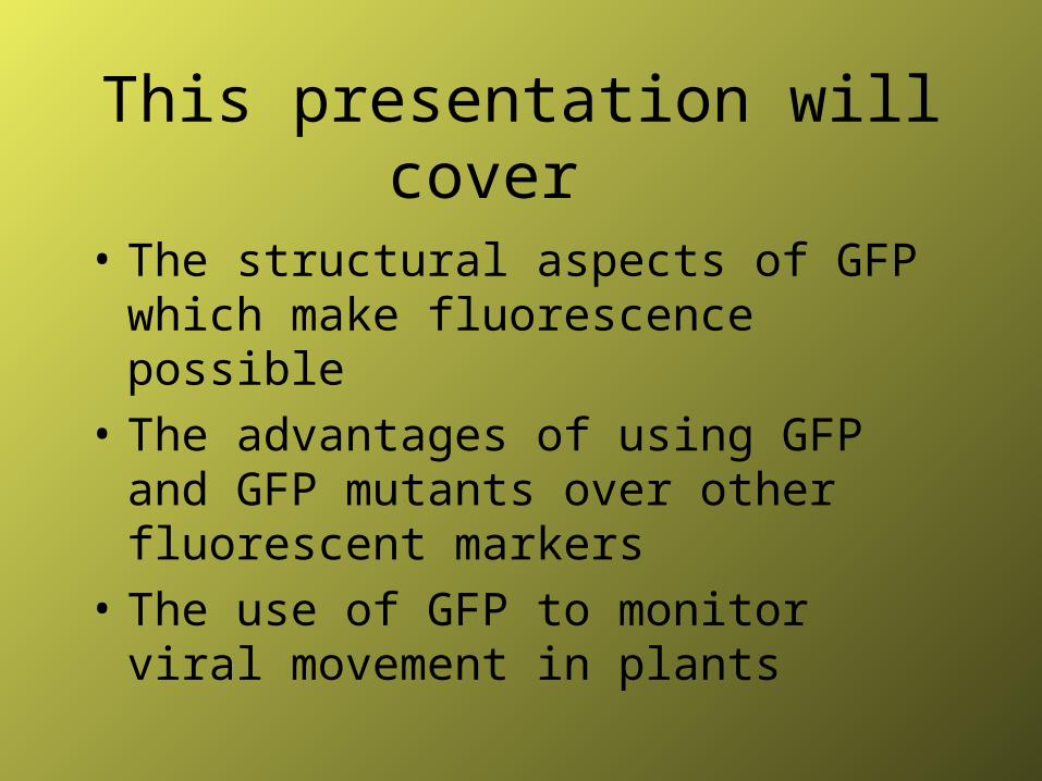
This presentation will cover
• The structural aspects of GFP which make fluorescence possible
• The advantages of using GFP and GFP mutants over other fluorescent markers
• The use of GFP to monitor viral movement in plants
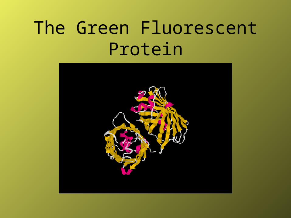
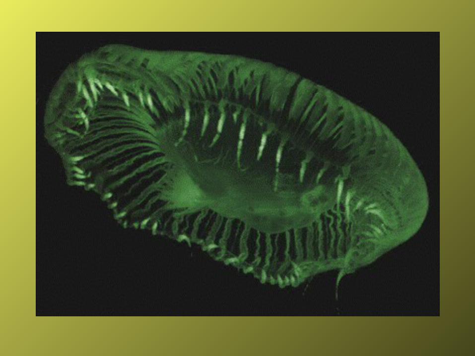
The Green Fluorescent Protein
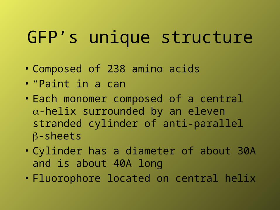
GFP’s unique structure
• Composed of 238 amino acids
• “Paint in a can”
• Each monomer composed of a central -helix surrounded by an eleven stranded cylinder of anti-parallel -sheets
• Cylinder has a diameter of about 30A and is about 40A long
• Fluorophore located on central helix

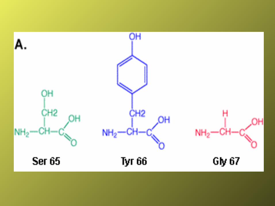
The Active Site
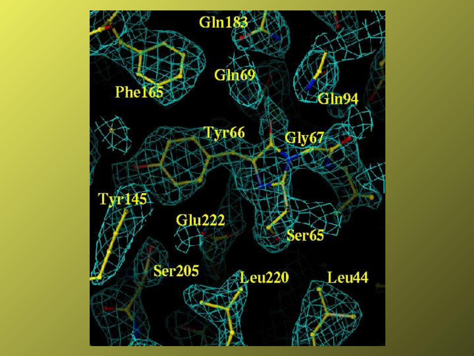
The Fluoropore Active Site
• Ser65-Tyr66-Gly67
• Deprotonated phenolate of Tyr66 is cause of fluorescence
• Forster Cycle (1949-Theodor Forster)
• Proton transfer to His148
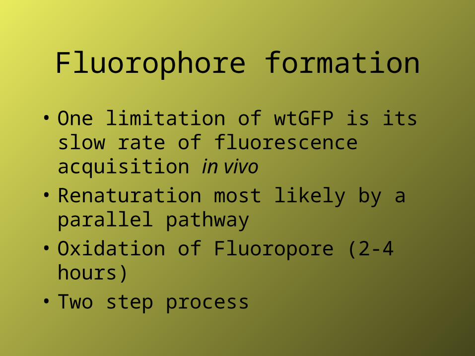
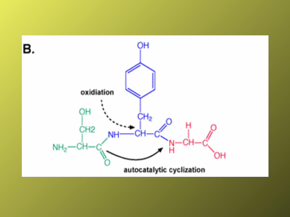
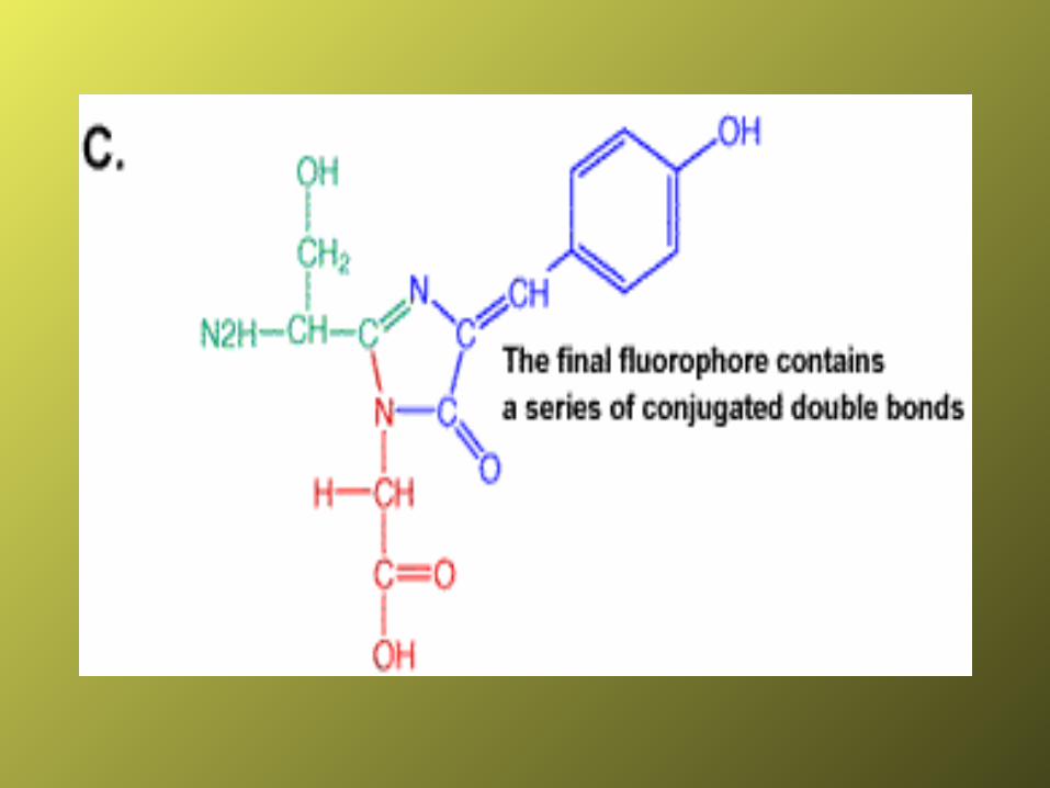
Fluorophore formation
• One limitation of wtGFP is its slow rate of fluorescence acquisition in vivo
• Renaturation most likely by a parallel pathway
• Oxidation of Fluoropore (2-4 hours)
• Two step process
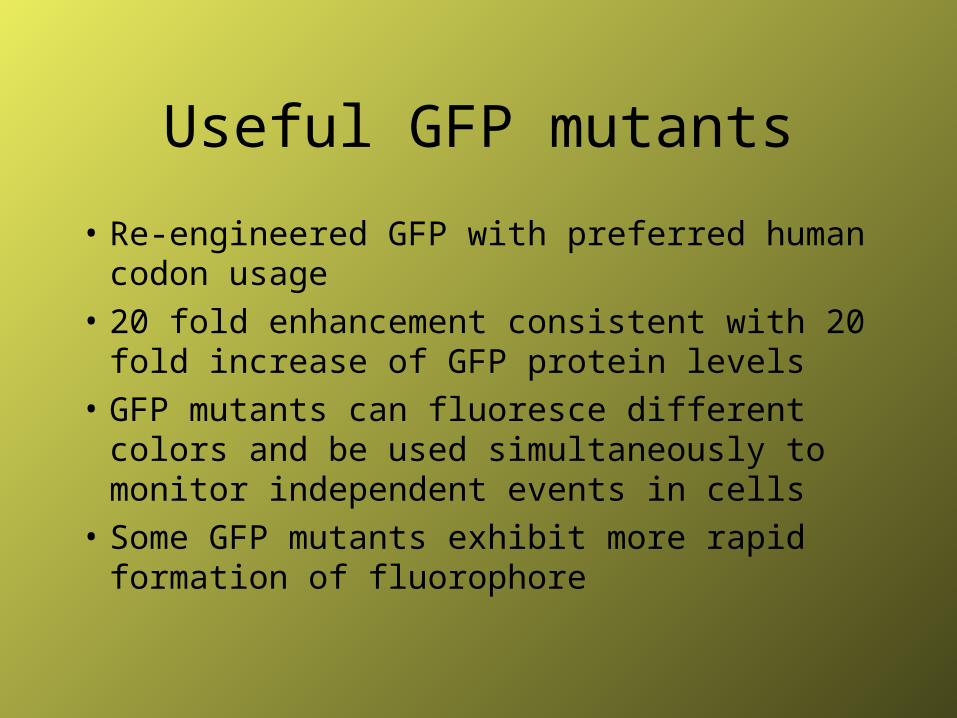
Useful GFP mutants
• Re-engineered GFP with preferred human codon usage
• 20 fold enhancement consistent with 20 fold increase of GFP protein levels
• GFP mutants can fluoresce different colors and be used simultaneously to monitor independent events in cells
• Some GFP mutants exhibit more rapid formation of fluorophore
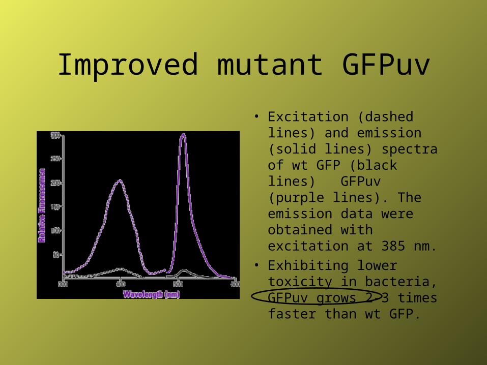
Improved mutant GFPuv
• Excitation (dashed lines) and emission (solid lines) spectra of wt GFP (black lines) GFPuv (purple lines). The emission data were obtained with excitation at 385 nm.
• Exhibiting lower toxicity in bacteria, GFPuv grows 2-3 times faster than wt GFP.
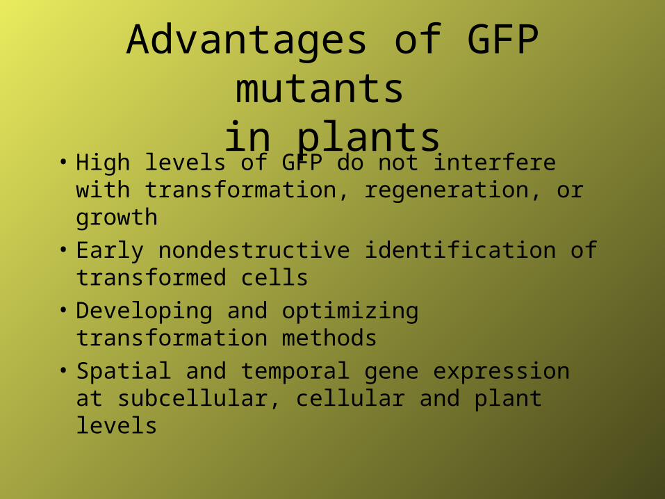
Advantages of GFP mutants in plants
• High levels of GFP do not interfere with transformation, regeneration, or growth
• Early nondestructive identification of transformed cells
• Developing and optimizing transformation methods
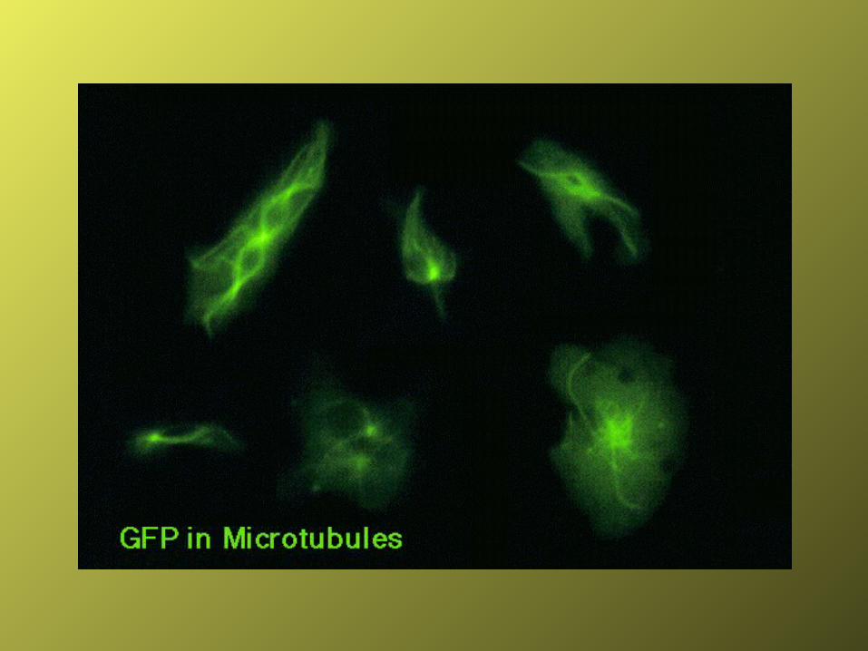
• Spatial and temporal gene expression at subcellular, cellular and plant levels
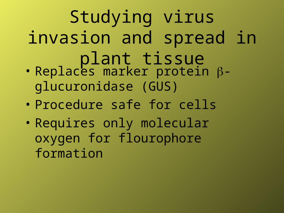
Studying virus invasion and spread in plant tissue
• Replaces marker protein -glucuronidase (GUS)
• Procedure safe for cells
• Requires only molecular oxygen for flourophore formation
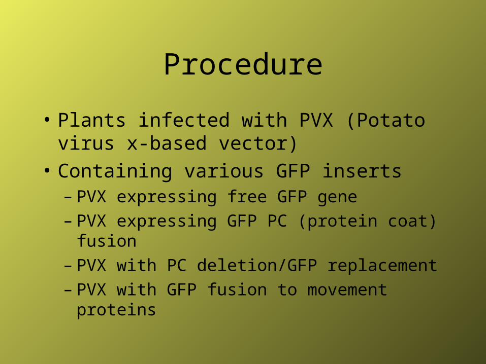
Procedure
• Plants infected with PVX (Potato virus x-based vector)
• Containing various GFP inserts– PVX expressing free GFP gene– PVX expressing GFP PC (protein coat) fusion– PVX with PC deletion/GFP replacement – PVX with GFP fusion to movement proteins
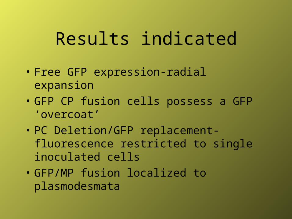
Results indicated
• Free GFP expression-radial expansion
• GFP CP fusion cells possess a GFP ‘overcoat’
• PC Deletion/GFP replacement- fluorescence restricted to single inoculated cells
• GFP/MP fusion localized to plasmodesmata
Bibliography
• Nina, Haruki, et al. "Chemical nature of light emitter of Aequorea green fluorescent protein" (1996) Proceedings Natl. Acad. Sci. USA vol. 93 p.13671-13622
• Oparka, Karl, et al. "Using GFP to study virus invasion and spread in plant tissues"(1997) Nature vol. 388 p. 401-402
• Reid, Brian, Gregory Flynn. "Chromophore formation in Green Fluorescent Protein" (1997) Biochemistry vol. 36 p. 6786-6791
• Yang, F., L. Moss, G. Phillips. "The Molecular Structure of GFP" (1996) Nature Biotechnology vol. 14 p. 1219-1220
• Youvan, Douglas., Gregory Flynn. "Chromophore formation in Green Fluorescent Protein" (1997)
Biochemistry vol. 36 6786-6791

Sources on the World Wide Web
• Medical College of Wisconsin (www.biochem.mcw.edu/science_ed/Pages/gfp/index.html
• Clonetech (www.gfp.clontech.com)
• www.biorad.com/889168.html
• www.bio.cmu.edu/Courses/03740/GFPTest/GFP.html