graves’ disease, celiac disease and liver function ... · graves’ disease, celiac disease and...
TRANSCRIPT
Regular paper
Graves’ disease, celiac disease and liver function abnormalities in a patient — clinical manifestation and diagnostic difficultiesMagdalena Góra-Gębka1*, Małgorzata Woźniak2, Joanna Cielecka-Kuszyk3, Maria Korpal- Szczyrska4, Katarzyna Sznurkowska1, Maciej Zagierski1, Irena Jankowska2, Katarzyna Plata-Nazar1, Barbara Kamińska1 and Anna Liberek5
1Department of Pediatrics, Pediatric Gastroenterology, Hepatology and Nutrition, Medical University of Gdansk, Gdańsk, Poland; 2Department of Gastroenterology, Hepatology and Eating Disorders Children’s Memorial Hospital in Warsaw, Warsaw, Poland; 3Department of Patology, Chil-dren’s Memorial Hospital in Warsaw, Warsaw, Poland; 4Department of Pediatrics, Hematology, Oncology and Endocrinology, Medical University of Gdansk, Gdańsk, Poland; 5Faculty of Health Sciences with Subfaculty of Nursing Medical University of Gdansk, Gdańsk, Poland
Autoimmune diseases due to probable common patho-genesis tend to coexist in some patients. Complex clini-cal presentation with diverse timing of particular symp-toms and sophisticated treatment with numerous side effects, may cause diagnostic difficulties, especially in children. The paper presents diagnostic difficulties and pitfalls in a child with Graves’ disease, celiac disease and liver function abnormalities.
Key words: autoimmune disease, Graves’ disease, celiac disease, liver abnormalities, children
INTRODUCTION
Graves’ disease is a common autoimmune disease with a prevalence of 1/1000 in children (Jacobson et al., 1997). Autoimmune diseases such as Graves’ disease, ce-liac disease and autoimmune hepatitis, due to probable common etiopathogenesis tend to coexist in some pa-tients (Meloni et al., 2001; Strassburger et al., 2002).
Complex clinical presentation with diverse timing of occurrence of particular symptoms and treatment with numerous side effects, may cause diagnostic difficulties. Liver function abnormalities are common in this group of patients. Children appear to be a special group of patients with immature immune system, altered pharma-cokinetics of some drugs and compliance.
The aim of this paper is to present diagnostic diffi-culties and pitfalls in a child with Graves’ disease, celiac disease and liver function abnormalities.
CASE REPORT
A 12-year-old girl with no evident burden in perinatal and familial medical history is presented. At the age of 8 the diagnosis of Graves’ hyperthyroidism was established — with thyroid hormone fT4 50 pmol/l, undetectable thyroid stimulating hormone (TSH), anti-thyroid peroxi-dase (TPO) antibodies — 598 IU/ml, anti-thyroglobulin (TG) antibodies 1837 IU/ml, and no nodules in the ul-trasound scan of thyroid gland. Screening for celiac dis-ease was negative by then and decreased IgA level-0, 16 g /l was noted. Initially, the patients was treated with methimazol (no consent for surgical therapy was ob-
tained), and next, because of an ophtalmopathy the ster-oids were introduced in a primary full dose of 40 mg (1 mg/kg/d) for 6 weeks, and then in the reduced doses for 4 months. Although the therapy brought some im-provement, full remission was not achieved - every at-tempt to withdraw from methimazol was unsuccessful. Still, no consent for surgical therapy was obtained. After 3 years propylthiouracyl (PTU) and Levothyroxine so-dium were introduced.
At the age of 12 the girl was transferred to our de-partment from Department of Pediatric Endocrinology because of liver injury with cholestasis. In medical his-tory infection of respiratory tract with cephalosporins therapy prior to the occurrence of jaundice was noted. On admission the girl was jaundiced, with enlargement of thyroid gland, exophthalmos and hepatosplenomegaly. Laboratory tests showed increased transaminases activ-ity (AlAT 977 U/l, Aspat 629 U/l), bilirubin levels (2, 3 mg/dl, direct bilirubin 1, 7mg/dl), increased activity of gamma-glutamyl transpeptidase (GGTP 120 U/l) and bile acids levels (179 micrmol/l). The full blood count, acute phase proteins, sedimentation rate, protein pat-tern, coagulation and thyroid function tests were within normal ranges; merely elevated immunoglobulin G level (IgG 17, 43 g/l), and slightly decreased immunoglobu-lin A (IgA-0, 2 g/l) were noted. The ultrasound scans showed enlarged liver with no features of cholestasis, no lymphadenopathy. In the further investigations the most common infectious (HBV, HCV, HAV, CMV, E-BV, HSV-1, HSV-2, Toxoplasma gondi) and metabolic (alfa-1-antitripsin deficiency, Wilson’s disease) diseases were excluded. Immunological test were negative for anti-liver-kidney microsomal (LKM-1), anti-smooth muscle (SMA), anti-nuclear (ANA), anti-neutrophil cytoplasmatic (ANCA), bile ductless (BDA) anibodies next to the test for anti-transglutaminase (tTG) and anti-endomysium (IgG-EMA) antybodies. Due to normal thyroid gland
*e-mail: [email protected]: AILD, autoimmune liver disease; AIH, autoimmune hepatitis; PSC, primary slerosing cholangitis; ANA, anti-nuclear an-tibododies; ANCA, anti-neutrophil cytoplasmatic antibodies; AlAT, alanine aminotransferase; Aspat, aspartate aminotransferase; BDA, bile ductless antibodies; CD, celiac disease; CMV, cytomegalovirus; EMA, antiendomysium antibodies; E-BV, Ebstein-Barr virus; fT4, free thyroxine 4; GGTP, gamma-glutamyl transpeptidase; HAV, hepatitis A virus; HBV, hepatitis B virus; HCV, hepatitis C virus; HSV, herpes simplex virus; IgA, immunoglobulin A; LKM, anti-liver-kidney micro-somal antibodies; PTU, propylthiouracyl; SMA, anti-smooth muscle antibodies; TSH, thyroid stimulating hormone; TPO, thyroid peroxi-dase; TG, thyroglobulin; tTG, anti-transglutaminase antibodies
Received: 13 October, 2013; revised: 12 March, 2014; accepted: 06 April, 2014; available on-line: 06 June, 2014
Vol. 61, No 2/2014281–284
on-line at: www.actabp.pl
282 2014M. Góra-Gębka and others
function test and possible risk of side effects, PTU was withdrawn and conservative hepatoprotective therapy (ursodeoxycholic acid, ornithine, essential fosfolipids, dextrose infusions) were introduced to the cure, which resulted in the fast liver function tests normalization.
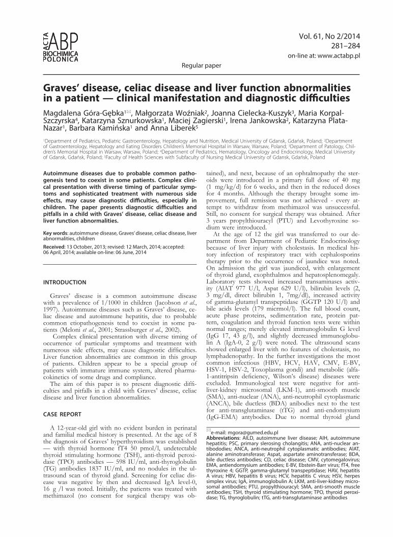
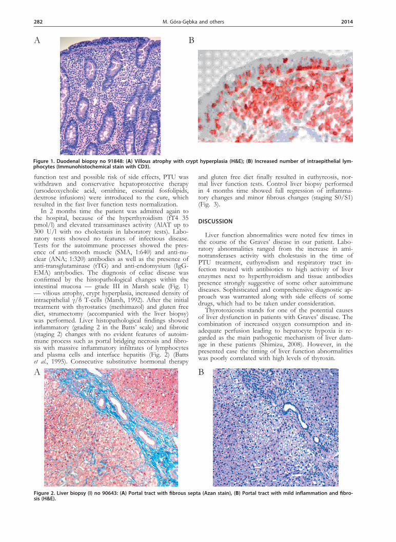
In 2 months time the patient was admitted again to the hospital, because of the hyperthyroidism (fT4 35 pmol/l) and elevated transaminases activity (AlAT up to 300 U/l with no cholestasis in laboratory tests). Labo-ratory tests showed no features of infectious disease. Tests for the autoimmune processes showed the pres-ence of anti-smooth muscle (SMA, 1:640) and anti-nu-clear (ANA; 1:320) antibodies as well as the presence of anti-transglutaminase (tTG) and anti-endomysium (IgG-EMA) antybodies. The diagnosis of celiac disease was confirmed by the histopathological changes within the intestinal mucosa — grade III in Marsh scale (Fig. 1) — vilious atrophy, crypt hyperplasia, increased density of intraepithelial γ/δ T-cells (Marsh, 1992). After the initial treatment with thyrostatics (methimazol) and gluten free diet, strumectomy (accompanied with the liver biopsy) was performed. Liver histopathological findings showed inflammatory (grading 2 in the Batts’ scale) and fibrotic (staging 2) changes with no evident features of autoim-mune process such as portal bridging necrosis and fibro-sis with massive inflammatory infiltrates of lymphocytes and plasma cells and interface hepatitis (Fig. 2) (Batts et al., 1995). Consecutive substitutive hormonal therapy
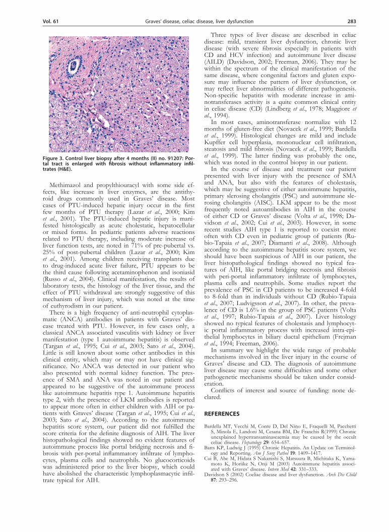
and gluten free diet finally resulted in euthyreosis, nor-mal liver function tests. Control liver biopsy performed in 4 months time showed full regression of inflamma-tory changes and minor fibrous changes (staging S0/S1) (Fig. 3).
DISCUSSION
Liver function abnormalities were noted few times in the course of the Graves’ disease in our patient. Labo-ratory abnormalities ranged from the increase in ami-notransferases activity with cholestasis in the time of PTU treatment, euthyrodism and respiratory tract in-fection treated with antibiotics to high activity of liver enzymes next to hyperthyroidism and tissue antibodies presence strongly suggestive of some other autoimmune diseases. Sophisticated and comprehensive diagnostic ap-proach was warranted along with side effects of some drugs, which had to be taken under consideration.
Thyrotoxicosis stands for one of the potential causes of liver dysfunction in patients with Graves’ disease. The combination of increased oxygen consumption and in-adequate perfusion leading to hepatocyte hypoxia is re-garded as the main pathogenic mechanism of liver dam-age in these patients (Shimizu, 2008). However, in the presented case the timing of liver function abnormalities was poorly correlated with high levels of thyroxin.
Figure 1. Duodenal biopsy no 91848: (A) Villous atrophy with crypt hyperplasia (H&E); (B) Increased number of intraepithelial lym-phocytes (Immunohistochemical stain with CD3).
A B
Figure 2. Liver biopsy (I) no 90643: (A) Portal tract with fibrous septa (Azan stain), (B) Portal tract with mild inflammation and fibro-sis (H&E).
A B
Vol. 61 283Graves’ disease, celiac disease, liver dysfunction
Methimazol and propylthiouracyl with some side ef-fects, like increase in liver enzymes, are the antithy-roid drugs commonly used in Graves’ disease. Most cases of PTU-induced hepatic injury occur in the first few months of PTU therapy (Lazar et al., 2000; Kim et al., 2001). The PTU-induced hepatic injury is mani-fested histologically as acute cholestatic, hepatocellular or mixed forms. In pediatric patients adverse reactions related to PTU therapy, including moderate increase of liver function tests, are noted in 71% of pre-pubertal vs. 25% of post-pubertal children (Lazar et al., 2000; Kim et al., 2001). Among children receiving transplants due to drug-induced acute liver failure, PTU appears to be the third cause following acetaminophenon and isoniasid (Russo et al., 2004). Clinical manifestation, the results of laboratory tests, the histology of the liver tissue, and the effect of PTU withdrawal are strongly suggestive of this mechanism of liver injury, which was noted at the time of euthyrodism in our patient.
There is a high frequency of anti-neutrophil cytoplas-matic (ANCA) antibodies in patients with Graves’ dis-ease treated with PTU. However, in few cases only, a classical ANCA associated vasculitis with kidney or liver manifestation (type 1 autoimmune hepatitis) is observed (Targan et al., 1995; Cui et al., 2003; Sato et al., 2004). Little is still known about some other antibodies in this clinical entity, which may or may not have clinical sig-nificance. No ANCA was detected in our patient who also presented with normal kidney function. The pres-ence of SMA and ANA was noted in our patient and appeared to be suggestive of the autoimmune process like autoimmune hepatitis type 1. Autoimmune hepatitis type 2, with the presence of LKM antibodies is reported to appear more often in either children with AIH or pa-tients with Graves’ disease (Targan et al., 1995; Cui et al., 2003; Sato et al., 2004). According to the autoimmune hepatitis score system, our patient did not fulfilled the score criteria for the definite diagnosis of AIH. The liver histopathological findings showed no evident features of autoimmune process like portal bridging necrosis and fi-brosis with per-portal inflammatory infiltrate of lympho-cytes, plasma cells and neutrophils. No glucocorticoids was administered prior to the liver biopsy, which could have abolished the characteristic lymphoplasmacytic infil-trate typical for AIH.
Three types of liver disease are described in celiac disease: mild, transient liver dysfunction, chronic liver disease (with severe fibrosis especially in patients with CD and HCV infection) and autoimmune liver disease (AILD) (Davidson, 2002; Freeman, 2006). They may be within the spectrum of the clinical manifestation of the same disease, where congenital factors and gluten expo-sure may influence the pattern of liver dysfunction, or may reflect liver abnormalities of different pathogenesis. Non-specific hepatitis with moderate increase in ami-notransferases activity is a quite common clinical entity in celiac disease (CD) (Lindberg et al., 1978; Maggiore et al., 1994).
In most cases, aminotransferase normalize with 12 months of gluten-free diet (Novacek et al., 1999; Bardella et al., 1999). Histological changes are mild and include Kupffer cell hyperplasia, mononuclear cell infiltration, steatosis and mild fibrosis (Novacek et al., 1999; Bardella et al., 1999). The latter finding was probably the one, which was noted in the control biopsy in our patient.
In the course of disease and treatment our patient presented with liver injury with the presence of SMA and ANA, but also with the features of cholestasis, which may be suggestive of either autoimmune hepatitis, primary slerosing cholangitis (PSC) and autoimmune sle-rosing cholangitis (AISC). LKM appear to be the most frequently noted autoantibodies in AIH in the course of either CD or Graves’ disease (Volta et al., 1998; Da-vidson et al., 2002; Cui et al., 2003). However, in some recent studies AIH type 1 is reported to coexist more often with CD even in pediatric group of patients (Ru-bio-Tapaia et al., 2007; Diamanti et al., 2008). Although according to the autoimmune hepatitis score system, we should have been suspicious of AIH in our patient, the liver histopathological findings showed no typical fea-tures of AIH, like portal bridging necrosis and fibrosis with peri-portal inflammatory infiltrate of lymphocytes, plasma cells and neutrophils. Some studies report the prevalence of PSC in CD patients to be increased 4-fold to 8-fold than in individuals without CD (Rubio-Tapaia et al., 2007; Ludvigsson et al., 2007). In other, the preva-lence of CD is 1.6% in the group of PSC patients (Volta et al., 1997; Rubio-Tapaia et al., 2007). Liver histology showed no typical features of cholestasis and lymphocyt-ic portal inflammatory process with increased intra-epi-thelial lymphocytes in biliary ductal epithelium (Frejman et al., 1994; Freeman, 2006).
In summary we highlight the wide range of probable mechanisms involved in the liver injury in the course of Graves’ disease and CD. The diagnosis of autoimmune liver disease may cause some difficulties and some other pathogenetic mechanisms should be taken under consid-eration.
Conflicts of interest and source of funding: none de-clared.
REFERENCES
Bardella MT, Vecchi M, Conte D, Del Nino E, Fraquelli M, Pacchetti S, Minola E, Landoni M, Cesana BM, De Franchis R(1999) Chronic unexplained hypertransaminaseaemia may be caused by the occult celiac disease. Hepatology 29: 654–657.
Batts KP, Ludwig J (1995) Chronic Hepatitis. An Update on Terminol-ogy and Reporting. Am J Surg Pathol 19: 1409–1417.
Cui B, Abe M, Hidata S Nakanishi S, Matsuura B, Michitaka K, Yama-moto K, Horiike N, Onji M (2003) Autoimmune hepatitis associ-ated with Graves’ disease. Intern Med 42: 331–333.
Davidson S (2002) Coeliac disease and liver dysfunction. Arch Dis Child 87: 293–296.
Figure 3. Control liver biopsy after 4 months (II) no. 91207: Por-tal tract is enlarged with fibrosis without inflammatory infil-trates (H&E).
284 2014M. Góra-Gębka and others
Diamanti A, Basso MS, Pietrobatista A, Nobili V (2008) Prevalence of celiac disease in children with autoimmune hepatitis. Dig Liv Dis 40: 965.
Freeman HJ (2006) Hepatobiliary and pancreatic disorders in celiac dis-ease. World J Gastroenterol 12: 1503–1508.
Frejman HJ, Kwan WC (1994) Occult celiac disease associated with lymphocytic sclerosing cholangitis. Can J Gastroenterol 8: 249–252.
Jacobson DL, Gange SJ, Rose NR, Graham NM.(1997) Epidemiology and estimated population burden of selected autoimmune diseases in the United States. Clin Immunol Immunopathol 84: 223–243.
Kim HJ, Kim BH, Han YS Yang I, Kim KJ, Dong SH, Kim HJ, Chang YW, Lee JI, Chang R (2001) The incidence and clinical char-acteristics of symptomatic propylthiouracyl-induced hepatic injury in patients with hyperthyroidism: a single-center retrospective study. Am J Gastroenterol 96: 165–169.
Lazar L, Kalter-Leibovici O, Pertzelan A, Weintrob N, Josefsberg Z, Phillip M (2000) Thyrotoxicosis in prepubertal children compared to with pubertal and postpubertal patients, J Clin Endocrinol Metab 85: 3678–3682.
Lindberg T, Berg NO, Borulf S, Jacobson I.(1978) Liver damage in coeliac disease or other food intolerance in childhood. Lancet 1: 390–391.
Ludvigsson JF, Elfstrom P, Broome U, Ekbom A, Montgomery SM.(2007) Celiac disease and risk of liver disease: a general popula-tion-based study. Clin Gastroenterol Hepatol 5: 63–69.
Maggiore G, Ceccarelli M, Colombo Giacomo C De, Virgilis S De, Musumeci S, Ventura A (1994) 141 Hepatic lesions in childhood ce-liac disease: a multicentric retrospective study. J Pediatr Gastroenterol Nutr 19: 691–696.
Marsh M (1002) Gluten, major histocompatibility complex and the small intestine. A molecular and immunobiologic approach to the
spectrum o gluten sensitivity(‘celiac sprue’). Gastroenterology 102: 330–354.
Meloni GF, Thomasi PA, Bertoncelli A Fanciulli G, Delitala G, Meloni T (2001) Prevalence of silent celiac disease in patients with auto-immune thyroiditis from Northern Sardinia. J Endocrinol Invest 24: 298–302.
Novacek G, Miehsler W, Wrba F, Ferenci, P, Penner E., Vogelsang H (1999) Prevalence and clinical importance of hypertransaminaseae-mia in celiac disease. Eur J Gastroenterol Hepatol 11: 283–288.
Rubio-Tapaia A, Murray J (2007) The liver in Celiac Disease. Hepatology 46: 1650–1658
Russo MW, Galanko JA, Shrestha R, Fried MW, Watkins P.(2004) Liver transplantation for acute liver failure from drug induced liver injury in the Unitated States. Liver Transplant 10: 1018–1023.
Sato H, Hattori M, Fujieda M, Sugihara S, Inomata H, Hoshi M, Miy-amoto S (2000) High prevalence of antineutrophil cytoplasmatic antibody in childhood onset Graves’ disease treatment with propyl-tiouracil. J Cin Endocrinol Metab 85: 4270–4273.
Shimizu Y (2008). Liver in systemic disease World J Gastroenterol 14: 4111–4119.
Strassburg CP, Manns MP( 2002) Autoantibodies and autoantigens in autoimmune hepatitis. Sem Liver Dis 22: 339–352.
Targan SR, Landers C, Vidrich A, Czaja AJ (1995) High-titer antineu-trophil cytoplasmatic antibodies in type-1 autoimmune hepatitis. Gastroenterology 108: 1159–1166.
Volta U, De Franceschi L, Molinaro N, Cassani F, Muratori L, Lenzi M, Bianchi FB, Czaja AJ (1998) Frequency and significance of anti-gliadin and anti-endomysial antibodies in autoimmune hepatitis. Dig Dis Sci 43: 2190–2195.