glucose biosensors based on ag nanoparticles modified tio2 nanotube arrays
TRANSCRIPT
ORIGINAL PAPER
Glucose biosensors based on Ag nanoparticles modified TiO2
nanotube arrays
Chunxiao Feng & Guangqing Xu & Haipeng Liu & Jun Lv &
Zhixiang Zheng & Yucheng Wu
Received: 16 May 2013 /Revised: 15 August 2013 /Accepted: 8 September 2013 /Published online: 21 September 2013# Springer-Verlag Berlin Heidelberg 2013
Abstract A GOx/Ag/TiO2 glucose biosensor was achievedby photoreducing Ag nanoparticles on TiO2 nanotube arrays(NTAs) following with adsorption of GOx. The morphology,structure, and element component of Ag/TiO2 NTAs werecharacterized by scanning electron microscope, transmissionelectron microscope, and X-ray diffraction. Ag nanoparticleswere uniformly deposited on surface of TiO2 NTAs withaverage size of 15 nm and the size and distribution changedwith the immersing time of TiO2 NTAs in AgNO3 aqueoussolution. Electrochemical properties of Ag/TiO2 NTAs werecharacterized by cyclic voltammetry and amperometric detec-tion of H2O2, revealing that TiO2 NTAs with immersing timeof 30 min achieve the best electrochemical activity. The GOx/Ag/TiO2 NTAs biosensor with optimum conditions achieves asensitivity of 0.39μAmM−1 cm−2 with liner range from 0.1 to4 mM.
Keywords TiO2nanotubearrays .Agnanoparticles .Glucosebiosensors
Introduction
Amperometric biosensors based on glucose oxidase (GOx)have attracted great attention since their development byLeland et al. [1]. The sensitivity, response speed, and selec-tivity of the enzyme biosensors are always highly related tothe electrode surface area; therefore, various methods havebeen used to increase the electrode surface area, such as themodification of electrode with carbon nanofibers [2, 3],
carbon nanotubes [4, 5], and using nanoporous electrode [6,7]. However, the considerable toxicity of carbon materials isstill a controversial issue and limits its biological application[8]. In recent years, anodized TiO2 nanotube arrays (NTAs)attract increasing attention for enzyme biosensors due to thesuperior properties such as easy preparation, large specificarea, chemical inertness, and excellent biocompability [9–11].
It has been demonstrated that electrodes based on combi-nation of metal nanoparticles and TiO2 nanomaterials exhibithighly sensitive and selective response to glucose. Kang et al.[12] fabricated an enzyme biosensor by modifying TiO2
nanotubes with Au and Pt nanoparticles, of which the sensitiv-ity to H2O2 is 2.92 μAmM−1 and to glucose is 0.08 μAmM−1.Han et al. [13] synthesized Pt doped TiO2 nanotubes catalystsfor glucose determination, achieving a detection limit of 1 mΜat signal-to-noise ratio of 3, almost ten times lower than that ofun-doped TiO2 nanotubes catalysts. Results indicate that noblemetals such as Au and Pt nanoparticles play an important rolein the modified TiO2 nanotubes catalysts ascribe to their ex-cellent conductivity, catalytic property, and biocompatibility.They can perform as “electron transfer channel” to enhance theelectron transfer between redox center in enzyme and electrodesurface and as catalysts to increase electrochemical reactions.Ag nanoparticles, having a similar electrochemical propertywith Au and Pt nanoparticles, is the best conductor amongmetals and may assist more efficient electron transfer thanother nanoparticles in biosensors. In addition, the simple prep-aration and low cost make Ag nanoparticles to be a promisingmaterial for sensors. Though Ag nanoparticles applied in elec-trochemical sensors have been investigated [14–16], TiO2
NTAs modified with Ag nanoparticles is seldom reported.In this work, Ag nanoparticles were loaded on TiO2 NTAs
by photocatalytic reduction to form Ag/TiO2 nanocompositeelectrodes and the electrochemical response to H2O2 wasinvestigated by clyclic voltammetry and amperometry. En-zyme biosensors were prepared by immobilizing Ag/TiO2
C. Feng :G. Xu (*) :H. Liu : J. Lv : Z. Zheng :Y. Wu (*)Laboratory of Functional Nanomaterials and Devices, School ofMaterials Science and Engineering, Hefei University of Technology,Hefei 230009, Chinae-mail: [email protected]: [email protected]
J Solid State Electrochem (2014) 18:163–171DOI 10.1007/s10008-013-2257-2
NTAs with GOx, of which the sensitivity was obtained byamperometric response to successively injection of glucose.
Experimental
Chemicals
GOx was purchased from Sigma and used as received. Theenzyme solution was prepared by dissolving GOx in 0.05 Mphosphate buffer solution (PBS) to make a 500 U mL−1 solu-tion and was kept at 4 °C in the fridge. Ammonium fluoride,ethylene glycol, silver nitrate, hydrogen peroxide, glucose,phosphoric acid, and other chemicals, of analytical reagentgrade, were purchased fromEnterprise group chemical reagentCo., Ltd. All the solutions were preparedwith deionized water.
Titanium sheets (0.1 mm thickness, 99.6 % purity) werepurchased from Beijing Cuibolin Non-Ferrous TechnologyDeveloping Co., Ltd. and used as received. A 0.05 M PBSconsisting of Na2HPO4 and NaH2PO4 was employed assupporting electrolyte.
Instruments
Anodization of TiO2 NTAs was performed in a self-madeelectrolytic cell with a traditional two electrode system(DH1722A-3), and Ag modification was carried out with aultraviolet lamp (luminous power of 600 mW cm−2). Mor-phology investigation of the as-prepared samples was obtainedwith scanning electron microscope (SEM, SU8020) and trans-mission electron microscope (TEM, JAPAN/HITACHIH800). The valency of Ag particles on the TiO2 was deter-mined by an X-ray photoelectron spectroscophotometer (XPS,CALAB250). Cyclic voltammetric experiments and glucose(or H2O2) detection were performed with a three-electrodeelectrochemical workstation (CHI660D), comprising of anAg/AgCl (3M KCl) reference electrode, a Pt/Ti wire auxiliaryelectrode and a Pt disc modified with Ag/TiO2 NTAs (or GOx/Ag/TiO2 NTAs) working electrode.
Fabrication of TiO2 NTAs
TiO2 NTAs were fabricated with an anodic oxidation methodcarried out as following specific steps. Prior to anodization,titanium foils with diameter of 2 cm were ultrasonicallycleaned in acetone and ethanol for each 15 min, respectively.The cleaned titanium foils were anodized in an electrolytecontaining 0.15 M NH4F ethylene glycol solution at roomtemperature for 6 h in a two electrode configuration with agraphite cathode for which one side of the titanium foil waspartially immersed in electrolyte, with the back un-anodizedused as an electrical contact. Then, the oxidation films wereremoved from the titanium substrate by rinsing with deionized
water and TiO2 NTAs without barrier layer were thusachieved. The vertical nanotube arrays formed on the titaniumsubstrate were amorphous, exhibiting relatively good conduc-tivity in potential electric application [17, 18].
Modification with Ag nanoparticles and electrochemicalmeasurements
Ag nanoparticles loaded on TiO2 NTAs were carried out withan ultraviolet lamp with a fixed wavelength of 365 nm. Asprecursor and hole sacrifice agent, 5 mM AgNO3 and 1 Mmethanol aqueous solution were used, respectively. To adjustthe pH value of about 8~9, 1 M NH3·H2O was applied. TiO2
NTAs were subsequently immersed in the solution prior tophotoreduction, which was kept at 50 °C for different timeranging from 5 to 60 min, following with illumination underan UV lamp for 30 min. The size and distribution of Agnanoparticles loaded on TiO2 nanotubes were controlled bythe immersing time.
The catalytic activity of Ag/TiO2 NTAs electrode in oxida-tion of H2O2 was determined by cyclic voltammetry in 50mMPBS (pH 7) containing H2O2 of different concentrations rang-ing from 0 to 10 mM at a scan rate of 10 mV s−1, andamperometric detection was performed at an optimized poten-tial of 0.7 V with successively injection of 0.1 mM H2O2. Allthe measurements were performed in a conventional three-electrode system with an Ag/TiO2 NTAs working electrode,a Pt/Ti auxiliary electrode, and an Ag/AgCl (3 M KCl) refer-ence electrode.
GOx immobilization and glucose detection
Physical adsorption was used for GOx immobilization toachieve GOx/Ag/TiO2 NTAs electrode. The enzyme solutionwas prepared by dissolving certain amount of GOx in 10 mL50 mM PBS to achieve a concentration of 500 U mL−1.Subsequently, 10 μL of the prepared solution was droppedonto the Ag/TiO2 NTAs electrode, which was dried in the airand kept in fridge at 4 °C overnight following with rinsing inPBS to remove free GOx. Cyclic voltammetry was carried outin 4 mL PBS in the presence and absence of glucose, andamperometric response was performed under constant stirring(100 rpm) at room temperature to the changes of glucoseconcentration.
Results and discussion
Characterization of Ag/TiO2 NTAs
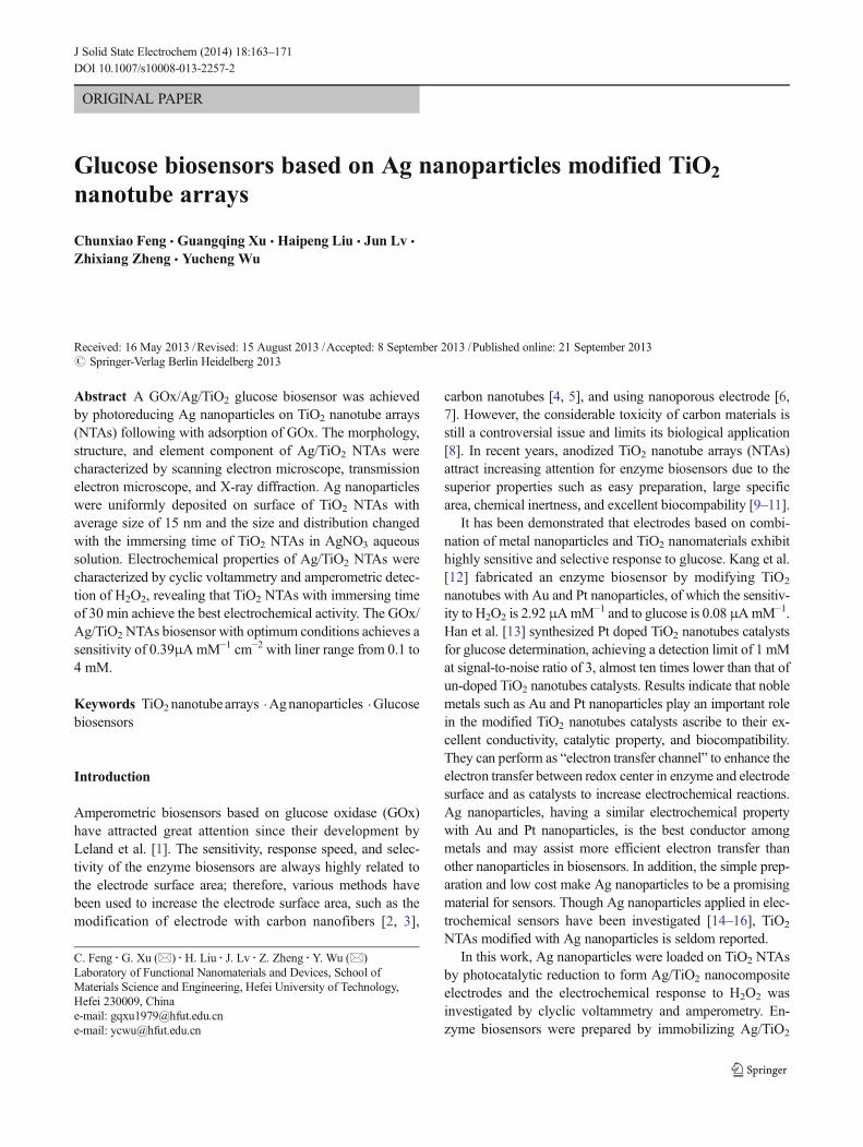
SEM and EDS are two frequently used approaches for micro-structure and composition characterization of the as synthe-sized Ag/TiO2 NTAs. Figure 1 shows the morphologies of
164 J Solid State Electrochem (2014) 18:163–171
TiO2 NTAs and Ag/TiO2 NTAs. TiO2 nanotubes of about110 nm in inner diameter and a length of 15 μm uniformlystand on Ti substrate (Fig. 1a). The relatively large pore sizeand regularly hollow structure are attributed to the matureanodic oxidation process bywhich the TiO2 nanotubes formedsteadily under the electric field force with presence of F− andH+. Figure 1b reveals TiO2 nanotubes modified with Agnanoparticles. The inner diameter of TiO2 nanotubes is small-er than that of TiO2 nanotubes unmodified from the top sideview due to the uniform distribution of Ag nanoparticles onthe mouth of the TiO2 nanotubes, which is verified further byFig. 1c. Figure 1c shows a view of the above sample inclinedat 45°, and Ag nanoparticles with size ranging from 10 to18 nm distributed uniformly on TiO2 nanotubes both of theinner and outer walls are obviously observed. The results aredifferent from former report [19] that the Ag nanoparticlesmainly loaded onto the outer surface due to the inconveniencefor Ag+ traveling into the nanotubes of small size in diameter(~10 nm). The corresponding EDX spectrum given in Fig. 1dreveals the presence of Ti, O, Ag, and C, with a Ti:O atomicratio of 1:2 and a Ag:Ti atomic ratio of 1:34. The uniformlydispersed small Ag nanoparticles are essential to a high cata-lytic activity.
With the purpose of achieving TiO2 NTAs decorated withdispersed Ag nanoparticles, TiO2 nanotubes immersed inAgNO3 aqueous solution for different time was investigated,as shown by TEM morphologies in Fig. 2. The loadingdensity and contents of Ag particles can be roughly estimatedaccording to the number and the size of the particles on theTiO2 nanotubes.
As it is shown, TiO2 nanotubes of 150 nm in diameter withsmooth surface are obtained (Fig. 2a) and almost no particlescan be seen when the immersing time is 5 min (Fig. 2b), then
the particles are sporadically distributed on the nanotubes astime increase to 15 min (Fig. 2c). In particular, highly dis-persed Ag nanoparticles of size ranging from 10 to 18 nm indiameter are clearly seen when the immersing time keeps30 min and the black cluster is due to the overlying of brokennanotubes during the ultrasonic process for TEM test, asshown in Fig. 2d. However, when the immersing time in-creases to 60 min, Ag nanoparticles agglomerate to formcluster and size reaches up to 40 nm (shown in Fig. 2e). Theresults are possible to be associated with Brownian movementand capillarity. As time increases, Ag+ ions and Ag2O crys-tallites make a moderate contact with nanotubes on atemperature-controlled (50 °C) condition, thus, highly dis-persible Ag nanoparticles can be achieved. Nevertheless, astime increases further, Ag2O crystallite severely agglomerate,resulting in large size of Ag nanoparticles after beingphotoreduced. The conglobated Ag nanoparticles tend to bethe mass-transfer-block, and the electronic signal acquired byexternal circuit thus is held up, which may cause decrease insensitivity of biosensors.
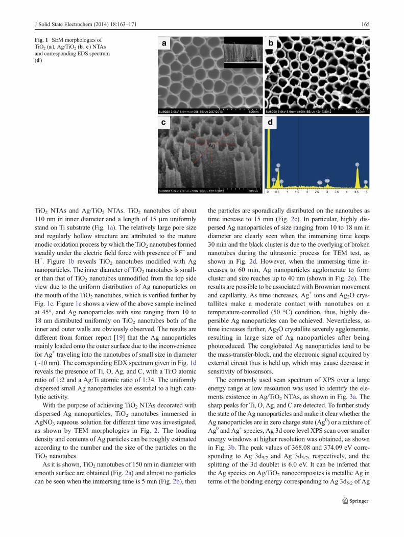
The commonly used scan spectrum of XPS over a largeenergy range at low resolution was used to identify the ele-ments existence in Ag/TiO2 NTAs, as shown in Fig. 3a. Thesharp peaks for Ti, O, Ag, and C are detected. To further studythe state of the Ag nanoparticles and make it clear whether theAg nanoparticles are in zero charge state (Ag0) or a mixture ofAg0 and Ag+ species, Ag 3d core level XPS scan over smallerenergy windows at higher resolution was obtained, as shownin Fig. 3b. The peak values of 368.08 and 374.09 eV corre-sponding to Ag 3d5/2 and Ag 3d3/2, respectively, and thesplitting of the 3d doublet is 6.0 eV. It can be inferred thatthe Ag species on Ag/TiO2 nanocomposites is metallic Ag interms of the bonding energy corresponding to Ag 3d5/2 of Ag
a b
c d
Fig. 1 SEM morphologies ofTiO2 (a), Ag/TiO2 (b , c) NTAsand corresponding EDS spectrum(d)
J Solid State Electrochem (2014) 18:163–171 165

(Ag0 368.25 eV), while the energy of Ag+ in Ag2O is367.70 eV [20]. Results demonstrate that Ag mainly existsin the Ag0 state on the TiO2 nanotube surface instead of Ag+
species [21]. The highest peak corresponding to Ag 3d otherthan O 1s is attributed to the XPS technology by whichsamples can be tested for a surface thickness of ~10 nm, whilethe surface of the nanotubes is modified with uniformly dis-tributed Ag nanoparticles (~15 nm), which is in accordancewith Fig. 1b, c.
The formation process of TiO2 NTAs modified with dis-persible Ag nanoparticles can be explained as following. Onthe one hand, a controlled temperature (using water bath) washeld at 50 °C when TiO2 NTAs were immersed in AgNO3
aqueous solution to accelerate the thermal motion of Ag+ ionsand induce an adequate contact between TiO2 nanotubes andAgNO3 solution. On the other hand, the ultraviolet lamp withfixed wavelength of 365 nm provides reliable energy forAg2O reducing to Ag0. The main reduction mechanism isgiven below.
Agþ→Agþadsorbed ð1Þ
Ag+ ions are inclined to be adsorbed onto the outer walls ofTiO2 nanotubes at room temperature, but the Brownian
movement is aggravated by a controlled temperature at50 °C, which gives promotion for adsorption of Ag+ ions ontoboth of the inner and outer walls.
2Agþadsorbed þ 2OH−→Ag2Oþ H2O ð2ÞTo adjust pH value to 8~9, 1 M NH3·H2O was introduced
and the Ag+adsorbed reacted withOH− to form sparingly soluble
Ag2O, but not obvious precipitation.
2Ag2O→hv4Ag þ O2 ð3Þ
Subsequently, the resulted Ag2O particles were reducedinto Ag0 by ultraviolet lamp. As for the Ag2O particles locat-ing inside TiO2 nanotubes, the orderly hollow structure whichallows for UV light penetration are favorable for theirreduction.
Electrochemical activity of Ag/TiO2 NTAs
In this work, the voltammetric behaviors of the pristine TiO2
NTAs and Ag/TiO2 nanocomposite electrodes were investi-gated by cyclic voltammetry in potentials ranging from −0.4to 0.7 V at a scan rate of 10 mV s−1. As the intermediateproduct for glucose oxidation, H2O2 was used as probe for
Fig. 2 TEM images of Agdeposition for differentimmersing time: 0 min (a), 5 min(b), 15 min (c), 30 min (d), and60 min (e)
166 J Solid State Electrochem (2014) 18:163–171
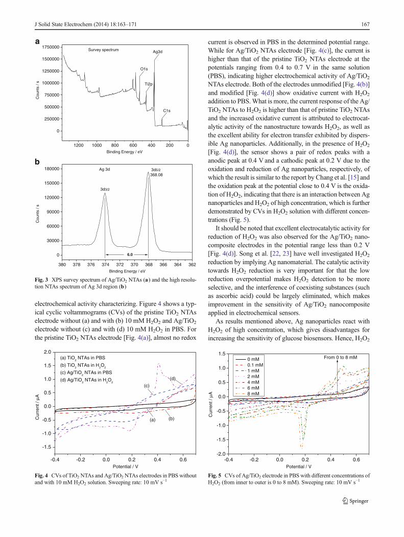
electrochemical activity characterizing. Figure 4 shows a typ-ical cyclic voltammograms (CVs) of the pristine TiO2 NTAselectrode without (a) and with (b) 10 mM H2O2 and Ag/TiO2
electrode without (c) and with (d) 10 mM H2O2 in PBS. Forthe pristine TiO2 NTAs electrode [Fig. 4(a)], almost no redox
current is observed in PBS in the determined potential range.While for Ag/TiO2 NTAs electrode [Fig. 4(c)], the current ishigher than that of the pristine TiO2 NTAs electrode at thepotentials ranging from 0.4 to 0.7 V in the same solution(PBS), indicating higher electrochemical activity of Ag/TiO2
NTAs electrode. Both of the electrodes unmodified [Fig. 4(b)]and modified [Fig. 4(d)] show oxidative current with H2O2
addition to PBS.What is more, the current response of the Ag/TiO2 NTAs to H2O2 is higher than that of pristine TiO2 NTAsand the increased oxidative current is attributed to electrocat-alytic activity of the nanostructure towards H2O2, as well asthe excellent ability for electron transfer exhibited by dispers-ible Ag nanoparticles. Additionally, in the presence of H2O2
[Fig. 4(d)], the sensor shows a pair of redox peaks with aanodic peak at 0.4 V and a cathodic peak at 0.2 V due to theoxidation and reduction of Ag nanoparticles, respectively, ofwhich the result is similar to the report by Chang et al. [15] andthe oxidation peak at the potential close to 0.4 V is the oxida-tion of H2O2, indicating that there is an interaction between Agnanoparticles and H2O2 of high concentration, which is furtherdemonstrated by CVs in H2O2 solution with different concen-trations (Fig. 5).
It should be noted that excellent electrocatalytic activity forreduction of H2O2 was also observed for the Ag/TiO2 nano-composite electrodes in the potential range less than 0.2 V[Fig. 4(d)]. Song et al. [22, 23] have well investigated H2O2
reduction by implying Ag nanomaterial. The catalytic activitytowards H2O2 reduction is very important for that the lowreduction overpotential makes H2O2 detection to be moreselective, and the interference of coexisting substances (suchas ascorbic acid) could be largely eliminated, which makesimprovement in the sensitivity of Ag/TiO2 nanocompositeapplied in electrochemical sensors.
As results mentioned above, Ag nanoparticles react withH2O2 of high concentration, which gives disadvantages forincreasing the sensitivity of glucose biosensors. Hence, H2O2
-0.4 -0.2 0.0 0.2 0.4 0.6
-1.5
-1.0
-0.5
0.0
0.5
1.0
1.5
2.0
Cur
rent
/ μA
Potential / V
(b)
(c)
(a)
(d)
(a) TiO2 NTAs in PBS
(b) TiO2 NTAs in H
2O
2
(c) Ag/TiO2 NTAs in PBS
(d) Ag/TiO2 NTAs in H
2O
2
Fig. 4 CVs of TiO2 NTAs and Ag/TiO2 NTAs electrodes in PBS withoutand with 10 mM H2O2 solution. Sweeping rate: 10 mV s−1
-0.4 -0.2 0.0 0.2 0.4 0.6-2.0
-1.5
-1.0
-0.5
0.0
0.5
1.0
1.5 0 mM 0.1 mM 1 mM 2 mM 4 mM 6 mM 8 mM
Cur
rent
/ μA
Potential / V
From 0 to 8 mM
Fig. 5 CVs of Ag/TiO2 electrode in PBS with different concentrations ofH2O2 (from inner to outer is 0 to 8 mM). Sweeping rate: 10 mV s−1
1200 1000 800 600 400 200 0
0
250000
500000
750000
1000000
1250000
1500000
1750000
Cou
nts
/ s
Binding Energy / eV
O1s
Ti2p
Ag3d
C1s
aSurvey spectrum
380 378 376 374 372 370 368 366 364 362
0
30000
60000
90000
120000
150000
180000
Cou
nts
/ s
Binding Energy / eV
6.0
3d5/2
368.08
3d3/2
Ag 3d
b
Fig. 3 XPS survey spectrum of Ag/TiO2 NTAs (a) and the high resolu-tion NTAs spectrum of Ag 3d region (b)
J Solid State Electrochem (2014) 18:163–171 167
of different concentration was detected by cyclic voltammetryat a sweeping rate of 10 mV s−1 shown in Fig. 5. None of anyredox peaks is obtained for Ag/TiO2 NTAs electrode towardsH2O2 in the concentration range from 0.1 to 2 mM, but theoxidative peaks at ~0.5 V appear evidently as the H2O2
concentration further increase due to the oxidation of H2O2.At the same time, almost no redox reaction of Ag nanoparticlescan be observed until the concentration of H2O2 is higher than4 mM for which the oxidative peaks at 0.4 Vand the reductiveat 0.2 V turn to be more obvious. This gives an indication ofthe interaction between Ag nanoparticles and H2O2 of highconcentration (higher than 4 mM) due to its strong oxidability,but the interaction can be ignorable for the Ag/TiO2 electrodesin addition with H2O2 of low concentration. Additionally, theoxidation of Ag nanoparticles in presence of H2O2 occurs inpotentials ranging from 0.35 to 0.45 Vand so it is necessary toavoid this region in H2O2 and glucose determination. Since thecurrent response towards H2O2 oxidation increases with anincreasing potential in the determined potential range, 0.7 V ischosen to be the applied potential in the following amperometrydetection.
As the GOx/Ag/TiO2 electrode towards glucose is a surface-controlled process, the H2O2 generated in the glucose oxidationreaction by GOx is of a miniscule amount, which makes littlenegative influence on oxidation of Ag nanoparticles. There-fore, the excellent conductive ability for electron transfer andpositive electrocatalytic effects of Ag nanoparticles on glucosedetection can be taken to the extreme, thus, to provide anefficient way for enzyme biosensor via application of Ag/TiO2 nanocomposite matrix.
The content and distribution of Ag nanoparticles on TiO2
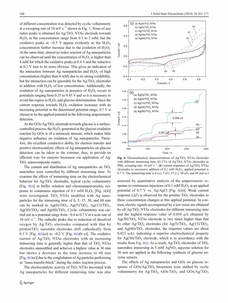
nanotubes were controlled by different immersing time. Toexamine the effects of immersing time on the electrochemicalbehavior for Ag/TiO2 electrodes, typical cyclic voltammetry[Fig. 6(i)] in buffer solution and chronoamperometric res-ponse to continuous injection of 0.1 mM H2O2 [Fig. 6(ii)]were investigated. TiO2 NTAs modified with Ag nano-particles for the immersing time of 0, 5, 15, 30, and 60 mincan be marked as Ag(0)/TiO2, Ag(5)/TiO2, Ag(15)/TiO2,Ag(30)/TiO2, and Ag(60)/TiO2. Cyclic voltammetry was car-ried out in a potential range from −0.4 to 0.7 Vat a scan rate of10 mV s−1. The cathodic peaks due to reduction of dissolvedoxygen by Ag/TiO2 electrodes compared with that bypristineTiO2 nanotube electrodes shift cathodically from0.1 V [Fig. 6(i)(a)] to ~0.2 V [Fig. 6(i)(b–e)]. The oxidativecurrent of Ag/TiO2 NTAs electrodes with an increasingimmersing time is generally higher than that of TiO2 NTAselectrodes unmodified and achieves a highest value at 30 minbut shows a decrease as the time increase to 60 min[Fig. 6(i)(e)] due to the conglobation of Ag particles performingas “mass-transfer-block” during the redox reaction process.
The electrocatalytic activity of TiO2 NTAs decorated withAg nanoparticles for different immersing time was also
assessed by quantitative analysis of the amperometric re-sponse to continuous injections of 0.1 mMH2O2 at an appliedpotential of 0.7 V vs. Ag/AgCl [Fig. 6(ii)]. Weak currentresponse (ΔI ) is observed for the pristine TiO2 electrodes tothese concentration changes at this applied potential. In con-trast, electric signals accompanied by a low noise are obtainedby all Ag/TiO2 NTAs electrodes for different immersing timeand the highest response value of 0.058 μA obtained byAg(30)/TiO2 NTAs electrode is two times higher than thatby other Ag/TiO2 electrodes (for Ag(5)/TiO2, Ag(15)/TiO2,and Ag(60)/TiO2 electrodes, the response values are about0.021 μA), indicating a superior electrochemical propertyfor Ag(30)/TiO2 electrode, which is in accordance with theresults from Fig. 6(i). As a result, Ag/TiO2 electrodes of TiO2
nanotubes immersing in 5 mM AgNO3 aqueous solution for30 min are applied in the following synthesis of glucose en-zyme sensors.
The effects of Ag nanoparticles and GOx on glucose re-sponse of GOx/Ag/TiO2 biosensors were studied by cyclicvoltammetry for Ag/TiO2, GOx/TiO2, and GOx/Ag/TiO2
-0.4 -0.2 0.0 0.2 0.4 0.6-0.4
-0.2
0.0
0.2
0.4
0.6(a) Ag(0)/TiO
2 NTAs
(b) Ag(5)/TiO2 NTAs
(c) Ag(15)/TiO2 NTAs
(d) Ag(30)/TiO2 NTAs
(e) Ag(60)/TiO2 NTAs (e)
(d)
(c)
(b)
Potential / V
(a)
(i)
1000 1500 2000 2500 3000
0.00
0.05
0.10
0.15
0.20
0.25
0.30
(a) Ag(0)/TiO2 NTAs
(b) Ag(5)/TiO2 NTAs
(c) Ag(15)/TiO2 NTAs
(d) Ag(30)/TiO2 NTAs
(e) Ag(60)/TiO2 NTAs
(e)
(d)
(c)
(b)
Cur
rent
/ μA
C
urre
nt /
μA
Time / s
(a)
(ii)
0.058 μA
0.021 μA
Fig. 6 Electrochemical characterizations of Ag/TiO2 NTAs electrodeswith different immersing time, (i) CVs of Ag/TiO2 NTAs electrodes inPBS, sweeping rate: 10 mV s−1; (ii) current responses of Ag/TiO2 NTAselectrodes to successive addition of 0.1 mM H2O2, applied potential is0.7 V. The immersing time is 0 (a), 5 (b), 15 (c), 30 (d), and 60 min (e)
168 J Solid State Electrochem (2014) 18:163–171
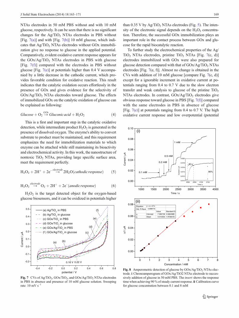
NTAs electrodes in 50 mM PBS without and with 10 mMglucose, respectively. It can be seen that there is no significantchanges for the Ag/TiO2 NTAs electrodes in PBS without[Fig. 7(a)] and with [Fig. 7(b)] 10 mM glucose, which indi-cates that Ag/TiO2 NTAs electrodes without GOx immobili-zation give no response to glucose in the applied potential.Comparatively, evident oxidative current response appears forthe GOx/Ag/TiO2 NTAs electrodes in PBS with glucose[Fig. 7(f)] compared with the electrodes in PBS withoutglucose [Fig. 7(e)] at potentials higher than 0.4 V accompa-nied by a little decrease in the cathodic current, which pro-vides favorable condition for oxidative reaction. This resultindicates that the catalytic oxidation occurs effortlessly in thepresence of GOx and gives evidence for the selectivity ofGOx/Ag/TiO2 NTAs electrodes toward glucose. The effectsof immobilized GOx on the catalytic oxidation of glucose canbe explained as following:
Glucoseþ O2 →GOx
Gluconicacid þ H2O2 ð4ÞThis is a first and important step in the catalytic oxidative
detection, while intermediate product H2O2 is generated in thepresence of dissolved oxygen. The enzyme's ability to convertsubstrate to product must be maintained, and this requirementemphasizes the need for immobilization materials to whichenzyme can be attached while still maintaining its bioactivityand electrochemical activity. In this work, the nanostructure ofnontoxic TiO2 NTAs, providing large specific surface area,meet the requirement perfectly.
H2O2 þ 2Hþ þ 2e− →electrode
2H2O cathodic responseð Þ ð5Þ
H2O2 →electrode
O2 þ 2Hþ þ 2e− anodicresponseð Þ ð6ÞH2O2 is the target detected object for the oxygen-based
glucose biosensors, and it can be oxidized in potentials higher
than 0.35 V by Ag/TiO2 NTAs electrodes (Fig. 5). The inten-sity of the electronic signal depends on the H2O2 concentra-tion. Therefore, the successful GOx immobilization plays animportant role in the contact process between GOx and glu-cose for the rapid biocatalytic reaction.
To further study the electrochemical properties of the Ag/TiO2 NTAs electrodes, pristine TiO2 NTAs [Fig. 7(c, d)]electrodes immobilized with GOx were also prepared forglucose detection compared with that of GOx/Ag/TiO2 NTAselectrodes [Fig. 7(e, f)]. Almost no change is obtained in theCVs with addition of 10 mM glucose [compare Fig. 7(c, d)]except for a ignorable increment in oxidative current at po-tentials ranging from 0.4 to 0.7 V due to the slow electrontransfer and weak catalysis to glucose of the pristine TiO2
NTAs electrodes. In contrast, GOx/Ag/TiO2 electrodes giveobvious response toward glucose in PBS [Fig. 7(f)] comparedwith the same electrodes in PBS in absence of glucose[Fig. 7(e)] at potentials ranging from 0.4 to 0.7 V. The highoxidative current response and low overpotential (potential
-0.4 -0.2 0.0 0.2 0.4 0.6 0.8
-0.2
-0.1
0.0
0.1
0.2
0.3
0.4
0.5
Cur
rent
/ μA
potential / V
(a) Ag/TiO2 in PBS
(b) Ag/TiO2 in glucose
(c) GOx/TiO2 in PBS
(d) GOx/TiO2 in glucose
(e) GOx/Ag/TiO2 in PBS
(f ) GOx/Ag/TiO2 in glucose
(a) (b)
(c)
(d)
(e)
(f)
0.16 V 0.22 V
Fig. 7 CVs of Ag/TiO2, GOx/TiO2, and GOx/Ag/TiO2 NTAs electrodesin PBS in absence and presence of 10 mM glucose solution. Sweepingrate: 10 mV s−1
1000 1500 2000 2500 3000 3500 4000
1 mM
0.5 mM
0.2 mM
Cur
rent
/ μA
Time / s
0.1 mM
(i)
2000 2050 2100 21500.010
0.012
0.014
0.016
0.018
0.020
Cur
rent
/ μA
Time/ s
40 s
90%
0 1 2 3 4 5 6 7 8
ΔI /
μA
Concentration / mM
Equation y = a + b*x
Adj. R-Squa 0.998715
Value Standard Er
a Intercept -7.992728 3.566498E-
b Slope 0.014554 1.972651E-
(ii)
0.00
0.02
0.04
0.06
0.08
0.10
0.00
0.02
0.04
0.06
0.08
Fig. 8 Amperometric detection of glucose by GOx/Ag/TiO2 NTAs elec-trode. i Chronoamperogram of GOx/Ag/TiO2 NTAs electrode to succes-sively addition of glucose in 50 mM PBS. The insert shows the responsetimewhen achieving 90% of steady current response. ii Calibration curvefor glucose concentration between 0.1 and 8 mM
J Solid State Electrochem (2014) 18:163–171 169
from 0.16 to 0.22 V) for reduction of dissolved O2 of GOx/Ag/TiO2 electrodes compared with that of GOx/TiO2 elec-trodes give evidence for the electrocatalytic activity of Agnanoparticles on the enzyme glucose biosensors. On the onehand, Ag particles of nanostructure possess nanometer cata-lytic effect for reduced–oxidized reaction of H2O2 and the sizeof about 15 nm in this study not only increase surface area forGOx immobilization but also improve the reaction activity ofH2O2 oxidation as mentioned above, which is similar to thatreported by Campbell et al. [24]. On the other hand, Ag is thebest conductor among metals and so the dispersible Agnanoparticles may assist more efficient electron transfer thanother nanoparticles in biosensors.
A glucose biosensor was fabricated by modified Ag/TiO2
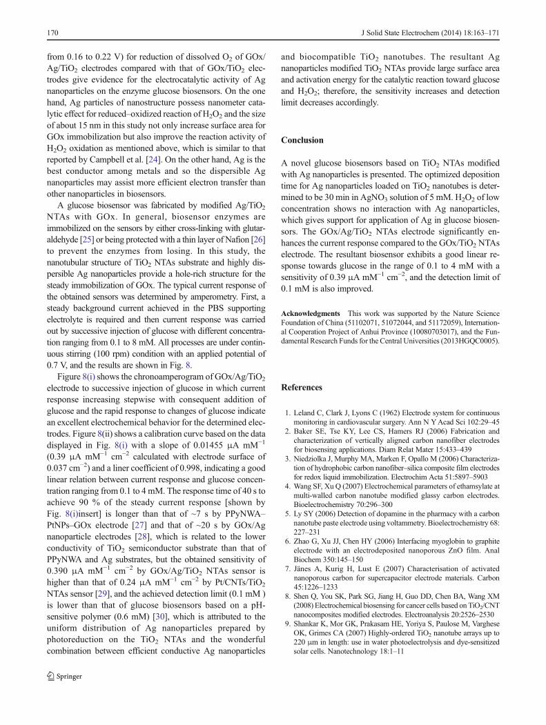
NTAs with GOx. In general, biosensor enzymes areimmobilized on the sensors by either cross-linking with glutar-aldehyde [25] or being protected with a thin layer of Nafion [26]to prevent the enzymes from losing. In this study, thenanotubular structure of TiO2 NTAs substrate and highly dis-persible Ag nanoparticles provide a hole-rich structure for thesteady immobilization of GOx. The typical current response ofthe obtained sensors was determined by amperometry. First, asteady background current achieved in the PBS supportingelectrolyte is required and then current response was carriedout by successive injection of glucose with different concentra-tion ranging from 0.1 to 8 mM. All processes are under contin-uous stirring (100 rpm) condition with an applied potential of0.7 V, and the results are shown in Fig. 8.
Figure 8(i) shows the chronoamperogram of GOx/Ag/TiO2
electrode to successive injection of glucose in which currentresponse increasing stepwise with consequent addition ofglucose and the rapid response to changes of glucose indicatean excellent electrochemical behavior for the determined elec-trodes. Figure 8(ii) shows a calibration curve based on the datadisplayed in Fig. 8(i) with a slope of 0.01455 μA mM−1
(0.39 μA mM−1 cm−2 calculated with electrode surface of0.037 cm−2) and a liner coefficient of 0.998, indicating a goodlinear relation between current response and glucose concen-tration ranging from 0.1 to 4 mM. The response time of 40 s toachieve 90 % of the steady current response [shown byFig. 8(i)insert] is longer than that of ~7 s by PPyNWA–PtNPs–GOx electrode [27] and that of ~20 s by GOx/Agnanoparticle electrodes [28], which is related to the lowerconductivity of TiO2 semiconductor substrate than that ofPPyNWA and Ag substrates, but the obtained sensitivity of0.390 μA mM−1 cm−2 by GOx/Ag/TiO2 NTAs sensor ishigher than that of 0.24 μA mM−1 cm−2 by Pt/CNTs/TiO2
NTAs sensor [29], and the achieved detection limit (0.1 mM )is lower than that of glucose biosensors based on a pH-sensitive polymer (0.6 mM) [30], which is attributed to theuniform distribution of Ag nanoparticles prepared byphotoreduction on the TiO2 NTAs and the wonderfulcombination between efficient conductive Ag nanoparticles
and biocompatible TiO2 nanotubes. The resultant Agnanoparticles modified TiO2 NTAs provide large surface areaand activation energy for the catalytic reaction toward glucoseand H2O2; therefore, the sensitivity increases and detectionlimit decreases accordingly.
Conclusion
A novel glucose biosensors based on TiO2 NTAs modifiedwith Ag nanoparticles is presented. The optimized depositiontime for Ag nanoparticles loaded on TiO2 nanotubes is deter-mined to be 30 min in AgNO3 solution of 5 mM. H2O2 of lowconcentration shows no interaction with Ag nanoparticles,which gives support for application of Ag in glucose biosen-sors. The GOx/Ag/TiO2 NTAs electrode significantly en-hances the current response compared to the GOx/TiO2 NTAselectrode. The resultant biosensor exhibits a good linear re-sponse towards glucose in the range of 0.1 to 4 mM with asensitivity of 0.39 μA mM−1 cm−2, and the detection limit of0.1 mM is also improved.
Acknowledgments This work was supported by the Nature ScienceFoundation of China (51102071, 51072044, and 51172059), Internation-al Cooperation Project of Anhui Province (10080703017), and the Fun-damental Research Funds for the Central Universities (2013HGQC0005).
References
1. Leland C, Clark J, Lyons C (1962) Electrode system for continuousmonitoring in cardiovascular surgery. Ann N YAcad Sci 102:29–45
2. Baker SE, Tse KY, Lee CS, Hamers RJ (2006) Fabrication andcharacterization of vertically aligned carbon nanofiber electrodesfor biosensing applications. Diam Relat Mater 15:433–439
3. Niedziolka J, Murphy MA, Marken F, Opallo M (2006) Characteriza-tion of hydrophobic carbon nanofiber–silica composite film electrodesfor redox liquid immobilization. Electrochim Acta 51:5897–5903
4. Wang SF, Xu Q (2007) Electrochemical parameters of ethamsylate atmulti-walled carbon nanotube modified glassy carbon electrodes.Bioelectrochemistry 70:296–300
5. Ly SY (2006) Detection of dopamine in the pharmacy with a carbonnanotube paste electrode using voltammetry. Bioelectrochemistry 68:227–231
6. Zhao G, Xu JJ, Chen HY (2006) Interfacing myoglobin to graphiteelectrode with an electrodeposited nanoporous ZnO film. AnalBiochem 350:145–150
7. Jänes A, Kurig H, Lust E (2007) Characterisation of activatednanoporous carbon for supercapacitor electrode materials. Carbon45:1226–1233
8. Shen Q, You SK, Park SG, Jiang H, Guo DD, Chen BA, Wang XM(2008) Electrochemical biosensing for cancer cells based on TiO2/CNTnanocomposites modified electrodes. Electroanalysis 20:2526–2530
9. Shankar K, Mor GK, Prakasam HE, Yoriya S, Paulose M, VargheseOK, Grimes CA (2007) Highly-ordered TiO2 nanotube arrays up to220 μm in length: use in water photoelectrolysis and dye-sensitizedsolar cells. Nanotechnology 18:1–11
170 J Solid State Electrochem (2014) 18:163–171
10. Yang LX, He DM, Cai QY, Grimes CA (2007) Fabrication andcatalytic performances of TiO2 nanotube array-supported Co–Ag–Pt nanoparticles. J Phys Chem C 111:8214–8217
11. Roy SC, Paulose M, Grimes CA (2007) The effect of TiO2 nanotubesin the enhancement of blood clotting for the control of hemorrhage.Biomaterials 28:4667–4672
12. Kang Q, Yang LX, Cai QY (2008) An electro-catalytic biosensorfabricated with Pt–Au nanoparticle-decorated titania nanotube array.Bioelectrochemistry 74:62–65
13. Han X, Zhu YH, Yang XL, Li CZ (2010) Electrocatalytic activity ofPt doped TiO2 nanotubes catalysts for glucose determination. JAlloys Compd 500:247–251
14. Ngece RF,West N, Ndangili PM, Olowu RA,Williams A, HendricksN, Mailu S, Baker P, Iwuoha E (2011) a silver nanoparticle/poly(8-anilino-1-naphthalene sulphonic acid) bioelectrochemical biosen-sor system for the analytical determination of ethambutol. Int JElectrochem Sci 6:1820–1834
15. Chang GH, Luo YL, Lu WB, Liao F, Sun XP (2011) Hydrothermalsynthesis of ultra-highly concentrated, well-stable Ag nanoparticlesand their application for enzymeless hydrogen peroxide detection. JNanoparticle Res 13:2689–2695
16. Khan MJ, Husain Q, Ansari SA (2013) Polyaniline-assisted silvernanoparticles: a novel support for the immobilization of α-amylase.Appl Microbiol Biotechnol 97:1513–1522
17. Varghese OK, Paulose M, Gong DW, Grimes CA, Dickey EC (2003)Crystallization and high-temperature structural stability of titaniumoxide nanotube arrays. J Mater Res 18:156–165
18. VargheseOK,MorGK,GrimesCA, PauloseM,MukherjeeN (2004)Atitania nanotube-array room-temperature sensor for selective detectionof hydrogen at low concentrations. J Nanosci Nanotechnol 4:733–737
19. Li HB, Duan XC, Liu GC, Liu XQ (2008) Photochemical synthesisand characterization of Ag/TiO2 nanotube composites. J Mater Sci43:1669–1676
20. Jin M, Zhang XT, Nishimoto S, Liu ZY, Tryk DA, Emeline AV,Murakami T, Fujishima A (2007) Light-stimulated composition con-version in TiO2-based nanofibers. J Phys Chem C 111:658–665
21. Yu JG, Xiong JF, Cheng B, Liu SW (2005) Fabrication andcharacterization of Ag–TiO2 multiphase nanocomposite thin filmswith enhanced photocatalytic activity. Appl Catal B Environ 60:211–221
22. Song XC, Wang X, Zheng YF, Ma R, Yin HY (2011) Ahydrogen peroxide electrochemical sensor based on Agnanoparticles grown on ITO substrate. J Nanoparticle Res 13:5449–5455
23. Song XC, Tong YJ, Zheng YF, Yin HY (2012) Hydrothermal syn-thesis and electrocatalytic application of the Ag nanorods. CurrNanosci 8:608–611
24. Campbell FW, Belding SR, Baron R, Xiao L, Compton RG (2009)Hydrogen peroxide electroreduction at a silver-nanoparticle array:investigating nanoparticle size and coverage effects. J Phys Chem C113:9053–9062
25. Zhang ZJ, Xie YB, Liu Z, Rong F,WangY, FuDG (2011) Covalentlyimmobilized biosensor based on gold nanoparticles modified TiO2nanotube arrays. J Electroanal Chem 650:241–247
26. Fan Y, Liu JH, Lu HT, Zhang Q (2011) Electrochemistry andvoltammetric determination of L-tryptophan and L-tyrosine using aglassy carbon electrode modified with a Nafion/TiO2-graphene com-posite film. Microchim Acta 173:241–247
27. Xu GQ, Adeloju SB, Wu YC, Zhang XY (2012) Modification ofpolypyrrole nanowires array with platinum nanoparticles and glucoseoxidase for fabrication of a novel glucose biosensor. Anal Chim Acta755:100–107
28. Tang FQ, Ren XL, Meng XW, Chen D, Jiao J (2005) Using silvernanoparticle to enhance current response of biosensor. BiosensBioelectron 21:433–437
29. Pang XY, He DM, Luo SL, Cai QY (2009) An amperometric glucosebiosensor fabricated with Pt nanoparticle-decorated carbonnanotubes/TiO2 nanotube arrays composite. Sensors Actuators B137:134–138
30. Cai QY, Zeng KF, Ruan CM, Desai TA, Grimes CA (2004) Awireless, remote query glucose biosensor based on a pH-sensitivepolymer. Anal Chem 76:4038–4043
J Solid State Electrochem (2014) 18:163–171 171