glp-1r-targeting magnetic nanoparticles for pancreatic
TRANSCRIPT
Ping Wang,1 Byunghee Yoo,1 Jingsheng Yang,2 Xueli Zhang,1,3 Alana Ross,1 Pamela Pantazopoulos,1
Guangping Dai,2 and Anna Moore1
GLP-1R–Targeting MagneticNanoparticles for PancreaticIslet ImagingDiabetes 2014;63:1465–1474 | DOI: 10.2337/db13-1543
Noninvasive assessment of pancreatic b-cell masswould tremendously aid in managing type 1 diabetes(T1D). Toward this goal, we synthesized an exendin-4conjugated magnetic iron oxide–based nanoparticleprobe targeting glucagon-like peptide 1 receptor (GLP-1R), which is highly expressed on the surface of pancre-atic b-cells. In vitro studies in bTC-6, the b-cell line,showed specific accumulation of the targeted probe(termed MN-Ex10-Cy5.5) compared with nontargeted(termed MN-Cy5.5). In vivo magnetic resonance imagingshowed a significant transverse relaxation time (T2)shortening in the pancreata of mice injected with theMN-Ex10-Cy5.5 probe compared with control animalsinjected with the nontargeted probe at 7.5 and 24 h afterinjection. Furthermore, DT2 of the pancreata of predia-betic NOD mice was significantly higher than that of di-abetic NOD mice after the injection of MN-Ex10-Cy5.5,indicating the decrease of probe accumulation in theseanimals due to b-cell loss. Of note, DT2 of prediabeticand diabetic NOD mice injected with MN-Cy5.5 was notsignificantly changed, reflecting the nonspecific modeof accumulation of nontargeted probe. We believe ourresults point to the potential for using this agent formonitoring the disease development and response ofT1D to therapy.
Diabetes, a metabolic disorder, results from an inadequatemass of functional b-cells. In type 1 diabetes (T1D),b-cells are destroyed by the immune system. Type 2
diabetes (T2D) is associated with insulin resistance andb-cell dysfunction (1). The reduction in b-cell mass is a com-mon feature of both T1D and T2D (2). Although monitor-ing autoantibody titers and C-peptide concentrations couldprovide useful information for diabetes treatment (3),a noninvasive method of directly monitoring the diseaseprogression by quantifying b-cell mass and/or functionwould provide unprecedented advantages for disease treat-ment. Various imaging methods allowing for b-cell visuali-zation have been developed (4). However, despite intenseresearch efforts, only one tracer (dihydrotetrabenazine) forpositron emission tomography has been developed and iscurrently under clinical evaluation (5). Therefore, there isan urgent need to develop an imaging probe that wouldenable simple, reproducible, and safe in vivo imaging ofislet b-cells.
Glucagon-like peptide 1 (GLP-1) is a peptide hormonesynthesized in the small intestine in response to nutrientingestion. GLP-1 binds to the GLP-1 receptor (GLP-1R), whichis expressed on islet b-cells and belongs to a G protein–coupled receptor family. Among the functional effectsof GLP-1 in pancreatic islet b-cells are augmentation ofglucose-induced insulin secretion, upregulation of insulinbiosynthesis, and antiapoptotic influences of pancreaticb-cells, known as the “incretin effect.” GLP-1R agonistshave been considered for diabetes therapy, which wasfirstly demonstrated in proof-of-concept studies that in-cluded administration of GLP-1 to T2D patients (6,7).Because of its short biological half-life due to pipeptidyl
1Molecular Imaging Laboratory, Massachusetts General Hospital/MassachusettsInstitute of Technology/Harvard Medical School Athinoula A. Martinos Center forBiomedical Imaging, Department of Radiology, Massachusetts General Hospital,Harvard Medical School, Boston, MA2Massachusetts General Hospital/Massachusetts Institute of Technology/HarvardMedical School Athinoula A. Martinos Center for Biomedical Imaging, Depart-ment of Radiology, Massachusetts General Hospital, Harvard Medical School,Boston, MA3Center for Drug Discovery, School of Pharmacy, China Pharmaceutical Univer-sity, Nanjing, China
Corresponding author: Anna Moore, [email protected].
Received 7 October 2013 and accepted 19 January 2014.
This article contains Supplementary Data online at http://diabetes.diabetesjournals.org/lookup/suppl/doi:10.2337/db13-1543/-/DC1.
© 2014 by the American Diabetes Association. See http://creativecommons.org/licenses/by-nc-nd/3.0/ for details.
Diabetes Volume 63, May 2014 1465
TECHNOLOGIC
ALADVANCES
Dow
nloaded from http://diabetesjournals.org/diabetes/article-pdf/63/5/1465/578174/1465.pdf by guest on 12 January 2022
peptidase IV (DPP IV) cleavage, long-acting analogs havebeen developed. Exendin-4, a GLP-1R agonist and GLP-1analog resistant to DPP IV (8), displays biological proper-ties similar to human GLP-1, with which it shares 53%sequence identity. Because of GLP-1R specific expressionon b-cells, GLP-1 and its analogs have been proposed astargeting ligands for imaging. As such, exendin-4 deriva-tives have been tested for fluorescence imaging or nuclearimaging of endogenous b-cells (1,9–12), transplantedislets (13–15), and insulinomas (16–22).
Compared with fluorescence and nuclear imaging mo-dalities, magnetic resonance imaging (MRI) has certainadvantages, which include high spatial resolution, truetomographic capabilities, ease of clinical translation, andthe absence of ionizing radiation. Furthermore, MRIprobes could be used as theranostic agents that deliverdiagnostic imaging probes and therapeutics at the sametime. We see the application of theranostics for diabetestreatment as an ultimate goal of our research.
The first step in designing agents with image-guidedcapabilities is to identify a targeting component thatdirects the imaging probe to the tissue of interest. Fortargeting pancreatic b-cells, we used as an MRI contrastagent iron oxide–based magnetic nanoparticles (MN) con-jugated to exendin-4 (targeting ligand). In the currentstudy, we describe the synthesis, characterization, andtesting of magnetic conjugates that mediate their accu-mulation in pancreatic b-cells through receptor-mediatedendocytosis (23,24). The results of our studies demon-strated preferential uptake of the MN–exendin-4 conju-gate by a b-cell cell line in vitro and by pancreatic b-cellsin vivo after an intravenous injection. Furthermore, accu-mulation of this probe in the pancreas of diabetic NODmice was significantly reduced, as reflected by the changesof transverse relaxation time (DT2) on MRIs, presumablydue to reduced b-cell mass in these animals. We believeour findings will serve as a building block in creatinga theranostic approach for treating the diabetic pancreas.
RESEARCH DESIGN AND METHODS
Animals, Cells Lines, and Mouse Islet IsolationAll animal experiments were performed in compliancewith institutional guidelines and approved by the Massa-chusetts General Hospital (MGH) Institutional AnimalCare and Use Committee. Female mice (Balb/c, 6–10 weeksold; A/J, 10 weeks old; and NOD, 5 and 15 weeks old) werepurchased from The Jackson Laboratories (Bar Harbor,ME). Mice with a serum glucose concentration above 250mg/dL on two consecutive measurements were considereddiabetic.
The mouse insulinoma b-cell line bTC6, purchased fromAmerican Type Culture Collection (Manassas, VA), was grownin Dulbecco’s modified Eagle’s medium supplemented with15% (vol/vol) FBS and 1% penicillin-streptomycin. Themouse pancreatic ductal adenocarcinoma cell line Ptf1-Cre (non–b-cell line; a gift from Dr. Nabeel Bardeesy,MGH) was used as the control. GLP-1R expression in
pancreatic islets has been shown previously (25). To usebTC6 cells for targeting, we confirmed GLP-1R expressionin these cells by staining with rabbit anti–GLP-1R anti-body (Abcam, Cambridge, MA), followed by fluoresceinisothiocyanate (FITC)-conjugated anti-rabbit IgG (H+L)secondary antibody (Vector Laboratories, Burlingame,CA). After incubation, cover slips were mounted withDAPI-containing mounting medium (Vectashield; VectorLaboratories). GLP-1R expression was clearly visualizedusing fluorescence microscopy (Eclipse 50i; Nikon Metrol-ogy, Brighton, MI) (Supplementary Fig. 1).
Murine islets were isolated by collagenase P (RocheApplied Science, Indianapolis, IN) digestion (26), followedby purification using Histopaque-1077 (Sigma-Aldrich,St. Louis, MO) density centrifugation, as described (27).Islets were cultured in CMRL-1066 medium (Gibco, GrandIsland, NY) supplemented with 10% FBS and 1% penicillin-streptomycin.
Probe Synthesis and CharacterizationIn this current project, we synthesized a nanoparticleprobe carrying exendin-4 for selective targeting of pan-creatic b-cells. Briefly, the N-terminus of exendin-4 (1 mg,239 nmol; AnaSpec, Inc., San Jose, CA) was protected bythe Fmoc group through the conjugation with Fmoc-NHS (121 mg in 100 mL DMSO, 1.5 equiv.) in PBS(50 mmol/L, pH 6.5) for 24 h. To introduce the thiolgroup, N-succinimidyl-S-acetylthioacetate hydrochlo-ride (SATA; 0.11 mg, 2 equiv.), after deacetylation byhydroxylamine, was introduced to the side chain of lysinein Fmoc-protected exendin-4 in PBS buffer (pH 8.5) for24 h. The Fmoc group was then deprotected by 20% pi-peridine in dimethylformamide for 30 min. Exendin-4derivatives in each step were purified by high-performanceliquid chromatography–photo-diode array. Iron oxidenanoparticles were synthesized in our laboratory, as de-scribed previously (28), and labeled with Cy5.5 (AmershamBiosciences, Piscataway, NJ), followed by modification withN-[g-maleimidobutyryloxy] succinimide ester (GMBS).SATA-conjugated exendin-4 was loaded on GMBS-modifiediron nanoparticles through thioether formation. Surfacemodified MNs were purified by size exclusion column(PD-10, GE Healthcare) in each conjugation step. Conju-gated nanoparticles were termed MN-Ex5-Cy5.5 or MN-Ex10-Cy5.5, reflecting the number of exendin-4 moleculesper nanoparticle.
The size of the conjugated nanoparticles was measuredusing dynamic light scattering (Zetasizer Nano ZS; MalvernInstruments Ltd., Worcestershire, U.K.). Nanoparticle relax-ivity was measured using a 20-MHz nuclear MR spectrom-eter (Bruker MiniSpec). The nontargeted control MN-Cy5.5probe was synthesized and characterized as well.
In Vitro Cell Viability, Insulin Secretion, Uptake, andCompetitive Inhibition AssaysCell viability was determined by colorimetric assay usingthe CellTiter 96 nonradioactive cell proliferation assay kit(Promega, Madison, WI) according to the manufacturer’s
1466 Exendin-4 Nanoparticles for Islet Imaging Diabetes Volume 63, May 2014
Dow
nloaded from http://diabetesjournals.org/diabetes/article-pdf/63/5/1465/578174/1465.pdf by guest on 12 January 2022
manual. Cells were seeded into 96-well plates and incubatedwith experimental and control probes for 3 h, followed byaddition of a tetrazolium dye solution. After overnightincubation with solubilization solution/stop mix, the platewas read on a SpectraMax M2 spectrophotometer (l test =570 nm; Molecular Devices, Sunnyvale, CA).
Glucose-stimulated insulin secretion in bTC-6 cells wasevaluated using a rodent insulin ELISA kit (Mercodia,Uppsala, Sweden) according to the manufacturer’s instruc-tions. Cells were seeded into 12-well plates (2 3 105/well)and incubated with MN-Ex10-Cy5.5 or with controlprobes (50 mg iron/well) at low (1.7 mmol/L) and high(20 mmol/L) glucose concentrations. Insulin concentrationsin supernatants and the insulin content in cells were mea-sured. A stimulation index was calculated as the ratio ofglucose-stimulated insulin secretion to basal insulin secre-tion normalized by the insulin content (29,30).
For the uptake assay, two cell lines were seeded into96-well plates and incubated with MN-Ex5-Cy5.5 andMN-Ex10-Cy5.5 at various concentrations for 1.5 or 3 hat 378C. After incubation, fluorescence levels were measuredusing the IVIS Spectrum imaging system (PerkinElmer/Caliper LifeSciences, Hopkinton, MA), at lex = 675 nmand lem = 720 nm, and normalized to total cell proteindetermined by bicinchoninic acid protein assay (Pierce,Thermo Fisher Scientific, Inc., Rockford, IL).
For competitive inhibition assay, cell lines were seededinto 96-well plates and incubated with MN-Ex10-Cy5.5(50 mg iron/well) for 1.5 h alone or with increasing dosesof exendin-4 (0–4,330 nmol/L) added 1 h before incuba-tion with the probe. After incubation, cells were washedthree times with PBS, and fluorescence intensities wereread and normalized to total cell protein, as described above.
Fluorescence microscopy was performed using bTC-6cells or isolated murine islets after incubation with MN-Ex10-Cy5.5 probe. Additional confirmation of probe accu-mulation in bTC-6 cells was obtained by fluorescencemicroscopy by colocalizing the Cy5.5 signal from theprobe with the staining for GLP-1R, as described above.
Biodistribution StudiesFor biodistribution studies, MN-Ex10-Cy5.5 or controlMN-Cy5.5 probe (10 mg Fe/kg) was injected intrave-nously in Balb/c mice (n = six). Ex vivo fluorescenceimages of isolated organs were obtained 7.5 or 24 h afterintravenous probe injection. Spleen, kidney, liver, lung,heart, intestine, muscle, brain, and pancreas were excised,scanned, and analyzed using an IVIS Spectrum animalimaging system (PerkinElmer/Caliper LifeSciences). Imageanalysis was conducted using LivingImage 4.2 software(PerkinElmer/Caliper LifeSciences). To evaluate imagingresults, a region of interest (ROI) was drawn around theselected tissue. Average signal efficiency, which is definedas fluorescence emission normalized to the incident exci-tation intensity (radiance of the subject/illuminationintensity), was used for quantification. Data were alsonormalized to the organ weights.
Ex Vivo MRI of Pancreatic Tissue From Healthy MiceSix-week-old Balb/c mice were injected intravenously withMN-Ex10-Cy5.5 or with control MN-Cy5.5 probe (10 mgFe/kg, n = four in each group) and sacrificed 7.5 or 24 hafter injection. The pancreatic tissues were excised andfixed for ex vivo MRI performed using a Magnex/Agilent13-cm horizontal bore scanner operating at 15 T inter-faced to a Siemens clinical MR console. T2 maps wereacquired (pulse sequences are described in detail inthe Supplementary Materials). T2 maps were analyzedvoxel-by-voxel by fitting the T2 measurements to a stan-dard exponential decay curve, defined by the followingequation: [y = A(–t/ T2)]. T2 times in ROIs surroundingthe pancreas were calculated using Marevisi 3.5 soft-ware (Institute for Biodiagnostics, National ResearchCouncil Canada).
In Vivo MRI of Pancreata in Healthy and Diabetic MiceTo evaluate probe accumulation in vivo, we performedMRI in healthy and diabetic mice. Six-week-old Balb/cmice and 10-week-old A/J mice were intravenouslyinjected with MN-Ex10-Cy5.5 or with control MN-Cy5.5probe (10 mg Fe/kg, n = six in each group). In vivo MRIwas performed 1.5, 3, 7.5, and 24 h later using a 9.4TBruker horizontal bore scanner equipped with a home-built radio frequency transmit and receive 3 3 4 cm ellip-tical surface coils. The imaging protocol consisted ofmultislice multiecho T2-weighted map and T2*-weightedthree-dimensional (3D) sequences (described in detail inthe Supplementary Materials). T2 map images were ana-lyzed using a similar procedure described above. Twoinvestigators blinded to sample identity analyzed theobtained results independently. Female NOD mice(5-week-old prediabetic and 15-week-old diabetic, n =three in each group) were imaged before and 7.5 h afteran intravenous injection of MN-Ex10-Cy5.5 or MN-Cy5.5.T2 relaxation times derived from ROIs surrounding thepancreas were analyzed as described above. DT2 was cal-culated according to the equation DT2 = (T2preinjection 2T2postinjection).
Ex Vivo Histology, Immunostaining, and FluorescenceMicroscopyHistology was performed on pancreatic frozen sectionsfrom healthy mice by staining for insulin and colocalizingit with Cy5.5 signal derived from the probe 1.5, 7.5, and24 h after injection. Sections were stained with anti-insulin antibody (Santa Cruz Biotechnology, Santa Cruz,CA), followed by incubation with FITC-labeled secondarygoat anti-rabbit IgG (Vector Laboratories, Inc., Burlingame,CA) and counterstained with DAPI (Vectashield; VectorLaboratories). Pancreatic frozen sections from prediabeticand diabetic NOD mice were also stained using anti–GLP-1R antibody (Abcam) or anti-insulin antibody (SantaCruz Biotechnology) and colocalized with Cy5.5 signal.Observers blinded to the treatments evaluated all tissuesections.
diabetes.diabetesjournals.org Wang and Associates 1467
Dow
nloaded from http://diabetesjournals.org/diabetes/article-pdf/63/5/1465/578174/1465.pdf by guest on 12 January 2022
Statistical AnalysisData are presented as mean 6 SD. Statistical comparisonsbetween two groups were evaluated by Student t test andcorrected by the repeated two-way ANOVA for the timecourse analysis using GraphPad Prism 5 software (GraphPadSoftware, Inc., La Jolla, CA). A value of P ,0.05 was con-sidered statistically significant.
RESULTS
Characterizations of the ProbesA schematic representation of GLP-1R-specific probes isshown in Supplementary Fig. 2. Exendin-4 was conjugatedto iron nanoparticles using the side chain of Lys27, whichwas the dominant reaction site on the peptide for conju-gation to nanoparticles. The second potential reaction siteon Lys12 side chain is hindered and could not serve as themajor reaction site (31,32). The ratio of Cy5.5:exendin-4:nanoparticles was determined as 2.5:5:1 or 2.5:10:1 byspectrophotometric analysis and bicinchoninic acid proteinassay. The core size of the control probe was 22.31 6 0.54nm, whereas the core size of the exendin-4–conjugatednanoparticles was 32.65 6 1.09 nm. R2 relaxivity deter-mined at 20 MHz was 103.04 mmol/L21 $ s21.
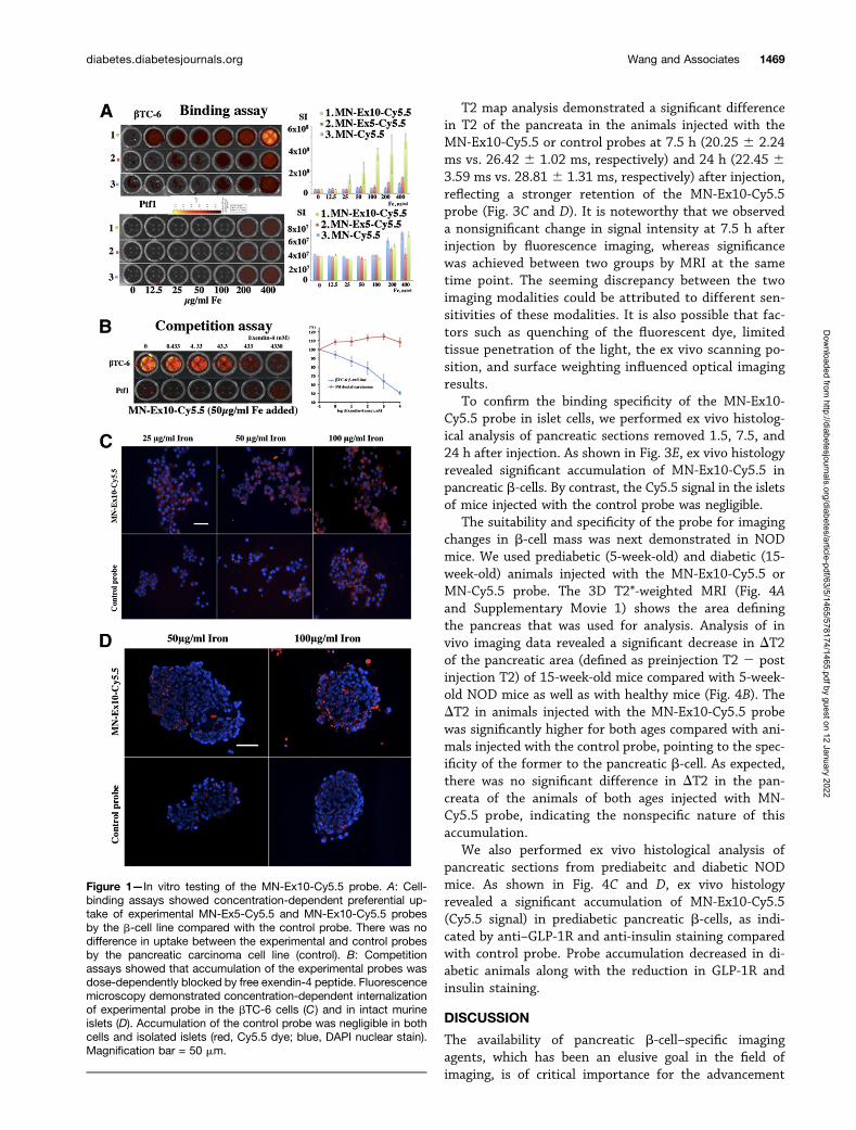
Cell Viability, Insulin Secretion, Probe Uptake, andCompetition Inhibition AssayTo evaluate toxicity associated with the probes, we per-formed a 3-(4,5-dimethylthiazol-2-yl)-2,5-diphenyltetrazoliumbromide (MTT) assay on bTC-6 cells incubated with dif-ferent concentrations of the probes. The results showedthat treatment with the probes did not have any signifi-cant effect on cell viability in vitro (Supplementary Fig. 3).The insulin-secretion assay showed that the presence ofexendin-4 on the nanoparticles did not change glucose-stimulated insulin secretion in the b-cell line as chargedby the stimulation index (Supplementary Fig. 4). Our invitro studies also demonstrated a concentration-dependentuptake of MN–exendin-4 conjugated probes by bTC-6cells and a background signal with control MN-Cy5.5probe. The uptake of the probe that contained 10 exendin-4 molecules/nanoparticle showed higher accumulation andwas therefore selected for subsequent in vivo studies. Theintensity of a background signal observed in control Ptf1-Creductal pancreatic carcinoma cells was an order of magnitudelower than in the b-cell line (Fig. 1A). A competition assayusing free exendin-4 peptide as a blocking agent showedspecific binding to bTC-6 cells and no competition withcontrol cells (Fig. 1B). Concentration-dependent internal-ization of the probe in the bTC-6 cells and intact murineislets was confirmed by fluorescence microscopy (Fig. 1Cand D) and showed colocalization with GLP-1R (Supple-mentary Fig. 5).
Biodistribution of Systemically DeliveredMN-Ex10-Cy5.5 ProbeBiodistribution study in vivo was performed in BALB/cmice after an intravenous injection of the MN-Ex10-Cy5.5
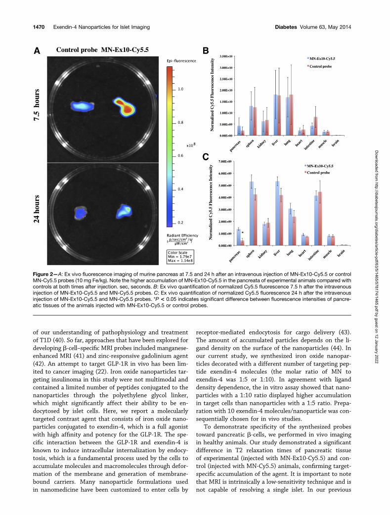
or control probe. We observed preferential accumulationof the MN-Ex10-Cy5.5 probe in the pancreas at 24 h afterinjection compared with the control probe (Fig. 2 andSupplementary Fig. 6). The higher accumulation of bothprobes in the pancreas at 7.5 h after injection comparedwith the accumulation at 24 h reflects the persistence oflong-circulated nanoparticles in the blood pool at thisearlier time and is in accordance with their known kinet-ics (33). Prototype nanoparticles (;5% injected dose/g)have been shown to be excreted by the urinary and fecalroute at 7.5 h (33). Note that the accumulation of theMN-Ex10-Cy5.5 probe was overall higher than that of thecontrol probe at 24 h but did not reach statistical significanceusing this method (Fig. 2B and Supplementary Fig. 6A).However, accumulation of MN-Ex10-Cy5.5 at 24 h was3.29 times higher than that of the control probe (1.373109 vs. 4.17 3 108 as normalized Cy5.5 fluorescence in-tensity), presumably due to the retention of the formerand the clearance of the later (Fig. 2C and SupplementaryFig. 6B and D). The rest of the organs showed accumula-tion consistent with the literature on related iron oxidenanoparticles (34,35), which could be taken up by macro-phages, mostly in liver, spleen, and lymph nodes, within24 h after a bolus injection and undergo progressive me-tabolism (36). Studies showed that several other tissues,including heart, intestine, and the central nervous system,also expressed GLP-1R (37–39), which might be the rea-son the accumulation of the probes was also observed inother organs in our biodistribution studies. Although thiswould not play a role in MRI studies, it could potentiallyresult in off-target effects if these probes were used fordrug-delivery purposes.
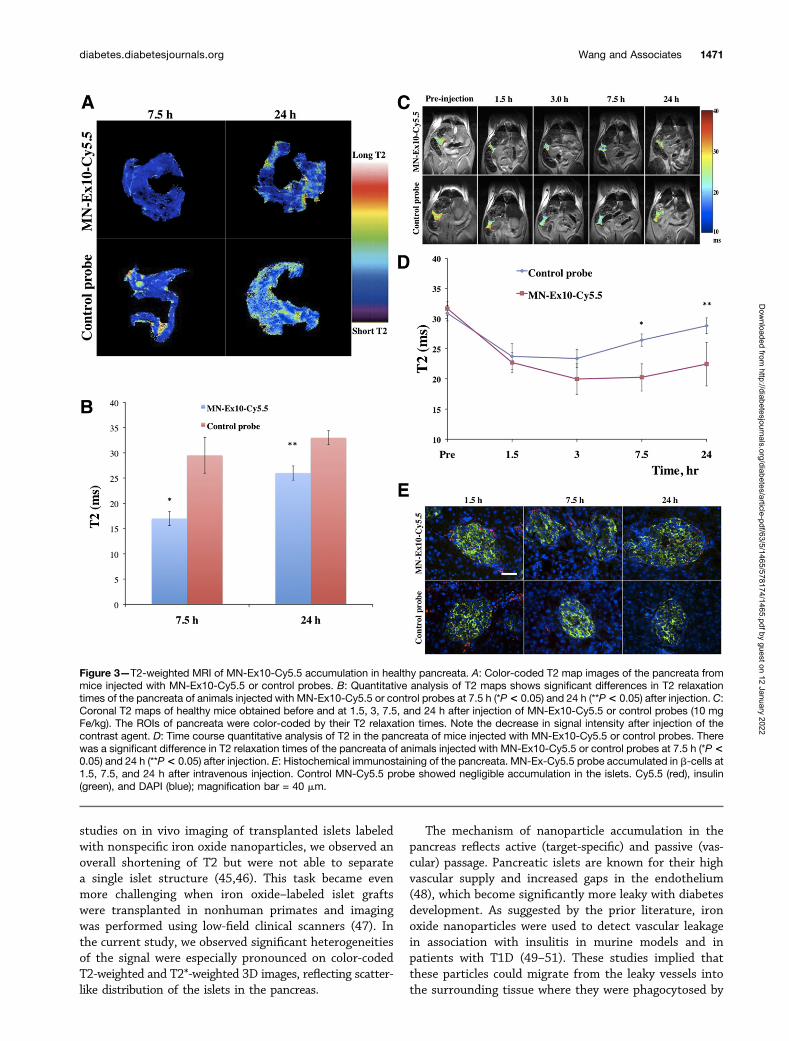
Ex Vivo and In Vivo MRI of Murine PancreataEx vivo imaging of mouse pancreata was performed onexcised organs from healthy mice injected with experi-mental or control probes using a 15-T MRI scanner. TheT2 relaxation times of the pancreatic tissues at 7.5 h or 24 hafter injection are summarized in Fig. 3A and B. Theresults showed the significant decrease of T2 in the pan-creata of the animals injected with MN-Ex10-Cy5.5 atboth time points compared with pancreatic tissues fromthe animals injected with the control probe.
Next, we performed experiments demonstrating thatthe MN-Ex10-Cy5.5 probe could be used for in vivoimaging of pancreatic b-cells. Quantitative analysis of T2relaxation times of the pancreata of mice intravenouslyinjected with the MN-Ex10-Cy5.5 or control probe showedthat a drop occurred in T2 of the pancreata starting at 1.5 hafter injection in both groups. In the control group, thedrop was most pronounced between 1.5 and 3 h afterinjection, followed by a gradual T2 increase, consistentwith contrast agent washout. In the MN-Ex10-Cy5.5 group,the drop reached its lowest point at 3 h after injection.The signal remained at that level up to 7 h after injection,followed by a much slower T2 increase compared with thecontrol group.
1468 Exendin-4 Nanoparticles for Islet Imaging Diabetes Volume 63, May 2014
Dow
nloaded from http://diabetesjournals.org/diabetes/article-pdf/63/5/1465/578174/1465.pdf by guest on 12 January 2022
T2 map analysis demonstrated a significant differencein T2 of the pancreata in the animals injected with theMN-Ex10-Cy5.5 or control probes at 7.5 h (20.25 6 2.24ms vs. 26.42 6 1.02 ms, respectively) and 24 h (22.45 63.59 ms vs. 28.81 6 1.31 ms, respectively) after injection,reflecting a stronger retention of the MN-Ex10-Cy5.5probe (Fig. 3C and D). It is noteworthy that we observeda nonsignificant change in signal intensity at 7.5 h afterinjection by fluorescence imaging, whereas significancewas achieved between two groups by MRI at the sametime point. The seeming discrepancy between the twoimaging modalities could be attributed to different sen-sitivities of these modalities. It is also possible that fac-tors such as quenching of the fluorescent dye, limitedtissue penetration of the light, the ex vivo scanning po-sition, and surface weighting influenced optical imagingresults.
To confirm the binding specificity of the MN-Ex10-Cy5.5 probe in islet cells, we performed ex vivo histolog-ical analysis of pancreatic sections removed 1.5, 7.5, and24 h after injection. As shown in Fig. 3E, ex vivo histologyrevealed significant accumulation of MN-Ex10-Cy5.5 inpancreatic b-cells. By contrast, the Cy5.5 signal in the isletsof mice injected with the control probe was negligible.
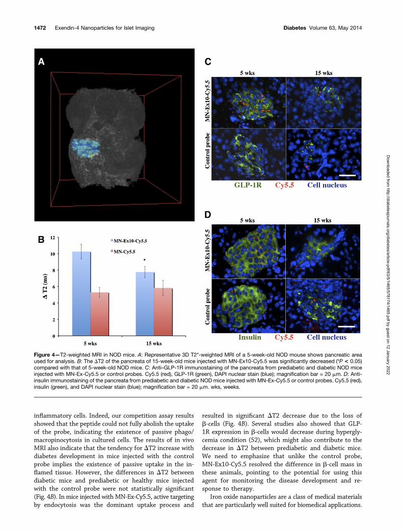
The suitability and specificity of the probe for imagingchanges in b-cell mass was next demonstrated in NODmice. We used prediabetic (5-week-old) and diabetic (15-week-old) animals injected with the MN-Ex10-Cy5.5 orMN-Cy5.5 probe. The 3D T2*-weighted MRI (Fig. 4Aand Supplementary Movie 1) shows the area definingthe pancreas that was used for analysis. Analysis of invivo imaging data revealed a significant decrease in DT2of the pancreatic area (defined as preinjection T2 2 postinjection T2) of 15-week-old mice compared with 5-week-old NOD mice as well as with healthy mice (Fig. 4B). TheDT2 in animals injected with the MN-Ex10-Cy5.5 probewas significantly higher for both ages compared with ani-mals injected with the control probe, pointing to the spec-ificity of the former to the pancreatic b-cell. As expected,there was no significant difference in DT2 in the pan-creata of the animals of both ages injected with MN-Cy5.5 probe, indicating the nonspecific nature of thisaccumulation.
We also performed ex vivo histological analysis ofpancreatic sections from prediabeitc and diabetic NODmice. As shown in Fig. 4C and D, ex vivo histologyrevealed a significant accumulation of MN-Ex10-Cy5.5(Cy5.5 signal) in prediabetic pancreatic b-cells, as indi-cated by anti–GLP-1R and anti-insulin staining comparedwith control probe. Probe accumulation decreased in di-abetic animals along with the reduction in GLP-1R andinsulin staining.
DISCUSSION
The availability of pancreatic b-cell–specific imagingagents, which has been an elusive goal in the field ofimaging, is of critical importance for the advancement
Figure 1—In vitro testing of the MN-Ex10-Cy5.5 probe. A: Cell-binding assays showed concentration-dependent preferential up-take of experimental MN-Ex5-Cy5.5 and MN-Ex10-Cy5.5 probesby the b-cell line compared with the control probe. There was nodifference in uptake between the experimental and control probesby the pancreatic carcinoma cell line (control). B: Competitionassays showed that accumulation of the experimental probes wasdose-dependently blocked by free exendin-4 peptide. Fluorescencemicroscopy demonstrated concentration-dependent internalizationof experimental probe in the bTC-6 cells (C) and in intact murineislets (D). Accumulation of the control probe was negligible in bothcells and isolated islets (red, Cy5.5 dye; blue, DAPI nuclear stain).Magnification bar = 50 mm.
diabetes.diabetesjournals.org Wang and Associates 1469
Dow
nloaded from http://diabetesjournals.org/diabetes/article-pdf/63/5/1465/578174/1465.pdf by guest on 12 January 2022
of our understanding of pathophysiology and treatmentof T1D (40). So far, approaches that have been explored fordeveloping b-cell–specific MRI probes included manganese-enhanced MRI (41) and zinc-responsive gadolinium agent(42). An attempt to target GLP-1R in vivo has been lim-ited to cancer imaging (22). Iron oxide nanoparticles tar-geting insulinoma in this study were not multimodal andcontained a limited number of peptides conjugated to thenanoparticles through the polyethylene glycol linker,which might significantly affect their ability to be en-docytosed by islet cells. Here, we report a molecularlytargeted contrast agent that consists of iron oxide nano-particles conjugated to exendin-4, which is a full agonistwith high affinity and potency for the GLP-1R. The spe-cific interaction between the GLP-1R and exendin-4 isknown to induce intracellular internalization by endocy-tosis, which is a fundamental process used by the cells toaccumulate molecules and macromolecules through defor-mation of the membrane and generation of membrane-bound carriers. Many nanoparticle formulations usedin nanomedicine have been customized to enter cells by
receptor-mediated endocytosis for cargo delivery (43).The amount of accumulated particles depends on the li-gand density on the surface of the nanoparticles (44). Inour current study, we synthesized iron oxide nanopar-ticles decorated with a different number of targeting pep-tide exendin-4 molecules (the molar ratio of MN toexendin-4 was 1:5 or 1:10). In agreement with liganddensity dependence, the in vitro assay showed that nano-particles with a 1:10 ratio displayed higher accumulationin target cells than nanoparticles with a 1:5 ratio. Prepa-ration with 10 exendin-4 molecules/nanoparticle was con-sequentially chosen for in vivo studies.
To demonstrate specificity of the synthesized probestoward pancreatic b-cells, we performed in vivo imagingin healthy animals. Our study demonstrated a significantdifference in T2 relaxation times of pancreatic tissueof experimental (injected with MN-Ex10-Cy5.5) and con-trol (injected with MN-Cy5.5) animals, confirming target-specific accumulation of the agent. It is important to notethat MRI is intrinsically a low-sensitivity technique and isnot capable of resolving a single islet. In our previous
Figure 2—A: Ex vivo fluorescence imaging of murine pancreas at 7.5 and 24 h after an intravenous injection of MN-Ex10-Cy5.5 or controlMN-Cy5.5 probes (10 mg Fe/kg). Note the higher accumulation of MN-Ex10-Cy5.5 in the pancreata of experimental animals compared withcontrols at both times after injection. sec, seconds. B: Ex vivo quantification of normalized Cy5.5 fluorescence 7.5 h after the intravenousinjection of MN-Ex10-Cy5.5 and MN-Cy5.5 probes. C: Ex vivo quantification of normalized Cy5.5 fluorescence 24 h after the intravenousinjection of MN-Ex10-Cy5.5 and MN-Cy5.5 probes. *P < 0.05 indicates significant difference between fluorescence intensities of pancre-atic tissues of the animals injected with MN-Ex10-Cy5.5 or control probes.
1470 Exendin-4 Nanoparticles for Islet Imaging Diabetes Volume 63, May 2014
Dow
nloaded from http://diabetesjournals.org/diabetes/article-pdf/63/5/1465/578174/1465.pdf by guest on 12 January 2022
studies on in vivo imaging of transplanted islets labeledwith nonspecific iron oxide nanoparticles, we observed anoverall shortening of T2 but were not able to separatea single islet structure (45,46). This task became evenmore challenging when iron oxide–labeled islet graftswere transplanted in nonhuman primates and imagingwas performed using low-field clinical scanners (47). Inthe current study, we observed significant heterogeneitiesof the signal were especially pronounced on color-codedT2-weighted and T2*-weighted 3D images, reflecting scatter-like distribution of the islets in the pancreas.
The mechanism of nanoparticle accumulation in thepancreas reflects active (target-specific) and passive (vas-cular) passage. Pancreatic islets are known for their highvascular supply and increased gaps in the endothelium(48), which become significantly more leaky with diabetesdevelopment. As suggested by the prior literature, ironoxide nanoparticles were used to detect vascular leakagein association with insulitis in murine models and inpatients with T1D (49–51). These studies implied thatthese particles could migrate from the leaky vessels intothe surrounding tissue where they were phagocytosed by
Figure 3—T2-weighted MRI of MN-Ex10-Cy5.5 accumulation in healthy pancreata. A: Color-coded T2 map images of the pancreata frommice injected with MN-Ex10-Cy5.5 or control probes. B: Quantitative analysis of T2 maps shows significant differences in T2 relaxationtimes of the pancreata of animals injected with MN-Ex10-Cy5.5 or control probes at 7.5 h (*P< 0.05) and 24 h (**P< 0.05) after injection. C:Coronal T2 maps of healthy mice obtained before and at 1.5, 3, 7.5, and 24 h after injection of MN-Ex10-Cy5.5 or control probes (10 mgFe/kg). The ROIs of pancreata were color-coded by their T2 relaxation times. Note the decrease in signal intensity after injection of thecontrast agent. D: Time course quantitative analysis of T2 in the pancreata of mice injected with MN-Ex10-Cy5.5 or control probes. Therewas a significant difference in T2 relaxation times of the pancreata of animals injected with MN-Ex10-Cy5.5 or control probes at 7.5 h (*P <0.05) and 24 h (**P< 0.05) after injection. E: Histochemical immunostaining of the pancreata. MN-Ex-Cy5.5 probe accumulated in b-cells at1.5, 7.5, and 24 h after intravenous injection. Control MN-Cy5.5 probe showed negligible accumulation in the islets. Cy5.5 (red), insulin(green), and DAPI (blue); magnification bar = 40 mm.
diabetes.diabetesjournals.org Wang and Associates 1471
Dow
nloaded from http://diabetesjournals.org/diabetes/article-pdf/63/5/1465/578174/1465.pdf by guest on 12 January 2022
inflammatory cells. Indeed, our competition assay resultsshowed that the peptide could not fully abolish the uptakeof the probe, indicating the existence of passive phago/macropinocytosis in cultured cells. The results of in vivoMRI also indicate that the tendency for DT2 increase withdiabetes development in mice injected with the controlprobe implies the existence of passive uptake in the in-flamed tissue. However, the differences in DT2 betweendiabetic mice and prediabetic or healthy mice injectedwith the control probe were not statistically significant(Fig. 4B). In mice injected with MN-Ex-Cy5.5, active targetingby endocytosis was the dominant uptake process and
resulted in significant DT2 decrease due to the loss ofb-cells (Fig. 4B). Several studies also showed that GLP-1R expression in b-cells would decrease during hypergly-cemia condition (52), which might also contribute to thedecrease in DT2 between prediabetic and diabetic mice.We need to emphasize that unlike the control probe,MN-Ex10-Cy5.5 resolved the difference in b-cell mass inthese animals, pointing to the potential for using thisagent for monitoring the disease development and re-sponse to therapy.
Iron oxide nanoparticles are a class of medical materialsthat are particularly well suited for biomedical applications.
Figure 4—T2-weighted MRI in NOD mice. A: Representative 3D T2*-weighted MRI of a 5-week-old NOD mouse shows pancreatic areaused for analysis. B: The DT2 of the pancreata of 15-week-old mice injected with MN-Ex10-Cy5.5 was significantly decreased (*P < 0.05)compared with that of 5-week-old NOD mice. C: Anti–GLP-1R immunostaining of the pancreata from prediabetic and diabetic NOD miceinjected with MN-Ex-Cy5.5 or control probes. Cy5.5 (red), GLP-1R (green), DAPI nuclear stain (blue); magnification bar = 20 mm. D: Anti-insulin immunostaining of the pancreata from prediabetic and diabetic NOD mice injected with MN-Ex-Cy5.5 or control probes. Cy5.5 (red),insulin (green), and DAPI nuclear stain (blue); magnification bar = 20 mm. wks, weeks.
1472 Exendin-4 Nanoparticles for Islet Imaging Diabetes Volume 63, May 2014
Dow
nloaded from http://diabetesjournals.org/diabetes/article-pdf/63/5/1465/578174/1465.pdf by guest on 12 January 2022
They are sized between 10 and 100 nm, and theirbiodegradable and biocompatible nature makes themsuitable for a wide variety of clinical contexts. Currently,several iron oxide nanoparticle–based agents are on themarket as MRI contrast probes or for iron supplementa-tion. The key advantage of using nanoparticles is the possi-bility of loading them with multiple copies of ligandmolecules for the targeting of specific tissues (53). Moreimportant, these nanoparticles are capable of carryingtherapeutic cargo, which makes them even more attrac-tive for theranostic imaging applications. We have alreadydemonstrated suitability of prototype nanoparticles fornucleic acid–based therapy in cancer (54–56) and in thecontext of islet transplantation (29,30,57). Our intentionis to apply similar strategies in the future for therapeutictargeted delivery to endogenous pancreatic islets.
Funding. This work was partly supported by a JDRF award (JDRF 37-2009-30) to A.M. and through the Harvard/Massachusetts General Hospital NuclearMedicine Training Program funded by the U.S. Department of Energy (DE-SC0008430).Duality of Interest. No potential conflicts of interest relevant to this articlewere reported.Author Contributions. P.W. performed the in vitro experiments, MRIscanning, ex vivo histology staining, and participated in writing the manuscript.B.Y. synthesized the probe. J.Y. participated in MRI data analysis. X.Z. assistedwith optical imaging. A.R., P.P., and G.D. researched the data. A.M. conceivedthe idea of the project and wrote the manuscript. A.M. is the guarantor of thiswork and, as such, had full access to all the data in the study and takesresponsibility for the integrity of the data and the accuracy of the data analysis.
References1. Mukai E, Toyoda K, Kimura H, et al. GLP-1 receptor antagonist as a potentialprobe for pancreatic beta-cell imaging. Biochem Biophys Res Commun 2009;389:523–5262. Mathis D, Vence L, Benoist C. beta-Cell death during progression to di-abetes. Nature 2001;414:792–7983. Fu W, Wojtkiewicz G, Weissleder R, Benoist C, Mathis D. Early window ofdiabetes determinism in NOD mice, dependent on the complement receptor CRIg,identified by noninvasive imaging. Nat Immunol 2012;13:361–3684. Wang P, Moore A. Translational molecular imaging of diabetes. Curr RadiolRep 2013;1:205–2155. Andralojc K, Srinivas M, Brom M, et al. Obstacles on the way to the clinicalvisualisation of beta cells: looking for the Aeneas of molecular imaging to nav-igate between Scylla and Charybdis. Diabetologia 2012;55:1247–12576. Nauck MA, Kleine N, Orskov C, Holst JJ, Willms B, Creutzfeldt W. Nor-malization of fasting hyperglycaemia by exogenous glucagon-like peptide 1 (7-36amide) in type 2 (non-insulin-dependent) diabetic patients. Diabetologia 1993;36:741–7447. Rachman J, Gribble FM, Barrow BA, Levy JC, Buchanan KD, Turner RC.Normalization of insulin responses to glucose by overnight infusion of glucagon-likepeptide 1 (7-36) amide in patients with NIDDM. Diabetes 1996;45:1524–15308. Kim TH, Jiang HH, Lee S, et al. Mono-PEGylated dimeric exendin-4 as highreceptor binding and long-acting conjugates for type 2 anti-diabetes therapeu-tics. Bioconjug Chem 2011;22:625–6329. Reiner T, Kohler RH, Liew CW, et al. Near-infrared fluorescent probe forimaging of pancreatic beta cells. Bioconjug Chem 2010;21:1362–136810. Reiner T, Thurber G, Gaglia J, et al. Accurate measurement of pancreaticislet beta-cell mass using a second-generation fluorescent exendin-4 analog.Proc Natl Acad Sci U S A 2011;108:12815–12820
11. Wang Y, Lim K, Normandin M, Zhao X, Cline GW, Ding YS. Synthesis andevaluation of [18F]exendin (9-39) as a potential biomarker to measure pancreaticb-cell mass. Nucl Med Biol 2012;39:167–17612. Selvaraju RK, Velikyan I, Johansson L, et al. In vivo imaging of the glucagonlikepeptide 1 receptor in the pancreas with 68Ga-labeled DO3A-exendin-4. J Nucl Med2013;54:1458–146313. Pattou F, Kerr-Conte J, Wild D. GLP-1-receptor scanning for imaging ofhuman beta cells transplanted in muscle. N Engl J Med 2010;363:1289–129014. Wu Z, Todorov I, Li L, et al. In vivo imaging of transplanted islets with64Cu-DO3A-VS-Cys40-Exendin-4 by targeting GLP-1 receptor. Bioconjug Chem2011;22:1587–159415. Wu Z, Liu S, Hassink M, et al. Development and evaluation of 18F-TTCO-Cys40-Exendin-4: a PET probe for imaging transplanted islets. J Nucl Med2013;54:244–25116. Wild D, Béhé M, Wicki A, et al. [Lys40(Ahx-DTPA-111In)NH2]exendin-4,a very promising ligand for glucagon-like peptide-1 (GLP-1) receptor targeting.J Nucl Med 2006;47:2025–203317. Christ E, Wild D, Forrer F, et al. Glucagon-like peptide-1 receptor imagingfor localization of insulinomas. J Clin Endocrinol Metab 2009;94:4398–440518. Wild D, Wicki A, Mansi R, et al. Exendin-4-based radiopharmaceuticals forglucagonlike peptide-1 receptor PET/CT and SPECT/CT. J Nucl Med 2010;51:1059–106719. Kiesewetter DO, Gao H, Ma Y, et al. 18F-radiolabeled analogs of exendin-4for PET imaging of GLP-1 in insulinoma. Eur J Nucl Med Mol Imaging 2012;39:463–47320. Kiesewetter DO, Guo N, Guo J, et al. Evaluation of an [(18)F]AlF-NOTAAnalog of Exendin-4 for imaging of GLP-1 receptor in insulinoma. Theranostics2012;2:999–100921. Sowa-Staszczak A, Pach D, Mikołajczak R, et al. Glucagon-like peptide-1receptor imaging with [Lys40(Ahx-HYNIC- 99mTc/EDDA)NH2]-exendin-4 for thedetection of insulinoma. Eur J Nucl Med Mol Imaging 2013;40:524–53122. Zhang B, Yang B, Zhai C, Jiang B, Wu Y. The role of exendin-4-conjugatedsuperparamagnetic iron oxide nanoparticles in beta-cell-targeted MRI. Bio-materials 2013;34:5843–585223. Lunov O, Zablotskii V, Syrovets T, et al. Modeling receptor-mediated en-docytosis of polymer-functionalized iron oxide nanoparticles by human macro-phages. Biomaterials 2011;32:547–55524. Duncan R, Richardson SC. Endocytosis and intracellular trafficking asgateways for nanomedicine delivery: opportunities and challenges. Mol Pharm2012;9:2380–240225. Tornehave D, Kristensen P, Rømer J, Knudsen LB, Heller RS. Expression ofthe GLP-1 receptor in mouse, rat, and human pancreas. J Histochem Cytochem2008;56:841–85126. Szot GL, Koudria P, Bluestone JA. Murine pancreatic islet isolation. J Vis Exp2007:25527. Zmuda EJ, Powell CA, Hai T. A method for murine islet isolation andsubcapsular kidney transplantation. J Vis Exp 2011:2096.28. Medarova Z, Evgenov NV, Dai G, Bonner-Weir S, Moore A. In vivo multi-modal imaging of transplanted pancreatic islets. Nat Protoc 2006;1:429–43529. Wang P, Yigit MV, Medarova Z, et al. Combined small interfering RNAtherapy and in vivo magnetic resonance imaging in islet transplantation. Diabetes2011;60:565–57130. Wang P, Yigit MV, Ran C, et al. A theranostic small interfering RNAnanoprobe protects pancreatic islet grafts from adoptively transferred immunerejection. Diabetes 2012;61:3247–325431. Jin CH, Chae SY, Son S, et al. A new orally available glucagon-like peptide-1receptor agonist, biotinylated exendin-4, displays improved hypoglycemic effectsin db/db mice. J Control Release 2009;133:172–17732. Chae SY, Choi YG, Son S, Jung SY, Lee DS, Lee KC. The fatty acid con-jugated exendin-4 analogs for type 2 antidiabetic therapeutics. J Control Release2010;144:10–16
diabetes.diabetesjournals.org Wang and Associates 1473
Dow
nloaded from http://diabetesjournals.org/diabetes/article-pdf/63/5/1465/578174/1465.pdf by guest on 12 January 2022
33. Wunderbaldinger P, Josephson L, Weissleder R. Crosslinked iron oxides(CLIO): a new platform for the development of targeted MR contrast agents. AcadRadiol 2002;9(Suppl. 2):S304–S30634. Moore A, Medarova Z, Potthast A, Dai G. In vivo targeting of under-glycosylated MUC-1 tumor antigen using a multimodal imaging probe. CancerRes 2004;64:1821–182735. Moore A, Marecos E, Bogdanov A Jr, Weissleder R. Tumoral distribution oflong-circulating dextran-coated iron oxide nanoparticles in a rodent model. Ra-diology 2000;214:568–57436. Bourrinet P, Bengele HH, Bonnemain B, et al. Preclinical safety and phar-macokinetic profile of ferumoxtran-10, an ultrasmall superparamagnetic ironoxide magnetic resonance contrast agent. Invest Radiol 2006;41:313–32437. Ban K, Noyan-Ashraf MH, Hoefer J, Bolz SS, Drucker DJ, Husain M.Cardioprotective and vasodilatory actions of glucagon-like peptide 1 receptorare mediated through both glucagon-like peptide 1 receptor-dependent and-independent pathways. Circulation 2008;117:2340–235038. Kedees MH, Guz Y, Grigoryan M, Teitelman G. Functional activity of murineintestinal mucosal cells is regulated by the glucagon-like peptide-1 receptor.Peptides 2013;48:36–4439. Richards P, Parker HE, Adriaenssens AE, et al. Identification and charac-terization of GLP-1 receptor–expressing cells using a new transgenic mousemodel. Diabetes 2014;63:1224–123340. Arifin DR, Bulte JW. Imaging of pancreatic islet cells. Diabetes Metab ResRev 2011;27:761–76641. Antkowiak PF, Stevens BK, Nunemaker CS, McDuffie M, Epstein FH.Manganese-enhanced magnetic resonance imaging detects declining pancreaticb-cell mass in a cyclophosphamide-accelerated mouse model of type 1 diabetes.Diabetes 2013;62:44–4842. Lubag AJ, De Leon-Rodriguez LM, Burgess SC, Sherry AD. Noninvasive MRIof b-cell function using a Zn2+-responsive contrast agent. Proc Natl Acad SciU S A 2011;108:18400–1840543. Canton I, Battaglia G. Endocytosis at the nanoscale. Chem Soc Rev 2012;41:2718–273944. Yuan H, Li J, Bao G, Zhang S. Variable nanoparticle-cell adhesion strengthregulates cellular uptake. Phys Rev Lett 2010;105:138101
45. Evgenov NV, Medarova Z, Dai G, Bonner-Weir S, Moore A. In vivo imaging ofislet transplantation. Nat Med 2006;12:144–14846. Evgenov NV, Medarova Z, Pratt J, et al. In vivo imaging of immune rejectionin transplanted pancreatic islets. Diabetes 2006;55:2419–242847. Medarova Z, Vallabhajosyula P, Tena A, et al. In vivo imaging of autologousislet grafts in the liver and under the kidney capsule in non-human primates.Transplantation 2009;87:1659–166648. Bonner-Weir S. The microvsaculature of the pancreas, with emphasis onthat of the islets of langerhans. In The Pancreas: Biology, Pathobiology, andDisease. Go VL, DiMagno EP, Gardner JD, Lebenthal E, Reber HA, Scheele GA,Eds. New York, Raven Press, 1993, p. 759–76849. Denis MC, Mahmood U, Benoist C, Mathis D, Weissleder R. Imaging in-flammation of the pancreatic islets in type 1 diabetes. Proc Natl Acad Sci U S A2004;101:12634–1263950. Turvey SE, Swart E, Denis MC, et al. Noninvasive imaging of pancreatic in-flammation and its reversal in type 1 diabetes. J Clin Invest 2005;115:2454–246151. Gaglia JL, Guimaraes AR, Harisinghani M, et al. Noninvasive imaging ofpancreatic islet inflammation in type 1A diabetes patients. J Clin Invest 2011;121:442–44552. Xu G, Kaneto H, Laybutt DR, et al. Downregulation of GLP-1 and GIP re-ceptor expression by hyperglycemia: possible contribution to impaired incretineffects in diabetes. Diabetes 2007;56:1551–155853. Jokerst JV, Gambhir SS. Molecular imaging with theranostic nanoparticles.Acc Chem Res 2011;44:1050–106054. Medarova Z, Pham W, Farrar C, Petkova V, Moore A. In vivo imaging ofsiRNA delivery and silencing in tumors. Nat Med 2007;13:372–37755. Kumar M, Yigit M, Dai G, Moore A, Medarova Z. Image-guided breast tumortherapy using a small interfering RNA nanodrug. Cancer Res 2010;70:7553–756156. Yigit MV, Ghosh SK, Kumar M, et al. Context-dependent differences in miR-10b breast oncogenesis can be targeted for the prevention and arrest of lymphnode metastasis. Oncogene 2013;32:1530–153857. Medarova Z, Kumar M, Ng SW, et al. Multifunctional magnetic nanocarriersfor image-tagged SiRNA delivery to intact pancreatic islets. Transplantation 2008;86:1170–1177
1474 Exendin-4 Nanoparticles for Islet Imaging Diabetes Volume 63, May 2014
Dow
nloaded from http://diabetesjournals.org/diabetes/article-pdf/63/5/1465/578174/1465.pdf by guest on 12 January 2022