giant cell-rich osteosarcoma: a case … · case reports nagoya j. med. sci. 59. 151 - 157, 1996...
TRANSCRIPT

CASE REPORTS
Nagoya J. Med. Sci. 59. 151 - 157, 1996
GIANT CELL-RICH OSTEOSARCOMA:A CASE REPORT
KEIJI SATO!, SHIGEKI YAMAMURA!, HISASHI IWATA!, HIDESHI SUGIURA2,
NOBUO NAKASHIMA3 and TETSURO NAGASAKA3
JDepartment of Orthopaedic Surgery, Nagoya University School of Medicine2Division of Orthopaedic Surgery, Nagoya Memorial Hospital3Division of Clinical Laboratory, Nagoya University Hospital
ABSTRACT
This report discusses a rare case of giant cell-rich osteosarcoma. The patient, a 19-year-old male, was diagnosed with a metadiaphyseal osteolytic lesion when he consulted a local doctor complaining of motionpain without swelling. Radiography revealed a geographic osteolytic lesion, cortical thinning and ballooningwithout obvious cortical destruction. However, a fine onion skin-like periosteal reaction was observed on thelateral side of the femur. The transitional zone was narrow and endosteal scalloping was also noted. Needlebiopsied material clearly showed nuclear atypism of the stromal tumor cells with numerous osteoclast-likegiant cells. Using a combination of pathological examination, radiography, computed tomography (CT) andmagnetic resonance imaging (MRI), a diagnosis of giant cell-rich osteosarcoma was reached. After chemotherapy, resection and limb salvage surgery with an autogeneous autoclaved bone graft, a vascularizedfibular graft were performed, and the patient has shown excellent limb function without local recurrence ordistant metastasis during the past 72 months.
Key Words: Giant cell-rich osteosarcoma, Autoclaved bone graft, Vasculalized fibular graft
INTRODUCTION
Giant cell-rich osteosarcoma, first described by Bathurst et al.,I) is a rare variant of osteosar
coma, accounting for 1 to 3% of conventional osteosarcoma cases, though differential diagnosis
from malignant giant cell tumor (GeT) is difficult in some cases.
CLINICAL INFORMATION
History of the present illness:The patient was a 19-year-old male whose chief complaint was left distal thigh pain. The pain
occurred during his job in July 1989 and gradually worsened until the middle of August when
the pain continued at night. The patient consulted a local doctor who detected an abnormal sha
dow on a radiograph of the left femur. Needle biopsy was done on August 19, and the diagnosis
of a giant cell tumor in the distal metadiaphyseal region of the left femur was made. The patient
was then referred to Nagoya University Hospital on September 5, 1989.
Correspondence: Dr. Keiji Sato, Department of Orthopaedic Surgery, Nagoya University School of Medicine,
65 Tsururnai-cho, Showa-ku, Nagoya 466, Japan
Tel: 81-52-741-2111, ext 5081 Fax: 81-52-744-2260
151

152
Keiji Sato et al.
Physical examination:Physical examination revealed a hard, bone like protrusion on the distal part of the left femur
with atrophy of the quadriceps muscle. There was slight tenderness over the bony lump withlocal warmth. A limp was present, but the range of the motion of the hip and knee joint wasnormal. The inguinal lymph nodes could not be palpated.
Characteristic imaging:A radiograph of the femur clearly disclosed a geographic osteolytic lesion with a sharp mar
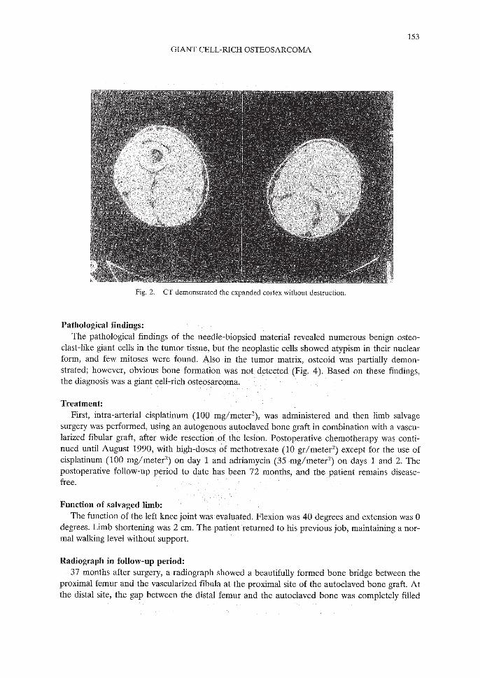
gin in the distal metadiaphyseal region. The transitional zone was narrow. Endosteal scallopingwas observed; however, obvious cortical destruction was not detected. A fine onion skin-likeperiosteal reaction was observed on the lateral side of the femur. Soft tissue extension could notbe detected (Fig. 1). cr clearly demonstrated that the cortex was thinned and ballooned without destruction (Fig. 2). MR images (GE Signa 1.5T) showed that the lesion had a low signalintensity on Tl-weighted images, intermediate intensity mixed with low signal intensity onproton density images and very high signal intensity (suggestive of hematoma) with a low signalintensity area on the T2-weighted images (Fig. 3).
Fig.!. Radiography of the left femur in antero-posterior and lateral views clearly disclosed a geographic osteolytic lesion with a sharp margin shown in the distalmetadiaphyseal region. The transitional zone was narrow. Endosteal scallopingwas observed and a faint onion skin-like periosteal reaction was also found onthe lateral side of the lesion.
153
GIANT CELL-RICH OSTEOSARCOMA
Fig. 2. cr demonstrated the expanded cortex without destruction.
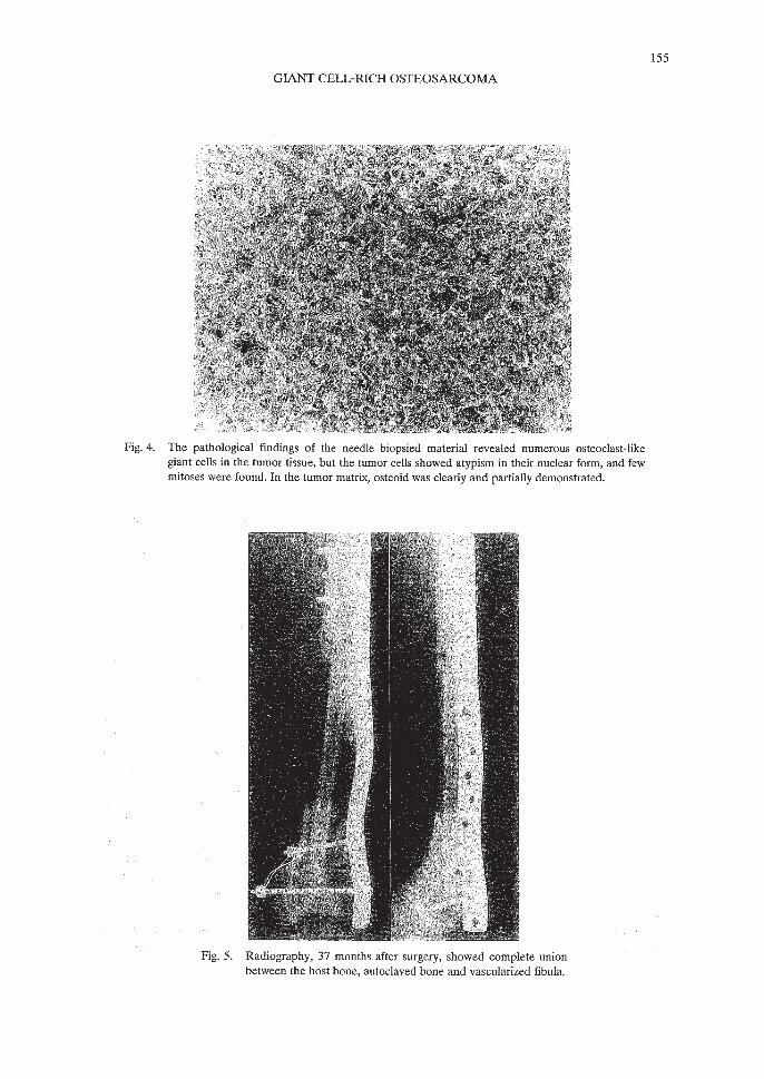
Pathological findings:The pathological findings of the needle-biopsied material revealed numerous benign osteo
clast-like giant cells in the tumor tissue, but the neoplastic cells showed atypism in their nuclearform, and few mitoses were found. Also in the tumor matrix, osteoid was partially demonstrated; however, obvious bone formation was not detected (Fig. 4). Based on these findings,the diagnosis was a giant cell-rich osteosarcoma.
Treatment:First, intra-arterial cisplatinum (100 mg/meter2), was administered and then limb salvage
surgery was performed, using an autogenous autoclaved bone graft in combination with a vascularized fibular graft, after wide resection of the lesion. Postoperative chemotherapy was continued until August 1990, with high-doses of methotrexate (10 gr/meter2) except for the use ofcisplatinum (100 mg/meter2) on day 1 and adriamycin (35 mg/meter2) on days 1 and 2. Thepostoperative follow-up period to date has been 72 months, and the patient remains diseasefree.
Function of salvaged limb:The function of the left knee joint was evaluated. Flexion was 40 degrees and extension was 0
degrees. Limb shortening was 2 cm. The patient returned to his previous job, maintaining a normal walking level without support.
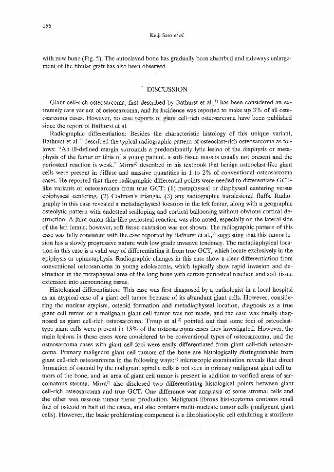
Radiograph in follow-up period:37 months after surgery, a radiograph showed a beautifully formed bone bridge between the
proximal femur and the vascularized fibula at the proximal site of the autoclaved bone graft. Atthe distal site, the gap between the distal femur and the autoclaved bone was completely filled

154
Keiji Sato et al.
3-A 3-B 3-C
3-D 3-E
Fig. 3. MR images of the coronal plane of the femur (3-A: T1-weighted images, TR400/TE20. 3-B: Proton density, TR2000/TE20. 3-C: T2-weighted images, TR2000/TE80) On Tl-weighted images, the lesionshowed a homogeneous low signal intensity, but the T2-weighted images demonstrated a marked high signal intensity mixed with an area of low signal intensity. MR images of the sagittal view (3-D: Proton density, TR2000/TE20. 3-E: T2-weighted images, TR2000/TE80) demonstrated the same changes.
GIANT CELL-RICH OSTEOSARCOMA
Fig. 4. The pathological findings of the needle biopsied material revealed numerous osteoclast-likegiant cells in the tumor tissue, but the tumor cells showed atypism in their nuclear form, and fewmitoses were found. In the tumor matrix, osteoid was clearly and partially demonstrated.
Fig. 5. Radiography, 37 months after surgery, showed complete unionbetween the host bone, autoclaved bone and vascularized fibula.
155
156
Keiji Sato et al.
with new bone (Fig. 5). The autoclaved bone has gradually been absorbed and sideways enlargement of the fibular graft has also been observed.
DISCUSSION
Giant cell-rich osteosarcoma, first described by Bathurst et aI.,I) has been considered an extremely rare variant of osteosarcoma, and its incidence was reported to make up 3% of all osteosarcoma cases. However, no case reports of giant cell-rich osteosarcoma have been publishedsince the report of Bathurst et aI.
Radiographic differentiation: Besides the characteristic histology of this unique variant,Bathurst et aI.l) described the typical radiographic pattern of osteoclast-rich osteosarcoma as follows: "An ill-defined margin surrounds a predominantly lytic lesion of the diaphysis or metaphysis of the femur or tibia of a young patient, a soft-tissue mass is usually not present and theperiosteal reaction is weak." Mirra2) described in his textbook that benign osteoclast-like giantcells were present in diffuse and massive quantities in 1 to 2% of conventional osteosarcomacases. He reported that three radiographic differential points were needed to differentiate GCflike variants of osteosarcoma from true GCf: (1) metaphyseal or diaphyseal centering versusepiphyseal centering, (2) Codman's triangle, (3) any radiographic intralesional fluffs. Radiography in this case revealed a metadiaphyseallocation in the left femur, along with a geographicosteolytic pattern with endosteal scalloping and cortical ballooning without obvious cortical destruction. A faint onion skin-like periosteal reaction was also noted, especially on the lateral sideof the left femur; however, soft tissue extension was not shown. The radiographic pattern of thiscase was fully consistent with the case reported by Bathurst et aI., 1) sugge~ting that this tumor lesion has a slowly progressive nature with low grade invasive tendency. The metadiaphyseallocation in this case is a valid way of differentiating it from true GCf, which locate exclusively in theepiphysis or epimetaphysis. Radiographic changes in this case show a clear differentiation fromconventional osteosarcoma in young adolescents, which typically show rapid invasion and destruction in the metaphyseal area of the long bone with certain periosteal reaction and soft tissueextension into surrounding tissue.
Histological differentiation: This case was first diagnosed by a pathologist in a local hospitalas an atypical case of a giant cell tumor because of its abundunt giant cells. However, considering the nuclear atypism, osteoid formation and metadiaphyseal location, diagnosis as a truegiant cell tumor or a malignant giant cell tumor was not made, and the case was finally diagnosed as giant cell-rich osteosarcoma. Troup et aI.3) pointed out that some foci of osteoclasttype giant cells were present in 13% of the osteosarco;na cases they investigated. However, themain lesions in these cases were considered to be conventional types of osteosarcoma, and theosteosarcoma cases with giant cell foci were easily differentiated from giant cell-rich osteosarcoma. Primary malignant giant cell tumors of the bone are histologically distinguishable fromgiant cell-rich osteosarcoma in the following ways:4) microscopic examination reveals that directformation of osteoid by the malignant spindle cells is not seen in primary malignant giant cell tumors of the bone, and an area of giant cell tumor is present in addition to verified areas of sarcomatous stroma. Mirra2) also disclosed two differentiating histological points between giantcell-rich osteosarcoma and true GCf. One difference was anaplasia of some stromal cells andthe other was osseous tumor tissue production. Malignant fibrous histiocytoma contains smallfoci of osteoid in half of the cases, and also contains multi-nucleate tumor cells (malignant giantcells). However, the basic proliferating component is a fibrohistiocytic cell exhibiting a storiform

157
GlANT CELL-RICH OSTEOSARCOMA
or cartwheel pattern; this pattern helps to differentiate malignant fibrous histiocytoma fromgiant cell-rich osteosarcoma.5)
Recently Hill et a1. 6) reported that osteoblast cells or human osteosarcoma cells (MG63)mediate insulin-like growth factor (IGF)-l and -II stimulation of osteoclast formation in a culture system. Giant cell-rich osteosarcoma might be interpreted as a unique osteosarcoma whichproduces rich IGFs, thereby stimulating osteoclast differentiation. From now on, research effortsshould focus on humeral factors produced by osteosarcoma cells which might moderate both thehistology of osteosarcoma and the natural course of the osteosarcoma bearing host.
REFERENCES
1) Bathurst, N. and Sanerkin, N.: Osteoclast-rich osteosarcoma. BiR, 59, 667-673 (1986).2) Mirra, J.M.: Bone tumors: clinical, radiologic and pathologic correlations. pp.326-331 (1989), Lea &
Febiger, Philadelphia.3) Troup, J.B. and Dahlin, D.C.: The significance of giant cells in osteogenic sarcoma: do they indicate a rela
tionship between osteogenic sarcoma and giant cell tumors of bone? Proceedings of the Staff Meetings of theMayo Clinic, 35,179-186 (1960).
4) Nascimento, A.G. and Huvos, A.G.: Primary malignant giant cell tumor of bone. A study of eight cases andreview of the literature. Cancer, 44, 1393-1402 (1979).
5) Dahlin, D.C.: Bone tumors: general aspects and data on 6221 cases. pp.307-314 (1978), Charles C Thomas,lllinois.
6) Hill, P.A., Reynolds, J.1. and Meikle, M.C.: Osteoblasts mediate insulin-like growth factor-l and -ll stimulation of osteoclast formation and function. Endocrinology, 136, 124-131 (1995).