genetic associations with diminished ovarian reserve: a systematic review of the literature
TRANSCRIPT
REVIEW
Genetic associations with diminished ovarian reserve:a systematic review of the literature
Alexis D. Greene & George Patounakis & James H. Segars
Received: 17 January 2014 /Accepted: 8 May 2014# Springer Science+Business Media New York (outside the USA) 2014
AbstractPurpose Diminished ovarian reserve (DOR) affects 10 % ofwomen seeking fertility treatment. Although it is much moreprevalent than premature ovarian failure, less is known aboutits etiology. The purpose of this article is to review the possiblegenetic causes of, and associations with, pathologic DOR.Methods A systematic review was conducted using PubMedfrom 1966 through November 2013.Results Twenty-one articles identified genes associated withDOR: one gene mutation (FMR1), three polymorphisms(GDF9, FSHR, and ESR1), and seven genes differentiallyexpressed between women with DOR and controls (AMH,LHCGR, IGF1, IGF2, IGF1R, IGF2R and GREM1). Sixcandidate genes were discovered in mice, including Foxl2,Gdf9, Bmp15, Aire, Wnt4, and Gpr3. Two case reports ofchromosomal translocations were also identified.Conclusions While the etiology of pathologic DOR is likelymultifactorial, it is possible that many cases attributed to anidiopathic cause may have a genetic component. Larger stud-ies are needed to expose the impact gene mutations, polymor-phisms, and epigenetics have on pathologic DOR.
Keywords DOR . Poor ovarian reserve . Genetic causes .
Genes . Premature ovarian aging . POR
Introduction
Approximately 10 % of women seeking fertility treatmenthave diminished ovarian reserve (DOR), defined as a de-creased number or quality of oocytes [1–4]. While the defini-tion is imprecise, DOR is associated with infertility and poorovarian response to controlled ovarian hyperstimulation(COH). The Bologna Criteria published in 2011 defined poorovarian response as having two of three of the followingcriteria: older than 39 years of age, prior poor ovarian responseto conventional stimulation protocol (less than three oocytesretrieved), and abnormal ovarian reserve testing [5]. Of note,we draw a distinction between the age-related physiologicDOR and the pathologicDOR, as explained below. Abnormalovarian reserve testing may be based on decreased antralfollicle count (AFC <5) on ultrasound, decreased anti-müllerian hormone (AMH <0.5–1 ng/mL), or elevated levelsof follicle stimulating hormone (FSH >10 IU/L on cycle day 2to 4) [6, 5, 7, 8]. Inconsistency within the literature also existsregarding the definition of many terms associated with DOR.For the purpose of this review, primary ovarian insufficiency(POI) is equivalent to premature ovarian failure (POF) [9–11].POI is defined as women under age 40 with amenorrhea for atleast 6 months and a day 3 FSH level in the menopausal range(>40 IU/L). It is possible in some patients that DOR and POIoccur within a spectrum, as they share many etiologies andfeatures. Furthermore, studies of poor ovarian response andabnormal ovarian reserve testing in in vitro fertilization (IVF)cycles suggest an earlier age of menopause in some patientswith DOR [12–14]. Despite an earlier age of menopause, mostaffected patients will not meet the definition for POI, yet somemay become menopausal before the age of 45 years old. POIaffects 1 % of women, whereas DOR affects a much largerportion of the infertile population [9–11, 15–18].
DOR can have multiple etiologies including genetic, auto-immune, idiopathic, and iatrogenic [19]. Ovarian surgery,
Capsule Diminished ovarian reserve (DOR) can be a normal process ofaging. In other cases DOR occurs at earlier ages, pathologic DOR, amultifactorial condition which may be caused by genetic or epigeneticanomalies. We review what is currently known about the contribution ofgenetic causes to the clinical condition of DOR.
A. D. GreeneObstetrics & Gynecology Department, St Luke’s Roosevelt HospitalCenter, 1000 Tenth Ave, Suite 10 C, New York, NY 10019, USA
G. Patounakis : J. H. Segars (*)Program in Reproductive and Adult Endocrinology, NICHD, NIH,10 Center Drive, Bethesda, MD 20892, USAe-mail: [email protected]
J Assist Reprod GenetDOI 10.1007/s10815-014-0257-5
chemotherapy, and radiation are iatrogenic causes of DORresulting from direct destruction of tissue or the toxic effectsof therapy on the follicular pool. These risk factors for DORare easily detected through a careful history. Age is anotherrisk factor for DOR. At some point in a woman’s reproductivelife span, she will meet the criteria for DOR as she approachesmenopause. It is expected that her risk of DOR varies directlywith increasing age. Therefore, we regard the age-related typeof DOR as physiologicDOR [20]. Other causes of DOR, suchas autoimmune and idiopathic, will be superimposed on thisphysiologic DOR and may thus be considered pathologicDOR, because they reflect abnormal ovarian aging [20]. Stat-ed differently, pathologic DOR reflects a process that acceler-ates the normal physiologic decline in ovarian function withage. It is important to differentiate between the two processesin the clinic, as they represent different groups of patients thatrequire different counseling clinically. Identifying pathologicDOR prior to undergoing an IVF cycle with ovarian stimula-tion would allow for proper counseling and may help to setappropriate expectations for the patient.
The cause for the prematurely lower number of folliclescan occur at many different levels of follicular and oocytedevelopment. There could be a smaller pool of primordialfollicles due to a decreased production of oocytes embryolog-ically, abnormal follicular development, increased follicularatresia, or defects in the communication between oocytes andgranulosa cells. Multiple genes play a role in folliculogenesis.The impact of mutations and polymorphic variant genes in-volved in folliculogenesis will be reviewed since they mayplay a role in pathologic DOR.
It is possible that an undetermined percentage of pathologicDOR has an underlying genetic etiology, as in those womenwith a family history of premature and early menopause whoare at increased risk for early menopause, DOR, and/or POI[2, 10, 21]. For example, a study examining 71 chromosom-ally normal patients characterized as idiopathic POI found31 % of cases to be familial, exemplifying the large geneticbasis of POI [22]. The purpose of this review is to identify thegenes that may be associated with pathologic DOR, includingthose found in humans, as well as candidate genes found inmice, to achieve the goal of ultimately developing a geneticscreening panel for pathologic DOR prior to initiating an IVFcycle. The murine model of DOR approximates but does notmirror the human DOR phenotype; however, with decreasednumber of follicles, increased atretic follicles, elevated FSHlevels, decreased litter size, and earlier diestrus or menopause,we believe it best mimics DOR.
Materials and methods
A systematic review of the literature was conducted from1966 through November 2013 using PubMed to find relevant
studies on genetic causes of or associations with DOR. Thecombination of keywords used for the searches were “dimin-ished ovarian reserve and genes”, “diminished ovarian reserveand genetic causes”, “premature ovarian aging and genes”,and “premature ovarian aging and genetic causes.” Eighty-nine articles were found with nine duplicates for a total of 80articles. All abstracts were read and the articles chosen includ-ed original research examining specific genes associated withDOR. Exclusion criteria were those written on POI and reviewarticles or editorials. We supplemented our search with areview of the references of the included articles. This resultedin identifying 21 total studies describing eight genes inhumans and six candidate genes in mice. There were two casereports on chromosomal translocations that were also relevantto this review.
Results
Whereas many genetic causes of POI are well established,little is known about definitive gene mutations associated withDOR. One of the known genetic causes of POI, a mutation inthe FMR1 gene, has been implicated as a potential cause ofDOR. Additionally, many known genetic causes of POI [10]have been implicated as being associated with DOR in mice,including autoimmune regulator gene (Aire) [23], forkheadtranscription factor forkhead protein (Foxl2) [24], growthdifferentiation factor 9 (Gdf9) [25], and bone morphogeneticprotein 15 (Bmp15) [26, 27]. Here, we focused on DOR toexamine the strength of evidence for the association. Thefollowing sections summarize current information organizedinto gene mutations and polymorphisms, differential geneexpression, candidate genes, as well as epigenetics associatedwith DOR.
Gene mutations associated with DOR
Fragile X mental retardation 1 (FMR1)
The strongest genetic association with DOR is the FMR1 gene(Table 1). The FMR1 gene is responsible for Fragile X Syn-drome, a form of X-linked mental retardation, which occurswhen >200 trinucleotide CGG (Cytosine Guanine Guanine)repeats are located at the 5′ untranslated region of the gene.This mutation causes hypermethylation of the promoter,resulting in complete absence of the FMR protein [10, 28,29]. Although women with Fragile X Syndrome with over200 CGG repeats do not have any form of ovarian dysfunc-tion, those with polymorphic triplet repeats above normal (35–54 repeats) have been associatedwith POI [2].FMR1 has beenshown to be the most significant single gene associated withPOI [30]. Women with premutation alleles, or 55–200 CGGrepeats, have a higher incidence of POI, while also carrying
J Assist Reprod Genet
the risk of passing on the Fragile X Syndrome to futuregenerations. Premutation carriers have been found to haveshorter menstrual cycles secondary to a decreased follicularphase length, as well as hormonal reflections of prematureovarian aging displayed by an increased FSH and decreasedinhibin A and B levels [28].
Patients carrying the intermediate allele, which includes 35to 54 CGG triplet repeats, are considered to be in a “greyzone.” Although those with this allele were previously con-sidered to have no specific phenotype, Bretherik et al. [30]found an increased intermediate carrier prevalence of 16 %among POI patients compared to 4.8–7.1 % of controls.Contradictory to this, a study by Bennett et al. [31] reportedthat although the presence of a premutation allele was statis-tically significant in POI patients compared to controls, inter-mediate alleles were not found to be increased among POIwomen.
The FMR1 gene is also implicated in DOR patients. Sev-eral studies suggested an increased incidence of grey zone andpremutation carriers amongDOR patients [2, 29]. In a study inthe Basque region by Barasoain et al. [29], 13.65 % of DOR
patients had alleles with 35 or greater repeats compared toonly 4.17 % of controls. Similarly, a multicenter prospectivecohort study examined intermediate and premutation alleles inwomen with DOR and found 14.5 % of DOR patients hadintermediate alleles compared to 3.9 % of controls [2].
A pilot study found a positive correlation between risingFSH levels and the number of CGG repeats, as well as AMHlevels <1.0 ng/mL being significantly associated with CGGrepeats above 32 [32]. However, these results differ frompreviously reported literature and await validation with alarger sample size. Given current data, the majority of datasupport an association of DOR with intermediate alleles.
Gene polymorphisms associated with DOR
Polymorphisms exist within genes and are a source of varia-tion between individuals. Particularly, the promoter region ofgenes contains polymorphisms and such polymorphisms havebeen shown to contain a large amount of genetic variation[33]. Gene polymorphisms associated with DOR arediscussed in Table 1.
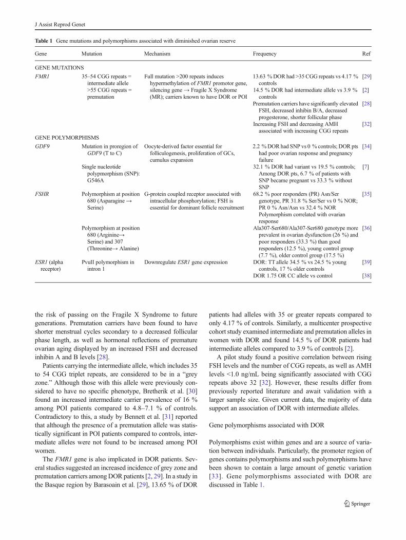
Table 1 Gene mutations and polymorphisms associated with diminished ovarian reserve
Gene Mutation Mechanism Frequency Ref
GENE MUTATIONS
FMR1 35–54 CGG repeats =intermediate allele>55 CGG repeats =premutation
Full mutation >200 repeats induceshypermethylation of FMR1 promotor gene,silencing gene→ Fragile X Syndrome(MR); carriers known to have DOR or POI
13.63 %DOR had >35 CGG repeats vs 4.17 %controls
[29]
14.5 % DOR had intermediate allele vs 3.9 %controls
[2]
Premutation carriers have significantly elevatedFSH, decreased inhibin B/A, decreasedprogesterone, shorter follicular phase
[28]
Increasing FSH and decreasing AMHassociated with increasing CGG repeats
[32]
GENE POLYMORPHISMS
GDF9 Mutation in proregion ofGDF9 (T to C)
Oocyte-derived factor essential forfolliculogenesis, proliferation of GCs,cumulus expansion
2.2 %DORhad SNP vs 0 % controls; DOR ptshad poor ovarian response and pregnancyfailure
[34]
Single nucleotidepolypmorphism (SNP):G546A
32.1 % DOR had variant vs 19.5 % controls;Among DOR pts, 6.7 % of patients withSNP became pregnant vs 33.3 % withoutSNP
[7]
FSHR Polymorphism at position680 (Asparagine→Serine)
G-protein coupled receptor associated withintracellular phosphorylation; FSH isessential for dominant follicle recruitment
68.2 % poor responders (PR) Asn/Sergenotype, PR 31.8 % Ser/Ser vs 0 % NOR;PR 0 % Asn/Asn vs 32.4 % NORPolymorphism correlated with ovarianresponse
[35]
Polymorphism at position680 (Arginine→Serine) and 307(Threonine→ Alanine)
Ala307-Ser680/Ala307-Ser680 genotype moreprevalent in ovarian dysfunction (26 %) andpoor responders (33.3 %) than goodresponders (12.5 %), young control group(7.7 %), older control group (17.5 %)
[36]
ESR1 (alphareceptor)
PvuII polymorphism inintron 1
Downregulate ESR1 gene expression DOR: TT allele 34.5 % vs 24.5 % youngcontrols, 17 % older controls
[39]
DOR 1.75 OR CC allele vs control [38]
J Assist Reprod Genet
Growth differentiation factor 9 (GDF9)
GDF9 is an oocyte-derived factor that is essential forfolliculogenesis, granulosa cell proliferation, and cumulusexpansion [25, 34, 7]. It is a member of the transforminggrowth factor-beta superfamily. Knockout mice unable toexpress the Gdf9 gene were infertile due to the inability toform follicles progressing beyond having a single layer ofgranulosa cells and were unresponsive to added FSH stimu-lation [25].
In humans, polymorphisms of GDF9 have been associatedwith DOR [7, 34]. One hundred three Chinese women withDOR were genetically analyzed for three single nucleotidepolymorphisms (SNPs) of GDF9 and compared to 123 age-matched women with infertility and normal ovarian reserve(NOR) [7]. NOR was defined as a day 3 FSH below 10 and/orestradiol level less than 80 pg/mL. Approximately 32 % ofwomen with DOR had the GA/AA genotype compared with19.5 % of those with NOR (p<0.05). A higher prevalence ofthe GA/AA genotype was found in those with poor ovarianresponse during IVF cycles.
Another study of Chinese women also analyzed the geno-type of the GDF9 gene [34]. Three out of 139 women (2.2 %)with DOR had a specific mutation (p.R146C) substitutingarginine for cystine, whereas this mutation was absent in the159 women in the control group. The women with this muta-tion also had poor quality oocytes and complete pregnancyfailure. Although a second mutation was found with differentfrequencies between the groups, the mutation was present inboth the DOR and control group. There was no difference inpregnancy outcomes, so it was thought to be a commonvariant in some Chinese women.
Follicle-stimulating hormone receptor (FSHR)
FSH is a glycoprotein produced by the anterior pituitarylargely responsible for folliculogenesis and recruitment ofthe dominant follicle prior to ovulation. FSHR is G-proteincoupled receptor, which is primarily located on granulosa cellsin the ovary. Polymorphisms have been identified within theFSHR gene that differed in frequency among poor ovarianresponders [35, 36].
In a study of patients referred to an infertility center under-going COH for an IVF cycle, 108 subjects were genotyped forpolymorphisms in the FSHR gene [35]. This study group wasdivided into 3 groups: NOR, poor ovarian responders, andhyper-ovarian responders. Poor ovarian responders were de-fined as womenwith less than fourmature follicles after COH.Hyper-responders at risk for ovarian hyperstimulation syn-drome were defined as those with more than 14 maturefollicles. The polymorphism in the FSHR gene the investiga-tors genotyped was at amino acid position 680. The SNPchanged the amino acid from asparagine (Asn) to serine
(Ser). Of the 22 poor responders, 15/22 (68.2 %) were hetero-zygotes for the SNP (Asn/Ser), while the remaining 31.8 %were homozygotes for the SNP (Ser/Ser). In the normal re-sponders group, a similar percentage 46/68 (67.6 %) wereheterozygotes for the SNP (Asn/Ser), however the remaining32.4 % did not have the SNP (Asn/Asn). As mentionedpreviously, a poor response to ovarian stimulation is a hall-mark of DOR. Therefore, based on this study, it can beextrapolated that patients with the polymorphism at position680 that substitutes a serine amino acid for asparagine are athigher risk of DOR, although the sample size is consideredsmall for a genotyping study.
A similar study performed by Livshyts et al. [36] examinedtwo polymorphisms within the FSHR gene. The SNP at posi-tion 680 was the same serine substitution for arginine as in thestudy by Sheikhha et al. [35]. Additionally, a second SNP atposition 307 was also found, which changed threonine (Thr)to alanine (Ala) [36]. The investigators compared poor re-sponders (n=39) together with the ovarian dysfunction group(n=102) to good responders (n=40), and two control groups,each normo-ovulatory and with children but stratified by age(control group I <35 years old, n=40; control group II>35 years old, n=130). The Ala307-Ser680/Ala307-Ser680genotype was more prevalent in those with ovarian dysfunc-tion (26 %) and poor responders (33.3 %) compared to goodresponders, control group I, and II (12.5 %, 7.7 %, 17.5 %,respectively). Although a small percentage of the controls andgood responders also have the genotype homozygous Ala307-Ser680, a higher prevalence is seen among ovarian dysfunc-tion (p<0.05) and poor responders (p<0.05) using pairwisecomparisons. In summary, the results of both of these studiesshowed that these polymorphisms in FSHR were found indifferent frequencies in patients with poor ovarian responseand suggest patients with the genotype have a higher preva-lence of DOR.
Estrogen alpha receptor (ESR1)
Estrogen binds to estrogen receptors, and plays an importantrole in granulosa cell proliferation, folliculogenesis, and fer-tility. Two types of nuclear estrogen receptors are found inhumans: alpha and beta. Within the ovary, the alpha-receptorsare typically expressed more in the theca cells, whereas thebeta-receptors are more highly expressed in granulosa cells[37].
Eleven estrogen receptor polymorphisms have been impli-cated in regulation of the menstrual cycle, low fecundity, andpolycystic ovarian disease.M’Rabet et al. [38] examined thesepolymorphisms in a large group of predominantly whiteAmerican infertile women (n=348), and compared them tothose with POI (n=48), while using fertile women (n=200) ascontrols. The SNPs were identified and amplified by PCR.The homozygous CC (Cytosine Cytosine) allele of the PvuII
J Assist Reprod Genet
polymorphic variant of the ESR1 gene was more prevalent ininfertile and POI women compared to fertile controls. Eighty-three women in the infertile group had elevated levels of FSHabove 9 IU/L and would meet criteria for DOR. These womenalso had a higher prevalence of this allele with an odds ratio of1.750 (95 % confidence interval=1.030–2.973).
Another study examined the same polymorphism in agroup of women in the Ukraine [39]. Women with ovariandysfunction were compared to two control groups. The groupswere stratified by age (control group I <35 years old, controlgroup II >35 years old) with both normo-ovulatory and havinga history of prior conception. Ovarian dysfunction was de-fined as women under age 40 years old with amenorrhea formore than 6 months and FSH levels above 25 IU/L. Thirty-nine out of 113 (34.5%) of those with ovarian dysfunction hadthe homozygous TT (Thymine Thymine) allele, whereas 25/102 (24.5 %) of control group I and 9/53 (17 %) of controlgroup II had the TT allele. These results were similar to aKorean study, which found a higher prevalence of the TTallele in women with POI [40].
Examination of IVF outcomes has shown that women withthe CC allele of the PvuII variant of ESR1 develop morefollicles (p=0.033) with COH [37]. However, no differencein clinical pregnancy outcome was observed. Although thisstudy supported the polymorphism as a risk factor for DORfrom the perspective of ovarian stimulation response, ulti-mately it did not alter pregnancy outcomes in this study. Thelack of difference in pregnancy outcome may have beenbecause of insufficient statistical power.
The four studies [37–40] were conducted in different ethnicpopulations, which may explain the discrepancy in results.Ethnic differences in this allele frequency, or other polymor-phisms dependent on ethnicity that interact with the PvuIIvariant of ESR1, might explain the different effects of thevariant on the diagnosis of DOR. The different definitionsused may have also affected the results. Therefore, it is im-portant to note that the first study [38] used the standard
definition of DOR, whereas the second study [39] used anovarian dysfunction group that was more similar to the POIphenotype. It appears that, at least within certain populations,the PvuII variant of ESR1 is associated with DOR.
Gene expression associated with DOR
Gene expression profiling with cDNA microarrays allows fordifferentiation between a diseased and normal state in largegroups. This technique permits identification of candidategenes potentially involved in the pathogenesis of disease.Chin et al. [41] performed a cDNA microarray analysis ofpooled follicular granulosa cells of 9 women with DOR and 9women with NOR. The investigators identified many genesthat were differentially expressed between the DOR and NORgroups, including forkhead-like transcription factor 13,Pumilio 2, Notch 3, and STAT-induced STAT inhibitor 3.Although the results were not confirmed by reverse transcrip-tase polymerase chain reaction (RT-PCR), it still suggestedthat differential gene expression might play a part indistinguishing those susceptible to DOR. May-Panloup et al.[3] performed a similar study examining genes differentiallyexpressed within the corona radiata cells between four patientswith DOR and four patients with NOR first using microarrayanalysis. The investigation found 16 candidate genes for val-idation based on the microarray analysis. The investigatorsthen validated the differential expression of the identifiedgenes using quantitative RT-PCR in a larger group of 20patients with DOR and 20 patients with NOR. Six genes wereultimately validated as differentially expressed including con-nective tissue growth factor (CTGF), CXXC finger protein 5(CXXC5), forkhead box C1 (FOXC1), follistatin-like 3(FSTL3), prostaglandin–endoperoxide synthase 2 (PTGS2),and suppressor of cytokine signaling 2 (SOCS2) [3]. However,each differentially expressed gene only showed a <2-folddifference in expression between the two groups. Other geneexpression studies identified four genes with at least a 2-fold
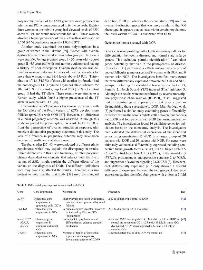
Table 2 Differential gene expression associated with DOR
Gene Gene Expression Mechanism Frequency Ref
AMH Differential geneexpression ingranulosa cells (GCs)
Higher levels associated with normalovarian reserve, produced by smallfollicles
2.02-fold higher in control vs DOR [42]
LHCGR Differential geneexpression in GCs
G-protein coupled receptor, known tobe induced by FSH on GCs(luteinization)
2.19-fold higher in DOR vs control [42]
IGF1, IGF2,IGF1R,IGF2R
Differential geneexpression incumulus and muralGCs
Stimulate GC proliferation anddifferentiation, enhance estradiolproduction
IGF1 and IGF2 downregulated 4.33- and 4.18- fold in DOR vscontrol pts in cumulus GCs; 4.35 and 3.89 fold in mural GCsIGF1R and IGF2R downregulated 5.32- and 2.13-fold incumulus GCs
[43]
GREM1 Differential geneexpression in GCs
Member of family of genes thatregulates folliculogenesis,downstream effector of GDF9
Downregulated 4.02-fold in DOR vs control pts [45]
J Assist Reprod Genet
differential gene expression between those with DOR andNOR (see Table 2). These genes with differential expressionbetween women with DOR and NOR represent candidates forfurther research.
Anti-müllerian hormone (AMH)
AMH is a hormone produced by pre-antral follicles, and is abiomarker of granulosa cell mass, and thus indirectly, ovarianreserve. Typically, serum AMH values below 0.8 ng/mL havebeen found to be consistent with elevated FSH levels andseverely diminished ovarian reserve [6]. Skiadas et al. [42]examined membrana granulosa cells from women with DOR.The membrana granulosa cells were obtained from 13 womenunder the age of 35 years old with DOR and 13 women withNOR. NOR patients were oocyte donors with day 3 FSH level<8 mIU/mL, day 3 estradiol level ≤50 pg/mL, and had ≥10follicles of ≥12 mm diameter at the time of ovulation trigger.The cells were screened for differential gene expression usingmicroarray and underwent confirmatory studies using quanti-tative RT-PCR. One of the genes identified was AMH, whichshowed a 2.02-fold increased expression in NOR patients overDOR patients. While this may have been expected given thatelevated serum AMH levels are correlated with NOR, it wasthe first study to show increased gene expression at the tissuelevel.
Luteinizing hormone/choriogonadotropin receptor (LHCGR)
Luteinizing hormone receptors (LHCGR) are found on thetheca cells, as well as on granulosa cells after induction byFSH. These receptors are important in ovarian steroidogene-sis, luteinization, and therefore, progesterone synthesis andfertility. Skiadas et al. [42] found a 2.19-fold increased ex-pression of the LHCGR gene in DOR patients over NORpatients. The increased gene expression of LHCGR in DORpatients could be explained by premature luteinization andupregulation of luteinizing hormone receptors on follicularcells, which might occur with DOR patients given the in-creased FSH in the follicular phase. It is also possible thatthe increased LHCGR gene expression could have been aresult of the higher levels of FSH administrated duringCOH, which is often required for DOR patients. Furtherstudies are needed to identify the reason for increased expres-sion of the LHCGR gene in patients with DOR.
Insulin-like growth factor 1 and 2 (IGF1/IGF2) and receptors(IGF1R/IGF2R)
IGF-1 and IGF-2 are hormones that have an autocrine andparacrine effect at the level of the granulosa and theca cells.These growth factors promote steroidogenesis and granulosacell proliferation. Greenseid et al. [43] examined granulosa
cell expression of the genes IGF1, IGF2, and their receptors,IGF1R and IGF2R. A prospective study of four patients withDOR and four patients with NOR was performed using mi-croarray and confirmatory quantitative RT-PCR of pooledgranulosa cells. Over one thousand statistically significantdifferentially expressed genes were identified between theDOR and NOR groups. The genes of the IGF family ligandswithin cumulus granulosa cells, IGF1 and IGF2, were down-regulated 4.33- and 4.18-fold, respectively, whereas the genesfor the corresponding receptors were downregulated 5.32- and2.13-fold, respectively. In mural granulosa cells, only IGF1and IGF2 genes showed a statistically significant downregu-lation, 4.35- and 3.89- fold, respectively. This differential geneexpression could be related to the diminished reproductivecapacity among DOR patients given the biologically plausiblerole the IGF family of genes play in ovarian folliculogenesis.
Gremlin 1 (GREM1)
GREM1 is a member of a family of genes that are highlyregulated in folliculogenesis and is a down-stream effector ofthe GDF9 gene, which is essential in folliculogenesis andfertility as mentioned previously. A 15-fold increased expres-sion of the GREM1 gene was seen in cumulus cells strippedfrom oocytes during IVF with intracytoplasmic sperm injec-tion (ICSI) that resulted in higher quality embryos whencompared to lower quality embryos [44]. Jindal et al. [45]performed a prospective study examining granulosa cell ex-pression of the GREM1 gene based on these findings usingmicroarray and confirmatory quantitative RT-PCR. Theyshowed a 4.02-fold decreased expression of the GREM1 genein DOR patients compared to NOR patients. Despite the smallsample size of only four patients per group of this study, thefinding suggested that decreased expression of the GREM1gene was found in the DOR group. Furthermore, it provides apotential gene linking DOR with embryo quality. Studies withlarger sample sizes are needed to confirm these associations.
Candidate genes in mice associated with DOR
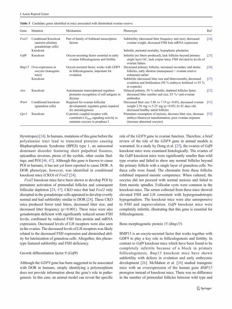
Many genes that are first identified in animal models are thenused to guide identification of similar genes in humans. Sixcandidate genes have been identified in mice that caused aDOR phenotype characterized by decreased number of folli-cles, increased atretic follicles, elevated FSH levels, decreasedlitter size and frequency, and earlier diestrus onset or meno-pause (Table 3). A few of these candidate genes have alsobeen implicated in humans.
Forkhead transcription factor forkhead protein L2 (Foxl2)
The FOXL2 gene is expressed in the eyelid, the ovary (gran-ulosa cells), and in the anterior pituitary (gonadotropes and
J Assist Reprod Genet
thyrotropes) [24]. In humans, mutations of this gene before thepolyalanine tract lead to truncated proteins causingBlepharophimosis Syndrome (BPES) type 1, an autosomaldominant disorder featuring short palpebral fissures,epicanthus inversus, ptosis of the eyelids, other ocular find-ings, and POI [46, 47]. Although this gene is known to causePOI in humans, it has not yet been reported to cause DOR. ADOR phenotype, however, was identified in conditionalknockout mice (CKO) of Foxl2 [24].
Foxl2 knockout mice have been shown to develop POI bypremature activation of primordial follicles and consequentfollicular depletion [24, 47]. CKO mice that had Foxl2 onlydisrupted in the gonadotrope cells appeared to develop overtlynormal and had subfertility similar to DOR [24]. These CKOmice produced fewer total litters, decreased litter size, anddecreased litter frequency (p<0.001). These mice were alsogonadotropin deficient with significantly reduced serum FSHlevels, confirmed by reduced FSH beta protein and mRNAexpression. Decreased levels of LH receptors were also seenin the ovaries. The decreased levels of LH receptors was likelyrelated to the decreased FSH expression and diminished abil-ity for luteinization of granulosa cells. Altogether, this pheno-type featured subfertility and FSH deficiency.
Growth differentiation factor 9 (Gdf9)
Although the GDF9 gene has been suggested to be associatedwith DOR in humans, simply identifying a polymorphismdoes not provide information about the gene’s role in patho-genesis. In this case, an animal model can reveal the specific
role of the GDF9 gene in ovarian function. Therefore, a briefreview of the role of the GDF9 gene in animal models iswarranted. In a study by Dong et al. [25], the ovaries of Gdf9knockout mice were examined histologically. The ovaries ofthe Gdf9 knockout mice were significantly smaller than wildtype ovaries and failed to show any normal follicles beyondthe primary follicle with a single layer of granulosa cells. Notheca cells were found. The chromatin from these folliclesexhibited impaired meiotic competence. When cultured, theoocytes did not proceed with normal meiosis and failed toform meiotic spindles. Follicular cysts were common in theknockout mice. The serum collected from these mice showedelevated FSH and LH consistent with hypergonadotropichypogonadism. The knockout mice were also unresponsiveto FSH and superovulation. Gdf9 knockout mice werecompletely infertile, illustrating that this gene is essential forfolliculogenesis.
Bone morphogenetic protein 15 (Bmp15)
BMP15 is an oocyte-secreted factor that works together withGDF9 to play a key role in folliculogenesis and fertility. Incontrast to Gdf9 knockout mice which have been found to becompletely infertile because of a block in primaryfolliculogenesis, Bmp15 knockout mice have shownsubfertility with defects in ovulation and early embryonicdevelopment [26]. McMahon et al. [26] studied transgenicmice with an overexpression of the human gene BMP15proregion instead of knockout mice. There was no differencein the number of primordial follicles between wild type and
Table 3 Candidate genes identified in mice associated with diminished ovarian reserve
Gene Mutation Mechanism Phenotype Ref
Foxl2 Conditional Knockout(anterior pituitarygonadotrope cells)
Part of family of forkhead transcriptionfactors
Subfertility (decreased litter frequency and size), decreasedovarian weight, decreased FSH beta mRNA expression
[24]
Knockout Infertile, perinatal mortality, hypoplastic pituitaries
Gdf9 Knockout Oocyte-secreting factor essential in earlyovarian folliculogenesis and fertility
Infertile (no litters produced), lack follicles beyond primarysingle layer GC, lack corpus lutea, FSH elevated to levels ofovarian failure
[25]
Bmp15 Over-expression inoocytes (transgenicmice)
Oocyte-secreted factor, works with GDF9in folliculogenesis, important forovulation
Decreased primary follicles, increased secondary and atreticfollicles, early diestrus (menopause) = ovarian reserveexhausted earlier
[26]
Knockout Subfertile (decreased litter size and litters/month), decreasedovulation and fertilization (50 % embryos fertilized vs 93 %in controls)
[27]
Aire Knockout Autoimmune transcriptional regulatorpromotes recognition of self antigens inthymus
Delayed puberty, 50 % infertile, depleted follicles faster,decreased litter number and size, 83 %+anti-ovarianantibodies
[23]
Wnt4 Conditional knockout(granulosa cells)
Required for ovarian folliculardevelopment; regulates genes requiredfor steroidogenesis
Decreased litter size 3.86 vs 7.19 (p<0.05), decreased ovarianweight 2.56 mg vs 5.25 mg (p<0.05) At 42 days old,decreased healthy antral follicles
[51]
Gpr3 Knockout G-protein coupled receptor withconstitutive Gstim signaling activity tomaintain oocytes in prophase I
Premature resumption of meiosis, decrease litter size, decreaseembryo blastocyst transformation, poor ovarian response(increase abnormal oocytes)
[53]
J Assist Reprod Genet
transgenic mice; however, a significant decrease in primaryfollicles and increase in secondary follicles in transgenic micewas observed in 25-day old mice. Additionally, an increasednumber of atretic follicles were also found in adult mice.These findings were consistent with accelerated folliculardevelopment in the transgenic mice. Bmp15 also decreasedthe FSH receptor mRNA expression in the transgenic mice.Litter frequency was the same in mice 1.5-5 months of age;however, older transgenic mice aged 6–11 months showed adecreased litter frequency and increased estrous cycle length.Litter size was the same between transgenic and wild typemice regardless of age. Transgenic mice began to show in-creased diestrus length earlier than wild type mice. Theyfound 87 % entered constant diestrus or menopause by20 months, whereas 94 % of wild type mice were still cyclingnormally at this age. In summary, overexpression of theBmp15 gene in mice led to accelerated follicle development,increased atresia, and decreased FSH receptor mRNA. Thisultimately led to premature ovarian aging and earlier declinein the follicular pool.
To analyze the function of BMP15, Yan et al. [27] createdmutant Bmp15 alleles in mice. Homozygous mutant micewere subfertile, showing a statistically significant decreasedlitter size and frequency. Bmp15 knockout mouse ovariesappeared relatively normal and showed signs of normalfolliculogenesis; however, when superovulated, produced adecreased number of eggs and developed denuded or trappedoocytes unable to ovulate. These findings suggested thatBMP15 was needed for proper ovulation in the mice. Inter-estingly, Bmp15 knockout sheep exhibited a similar pheno-type to the Gdf9 knockout mice: infertility due to a completeblock in folliculogenesis [27]. It is unclear why this differencebetween species exists, but it underscores the need to investi-gate the BMP15 gene as a pathogenic mechanism in womenwith DOR.
Autoimmune regulator (Aire)
Autoimmune causes of POI have been well documented in theliterature, however, its link to DOR has been less wellestablished [15, 17]. Several studies have suggested an auto-immune association with DOR [48, 49, 4, 50]. Unfortunately,no standard exists for defining those with autoimmune fea-tures. Most studies have used only one positive autoimmuneantibody to classify a patient as having abnormal autoimmunefunction [48, 4, 49].
AIRE gene mutations in humans are responsible for Auto-immune Polyglandular Syndrome type 1 (APS-1), whichmanifests as chronic mucocutaneous candidiasis, hypopara-thyroidism, adrenal insufficiency, and other autoimmune pro-cesses including infertility [23]. AIRE is a glycoprotein pro-duced in the medullary thymus where it promotes the expres-sion of tissue-restricted antigens to develop central tolerance,
or immunologic tolerance to self. It is responsible for negativeselection of self-reactive T cells. Failure to do so permitsautoreactive Tcells to escape into the peripheral system, whichcanmanifest as an autoimmune disease. Jasti et al. [23] studiedthe reproductive lifespan of Aire knockout mice. Delayedonset of puberty was observed in Aire knockout mice whencompared to wild type mice (p=0.0009). Although matingbehaviors were normal, only 50% of knockout mice produceda litter and only 17% produced a second litter. Of themice thatdid not develop a litter, 83 % showed impaired estrous cycles.These mice exhibited either complete depletion or a decreasednumber of follicles, which would correlate with DOR. FSHlevels were also elevated in 29 % of knockout mice. In themice that had elevated FSH, their ovaries displayed folliculardepletion. When histologic sections of the ovaries were exam-ined, 57 % of the knockout mice showed T cell infiltration ofthe ovary as early as 4 weeks of age. By 20weeks of age, 96%of AIRE-deficient mice had T cell infiltration. Although T cellinfiltration likely played a factor in follicular depletion, it didnot immediately cause depletion, as Tcell infiltration was seenprior to follicular depletion.
Next, Jasti et al. [23] analyzed the sera of the mice forautoimmune antibodies, specifically anti-ovarian antibodies(AOA). Eighty-three percent of AIRE-deficient mice hadpositive a AOA, whereas none of the wild type mice displayedpositive antibodies. There were no statistically significantdifferences in ovulation and fertilization outcomes betweenthe knockout and wild type mice. Interestingly, wild typeovaries were reimplanted subrenally into knockout micewhich resulted in 4/6 mice undergoing complete folliculardepletion and lymphocytic infiltration. Jasti et al. [23] con-cluded that infertility and subfertility could be caused bymutations in the Aire gene. These results suggest that thereduced fertility of the mice was due in part to an immune-mediated follicular decline supported by the positive autoim-mune antibodies and T cell infiltration in transplanted wildtype ovaries into Aire knockout mice.
Wingless-type MMTV integration site family, member 4(Wnt4)
Wnt4 is part of a family of genes that is important in manyembryonic processes. Wnt4 knockout mice have been shownto have a decreased ovarian reserve by depletion of primordialfollicles and display perinatal mortality secondary to kidneydefects [51]. Therefore, Boyer et al. [51] studied Wnt4 CKOmice by deleting the gene only in granulosa cells within theovary. Litter size was significantly decreased in CKOmice by54 %. Ovarian weight was approximately 50 % of wild typemice. At 5 days old, there was no difference in primordial orprimary follicles; however, by 42 days old, there was a statis-tically significant decreased number of follicles in the CKOmice. The number of healthy antral follicles was significantly
J Assist Reprod Genet
reduced (p<0.05). Expression of steroidogenic genes includ-ing steroidogenic acute regulatory (StAR) protein and aroma-tase enzyme was decreased among CKO mice; however, onlyserum progesterone, but not estrogen, was decreased. Thiswas the first study to show that Wnt4 gene expression isimportant in the normal adult murine ovary and that WNT4might affect fertility by decreasing the number of healthyfollicles and steroidogenic factors.
G protein-coupled receptor 3 (Gpr3)
Meiotic arrest of oocytes is maintained in the diplotene phaseof prophase I until the LH surge of ovulation. This arrest,maintained by cyclic adenosine monophosphate (cAMP)levels within the oocyte, is derived from a constitutive stimu-lating G-protein, the GPR3 [52]. Ledent et al. [53] generatedGpr3 knockout mice to assess their phenotype. A decreasedlitter size was seen in these knockout mice at all ages(p<0.001). Knockout mice displayed an increase in extrusionof the first polar body, which signified premature progressionof meiosis and an inability to maintain meiotic arrest. Al-though the number of oocytes produced during ovulationwas the same between controls and knockout mice, knockoutmice showed an increased number of fragmented oocytesduring superovulation. Embryos of GPR3 deficient mice werepoorer quality and showed a decreased progression to theblastocyst stage (p<0.001). Knockout mice also had signifi-cantly higher FSH levels and shorter estrous cycles thancontrol mice. This finding suggested premature ovarian agingand thus may be related to DOR.
Chromosomal translocations
Chromosomal translocations occur when parts of nonhomol-ogous chromosomes are rearranged and crossed. A transloca-tion can interrupt a gene and result in a loss of geneticinformation when rearrangement occurs. Two cases in theliterature have identified women with diminished ovarianreserve associated with a translocation [54, 19]. The firstwas the case of a mosaic balanced Robertsonian translocationbetween the long arms of chromosomes 13 and 21 [54]. Thepatient was classified as DOR based on elevated FSH levels,normal menstrual cycles, and a decreased AFC. This patientalso had a family history of early menopause, suggesting agenetic component of DOR. This phenotype may have beenrelated to a disruption in chromosome 21 because it has beenshown that women with trisomy 21 undergo menopause at anearlier age [55].
The second case report involved a family with an unbal-anced X;18 translocation that took place in the middle of thePOF1 locus [19]. Two loci on the X chromosome havepreviously been described that, when interrupted, were likelyto cause POI [17, 19]. The proband in this study [19] was
diagnosed with DOR based on a low AFC and low ovarianvolume, but with normal menses and FSH levels. As part ofher workup, the patient was tested for the FMR1 gene muta-tion, which showed an unexpected result. The normal FMR1allele showed an unmethylated band of 30 CGG repeats, butthe expectedmethylated inactive bandwas not identified. Thisunexpected result suggested that a rearrangement or deletionnear the FMR1 gene may have occurred to cause DOR, whichwas ultimately mapped to the POF1 locus. Fluorescence insitu hybridization (FISH) analysis of the chromosomes con-firmed this unbalanced translocation, which was also detectedin the proband’s mother. However, the proband’s mother hadnormal reproductive ability and did not show evidence ofDOR. The difference between the mother’s phenotype anddaughter’s phenotype was likely an example of incompletepenetrance or variable expressivity based on skewed X inac-tivation. These DOR phenotypes associated with chromosom-al translocations suggest that further genomic testing may helpidentify new genetic factors in ovarian reserve.
Epigenetic causes of DOR
Epigenetics is a term used to describe heritable changes ingene function that do not entail a change in DNA sequence[56]. Examples include DNA methylation, histones, and post-translational modification—all of which can be affected by theenvironment. Vinclozolin, a commonly used fungicide, haspreviously been shown to affect rat and rabbit sperm produc-tion [57]. To study the effects of vinclozolin along with othercommon environmental toxins including insecticides, plastics,and jet fuel on ovarian disease, these substances were injectedinto pregnant female rats [58]. The primordial follicle pool wasexamined in these rats as well as in the next three generationsof offspring. All generations showed a decreased number ofprimordial follicles (p<0.001). Differential DNA methylationwas seen between the controls and the vinclozolin lineage F3generation. Differential DNA methylation pattern demonstrat-ed that the F3 generation rats had changes in the epigenomethat persisted for generations following one exposure. Further-more, over 500 genes were differentially expressed betweenthe controls and vinclozolin lineage F3 generation, some ofwhich were involved in lipid metabolism and steroid precursorsynthesis. This animal study suggested that environmentaltoxins that affected the epigenome in female rats may alsocreate a similar DOR phenotype in humans.
A review article examining all environmental endocrine-disrupting chemicals (EDCs) affecting fertility disorders infemales identified similar toxins including methoxychlor(MTX, pesticide), genistein (phytoestrogen), and bisphenolA (BPA, plasticizer) [59]. While the EDCs directly affectedthe fertility of the female mouse exposed, they also affectedfuture generations and caused differential methylation patternssuggesting epigenetic effects. Further studies need to be
J Assist Reprod Genet

conducted to determine the epigenetic effects these ubiquitoustoxins have on human ovaries and reproduction.
Future directions
NextGen Sequencing technology and genome wide associa-tion studies (GWAS) can be used to identify genetic variantswithin specific patient populations. A GWAS database hasalready begun to emerge for POI cases [10]. POI has proven tobe largely familial [21, 22]. As we have shown here, evidencesupports that pathologic DOR behaves in a similar fashion.Genetic studies have revealed upregulated or downregulatedgene expression within the pathologic DOR population. Al-though small sample sizes were often examined, these studiessuggest specific genes that are associated with pathologicDOR. Therefore, we may be able to identify some womenwith pathologic DOR as having a genetic cause instead ofclassifying them as having an idiopathic cause of DOR. Wehold the view that it is essential to clearly distinguish physi-ologic age-related DOR from pathologic, or early onset DOR.The reason is that with early onset DOR, proper counseling ofpatients may ultimately depend on identification of geneticcauses, and thus the distinction is clinically important. Draw-ing such a clinical distinction has been suggested for geneticcauses of other reproductive disorders where practical consid-erations for genetic testing are entertained [60].
In summary, pathologic DOR affects a significant numberof women with infertility; however, in the majority of cases,the etiology remains obscure. As more gene polymorphisms,mutations, and epigenetic factors become associated withpathologic DOR, a panel of tests may be developed to screenfor these patients prior to undergoing ovarian stimulation forIVF. At the present time, this will be especially useful in thecounseling of women prior to undergoing these procedures sothey can make informed decisions about infertility treatment.In the future, as our understanding of folliculogenesis im-proves, identifying specific genes causing pathologic DORmay allow for novel targeted therapies for these women.
Acknowledgments We would like to thank Dr. Alan DeCherney andDr. Peter McGovern for their support.
Funding This research was supported, in part, by the Program inReproductive and Adult Endocrinology, NICHD, NIH and ZIA HD-008737- to JHS.
Conflicts of interest The authors have no conflicts to disclose.
References
1. Levi AJ, Raynault MF, Bergh PA, DrewsMR,Miller BT, Scott Jr RT.Reproductive outcome in patients with diminished ovarian reserve.Fertil Steril. 2001;76(4):666–9.
2. Pastore LM, Young SL, Baker VL, Karns LB, Williams CD,Silverman LM. Elevated prevalence of 35–44 FMR1 trinucleotiderepeats in women with diminished ovarian reserve. Reprod Sci.2012;19(11):1226–31. doi:10.1177/1933719112446074.
3. May-Panloup P, Ferre-L’Hotellier V, Moriniere C, MarcaillouC, Lemerle S, Malinge MC, et al. Molecular characterizationof corona radiata cells from patients with diminished ovarianreserve using microarray and microfluidic-based gene expres-sion profiling. Hum Reprod. 2012;27(3):829–43. doi:10.1093/humrep/der431.
4. Gleicher N, Weghofer A, Oktay K, Barad DH. Is the immunologicalnoise of abnormal autoimmunity an independent risk factor forpremature ovarian aging? Menopause. 2009;16(4):760–4. doi:10.1097/gme.0b013e318193c48b.
5. Ferraretti AP, La Marca A, Fauser BC, Tarlatzis B, Nargund G,Gianaroli L. ESHRE consensus on the definition of ‘poor response’to ovarian stimulation for in vitro fertilization: the Bologna criteria.Hum Reprod (Oxford, England). 2011;26(7):1616–24. doi:10.1093/humrep/der092.
6. Gleicher N, Weghofer A, Barad DH. Defining ovarian reserve tobetter understand ovarian aging. Reprod Biol Endocrinol. 2011;9:23.doi:10.1186/1477-7827-9-23.
7. Wang TT, Wu YT, Dong MY, Sheng JZ, Leung PC, Huang HF.G546A polymorphism of growth differentiation factor-9 contributesto the poor outcome of ovarian stimulation in women with dimin-ished ovarian reserve. Fertil Steril. 2010;94(6):2490–2. doi:10.1016/j.fertnstert.2010.03.070.
8. Sills ES, Alper MM, Walsh AP. Ovarian reserve screening in infer-tility: practical applications and theoretical directions for research.Eur J Obstet Gynecol Reprod Biol. 2009;146(1):30–6. doi:10.1016/j.ejogrb.2009.05.008.
9. Nelson LM. Clinical practice. Primary ovarian insufficiency. N EnglJ Med. 2009;360(6):606–14. doi:10.1056/NEJMcp0808697.
10. Persani L, Rossetti R, Cacciatore C. Genes involved in humanpremature ovarian failure. J Mol Endocrinol. 2010;45(5):257–79.doi:10.1677/jme-10-0070.
11. Ferrarini E, Russo L, Fruzzetti F, Agretti P, De Marco G, Dimida A,et al. Clinical characteristics and genetic analysis in women withpremature ovarian insufficiency. Maturitas. 2013;74(1):61–7. doi:10.1016/j.maturitas.2012.09.017.
12. Lawson R, El-Toukhy T, Kassab A, Taylor A, Braude P, Parsons J,et al. Poor response to ovulation induction is a stronger predictor ofearly menopause than elevated basal FSH: a life table analysis. HumReprod. 2003;18(3):527–33.
13. Nikolaou D, Lavery S, Turner C, Margara R, Trew G. Is there a linkbetween an extremely poor response to ovarian hyperstimulation andearly ovarian failure? Hum Reprod. 2002;17(4):1106–11.
14. de Boer EJ, den Tonkelaar I, te Velde ER, Burger CW, Klip H, vanLeeuwen FE, et al. A low number of retrieved oocytes at in vitrofertilization treatment is predictive of early menopause. Fertil Steril.2002;77(5):978–85.
15. Shamilova NN, Marchenko LA, Dolgushina NV, Zaletaev DV,Sukhikh GT. The role of genetic and autoimmune factors in prema-ture ovarian failure. J Assist Reprod Genet. 2013;30(5):617–22. doi:10.1007/s10815-013-9974-4.
16. Nikolaou D, Templeton A. Early ovarian ageing: a hypothesis detec-tion and clinical relevance. Hum Reprod. 2003;18(6):1137–9.
17. Vujovic S. Aetiology of premature ovarian failure. Menopause Int.2009;15(2):72–5. doi:10.1258/mi.2009.009020.
18. Goswami D, Conway GS. Premature ovarian failure. Horm Res.2007;68(4):196–202. doi:10.1159/000102537.
19. Fusco F, Paciolla M, Chen E, Li X, Genesio R, Conti A, et al. Geneticand molecular analysis of a new unbalanced X;18 rearrangement:localization of the diminished ovarian reserve disease locus in thedistal Xq POF1 region. Hum Reprod. 2011;26(11):3186–96. doi:10.1093/humrep/der266.
J Assist Reprod Genet
20. Sun W, Stegmann BJ, Henne M, Catherino WH, Segars JH. A newapproach to ovarian reserve testing. Fertil Steril. 2008;90(6):2196–202. doi:10.1016/j.fertnstert.2007.10.080.
21. Torgerson DJ, Thomas RE, Reid DM. Mothers and daughters men-opausal ages: is there a link? Eur J Obstet Gynecol Reprod Biol.1997;74(1):63–6.
22. Vegetti W, Grazia Tibiletti M, Testa G, de Lauretis Y, AlagnaF, Castoldi E, et al. Inheritance in idiopathic premature ovar-ian failure: analysis of 71 cases. Hum Reprod. 1998;13(7):1796–800.
23. Jasti S, Warren BD, McGinnis LK, Kinsey WH, Petroff BK, PetroffMG. The autoimmune regulator prevents premature reproductivesenescence in female mice. Biol Reprod. 2012;86(4):110. doi:10.1095/biolreprod.111.097501.
24. Tran S, Zhou X, Lafleur C, Calderon MJ, Ellsworth BS, Kimmins S,et al. Impaired fertility and FSH synthesis in gonadotrope-specificFoxl2 knockout mice. Mol Endocrinol. 2013;27(3):407–21. doi:10.1210/me.2012-1286.
25. Dong J, Albertini DF, Nishimori K, Kumar TR, Lu N, Matzuk MM.Growth differentiation factor-9 is required during early ovarianfolliculogenesis. Nature. 1996;383(6600):531–5. doi:10.1038/383531a0.
26. McMahon HE, Hashimoto O, Mellon PL, Shimasaki S. Oocyte-specific overexpression of mouse bone morphogenetic protein-15leads to accelerated folliculogenesis and an early onset of acyclicityin transgenic mice. Endocrinology. 2008;149(6):2807–15. doi:10.1210/en.2007-1550.
27. Yan C, Wang P, DeMayo J, DeMayo FJ, Elvin JA, Carino C, et al.Synergistic roles of bone morphogenetic protein 15 and growthdifferentiation factor 9 in ovarian function. Mol Endocrinol.2001;15(6):854–66.
28. Welt CK, Smith PC, Taylor AE. Evidence of early ovarian aging infragile X premutation carriers. J Clin Endocrinol Metab. 2004;89(9):4569–74. doi:10.1210/jc.2004-0347.
29. Barasoain M, Barrenetxea G, Huerta I, Telez M, Carrillo A, Perez C,et al. Study of FMR1 gene association with ovarian dysfunction in asample from the Basque country. Gene. 2013;521(1):145–9. doi:10.1016/j.gene.2013.03.032.
30. Bretherick KL, Fluker MR, Robinson WP. FMR1 repeat sizes in thegray zone and high end of the normal range are associated withpremature ovarian failure. Hum Genet. 2005;117(4):376–82. doi:10.1007/s00439-005-1326-8.
31. Bennett CE, Conway GS, Macpherson JN, Jacobs PA, Murray A.Intermediate sized CGG repeats are not a common cause of idiopathicpremature ovarian failure. Hum Reprod. 2010;25(5):1335–8. doi:10.1093/humrep/deq058.
32. Gleicher N, Weghofer A, Barad DH. A pilot study of prematureovarian senescence: I. Correlation of triple CGG repeats on theFMR1 gene to ovarian reserve parameters FSH and anti-mullerianhormone. Fertil Steril. 2009;91(5):1700–6.
33. Hoogendoorn B, Coleman SL, Guy CA, Smith K, Bowen T,Buckland PR, et al. Functional analysis of human promoter poly-morphisms. Hum Mol Genet. 2003;12(18):2249–54. doi:10.1093/hmg/ddg246.
34. Wang TT, Ke ZH, Song Y, Chen LT, Chen XJ, Feng C, et al.Identification of a mutation in GDF9 as a novel cause of diminishedovarian reserve in young women. Hum Reprod. 2013;28(9):2473–81. doi:10.1093/humrep/det291.
35. Sheikhha MH, Eftekhar M, Kalantar SM. Investigating theassociation between polymorphism of follicle-stimulating hor-mone receptor gene and ovarian response in controlled ovar-ian hyperstimulation. J Hum Reprod Sci. 2011;4(2):86–90.doi:10.4103/0974-1208.86089.
36. Livshyts G, Podlesnaja S, Kravchenko S, Sudoma I, Livshits L. Adistribution of two SNPs in exon 10 of the FSHR geneamong the women with a diminished ovarian reserve in
Ukraine. J Assist Reprod Genet. 2009;26(1):29–34. doi:10.1007/s10815-008-9279-1.
37. Altmae S, Haller K, Peters M, Hovatta O, Stavreus-Evers A,Karro H, et al. Allelic estrogen receptor 1 (ESR1) genevariants predict the outcome of ovarian stimulation inin vitro fertilization. Mol Hum Reprod. 2007;13(8):521–6.doi:10.1093/molehr/gam035.
38. M’Rabet N, Moffat R, Helbling S, Kaech A, Zhang H, de Geyter C.The CC-allele of the PvuII polymorphic variant in intron 1 of thealpha-estrogen receptor gene is significantly more prevalent amonginfertile women at risk of premature ovarian aging. Fertil Steril.2012;98(4):965–72.e1-5. doi:10.1016/j.fertnstert.2012.05.048.
39. Livshyts G, Podlesnaja S, Kravchenko S, Livshits L. Association ofPvuII polymorphism in ESR1 gene with impaired ovarian reserve inpatients from Ukraine. Reprod Biol. 2013;13(1):96–9. doi:10.1016/j.repbio.2013.01.178.
40. Yoon SH, Choi YM, Hong MA, Lee GH, Kim JJ, Im HJ, et al.Estrogen receptor {alpha} gene polymorphisms in patients withidiopathic premature ovarian failure. Hum Reprod. 2010;25(1):283–7. doi:10.1093/humrep/dep375.
41. Chin KV, Seifer DB, Feng B, Lin Y, Shih WC. DNA micro-array analysis of the expression profiles of luteinized granu-losa cells as a function of ovarian reserve. Fertil Steril.2002;77(6):1214–8.
42. Skiadas CC, Duan S, Correll M, Rubio R, Karaca N, Ginsburg ES,et al. Ovarian reserve status in young women is associated withaltered gene expression in membrana granulosa cells. Mol HumReprod. 2012;18(7):362–71. doi:10.1093/molehr/gas008.
43. Greenseid K, Jindal S, Hurwitz J, Santoro N, Pal L. Differentialgranulosa cell gene expression in young women with diminishedovarian reserve. Reprod Sci. 2011;18(9):892–9. doi:10.1177/1933719111398502.
44. McKenzie LJ, Pangas SA, Carson SA, Kovanci E, Cisneros P, BusterJE, et al. Human cumulus granulosa cell gene expression: a predictorof fertilization and embryo selection in women undergoing IVF. HumReprod. 2004;19(12):2869–74. doi:10.1093/humrep/deh535.
45. Jindal S, Greenseid K, Berger D, Santoro N, Pal L. Impaired gremlin1 (GREM1) expression in cumulus cells in young women withdiminished ovarian reserve (DOR). J Assist Reprod Genet.2012;29(2):159–62. doi:10.1007/s10815-011-9684-8.
46. Jiang H, Huang X, Su Z, Rao L, Wu S, Zhang T, et al. Geneticanalysis of the forkhead transcriptional factor 2 gene in three Chinesefamilies with blepharophimosis syndrome. Mol Vis. 2013;19:418–23.
47. Park M, Shin E, Won M, Kim JH, Go H, Kim HL, et al. FOXL2interacts with steroidogenic factor-1 (SF-1) and represses SF-1-induced CYP17 transcription in granulosa cells. Mol Endocrinol.2010;24(5):1024–36. doi:10.1210/me.2009-0375.
48. Gleicher N, Weghofer A, Barad DH. A pilot study of prematureovarian senescence: II Different genotype and phenotype for geneticand autoimmune etiologies. Fertil Steril. 2009;91(5):1707–11. doi:10.1016/j.fertnstert.2008.01.099.
49. Gleicher N, Weghofer A, Kushnir VA, Shohat-Tal A, Lazzaroni E,Lee HJ, et al. Is androgen production in association with immunesystem activation potential evidence for existence of a functionaladrenal/ovarian autoimmune system in women? Reprod BiolEndocrinol. 2013;11:58. doi:10.1186/1477-7827-11-58.
50. Lawrenz B, Henes J, Henes M, Neunhoeffer E, SchmalzingM, FehmT, et al. Impact of systemic lupus erythematosus on ovarian reserve inpremenopausal women: evaluation by using anti-Muellerian hor-mon e . L u p u s . 2 0 11 ; 2 0 ( 11 ) : 11 9 3 –7 . d o i : 1 0 . 11 7 7 /0961203311409272.
51. Boyer A, Lapointe E, Zheng X, Cowan RG, Li H, Quirk SM, et al.WNT4 is required for normal ovarian follicle development andfemale fertility. Faseb j. 2010;24(8):3010–25. doi:10.1096/fj.09-145789.
J Assist Reprod Genet
52. Conti M, Hsieh M, Zamah AM, Oh JS. Novel signaling mechanismsin the ovary during oocyte maturation and ovulation. Mol CellEndocrinol. 2012;356(1–2):65–73. doi:10.1016/j.mce.2011.11.002.
53. Ledent C, Demeestere I, Blum D, Petermans J, Hamalainen T, SmitsG, et al. Premature ovarian aging in mice deficient for Gpr3. ProcNatl Acad Sci U S A. 2005;102(25):8922–6. doi:10.1073/pnas.0503840102.
54. Kummer N, Martin JR, Pal L. Diminished ovarian reserve in awoman with a balanced 13;21 translocation. Fertil Steril.2009;91(3):931 e3-5. doi:10.1016/j.fertnstert.2008.07.1726.
55. Cosgrave MP, Tyrrell J, McCarron M, Gill M, Lawlor BA. Age atonset of dementia and age of menopause in women with Down’ssyndrome. J Intellect Disabil Res. 1999;43(Pt 6):461–5.
56. Dupont C, Armant DR, Brenner CA. Epigenetics: definition, mech-anisms and clinical perspective. Semin Reprod Med. 2009;27(5):351–7. doi:10.1055/s-0029-1237423.
57. Skinner MK, Anway MD. Seminiferous cord formation and germ-cell programming: epigenetic transgenerational actions of endocrinedisruptors. Ann N Y Acad Sci. 2005;1061:18–32. doi:10.1196/annals.1336.004.
58. Nilsson E, Larsen G, Manikkam M, Guerrero-Bosagna C,Savenkova MI, Skinner MK. Environmentally induced epi-genetic transgenerational inheritance of ovarian disease.PLoS One. 2012;7(5):e36129. doi:10.1371/journal.pone.0036129.
59. Zama AM, Uzumcu M. Epigenetic effects of endocrine-disruptingchemicals on female reproduction: an ovarian perspective. FrontNeuroendocrinol. 2010;31(4):420–39. doi:10.1016/j.yfrne.2010.06.003.
60. Layman LC. The genetic basis of female reproductive disorders:etiology and clinical testing. Mol Cell Endocrinol. 2013;370(1–2):138–48. doi:10.1016/j.mce.2013.02.016.
J Assist Reprod Genet