g-protein coupled receptor (gpcr) signaling (morgan...
TRANSCRIPT
G-protein coupled receptor (GPCR) signaling (Morgan Sheng)
N
C
C
N
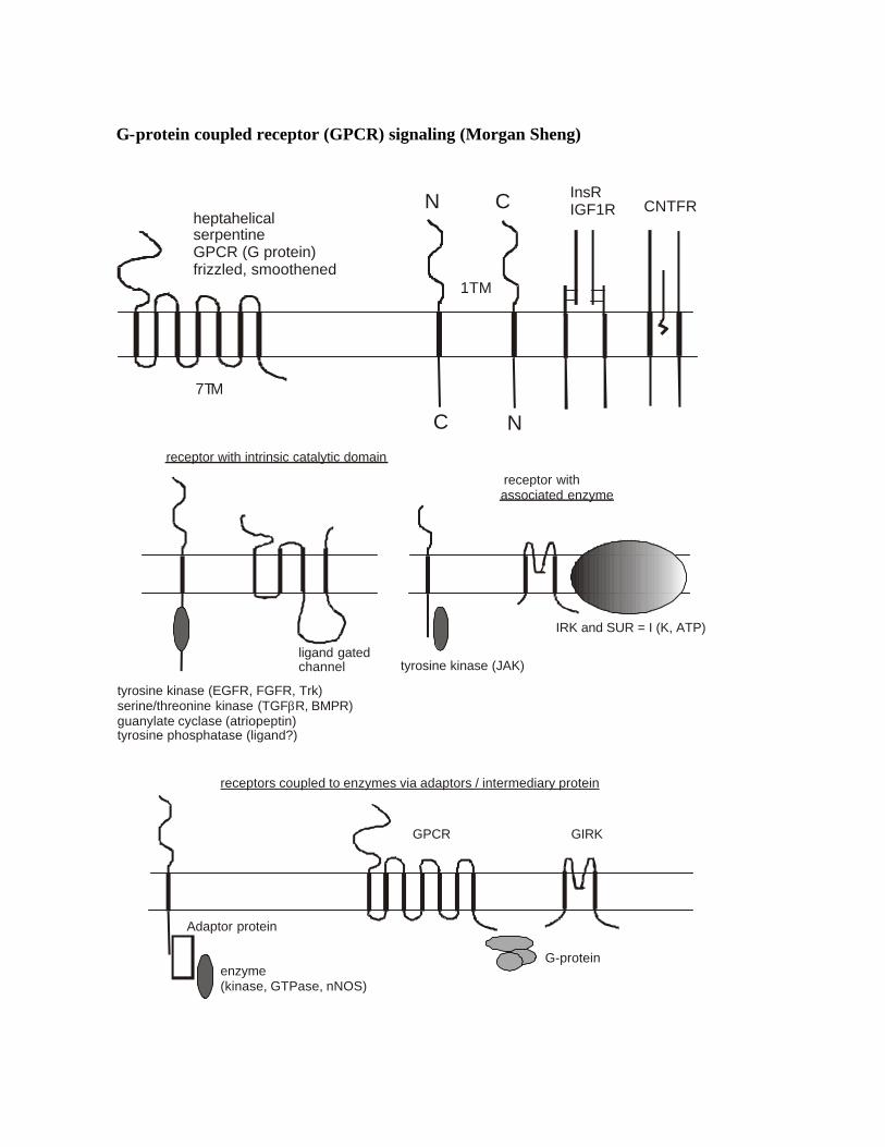
heptahelicalserpentineGPCR (G protein)frizzled, smoothened
7TM
1TM
InsRIGF1R CNTFR
receptor with intrinsic catalytic domain
associated enzyme
tyrosine kinase (EGFR, FGFR, Trk)serine/threonine kinase (TGF R, BMPR)guanylate cyclase (atriopeptin)tyrosine phosphatase (ligand?)
β
ligand gated channel tyrosine kinase (JAK)
IRK and SUR = I (K, ATP)
enzyme(kinase, GTPase, nNOS)
receptor with
receptors coupled to enzymes via adaptors / intermediary protein
G-protein
GIRKGPCR
Adaptor protein
Generalizations Cell surface receptors: 1. Ligand-gated ion channels (e.g. iGluRs), receptor tyrosine kinases, receptor tyrosine
phosphatases, receptor serine kinases, receptor guanylate cyclases: contain receptor and catalytic activity in one molecule
2. GPCRs have no catalytic domain and require intermediary (G-protein); thus function in a 3-component pathway: R à G à E (but not nec linear pathway 1R à 1G à 1E)
3. GPCRs highly versatile -- diversity of ligands: photons, inorganic ions, small chemicals eg odorants, monoamines; peptides to large glycoprotein hormones; even proteases
4. GPCRs involved in short-term regulation (seconds to hours) of cellular physiology rather than in long term regulation of growth or development (growth factors/RTKs) [but endothelin B receptor and Hirschsprungs disease (colonic aganglionosis); endothelin A receptor required for neural crest development; GPCRs can activate Src, Ras, MAPK, transcription, mitogenesis]
5. GPCRs mediate slower “modulatory” effects in neurons (seconds to hours) cf ligand-gated ion channels (millisec) [but phototransduction can be as fast as 10s of msec]
The importance of GPCRs 1. Number (C.elegans 1100; H. sapiens, ~1000; D. melanogaster, 160; reflects number of
olfactory receptor genes in worm [~1000] and mammal [several hundreds]), a few % of genome; 300-400 non-olfactory GPCRs),
2. Diversity (mostly small molecule ligands) 3. Evolutionarily conserved yeast to man (yeast Gα 45% identical to mammalian Giα) 4. Pharmaceutical importance: ~500 known molecular targets of drugs, 60% of these are
cell surface receptors, 75% of these are GPCRs (GPCRs = ~45% of all known drug targets)
[why are GPCRs such good drug targets?] [Are there any neurotransmitters that act on ligand-gated channels but do not act on GPCRs? And vice versa?] Architecture of GPCRs (Strader et al 1994 Annual Review of Biochem 63:101-132) 1. Heptahelical, “magnificent seven”, 7TM, serpentine 2. glycoproteins (glycosylation may be important for surface expression but not for
function) 3. Rhodopsin first cloned GPCR, then β2-adrenergic R 4. Cloning has revealed diversity unsuspected from classical pharmacology: e.g. 5
muscarinic, 9 adrenergic, 14 serotonin (cf 2-4 pharmacologic subtypes) 5. Highest conservation in TM domains (60-80% identity for subtypes of same receptor;
20-25% for unrelated Rs)
6. Poor conservation in N- and C- terminal regions and loops between TM segments 7. Conserved extracellular –S-S- that links first and second extracellular loops;
palmitoylation of C-term cysteine 8. GPCRs are composed of various independent fold ing units: receptor fragments (e.g.
rhodopsin, M3 muscarinic) can assemble to form functional receptors (e.g. TM1-5 and TM6-7). Consistent with idea that TM helices are tightly packed in ring like structure in membrane.
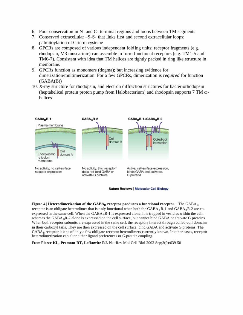
9. GPCRs function as monomers (dogma); but increasing evidence for dimerization/multimerization. For a few GPCRs, dimerization is required for function (GABA(B))
10. X-ray structure for rhodopsin, and electron diffraction structures for bacteriorhodopsin (heptahelical protein proton pump from Halobacterium) and rhodopsin supports 7 TM α-helices
Figure 4 | Heterodimerization of the GABAB receptor produces a functional receptor. The GABAB receptor is an obligate heterodimer that is only functional when both the GABABR-1 and GABABR-2 are co-expressed in the same cell. When the GABABR-1 is expressed alone, it is trapped in vesicles within the cell, whereas the GABABR-2 alone is expressed on the cell surface, but cannot bind GABA or activate G proteins. When both receptor subunits are expressed in the same cell, the receptors interact through coiled-coil domains in their carboxyl tails. They are then expressed on the cell surface, bind GABA and activate G proteins. The GABAB receptor is one of only a few obligate receptor heterodimers currently known. In other cases, receptor heterodimerization can alter either ligand preferences or G-protein coupling.
From Pierce KL, Premont RT, Lefkowitz RJ. Nat Rev Mol Cell Biol 2002 Sep;3(9):639-50
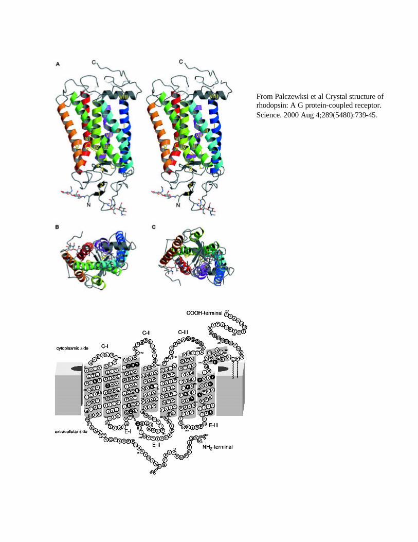
From Palczewksi et al Crystal structure of rhodopsin: A G protein-coupled receptor. Science. 2000 Aug 4;289(5480):739-45.
Ligand binding domain binding surface varies in different receptors: often in the TM domains for small molecule ligands; typically on the extracellular domains for large protein ligands Biogenic amines/retinal: Residues important for ligand binding identified by chimeric and site directed mutagenesis studies: catecholamines interact with residues in TM3, 5, 6 of adrenoreceptor (all located “deep” in bilayer); replacement of TM4-5 of β2 with β1 R sufficient to switch preference to NE from E; retinal covalently attached to Lys296 in TM7 of rhodopsin Peptides N-terminus and extracellular loops important in binding of many peptide ligands, but TM segments also involved, thus binding surface more diffuse Unusual ligands (Ca2+, thrombin) Parathyroid Ca2+ sensing receptor [cloned by functional expression cloning] and mGluRs – very long N-terminal region (600 aa); extracellular N-terminus involved in ligand binding Thrombin receptor releases tethered ligand by proteolysis of N-terminus; specificity is conferred by proteolysis site sequence 100-200 orphan GPCRs are known in human. “Reverse pharmacology” to identify possible ligands (of great pharmaceutical interest)
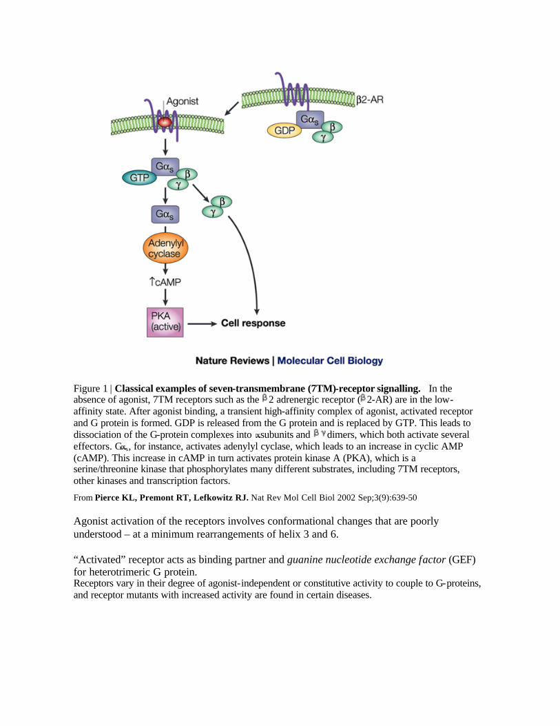
Figure 1 | Classical examples of seven-transmembrane (7TM)-receptor signalling. In the absence of agonist, 7TM receptors such as the 2 adrenergic receptor ( 2-AR) are in the low-affinity state. After agonist binding, a transient high-affinity complex of agonist, activated receptor and G protein is formed. GDP is released from the G protein and is replaced by GTP. This leads to dissociation of the G-protein complexes into subunits and dimers, which both activate several effectors. G s, for instance, activates adenylyl cyclase, which leads to an increase in cyclic AMP (cAMP). This increase in cAMP in turn activates protein kinase A (PKA), which is a serine/threonine kinase that phosphorylates many different substrates, including 7TM receptors, other kinases and transcription factors.
From Pierce KL, Premont RT, Lefkowitz RJ. Nat Rev Mol Cell Biol 2002 Sep;3(9):639-50 Agonist activation of the receptors involves conformational changes that are poorly understood – at a minimum rearrangements of helix 3 and 6. “Activated” receptor acts as binding partner and guanine nucleotide exchange factor (GEF) for heterotrimeric G protein. Receptors vary in their degree of agonist-independent or constitutive activity to couple to G-proteins, and receptor mutants with increased activity are found in certain diseases.
Receptor-G protein interactions (chimeras, mutagenesis, peptide blocking etc): 1. efficacy and selectivity of G-protein interactions probably depend on the proper
combination of multiple specific cytoplasmic receptor regions 2. residues at cytoplasmic ends of all helices and nearby loops have been implicated for
overall interaction and specificity determination of G-protein interaction: eg 3. cytoplasmic domains close to TM5, 6, 7 of adrenoreceptors involved in G coupling; 4. M3 receptor G-protein coupling preference due to second and third intracellular loops; 5. second intracellular loop of V1A vasopressin receptor required for coupling to Gq/11,
whereas third intracellular loop of V2 receptor responsible for coupling to Gs: thus differential G-protein coupling of related receptors may be mediated by different cytoplasmic domains.
6. Individual GPCRs can couple to more than one class of G-proteins, probably by using distinct receptor interaction sites for coupling to different G-proteins.
7. For instance: β2-AR predominantly couples via Gs to adenylate cyclase (AC)/cAMP, but it can also couple to Gi to MAPK (via Src/Ras). PKA phosphorylation required for Gi coupling. PKA phosphorylation site mutants of β2AR can activate AC but not MAPK.
8. Unknown how R transduces signal to G-protein: ligand binding may induce a conformational change in intracellular surface of R, allowing G-prot to interact.
Functional Diversity of GPCRs • Signaling molecules can cause a focused response in one cell and plethora of different
responses in another; single receptor can activate several pathways in a given cell, though the predominant pathway may vary from cell to cell.
• Multiple pathways can be activated by single extracellular stimulus acting on distinct related receptors e.g. 14 mammalian serotonin GPCRs with distinct expression patterns and signal transduction properties.
• Classic example: stimulation of AC by β-ARs (via Gs), inhibition by α2-ARs (via Gi) (these receptors can be pharmacologically differentiated by drugs)
Receptor subtypes: splice variants, RNA editing (Guderman et al 1996 Annual review of Pharmacology and Toxicology 36: 429-59) Initial GPCR genes lacked introns, but alternative splicing has been detected, affecting TM domains and C-terminal regions of GPCRs. Can affect ligand binding, G-protein coupling preference, signaling efficacy, glycosylation patterns, intracellular trafficking RNA editing of GPCRs: genomically encoded adenosine converted to inosine by dsRNA adenosine deaminases in second intracellular loop of 5-HT2CR, changing 3 aas (Burns et al 1997 Nature 387: 303). Edited and unedited Rs couple to PLC, but agonist 10-15x less potent in edited R, probably via reduced efficiency of R-G coupling.

Desensitization of GPCRs Termination of receptor signaling in the face of persistent ligand (“desensitization” is an umbrella term encompassing several distinct mechanisms that overlap in time course) Second messenger kinases: PKA (Gs), PKC (Gq) à β2AR phosphorylation on serines in 3rd cytoplasmic loop and C-tail à impaired interaction with G-proteins (“heterologous desensitization”; not specific for agonist, classical negative feedback inhibition) GRKs (7 genes) and β-arrestins (4 genes) Agonist-occupied GPCR à Recruitment of GRK (from cytosol via free Gβγ and PIP2 binding to GRK2 PH domain) à allosteric activation of GRK upon binding of GRK to activated-GPCR substrate à receptor phosphorylation on C-tail by GRK à binding of β-arrestin (10-30 fold increase in affinity of phospho-GPCR for β-arrestin = (“homologous desensitization”, agonist-specific, since requires receptor occupancy) 1. Ligand-stimulated phosphorylation of GPCR by associated GRK is probably a general
mechanism of desensitization of GPCRs; GPCRs are regulated by specific (degenerate) GRKs (e.g. rhodopsin kinase (GRK1), βARK1 (GRK2)); basis of specificity unknown
2. Phosphorylation of GPCR enables them to bind to inhibitory protein β-arrestin that sterically blocks GPCR from activating G protein
3. Thus 3 classes of proteins bind to GPCR in agonist-dependent fashion: G proteins, β-arrestins, GRKs
4. Receptor endocytosis also follows β-arrestin binding and may also contribute to desensitization (sequestration and degradation)
5. Knockout mice lacking specific GRK or arrestin show supersensitivity to GPCR ligand: e.g. β-arrestin-2 (mu-opioid receptor) --- increased and prolonged antinociceptive effect of morphine, absence of morphine- induced tolerance
GPCR internalization/sequestration (sometimes called “downregulation” 1. Functional significance and mechanisms controversial: probably not important for the
most rapid phase of desensitization; also involved in degradation, recycling/resensitization, MAPK signaling. Tolerance (?)
2. Typically (but not necessarily) via dynamin-dependent clathrin-coated vesicle pathway of endocytosis
3. β-arrestin mediated internalization of β2AR involves ubiquitination; β-arrestin interacts directly with a ubiquitin ligase [Shenoy et al 2001 Science 294, 1307].
GRK phosphorylation and β-arrestin binding crucial for GPCR internalization
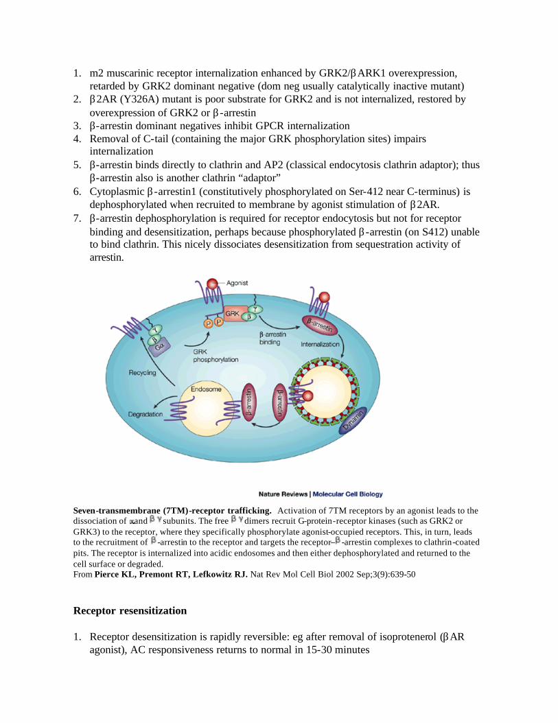
1. m2 muscarinic receptor internalization enhanced by GRK2/βARK1 overexpression, retarded by GRK2 dominant negative (dom neg usually catalytically inactive mutant)
2. β2AR (Y326A) mutant is poor substrate for GRK2 and is not internalized, restored by overexpression of GRK2 or β-arrestin
3. β-arrestin dominant negatives inhibit GPCR internalization 4. Removal of C-tail (containing the major GRK phosphorylation sites) impairs
internalization 5. β-arrestin binds directly to clathrin and AP2 (classical endocytosis clathrin adaptor); thus
β-arrestin also is another clathrin “adaptor” 6. Cytoplasmic β-arrestin1 (constitutively phosphorylated on Ser-412 near C-terminus) is
dephosphorylated when recruited to membrane by agonist stimulation of β2AR. 7. β-arrestin dephosphorylation is required for receptor endocytosis but not for receptor
binding and desensitization, perhaps because phosphorylated β-arrestin (on S412) unable to bind clathrin. This nicely dissociates desensitization from sequestration activity of arrestin.
Seven-transmembrane (7TM)-receptor trafficking. Activation of 7TM receptors by an agonist leads to the dissociation of and subunits. The free dimers recruit G-protein-receptor kinases (such as GRK2 or GRK3) to the receptor, where they specifically phosphorylate agonist-occupied receptors. This, in turn, leads to the recruitment of -arrestin to the receptor and targets the receptor– -arrestin complexes to clathrin-coated pits. The receptor is internalized into acidic endosomes and then either dephosphorylated and returned to the cell surface or degraded. From Pierce KL, Premont RT, Lefkowitz RJ. Nat Rev Mol Cell Biol 2002 Sep;3(9):639-50 Receptor resensitization 1. Receptor desensitization is rapidly reversible: eg after removal of isoprotenerol (βAR
agonist), AC responsiveness returns to normal in 15-30 minutes
2. Resensitization requires endocytosis and probably mediated by dephosphorylation of GPCR in an intracellular compartment of acid pH (endosome); resensitization blocked by dynamin and β-arrestin dom negatives, or NH4Cl (which inhibits acidification)
3. Thus the same molecules that initiate desensitization (GRK and β-arrestin) also cause internalization into acidified endosomes, which is required for dephosphorylation and resensitization
4. How the dephosphorylated receptor is recycled to the surface not understood, but may involve specific protein interactions of the cytoplasmic tail.
GPCR endocytosis and MAPK signaling [non-classical signaling by GPCR] Internalization not only important for late desensitization and resensitization, but also for other modes of signaling by GPCRs. 1. Many GPCRs can activate MAP kinases (eg Erk1/2), in some cases leading to mitogenic
responses 2. Best characterized pathway: Gi pathway, mediated by βγ à activation of Src- like
tyrosine kinase à tyrosine phosphorylation of adaptor (eg Shc) or scaffold proteins (EGFR) à recruitment of Grb2/SOS complex to plasma membrane à Ras à Raf à MEKà à Erks
3. MAPK activation by GPCR is blocked by β-arrestin- or dynamin- dom negatives and other inhibitors of endocytosis, suggesting endocytosis is required for GPCR signaling to MAPK cascade (but controversial). β-arrestin can bind to Src.
4. EGFR activation of MAPK also requires endocytosis of the receptor 5. Classical “membrane delimited” GPCR pathways (eg to AC or to PLC or ion channels)
not affected by dynamin or β-arrestin dominant negatives

β-arrestin scaffolding of mitogen-activated protein kinase (MAPK) cascades. β-arrestins function as adaptor/scaffolding molecules, which facilitate the activation of two MAPK cascades — the extracellular-signal regulated kinase (ERK)/MAPK cascade and the c-Jun-amino terminal kinase-3 (JNK3) cascade. a | After G-protein-regulated kinase (GRK)-dependent phosphorylation of the 7TM receptor, -arrestin is recruited to the receptor (b), and brings with it the components of the MAPK cascade; the MAPK kinase kinase (Raf), the MAPK kinase (MEK1), and the MAPK (ERK). c | This association facilitates the activation of ERK/MAPK and targets the active MAPK to the cytosol, thereby decreasing nuclear ERK/MAPK signalling. -arrestin-dependent scaffolding of the JNK3 is highly analogous — -arrestin both facilitates JNK3 activation and leads to the agonist-dependent co-localization of the receptor, -arrestin and the components of the JNK3 cascade in endosomal vesicles.
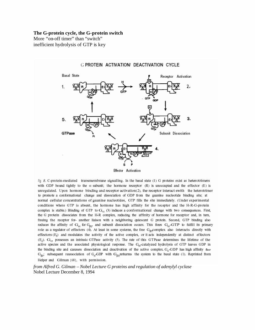
The G-protein cycle, the G-protein switch More “on-off timer” than “switch” inefficient hydrolysis of GTP is key
from Alfred G. Gilman – Nobel Lecture G proteins and regulation of adenylyl cyclase Nobel Lecture December 8, 1994
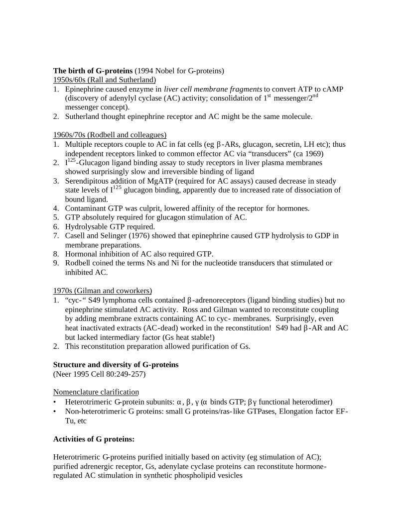
The birth of G-proteins (1994 Nobel for G-proteins) 1950s/60s (Rall and Sutherland) 1. Epinephrine caused enzyme in liver cell membrane fragments to convert ATP to cAMP
(discovery of adenylyl cyclase (AC) activity; consolidation of 1st messenger/2nd messenger concept).
2. Sutherland thought epinephrine receptor and AC might be the same molecule. 1960s/70s (Rodbell and colleagues) 1. Multiple receptors couple to AC in fat cells (eg β-ARs, glucagon, secretin, LH etc); thus
independent receptors linked to common effector AC via “transducers” (ca 1969) 2. I125-Glucagon ligand binding assay to study receptors in liver plasma membranes
showed surprisingly slow and irreversible binding of ligand 3. Serendipitous addition of MgATP (required for AC assays) caused decrease in steady
state levels of I125 glucagon binding, apparently due to increased rate of dissociation of bound ligand.
4. Contaminant GTP was culprit, lowered affinity of the receptor for hormones. 5. GTP absolutely required for glucagon stimulation of AC. 6. Hydrolysable GTP required. 7. Casell and Selinger (1976) showed that epinephrine caused GTP hydrolysis to GDP in
membrane preparations. 8. Hormonal inhibition of AC also required GTP. 9. Rodbell coined the terms Ns and Ni for the nucleotide transducers that stimulated or
inhibited AC. 1970s (Gilman and coworkers) 1. “cyc-“ S49 lymphoma cells contained β-adrenoreceptors (ligand binding studies) but no
epinephrine stimulated AC activity. Ross and Gilman wanted to reconstitute coupling by adding membrane extracts containing AC to cyc- membranes. Surprisingly, even heat inactivated extracts (AC-dead) worked in the reconstitution! S49 had β-AR and AC but lacked intermediary factor (Gs heat stable!)
2. This reconstitution preparation allowed purification of Gs. Structure and diversity of G-proteins (Neer 1995 Cell 80:249-257) Nomenclature clarification • Heterotrimeric G-protein subunits: α, β , γ (α binds GTP; βγ functional heterodimer) • Non-heterotrimeric G proteins: small G proteins/ras- like GTPases, Elongation factor EF-
Tu, etc Activities of G proteins: Heterotrimeric G-proteins purified initially based on activity (eg stimulation of AC); purified adrenergic receptor, Gs, adenylate cyclase proteins can reconstitute hormone-regulated AC stimulation in synthetic phospholipid vesicles
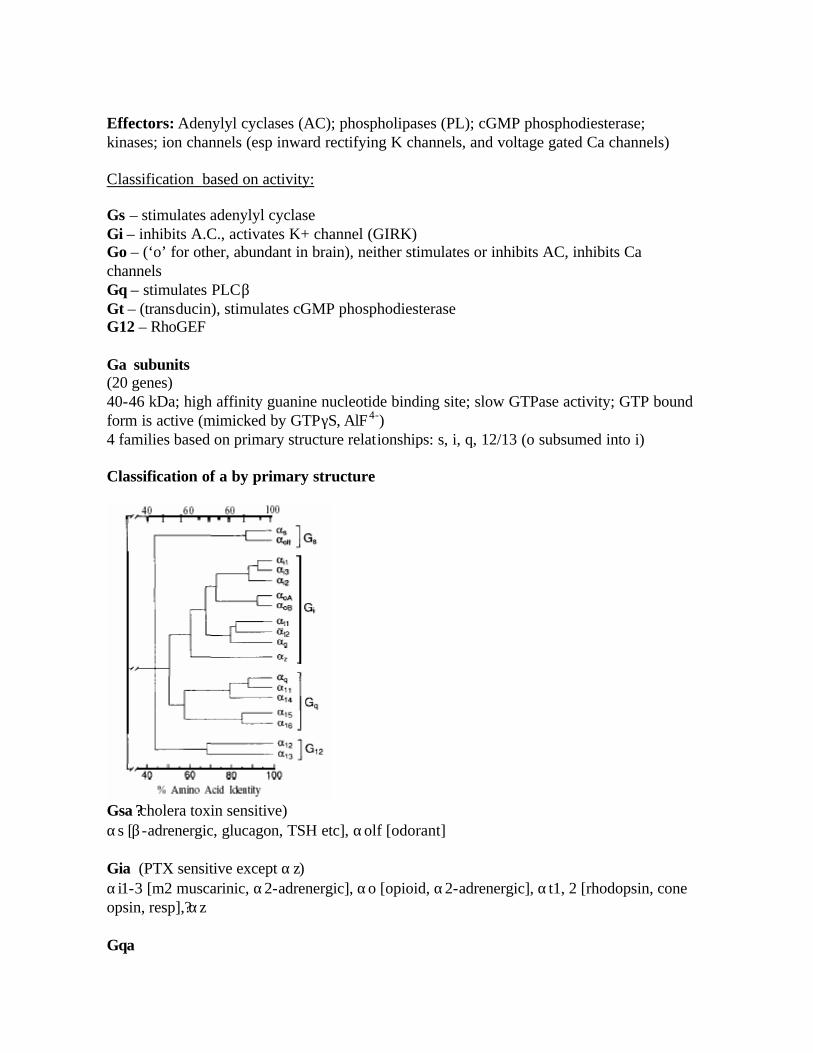
Effectors: Adenylyl cyclases (AC); phospholipases (PL); cGMP phosphodiesterase; kinases; ion channels (esp inward rectifying K channels, and voltage gated Ca channels) Classification based on activity: Gs – stimulates adenylyl cyclase Gi – inhibits A.C., activates K+ channel (GIRK) Go – (‘o’ for other, abundant in brain), neither stimulates or inhibits AC, inhibits Ca channels Gq – stimulates PLCβ Gt – (transducin), stimulates cGMP phosphodiesterase G12 – RhoGEF Gα subunits (20 genes) 40-46 kDa; high affinity guanine nucleotide binding site; slow GTPase activity; GTP bound form is active (mimicked by GTPγS, AlF4-) 4 families based on primary structure relationships: s, i, q, 12/13 (o subsumed into i) Classification of a by primary structure
Gsα?? cholera toxin sensitive) αs [β-adrenergic, glucagon, TSH etc], αolf [odorant] Giα (PTX sensitive except αz) αi1-3 [m2 muscarinic, α2-adrenergic], αo [opioid, α2-adrenergic], αt1, 2 [rhodopsin, cone opsin, resp],? αz Gqα
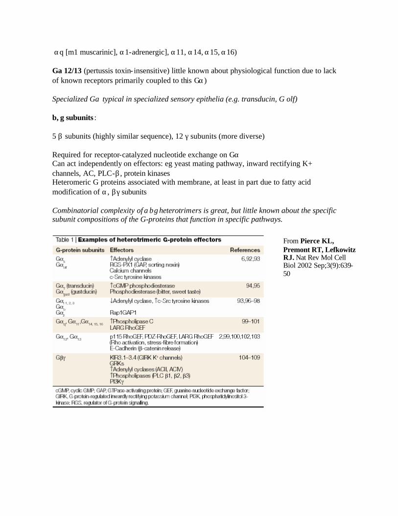
αq [m1 muscarinic], α1-adrenergic], α11, α14, α15, α16) Gα12/13 (pertussis toxin- insensitive) little known about physiological function due to lack of known receptors primarily coupled to this Gα) Specialized Gα typical in specialized sensory epithelia (e.g. transducin, G olf) β, γ subunits : 5 β subunits (highly similar sequence), 12 γ subunits (more diverse) Required for receptor-catalyzed nucleotide exchange on Gα Can act independently on effectors: eg yeast mating pathway, inward rectifying K+ channels, AC, PLC-β , protein kinases Heteromeric G proteins associated with membrane, at least in part due to fatty acid modification of α, βγ subunits Combinatorial complexity of αβγ heterotrimers is great, but little known about the specific subunit compositions of the G-proteins that function in specific pathways.
From Pierce KL, Premont RT, Lefkowitz RJ. Nat Rev Mol Cell Biol 2002 Sep;3(9):639-50
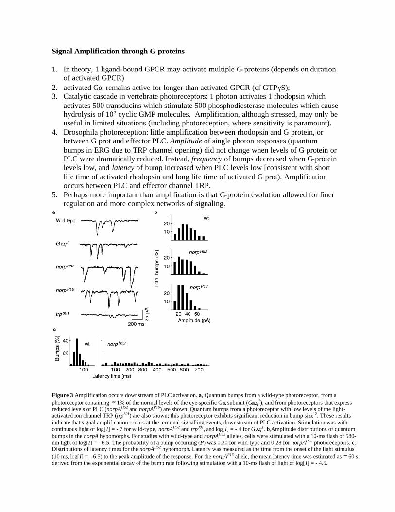
Signal Amplification through G proteins 1. In theory, 1 ligand-bound GPCR may activate multiple G-proteins (depends on duration
of activated GPCR) 2. activated Gα remains active for longer than activated GPCR (cf GTPγS); 3. Catalytic cascade in vertebrate photoreceptors: 1 photon activates 1 rhodopsin which
activates 500 transducins which stimulate 500 phosphodiesterase molecules which cause hydrolysis of 105 cyclic GMP molecules. Amplification, although stressed, may only be useful in limited situations (including photoreception, where sensitivity is paramount).
4. Drosophila photoreception: little amplification between rhodopsin and G protein, or between G prot and effector PLC. Amplitude of single photon responses (quantum bumps in ERG due to TRP channel opening) did not change when levels of G protein or PLC were dramatically reduced. Instead, frequency of bumps decreased when G-protein levels low, and latency of bump increased when PLC levels low [consistent with short life time of activated rhodopsin and long life time of activated G prot). Amplification occurs between PLC and effector channel TRP.
5. Perhaps more important than amplification is that G-protein evolution allowed for finer regulation and more complex networks of signaling.
Figure 3 Amplification occurs downstream of PLC activation. a, Quantum bumps from a wild-type photoreceptor, from a photoreceptor containing 1% of the normal levels of the eye-specific G subunit (G q1), and from photoreceptors that express reduced levels of PLC (norpAH52 and norpAP16) are shown. Quantum bumps from a photoreceptor with low levels of the light-activated ion channel TRP (trp301) are also shown; this photoreceptor exhibits significant reduction in bump size12. These results indicate that signal amplification occurs at the terminal signalling events, downstream of PLC activation. Stimulation was with continuous light of log[I] = - 7 for wild-type, norpAH52 and trp301, and log[I] = - 4 for G q1. b,Amplitude distributions of quantum bumps in the norpA hypomorphs. For studies with wild-type and norpAH52 alleles, cells were stimulated with a 10-ms flash of 580-nm light of log[I] = - 6.5. The probability of a bump occurring (P) was 0.30 for wild-type and 0.28 for norpAH52 photoreceptors. c, Distributions of latency times for the norpAH52 hypomorph. Latency was measured as the time from the onset of the light stimulus (10 ms, log[I] = - 6.5) to the peak amplitude of the response. For the norpAP16 allele, the mean latency time was estimated as 60 s, derived from the exponential decay of the bump rate following stimulation with a 10-ms flash of light of log[I] = - 4.5.
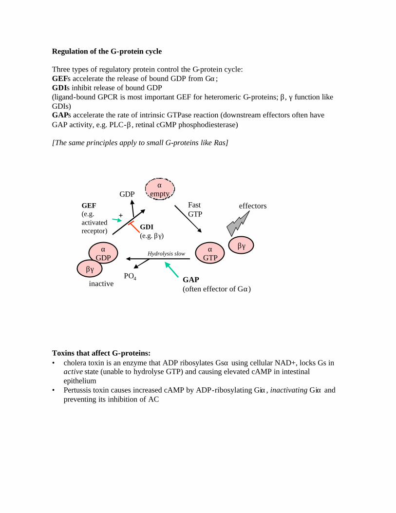
Regulation of the G-protein cycle Three types of regulatory protein control the G-protein cycle: GEFs accelerate the release of bound GDP from Gα; GDIs inhibit release of bound GDP (ligand-bound GPCR is most important GEF for heteromeric G-proteins; β, γ function like GDIs) GAPs accelerate the rate of intrinsic GTPase reaction (downstream effectors often have GAP activity, e.g. PLC-β , retinal cGMP phosphodiesterase) [The same principles apply to small G-proteins like Ras]
βγ
effectors
αGTP
FastGTP
αemptyGDP
GEF(e.g. activated receptor)
+GDI(e.g. βγ)
PO4
Hydrolysis slow
GAP(often effector of Gα)
αGDP
βγ
inactive
Toxins that affect G-proteins: • cholera toxin is an enzyme that ADP ribosylates Gsα using cellular NAD+, locks Gs in
active state (unable to hydrolyse GTP) and causing elevated cAMP in intestinal epithelium
• Pertussis toxin causes increased cAMP by ADP-ribosylating Giα, inactivating Giα and preventing its inhibition of AC
RGS (regulators of G-protein signaling) act as GAPs (probably only for Gi and Go) >25 RGS genes in mammals (characteristic RGS homology domain of 130 aa) Physiological roles poorly understood in mammals. The power of genetics Identification of a RGS (sst2 gene) in yeast 1. Mutant sst2 supersensitive to pheromone- induced cell cycle arrest in yeast mating
pathway --- respond to 100x lower conc of pheromone (pheromone receptor is GPCR) 2. genetic interaction with Gα suggests functions in desensitization of Gα, 3. biochemically associated with Gα; 4. sst2 is induced by pheromone (neg feedback); 5. high copy suppressors of sst2 also include kss1 (the first MAPK cloned, the first
connection between G-protein and MAPK pathway) and GPA1 (yeast Gα subunit). 6. Loss of function GPA1 mutants show constitutive activation of pheromone pathway
(first in vivo implication that βγ is the signal transmitter) 7. [How does GPA1 overexpression suppress sst2?] C. elegans : G-protein pathway controls egg laying (mediated by serotonergic motoneurons), egl-10 negative regulator of Goα?(overexpression of egl-10 increase egg laying frequency); loss of function of egl-10 similar phenotype to goa-1 (Goα??overexpression, suppressed by goa-1 loss of function GAIP is RGS identified by Y2hybrid with Gαi3 Multiple sst2/egl-10 homologs in higher eukaryotes All share a region of homology found at C-termini of sst2 and egl-10 RGS as GAP • Purified RGSs act as GAPs in vitro for Giα family (40x rate of GTP hydrolysis); • stimulate ‘single round’ of GTP hydrolysis but not steady state rate (which is limited by
GDP dissociation) [what does this say about RGS as GEF or GDI?]
Signal transduction by G-proteins Linear dogma of one receptor, one G-protein, one effector is inadequate. Most 7TM receptors interact with diverse G proteins and elicit multiple intracellular signals. However, with a given receptor in a certain cell, there may be high specificity in R-G interaction. G-protein signaling: complex signaling network, with divergent and convergent steps at ligand-R, R-G, and G-E interfaces. [NB G protein signaling pathways now intertwined with growth factor, ras MAPK cascades, and probably small GTPases, as well.] Divergence of signals: Both α?and βγ subunits are signaling mediators thus activation of one G-protein gives potentially bifurcating signal [Clapham and Neer 1993 Nature 365: 403]. Cloned GPCRs (esp Gi/o coupled) capable of dual signaling: ie inhibition of AC (Giα) and stimulation of PLC-β (βγ? released from Gi). [has been mainly studied in vitro/heterologous cells] Multiple coupling of receptors to different G-protein families Post-translational regulation of R-G coupling: βARs couple to both Gs (classical) and Gi (e.g. stimulation of MAPK via βγ, Src, ras). Gs mediated activation of PKA causes phosphorylation of βAR which then switches R coupling to Gi and stimulates a new signaling pathway (Daaka et al 1997 Nature 390: 88). Convergence of signals: different receptors acting on G proteins of one family (e.g. α2-adrenergic, A1 adenosine, M2 muscarinic, opioid and somatostatin receptors stimulating Gi/o) à common triad of effects: inhibition of AC (via Giα and/or βγ); activation of K channel (via βγ derived from Gi/o); inhibition of calcium channels via Go. Specificity of receptor-G protein interactions (Guderman et al 1996 Annual review of Pharmacology and Toxicology 36: 429-59) 100s of GPCRs use a fairly limited repertoire of G-proteins [how do you get specificity?] Antisense oligos directed against specific Gα, β, γ subunits show that selectivity of receptor-effector coupling is determined by specific αβγ heterotrimers (e.g. carbachol acts on m1 muscarinic receptor in basophil lymphocytes to stimulate PLC-β via a αq/α1-1β1/β4-γ4 complex).
Compartmentalization of GPCR signal transduction components 1. subsets of GPCRs, G-proteins and effectors reside within different cellular
compartments, perhaps poised to interact with each other (?”membrane delimited pathway”).
2. Examples: rod outer segment of photoreceptor contains all components needed for photon reception and signal transduction, most of these associated with the disc membrane. (Rhodopsin in vitro will interact with transducin plus related Gi);
3. asymmetrical distribution of GPCRs and G-proteins in growth cones, in polarized epithelia.
4. Targeting of G-proteins and GPCRs to specific signaling complexes likely to be important; probable existence of large complex containing Receptor, G protein, Effectors eg INAD complex for phototransduction in Drosophila: [Montell, 1998, Curr Opin Neurobiol 8:389]
5. Gαβγ? heterotrimers do not form randomly. Some specificity in βγ dimerization (differing affinities?). Preferential βγ interaction, in conjunction with differential expression of β and γ ?subunits in specific cells, may determine certain routes of signaling.