fuller albright and our current understanding of...
TRANSCRIPT

historia de la nefrología
346
http://www.revistanefrologia.com
© 2011 Revista Nefrología. Órgano Oficial de la Sociedad Española de Nefrología
Fuller Albright and our current understanding of calcium and phosphorus regulation and primary hyperparathyroidismA.J. Felsenfeld, B.S. Levine, C. R. KleemanDepartment of Medicine. VA Greater Los Angeles Healthcare System and the David Geffen School of Medicine at UCLA. Los Angeles, CA (USA)
Nefrologia 2011;31(3):346-57doi:10.3265/Nefrologia.pre2011.Mar.10774
Correspondence: A.J. Felsenfeld Department of Medicine. VA Greater Los Angeles Healthcare Systemand the David Geffen School of Medicine at UCLA.Nephrology Section (111L). West Los Angeles VA Medical Center,11301 Wilshire Boulevard, 900073, Los Angeles, CA, [email protected]
Fuller Albright y nuestro conocimiento actual sobre la regulación del calcio y del fósforo y el hiperparatiroidismo primario
RESUMEN
Se destacan las principales contribuciones de Fuller Albrightsobre el conocimiento de la regulación del calcio y del fósforoen el hiperparatiroidismo primario. Albright fue el primer in-vestigador que inició un estudio sistemático sobre el metabo-lismo de los minerales. Con unos recursos que se limitaban ala medición de la concentración de calcio y fósforo en suero yla infusión de extracto paratiroideo, Albright, a través de es-tudios del equilibrio, estableció unas bases para entender laregulación del calcio y del fósforo y el hiperparatiroidismo pri-mario. Albright fue el primero en afirmar que un adenoma ouna hiperplasia de las glándulas paratiroideas podrían ser lascausantes del hiperparatiroidismo primario. Además indicóque la litiasis sería una manifestación independiente del hi-perparatiroidismo primario. Albright observó también que:1) los pacientes con hipoparatiroidismo primario presentabanun valor umbral para la eliminación renal del calcio; 2) en pa-cientes con hipoparatiroidismo la rectificación de la hipocal-cemia con vitamina D tenía un efecto fosfatúrico; 3) en el hi-perparatiroidismo primario, la insuficiencia renal reducía laabsorción intestinal del calcio; 4) en el hiperparatiroidismo pri-mario grave, tras una paratiroidectomía se observaba el sín-drome del «hueso hambriento» y 5) es posible que un órganodiana deje de responder a una hormona. Albright también de-fendió la posibilidad de que un tumor maligno provocara laproducción ectópica de hormona. Por último, nuestra revisiónintegra las observaciones de Albright con los conocimientosactuales sobre la regulación y los trastornos del calcio.
Palabras clave: Calcio. Hiperparatiroidismo. Hipoparatiroidismo.Hormona paratiroidea. Fósforo.
INTRODUCTION
Fuller Albright’s academic career began in the late 1920s andended in 1956 after brain surgery for Parkinson’s diseaseresulted in a non-functional state until his death in 1969.
ABSTRACT
The major contributions of Fuller Albright to ourunderstanding of calcium and phosphorus regulation andprimary hyperparathyroidism are highlighted. Albrightwas the first investigator to initiate a systematic study ofmineral metabolism. With resources limited to themeasurement of serum calcium and phosphorus and theinfusion of parathyroid extract, Albright used balancestudies to establish a framework for our understanding ofcalcium and phosphorus regulation and primaryhyperparathyroidism. Albright was the first to show thatthe etiology of primary hyperparathyroidism could befrom either an adenoma or hyperplasia of the parathyroidglands and stone disease was a separate manifestation ofprimary hyperparathyroidism. Albright also showed that:1) a renal threshold for calcium excretion was present inhypoparathyroid patients; 2) correction of hypocalcemia inhypoparathyroid patients with vitamin D had a phosphaturicaction; 3) renal failure reduced the intestinal absorption ofcalcium in primary hyperparathyroidism; 4) the “hungrybone” syndrome developed after parathyroidectomy insevere primary hyperparathyroidism; and 5) a target organcan fail to respond to a hormone. He also suggested that amalignant tumor could be responsible for ectopic hormoneproduction. Finally, our review integrates the observations ofAlbright with our current knowledge of calcium regulationand disorders.
Keywords: Calcium. Hyperparathyroidism. Hypoparathyroidism.Parathyroid hormone. Phosphorus.
originales 10774 13/5/11 09:22 Página 346
historia de la nefrología
347
A.J. Felsenfeld et al. Fuller Albright and Ca regulation and disorders
Nefrologia 2011;31(3):346-57
Although increasingly disabled by Parkinson’s disease from themid 1930s, Albright continued to make important contributionsto our knowledge of calcium and phosphorus disorders1. Hisbook, “Parathyroid Glands and Metabolic Bone Disease”,published in 1948, is a testimony to his many importantobservations2. Our goal is to highlight some of the manycontributions made by Albright on calcium and phosphorusregulation and primary hyperparathyroidism and to integrate thefindings of Albright with more recent studies. Albright’scontributions to our understanding of renal phosphate transporthave been discussed elsewhere3. Virtually all that Albrightobserved remains valid today, but as often happens, theexplanations and their complexity continue to evolve.
Our retrospective highlights Albright’s enduring legacy tothe modern study of calcium and phosphorus regulationand primary hyperparathyroidism. The following topicsare discussed: 1) the resources available to Albright tostudy calcium and phosphorus regulation and primaryhyperparathyroidism; 2) Albright’s studies of calciumbalance; 3) phosphaturia resulting from the correction ofhypocalcemia in hypoparathyroid patients; 4) primaryhyperparathyroidism as a surgically curable disorder; and5) several clinical vignettes which include: a) a prelude tothe trade-off hypothesis of Slatopolsky and Brickeradvanced by Albright to explain the development ofsecondary hyperparathyroidism in renal failure; b) thesuggestion that a malignant tumor could be responsiblefor ectopic hormone production; c) the realization thatvitamin D deficiency can be associated with the failure torespond to parathyroid hormone (PTH); and d) theappreciation that immobilization can be a cause ofhypercalcemia.
TOOLS OF THE TRADE
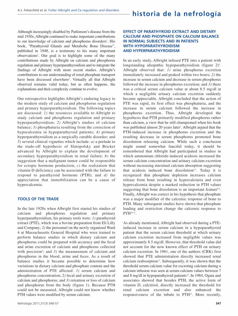
In the late 1920s when Albright first started his studies ofcalcium and phosphorus regulation and primaryhyperparathyroidism, his primary tools were: 1) parathyroidextract (PTE), which was a bovine preparation from Eli Lillyand Company; 2) the personnel on the newly organized Ward4 at Massachusetts General Hospital who were trained toperform balance studies in which dietary calcium andphosphorus could be prepared with accuracy and the fecaland urine excretion of calcium and phosphorus collectedwith precision4; and 3) the measurement of calcium andphosphorus in the blood, urine and feces. As a result ofbalance studies it became possible to determine howvariations in dietary calcium and phosphate content and theadministration of PTE affected: 1) serum calcium andphosphorus concentration; 2) fecal and urinary excretion ofcalcium and phosphorus; and 3) retention or loss of calciumand phosphorus from the body (figure 1). Because PTHcould not be measured, Albright could not know whetherPTH values were modified by serum calcium.
EFFECT OF PARATHYROID EXTRACT AND DIETARYCALCIUM AND PHOSPHATE ON CALCIUM BALANCEIN NORMAL SUBJECTS AND IN PATIENTS WITH HYPOPARATHYROIDISM AND HYPERPARATHYROIDISM
In an early study, Albright infused PTE into a patient withlongstanding idiopathic hypoparathyroidism (figure 2)5.Albright observed that: 1) urine phosphorus excretionimmediately increased and peaked within two hours; 2) theincrease in serum calcium and decrease in serum phosphorusfollowed the increase in phosphorus excretion; and 3) therewas a critical serum calcium value at about 8.5 mg/dl atwhich a negligible urinary calcium excretion suddenlybecame appreciable. Albright concluded that the action ofPTE was rapid, its first effect was phosphaturia, and theincrease in serum calcium followed the increase inphosphorus excretion. Thus, Albright developed thehypothesis that PTH primarily modified phosphorus ratherthan calcium, a view that he still championed when his bookwas published almost 20 years later2. Albright argued that thePTH-induced increase in phosphorus excretion and theresulting decrease in serum phosphorus promoted bonedissolution releasing calcium. While such a conclusionmight sound somewhat fanciful today, it should beremembered that Albright had participated in studies inwhich ammonium chloride-induced acidosis increased theserum calcium concentration and urinary calcium excretionwithout increasing intestinal calcium absorption suggestingthat acidosis induced bone dissolution6,7. Today it isrecognized that phosphate depletion increases calciumrelease from bone resulting in hypercalciuria and evenhypercalcemia despite a marked reduction in PTH valuessuggesting that bone dissolution is an important feature8,9.Finally, Albright was correct in his hypothesis that phosphatewas a major modifier of the calcemic response of bone toPTH. Many subsequent studies have shown that phosphateloading and restriction change the calcemic response toPTH10-12.
As already mentioned, Albright had observed during a PTE-induced increase in serum calcium in a hypoparathyroidpatient that the serum calcium threshold at which urinarycalcium excretion increased from negligible values wasapproximately 8.5 mg/dl. However, that threshold value didnot account for the now known effect of PTH on urinarycalcium excretion. In 1961, one of the authors (CRK) firstshowed that PTE administration directly increased renalcalcium reabsorption13. Subsequently, it was shown that thethreshold serum calcium value for excreting calcium during acalcium infusion was seen at serum calcium values between 7and 8 mg/dl in hypoparathyroid patients14. In 1984, Ogata andassociates showed that besides PTH, the active form ofvitamin D, calcitriol, directly increased the threshold forrenal calcium excretion and also enhanced theresponsiveness of the tubule to PTH15. More recently,
originales 10774 13/5/11 09:22 Página 347
historia de la nefrología
348
A.J. Felsenfeld et al. Fuller Albright and Ca regulation and disorders
Nefrologia 2011;31(3):346-57
Bindels and colleagues have shown that the stimulatoryeffect of both calcitriol and PTH on renal calciumreabsorption results from the activation of an epithelialcalcium channel (TRPV5) in the distal convoluted tubule16.
VITAMIN D TREATMENT AND CALCIUM INFUSIONAS PHOSPHATURIC AGENTS
In 1938 and in 1942, Albright used the newly available analog ofvitamin D, dihydrotachysterol, for the treatment of hypocalcemiain patients with hypoparathyroidism17 and also in the newlydescribed disorder of pseudohypoparathyroidism in which therewas a failure to respond to administered PTE18. Albrightobserved that the correction of hypocalcemia increased urinephosphate excretion. This observation had been made earlier byassociates of Albright19 and by Howland and Kramer20. In 1965,Eisenberg demonstrated in hypoparathyroid patients that thephosphaturia was independent of vitamin D by showing that aprolonged intravenous infusion of calcium sufficient to
normalize the serum calcium concentration at 48 hours wasphosphaturic and also lowered the serum phosphorusconcentration (figure 3A and B)21. Evidence has accumulatedduring the past several years that the recently discovered bone-derived phosphaturic hormone, fibroblast growth factor 23(FGF23) might be involved. High dietary calcium has beenshown to stimulate FGF2322 and the correlation between theserum calcium concentration and FGF23 seen in primaryhyperparathyroidism23-25 even remained significant afterparathyroidectomy24. However, a study has yet to be performedidentifying the specific mechanism for the observation mademore than 70 years ago by Albright and others.
PRIMARY HYPERPARATHYROIDISM, INTESTINALCALCIUM ABSORPTION, AND RENAL FAILURE
In patients with primary hyperparathyroidism, Albrightshowed that changes in dietary calcium and phosphateaffected calcium balance. The first patient studied was
Figure 1. Graphic representation of balance data.
The foundation of Albright’s studies of calcium and phosphorus was the balance study. Most of the time, the results were reported in long,detailed tables, but in this early study from 1929 of a patient with hypoparathyroidism, a figure is provided to show how parathormone(parathyroid extract) administration and changes in dietary phosphate affect the serum calcium and phosphorus concentration and urine calciumand phosphorus excretion. Each day was divided into three-eight hour periods. The patient was given the same meal three times a day at thebeginning of each period. Fifty units of parathormone were administered at the beginnings of periods 16, 19, 22 and 25 as indicated byasterisks. The diet was altered at period 43 and several times thereafter5.
Periods* 50 Units Parathormone
Serum P.
Serum Ca.
P. intake
Phos
phor
us in
urin
e
Cal
cium
in u
rine
Seru
m P
.: M
q.%
Seru
m C
a.: M
q.%10
8
6
4
800
700
600
500
400
300
200
100
0
12
8
4
0
140
120
100
80
60
40
20
01 3 5 7 9 11 13 15 17 19 21 23 25 27 2931 33 35 37 39 41 43 454749 51 53 55 57 59 61 63 6567 69 71 73 75 77 7981
originales 10774 13/5/11 09:22 Página 348

Captain Martell26, who was to have seven parathyroidoperations before an ectopic parathyroid gland was removedfrom the anterior mediastinum27. From the balance studies inCaptain Martell, who had serum calcium values between 13.1and 15.3 mg/dl, Albright made the following observations: 1)the patient’s negative calcium balance on a low calcium diet(0.1 grams/day) was greater than in normal controls (–0.46 vs–1.29 grams per 3 day study period); 2) the increasednegative calcium balance in the hyperparathyroid patient wasdue to increased urinary calcium excretion because fecalcalcium excretion was less than in normals; 3) an infusion ofPTE in normal volunteers sufficient to increase serumcalcium to between 11.5 and 12.8 mg/dl resulted in anegative calcium balance duplicating the results of thehyperparathyroid patient; and 4) a calcium diet of 1.07grams/day in the hyperparathyroid patient resulted in apositive calcium balance (0.36 grams per day) becauseurinary calcium excretion increased by only 0.06 grams/dayfrom that on a low calcium diet (0.1 grams/day). Thus,
Albright concluded that in primary hyperparathyroidism: 1)adaptation to a low calcium diet did not occur because highurine calcium losses persisted; 2) intestinal calciumabsorption was increased; and 3) high PTH values were thelikely cause of the negative calcium balance.
Albright also evaluated the effect of dietary phosphate oncalcium balance in hyperparathyroidism. He showed that ahigh phosphate diet improved calcium balance inhyperparathyroid patients on both low (0.07 g/day) andnormal (0.9 g/day) intakes of dietary calcium28. When highdietary phosphate was given to patients with primaryhyperparathyroidism, there was: 1) almost completeintestinal absorption of phosphate; 2) rapid excretion of theabsorbed phosphate by the kidneys; 3) a rise in thepreviously low serum phosphorus; 4) a fall in the previouslyelevated serum calcium; 5) a rise in the serum calcium-phosphorus product; and 6) a fall in urinary calciumexcretion. While a high phosphate diet seemed to havecertain beneficial effects such as lowering the serumcalcium concentration and decreasing urinary calciumexcretion, Albright recognized that there were two potentialdangers associated with increased phosphate ingestion inpatients with primary hyperparathyroidism: 1) parathyroidpoisoning with soft tissue calcium deposition including thekidney and lungs; and 2) calcium-phosphate kidney stones.Today, the recognition that hyperphosphatemia in CKDpatients and perhaps even high normal serum phosphorusvalues in the general population are associated withincreased vascular disease and mortality probably fromincreased vascular calcification29,30 could be considered anextension of the pioneering studies of Albright.
By 1934, Albright came to understand that renal failure had aspecific effect on calcium and phosphorus regulation31. Whena 13 year old girl with moderate renal failure was referredfor hypercalcemia (13.6 mg/dl), bone demineralization, amarkedly elevated serum alkaline phosphatase, and a serumphosphorus of 4.3 mg/dl, balance studies were performedbefore the parathyroid surgery during which an adenomawas removed. The balance studies showed that on a lowcalcium diet (0.1 g/day), urine calcium excretion despitehypercalcemia was only marginally greater than in controlson a similar diet (94 mg vs 63 mg/day). This value was muchless than in non-azotemic patients with primaryhyperparathyroidism and hypercalcemia (435 mg/day). Also,fecal calcium excretion was greater than in controls andmuch greater than in hyperparathyroid patients without renalfailure. Because of the results in this young girl with renalfailure, Albright reviewed the results of the series of balancestudies which had been performed on Captain Martell beforeand after he developed renal failure. As shown in table 1, fora similar magnitude of hypercalcemia (14 mg/dl vs 14.4mg/dl), for the same low calcium diet after the onset of renalfailure, intestinal calcium absorption was less (0.06 vs 0.26g/day) as was urine calcium excretion (0.09 vs 0.44 g/day).
historia de la nefrología
349
A.J. Felsenfeld et al. Fuller Albright and Ca regulation and disorders
Nefrologia 2011;31(3):346-57
Figure 2. Effect of parathyroid extract on urinary calcium and phosphorus excretion in a patient with idiopathichypoparathyroidism.
Hourly urinary calcium and phosphorus excretion are negligibleuntil parathyroid extract is given after which urinary phosphorusexcretion rapidly increases, serum phosphorus decreases and thenserum calcium slowly increases. Despite an increase in serumcalcium to almost 9 mg/dl, urine calcium excretion remainsnegligible5.
Time
Phos
phor
us in
urin
e
Cal
cium
in u
rine
* 75 Units Parathormone
Serum Ca.
*
P. m
q. %
Ca.
Mq.
%Serum P.
7 8 9 10 11 12 1 2 3 4 5 6 7
10
9
80
60
40
20
0
9
8
7
40
30
20
10
0
originales 10774 13/5/11 09:22 Página 349

historia de la nefrología
350
A.J. Felsenfeld et al. Fuller Albright and Ca regulation and disorders
Nefrologia 2011;31(3):346-57
Thus, Albright was the first to recognize that thedevelopment of renal failure in primary hyperparathyroidismdramatically decreased both intestinal absorption and therenal excretion of calcium. Today, it is known that PTHstimulates renal production of the active form of vitamin D,
calcitriol, which in turn enhances intestinal absorption ofcalcium. With loss of renal function, calcitriol production isdecreased despite high PTH values, a result which may inpart be due to increased FGF23 values. Consequently, bothintestinal calcium absorption and renal calcium excretion arereduced in renal failure. The latter results from both adecrease in the glomerular filtration of calcium andincreased tubular calcium reabsorption from high PTHvalues.
PRIMARY HYPERPARATHYROIDISM
In several patients in his original series of 17 patientspublished in 193432, Albright made the diagnosis ofhyperparathyroidism only because he had the insight tomeasure serum calcium and phosphorus values in all patientswho presented with kidney stones. In the first 14 patients toundergo parathyroid surgery, parathyroid adenomas werefound. Two of these patients had ectopic parathyroidadenomas located in the anterior mediastinum. Cases 15 to17 had hyperplasia of all the parathyroid glands. Becausehyperplasia had not been previously recognized as an entity,case 15 required three parathyroid operations to remove asufficient amount of the hyperplastic glands before thehypercalcemia resolved. Between the second and thirdoperations, estrogen treatment and irradiation of thepituitary and parathyroid glands were tried withoutsuccess33.
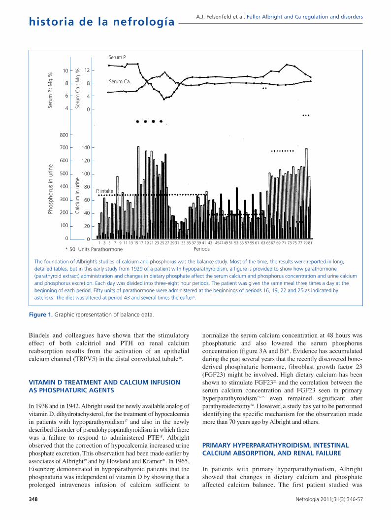
In this series of 17 patients, the dimensions, but not theweights of the removed parathyroid glands were provided32.The mean amount of parathyroid tissue removed per patientwas approximately 83 times greater than the combined sizeof four normal human parathyroid glands, whichsubsequently were shown to have a combined weight ofapproximately 140 mg34. Based on these results, the averageestimated weight of removed parathyroid tissue for eachpatient was approximately 11 grams. In severe cases ofprimary hyperparathyroidism with marked hypercalcemia,cachexia and debilitating fractures were sometimes seen andparathyroidectomy was life saving (figure 4). The differentmagnitude of primary hyperparathyroidism in patientspresenting in the 1930s and today is shown by the very highpreoperative serum calcium values in Albright’s patients(figure 5). Finally, Albright was the first to describe the“hungry bone syndrome” in which severe hypocalcemiadeveloped values shortly after parathyroidectomy (figure 6).He also showed that the decrease in serum calciumcorrelated with the pre-operative serum alkalinephosphatase value. When the serum calcium decreased toless than 7 mg/dl, Albright reported that tetany and visualdisturbances were often seen. In actuality, the subsequentrecognition of the “hungry bone syndrome” in dialysispatients after parathyroidectomy is an extension of theresults in primary hyperparathyroidism by Albright.
Figure 3. Effect of a 48 hour calcium infusion with normalization of the serum calcium concentration on phosphate excretion in hypoparathyroid patients.
The results shown from Robertson70 were recalculated from thedata in the study by Eisenberg21. A. The effect of a prolongedcalcium infusion on the maximal tubular reabsorption ofphosphate factored for the glomerular filtration rate (Tmp/GFR)in hypoparathyroid subjects is shown. The Tmp/GFR is theclassic test for evaluating the appropriateness of urinaryphosphate excretion. The dotted lines define the normal range.B. The relationship between the Tmp/GFR and the level ofplasma calcium in hypoparathyroid subjects is shown beforetreatment (open circle) and after a prolonged calcium infusion(closed circle) or vitamin D therapy (closed triangle). The dottedarea encloses the normal range.
A
Hypoparatiroid +Ca infusion
Plasma Calcium (mg/100ml)
Tmp/
GFR
(m
g/10
0 m
l GF)
Tmp/
GFR
(m
g/10
0 m
l GF)
4 6 8 10 12 14
8
6
4
2
0
8
6
4
2
0
B
originales 10774 13/5/11 09:22 Página 350

historia de la nefrología
351
A.J. Felsenfeld et al. Fuller Albright and Ca regulation and disorders
Nefrologia 2011;31(3):346-57
In 1934, Albright also recognized that patients with primaryhyperparathyroidism presented with either bone disease orstone disease, but rarely both together32. In patients withbone disease, skeletal symptoms associated with bone loss,bone cysts, brown tumors, and fractures predominated. Inpatients with stone disease, presenting symptoms were thoseassociated with nephrolithiasis and skeletal problems weregenerally absent. Albright questioned why there should betwo separate presentations for the same disease. Hehypothesized that the extent of bone disease was proportionalto the duration of disease times the daily loss of calcium32.Thus, according to Albright a short duration of disease wouldlessen the risk of bone disease. Moreover, a high calciumintake would make bone disease less likely because asAlbright had previously observed in studies of patients withprimary hyperparathyroidism, a high calcium diet resulted ina positive calcium balance26.
In 1945, Keating reported 24 patients with primaryhyperparathyroidism in whom the magnitude ofhypercalcemia, hypophosphatemia and serum alkalinephosphatase elevation was greater in patients with bonedisease than with stone disease35. In contradiction to thehypothesis advanced by Albright, the patients with bonedisease had a shorter duration of symptoms beforepresentation. In 1956, Dent also reported that patientswith bone disease had a shorter duration of symptoms36.Subsequently, Dent reported that there was no differencein dietary intake of calcium between patients with bone orstone disease37.
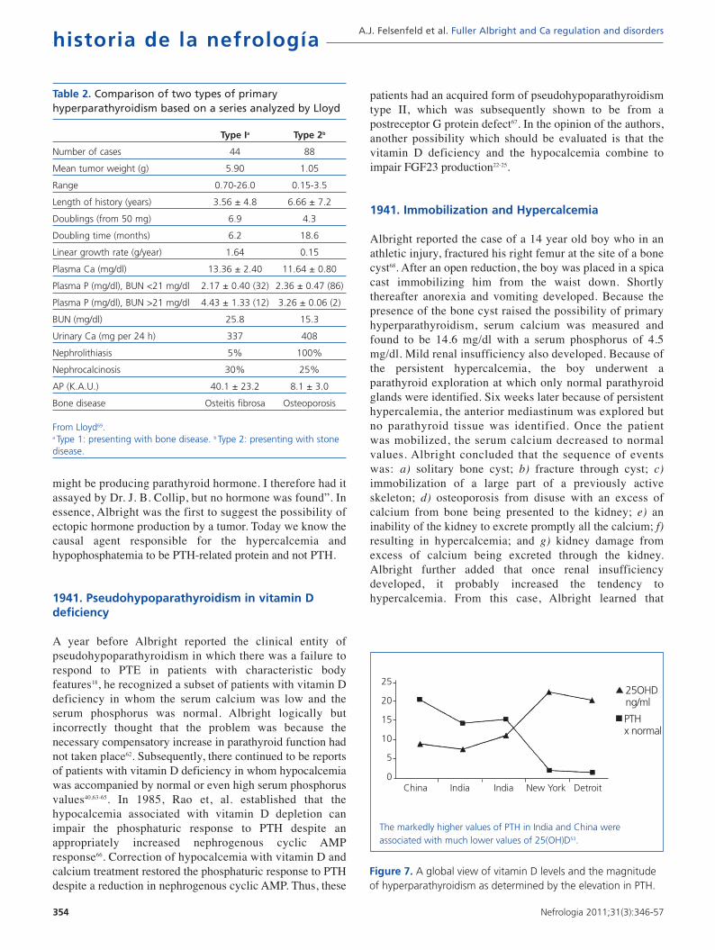
In 1968, Lloyd reviewed 138 consecutive cases of primaryhyperparathyroidism accumulated by Dent in London from1950 to 196538. Patients were divided into overt bone diseasewithout kidney stones (Type 1, n = 44) and kidney stoneswithout overt bone disease (Type 2, n = 88). The remainingsix patients had neither overt bone disease nor kidney stones.
As shown in table 2, the adenoma weight was greater, thegrowth rate of the parathyroid tissue more rapid, and the durationof symptoms was shorter in patients with bone disease. Theseresults contradicted Albright’s hypothesis that patients with bonedisease have a longer duration of disease.
One explanation for the shorter duration of disease togetherwith more severe hypercalcemia and larger adenomas inpatients with bone disease is simply a more rapid growth rateof the parathyroid adenomas in patients with bone disease. Inthe early 1970s, both Woodhouse et al39 and Lumb andStanbury40 suggested that the more rapid growth ofadenomas in patients with bone disease might be from a lackof vitamin D. In 1987, one of the authors (CRK) wrote aneditorial in support of this possibility41. Also in 1987,Paillard and associates reported that patients with bonedisease had lower values of the stored form of vitamin D,25-hydroxyvitamin D (25[OH]D), 3.4 ± 2.0 vs 17.6 ± 13.6ng/ml (P <0.001) and three-fold greater PTH values thanpatients with stone disease42. In more recent studies,Silverberg et al in a longitudinal study of patients with mildhyperparathyroidism, found that patients in the lowest tertileof 25(OH)D measurements (12 ± 3 ng/ml) had the highestPTH values43. Rao, et al., in a study of 148 consecutivepatients operated for primary hyperparathyroidism with amean resected parathyroid gland weight of 1.27 grams,reported an inverse correlation between 25(OH)D andparathyroid gland weight while the correlation between theactive form of vitamin D, calcitriol, and parathyroid glandweight was not significant44. These results suggest that asuboptimal vitamin D status may stimulate parathyroidadenoma growth. The presence of 1 alpha-hydroxylase, theenzyme responsible for conversion of 25(OH)D to calcitriol,in parathyroid cells suggests the possibility that 25(OH)Dmay directly affect PTH secretion and parathyroid glandgrowth45,46. Also contributing to the lowering of 25(OH)Dlevels in primary hyperparathyroidism is that high PTH
Table 1. A comparison of the calcium and phosphorus metabolism of a patient with hyperparathyroidism before and after the development of renal impairment
Calcium (g) Phosphorus (g) Serum
Output Output (mg per 100 cc)
Urine Feces Total Intake Balance Urine Feces Total Intake Balance Ca P
Control Seriesa 0.13 0.32 0.45 0.33 –0.12 1.21 0.60 1.81 2.07 0.26 10.0 4.0
Average of 3 1.31 0.19 1.50 0.31 –1.19 2.22 0.24 2.46 2.10 –0.36 14.0 2.7periods in 1926before renal impairment
Average of 4 0.27 0.79 1.06 0.30 –0.76 2.12 0.66 2.78 1.77 –1.01 14.4 4.2periods in 1932after renal impairment
The results for normal individuals are included for comparison31.a Each collection period was three days.
originales 10774 13/5/11 09:22 Página 351
historia de la nefrología
352
A.J. Felsenfeld et al. Fuller Albright and Ca regulation and disorders
Nefrologia 2011;31(3):346-57
levels decrease 25(OH)D levels by increasing conversion of25(OH)D to calcitriol47. In summary, even in the studies of
Silverberg and Rao in which the increased weight of theparathyroid adenoma was modest and the diagnosis ofprimary hyperparathyroidism was made relatively early inthe course of the disease, vitamin D status seemed to playa role43,44.
Since the 1970s, the clear demarcation between patients withbone and stone disease previously seen in patients with primaryhyperparathyroidism starting with the report of Albright in 1934and continuing through the 1960s has been lost32,35,38,48-51.Beginning in the 1930s and continuing for the next threedecades, the sequence of events in diagnosing bone disease inprimary hyperparathyroidism often was as described byAlbright, “appearance of a bone tumor, biopsy, diagnosis ofbenign giant cell tumor, local treatment, and finally recognitionof generalized disease only years later”2. However, theintroduction of multichannel analyzers in which serum calciumand phosphorus values were routinely measured resulted in thedetection of many asymptomatic hyperparathyroid patientswith mild hypercalcemia.
In a recent review of primary hyperparathyroidism, theaverage weight of the removed adenoma was 400 to 600mg, values which are only three to four times greater thanthe combined weight of four normal parathyroid glands52.In contrast, the mean weight of the removed parathyroidadenomas in the Albright study from 1934 wasapproximately 11 grams32. In 1947, Norris reviewed 322cases from the existing world literature and reported thatthe average weight of a removed parathyroid adenoma was8 grams48. In 1963, Hodgkinson reported that the mean
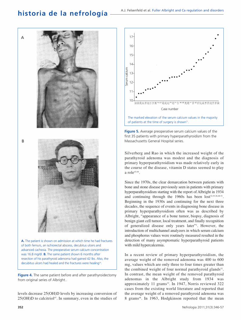
Figure 5. Average preoperative serum calcium values of thefirst 35 patients with primary hyperparathyroidism from theMassachusetts General Hospital series.
The marked elevation of the serum calcium values in the majorityof patients at the time of surgery is shown71.
Seru
m c
alci
um
Case number
17
16
15
14
13
12
11
10
32 33 29 31 16 22 11 12 34 2 5 20 25 21 3 23 7 17 4 8 24 28 1 10 6 15 27 35 14 19 22 13 16 30
Figure 4. The same patient before and after parathyroidectomyfrom original series of Albright..
A. The patient is shown on admission at which time he had fracturesof both femurs, an ischiorectal abscess, decubitus ulcers andadvanced cachexia. The preoperative serum calcium concentrationwas 16.8 mg/dl. B. The same patient shown 6 months afterresection of his parathyroid adenoma had gained 42 lbs. Also, thedecubitus ulcers had healed and the fractures were healing34.
A
B
originales 10774 13/5/11 09:22 Página 352

historia de la nefrología
353
A.J. Felsenfeld et al. Fuller Albright and Ca regulation and disorders
Nefrologia 2011;31(3):346-57
weight of the removed adenoma was 5.1 grams in patientswith bone disease and 1.4 grams in patients with stonedisease49. Similar differences in the weight of parathyroidadenomas between patients with bone and those with stonedisease were reported by Lloyd in his analysis of Dent’spatients38 and by O’Riordan51. The mean weight of theparathyroid adenoma in patients with bone disease was 5.9grams in the former and 4.2 grams in the latter series.
In areas of the world with limited access to medical care,vitamin D insufficiency/deficiency appears to be morecommon and the duration of primary hyperparathyroidismmuch longer before diagnosis and treatment. In recentstudies of primary hyperparathyroidism from India andChina, the presence of large parathyroid adenomas andbone disease has been associated with vitamin Dinsufficiency/deficiency (figure 7)53-57. In a study from China,the presenting PTH value was 21 times greater than normal55.Similarly, in a study from India, the mean weight of theremoved parathyroid adenoma was 10.75 g in hypercalcemicpatients and 3.9 g in normocalcemic patients54. In anotherstudy from India, the mean weight of the removedparathyroid adenoma was 7.9 g56. Moreover, the 25(OH)D
value was less than 10 ng/ml in the majority of thesepatients. In the cited studies, pathologic bone fractures, bonecysts, and brown tumors were commonly encountered54-56. Insummary, the severe form of primary hyperparathyroidismcharacterized by large adenomas and disabling bone disease,first described by Albright in the 1930s, is still commonlyencountered in areas of the world with limited access tomedical care. Furthermore, the severe form of primaryhyperparathyroidism seen in these patients is oftenassociated with vitamin D insufficiency/deficiency.
VIGNETTES IN WHICH OBSERVATIONS BY ALBRIGHTHAVE HAD CONTINUED CLINICAL RELEVANCE
1937. Prelude to the “Trade-Off Hypothesis” of Slatopolsky and Bricker10,58 which was Advancedto explain the development of secondaryhyperparathyroidism
Albright made the following statement in a 1937publication59: “It has been suggested above that theparathyroid hyperplasia is a compensation for thedisturbed equilibrium occasioned by phosphate retentionresulting from the renal insufficiency. In the absence of thehyperplasia there would probably be greater phosphateretention in the blood with a lowering of the blood calciumlevel and severe tetany. If these concepts are correct, thehyperplasia would have to be considered beneficial. Theone reservation might have to be made that, while helpinghomeostasis, the parathyroid hyperplasia may lead to bonedisease”. Confirmation of the Albright hypothesis has beenshown in many animal and clinical studies of phosphateloading. Also in studies of patients with stage 3 and 4CKD treated with the calcimimetic, cinacalcet, thereduction in PTH values has increased the serumphosphorus concentration60.
1941. Hypothesis that hypercalcemia in malignancycould be from ectopic hormone production
At a clinicopathological conference, a 51 year old malepresenting with hypercalcemia and hypophosphatemia wasdiscussed61. A neck exploration for presumedhyperparathyroidism was performed, but no abnormality wasfound. Bone x-rays showed a destructive lesion in the rightilium, which on biopsy was reported to originate from arenal cell carcinoma. The comment of Albright at the end ofthe conference was “Why a person should have high serumcalcium and low serum phosphorus when the cause of thedisturbance is a tumor destroying bone is an interestingtheoretical question. We treated this case by radiation of thetumor masses; the serum calcium went down to normal, andthe serum phosphorus went up to normal. Gradually, bothvalues became abnormal again. I suspected that the tumor
Figure 6. Fall in serum calcium values after parathyroidectomyin 35 cases of primary hyperparathyroidism (first demonstrationof hungry bone syndrome).
Serum calcium values are shown before and afterparathyroidectomy. The values connected by dotted lines are onpatients with high serum alkaline phosphatase. Albright tells thereader to note that cases with high alkaline phosphatase valuesand therefore, with bone disease, are evenly distributedpreoperatively at various levels of hypercalcemia. However, thecases with high alkaline phosphatase values all developedhypocalcemia postoperatively71.
Seru
m c
alci
um p
er 1
00 C
C
Days before operation Days after operation
Operation
3000
2000
1000 500
250
200
150
100 50 25 20 15 10 5 1 2 3 4 5 10 20 30 40 50 100
150
200
250
500
1000
2000
18.0
17.0
16.0
15.0
14.0
13.0
12.0
11.0
10.0
9.0
8.0
7.0
6.0
6.0
5.0
4.0
originales 10774 13/5/11 09:22 Página 353
historia de la nefrología
354
A.J. Felsenfeld et al. Fuller Albright and Ca regulation and disorders
Nefrologia 2011;31(3):346-57
might be producing parathyroid hormone. I therefore had itassayed by Dr. J. B. Collip, but no hormone was found”. Inessence, Albright was the first to suggest the possibility ofectopic hormone production by a tumor. Today we know thecausal agent responsible for the hypercalcemia andhypophosphatemia to be PTH-related protein and not PTH.
1941. Pseudohypoparathyroidism in vitamin Ddeficiency
A year before Albright reported the clinical entity ofpseudohypoparathyroidism in which there was a failure torespond to PTE in patients with characteristic bodyfeatures18, he recognized a subset of patients with vitamin Ddeficiency in whom the serum calcium was low and theserum phosphorus was normal. Albright logically butincorrectly thought that the problem was because thenecessary compensatory increase in parathyroid function hadnot taken place62. Subsequently, there continued to be reportsof patients with vitamin D deficiency in whom hypocalcemiawas accompanied by normal or even high serum phosphorusvalues40,63-65. In 1985, Rao et, al. established that thehypocalcemia associated with vitamin D depletion canimpair the phosphaturic response to PTH despite anappropriately increased nephrogenous cyclic AMPresponse66. Correction of hypocalcemia with vitamin D andcalcium treatment restored the phosphaturic response to PTHdespite a reduction in nephrogenous cyclic AMP. Thus, these
patients had an acquired form of pseudohypoparathyroidismtype II, which was subsequently shown to be from apostreceptor G protein defect67. In the opinion of the authors,another possibility which should be evaluated is that thevitamin D deficiency and the hypocalcemia combine toimpair FGF23 production22-25.
1941. Immobilization and Hypercalcemia
Albright reported the case of a 14 year old boy who in anathletic injury, fractured his right femur at the site of a bonecyst68. After an open reduction, the boy was placed in a spicacast immobilizing him from the waist down. Shortlythereafter anorexia and vomiting developed. Because thepresence of the bone cyst raised the possibility of primaryhyperparathyroidism, serum calcium was measured andfound to be 14.6 mg/dl with a serum phosphorus of 4.5mg/dl. Mild renal insufficiency also developed. Because ofthe persistent hypercalcemia, the boy underwent aparathyroid exploration at which only normal parathyroidglands were identified. Six weeks later because of persistenthypercalemia, the anterior mediastinum was explored butno parathyroid tissue was identified. Once the patientwas mobilized, the serum calcium decreased to normalvalues. Albright concluded that the sequence of eventswas: a) solitary bone cyst; b) fracture through cyst; c)immobilization of a large part of a previously activeskeleton; d) osteoporosis from disuse with an excess ofcalcium from bone being presented to the kidney; e) aninability of the kidney to excrete promptly all the calcium; f)resulting in hypercalcemia; and g) kidney damage fromexcess of calcium being excreted through the kidney.Albright further added that once renal insufficiencydeveloped, it probably increased the tendency tohypercalcemia. From this case, Albright learned that
Table 2. Comparison of two types of primaryhyperparathyroidism based on a series analyzed by Lloyd
Type Ia Type 2b
Number of cases 44 88
Mean tumor weight (g) 5.90 1.05
Range 0.70-26.0 0.15-3.5
Length of history (years) 3.56 ± 4.8 6.66 ± 7.2
Doublings (from 50 mg) 6.9 4.3
Doubling time (months) 6.2 18.6
Linear growth rate (g/year) 1.64 0.15
Plasma Ca (mg/dl) 13.36 ± 2.40 11.64 ± 0.80
Plasma P (mg/dl), BUN <21 mg/dl 2.17 ± 0.40 (32) 2.36 ± 0.47 (86)
Plasma P (mg/dl), BUN >21 mg/dl 4.43 ± 1.33 (12) 3.26 ± 0.06 (2)
BUN (mg/dl) 25.8 15.3
Urinary Ca (mg per 24 h) 337 408
Nephrolithiasis 5% 100%
Nephrocalcinosis 30% 25%
AP (K.A.U.) 40.1 ± 23.2 8.1 ± 3.0
Bone disease Osteitis fibrosa Osteoporosis
From Lloyd69.a Type 1: presenting with bone disease. b Type 2: presenting with stonedisease.
Figure 7. A global view of vitamin D levels and the magnitudeof hyperparathyroidism as determined by the elevation in PTH.
The markedly higher values of PTH in India and China wereassociated with much lower values of 25(OH)D53.
25OHDng/ml
PTHx normal
25
20
15
10
5
0China India India New York Detroit
originales 10774 13/5/11 09:22 Página 354

historia de la nefrología
355
A.J. Felsenfeld et al. Fuller Albright and Ca regulation and disorders
Nefrologia 2011;31(3):346-57
immobilization of an individual with active skeletalremodeling increases calcium efflux from bone and he alsorecognized that a decreased glomerular filtration rate reducesthe capacity to excrete calcium, which in turn, exacerbateshypercalcemia.
CONCLUSION
In the late 1920s, Albright joined Joseph Aub and WalterBauer to pursue studies of calcium and phosphorusmetabolism. First Aub and then Bauer were appointed toacademic positions in other subspecialties at Harvard, whichby 1930 left the young Albright as the primary investigatorof calcium and phosphorus metabolism in Boston. Throughinductive reasoning, which has been defined by the lateJacob Bronowski as that unpredictable blend of speculationand insight, Albright came to recognize in normal volunteersand in hypo- and hyperparathyroid patients, the presence ofconsistent patterns of response for calcium and phosphorusmetabolism. Fuller Albright was truly the first person toestablish a sense of order out of the existing chaos in thenew field of calcium and phosphorus metabolism. As oneof the 20th century’s preeminent philosophers of science,Karl Popper has stated, “Science does not rest upon rock-bottom. The bold structure of its theories rises, as it were,above a swamp, but not down to any natural or given base;and when we cease our attempts to drive our piles into adeeper layer, it is not because we have reached firmground. We simply stop when we are satisfied that they arefirm enough to carry the structure, at least for the timebeing”. Albright was the first to establish a functionalsystem which explained calcium and phosphorusmetabolism. His classic book, The Parathyroid Glands andMetabolic Bone Disease, published in 1948, became thestandard reference for a generation of students of calciumand phosphorus metabolism. Albright trained many futureinvestigators and became an inspiration for the nextgeneration of clinical investigators studying calcium andphosphorus disorders. As the importance of translationalresearch is again rightfully being emphasized, thecontributions of Fuller Albright should be recognized andhe should be celebrated as a role model for a newgeneration of young clinical investigators.
REFERENCES
1. Kleeman CR, Levine BS, Felsenfeld AJ. Fuller, Albright, the consum-
mate clinical investigator. Clin J Am Soc Nephrol 2009;4:1541-6.
2. Albright F, Reifenstein EC, Jr. The parathyroid glands and metabolic
bone disease, selected studies. Baltimore, MD: The Williams and
Wilkins Company; 1948.
3. Levine BS, Kleeman CR, Felsenfeld AJ. The journey from vitamin D-
resistant rickets to the regulation of renal phosphate transport. Clin
J Am Soc Nephrol 2009;4:1866-77.
4. Means JH. Ward 4 Cambridge: Harvard University Press; 1958:101.
5. Albright F, Ellsworth R. Studies on the physiology of the parathyroid
glands. I. Calcium and phosphorus studies on a case of idiopathic
hypoparathyroidism. J Clin Invest 1929;7:183-201.
6. Albright F, Bauer W, Ropes M, Aub JC. Studies of calcium and phos-
phorus metabolism. IV. The effect of the parathyroid hormone. J Clin
Invest 1929;7:139-81.
7. Albright F, Bauer W. The action of sodium chloride, ammonium chlo-
ride, and sodium bicarbonate on the total acid-base balance of a
case of chronic nephritis with edema. J Clin Invest 1929;7:465-86.
8. Raisz LG, Niemann I. Effect of phosphate, calcium, and magnesium
on bone resorption and hormonal responses in tissue culture. Endo-
crinology 1969;85:446-52.
9. Coburn JW, Massry SG. Changes in serum and urinary calcium du-
ring phosphate depletion: studies on mechanism. J Clin Invest
1970;49:1073-87.
10. Slatopolsky E, Caglar S, Pennell JP, et al. On the pathogenesis of
hyperparathyroidism in chronic experimental renal insuffficiency in
the dog. J Clin Invest 1971;50:492-99.
11. Rodríguez M, Martín Malo A, Martínez ME, Torres A, Felsenfeld AJ,
Llach F. Calcemic response to parathyroid hormone in renal failure: role
of phosphorus and its effect on calcitriol. Kidney Int 1991;40:1055-
62.
12. Krapf R, Glatz M, Hulter HN. Neutral phosphate administration ge-
nerates and maintains renal metabolic alkalosis and hyperparathy-
roidism. Am J Physiol 1995;268:F802-7.
13. Kleeman CR, Bernstein D, Rockney R, Dowling JT, Maxwell MH. Stu-
dies on the renal clearance of diffusible calcium and the role of the
parathyroid glands in its regulation. Yale J Biol Med 1961;34:1-30.
14. Peacock M, Robertson WG, Nordin BEC. Relation between serum
and urinary calcium with particular reference to parathyroid activity.
Lancet 1969;1:384-6.
15. Yamamoto M, Kawanobe Y, Takahashi H, Shimazawa E, Kimura S,
Ogata E. Vitamin D deficiency and renal calcium transport in the rat.
J Clin Invest 1984;74:507-13.
16. Hoenderop JGJ, Muller D, Van der Kemp AWCM, et al. Calcitriol
controls the epithelial calcium channel in kidney. J Am Soc Nephrol
2001;12:1342-9.
17. Albright F, Bloomberg E, Drake T, Sulkowitch HW. A comparison of
the effects of AT10 (dihydrotachysterol) and vitamin D on calcium
and phosphorus metabolism in hypoparathyroidism. J Clin Invest
1938;17:317-29.
18. Albright F, Burnett CH, Smith PH, Parson W. Pseudo-hypoparathy-
roidism - An example of Seabright-Bantam syndrome. Endocrino-
logy 1942;30:922-32.
19. Bauer W, Marble A, Claflin D. Studies on the mode of action of irra-
diated ergosterol IV. In hypoparathyroidism. J Clin Invest
1932;11:47-62.
20. Howland J, Kramer B. Calcium and phosphorus in the serum in re-
lation to rickets. Am J Dis Child 1921;22:105-19.
21. Eisenberg E. Effects of serum calcium and parathyroid extracts on
phosphate and calcium excretion in hypoparathyroid patients. J Clin
Invest 1965;44:942-6.
22. Shimada T, Yamazaki Y, Takahashi M, et al. Vitamin D receptor-in-
dependent FGF23 actions in regulating phosphate and vitamin D
metabolism. Am J Physiol Renal Physiol 2005;289:F1088-95.
originales 10774 13/5/11 09:22 Página 355
historia de la nefrología
356
A.J. Felsenfeld et al. Fuller Albright and Ca regulation and disorders
Nefrologia 2011;31(3):346-57
23. Yamashita H, Yamashita T, Miyamoto M, et al. Fibroblast growth fac-
tor (FGF)-23 in patients with primary hyperparathyroidism. Eur J En-
docrinol 2004;151:55-60.
24. Kobayashi K, Imanishi Y, Miyauchi A, et al. Regulation of plasma fi-
broblast growth factor 23 by calcium in primary hyperparathyroi-
dism. Eur J Endocrinol 2006;154:93-9.
25. Kawata T, Imanishi Y, Kobayashi K, et al. Parathyroid hormone re-
gulates fibroblast growth factor-23 in a mouse model of primary
hyperparathyroidism. J Am Soc Nephrol 2007;18:2683-8.
26. Bauer W, Albright F, Aub JC. A case of osteitis fibrosa cystica (oste-
omalacia?) with evidence of hyperactivity of the parathyroid bodies.
Metabolic study II. J Clin Invest 1930;8:229-48.
27. Bauer W, Federman DD. Hyperparathyroidism epitomized: the case
of Captain Charles E. Martell. Metabolism 1962;11:21-9.
28. Albright F, Bauer W, Claflin D, Cockrill JR. Studies in parathyroid
physiology III. The effect of phosphate ingestion in clinical hyperpa-
rathyroidism. J Clin Invest 1932;11:411-35.
29. Block GA, Hulbert-Shearon TE, Levin NW, Port FK. Association of se-
rum phosphorus and calcium x phosphate product with mortality
risk in chronic hemodialysis patients: A national study. Am J Kidney
Dis 1998;31:607-17.
30. Foley RN, Collins AJ, Herzog CA, Ishani A, Kalra PA. Serum phos-
phorus levels associate with coronary atherosclerosis in young
adults. J Am Soc Nephrol 2009;20:397-404.
31. Albright F, Baird PC, Cope O, Bloomberg E. Studies on the physio-
logy of the parathyroid glands. IV. Renal complications of hyperpa-
rathyroidism. Am J Med Sci 1934;187:49-65.
32. Albright F, Aub JC, Bauer W. Hyperparathyroidism: a common and
polymorphic condition as illustrated by seventeen proved cases from
one clinic. JAMA 1934;102:1276-87.
33. Albright F, Sulkowitch HW, Bloomberg E. Hyperparathyroidism due
to idiopathic hypertrophy (hyperplasia?) of parathyroid tissue. Arch
Intern Med 1938;62:199-215.
34. Churchill ED, Cope O. The surgical treatment of hyperparathyroi-
dism. Ann Surg 1936;104:9-35.
35. Keating FR, Jr., Cook EN. The recognition of primary hyperparathy-
roidism. JAMA 1945;129:994-1002.
36. Davies DR, Dent CE, Willcox A. Hyperparathyroidism and steator-
rhoea. Br Med J 1956;2:1133-7.
37. Dent CE, Hartland BV, Hicks J, Sykes ED. Calcium intake in patients
with primary hyperparathyroidism. Lancet 1961;2:336-8.
38. Lloyd HM. Primary hyperparathyroidism: an analysis of the role of
the parathyroid tumor. Medicine (Balt) 1968;47:53-71.
39. Woodhouse NJY, Doyle FH, Joplin GF. Vitamin-D deficiency and pri-
mary hyperparathyroidism. Lancet 1971;2:283-7.
40. Lumb GA, Stanbury SW. Parathyroid function in human vitamin D
deficiency and vitamin D deficiency in primary hyperparathyroidism.
Am J Med 1974;56:833-9.
41. Kleeman CR, Norris K, Coburn JW. Is the clinical expression of
primary hyperparathyroidism a function of the long-term vita-
min D status of the patients? Miner Electrolyte Metab
1987;13:305-10.
42. Patron P, Gardin JP, Paillard M. Renal mass and reserve of vitamin D: De-
terminants in primary hyperparathyroidism. Kidney Int 1987;31:1174-
80.
43. Silverberg SJ, Shane E, Dempster DW, Bilezikian JP. The effects of vi-
tamin D insufficiency in patients with primary hyperparathyroidism.
Am J Med 1999;107:561-7.
44. Rao DS, Honasoge M, Divine GW, et al. Effect of vitamin D nutrition
on parathyroid adenoma weight: Pathogenetic and clinical implica-
tions. J Clin Endocrinol Metab 2000;85:1054-8.
45. Segersten U, Correa P, Hewison M, et al. 25-Hydroxyvitamin
D3-1a-hydroxylase expression in normal and pathological
parathyroid glands. Nephrol Dial Transplant 2002;87:2967-
72.
46. Ritter CS, Armbrecht HJ, Slatopolsky E, Brown AJ. 25-Hydroxyvita-
min D3 suppresses PTH synthesis and secretion by bovine parathy-
roid cells. Kidney Int 2006;70:654-9.
47. Clements MR, Davies M, Fraser DR, Lumb GA, Mawer EB, Adams PH.
Metabolic inactivation of vitamin D is enhanced in primary hyperpa-
rathyroidism. Clin Sci 1987;73:659-64.
48. Norris EH. The parathyroid adenoma-a study of 322 cases. Int Abstr
Surg 1947;84:1-41.
49. Hodgkinson A. Biochemical aspects of primary hyperparathyroidism:
an analysis of 50 cases. Clin Sci 1963;25:231-42.
50. Cope O. The story of hyperparathyroidism at the Massachusetts Ge-
neral Hospital. N Engl J Med 1966;274:1174-82.
51. O’Riordan JLH, Watson L, Woodhead JS. Secretion of parathyroid
hormone in primary hyperparathyroidism. Clin Endocrinol (Oxf)
1972;1:149-55.
52. Bilezikian JP, Silverberg SJ. Clinical spectrum of primary hyperpa-
rathyroidism. Rev Endocrinol Metab Dis 2000;1:237-45.
53. Silverberg SJ. Vitamin D deficiency and primary hyperparathyroi-
dism. J Bone Miner Res 2007;22(Suppl.2):V100-4.
54. Harinarayan CV, Gupta N, Kochupillai N. Vitamin D status in primary
hyperparathyroidism. Clin Endocrinol (Oxf) 1995;43:351-8.
55. Bilezikian JP, Meng X, Shi Y, Silverberg SJ. Primary hyperparathyroi-
dism in women: a tale of two cities-New York and Beijing. Int J Fer-
til Women’s Health 2000;45:158-65.
56. Mishra SK, Agarwal G, Kar DK, Gupta SK, Mithal A, Rastad J. Uni-
que clinical characteristics of primary hyperparathyroidism in India.
Br J Surg 2001;88:708-14.
57. Rao DS, Agarwal G, Talpos GB, et al. Role of vitamin D and calcium
nutrition in disease expression and parathyroid tumor growth in pri-
mary hyperparathyroidism: a global perspective. J Bone Miner Res
2002;17(Suppl.2):N75-N80.
58. Bricker NS. On the pathogenesis of the uremic state. An exposition
of the “trade-off hypothesis”. N Engl J Med 1972;286:1093-9.
59. Albright F, Drake TG, Sulkowitch HW. Renal osteitis fibrosa cystica.
Bull Johns Hopkins Hosp 1937;60:377-99.
60. Charytan C, Coburn JW, Chonchol M, et al. Cinacalcet hydrochlori-
de is an effective treatment for secondary hyperparathyroidism in
patients with CKD not receiving dialysis. Am J Kidney Dis
2005;46:58-67.
61. Albright F. Case records of the Massachusetts General Hospital-case
27461. N Engl J Med 1941;225:789-91.
62. Albright F. The parathyroids-Physiology and therapeutics. JAMA
1941;117:527-33.
63. Salvesen HA, Boe J. Osteomalacia in sprue. Acta Med Scand
1953;146:290-9.
64. Taitz LS, De Lacy CD. Parathyroid function in vitamin D deficiency.
Part II. Pediatrics 1962;30:884-92.
originales 10774 13/5/11 09:22 Página 356

historia de la nefrología
357
A.J. Felsenfeld et al. Fuller Albright and Ca regulation and disorders
Nefrologia 2011;31(3):346-57
65. Taitz LS, De Lacy CD. Parathyroid function in vitamin D deficiency.
Part I. Pediatrics 1962;30:875-83.
66. Rao DS, Parfitt AM, Kleerekoper M, Pumo BS, Frame B. Dissocia-
tion between the effects of endogenous parathyroid hormone on
adenosine 3’5’-monophosphate generation and phosphate reab-
sorption in hypocalcemia due to vitamin D depeletion: an acqui-
red disorder resembling pseudohypoparathyroidism type II. J Clin
Endocrinol Metab 1985;61:285-90.
67. Mitchell J, Tenenhouse A, Warner M, Goltzman D. Parathyroid hormone
desensitization in renal membranes of vitamin D-deficient rats is associa-
ted with a postreceptor defect. Endocrinology 1988;122:1834-41.
68. Albright F, Burnett CH, Cope O, Parson W. Acute atrophy of bone (osteo-
porosis) simulating hyperparathyroidism. J Clin Endocrinol 1941;1:711-6.
Enviado a Revisar: 12 Dic. 2010 | Aceptado el: 14 Mar. 2011
69. Parfitt AM, Kleerekoper M. Clinical disorders of calcium, phos-
phorus, and magnesium metabolism. In: Maxwell MH, Kleeman
CR, eds. Clinical disorders of fluid and electrolyte metabolism.
Third ed. New York: McGraw-Hill Book Company; 1980:947-
1151.
70. Robertson WG. Plasma phosphate homeostasis. In: Nordin BEC,
ed. Calcium, phosphate and magnesium metabolism. Edin-
burgh, London and New York: Churchill Livingstone; 1976:217-
29.
71. Albright F, Sulkowitch HW, Bloomberg E. Further experience in
the diagnosis of hyperparathyroidism, including a discussion of
cases with a minimal degree of hyperparathyroidism. Am J Med
Sci 1937;193:800-12.
originales 10774 13/5/11 09:22 Página 357