from qtl to qtn - slu.sepub.epsilon.slu.se/719/1/thesis4.pdf · its expression was also influenced...
TRANSCRIPT

1
From QTL to QTN
Identification of a QuantitativeTrait Nucleotide Influencing Muscle Development and
Fat Deposition in Pig
Anne-Sophie Van LaereVeterinary Faculty
Department of Animal Breeding and GeneticsUppsala
Doctoral thesisSwedish University of Agricultural Sciences
Uppsala 2005

2
Acta Universitatis Agriculturae Sueciae
2005: 9
ISSN 1652-6880ISBN 91-576-7008-0© 2005 Anne-Sophie Van Laere, UppsalaTryck: SLU Service/Repro, Uppsala 2005

3
Abstract
Van Laere, A.S. 2004. From QTL to QTN. Identification of a Quantitative TraitNucleotide Influencing Muscle Development and Fat Deposition in Pig. Doctor’sdissertation.ISSN:1652-6880, ISBN:91-576-7008-0
Most traits of economical importance in animal production are quantitative i.e. they arecharacterized by a continuous variation of phenotypic values. Examples for such traitsare carcass weight, milk production and lean meat content. The phenotype of an animalfor a quantitative trait depends on its genotype at several loci (called quantitative traitloci, QTL) as well as on environmental factors. Up to date, a large number of QTLs havebeen identified in farm animals by segregation analysis either within commercialpopulations or in crossbreed populations. Animal geneticists face now the challenge toidentify the causative mutations lying behind these QTLs.In this thesis, we report the identification of the causative mutation for a major QTLinfluencing muscle development, fat deposition and heart size in pig. Previous studieshave mapped this locus to the distal end of pig chromosome 2p. Furthermore, theyhave hypothesized that the causative mutation(s) may lie in an element regulating theexpression of insulin-like growth factor 2 (IGF2). Firstly, we sequenced the IGF2region in the pig and made comparative sequence analysis with available human andmouse sequences. We then used an identity-by-descent approach and managed topinpoint the causative mutation to a GA transition located in an evolutionaryconserved CpG island in IGF2 intron 3 (IGF2-intron3-G3070A). Subsequently, we usedelectrophoretic mobility shift assay and transient transfection experiments and showedthat the QTN (quantitative trait nucleotide) abrogates the binding of a putativerepressor. We completed our study by determining the core binding site of this trans-acting factor and by performing DNase I footprinting of the CpG island containing theQTN. In addition, we identified an IGF2 antisense transcript (IGF2-AS) and showed thatits expression was also influenced by the QTN.The discovery of mutations causing QTLs in farm animals opens great future prospects.Besides evident practical breeding interests there are also major scientific interests, asunderstanding the mechanism causing the QTL effects will broaden our generalknowledge on how the genome operates.
Keywords: antisense transcript, CpG island, quantitative trait locus, quantitative traitnucleotide, repressor, Sus scrofa.
Author’s address: Anne-Sophie Van Laere, Department of Animal Breeding andGenetics, SLU, BMC, Box 597, S-751 24 UPPSALA, Sweden. Email: [email protected]

4

5
Contents
Introduction, 7I. Chromosomal location of IGF2, 8II. Effects of IGF2, 8
Receptors, 8Binding proteins, 9Biological actions, 9
III. Imprinting, 10Definition, 10Transmission of imprints, 10(Epi)genetic characteristics of imprinted genes, 11Regulation of expression at the IGF2 locus, 13
Aims of the thesis, 15
Methods, 16I. Transient transfection, 16II. Electrophoretic mobility shift assay, 16III. DNase I footprinting, 17
Results and discussion, 19I. Comparative sequence analysis of the INS-IGF2-H19 gene cluster inpigs (Paper 1), 19II. A regulatory mutation in IGF2 causes a major QTL effect on musclegrowth in the pig (Paper 2), 20III. IGF2 antisense transcript expression in porcine postnatal muscle isaffected by a quantitative trait nucleotide in intron 3 (Paper 3), 22IV. Molecular Characterization of a Region in IGF2 Intron 3 harbouringa Quantitative Trait Nucleotide affecting Muscle Growth in the Pig(Paper 4), 24
Future prospects, 26
References, 27
Acknowledgements, 31

6
Appendix
Papers I-IV
The present thesis is based on the following papers, which will be referred to bytheir Roman numerals:
I. Amarger, V., Nguyen, M., Van Laere, A.S., Braunschweig, M.,Nezer, C., Georges, M. & Andersson, L. 2002. Comparativesequence analysis of the INS-IGF2-H19 gene cluster in pigs.Mammelian Genome 13, 388-398.
I I . Van Laere, A.S., Nguyen, M., Braunschweig, M., Nezer, C.,Collette, C., Moreau, L., Archibald, A.L., Haley, C.S., Buys, N.,Tally, M., Andersson, G., Georges, M. & Andersson, L. 2003. Aregulatory mutation in IGF2 causes a major QTL effect on musclegrowth in the pig. Nature 425, 832-836.
III. Braunschweig, M.H., Van Laere, A.S., Buys, N., Andersson, L. &Andersson, G. 2004. IGF2 antisense transcript expression in porcinepostnatal muscle is affected by a quantitative trait nucleotide inintron 3. Genomics 84, 1021-1029.
IV. Van Laere, A.S., Andersson, G., Kindmark, A. & Andersson, L.Molecular Characterization of a Region in IGF2 Intron 3 harbouringa Quantitative Trait Nucleotide affecting Muscle Growth in the Pig.(Manuscript).
Papers I-III are reproduced by permission of the journals concerned.

7
Introduction
Most traits of economical importance in animal production are quantitative i.e.they are characterized by a continuous variation of phenotypic values. Examplesfor such traits are carcass weight, milk production and lean meat content. Thephenotype of an animal for a quantitative trait depends on its genotype at severalloci (called quantitative trait loci, QTL) as well as on environmental factors(Andersson, 2001). Up to date, a large number of QTLs have been identified infarm animals by segregation analysis either within commercial populations or incrossbreed populations. Animal geneticists face now the challenge to identify thecausative mutations lying behind these QTLs. The major obstacle is the poorprecision in the location of those loci. Indeed, the complex relation betweengenotype and phenotype complicates the detection of recombinants betweenmarkers and QTL as the genotype of an individual can only be determined byprogeny testing. In addition, the nature of QTL mutations might complicate theiridentification. QTLs are not responsible for disorders but only for mild variationin phenotypic value and are therefore expected to be caused by a variant geneproduct or an altered gene expression rather than by a defect in gene product or ingene expression. The causative mutation(s) can hence be regulatory or structuraland might be extremely difficult to distinguish from neutral linked mutations(Georges & Andersson, 1996).
During the last decades, overweight and metabolic disorders have beenincreasing in western countries. As a consequence, the demand on “lighter” morehealthy products has also increased. This led the pig industry to select for animalswith higher lean muscle and reduced fat deposition. This selection goes inopposite direction to the one occurring on wild boars. Indeed, natural selectionfavours animals that can store energy (i.e. fat), as those will be able to surviveperiods of starvation. A three-generation intercross was made between wild boarsand Large White domestic pigs in an attempt to discover QTLs responsible for thedifferences in growth and fat deposition observed between those animals(Andersson et al., 1994; Andersson-Eklund et al., 1998). This successful approachled to the discovery of several QTLs, including one influencing muscledevelopment, fat deposition and heart size. This locus maps to the distal end ofpig chromosome 2p (SSC2p) and has the particularity of being imprinted(maternally silenced). Early studies (Jeon et al., 1999; Nezer et al., 1999) havesuggested IGF2 (insulin-like growth factor 2) as a candidate gene for this QTLbecause of:
- its chromosomal location,- its paternal-specific expression,- its effect on myogenesis.

8
I. Chromosomal location of IGF2
The chromosomal location of IGF2 in pigs was unknown at the time the QTL wasdiscovered. However, it was suspected to co-localize with the QTL because of itsposition in the human genome. Indeed, IGF2 was known to map to humanchromosome 11p15.5 and bidirectional chromosome painting had shown thatHSA11pter-q13 corresponds to SSC2p (Goureau et al., 1996) . A FISH(fluorescent in situ hybridisation) experiment was consequently set up to confirmthe assignment of IGF2 to SSC2p. In this experiment, a porcine BAC clonecontaining IGF2 was hybridized to porcine metaphase chromosomes and gave aconsistent signal on the distal end of chromosome 2p (band 2p1.7) (Jeon et al.,1999). This confirmed that IGF2 and the QTL both mapped to the distal end ofpig chromosome 2.
II. Effects of IGF-II
Insulin-like growth factor II (IGF-II) is a 67 amino acid-long, single chainpolypeptide belonging to the insulin family. This family also includes insulin andinsulin-like growth factor I (IGF-I). The genes coding for these three proteins areorthologs (i.e. they have evolved from a common ancestral gene) and are the resultof two duplication events. The first duplication occurred approximately 600million years ago and gave raise to insulin and a common ancestor for the twoinsulin-like growth factors. This ancestor then led to the genes coding for IGF-Iand IGF-II after an additional duplication event that took place around 300 millionyears ago (Froesch et al., 1985).
IGF-II and insulin show 47% sequence identity at the amino acid level.Furthermore, they have the same three-dimensional structure since they have thesame three interchain disulphide bridges and hydrophobic core (O’Dell & Day,1998).
Receptors
IGF-II exerts its biological effects through three receptors:
IGF-I receptorThe IGF-I receptor binds IGF-I with highest affinity but it binds also IGF-II (with2-15 times lower affinity) and insulin (with 100-500 lower affinity). It is presentin a large variety of tissues where it mediates most of the effects of both IGF-I andIGF-II (Cohick & Clemmons, 1993).
IGF-II receptorThe IGF-II receptor has a high affinity for IGF-II. It can also bind IGF-I but with a100 to 500 times lower affinity and it does not bind insulin at all. This receptor ismainly known for its clearance role; hence, it internalizes IGF-II upon binding andtransports it to the lysosomes for degradation (Jones & Clemmons, 1995). Inaddition, it has been demonstrated to mediate part of the physiological actions ofIGF-II e.g. on myosarcoma cell motility (Minniti et al., 1992) and on extravillous

9
trophoblast cell migration (McKinnon et al., 2001). The IGF-II receptor possessestwo binding sites for mannose-6-phosphate (Man-6-P) in addition to its IGF-IIbinding site and is therefore also known as cation-independent mannose-6-phosphate receptor. These Man-6-P sites mediate the transport of lysosomalenzymes from the Golgi apparatus to the pre-lysosomes and the endocytosis ofligands containing Man-6-P e.g. thyroglobulin.
Insulin receptorThe insulin receptor binds both insulin and IGF-II, but it has a ten times loweraffinity for IGF-II compared to insulin. It was shown to mediate part of the growthpromoting function of IGF-II in human and mouse fetus (Louvi, Accili &Efstratiadis, 1997). The insulin and IGF-I receptors are structurally highly similarheterotetrameric glycoproteins composed of two alpha and two beta subunits(α2β2). Hybrid insulin/IGF-I receptors composed of one αβ IGF-I half receptor andone αβ insulin half receptor have even been found on cells expressing both typesof receptors (Jones & Clemmons, 1995).
Binding Proteins
More than 99% of circulating IGFs are bound by Insulin-like Growth FactorBinding Proteins (IGFBP) (Dupont et al., 2003). Up till now, six IGFBP(IGFBP1-6) have been described. They are characterized by conserved amino- andcarboxy-terminal but each of them has a unique central domain. Their main role isto modulate the biological effects of the IGFs by (1) maintaining a reservoir ofIGFs in circulation, (2) transporting IGFs across the capillary membrane, (3)localizing the IGFs to specific tissues, (4) modulating binding of the IGFs to theirreceptors and (5) prolonging the half-life of the IGFs (Wood, 1995). In addition,they have also been shown to have various IGF-independent actions e.g. as growthmodulators (Mohan & Baylink, 2002).
Biological actions
IGF-II acts both through endocrine and autocrine / paracrine pathways and has beenshown to:
- Promote feto-placental growth: IGF-II has metabolic, mitogenic anddifferentiative actions on a wide range of fetal tissues and on the placenta(Jones & Clemmons, 1995). Experiments using transgenic mice haveproven that IGF-II is a potent fetal growth factor. DeChiara, Robertson &Efstratiadis (1990) showed, for example, that knockout Igf2 mice weighonly 60% of the normal weight at birth.
- Promote both cell proliferation and cell differentiation. Hence, Floriniand co-workers (1991) demonstrated that autocrine secretion of IGF-IIplays a major role in skeletal muscle cell differentiation. Oksbjerg,Gondret & Vestergaard (2004) reported that, in muscle cells, thestimulation of proliferation and differentiation by the IGFs isconcentration- and time-dependant.
- Prevent apoptosis: this has been shown e.g. in cultures of myoblast,neurons and oligodendrocytes (Jones & Clemmons, 1995)

10
- Mediate insulin-like effects e.g. on glucose and fat metabolism (Jones &Clemmons, 1995).
- Increase cell motility (and hence malignancy) in myosarcoma (Minniti etal., 1992), to increase migration of extravillous trophoblastic cells(McKinnon et al., 2001).
In addition, over-expression of IGF-II has been shown to cause cellhyperproliferation associated with tumour formation (Wood, 1995).
III. Imprinting
Definition
Genomic imprinting has been defined as “an epigenetic modification that isparental-origin specific, and/or preferential expression of a specific parental allelein somatic cells of the offspring” (Feinberg, Cui & Ohlsson, 2002). The term“epigenetic” literally means outside conventional genetics (Jaenisch & Bird,2003); thus, epigenetic modifications are modifications of the chromatin (e.g.histone acetylation, DNA methylation) without modification of the DNA sequence(Wilkins & Haig, 2003). Those modifications are heritable trough many celldivisions but can also be reset (at least in germline).
Transmission of imprints
The exact nature of the primary epigenetic modification(s) responsible for theestablishment of imprinting is still unknown. Nevertheless, Li and co-worker’s(1993) study on knockout mice has proven that methylation is necessary at leastfor maintaining imprinting. The imprints causing parent-of-origin specificexpression have to be reset at each generation in order to correspond to thegermline of the new individual. Hence, imprints go through a three-step life cycle(Reik & Walter, 2001a):
ErasureThis first step occurs in the primordial germ cells. Imprints inherited from theparents are removed and DNA is totally unmethylated. Nuclear transplantationexperiments in mouse have shown that Igf2 is silenced at this stage whereas H19is expressed (Labosky et al., 1994).
EstablishmentThe new imprints specific to the germline (oocyte or sperm) are set up at a latefetal stage in males and after birth in females. De novo methylation takes place andresults in overall higher methylation in male germ cells than in oocytes.
MaintenanceThe new imprints have to be transmitted to both daughter cells at mitoses. This ismore challenging than it first appears as the new imprints have to resist thegenome-wide demethylation occurring after fertilization and the de novomethylation taking place after implantation.

11
(Epi)genetic characteristics of imprinted genes
ClustersEighty percents of imprinted genes are found in clusters. Genes linked in the samecluster are believed to be co-regulated (Reik & Walter, 2001a), notably throughImprinting Control Regions (ICR). These are CpG-rich cis-acting elements that arefound associated to approximately 50% of the known imprinted genes and areessential for the correct imprinting to occur (Fergusson-Smith & Surani, 2001).ICRs can be up to several kilobases long and are differentially methylated (usuallythe maternally-derived ICR is methylated) (Delaval & Feil, 2004).
CpG islandsCpG islands are not an exclusive characteristic of imprinted genes but imprintedgenes are much more often associated with CpG islands than non-imprinted genes(88% versus 47% in mouse) (Reik & Walter, 2001a). Gardiner-Garden andFrommer (1987) defined CpG islands as DNA stretches fulfilling the threefollowing criteria:
- length > 200 bp,- G + C content > 50%,- Observed CpG / Expected CpG > 0.6.
Direct repeatsThe presence of tandem direct repeats associated to GC-rich sequences is acommon characteristic among many imprinted genes. These repeats have beensuggested to attract the methylation machinery by mimicking foreign DNAstructure. Indeed, DNA methylation has been proposed to have evolved to protectthe host against the spreading of transposons and endogenous retroviruses. Themethylation and subsequent heterochromatization of the tandem repeats could leadto spreading of methylation to the nearby GC-rich region. Nevertheless, deletionexperiments realized with H19 transgenes have shown that the tandem repeatsalone are not sufficient to cause allele-specific methylation (Reik & Walter,2001a).
Except for their association with imprinted genes, the repeats themselves do nothave much in common. Their sequence, number of repetitions, length, positionrelatively to the gene, position relatively to the CpG island or DMR (differentiallymethylated region) varies. Consequently, if they are involved in the acquisitionand/or the maintenance of differential methylation they would probably actthrough their organization. This could be done by:
- Influencing the DNA secondary structure- Being recognized by protein complexes, e.g. methyltransferase
DNA methylationDNA methylation has been shown to be a key element to maintain imprinting(Brannan & Bartolomei, 1999; Tilghman, 1999). In eukaryotes, methylationoccurs on the carbon at position 5 of cytosines found in CpG dinucleotides (andmuch more rarely in CpNpG trinucleotides) (Strachan & Read, 1999).
12
5’ mCpG 3’ 5’ mCpNpG 3’3’ GpCm 5’ 3’ GpNpCm 5’
The resulting 5-methylcytosines are unstable and tend to deaminate into thymines.This phenomenon has resulted in a decrease of the frequency of CpG dinucleotidesin the genome over time so that the actual observed frequency only corresponds to23% and 19% of the expected frequency in human and mouse, respectively(Fazzari & Greally, 2004).
The majority of known imprinted genes have been shown to containdifferentially methylated regions (DMR). These CpG-rich regions can bemethylated on the active or silenced allele and can contain various types ofregulatory elements like enhancers, repressors and chromatin boundaries.Differential methylation results in allele-specific gene expression by modifying theprotein-DNA interactions. Indeed, the addition of a methyl group to the cytosinemodifies the aspect of the major groove of the DNA (which contains most of theDNA-protein recognition sites) (Constância et al., 1998; Fazzari & Greally, 2004)and can consequently:
- prevent binding of transcription factors,- allow methyl CpG binding proteins (e.g. MeCP1, MeCP2) to bind. These
proteins bind specifically to methylated DNA and mediate silencing throughhistone deacethylation and subsequent chromatin condensation (Jones, 1999;Jaenish & Bird, 2003).
Antisense transcriptsFifteen percent of known imprinted genes have an antisense transcript. Amazingly,this antisense gene is also imprinted and (almost) always maternally silenced,whether the sense transcript is maternally or paternally expressed (Reik & Walter,2001a). The hypothesis that the antisense transcript is important for the regulationof the sense gene was recently proven for Air (antisense Insulin-like growth factor2 receptor) (Sleutels, Zwart & Barlow, 2002) and Kcnq1ot1 (antisense Kcnq1)(Thakur et al., 2004).
It is amazing to notice that if (almost) all imprinted antisense genes arepaternally transcribed, the majority of the DMRs are maternally methylated. Reik& Walter (2001b) have linked these observations to the fact that the genome-widedemethylation occurring after fertilization is active on the paternally inheritedchromosomes but passive on the maternally inherited ones. Hence, they suggestedthat this active demethylation could be an attempt from the mother’s side toremove paternal imprints. Paternal imprints would then have evolved towardsanother type of silencing mechanism i.e. antisense transcripts.
Asynchronous replicationKitsberg and co-workers (1993) studied the timing of replication of imprintedgenes by in situ hybridization to interphase nuclei. They showed that replicationof imprinted genes is asynchronous and even allele-specific as the paternal allelealways replicates before the maternal allele. Hence, Kitsberg and co-workerssuggested that the different replication time is a necessary imprint to establish

13
allele-specific gene expression (e.g. by changing the accessibility of the DNA formethyltransferases).
Meiotic recombinationThe observation that regions actively transcribed during gametogenesis are moreprone to recombination led Thomas and Rothstein (1991) to the hypothesis thatthe sex-specific recombination frequencies observed at certain places of the genomemight be caused by sex-specific gene expression e.g. imprinting. Pàldi, Gyapay &Jami (1995) suggested that chromatin is organized in higher-order structures thatare responsible for:
- Asynchronous replication.- Different frequency of meiotic recombination between sexes: actively
transcribed regions have a more “open” chromatin structure which allowsmore recombinations (possibly because enzymes initiating crossing-overshave a better access to the DNA).
- Imprinting: genes display allele-specific expression if:o They have specific signals in their sequence.o Modifying enzymes can access those signals in one sex but not
in the other because of the different chromatin environment.
Regulation of expression at the IGF2 locus
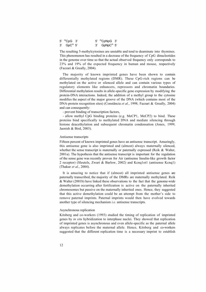
I G F 2 is part of a cluster of imprinted genes located on HSA11p15.5(corresponding to SSC2p1.7). Two ICRs (imprinted control regions) regulate theimprinting of these genes. The first one, Kv ICR, controls the imprinting of thecentromeric subcluster which contains KCNQ1, KCNQ1OT1 and CDKN1C. Thesecond one, H19 ICR, is located 2-4 kb upstream of the H19 promoter andcontrols imprinting at the telomeric subcluster which contains H19 and IGF2 (Duet al., 2003). These two genes are reciprocally imprinted so that in most tissuesH19 is maternally expressed (Bartolomei, Zemel & Tilghman, 1991) and IGF2 ispaternally expressed (DeChiara, Robertson & Efstratiadis, 1991; Nezer et al.,1999). Furthermore, Li et al. (1998) suggested that the human H19 gene is anantagonist of IGF2 expressivity in trans. The mechanisms controlling imprintingat the IGF2-H19 domain are complex and appear to be tissue-specific. In theendoderm, expression of IGF2 and H19 depends on activation of their promotersby a set of shared enhancers located 3’ of H19 (Leighton et al., 1995). On thematernal chromosome, the unmethylated H19 ICR is bound by CCCTC-bindingfactors (CTCF). This creates a chromatin boundary which isolates the IGF2promoters from the endodermal enhancers and results in silencing of IGF2 andexpression of H19. On the paternal chromosome, methylation of the ICR preventsCTCF from binding which results in activation of the IGF2 promoters by theendodermal-specific enhancers and IGF2 transcription (Bell & Felsenfeld, 2000;Hark et al., 2000; Kanduri et al., 2000a, b). In addition, the methylated ICRdirects methylation and subsequent silencing of the H19 promoter (Srivastava etal., 2000) (Figure 1). The situation appears to be more complex in mesodermaltissues. In addition to mesodermal-specific enhancers located 3’ of H19 (Ishihara etal., 2000), a series of other control elements have also been found as e.g. asilencer located in DMR1 (IGF2 intron 3) (Eden et al., 2001) and a muscle-
14
specific silencer situated in the IGF2-H19 intergenic region (Ainscough et al.,2000).
F i g u r e 1Representationof the boundarymodelexplainingimprinting atthe I G F 2 - H 1 9locus. The H19I C R i sunmethylatedon the maternalchromosomea n d i sconsequently
bound by CTCF. This creates a chromatin boundary that prevents the endodermalenhancers (represented as triangles) to activate IGF2 expression. The promoter of H19is unmethylated and its expression can hence be activated by the endodermalenhancers. On the contrary, the H19 ICR is methylated on the paternal chromosome(methyl groups are represented as stars). This methylation spreads to the H19 promoterwhich becomes silenced. In addition CTCF cannot bind to the methylated ICR and theendodermal enhancers can activate transcription of IGF2 (modified from Rand & Cedar,2003).
At imprinted loci, the level of transcription of the transcribed allele is stillcontrolled by transcription factors like at non-imprinted loci. Most informationavailable on the regulation of IGF2’s four promoters (P1-4) (Sussenbach 1989; vanDijk et al., 1991) comes from studies made in different human liver cell lines.Hence, promoter P1 has been shown to be activated by the ubiquitoustranscription factor Sp1 (Rodenburg, Holthuizen & Sussenbach, 1997) and by theCCAAT/enhancer binding protein (C/EBP) (van Dijk. et al., 1992). Promoters P3and P4 have been shown to be regulated by the zinc finger transcription factorsEgr-1 and WT1. These two proteins bind to the same DNA element but binding ofEgr-1 results in transcriptional activation whereas binding of WT1 results intranscriptional repression (Bae et al., 1999; Lee et a l . , 1998). In addition,Rietveld et al. (1999) have demonstrated that transcription from promoter P3responds to AP-2 binding so that overexpression of AP-2 results in activation ofP3 in cells with low endogenous level of AP-2 and repression of P3 in cells withhigh endogenous level of AP-2. P3 has also been shown to be activated by thezinc finger oncogene PLAG1 (Zatkova et al., 2004). Finally, Sp1 was shown tobind to promoter P4 and to cooperate with Egr-1 to mediate maximal activity ofthis promoter (Lee, Park & Lee, 2001). The regulation of IGF2 transcription inskeletal muscle cells has been less studied and is poorly understood. However,Erbay et al. (2003) have demonstrated that the Ser/Thr kinase mTOR initiatesmyoblast differentiation by regulating the expression of IGF2. In addition, Zhanget al. (1998) have suggested that AP-2 may contribute to IGF-II overexpression inan embryonal skeletal muscle tumor (rhabdomyosarcoma).

15
Aims of the thesis
The objectives of this thesis were:- To identify the causative mutation(s) for a major QTL in the pig
influencing muscle growth, fat deposition and heart size located onSSC2p.
- To characterize the molecular mechanism(s) through which the mutationexerts its effects.

16
Methods
I. Transient transfection
This method can be used to determine if a DNA element is involved intranscriptional regulation of gene expression e.g. if it acts as promoter or enhancer.Firstly, the element of interest is cloned in a plasmid containing a reporter genewhich expression can be easily assayed (e.g. luciferase, green fluorescent protein,chloramphenicol acetyl transferase). If the element is a putative promoter, it willbe inserted in a plasmid containing a strong enhancer (e.g. SV40 enhancer). On theother hand, if the experimentator wants to test a supposed enhancer or silencer, itwill be cloned in a plasmid containing a promoter. In this case, it is generallyrecommended to use the homologous promoter. Secondly, the plasmid istransfected into a suitable cell line. This can be done by a biochemical (e.g. cationlipid, calcium phosphate), physical (e.g. electroporation) or virus-mediatedmethod. It is important to simultaneously transform the cells with a control vector(expressing a different reporter) to be able to monitor differential cell growth andtransfection efficiency. Finally, the activity of the reporter is assayed after one tofour days incubation and the observed reporter signal is normalized to the control.
II. Electrophoretic Mobility Shift Assay (EMSA)
EMSA is a standard biochemical in vitro method to detect protein-DNAinteractions (Fried & Crothers, 1981). This assay is based on the fact thatmigration of DNA through a native polyacrylamide gel is retarded upon proteinbinding. First, a short double stranded DNA fragment (called the probe) isradioactively labelled. Second, the probe is incubated with proteins to allow DNA-protein complexes to form. Proteins from diverse origins can be used in EMSAe.g. nuclear or whole-cell extracts from cells or tissues (Dignam, Lebovitz &Roeder, 1983) and purified recombinant proteins. Third, the protein-DNA bindingreactions are electrophoresed on a native polyacrylamide gel to separate free andprotein-bound DNA. After autoradiography, the band corresponding to the DNA-protein complex appears higher on the gel compared to the free probe; the probehas also been “shifted” (Fig. 2). The migration of the DNA-protein complexdepends mainly on the charge, shape and multimeric state of the protein. Thespecificity of the obtained complexes has to be tested by the addition of an excessof cold probe to the binding reaction. If the protein binds specifically it has thesame affinity for the cold and for the radioactively labelled probe. Hence, bothprobes will compete for its binding and as the cold probe is in large molar excessthere will be no protein left to bind to the labelled probe, which results in thedisappearance of the complex.

17
Figure 2 Hypothetical EMSA. The free (unbound) probe as well as two specific and oneunspecific complex are represented (modified from Carey & Smale, 2000)
The major advantages of EMSA are its simplicity and its high sensitivity. Inaddition, it allows the detection of complexes of different composition, eachcomplex appearing as a band with a specific migration. Furthermore, EMSA givesthe possibility to check the identity of proteins included in a complex by usingantibodies. Hence, a specific antibody can be added to the protein-DNA bindingreaction and its binding to the protein will result in an antibody-protein-DNAcomplex which gel migration will be even more retarded (this is called asupershift). Alternatively, the antibody can cover the DNA binding site of theprotein and thereby prevent the formation of the complex, resulting in thedisappearance of the shifted band (Carey & Smale, 2000).
III. DNase I Footprinting
DNase I footprinting allows the detection of protein-DNA interactions in vitro(Galas and Schmitz, 1978). This method is based on the principle that DNAregions bound by proteins are protected from digestion by DNase I. Basically, adouble-stranded DNA probe corresponding to the region of interest is radioactivelylabelled on one end and used to set up two parallel reactions: one with proteins,the other without. After incubation, a specific amount of DNase I is added to bothreactions so that each DNA molecule is cut only once. DNase I cuts randomly, andin the absence of proteins the probe will be digested in a series of labelledfragments ranging from one bp to full length probe. On the other hand, if DNA-protein complexes form, the DNA bound by the proteins will not be accessible to

18
the enzyme and this will result in the absence of DNA fragments of specific sizes.Both reactions are then run on a denaturing polyacrylamide gel to separate theDNA fragments according to their length. After autoradiography of the gel, theprobe incubated without proteins will appear as a continuous series of bands. Theprobe incubated with proteins will also appear as a series of bands, but if protein-DNA complexes have formed the areas corresponding to the complexes will bedevoid of bands. Those regions are called “footprints” (Carey & Smale, 2000).Usually, a Maxam-Gilbert sequencing reaction of the probe is run together withthe DNase I digestions to enable the localization of the footprints (Fig. 3). Themain advantage of this method is that it gives the approximate binding site ofeach protein binding to the probe. Furthermore, it is possible to analyse a quitelong DNA region in a single experiment (the probes are generally at least 300 bplong).
Figure 3 Hypothetical DNase Ifootprinting experiment allowingthe detection of two protectedregions. Footprint 1 appears as aregion devoid of bands whereasin footprint 2 the bands are onlyweakened compared to the onesfrom the probe incubated withoutproteins. A ladder (which i susually a Maxam- Gilbert A+Gs e q u e n c i n g r e a c t i o n ) i selectrophoresed together with thedigested probes to allowlocalization of the footprints.

19
Results and discussion
I. Comparative sequence analysis of the INS-IGF2-H19 genecluster in pigs (Paper 1)
The aim of this work was to further characterize the region containing themutation(s) causing the QTL. Here we report the sequence analysis of two pigcontigs. The first one is 32 kb long and contains the five last exons of TH(Tyrosine hydroxylase) as well as the entire INS (insulin) and IGF2 genes. Thesecond one contains H19 and covers 56 kb.
We started by characterizing the order and the structure of INS, IGF2 and H19 inpig and showed that they were identical to the ones in human. Hence, the geneorder is as follows: TH - 1.9 kb - INS - 0.7 kb - IGF2 - 88.1 kb - H19. IGF2 iscomposed of ten exons (1-9 and 4b) that display high sequence identity betweenhuman and pig (Fig. 4).
Figure 4. Genomic structure of porcine IGF2. The ten exons of IGF2 are represented asboxes, black boxes correspond to the translated exons. The four promoters of IGF2 (P1-4) are represented by arrows.
Nezer et al. (1999) showed that the coding region of IGF2 was identical betweenpigs with different QTL genotypes. We therefore suspected the causativemutation(s) to lie in (a) regulatory element(s). Such elements tend to be wellconserved between species. This results from natural selection as individualscarrying mutations in a regulatory element might display erratic gene expressionand lower fitness. Consequently, we compared our pig sequence with availablehuman and mouse sequences to find these conserved regions. We report 59evolutionary conserved elements (outside exons, promoters and simple repeats) inthe INS-IGF2 region and 38 in the H19 region. Most of them have an unknownfunction but some have been assigned an important role in regulating theexpression of IGF2 in human and mouse e.g. DMR1, CTCF binding sites,endodermal enhancers (see introduction).
As expected from phylogenetic studies, the overall sequence similarity washigher between pig and human than between pig and mouse or human and mouse.The pig sequence displays an amazingly large number of CpG islands: nine in theINS-IGF2 region and sixteen in the H19 region. This can be put into relation withthe imprinting of the region. Indeed, CpG islands are more often found associatedwith imprinted than with non-imprinted genes. The sequence is also characterizedby its low abundance of interspersed repeats. Once again, this could be related to

20
the imprinting of IGF2 and H19 as the introduction of foreign sequence mightperturb the complex regulatory mechanisms controlling their expression.
Another important part of this work was to characterize IGF2 transcripts andpromoter usage in different fetal and adult tissues. We found that IGF2transcription is tissue- and development-specific and that it can be initiated fromfour promoters (P1-4 located upstream of exons 1, 4, 5 and 6, respectively).
II. A regulatory mutation in IGF2 causes a major QTL effect onmuscle growth in the pig (Paper 2)
Nezer and co-workers (2003) refined the position of the QTL to a 250 kb-longinterval between the markers 370SNP6/15 and SWC9 (located in the 3’untranslated region of IGF2). The only known paternally expressed genes mappingto this region were insulin and IGF2. Therefore, we decided to re-sequence 28.6 kbcovering these two genes on 15 chromosomes which QTL status could bedetermined by progeny testing and marker-assisted segregation analysis. One ofthe chromosomes (H254) appeared to be recombinant and allowed us to localizethe QTL downstream of the first exon of IGF2. Among the 258 polymorphismsdifferentiating the 15 chromosomes, we only found one SNP co-segregatingperfectly with the QTL status of the chromosome. Therefore, this SNP, a GAtransition at position IGF2-intron 3-nt 3072, has to be the causative mutation. Thewild type allele (G) is associated with lower muscle mass and was therefore named”q” while the mutant allele (A) causes higher muscle development and was called”Q”. The quantitative trait nucleotide (QTN) is located in an evolutionaryconserved CpG island of unknown function. Consequently, we set up EMSA andtransient transfection experiments to uncover its mechanism of action. In addition,we studied the methylation status of the CpG island by bisulphite sequencing.
We carried out EMSA with nuclear extracts from three different cell types(C2C12 murine myoblasts, HepG2 human hepatocytes and HEK 293 humanembryonic kidney cells) and three different 27 bp-long probes:
- q: wild-type probe- Q: mutant probe- q*: wild-type probe with a methylated CpG at the QTN. As methylation
is important for expression of imprinted genes we designed this probe inorder to test the influence of methylation on in vitro binding to the QTN.
We demonstrated the existence of a specific complex forming only with the wild-type probe but not with the mutant probe nor with the methylated probe.
We transfected C2C12 myoblast cells with reporter plasmids expressing fireflyluciferase under the control of the thymidine kinase minimal promoter (TK) and a578 bp-long fragment corresponding to the q or Q genotype at the QTN. Afternormalization, we found that the q insert doubles the basal TK transcriptionwhereas the Q insert increases it seven times.

21
Figure 5 Results of transientt ransfec t ion exper imentscarried out with reporterplasmids expressing fireflyluciferase under the control ofTK and an insert correspondingto the q (q+TK) or Q (Q+TK)allele at the QTN. Results werenormalised to a Renillaluciferase control plasmid andare expressed as relativeactivit ies to a plasmidexpressing firefly luciferaseunder the sole control of TK.The triple asterisk indicatesthat the differences observedbetween the three plasmids arehighly significant (P<0.01).
These results were quite difficult to conciliate with the results of the EMSA.For this reason, we replaced the TK promoter with IGF2 promoter 3 (P3) andrepeated the experiment. We choose P3 because it is the most actively transcribedpromoter in muscle cells and because it is influenced by the QTN in vivo (seebelow). This time, we found that q reduces the basal P3 transcription with 70%,whereas Q only reduces it with 30%.
Figure 6 Results of transienttransfections carried out likein Fig. 6 but plasmidsexpress luciferase under thecontrol of the P3 instead ofTK.
Taken together with the EMSA these new results suggest that the QTNabrogates the binding of a repressor to a cis-element. In addition, our transienttransfection experiments illustrate how important it is to use a homologous ratherthen a heterologous promoter in this kind of experiments.
We analyzed the methylation of the CpG island containing the QTN and foundthat it is independent from the genotype at the QTN and from the parental originof the allele. However, we found that it is tissue-specific. Hence, on average, 26%of the CpG dinucleotides are methylated in liver but only 3.4% are methylated inskeletal muscle. Interestingly, the effect of the QTN is observed in muscle i.e. in a

22
non-methylated tissue where the putative repressor is able to bind, but no effect isseen in liver which is more methylated and where the putative repressor mightconsequently not be able to bind so efficiently (according to the EMSA results). Itwould be very exciting to analyze the situation in other tissues to check if thisassociation between methylation status and QTN effect holds.
We quantified the expression of IGF2 in vivo and found a significantly higherexpression in postnatal muscle samples from QQ and Qpatqmat animals compared toqpatQmat and qq animals. A weaker (but significant) difference could also beobserved in postnatal heart samples but not in postnatal liver nor in any testedprenatal tissue sample. Furthermore, we showed that IGF2 transcription wasincreased from all three promoters located downstream of the QTN (i.e. P2-4).
Finally, we genotyped the progeny of 13 heterozygous sires (Qq) and of 50homozygous sires (QQ or qq) and used this data in segregation analyses. Wefound evidence for segregation in all heterozygous families but we could not findany indication of segregation among progeny sired by homozygous males.Furthermore, Jungerius et al. (2005) showed that the QTN also controls the QTLfor backfat thickness found in a Meishan x European Whites cross. In conclusion,we demonstrated that the SSC2p QTL is caused by a GA transition at positionIGF2-intron 3-nt 3072 and that this mutation influences IGF2 expression. Hence,we showed that, in addition to its well-known fetal role, IGF2 is involved inpostnatal muscle development.
III. IGF2 antisense transcript expression in porcine postnatalmuscle is affected by a quantitative trait nucleotide in intron 3(Paper 3)
The aim of this study was to search for an IGF2 antisense (IGF2-AS) gene in pig,and upon its existence to:
- Characterize and quantify its transcript(s).- Examine its imprinting status.- Determine whether its expression was influenced by the IGF2 QTN.
We have shown by RT-PCR and RNase protection assay (RPA) that IGF2-ASindeed exists in pig. Furthermore, we have shown that it has two differenttranscription start sites; a major site located around IGF2 intron4-nt70 and a minorsite located approximately at IGF2 intron3-nt2294 (RPA results) or intron3-nt2205 (5’ RACE results). The 3’ end of the transcripts was mapped to positionIGF2 intron2–nt1236 by RACE. We found three different transcripts originating atthe major start site. These transcripts contain from three to five exons and share allthe same first and last exons (Fig. 7).

23
Figure 7. Genomic structure of porcine IGF2 and multiple IGF2-AS transcripts(a-d). Exons are represented as boxes, black boxes correspond to the translatedexons. Promoters are represented by arrows
Hence, we can conclude that the structure of IGF2-AS is not well conservedbetween pig, human and mouse as this gene only has three exons in human andfour in mouse. However, some of the exons are quite well conserved betweenspecies (Table 1).
Table1. Sequence identities between IGF2-AS exons in pig and human or mouse.
Pig exon Human exon Mouse exon % Identity1 1 722 2 605 3 63
Northern blot analyses revealed the existence of three transcripts in fetal muscle(4.7 kb, 3.3 kb and 2.1 kb), two in fetal liver (3.5 kb and 2.1 kb) and one in fetalkidney (3.3 kb). Surprisingly, the shortest band observed on the northern blot islonger than the longest transcript predicted by the RT-PCR and 3’ RACE results.This could result from preferential amplification of short truncated transcripts bythe nested RACE PCR or from a real heterogeneity of the transcripts.
Next, we examined the imprinting status of IGF2-AS in liver and musclesamples from fetal, 3-weeks- and 4-months-old piglets. This was done bysequencing an A to C transversion at IGF2-AS exon2-nt32 which allowed us todiscriminate between Q and q alleles at the QTN. We found that IGF2-AS isimprinted and only expressed from the paternal allele. This reflects the status atother imprinted loci as most antisense transcripts found in imprinted genes arematernally silenced. However, we should note that in muscle of 4-months-old pigs

24
we could detect some transcription from maternal origin which indicates thatimprinting is partially released.
Finally, we used real-time PCR analysis to quantify IGF2-AS transcripts inmuscle and liver from fetal, 3-weeks- and 4-months old pigs carrying the q or Qallele at the QTN. Firstly, we found that the expression of IGF2-AS decreasesnoticeably after birth. Secondly, we found that in 3-weeks- and 4-months-old pigmuscle it depends on the genotype at the QTN. Indeed, at these stages, IGF2-ASexpression was significantly higher in Q than in q muscle samples. Hence, theputative repressor binding at the QTN seems to influence both IGF2 and IGF2-ASexpression.
The function of IGF2-AS is still unknown, but it has been suggested that itcould take part in the regulation of IGF2 expression. Indeed, it is noteworthy thatin pig as well as in human, the first and the last exon of IGF2-AS overlap part ofIGF2 exon 4 and the entire exon 3, respectively. Consequently, IGF2-AS couldinterfere with transcripts originating from P1 and P2.
IV. Molecular Characterization of a Region in IGF2 Intron 3harbouring a Quantitative Trait Nucleotide affecting MuscleGrowth in the Pig (Paper 4)
The aim of this study was to:- Determine the binding site of the transcription factor binding to the QTN.- Characterize the CpG island containing the QTN.- Search for polymorphisms in the CpG island in human.
We used EMSA to determine which nucleotides were important for the bindingof the putative repressor described in paper 2. Firstly, we performed EMSA with aseries of mutated probes to determine if these mutations could abolish formationof the specific complex obtained with the wild-type probe. Secondly, we used themutated oligonucleotides in a competition assay to determine if they were stillable to compete against the wild-type probe. Taking these results together, wefound that the core binding site of the transcription factor is: 5’-GCTCG-3’. Newdatabase searches did not reveal any factor with similar binding capacities.
DNase I footprinting of the 333 bp surrounding the QTN revealed two protectedregions, FP1 and FP2:
- FP1 covers ~50 bp (from nucleotide position ~3027 to ~3076) andincludes the core binding site of the putative repressor and a perfectconsensus AP-2 binding site.
- FP2 is a ~ 30 bp-long footprint located ~20 bp upstream of FP1 (fromnucleotide position ~2981 to ~3008) and covers a putative Sp1 bindingsite.
AP-2 has previously been shown to regulate IGF2 transcription from P3 and itwould be particularly interesting to confirm it’s binding to FP2. Even moreinteresting would be to know if it binds DNA as a heteromer with the putativerepressor. Hence, if they bind together, one could try to purify the repressor by co-immunoprecipitating it with AP-2.
25
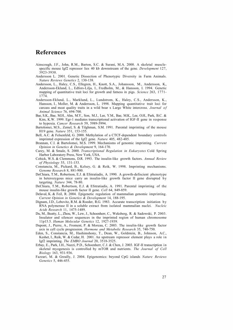
Comparative sequence analyses of the CpG island containing the QTN haverevealed a high sequence conservation between pig and human. This led us to thehypothesis that the human DNA sequence could contain cis-elements involved inmuscle development, as is the case in pig. Therefore, we resequenced this regionin individuals with low versus high muscle mass. We discovered three SNPs andone insertion/deletion. Interestingly, one of the mutations (CT, at positionIGF2-intron 3-3462) lies in the putative AP-2 site included in FP2. Consequently,we analysed the in vitro binding capacity of this site by running EMSA with awild-type and a mutant probe of the region. We were able to detect binding ofspecific complexes with similar mobility using both wild-type and mutantsequences. It would be very interesting to complement this experiment by asupershift assay with AP-2-specific antibodies to confirm the involvement of thistranscription factor.

26
Future prospects
This thesis summarizes the work that led to the identification of the causativemutation for a QTL influencing muscle development, fat deposition and heart sizein pig. In addition, it presents how we have started to uncover the molecularmechanisms by which this QTN mediates its effects. We have shown that themutation abrogates the binding of a putative repressor element, which results inincreased expression of IGF2 and IGF2-AS. We have also demonstrated that othertranscription factors bind DNA in the immediate vicinity of this repressor. Theobvious next step in this project will be to clone and characterize this repressor.Different methods could be considered to achieve this goal e.g. one-hybrid screen,in vitro expression library screening, biochemical purification. In order to choosethe most appropriate approach, it is important to collect as much information aspossible on this transcription factor and on its possible interactions with theneighbouring DNA-binding factors. It is for example essential to know if it bindsDNA by itself or as a heteromer (as could be suspected from the DNase Ifootprinting results). Indeed, some of the methods cited above are only capable todeal with proteins binding as monomer, homodimer or homopolymer. In addition,identifying a known factor binding DNA together with the repressor could beextremely useful; antibodies against this factor could be used to co-immunoprecipitate both factors as a first step towards purification of the repressor.
The identification of the gene coding for a transcription factor might be adifficult task, but it would be worth the effort as this repressor could havefascinating therapeutic uses in the future. Indeed, it could be inactivated to increasethe expression of IGF2 in specific tissues. Our study has demonstrated that it isactive in skeletal muscle and heart but not in liver. In addition, it may also beactive in adipocytes as the IGF2-QTL also influences backfat thickness. Hence,one could imagine to inactivate it in order to increase the muscle mass of patientssuffering from muscular degenerative diseases. Furthermore, it could be transientlyinactivated in patients confined in bed to avoid the muscle loss accompanyinglong periods of inactivity. In addition, if its activity is confirmed in adipocytes, itcould even be used to treat obese patients.
Finally, the identification of the causative mutation for the IGF2-QTL makes itvery easy to select pigs with the favourable Q allele to breed for the nextgenerations and hence to fix the mutation in populations were it is present. Inaddition, it will also facilitate the introgression of the mutant allele in pig breedswhere it is absent. Indeed, when the introgression of a favourable gene variant isbased on linked markers there is always a risk to loose it because of arecombination between the markers and the actual causative mutation. Thisproblem has now been eliminated and makes the IGF2-QTL a very attractivecandidate for introgression into certain commercial populations.
27
References
Ainscough, J.F., John, R.M., Barton, S.C. & Surani, M.A. 2000. A skeletal muscle-specific mouse Igf2 repressor lies 40 kb downstream of the gene. Development 127,3923-3930.
Andersson L. 2001. Genetic Dissection of Phenotypic Diversity in Farm Animals.Nature Reviews Genetics 2, 130-138.
Andersson, L., Haley, C.S., Ellegren, H., Knott, S.A., Johansson, M., Andersson, K.,Andersson-Eklund, L., Edfors-Lilja, I., Fredholm, M., & Hansson, I. 1994. Geneticmapping of quantitative trait loci for growth and fatness in pigs. Science 263, 1771-1774.
Andersson-Eklund, L., Marklund, L., Lundstrom, K., Haley, C.S., Andersson, K.,Hansson, I., Moller, M. & Andersson, L. 1998. Mapping quantitative trait loci forcarcass and meat quality traits in a wild boar x Large White intercross. Journal o fAnimal Science 76, 694-700.
Bae, S.K., Bae, M.H., Ahn, M.Y., Son, M.J., Lee, Y.M., Bae, M.K., Lee, O.H., Park, B.C. &Kim, K.W. 1999. Egr-1 mediates transcriptional activation of IGF-II gene in responseto hypoxia. Cancer Research 59, 5989-5994.
Bartolomei, M.S., Zemel, S. & Tilghman, S.M. 1991. Parental imprinting of the mouseH19 gene. Nature 351, 153-155.
Bell, A.C. & Felsenfeld, G. 2000. Methylation of a CTCF-dependent boundary controlsimprinted expression of the Igf2 gene. Nature 405, 482-485.
Brannan, C.I. & Bartolomei, M.S. 1999. Mechanisms of genomic imprinting. CurrentOpinion in Genetics & Development 9, 164-170.
Carey, M. & Smale, S. 2000. Transcriptional Regulation in Eukaryotes Cold SpringHarbor Laboratory Press, New York, USA.
Cohick, W.S. & Clemmons, D.R. 1993. The insulin-like growth factors. Annual Reviewof Physiology 55, 131-153.
Constancia, M., Pickard, B., Kelsey, G. & Reik, W. 1998. Imprinting mechanisms.Genome Research 8, 881-900.
DeChiara, T.M., Robertson, E.J. & Efstratiadis, A. 1990. A growth-defiscient phenotypein heterozygous mice carry an insulin-like growth factor II gene disrupted bytargeting. Nature 344, 78-80.
DeChiara, T.M., Robertson, E.J. & Efstratiadis, A. 1991. Parental imprinting of themouse insulin-like growth factor II gene. Cell 64, 849-859.
Delaval, K. & Feil, R. 2004. Epigenetic regulation of mammalian genomic imprinting.Current Opinion in Genetics & Development 14, 188-195.
Dignam, J.D., Lebovitz, R.M. & Roeder, R.G. 1983. Accurate transcription initiation byRNA polymerase II in a soluble extract from isolated mammalian nuclei. NucleicAcids Research 11, 1475-1489.
Du, M., Beatty, L., Zhou, W., Lew, J., Schoenherr, C., Weksberg, R. & Sadowski, P. 2003.Insulator and silencer sequences in the imprinted region of human chromosome11p15.5. Human Molecular Genetics 12, 1927-1939.
Dupont, J., Pierre, A., Froment, P. & Moreau, C. 2003. The insulin-like growth factoraxis in cell cycle progression. Hormone and Metabolic Research 35, 740-750.
Eden, S., Constancia, M., Hashimshony, T., Dean, W., Goldstein, B., Johnson, A.C.,Keshet, I., Reik, W. & Cedar, H. 2001. An upstream repressor element plays a role inIgf2 imprinting. The EMBO Journal 20, 3518-3525.
Erbay, E., Park, I.H., Nuzzi, P.D., Schoenherr, C.J. & Chen, J. 2003. IGF-II transcription inskeletal myogenesis is controlled by mTOR and nutrients. The Journal of CellBiology 163, 931-936.
Fazzari, M. & Greally, J. 2004. Epigenomics: beyond CpG islands Nature ReviewsGenetics 5, 446-455.

28
Feinberg, A.P., Cui, H. & Ohlsson, R. 2002 DNA methylation and genomic imprinting:insights from cancer into epigenetic mechanisms. Seminars in Cancer Biology 12,389-398.
Ferguson-Smith, A.C. & Surani, M.A. 2001. Imprinting and the epigenetic asymmetrybetween parental genomes. Science 293, 1086-1089.
Florini, J.R., Magri, K.A., Ewton, D.Z., James, P.L., Grindstaff, K. & Rotwein, P.S. 1991."Spontaneous" differentiation of skeletal myoblasts is dependent upon autocrinesecretion of insulin-like growth factor-II. The Journal of Biological Chemistry 266,15917-1523.
Fowden, A.L. 2003. The insulin-like growth factors and feto-placental growth.Placenta. 24, 803-812.
Fried, M. & Crothers, D.M. 1981. Equilibria and kinetics of lac repressor-operatorinteractions by polyacrylamide gel electrophoresis. Nucleic Acids Research, 9, 6505-6525.
Froesch, E.R., Schmid, C., Schwander, J. & Zapf, J. 1985. Actions of insulin-like growthfactors. Annual Review of Physiology 47, 443-467.
Galas, D.J. & Schmitz, A. 1978. DNAse footprinting: a simple method for the detectionof protein-DNA binding specificity. Nucleic Acids Research 5, 3157-3170.
Gardiner-Garden, M. & Frommer, M. 1987. CpG islands in vertebrate genomes. Journalof Molecular Biology 196, 261-282.
Georges, M. & Andersson, L. 1996. Livestock Genomics Comes of Age. GenomeResearch 6, 907-921.
Goureau, A., Yerle, M., Schmitz, A., Riquet, J., Milan, D., Pinton, P., Frelat, G. & Gellin, J.1996. Human and porcine correspondence of chromosome segments usingbidirectional chromosome painting. Genomics 36, 252-262.
Hark, A.T., Schoenherr, C.J., Katz, D.J., Ingram, R.S., Levorse, J.M. & Tilghman, S.M.2000. CTCF mediates methylation-sensitive enhancer-blocking activity at theH19/Igf2 locus. Nature 405, 486-489.
Ishihara, K., Hatano, N., Furuumi, H., Kato, R., Iwaki, T., Miura, K., Jinno, Y. & Sasaki, H.2000. Comparative genomic sequencing identifies novel tissue-specific enhancersand sequence elements for methylation-sensitive factors implicated in Igf2/H19imprinting. Genome Research 10, 664-671.
Jaenish, R. & Bird, A. 2003. Epigenetic regulation of gene expression: how the genomeintegrates intrinsic and environmental signals. Nature Genetics Supplement 33, 245-154.
Jeon, J.T., Carlborg, O., Tornsten, A., Giuffra, E., Amarger, V., Chardon, P., Andersson-Eklund, L., Andersson, K., Hansson, I., Lundstrom, K. & Andersson, L. 1999. Apaternally expressed QTL affecting skeletal and cardiac muscle mass in pigs maps tothe IGF2 locus. Nature Genetics 21, 157-158.
Jones, J.I. & Clemmons, D.R. 1995. Insulin-like growth factors and their bindingproteins: biological actions. Endocrine Reviews 16, 3-34.
Jones, P. 1999. The DNA methylation paradox. Trends in Genetics 15, 34-37.Jungerius, B., Van Laere, A.S., te Pas, M., van Oost, B., Andersson, L. & Groenen, M.
2005. The IGF2-intron 3-G3072A substitution explains a major imprinted QTL effecton backfat thickness in a Meishan X European white pig intercross. GeneticalResearch in press.
Kanduri, C., Holmgren, C., Pilartz, M., Franklin, G., Kanduri, M., Liu, L., Ginjala, V.,Ulleras, E., Mattsson, R. & Ohlsson, R. 2000a. The 5' flank of mouse H19 in anunusual chromatin conformation unidirectionally blocks enhancer-promotercommunication. Current Biology 10, 449-457.
Kanduri, C., Pant, V., Loukinov, D., Pugacheva, E., Qi, C.F., Wolffe, A., Ohlsson, R. &Lobanenkov, V.V. 2000b. Functional association of CTCF with the insulatorupstream of the H19 gene is parent of origin-specific and methylation-sensitive.Current Biology 10, 853-856.
Kitsberg, D., Selig, S., Keshet, I. & Cedar, H. 1993. Replication structure of the humanbeta-globin gene domain. Nature 366, 588-590.

29
Labosky, P.A., Barlow, D.P. & Hogan, B.L. 1994. Mouse embryonic germ (EG) cell lines:transmission through the germline and differences in the methylation imprint ofinsulin-like growth factor 2 receptor (Igf2r) gene compared with embryonic stem(ES) cell lines. Development 120, 3197-3204.
Lee, S., Park, U. & Lee, Y.I. 2001. Hepatitis C virus core protein transactivates insulin-like growth factor II gene transcription through acting concurrently on Egr1 and Sp1sites. Virology 283,167-177.
Lee, Y.I., Lee, S., Lee, Y., Bong, Y.S., Hyun, S.W., Yoo, Y.D., Kim, S.J., Kim, Y.W. & Poo,H.R. 1998. The human hepatitis B virus transactivator X gene product regulates Sp1mediated transcription of an insulin-like growth factor II promoter 4. Oncogene 16,2367-2380.
Leighton PA, Saam JR, Ingram RS, Stewart CL, Tilghman SM. 1995. An enhancerdeletion affects both H19 and Igf2 expression. Genes & Development 9, 2079-2089.
Li, E., Beard, C. & Jaenisch, R. 1993. Role for DNA methylation in genomic imprintingNature 366, 362-365.
Louvi, A., Accili, D. & Efstratiadis, A. 1997. Growth-promoting interaction of IGF-IIwith the insulin receptor during mouse embryonic development. DevelopmentalBiology 189, 33-48.
McKinnon, T., Chakraborty, C., Gleeson, L.M., Chidiac, P. & Lala, P.K. 2001.Stimulation of human extravillous trophoblast migration by IGF-II is mediated byIGF type 2 receptor involving inhibitory G protein(s) and phosphorylation ofMAPK. The Journal of Clinical Endocrinology and Metabolism 86, 3665-3674.
Minniti, C.P., Kohn, E.C., Grubb, J.H., Sly, W.S., Oh, Y., Muller, H.L., Rosenfeld, R.G. &Helman, L.J. 1992. The insulin-like growth factor II (IGF-II)/mannose 6-phosphatereceptor mediates IGF-II-induced motility in human rhabdomyosarcoma cells. TheJournal of Biological Chemistry 267, 9000-9004.
Mohan, S. & Baylink, D.J. 2002. IGF-binding proteins are multifunctional and act viaIGF-dependent and -independent mechanisms. Journal of Endocrinology 175, 19-31.
Nezer, C., Collette, C., Moreau, L., Brouwers, B., Kim, J.J., Giuffra, E., Buys, N.,Andersson, L. & Georges, M. 2003. Haplotype sharing refines the location of animprinted quantitative trait locus with major effect on muscle mass to a 250-kbchromosome segment containing the porcine IGF2 gene. Genetics 165, 277-285.
Nezer, C., Moreau, L., Brouwers, B., Coppieters, W., Detilleux, J., Hanset, R., Karim, L.,Kvasz, A., Leroy, P. & Georges, M. 1999. An imprinted QTL with major effect onmuscle mass and fat deposition maps to the IGF2 locus in pigs. Nature Genetics 21,155-156.
O'Dell, S.D. & Day, I.N. 1998. Insulin-like growth factor II (IGF-II). The InternationalJournal of Biochemistry & Cell Biology 30, 767-771.
Oksbjerg, N., Gondret, F. & Vestergaard, M. 2004. Basic principles of muscledevelopment and growth in meat-producing mammals as affected by the insulin-likegrowth factor (IGF) system. Domestic Animal Endocrinology 27, 219-240.
Paldi, A., Gyapay, G. & Jami, J. 1995. Imprinted chromosomal regions of the humangenome display sex-specific meiotic recombination frequencies. Current Biology 5,1030-1035.
Rand, E. & Cedar, H. 2003. Regulation of Imprinting: A Multi-Tiered Process. Journalof Cellular Biochemestry 88, 400-407.
Reik, W. & Walter, J. 2001a. Evolution of imprinting mechanisms: the battle of thesexes begins in the zygote. Nature Genetics 27, 255-256.
Reik, W. & Walter. J. 2001b. Genomic imprinting: parental influence on the genome.Nature Reviews Genetics 2, 21-32.
Rietveld, L.E., Koonen-Reemst, A.M., Sussenbach, J.S. & Holthuizen, P.E. 1999. Dualrole for transcription factor AP-2 in the regulation of the major fetal promoter P3 ofthe gene for human insulin-like growth factor II. The Biochemical Journal 338, 799-806.

30
Rodenburg, R.J., Holthuizen, P.E. & Sussenbach, J.S. 1997. A functional Sp1 bindingsite is essential for the activity of the adult liver-specific human insulin-like growthfactor II promoter. Molecular Endocrinology 11, 237-250.
Sleutels, F., Zwart, R. & Barlow, D.P. 2002. The non-coding Air RNA is required forsilencing autosomal imprinted genes. Nature 415, 810-813.
Srivastava, M., Hsieh, S., Grinberg, A., Williams-Simons, L., Huang, S.P. & Pfeifer, K.2000. H19 and Igf2 monoallelic expression is regulated in two distinct ways by a
shared cis acting regulatory region upstream of H19. Genes & Development 14, 1186-1195.
Strachan, T. & Read, A. 1999. Human molecular genetics 2, BIOS Scientific PublishersLtd, Oxford, UK.
Sussenbach, J.S. 1989. The gene structure of the insulin-like growth factor family.Progress in Growth Factor Research 1, 33-48.
Thakur, N., Tiwari, V.K., Thomassin, H., Pandey, R.R., Kanduri, M., Gondor, A., Grange,T., Ohlsson, R. & Kanduri, C. 2004. An antisense RNA regulates the bidirectionalsilencing property of the Kcnq1 imprinting control region. Molecular and CellularBiology 24, 7855-7862.
Thomas, B.J. & Rothstein, R.. 1991. Sex, maps, and imprinting. Cell 64, 1-3.Tilghman, S.M. 1999. The sins of the fathers and mothers: genomic imprinting in
mammalian development. Cell 96, 185-193.van Dijk, M.A., Rodenburg, R.J., Holthuizen, P. & Sussenbach, J.S. 1992. The liver-
specific promoter of the human insulin-like growth factor II gene is activated byCCAAT/enhancer binding protein (C/EBP). Nucleic Acids Research 20, 3099-3104.
van Dijk, M.A., van Schaik, F.M., Bootsma, H.J., Holthuizen, P. & Sussenbach, J.S. 1991.Initial characterization of the four promoters of the human insulin-like growth factorII gene. Molecular and Cellular Endocrinology 81, 81-94.
Wilkins, J.F. & Haig, D. 2003. What good is genomic imprinting: the function ofparent-specific gene expression. Nature Reviews Genetics 4, 359-368.
Wood, T.L. 1995. Gene-targeting and transgenic approaches to IGF and IGF bindingprotein function. The American Journal of Physiology 269, E613-622.
Zatkova, A., Rouillard, J.M., Hartmann, W., Lamb, B.J., Kuick, R., Eckart, M., vonSchweinitz, D., Koch, A., Fonatsch, C., Pietsch, T., Hanash, S.M. & Wimmer, K. 2004.Amplification and overexpression of the IGF2 regulator PLAG1 in hepatoblastoma.Genes, Chromosomes & Cancer 39, 126-137.
Zhang, L., Zhan, S., Navid, F., Li, Q., Choi, Y.H., Kim, M., Seth, P. & Helman, L.J. 1998.AP-2 may contribute to IGF-II overexpression in rhabdomyosarcoma. Oncogene 17,1261-1270.

31
ACKNOWLEDGEMENTS
I would like to take thank the following persons:
My supervisors:Leif Andersson. Thank you for giving me the chance to work in the fascinatingfield of molecular genetics; but most of all, thank your enthusiasms, youroptimism and for the great independence you gave me.Göran Andersson. Thank you for always taking the time to answer my questionsand for making me see the world from your transcriptional point of view.Valérie Amarger. C’est toi qui m’as mis le pied à l’étrier, merci!
The past and present members of the lab:Gabriella Lindgren, Carl-Gustaf Thulin, Stefan Marklund, Siw Johansson,Elisabetta Giuffra, Jin-Tae Jeon, Anna Törnsten, Göran Hjälm, Anna-KarinFridolfsson, Maria Moller, Örjan Carlborg, Gabriella Nyman, Teresa Casals,Jessica PeterssonAmélie Johansson. I am looking forward to the next ISAG conference.Ana Fenandez and Meiying Fang. You saved me each time I was looked out ofmy office.Carolyn Fitzsimmons. I will miss these Ph.D. student parties.Emmanuelle Bourneuf. You have a great sense of humor (et merci d’avoir sauvémon gel!)Erik Bongcam-Rudloff. You saved my computer from falling out through thewindow.Frida Berg. Where are you taking this amazing energy from?Lina Jacobsson and Susanne Kerje. I would never have made it to the Big Applewithout you.Martin Braunschweig. The IGF2 group was not the same anymore after you leftMy brother, Ben. It was great having you here for four month (même si Voldos’est pris quelque raclées).My office-mate, Hee-Bok Park. I enjoyed sharing the office with you and the“little guy”, good luck in Malmö.Nicolette Sammon Hillbertz. I will miss the hugs, the week-ends at thecountryside, the dogs and all the rest.Sonchita Bagchi. You are a real sunshine.The first person I met when I arrived, Gerli Pielberg. You have been a real friendduring these five years. Thank you for defending your thesis two month before mein order to figure out all the administrative mess!The new generation of Ph.D. students: Ullrika Gunnarsson, Per Wahlberg andEllen Markljung. Time is flying so fast that you will soon be writing your ownlittle book; enjoy pipetting as long as it lasts!Ulla Gustafson and Gudrun Wieselander. The lab would not survive without you.All the Swedes. Tack för att ni hjälpte mig att lära mig språket!

32
All the people in my “adoptive” labs:Cihan Cetinkaya, Natalie von der Lehr and Fuad Bahram. I will never forget thatthe cells are “my children”.Susanna Tronnersjö. You made me discover the fascinating world of the buddingyeasts.Beat Bornhauser. Thank you for always having the time to show me how to usethe nice machines you have down there.
The AgriFunGen program at SLU.I am grateful for my founding, but also for all the interesting courses and for thegreat meetings!
Ma famille et mes amis.Merci pour tous ces bons moments passés ensemble pendant mes vacances et pourvos encouragements pendant le restant de l’année.