from 1987 to 1999 by - core.ac.uk · latar belakang : kancer kolon dan rektum adalah salah satu...
TRANSCRIPT

Ul'41 Vt:H;:)I II ~A l i\J~ IVIALA Y::iiA
D!TERI:vlA ln-.·-· .. ) t) r~ ~ c J J I
L...----· -
COLORECTAL CARCINOMA IN HOSPITAL KUALA TERENGGANU
FROM 1987 TO 1999
BY
DR UMASANGAR AIL RAMASAMY M.D(USM)
Dissertation Submitted In Partial Fulfillment Of The Requirement For
The Degree of Master of Medicine ( General Surgery )
UNIVERSITI SAINS MALAYSIA
YEAR2002
Background : Colorectal carcinoma is one of the most common malignant
neoplasm in this country . Its incidence is rising . New development in
genetic studies has brought better understanding ofcolorectal carcinoma.
·Newer treatment option has increased survival and quality of life .
However the impact of this development has not been fully
utilized especially in the rural areas in this country.
Objective :To study the epidemiology of colorectal carcinoma in the
Kuala Terengganu Hospital and the state ofTerengganu. To determine the
clinical presentation of colorectal carcinoma in this hospital and to
evaluate the mode of investigation and treatment received by the patients
with colorectal carcinoma . The primary objective is to assess the
treatment outcome in tenns of recurrence and survival .
Methods : Between 19 87 to 1999 , 90 patients were treated in Hospital
Kuala Terengganu for colorectal carcinoma. The sex, age , duration of
symptom , the main and subsequent presenting problems , the type of
special investigation and the location of the tumour were determined from
patients record . Type of surgery , method of resection and presence of
metastases were noted at the time of surgery . Specimens were sent to the
pathologist for assessment of the tumour grade .
1J
Survival duration were obtained from the patients record . 2 year survival
rate was assessed and factors affecting survival were determined using
nonparametric statistical analysis .
Result: There was almost equal sex distribution among the patients. 53%
of the patients were more then 60 years and young colorectal carcinoma
consist of6%. Median duration of symptom was 4.3 months. 43.3% of
the patients presented with intestinal obstruction and a almost equal
number of them underwent emergency surgery. 68.7% of
the patients were in Duke's C and D at presentation . At the time of
presentation 24.4 % already had liver metastases . 30 day mortality and
morbidity was 2.2 % and 11.1 % respectively . Overall swvival was 3 7.8
%. Statistically Duke's staging, stage of metastases, initial clinical
presentation , type of operation and use of adjuvant therapy
significantly influences( p <0.05) the 2 year survival in this study.
Conclusion : In this study , late presentation of colorectal carcinoma was
common . Duke' s staging , initial clinical presentation , type of opemtion
and the use of chemotherapy influences survival . A better follow up of
colorectal carcinoma patient is needed.
Ill
III ABSTRAK
Latar Belakang : Kancer kolon dan rektum adalah salah satu ketumbuhan
malignan yang utama di negara ini . Kini insiden kanser in sedang
meningkat . Kefahaman kita berkenaan penyakit kanser kolon dan rectum
bertambah dengan perkembangan terbaru cialam bidang genetic. Kaedah
perubatan yang terbaru membolehkan survival dan kualiti hidup
ditingkatkan . Walaubagaimanapun perkembangan ini tidak dipergunakan
sepenuhnya terutama di kawasan pendalaman di negara ini .
Objektif: Kajian in bertujuan untuk mengetahui epidemiologi penyakit
kanser kolon dan rectum di Hospital Kuala Terengganu dan di negeri
Terengganu. Kajian inijuga melihat pada riwayat clinical penyakit ini dan
menilai cara penyiasatan dan rawatan yang diterima oleh pesakit penyakit
kanser kolon dan rectum . Objektif utama ialah untuk mengetahui kadar
survival dan rekuren .
Tatacara : Di antara tahun 1987 dan 1990 , seramai 90 pesakit kanser
kolon dan rectum telah dirawat di Hospital Kuala Terengganu. Jantina, umur
, jangkamasa gejala , riwayat penyakit yang utama , jenis penyiasatan yang
digunakan dan lokasi kanser dipastikan melalui rekod pesakit. Jenis
pembedahan , cara pembedahan dan metastasis tumor pada
masa pembedahan dicatitkan .
IV

Pengradan tumor oleh pakar patolo!:,ti juga dicatitkan . Jangkamasa pesakit
hidup selepas pembedahan didapatkan melalui rekod pesakit. Kadar
sutvival 2 tahun dikira dan faktor- faktor yang mempengaruhinya
dipastikan melalui pengiraan statistik .
Keputusan : Distribusi jan tina dikalangan penyakit adalah hampir sama rata .
53 % pesakit berumur 53 tahun keatas dan 6 % adalah pesakit di bawah 40
tahun. Kadar medianjangkamasa gejala adalah 4.3 bulan. 43.3 o/o pesakit
datang dengan keadaan usus tersumbat dan jumlah yang sama menjalani
pembedahan kecemasan. Semasa kali pertama pesakit dinilai, 68.7 o/o dari
mereka berada dalam peringkat Dukes' C dan D . Pada masa yang sama
24.4 % dari mereka sudah mempunyai metastasis di hepar . Kadar
mortaliti dalam 30 hari dan mobiliti adalah 2.2 %dan 11/1 % masing
masing . Kadar survival keseluruhan adalah 3 7.8 % . Klasifikasi Dukes' ,
peringkat metastasis ,gejala utama , jenis pembedahan dan pemberian
terapi adjuvan mempengaruhi kadar survival dalam masa 2 tahun di
dalam kajian ini .
Kesimpulan: Dalam kajian ini pesakit kanserkolon dan rektum hanya
datang untuk rawatan apabila penyakit mereka sudah merebak .
Klasifikasi Dukes' , peringkat metastasis ,gejala utama , jenis pembedahan
dan pemberian terapi adjuvant mempengaruhi kadar survival.Cara yang
lebih baik diperlukan untuk mengikuti pesakit yang sudah menjalani
rawatan.
v
VI
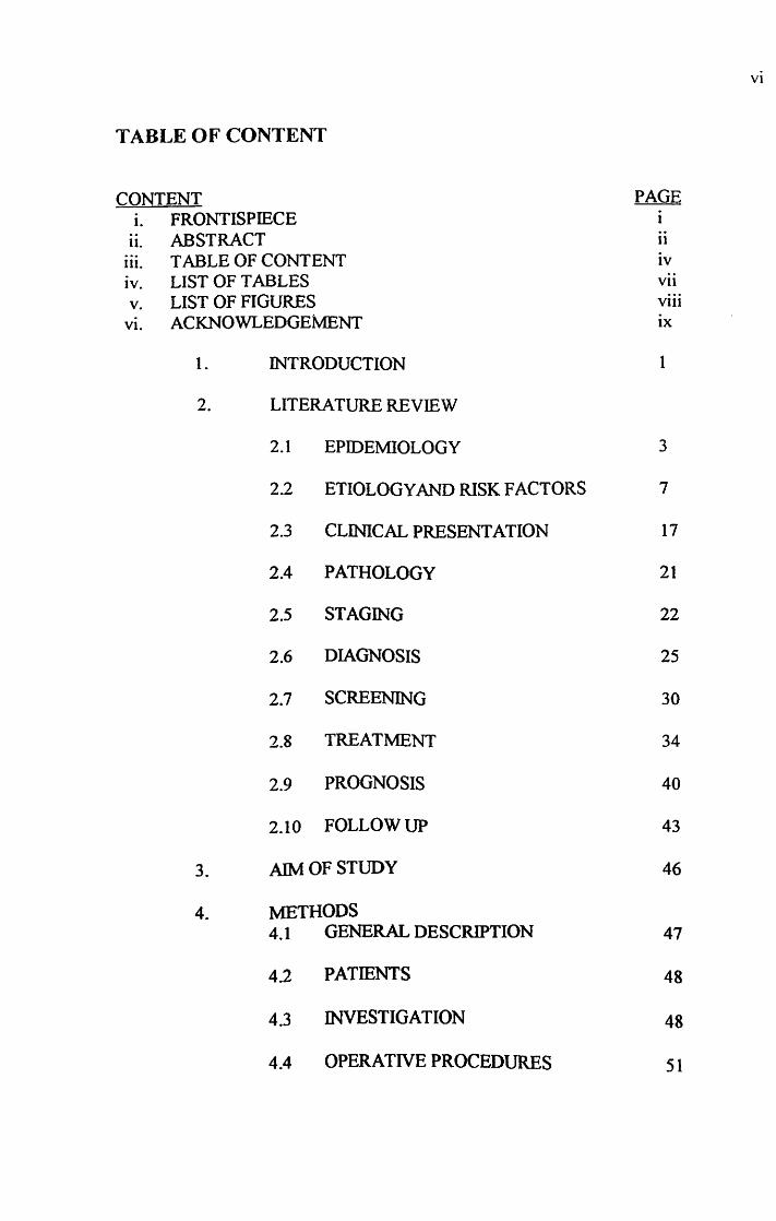
TABLE OF CONTENT
CONTENT PAGE 1. FRONTISPIECE
11. ABSTRACT 11
111. TABLE OF CONTENT IV
IV. LIST OF TABLES Vll
v. LIST OF FIGURES Vlll
Vt. ACKNOWLEDGEMENT IX
1. INTRODUCTION 1
2. LITERATURE REVIEW
2.1 EPIDEMIOLOGY 3
2.2 ETIOLOGY AND RISK FACTORS 7
2.3 CLINICAL PRESENTATION 17
2.4 PATHOLOGY 21
2.5 STAGING 22
2.6 DIAGNOSIS 25
2.7 SCREENING 30
2.8 TREATMENT 34
2.9 PROGNOSIS 40
2.10 FOLLOW UP 43
3. AIM OF STUDY 46
4. METHODS 4.1 GENERAL DESCRIPTION 47
4.2 PATIENTS 48
4.3 INVESTIGATION 48
4.4 OPERATIVE PROCEDURES 51
VII
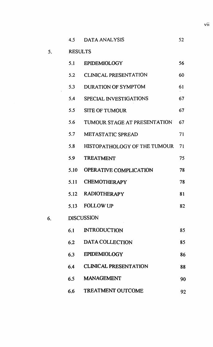
4.5 DATA ANALYSIS 52
5. RESULTS
5.1 EPIDEMIOLOGY 56
5.2 CLINICAL PRESENT AT ION 60
5.3 DURATION OF SYMPTOM 61
5.4 SPECIAL INVESTIGATIONS 67
5.5 SITE OF TUMOUR 67
5.6 TUMOUR STAGE AT PRESENT AT ION 67
5.7 METASTATIC SPREAD 71
5.8 HISTOPATHOLOGY OF THE TUMOUR 71
5.9 TREATMENT 75
5.10 OPERATIVE COMPLICATION 78
5.11 CHEMOTHERAPY 78
5.12 RADIOTHERAPY 81
5.13 FOLLOW UP 82
6. DISCUSSION
6.1 INTRODUCTION 85
6.2 DATA COLLECTION 85
6.3 EPIDEMIOLOGY 86
6.4 CLINICAL PRESENTATION 88
6.5 MANAGEMENT 90
6.6 TREATMENT OUTCOME 92

7. CONCLUSIONS
7.1 CONCLUSION FROM THIS STUDY
7.2 LIMITATIONS
7.3 RECOMMENDATIONS
8. REFERENCES
94
96
97
99
Vlll

IX
LIST OF TABLES
TABLE TITLE PAGE
1 Dukes' Classification 27
2 AJCCIUICC TNM pathological staging, 1997 version 28
3 Stage Grouping 28
4 Common ' T stage ' penetration 29
5 Survival at 2 years comparing with the main clinical presentation 61
6 Survival at 2 years based on the initial stage of the disease at the time of presentation 71
7 Survival at 2 years comparing with histological grade of the tumour 72
8 Dukes' classification and the number of patients receiving adjuvant treatment 81
9 Survival at 2 years based on Dukes' staging in colorectal carcinoma 82
10 Survival at 2 years for patients after adjuvant therapy 83
11 Follow up investigations for surviving patients 84
12 Survival at 2 years based on type of surgery 84
X
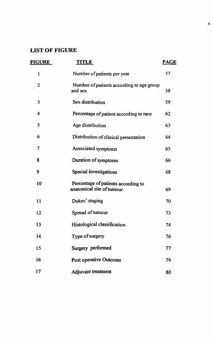
LIST OF FIGURE
FIGURE TITLE PAGE
Number of patients per year 57
2 Number of patients according to age ~oup and sex 58
3 Sex distribution 59
4 Percentage of patient according to race 62
5 Age distribution 63
6 Distribution of clinical presentation 64
7 Associated symptoms 65
8 Duration of symptoms 66
9 Special investigations 68
10 Percentage of patients according to anatomical site of tumour 69
11 Dukes' staging 70
12 Spread of tumour 73
13 Histological classification 74
14 Type of surgery 76
15 Surgery performed 77
16 Post operative Outcome 79
17 Aldjuvanttteattnent 80
VII ACKNOWLEDGEMENT
I wish to express my gratitude and appreaciation to my supervisors Datuk Jamil
Abdullah from the Department of Surgery Hospital Kuala Terengganu and
Dr Myint Tun from the Department of Surgery Hospital Universiti Sains
Malaysia , for their guidance and supervision in the preparation of this
dissertation . I thank them for the valuable time they spent with me to put
through this study .
I also wish to thank my Head of Department , Associate Professor Dr. Abdul
Hamid , for his suggestions and support .
I wish to thank all the lecturers in Hospital Universiti Sains Malaysia , clinical
specialists from Hospital Kuala Terengganu and my colleagues for their
encouragement and support
XI

In Malaysia more and more cases of colorectal carcinoma are being identified
and treated. With establishment of a colorectal unit in Hospital Selayang and
Hospital Universiti Kebangsaan Malaysia , proper research can be done
on the Malaysian population in relation to colorectal carcinoma. As this
country is a multiracial society , the differences in colorectal carcinoma
within races may exist . This may be compounded with different levels of
development in various parts of the country .
This dissertation is a retrospective study of patients with colo rectal
carcinomas seen in Hospital Kuala Terengganu, Kuala Terengganu from
1987 to 1999. This hospital is a tertiacy referral center serving the rural
population ofTerengganu state in the east coast of peninsular Malaysia. This
study will assess the presentation and outcome of treatment of
colorectal carcinoma in a general hospital where there is no specialized
colorectal unit . From here we will know how to improve the service for
the benefit of patients in the treatment of colorectal carcinoma .
2

2.0 LITERATURE REVIEW
2.1 EPIDEMIOWGY
Colorectal carcinoma is the fourth commonest form of cancer occurring
world-wide , with an estimated 783,000 new cases diagnosed in 1990 . It affects
men and women equally. World-wide, colorectal carcinoma represents
9.4 % of all incident cancer in men and 10.1 % in women ( 3 ) .
However it is not equally common throughout the world .If the
western countries (North America ; Northern , southern and western
Europe ; Austmlia ; New Zealand ) are combined , colorectal cancer
represents 12.6% of man and 14.1 %of women of all incident cancers.
Elsewhere colo rectal cancer represents 7. 7 % and 7.9 %
of all incident cases in men and women respectively ( 3 ).
Different populations worldwide experience different levels of colorectal
cancer , and these levels change with time . Population living in one
community also experience difrerent levels of colorectal cancers ( 4 ).
Groups of migrants quickly lose the risk associated with their original home
community and acquire the patterns of the new community , often starting
within one generation of arrival( 4 ).
3

Ethnic and racial differences in colorectal cancer suggest that environmental
factors play a major part in the etiology of the disease. In Israel male Jews
born in Europe or the United States are at a higher risk of colon cancer
compared to those born in Asia or Africa . Incidence in the offsprings
now approaches or surpasses that in white people in the same
population and is three or four times higher than Japanese in Japan . For
these reason colorectal cancer is widely believed to be an" environmental"
disease ( 4 ) .
The incidence of colorectal carcinoma is a rising trend(5) . The incidence of
colorectal cancer in eastern part of the Netherlands (700,000 inhabitants)
was determined for two years, 1981 and 1996. In 1981 the diagnosis of
colorectal cancer was seen in 232 patients in this region and in 1996, it was
seen in 410 patients. The population remained almost stable during this
time. Therefore, the incidence rose from 33 to 55 per 100,000 inhabitants
from 1981 to 1996, respectively.ln 1981, 25 percent of the carcinomas were
proximal (to the sigmoid colon); this increased to 37 percent in 1996.
The location of the tumour is also more proximal now ( 6 ) .
A similar trend is also observed in Bulgaria . It showed the
incidence rate of colorectal carcinoma from 1985 to 1998 increased steadily
from 22.14/100,000 to 37.18/100,000 (an increment ofl5.04/100,000).
Most of the patients in this study ( 60 % ) were diagnosed at a later stage of
the disease and this trend continues to 1998 ( 7 ) .
4
In Malaysia a similar trend is observed .Data from the ministry of health shows an
increase of colorectal carcinoma rate from 8.1 % ( 1973 cases ) in 1987 to
11.9 % {4215 cases in 1995 . Colorectal cancer was the third conunonest
cancer death in Malaysia from 1987 to 1995 ( 8 ) .
Rectal cancer is slightly more common in men, whereas there is a slight
predominance of colon cancer in women( I). An American has
approximately a 5 percent probability of developing colorectal cancer during
a 70-year life span. Most cases of colorectal cancer are diagnosed in patients
over the age of 50, and the incidence of the disease rises steadily after that
age. Despite clear relationship with aging, colorectal cancer is not stricdy
a disease of the elderly and between 6 to 8 percent of cases occur in
individuals under the age of 40(1 ). In the Far East ,the incidence of colo rectal
carcinoma is more in younger population compared to Western
countries ( 9).
In a retrospective study in Egypt, 38% of the colorectal cases were below the
age of40 years old. The onset offiunilial and hereditary forms of the
disease occurs at a much earlier age, typically around the third decade {10).
5

There are few studies in assessing the awareness of colo rectal carcinoma in
an adult population. On assessment of public awareness ofcolorectal
carcinoma , it was found that even in countries with high literacy rate only a
minority ( I 1 % ) were aware of colorectal screening ( 1 1 ) .
Similarly Nadel et ai noted only 22.9 %of Americans aged more than
50 years old had gone for screening for colorectal carcinoma (12).
6
2.2 : ETIOLOGY AND RISK FACfORS
2.2.1: Diet
Evidence from epidemiological studies seems to show consistendy that
intake of dietary fut and meat is positively related to the
risk of colo rectal cancer . This evidence was obtained from ecological studies ,
animal experiments , and case control and cohort studies ( 13 ).
In 1990 Willett et al published the results from the United States nurse
health study involving a fullow-up of888,751 women aged 34-59 years who
were without cancer or inflammatory bowel disease at recruitment . After
adjustment for total energy intake , consumption of animal filt was found to
be associated with increased risk of colon cancer. No association was found
with vegetable mt . The relative risk in women who ate beef, pork , or lamb
as a main dish every day was 2.49 compared with women reporting
consumption less than once a month . The authors suggested that intake of
animal fat increases the risk of colon cancer ( 13) . In the Asian population a
similar trend could be found . Zhang et al (2002) in a study involving a rural
region in China found that meat intake and saturated fat were prominent
risk filctors for colorectal carcinoma (14).
7
Dietary fiber has been proposed as accounting for differences in the rates of
colorectal cancer between Africa and westernized countries- on the basis
that increased intake of dietary fiber may increase faecal bulk and reduce
transit time . Many studies found no protective effect of fiber in cereals but
have consistently found a protective effect of fiber in vegetables
and fruits ( 13 , 14 ).
Folate deficiency enhances intestinal carcinogenesis in several animal
models. An increasing number of epidemiologic studies indicate that higher
intakes of folate either from dietary sources or from supplements may lower
the risk of colorectal adenoma and cancer( IS). More limited data also suggest
that dietary methionine, which might also influence methylation, may have a
similar protective role. High alcohol consumption, which has a strong
antifolate effect, also has been related to higher risk of colorectal neoplasia
The deleterious effects of alcohol are accentuated when folate or methionine
intake is low (15).
2.2.2 : Physical activity , body mass index , and energy intake
Evidence from epidemiological studies show that men with high
occupational or recreational physical activity seem to have a decreased risk
of colon cancer. Giovannucci et al reported that activities from moderate
intensity such as brisk walking is associated inversely with the risk of large
adenoma (16).However there is no evidence to show a consistent association
8

between obesity and the risk of colo rectal cancer.
2.2.3 : Hormone replacement therapy
Increasing evidence supports an association between hormone replacement
therapy and a reduced risk of colorectal cancer . The risk is halved with
5 - 10 years of use . Whether this association is casual or is associated
with some other factor is not known ( 17 ).
2.2.4 : Familial risk factors
A :family histocy of colorectal polyps is associated with increased risk of
colo rectal carcinoma in other fiunily members . However 80 % of patients
have " sporadic " colorectal carcinoma, with no familial risk factor identified.
First degree relatives of patients with colorectal carcinoma are at a
high risk to develop colorectal neoplasia. Orrom WJ et al(l990), studied
colonoscopic finding in patients with colorectal carcinoma and found
9
that 21 % of them had neoplastic disease and another 28 % had adenoma beyond
the splenic flexure(l9) . 25 % of these patients who were positive for adenoma are
below 40 years old(l9) . Kesani et al (2002) reported 44 %patients under 40
years old in their study had finnily history of colorectal carcinoma and these
patients usually presents with more advanced stage of disease ( 18) .

• Polyposis syndrome
Familial syndrome takes the form of familial adenomatous
polyposis syndromes (FAP). F AP accounts about 0.5% of all
colonic carcinoma. It is autosomal dominant inherited propensity
to develop numerous adenomas throughout the colon ' some of
which in time will become malignant (2). The polyp appears at
puberty and nearly all patients will have polyp by the early thirties.
• Hereditaty non polyposis colonic carcinoma ( HNPCC )
This is more common but less obvious than F AP and is autosomal
dominant inheritance . It is clinically similar to that of sporadic
cases but more common in younger age groups. Lynch (1996)
classified HNPCC into 2 subtypes ; site specific colon cancer
where individuals of a family are susceptible to colonic cancer but
not cancers of other organs (Lynch type I) ; cancer family
syndrome where female members of the family are prone to breast
and uterine cancer as well as colonic cancer (Lynch type 2) . The
cancers occur predominantly on the right side of the colon and are
of low malignancy in contrast to sporadic cases (20).
10
2.2.5 : Molecular Genetics
Colorectal adenoma carcinoma sequence
One of the most important concepts in colorectal carcinoma to emerge in
recent years has been the adenoma- carcinoma sequence , a term that
describes the stepwise progression from normal to dysplastic epithelium to
carcinoma associated with the accumulation of multiple clonally selected
genetic alterations.
Although the adenoma- carcinoma sequence has not been proven directly ,
there is considerable indirect evidence to support it from a range of
epidemiological, clinical, histopathological and genetic studies.
1. Epidemiological and clinico-pathological evidence - age distribution
cwves for adenoma and carcinoma show that the prevalence of both
increases with increasing age , but adenomas are recognized and their
prevalence peaks at least 5 years earlier than that of colorectal
carcinomas ( 21 ). In surgical resection specimens and during
endoscopic examination adenomas are found to coexist with carcinoma
in about 30 % of cases and patients who have colo rectal carcinoma and
simultaneous adenomas have been shown to have increased risk of
synchronous and metachronous carcinoma ( 22 ).
I I

12
The anatomical distribution of adenomas and carcinoma is similar , occurring
more frequendy distal to splenic flexure; adenoma ofleft colon often
contain more severe dysplasia or invasive adenocarcinoma ( 23 ).
Finally endoscopic removal of adenomatous polyps appear to reduce the
long term risk of colorectal carcinoma ( 24 ).
2. Genetic evidence - the concept of carcinoma arising from genetic
abnormality has existed fur many years. In colo rectal carcinoma, the
genes of interest that are involved in genetic alterations may be classified
into three types : oncogenes , tumour suppressor genes and DNA repair
genes ( 25 ). In 1990 Fearon and Vogelstein proposed a genetic model for
colorectal tumorigenesis ( 26). This model postulated that mutational
activation of oncogenes , coupled with mutational inactivation of tumour
suppressor genes , leads to the development of colorectal tumours . Although
these alterations occur in sequence , it is the total accumulation of changes
which is important. The key oncogene in this model was ras and the key
tumour suppressor genes were proposed residing on chromosome 5q , l7p , and
18q . However , now there is an alternative pathway in a subset of colorectal
tumours that less frequently involves the above mentioned genes and often
involves mismatched repair genes(26,27)

• Adenomatous Polyposis coli ( APC ) -is one mutation known to
occur early in the adenoma- carcinoma sequence affects the
adenomatous polyposis coli ( APC) tumour suppressor gene
located on chromosome 5q21 .
Mutation of this gene is responsible for familial adenomatous
polyposis (F AP) , an autosomal dominant disorder characterized
by development of thousands ofcolorectal adenomas appearing in
adolescence or early adulthood ( 28 ). If untreated it
leads to colorectal carcinoma at the third or fourth decade of life.
APC mutation or allelic losses of Sq are observed in 40 - 80 % of
colorectal carcinomas and are fomtd at a similar frequency in
adenomas ( 29 ) .
• K-ras- Activating mutation ofthe oncogene K-ras also occurs
early in the adenoma- carcinoma sequence . This gene is involved
in signal transduction of regulatory pathways critical for normal
proliferation and differentiation ( 30 ) .
Activating K-ras mutation occurs in 35 - 42 % of colorectal
careinoma and were also observed at similar frequency in large
adenomas . It is also being investigated as a potential predictors of
metachronous adenomas ( 29 , 32 ).
13
• P53 -located on the short ann of chromosome 17 , p53 was
initially implicated in colorectal carcinoma as a result of the
frequent loss of 17 p in allelic loss and cytogenic studies . It
functions as a sequence specific DNA binding protein and
transcription factor controlling the expression of a large number of
genes (29 , 33 ). It has been labeled as the
guardian of the genome because of its ability to block cell
proliferation in the presence of DNA damage , to stimulate DNA
repair and to promote apoptotic cell death if repair is insufficient .
The alteration in p53 or 17p allelic loss has been reported to occur
in 50- 75 % of adenocarcinoma and in 4 - 26 % of adenoma
(29 ,31).
• 18q loss - this is the second most common region of allelic loss
in colorectal carcinoma occuning in about 70 % of cases . 18q
loss is also observed in 10-30% of early adenomas and it is
raised to 60 % in late adenomas . The original tumour suppressor
gene in this region was thought to be the " deleted in colorectal
cancer" (29, 31 , 34).
14

Microsatellite instability
A recently proposed alternative pathway for a subset of colorectal tumours is
characterized by the presence ofmicrosatellite instability (MSI).
Microsatellite are a type ofDNA that consists of tandem repeats, usually
between one and five base pairs , repeated many times (3 5 ).
Hundreds ofthousands ofmicrosatellites are found interspersed throughout
the human genome and are particularly prone to errors during DNA
replication .
Such errors are usually repaired by mismatch repair (MMR)
proteins but , in the absence of competent MMR function , microsatellite
eiTOr accumulates . When these errors are sufficiently frequent , the term
MSI or replication error positive is applied .
Thus MSI can be interpreted as a marker for a state ofhypermutability (36,37).
MSI is obsetVed in almost all adenocarcinomas from patients with hereditary
non -polyposis colorectal cancer (HNPCC) that occurs in 1 0 - 15 % of
sporadic colorectal carcinoma (38). The presence ofMSI in
sporadic colorectal carcinoma and HNPCC correlates significantly with a
number of clinical and pathological features including proximal location ,
diploid DNA content , a favourable Dukes' staging , improved survival
and the presence of a Crohn~s like inflammatory infiltrate (38, 39,42).
15

The presence of MSI has a favorable genetic prognostic markers for
colo rectal carcinoma and may imply an increased risk of a second primary
tumour in the colon or an endometrial tumour in the patient or family
members.
In the future this genetic testing will come into common use for
the prognosis of colorectal carcinoma and will affect their management
( 40 '41 ).
16

2J:CL~CALPRESENTATION
Patients with colonic and rectal carcinoma have a broad range of clinical
presentation that can be sub-classified according to the anatomic site of the
primary . In the earliest stage they may be asymptomatic . Caecal and right
sided tumours accounts for 20 % of large bowel carcinoma , 70 % occur
distal to the splenic flexure , and about 45 % are at or below the recto-
17
sigmoid junction ( 1,2 ). Kullavanijaya (2002) in a retrospective analysis reported
distal colorectal carcinoma was about 72 % ( 45) . However there is a shift from
left sided toward right sided carcinoma confirmed by epidemiological study
by Cucino et al ( 44 ). Bowel habit change , weight loss and mucous
bloody dianhea are the most common presentations( 45).
Caecal and right sided carcinoma
Right sided tumours are often remarkably silent and many patients present
with only the symptom and signs of iron deficiency from protracted occult
blood loss .Preoperative anaemia was the most common finding with right sided
tumour present in 58% of patients ( 46). As the lumen becomes
narrowed the patient complains of intermittent colic , centrally or in the right
iliac fossa, which is often post-prandial, stimulated by gastro-colic reflex.

Typical distal ileal obstruction occurs if the tumour blocks the ileo-caecal
valve causing incompetence of the valve characterized by progressive
central abdominal colicky pain , faeculent vomiting and abdominal
distension . Not infrequently a palpable mass may be the presenting
symptom.
Patients can occasionally present with acute appendicitis when the carcinoma
occludes the appendicular orifice and produce acute inflammation or from a
perforated carcinoma .
The tumour may penetiate the bowel wall , producing a sealed perforation
and fonnation of abscess on the psoas muscle which may manifest as painful
right iliac fossa mass ( 2 ).
Left sided and sigmoid colon
Patients commonly present with change of bowel habit, often constipation
alternating with diarrhea , usually accompanied by lower abdominal colicky
pain . The symptoms progressively become worse and later patients may
develop obstruction.
Change in bowel habit is often accompanied by passage of altered blood ,
sometimes mucus , in the stool or on its surfilce .
Occasionally they may present as frank per-rectal bleeding which is usually
intermittent and brisk .
18

A few patients may have localized left iliac fossa pain associated with a
palpable mass . At the splenic flexure presence of tumour must be
distinguished from a palpable left kidney .
Some patients have few symptoms until they present with intestinal
obstruction . If the ileo-caecal valve is incompetent the obstructed large
bowel decompresses into the small bowel and produces a mixed picture of
small and large bowel obstruction .
If the ileo-caecal valve is competent , the caecum will be distended and this
will lead to perforation . Occasionally the tumour itself can perforate and
present as diffuse peritonitis . Perforation of colon associated with
malignancy is reported to be between 3 - 8 % ( 43 ) .
At times the tumour may become attached to the adjacent organ such as
lateral abdominal wall , uterus , bladder and vagina . The tumour may
perforate the organs and present as a fistula . Colonic carcinoma is the
19
second most common cause of colovesical fistula after diverticular disease ( 1 , 2 ).

Rectal carcinoma
Most patients with rectal carcinoma present with per-rectal bleeding and
change ofbowel habits . The blood may be dark in color, mixed with stool
or bright red separate fiom the stool. For this reason patients attribute it to
hemonhoid, a symptom produced by advanced rectal tumour (1 ,2).
The patients also may have tenesmus and spurious diarrhea .
Penetration of tumour to the sacrum will produce perineal pain
shooting down the thigh . Intestinal obstruction is a late sign , indicating
advanced disease {1).
Metastatic symptoms from colorectal carcinoma are rare presentations.
20
21
2.4 PATHOLOGY
The vast majority ofcolorectal carcinomas are adenocarcinoma. They may be well
differentiated ( 20 % ) , moderately differentiated ( 60 % ) or poorly
differentiated ( 20 % ) . The epithelial cells lining the glands show nuclear
stratification , pleomorphism , hyperchromasia and high mitotic rate ( 4 7 ). If large
amount of mucin is produced by the tumour and more then 70 % of the tumour is
occupied by mucin lakes , it is classified as a mucinous carcinoma ( 4 7 ).
Lee YS( 1988 ) in a study of large bowel carcinoma in Singapore noted that non
mucinous carcinoma is by fur the commonest histological type ( 74.7%) followed
by mucinous carcinoma ( 20.7 % ) . Other histological type were relatively
uncommon ( 48 ). They include carcinoid tumours (1.8 % ) , signet ring cell
carcinoma ( 1.5 % ) , squamous cell carcinoma ( 0. 7 % ) , undifferentiated
carcinoma ( 0.4%) and adenosquamous carcinoma ( 0.2%). The proportion of
mucinous carcinoma was greatest among the Malay and Indian ( 48 ). The right
colon had a greater proportions of poorly differentiated carcinoma than the left
colon ( 48 , 49 , 50). This tendency is more evident in the female . Mucinous
carcinoma occurs more frequent in younger age group and in populations with with
low risk for colorectal carcinoma ( 48 , 49 , 50 ). A greater proportion of mucinous
caroinoma located in the rectum and right colon ( 48 , 49 , 50 ). Mucinous
carcinoma usually present in more advanced stage and have a low curative resection
rate ( 49 ). The overall survival rate of patients with mucinous carcinoma is worse
than that of non-mucinous carcinoma ( 48 , 49 , 50 ) .

2.5: STAGING
Several staging methods are in use throughout the world, and each has its own
strengths and weaknesses. The most commonly used are Duke's
classification, American Joint Committee on Cancer (AJCC) and the Union
Intemationale Contre Cancer (UICC) TNM classification. Based on
AJCC/UICC , several variations developed but the most commonly used is
pathological (pTNM) staging (51 , 52).
Dukes' classification introduced in 1929, has the advantage of simplicity but
it lacks precision. It does not reflect accurately the
depth of tumour penetration, the extent of spread outside the bowel wall, the
number oflymph nodes involved and the presence or absence of metastases.
Staging gives information about prognosis in general, but particularly
indicates the probability of occult hepatic metastases, which is the major
fitctor affecting swvival.
22
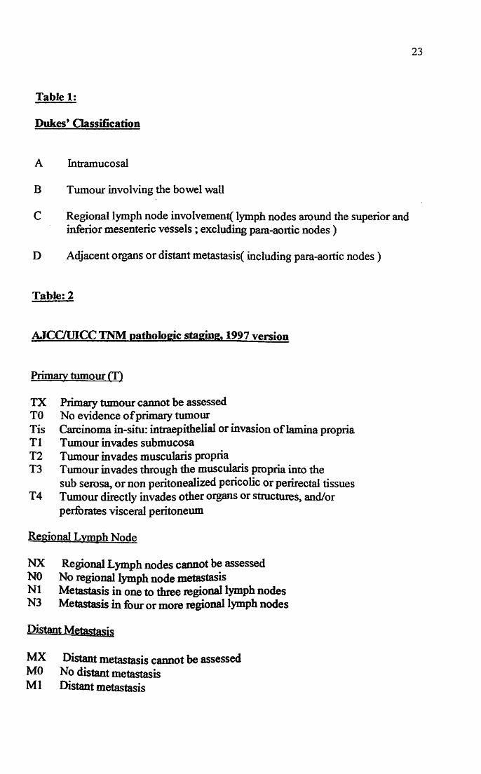
Table 1:
Dukes' Classification
A Intramucosal
B Tumour involving the bowel wall
C Regional lymph node involvement( lymph nodes around the superior and inferior mesenteric vessels ; excluding para-aortic nodes )
D Adjacent organs or distant metastasis( including para-aortic nodes )
Table: 2
AJCauiCC TNM pathologic staging, 1997 version
PrimaJy tumour (T)
TX Primary twnour cannot be assessed TO No evidence of primary tumour Tis Carcinoma in-situ: intraepithelial or invasion of lamina propria Tl Tumour invades submucosa T2 Tumour invades muscularis propria T3 Tumour invades through the muscularis propria into the
sub serosa, or non peritonealized pericolic or perirectal tissues T 4 Tumour directly invades other organs or structures, and/or
perforates visceral peritoneum
Regional Lvmph Node
NX Regional Lymph nodes cannot be assessed NO No regional lymph node metastasis Nl Metastasis in one to three regional lymph nodes N3 Metastasis in four or more regional lymph nodes
Distant Metastasis
MX Distant metastasis cannot be assessed MO No distant metastasis M 1 Distant metastasis
23

Table: 3
Stage Groupine
AJCC/lHCC
Stage 0 Tis NO MO
Stage 1 Tl NO MO T2 NO MO
Stage2 T3 NO MO T4 NO MO
Stage 3 AnyT Nl MO AnyT N2 MO
Stage4 AnyT AnyT Ml
Table: 4
Common ' T stage ' penetration
Colon Transverse , Sigmoid . Recto-sigmoid
pTl invasive into submucosa p T2 muscularis propria pT3 through the muscle , not through serosa pT4 through the mucosa pT4 into contiguous organ
Extra-peritoneum rectum
pTl Invasive into the submucosa pT2 Muscularis propria pT3 Transmural into peri-rectal :fat pT4 lntocontigous organ
Dukes'
A
B
c
D
For this dissertation Duke's Classification was used as the staging system .
24