freshwater rhabdocoela (platyhelminthes) from ephemeral ... · zoological science 2004 zoological...
TRANSCRIPT
BioOne Complete (complete.BioOne.org) is a full-text database of 200 subscribed and open-access titles in the biological, ecological, and environmental sciences published by nonprofit societies, associations, museums, institutions, and presses.
Your use of this PDF, the BioOne Complete website, and all posted and associated content indicates your acceptance of BioOne’s Terms of Use, available at www.bioone.org/terms-of-use.
Usage of BioOne Complete content is strictly limited to personal, educational, and non-commercial use. Commercial inquiries or rights and permissions requests should be directed to the individual publisher as copyright holder.
BioOne sees sustainable scholarly publishing as an inherently collaborative enterprise connecting authors, nonprofit publishers, academic institutions, research libraries, and research funders in the common goal of maximizing access to critical research.
Freshwater Rhabdocoela (Platyhelminthes) from Ephemeral Rock Poolsfrom Botswana, with the Description of Four New Species and One NewGenusAuthors: Tom Artois, Wim Willems, Els De Roeck, Merlijn Jocqué, and Luc BrendonckSource: Zoological Science, 21(10) : 1063-1072Published By: Zoological Society of JapanURL: https://doi.org/10.2108/zsj.21.1063
Downloaded From: https://bioone.org/journals/Zoological-Science on 25 Apr 2019Terms of Use: https://bioone.org/terms-of-use
2004 Zoological Society of JapanZOOLOGICAL SCIENCE
21
: 1063–1072 (2004)
Freshwater Rhabdocoela (Platyhelminthes) from Ephemeral RockPools from Botswana, with the Description of
Four New Species and One New Genus
Tom Artois
1
*, Wim Willems
1
, Els De Roeck
2
, Merlijn Jocqué
2
and Luc Brendonck
2
1
Limburgs Universitair Centrum, Centre for Environmental Sciences, Research Group Biodiversity,Phylogeny and Population Studies, Dept. SBG, Universitaire Campus Gebouw D,
B-3590 Diepenbeek, Belgium.
2
Catholic University of Leuven, Laboratory of Aquatic Ecology, Charles deBériotstraat 32, B-3000 Leuven, Belgium
ABSTRACT
—Four new species of freshwater rhabdocoel flatworms from ephemeral rock pools in south-eastern Botswana are described and discussed. Two of them,
Syringoplana
kolasai
n. gen. n. sp. and
Mesostoma
thamagai
n. sp. belong to the Typhloplanidae Graff, 1905. The unique construction of theexcretory system is the main characteristic of
S.
kolasai
.
M.
thamagai
can be separated from other
Mesos-toma
Ehrenberg, 1837 species by the presence of a bundle of eosinophilic glands at the transition fromoviduct to seminal receptacle. The other two taxa,
Gieysztoria
isoldeae
n. sp. and
G. faubeli
n. sp. belongto the Dalyelliidae Graff, 1905.
G.
isoldeae
is characterised by the presence of four separate hollow spinesin the male atrium, which are connected to two accessory glandular organs.
G.
faubeli
can be separatedfrom other
Gieysztoria
Ruebush and Hayes, 1939 species by the detailed construction of the stylet. Apartfrom these two species the occurrence of an unidentified
Microdalyellia
Gieysztor, 1938 species is men-tioned.
Key words:
Rhabdocoela,
Syringoplana
,
Mesostoma
,
Gieysztoria
, new taxa
INTRODUCTION
Although free-living flatworms (“Turbellaria”) are ofmajor importance in freshwater and marine ecosystems, ourknowledge of this group is very scant. This is especially truefor tropical and subtropical areas, such as South America,Australia and Africa. Knowledge of the African freshwaterTurbellaria mostly comes from older literature. A compre-hensive list of the African species of freshwater Turbellariaand of the literature concerning them is given by Young(1976). This author recognises 83 valid species. Since thennot much work on African freshwater Turbellaria has beendone (De Vries, 1988; Kolasa, 1976; Kolasa and Mead,1981; Young, 1977).
In this contribution we describe four new rhabdocoelspecies from ephemeral rock pools in southeasternBotswana. These pools were sampled within the frameworkof an intercontinental comparative study on the faunal com-munity structures of ephemeral rock pools in subtropical
areas. Two of the species described here,
Mesostomathamagai
n. sp. and
Syringoplana
kolasai
n. gen. n. sp.,belong to the large and widespread rhabdocoel taxonTyphloplanidae Graff, 1905. The other two,
Gieysztoria
isol-deae
n. sp. and
G.
faubeli
n. sp. fit into the DalyelliidaeGraff, 1905, another species-rich and widespread taxon ofrhabdocoel flatworms. A third representative of the Dalyelli-idae, a species of
Microdalyellia
Gieysztor, 1938, was alsocollected. However, the only whole mounted specimen ofthis species is in such bad condition that it does not allowfurther identification.
MATERIAL AND METHODS
All turbellarians were hatched from dry sediment samples con-taining the resting propagules collected from ephemeral rock poolson a granite escarpment in southeastern Botswana. The sampleswere taken with a spoon and a little brush during the dry phase ofthe pools. Sediment was transported to Belgium in plastic bags. Toobtain hatchlings, about 200 g of sediment was incubated in dis-tilled water, under 24-hour light conditions and at a temperature of25
°
C. Usually the first turbellarians were visible after one or twodays. They were fed with living
Daphnia
and fairy shrimps (fresh-water Anostraca).
* Corresponding author: Tel. +32-11-268383;FAX. +32-11-268301.E-mail: [email protected]
Downloaded From: https://bioone.org/journals/Zoological-Science on 25 Apr 2019Terms of Use: https://bioone.org/terms-of-use
T. Artois
et al
.1064
Specimens of most species could be studied alive. If hard partswere present, whole mounts were prepared using lactophenol.Specimens intended for sectioning were fixed using hot (50
°
C)Bouin’s fixative. They were embedded in paraffin and serially sec-tioned (5
µ
m), then stained with Heidenhain’s haematoxylin, usingerythrosine as counterstain.
Hard parts were measured axially. Drawings without a scaleare freehand.
Type material will be deposited in the collections of theresearch group Biodiversity, Phylogeny and Population Studies ofthe Limburgs Universitair Centrum (LUC), Diepenbeek, Belgium.
TAXONOMIC ACCOUNT
TYPHLOPLANIDAE GRAFF, 1905
Syringoplana
kolasai
n. gen. n. sp.
(Fig. 1)
Locality
. Botswana, Thamaga (24
°
41’50’’S, 25
°
31’00’’E),ephemeral rock pools on isolated granite domes (type local-ity). Sediment collected 22/12/1998, inundated in the labo-ratory 4/10/2002.
Material
. Observations on a live animal. Several sectionedspecimens, a sagitally-sectioned one designated holotype(LUC nr. 232), the others paratypes (LUC nr. 233–238).
Etymology
. The genus is named after Syrinx, one of theNaiads (water nymphs) of Greek mythology. Species epithetin honour of Dr. Jurek Kolasa (Hamilton, Canada).
Description
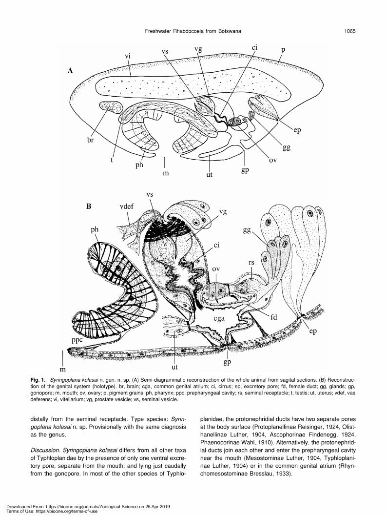
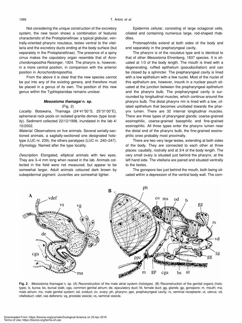
. The animals are relatively small, oval-shaped,with a blunt rostral and a pointy caudal end (“tail”). Animalscoloured dark brownish green, owing to the presence of asubepidermal pigment, which is less dense at the ventralside. Eyes absent.
Epidermis cellular, ciliated all over the body surface.Dermal rhabdites absent. Anteriorly there are some largerhabdite glands that produce large, adenal rhabdites.
The excretory system consists of two protonephridialducts, situated at both sides of the body. They end in a com-mon excretory pore, which is just caudal from the gonoporeand lies in a ventro-caudal pit. Several large, coarse-grainedbasophilic glands surround this pit. The excretory pore canbe closed by a sphincter.
The mouth is at 1/3 of the body length and is sur-rounded by a weak sphincter. The prepharyngeal cavity isrelatively small and lined with a very low epithelium withoutnuclei. It is surrounded by a weak internal circular and astrong external longitudinal muscle layer. The globular phar-ynx is very large and oriented vertically. Its distal border islined with a very low, membranous, anucleated epithelium,with very short cilia. The pharynx lumen is also lined with avery low, anucleated epithelium that has no cilia. The phar-ynx musculature consists of a weak internal circular musclelayer (around the pharynx lumen), a thick internal longitudi-nal muscle layer consisting of 24–26 muscles, an externalcircular and an external longitudinal muscle layer under-neath the septum of the bulb, and a longitudinal musclelayer just outside the septum, continuous with the longitudi-
nal muscles of the prepharyngeal cavity. Strong radial mus-cles connect the wall of the pharynx lumen with the septumof the bulb. There are two types of pharyngeal glands, withthe eosinophilic fine-grained ones entering the pharynxlumen proximally from the basophilic coarse-grained ones.
The genital system lies just caudally from the pharynx,the gonopore situated ventrally at about 2/3 of the bodylength, just in between the excretory pore and the mouth.The two large testes are situated at both sides of the body.The single ovary is situated at the level of the gonopore. Thevitellaria are long and narrow, extending dorsally from thetestes at both sides of the body. The common genital atriumis surrounded by an inner circular and an outer longitudinalmuscle layer, and lined with a high, nucleated, lightly scle-rotised epithelium.
The copulatory organ enters the genital atrium dorsally.It is a large, ovoid bulb, with a septum enclosing the seminalvesicle, the prostate glands and the cirrus (conjuncta-duplexcopulatory organ; terminology of Karling, 1956). Two spi-rally-running muscle layers surround the bulb. The vasa def-erentia join each other just before entering the copulatorybulb at its proximal end. Within the bulb the seminal ductenlarges to form a seminal vesicle, which is surrounded bya weak circular muscle layer. The seminal duct enters thecirrus about midway along the bulb. The cirrus consists of arather broad tube armed with small, sharp spines and is sur-rounded by an inner circular and an outer longitudinal mus-cle layer. The coarse-grained basophilic prostate glandsenter the copulatory bulb at the same place as do the vasadeferentia, feeding into the prostate vesicle, which in turnenters the cirrus at the same place as does the seminal ves-icle. The internal prostate vesicle is surrounded by a weakcircular muscle layer.
The female duct enters the genital atrium caudally,while proximally it ends in the small ovary. Somewhat half-way, it makes a 180
°
turn and starts running anteriorly. Atthe turn, a large bundle of fine-grained eosinophilic glandsenters the duct. Just before it reaches the ovary it widens toa seminal receptacle, containing eosinophilic glandularsecretion and many sperm. The duct is lined with a high,nucleated epithelium and surrounded by an inner circularand an outer longitudinal muscle layer.
The vitelloduct could not be seen. The single uterusleaves the common genital atrium anteriorly.
Diagnosis
.
Syringoplana
n. gen. Typhloplanidae with phar-ynx rosulatus in the first 1/3 of body. Dermal rhabdites lack-ing. Excretory system with two lateral nephridial ducts end-ing in a single ventral excretory pore, caudal from thegonopore. Excretory pore surrounded by large basophilicglands. Testes paired, lying laterally from the pharynx, ven-trally from the vitellaria. Copulatory organ of the duplex-type,caudal to the pharynx. Seminal vesicle single, intracapsular.Prostate vesicle intracapsular. With a cirrus with fine spines.Ovary single. Oviduct enlarged to a seminal receptacle.Large bundle of eosinophilic glands entering the female duct
Downloaded From: https://bioone.org/journals/Zoological-Science on 25 Apr 2019Terms of Use: https://bioone.org/terms-of-use
Freshwater Rhabdocoela from Botswana 1065
distally from the seminal receptacle. Type species:
Syrin-goplana kolasai
n. sp. Provisionally with the same diagnosisas the genus.
Discussion
.
Syringoplana kolasai
differs from all other taxaof Typhloplanidae by the presence of only one ventral excre-tory pore, separate from the mouth, and lying just caudallyfrom the gonopore. In most of the other species of Typhlo-
planidae, the protonephridial ducts have two separate poresat the body surface (Protoplanellinae Reisinger, 1924, Olist-hanellinae Luther, 1904, Ascophorinae Findenegg, 1924,Phaenocorinae Wahl, 1910). Alternatively, the protonephrid-ial ducts join each other and enter the prepharyngeal cavitynear the mouth (Mesostominae Luther, 1904, Typhloplani-nae Luther, 1904) or in the common genital atrium (Rhyn-chomesostominae Bresslau, 1933).
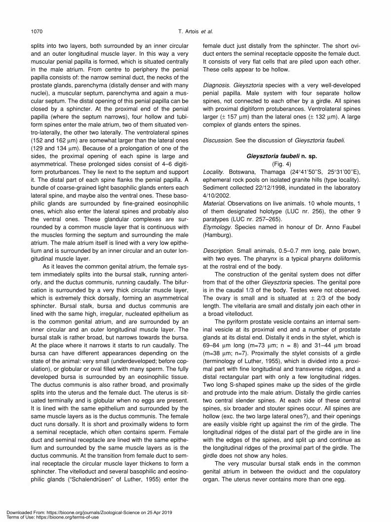
Fig. 1.
Syringoplana kolasai
n. gen. n. sp. (A) Semi-diagrammatic reconstruction of the whole animal from sagital sections. (B) Reconstruc-tion of the genital system (holotype). br, brain; cga, common genital atrium; ci, cirrus; ep, excretory pore; fd, female duct; gg, glands; gp,gonopore; m, mouth; ov, ovary; p, pigment grains; ph, pharynx; ppc, prepharyngeal cavity; rs, seminal receptacle; t, testis; ut, uterus; vdef, vasdeferens; vi, vitellarium; vg, prostate vesicle; vs, seminal vesicle.
Downloaded From: https://bioone.org/journals/Zoological-Science on 25 Apr 2019Terms of Use: https://bioone.org/terms-of-use
T. Artois
et al
.1066
Not considering the unique construction of the excretorysystem, the new taxon shows a combination of featurescharacteristic of the Protoplanellinae: a typical globular, ven-trally-oriented pharynx rosulatus, testes ventral to the vitel-laria and the excretory ducts ending at the body surface (butseparately in the Protoplanellinae). The presence of a spinycirrus makes the copulatory organ resemble that of
Acro-chordonoposthia
Reisinger, 1924. The pharynx is, however,in a more central position, in comparison with the anteriorposition in
Acrochordonoposthia
.From the above it is clear that the new species cannot
be put into any of the existing genera, and therefore mustbe placed in a genus of its own. The position of this newgenus within the Typhloplanidae remains unclear.
Mesostoma
thamagai
n. sp.
(Fig. 2)
Locality
. Botswana, Thamaga (24
°
41’50’’S, 25
°
31’00’’E),ephemeral rock pools on isolated granite domes (type local-ity). Sediment collected 22/12/1998, inundated in the lab 4/10/2002.
Material
. Observations on live animals. Several serially-sec-tioned animals, a sagitally-sectioned one designated holo-type (LUC nr. 239), the others paratypes (LUC nr. 240–247).
Etymology
. Named after the type locality.
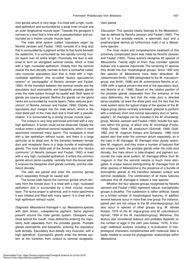
Description
. Elongated, elliptical animals with two eyes.They are 3–4 mm long when reared in the lab. Animals col-lected in the field were not measured, but appear to besomewhat larger. Adult animals coloured dark brown bysubepidermal pigment. Juveniles are somewhat lighter.
Epidermis cellular, consisting of large octagonal cells,ciliated and containing numerous large, rod-shaped rhab-dites.
Protonephridia extend at both sides of the body andend separately in the prepharyngeal cavity.
The pharynx is of the rosulatus type and is identical tothat of other
Mesostoma
Ehrenberg, 1837 species. It is sit-uated at 1/3 of the body length. The mouth is lined with adegenerating, ruffled epithelium (pseudociliation) and canbe closed by a sphincter. The prepharyngeal cavity is linedwith a low epithelium with a few nuclei. Most of the nuclei ofthis epithelium are, however, insunk in a nuclear pouch sit-uated at the junction between the prepharyngeal epitheliumand the pharynx bulb. The prepharyngeal cavity is sur-rounded by longitudinal muscles, which continue around thepharynx bulb. The distal pharynx rim is lined with a low, cil-iated epithelium that becomes unciliated towards the phar-ynx lumen. There are 32 internal longitudinal muscles.There are three types of pharyngeal glands: coarse-grainedeosinophilic, coarse-grained basophilic and fine-grainedeosinophilic. All three types enter the pharynx lumen nearthe distal end of the pharynx bulb, the fine-grained eosino-philic ones probably most proximally.
There are two very large testes, extending at both sidesof the body. They are connected to each other at threeplaces: caudally, rostrally and at 3/4 of the body length. Thevery small ovary is situated just behind the pharynx, at theleft hand side. The vitellaria are paired and situated ventrallyto the testes.
The gonopore lies just behind the mouth, both being sit-uated within a depression of the ventral body wall. The com-
Fig. 2
. Mesostoma thamagai
n. sp. (A) Reconstruction of the male atrial system (holotype). (B) Reconstruction of the genital organs (holo-type). b, bursa; bs, bursal stalk; cga, common genital atrium; de, ejaculatory duct; fd, female duct; gg, glands; gp, gonopore; m, mouth; ma,male atrium; ms, male genital system; od, oviduct; ov, ovary; ph, pharynx; ppc, prepharyngeal cavity; rs, seminal receptacle; ut, uterus; vd,vitelloduct; vdef, vas deferens; vg, prostate vesicle; vs, seminal vesicle.
Downloaded From: https://bioone.org/journals/Zoological-Science on 25 Apr 2019Terms of Use: https://bioone.org/terms-of-use
Freshwater Rhabdocoela from Botswana 1067
mon genital atrium is very large. It is lined with a high, nucle-ated epithelium and surrounded by a weak inner circular andan outer longitudinal muscle layer. Towards the gonopore itnarrows to a duct that is lined with a pseudociliation and sur-rounded by a thicker circular muscle layer.
The male atrial system (called “copulatory organ” byNoreña Janssen and Faubel, 1992) consists of a long ductthat is surrounded by a pigment similar to that found beneaththe epidermis. It is surrounded by two spirally-running mus-cle layers over its entire length. Proximally the duct is wid-ened to form an elongated seminal vesicle, which is linedwith a high, nucleated epithelium. Distally from the seminalvesicle the duct abruptly narrows to form a somewhat coiled,very muscular ejaculatory duct that is lined with a high,nucleated epithelium (the so-called “ductus ejaculatoriusexterior” or “penispapilla” of Noreña Janssen and Faubel,1992). At the transition between the seminal vesicle and theejaculatory duct eosinophilic and basophilic prostate glandsenter the male system through its caudal wall. Both types ofglands are coarse-grained. Neither the glands nor the glandnecks are surrounded by muscle layers (“false vesicula gran-ulorum” of Noreña Janssen and Faubel, 1992). Distally, theejaculatory duct merges into the male atrium, which is themost distal part of the male duct and is lined with a pseudo-ciliation. It is surrounded by a strong circular muscle layer.
The oviduct is very long and broad and lined with a veryhigh epithelium. A lumen could not be observed. Distally theoviduct enters a spherical seminal receptacle, which in mostspecimens contained many sperm. The receptacle is linedwith a low epithelium without nuclei, and there is a largesphincter at its distal end. At the transition between femaleduct and receptacle there is a large bundle of eosinophilicglands. The most distal part of the female duct (the “ductuscommunis” of Noreña Janssen and Faubel, 1992) is linedwith a very high, nucleated epithelium. It enters the commongenital atrium dorso-caudally, ventrally from the bursal stalk.It receives the basophilic shell glands ventrally, and the vitel-loduct dorsally.
The uteri are paired and enter the common genitalatrium separately through its caudal wall.
The bursal stalk leaves the common genital atrium dor-sally from the female duct. It is lined with a high, nucleatedepithelium and is surrounded by a thick circular musclelayer. The bursa proper is spherical, and in many specimensit was inflated and filled with many sperm. It is lined with ahigh epithelium without nuclei.
Diagnosis
.
Mesostoma thamagai
n. sp.
Mesostoma
specieswith dark brown, subepidermal pigment. Same pigmentpresent around the male genital system. Gonopore veryclose behind the mouth. Vasa deferentia entering the copu-latory bulb separately from the prostate glands. Prostateglands eosinophilic and basophilic, entering the copulatorybulb dorsally. Ejaculatory duct distally very muscular, with ahigh epithelium. Eosinophilic glands enter the female sys-tem at the transition from oviduct to seminal receptacle.
Uteri paired.
Discussion
. This species clearly belongs to the Mesostomi-nae as defined by Noreña Janssen and Faubel (1992). Thelack of a true prostate vesicle, a spermatic duct and aprepharyngeal dermal pit furthermore mark it as a
Mesos-toma
species.The most recent and comprehensive treatment of this
extremely complicated taxon was made by Noreña Janssenand Faubel (1992). These authors recognise 60 species of
Mesostoma
. Twenty eight of them they consider
speciesdubiae
, one a
species inquirenda
. The remaining 31 speciesthey divide into four morphological groups. Since 1992 onlytwo species of
Mesostoma
have been described:
M.tubiseminalis
Smith, 1998 (designated to the
M. maculatum
-group; see Smith, 1998) and
M. extremiensis
Noreña
et al
.,1999 (with a typical pincers-like end of the ejaculatory duct;see Noreña
et al
., 1999). Based on the relative position ofthe prostate glands (separated from the entrance of thevasa deferentia), the fact that the male duct is directeddorso-caudally (at least the distal part) and the fact that themale system lacks the typical shape of the species of the
M.lingua
-group (dorso-ventrally orientated, globular to pyriformwith a relatively short and conical ejaculatory duct or “penis-papilla”),
M.
thamagai
can be included in the
M. ehrenbergi
-group. Noreña Janssen and Faubel (1992) include five spe-cies within this group, of which only
M. ehrenbergi
(Focke,1836) Örsted, 1844,
M. productum
(Schmidt, 1848) Graff,1882 and
M. magnum
Kolasa and Schwartz, 1988 havepaired uteri that enter the common atrium separately, as isthe case in
M
.
thamagai
.
M.
thamagai
most closely resem-bles
M. magnum
, and they share a number of features thatare unique to both: the prostate glands enter the male ductdorsally, the male atrium is tube-shaped, and pigment sur-rounds the male atrial system.
M.
thamagai
differs from
M.magnum
in that the seminal vesicle is much more elon-gated. A unique feature distinguishing
M
.
thamagai
from allother species of
Mesostoma
is the presence of a bundle ofeosinophilic glands at the transition between oviduct andseminal receptacle. The combination of all these featuresindicates that
M
.
thamagai
is indeed a new species.Whether the four species-groups recognised by Noreña
Janssen and Faubel (1992) represent natural, monophyleticgroups is doubtful. The subdivision is rather artificial, basedon a limited number of morphological features. In additionseveral features occur in more than one group. For instance,paired uteri are not unique to the
M. ehrenbergi
-group, butalso occur in species of other groups (e.g.
M. sibolae
Kolasa, 1976 of the
M. lingua
-group and
M. columbianum
Hyman, 1939 of the
M. maculatum-group). Moreover, thisfeature was considered dubious and probably depends onthe number of eggs in the uteri (see Kolasa, 1976). A thor-ough cladistical analysis including a re-evaluation of mor-phological characters complemented with molecular data isbadly needed to reveal the phylogenetic relationships withinMesostoma.
Downloaded From: https://bioone.org/journals/Zoological-Science on 25 Apr 2019Terms of Use: https://bioone.org/terms-of-use
T. Artois et al.1068
Young (1976) mentioned seven valid Mesostoma spe-cies occurring on the African continent: M. brincki Marcus,1970, M. ehrenbergi, M. evelinae Marcus, 1955, M. ewerumDu Bois-Reymond Marcus, 1951, M. inversum Beau-champs, 1954, M. lacteum Neppi, 1904 and M. lingua(Abildgaard, 1789) Schmidt 1848. Additionally, he men-tioned two other South African species as species dubiae:M. antarcticum Dreyer, 1918 and M. karrooense Dreyer,1914. Of these two, especially M. karrooense has oftenbeen considered insufficiently described (Ruebush, 1939;Du Bois-Reymond Marcus, 1951; Marcus, 1955), and there-fore has not been considered in more recent faunistic or tax-onomic studies. In the same year, Kolasa (1976) describedM. africanum Kolasa, 1976 from south Algeria and gave alist of six “undoubtedly valid” African species: M. africanum,M. brincki, M. evelinae, M. ewerum, M. lingua, and M. pro-ductum. His list and that of Young (1976) differ in the factthat Kolasa (1976), at the time of his writing, was not awareof the record of M. ehrenbergi by Young and Young (1976)and (probably) considered M. inversum a junior synonym ofM. productum as later also did Noreña Janssen and Faubel(1992), although Kolasa did not mention this explicitly.Moreover, he apparently considered M. lacteum to be a spe-cies dubia. In 1981, Kolasa and Mead described a new spe-cies from Nigeria: M. zariae Kolasa and Mead, 1981. In theirmonograph of the Mesostominae, Noreña Janssen andFaubel (1992) erected a new monospecific genus to classifythe somewhat aberrant species M. evelinae: Marcomesos-toma Noreña Janssen and Faubel, 1992. Moreover, theyconsidered M. antarcticum a junior synonym of M. mutabileBöhmig, 1902, probably based on the statement by Dreyer(1918) that there was virtually no difference between the twospecies except for their geographical distribution and forsome minute differences in the female system. From whatwe could retrieve from literature, this synonymisation seemsjustified. They also explicitly considered M. lacteum a spe-cies dubia. Following these changes and additions, ninevalid Mesostoma species are now recorded from Africa: M.africanum, M. brincki, M. ehrenbergi, M. ewerum, M. lingua,M. mutabile, M. productum, M. thamagai and M. zariae.Three of these are cosmopolitan (M. ehrenbergi, M. produc-tum, M. lingua), while M. mutabile also occurs in SouthAmerica. The remaining five are only known from the Africancontinent.
DALYELLIIDAE GRAFF, 1905Gieysztoria isoldeae n. sp.
(Fig. 3)Locality. Botswana, Thamaga (24°41’50’’S, 25°31’00’’E),ephemeral rock pools on isolated granite domes (type local-ity). Sediment collected 22/12/1998, inundated in the labo-ratory 4/10/2002.Material. Observations on live animals. One whole mount,designated holotype (LUC nr. 248). Seven sectioned speci-mens, designated paratypes (LUC nr. 249–255).Etymology. Species named after the first author’s daughter
Isolde.
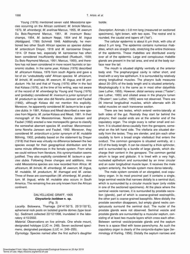
Description. Animals ± 0.8 mm long (measured on sectionedspecimens), light brown, with two eyes. The rostral end isrounded, the caudal end tapers off (“tail”).
The cellular epidermis is about 6 µm thick, with cilia ofabout 5 µm long. The epidermis contains numerous rhab-dites, which are straight rods, stretching the entire thicknessof the epidermis. These rhabdites are distributed evenlyover all of the epidermis. Large and conspicuous rhabditeglands are present in the tail area, and end at the body sur-face near the tail.
The mouth is situated slightly ventrally at the anteriorbody end. The prepharyngeal cavity is rather short, andlined with a very low epithelium. It is surrounded by relativelystrong longitudinal muscles. The pharynx bulb measuresabout 20–25% of the body length and is situated anteriorly.Morphologically it is the same as in most other dalyelliids(see Luther, 1955). However, distal sensory areas (“Taster”;see Luther, 1955) and ciliated areas in the pharynx lumen(“Brusten”; see Luther, 1955) were not observed. There are26 internal longitudinal muscles, which alternate with 26radial muscles on each transverse section.
There are two testes, which extend ventro-laterally atboth sides of the gut. Rostrally they start far behind thepharynx; their caudal ends are at the anterior end of thecopulatory organ. The single ovary is rather small and ovi-form. It is situated dorso-caudally from the gonopore, some-what on the left hand side. The vitellaria are situated dor-sally from the testes. They are slender, and join each othercaudally to form a broad single vitelloduct that enters theoviduct. The common gonopore is situated ventrally, at ±2/3 of the body length. It can be closed by a thick sphincter,and is surrounded by a bundle of large glands, which dis-charge their content in the gonopore. The common genitalatrium is large and globular. It is lined with a very high,nucleated epithelium and surrounded by an inner circularand an outer longitudinal muscle layer. It receives the malesystem anteriorly, the female system more dorso-laterally.
The male system consists of an elongated, oval copu-latory organ. In its most proximal part it contains a single,large seminal vesicle that narrows distally to a seminal duct,which is surrounded by a circular muscle layer (only visiblein one of the sectioned specimens). At the place where theseminal vesicle narrows, it is surrounded by prostate secre-tion (glands), part of which is coarse-grained eosinophilic,the other part is coarse-grained basophilic. More distally theprostate secretion disappears, but empty gland necks con-spicuously surround the seminal duct. The nuclei of theprostate glands were not observed. Seminal vesicle andprostate glands are surrounded by a muscular septum, con-sisting of at least two muscle layers which cross each other.Between seminal vesicle/prostate glands and the septumthere is a space filled with loose parenchyma. As such thecopulatory organ is clearly of the conjuncta-duplex type (ter-minology of Karling, 1956). Distally the septum narrows and
Downloaded From: https://bioone.org/journals/Zoological-Science on 25 Apr 2019Terms of Use: https://bioone.org/terms-of-use
Freshwater Rhabdocoela from Botswana 1069
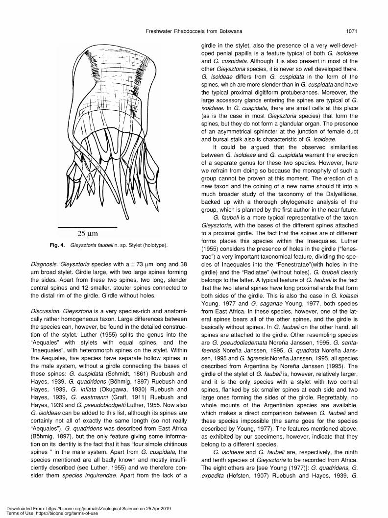
Fig. 3. Gieysztoria isoldeae n. sp. (A) Habitus (from a living animal). (B) Spines of the male system (holotype). (C) Reconstruction of the malegenital organs (from several paratypes). (D) Reconstruction of the genital system (from several paratypes). acg, accessory glands; b, bursa;bs, bursal stalk; cb, copulatory bulb; cga, common genital atrium; dc, ductus communis; e, eye; fd, female duct; gg, glands; gp, gonopore; i,intestine; m, mouth; ma, male atrium; od, oviduct; ov, ovary; pg, prostate glands; ph, pharynx; pp, penial papilla; rs, seminal receptacle; sph,sphincter; sp, spine; ut, uterus; vd, vitelloduct; vi, vitellarium; vs, seminal vesicle.
Downloaded From: https://bioone.org/journals/Zoological-Science on 25 Apr 2019Terms of Use: https://bioone.org/terms-of-use
T. Artois et al.1070
splits into two layers, both surrounded by an inner circularand an outer longitudinal muscle layer. In this way a verymuscular penial papilla is formed, which is situated centrallyin the male atrium. From centre to periphery the penialpapilla consists of: the narrow seminal duct, the necks of theprostate glands, parenchyma (distally denser and with manynuclei), a muscular septum, parenchyma and again a mus-cular septum. The distal opening of this penial papilla can beclosed by a sphincter. At the proximal end of the penialpapilla (where the septum narrows), four hollow and tubi-form spines enter the male atrium, two of them situated ven-tro-laterally, the other two laterally. The ventrolateral spines(152 and 162 µm) are somewhat larger than the lateral ones(129 and 134 µm). Because of a prolongation of one of thesides, the proximal opening of each spine is large andasymmetrical. These prolonged sides consist of 4–6 digiti-form proturbances. They lie next to the septum and supportit. The distal part of each spine flanks the penial papilla. Abundle of coarse-grained light basophilic glands enters eachlateral spine, and maybe also the ventral ones. These baso-philic glands are surrounded by fine-grained eosinophilicones, which also enter the lateral spines and probably alsothe ventral ones. These glandular complexes are sur-rounded by a common muscle layer that is continuous withthe muscles forming the septum and surrounding the maleatrium. The male atrium itself is lined with a very low epithe-lium and is surrounded by an inner circular and an outer lon-gitudinal muscle layer.
As it leaves the common genital atrium, the female sys-tem immediately splits into the bursal stalk, running anteri-orly, and the ductus communis, running caudally. The bifur-cation is surrounded by a very thick circular muscle layer,which is extremely thick dorsally, forming an asymmetricalsphincter. Bursal stalk, bursa and ductus communis arelined with the same high, irregular, nucleated epithelium asis the common genital atrium, and are surrounded by aninner circular and an outer longitudinal muscle layer. Thebursal stalk is rather broad, but narrows towards the bursa.At the place where it narrows it starts to run caudally. Thebursa can have different appearances depending on thestate of the animal: very small (underdeveloped; before cop-ulation), or globular or oval filled with many sperm. The fullydeveloped bursa is surrounded by an eosinophilic tissue.The ductus communis is also rather broad, and proximallysplits into the uterus and the female duct. The uterus is sit-uated terminally and is globular when no eggs are present.It is lined with the same epithelium and surrounded by thesame muscle layers as is the ductus communis. The femaleduct runs dorsally. It is short and proximally widens to forma seminal receptacle, which often contains sperm. Femaleduct and seminal receptacle are lined with the same epithe-lium and surrounded by the same muscle layers as is theductus communis. At the transition from female duct to sem-inal receptacle the circular muscle layer thickens to form asphincter. The vitelloduct and several basophilic and eosino-philic glands (“Schalendrüsen” of Luther, 1955) enter the
female duct just distally from the sphincter. The short ovi-duct enters the seminal receptacle opposite the female duct.It consists of very flat cells that are piled upon each other.These cells appear to be hollow.
Diagnosis. Gieysztoria species with a very well-developedpenial papilla. Male system with four separate hollowspines, not connected to each other by a girdle. All spineswith proximal digitiform protuberances. Ventrolateral spineslarger (± 157 µm) than the lateral ones (± 132 µm). A largecomplex of glands enters the spines.
Discussion. See the discussion of Gieysztoria faubeli.
Gieysztoria faubeli n. sp.(Fig. 4)
Locality. Botswana, Thamaga (24°41’50’’S, 25°31’00’’E),ephemeral rock pools on isolated granite hills (type locality).Sediment collected 22/12/1998, inundated in the laboratory4/10/2002.Material. Observations on live animals. 10 whole mounts, 1of them designated holotype (LUC nr. 256), the other 9paratypes (LUC nr. 257–265).Etymology. Species named in honour of Dr. Anno Faubel(Hamburg).
Description. Small animals, 0.5–0.7 mm long, pale brown,with two eyes. The pharynx is a typical pharynx doliiformisat the rostral end of the body.
The construction of the genital system does not differfrom that of the other Gieysztoria species. The genital poreis in the caudal 1/3 of the body. Testes were not observed.The ovary is small and is situated at ± 2/3 of the bodylength. The vitellaria are small and distally join each other ina broad vitelloduct.
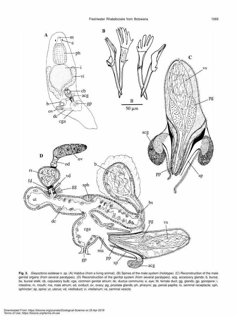
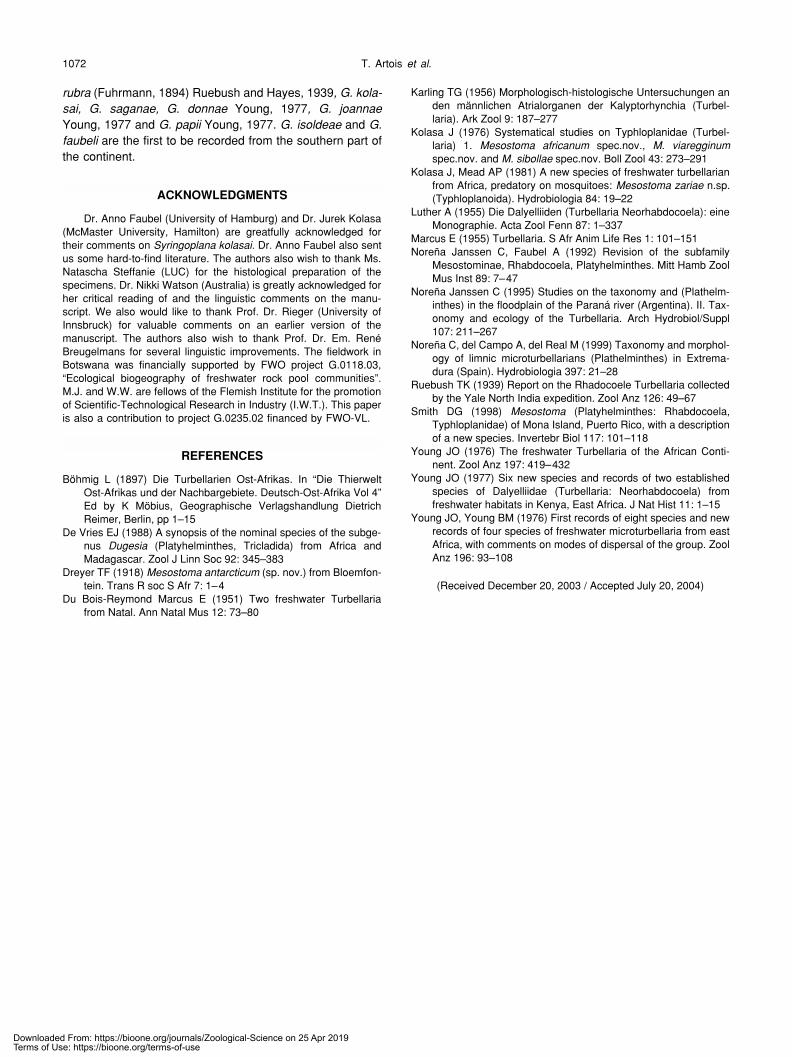
The pyriform prostate vesicle contains an internal sem-inal vesicle at its proximal end and a number of prostateglands at its distal end. Distally it ends in the stylet, which is69–84 µm long (m=73 µm; n = 8) and 31–44 µm broad(m=38 µm; n=7). Proximally the stylet consists of a girdle(terminology of Luther, 1955), which is divided into a proxi-mal part with fine longitudinal and transverse ridges, and adistal rectangular part with only a few longitudinal ridges.Two long S-shaped spines make up the sides of the girdleand protrude into the male atrium. Distally the girdle carriestwo central slender spines. At each side of these centralspines, six broader and stouter spines occur. All spines arehollow (exc. the two large lateral ones?), and their openingsare easily visible right up against the rim of the girdle. Thelongitudinal ridges of the distal part of the girdle are in linewith the edges of the spines, and split up and continue asthe longitudinal ridges of the proximal part of the girdle. Thegirdle does not show any holes.
The very muscular bursal stalk ends in the commongenital atrium in between the oviduct and the copulatoryorgan. The uterus never contains more than one egg.
Downloaded From: https://bioone.org/journals/Zoological-Science on 25 Apr 2019Terms of Use: https://bioone.org/terms-of-use
Freshwater Rhabdocoela from Botswana 1071
Diagnosis. Gieysztoria species with a ± 73 µm long and 38µm broad stylet. Girdle large, with two large spines formingthe sides. Apart from these two spines, two long, slendercentral spines and 12 smaller, stouter spines connected tothe distal rim of the girdle. Girdle without holes.
Discussion. Gieysztoria is a very species-rich and anatomi-cally rather homogeneous taxon. Large differences betweenthe species can, however, be found in the detailed construc-tion of the stylet. Luther (1955) splits the genus into the“Aequales” with stylets with equal spines, and the”Inaequales”, with heteromorph spines on the stylet. Withinthe Aequales, five species have separate hollow spines inthe male system, without a girdle connecting the bases ofthese spines: G. cuspidata (Schmidt, 1861) Ruebush andHayes, 1939, G. quadridens (Böhmig, 1897) Ruebush andHayes, 1939, G. inflata (Okugawa, 1930) Ruebush andHayes, 1939, G. eastmanni (Graff, 1911) Ruebush andHayes, 1939 and G. pseudoblodgetti Luther, 1955. Now alsoG. isoldeae can be added to this list, although its spines arecertainly not all of exactly the same length (so not really“Aequales”). G. quadridens was described from East Africa(Böhmig, 1897), but the only feature giving some informa-tion on its identity is the fact that it has “four simple chitinousspines ” in the male system. Apart from G. cuspidata, thespecies mentioned are all badly known and mostly insuffi-ciently described (see Luther, 1955) and we therefore con-sider them species inquirendae. Apart from the lack of a
girdle in the stylet, also the presence of a very well-devel-oped penial papilla is a feature typical of both G. isoldeaeand G. cuspidata. Although it is also present in most of theother Gieysztoria species, it is never so well developed there.G. isoldeae differs from G. cuspidata in the form of thespines, which are more slender than in G. cuspidata and havethe typical proximal digitiform protuberances. Moreover, thelarge accessory glands entering the spines are typical of G.isoldeae. In G. cuspidata, there are small cells at this place(as is the case in most Gieysztoria species) that form thespines, but they do not form a glandular organ. The presenceof an asymmetrical sphincter at the junction of female ductand bursal stalk also is characteristic of G. isoldeae.
It could be argued that the observed similaritiesbetween G. isoldeae and G. cuspidata warrant the erectionof a separate genus for these two species. However, herewe refrain from doing so because the monophyly of such agroup cannot be proven at this moment. The erection of anew taxon and the coining of a new name should fit into amuch broader study of the taxonomy of the Dalyelliidae,backed up with a thorough phylogenetic analysis of thegroup, which is planned by the first author in the near future.
G. faubeli is a more typical representative of the taxonGieysztoria, with the bases of the different spines attachedto a proximal girdle. The fact that the spines are of differentforms places this species within the Inaequales. Luther(1955) considers the presence of holes in the girdle (“fenes-trae”) a very important taxonomical feature, dividing the spe-cies of Inaequales into the “Fenestratae”(with holes in thegirdle) and the “Radiatae” (without holes). G. faubeli clearlybelongs to the latter. A typical feature of G. faubeli is the factthat the two lateral spines have long proximal ends that formboth sides of the girdle. This is also the case in G. kolasaiYoung, 1977 and G. saganae Young, 1977, both speciesfrom East Africa. In these species, however, one of the lat-eral spines bears all of the other spines, and the girdle isbasically without spines. In G. faubeli on the other hand, allspines are attached to the girdle. Other resembling speciesare G. pseudodiademata Noreña Janssen, 1995, G. santa-feensis Noreña Janssen, 1995, G. quadrata Noreña Jans-sen, 1995 and G. tigrensis Noreña Janssen, 1995, all speciesdescribed from Argentina by Noreña Janssen (1995). Thegirdle of the stylet of G. faubeli is, however, relatively larger,and it is the only species with a stylet with two centralspines, flanked by six smaller spines at each side and twolarge ones forming the sides of the girdle. Regrettably, nowhole mounts of the Argentinian species are available,which makes a direct comparison between G. faubeli andthese species impossible (the same goes for the speciesdescribed by Young, 1977). The features mentioned above,as exhibited by our specimens, however, indicate that theybelong to a different species.
G. isoldeae and G. faubeli are, respectively, the ninthand tenth species of Gieysztoria to be recorded from Africa.The eight others are [see Young (1977)]: G. quadridens, G.expedita (Hofsten, 1907) Ruebush and Hayes, 1939, G.
Fig. 4. Gieysztoria faubeli n. sp. Stylet (holotype).
Downloaded From: https://bioone.org/journals/Zoological-Science on 25 Apr 2019Terms of Use: https://bioone.org/terms-of-use
T. Artois et al.1072
rubra (Fuhrmann, 1894) Ruebush and Hayes, 1939, G. kola-sai, G. saganae, G. donnae Young, 1977, G. joannaeYoung, 1977 and G. papii Young, 1977. G. isoldeae and G.faubeli are the first to be recorded from the southern part ofthe continent.
ACKNOWLEDGMENTS
Dr. Anno Faubel (University of Hamburg) and Dr. Jurek Kolasa(McMaster University, Hamilton) are greatfully acknowledged fortheir comments on Syringoplana kolasai. Dr. Anno Faubel also sentus some hard-to-find literature. The authors also wish to thank Ms.Natascha Steffanie (LUC) for the histological preparation of thespecimens. Dr. Nikki Watson (Australia) is greatly acknowledged forher critical reading of and the linguistic comments on the manu-script. We also would like to thank Prof. Dr. Rieger (University ofInnsbruck) for valuable comments on an earlier version of themanuscript. The authors also wish to thank Prof. Dr. Em. RenéBreugelmans for several linguistic improvements. The fieldwork inBotswana was financially supported by FWO project G.0118.03,“Ecological biogeography of freshwater rock pool communities”.M.J. and W.W. are fellows of the Flemish Institute for the promotionof Scientific-Technological Research in Industry (I.W.T.). This paperis also a contribution to project G.0235.02 financed by FWO-VL.
REFERENCES
Böhmig L (1897) Die Turbellarien Ost-Afrikas. In “Die ThierweltOst-Afrikas und der Nachbargebiete. Deutsch-Ost-Afrika Vol 4”Ed by K Möbius, Geographische Verlagshandlung DietrichReimer, Berlin, pp 1–15
De Vries EJ (1988) A synopsis of the nominal species of the subge-nus Dugesia (Platyhelminthes, Tricladida) from Africa andMadagascar. Zool J Linn Soc 92: 345–383
Dreyer TF (1918) Mesostoma antarcticum (sp. nov.) from Bloemfon-tein. Trans R soc S Afr 7: 1–4
Du Bois-Reymond Marcus E (1951) Two freshwater Turbellariafrom Natal. Ann Natal Mus 12: 73–80
Karling TG (1956) Morphologisch-histologische Untersuchungen anden männlichen Atrialorganen der Kalyptorhynchia (Turbel-laria). Ark Zool 9: 187–277
Kolasa J (1976) Systematical studies on Typhloplanidae (Turbel-laria) 1. Mesostoma africanum spec.nov., M. viaregginumspec.nov. and M. sibollae spec.nov. Boll Zool 43: 273–291
Kolasa J, Mead AP (1981) A new species of freshwater turbellarianfrom Africa, predatory on mosquitoes: Mesostoma zariae n.sp.(Typhloplanoida). Hydrobiologia 84: 19–22
Luther A (1955) Die Dalyelliiden (Turbellaria Neorhabdocoela): eineMonographie. Acta Zool Fenn 87: 1–337
Marcus E (1955) Turbellaria. S Afr Anim Life Res 1: 101–151Noreña Janssen C, Faubel A (1992) Revision of the subfamily
Mesostominae, Rhabdocoela, Platyhelminthes. Mitt Hamb ZoolMus Inst 89: 7–47
Noreña Janssen C (1995) Studies on the taxonomy and (Plathelm-inthes) in the floodplain of the Paraná river (Argentina). II. Tax-onomy and ecology of the Turbellaria. Arch Hydrobiol/Suppl107: 211–267
Noreña C, del Campo A, del Real M (1999) Taxonomy and morphol-ogy of limnic microturbellarians (Plathelminthes) in Extrema-dura (Spain). Hydrobiologia 397: 21–28
Ruebush TK (1939) Report on the Rhadocoele Turbellaria collectedby the Yale North India expedition. Zool Anz 126: 49–67
Smith DG (1998) Mesostoma (Platyhelminthes: Rhabdocoela,Typhloplanidae) of Mona Island, Puerto Rico, with a descriptionof a new species. Invertebr Biol 117: 101–118
Young JO (1976) The freshwater Turbellaria of the African Conti-nent. Zool Anz 197: 419–432
Young JO (1977) Six new species and records of two establishedspecies of Dalyelliidae (Turbellaria: Neorhabdocoela) fromfreshwater habitats in Kenya, East Africa. J Nat Hist 11: 1–15
Young JO, Young BM (1976) First records of eight species and newrecords of four species of freshwater microturbellaria from eastAfrica, with comments on modes of dispersal of the group. ZoolAnz 196: 93–108
(Received December 20, 2003 / Accepted July 20, 2004)
Downloaded From: https://bioone.org/journals/Zoological-Science on 25 Apr 2019Terms of Use: https://bioone.org/terms-of-use