françois fagotto - rero · j(jlos yolk degradation in tick eggs françois fagotto doctoral thesis...
TRANSCRIPT

J(JlOS
Yolk degradation in tick eggs
François Fagotto
Doctoral thesis at Neuchâtel University, Faculty of Sciences
Neuchâtel ,1990

IMPRIMATUR POUR LA THESE
..YpIk..pegradatipn in...Tick,...Eggs
de M.onsieur .Francois..Fagotto.
UNIVERSITE DE NEUCHATEL
FACULTÉ DES SCIENCES
La Faculté des sciences de l'Université de Neuchâtel
sur le rapport des membres du jury,
..Me.s^s.i.e.urs.P.v Schu.rman
...P..A.,..pi.eM.>...M.v„
. B......Sordat.. (EpalJ.ng.es)
autorise l'impression de la présente thèse.
Neuchâtel, le L.nQy.?É>rçJ.99.$
Le doyen :
'CU^Me^mod /

1
CONTENTS
Summary Preface Abbreviations Introduction:
The yolk Yolk degradation Proteinases in eggs The tick model
Results and discussion Detection, localization and characterization of the major acid proteinase activity Cathepsin L activation Yolk spheres acidification Egg neutral proteinase Endodermal cells and gut morphogenesis Model for yolk degradation in tick eggs Unknowns and perspectives Tentative unifying model for vitellin degradation
Conclusion Acknowledgements References cited Résumé Annexes I-VI
Page
2 4 4
5 6 8 9
12
14 16 18 18 18 19 21 22 23 24 31
Cover page: Nomarski-interference microphotograph of a yolk-filled endodermal cell.

2
SUMMARY
I. OCCURRENCE OF A CATHEPSIN L-LIKE ACID PROTEINASE IN THE YOLK SPHERES Fagotto, R1 Arch. Insect Biochem. Physiol, 14, 1990, 217-235.
In crude extracts of eggs of the soft tick Ornitkodoros moubata, maximum degradation of vitellin is at a pH 3-3.5, whereas no proteolysis is detected at neutral or weakly acidic pH's. Acidic proteolysis is maintained at high level throughout embryonic development, and rapidly decreases in the larva, during the high phase of yolk degradation. Proteinase, acid phosphatase and N-acetylglucosaminidase are localized within the yolk spheres. These can be considered as lysosomal-like organelles containing both substrate (vitellin) and the degradative machinery. Proteolytic activity has been essentially attributed to a cathepsin L-like enzyme through substrate specificity and inhibitors. The molecular weight is 37,000 to 39,000 as shown using gelatin-containing SDS-PAGE activity gels. At neutral pH the enzyme binds to vitellin, as demonstrated by gel filtration and PAGE under non-denaturing conditions. Acid proteinase activity at pH 5-6 is undetectable both with proteins and synthetic substrates, but is strongly increased after preincubation at pH 3-4. Activation at low pH could be important in the regulation of yolk degradation.
II. EVIDENCE THAT THE CATHEPSIN L-LIKE PROTEINASE IS STORED AS A LATENT, ACID ACTIVABLE PROENZYME Fagotto, F., Arch. Insect Biochem. Physiol, 14, 1990, 237-252.
Cathepsin L-like proteinase found in the eggs of the tick Ornithodoros moübata is latent during embryogenesis, but can be activated by acid treatment. In crude extracts as well as in partially purified fractions, activation requires reducing conditions and is inhibited by leupeptin, which indicates that it is mediated by a thiol proteinase, probably by the cathepsin L itself. Latency disappears in vivo at the time of the acute phase of yolk digestion, that takes place during late embryonic development and larval life. When egg cathepsin L is localized through its gelatinolytic activity on SDS-PAGE, the activated enzyme migrates as lower Mr bands than the latent form. Disappearance of the higher Mr bands corresponding to the latent form is directly related to appearance of the lower Mr bands characteristic of the active one; transition from one pattern to the other and enzymatic activation are in perfect agreement with regards to kinetics and sensitivity to inhibitors. The same pattern change occurs in vivo, parallel to latency removal and intense yolk degradation. These results strongly suggest that egg cathepsin L is stored in the yolk as a proenzyme which is activated by partial proteolysis at low pH.
ID. DEVELOPMENTALLY REGULATED ACIDIFICATION OF THE YOLK SPHERES Fagotto, F., Develop. Growth & Differ., 1990, in press.
Yolk spheres in tick eggs contain a latent procathepsin L, which is activated in vivo, in parallel with yolk degradation, and in vitro by acid treatment (Fagotto, F., Arch. Insect Biochem. Physiol, 14, 1990, 236). Mature cathepsin L hydrolyzes vitellin at acidic pH (Fagotto, F., Arch. Insect Biochem. Physiol, 14, 1990, 217). Here, yolk spheres' pH has been estimated using acridine orange. In the early development, all yolk spheres are neutral, then an increasing number acidify, until hatching, where general acidification seems to occur. This fits well with vitellin utilized slowly during embryogenesis, more intensely at hatching (Chinzei, Y. and I. Yano, Experientia, 41, 1985, 948), and can be related to sequential degradation of individual spheres during embryonic development, then extensive yolk liquefaction in the larva. Different yolk sphere populations have been separated on Percoli density gradients. In freshly laid eggs, yolk spheres are dense, neutral, undegraded and contain exclusively the precursor of cathepsin L. As development proceeds, yolk spheres are progressively recovered in lower density fractions, displaying acidic interior and cytological signs of degradation. They co-sediment with mature cathepsin L. It is concluded that acidification initiates yolk degradation through procathepsin L activation.
3
IV. LIMITED PROTEOLYSIS OF VITELLIN BY A NEUTRAL PROTEINASE (Unpublished results). ': '* •• ;
In freshly laid eggs, vitellin is not degraded at all when incubated under neutral conditions. However, from day 4 onwards, the embryos possess a neutral proteinase activity, which specifically cleaves the higher vitellin subunit into two lower fragments. Characterization of this activity remains so far elusive.
V. FATE OF ENDODERMAL CELLS AND GUT MORPHOGENESIS (Unpublished results),
The yolk-filled primary endodermal cells appear essentially unchanged during embryonic development, but in the larva, they exocytoze the rest of their yolk content. The gut epithelium probably originate from the primary endoderm. Possible models for gut morphogenesis are proposed.
VL THE EARLY DEVELOPMENT OF THE ARGASID TICK ORNITHODOROS MOUBATA (ACARINA: IXODOIDEA: ARGASIDAE) Fagotto, F., Hess, E. and A. Aeschlimann, Entomol. Gener., 75, 1988, 1-8.
The mode of cleavage of the centrolecithal tick eggs has so far been considered to be superficial. Evidence could be obtained from an electron-microscopic study of Ornithodoros moubaia Murray 1877 that cleavage of this species is total. The early development leads to a diploblastic stage composed of a superficial layer of yolk-free micromeres (ecto-mesoderm) and a core of yolk-rich macromeres (primary endoderm).

4
PREFACE
This thesis is a study of the modalities of yolk degradation and its regulation during embryogenesis. Using the tick egg as a model, it has been attempted to present evidence for possible general mechanisms governing yolk utilization, at the enzymatic, ultrastructural and physiological levels. This thesis is based on the following original articles, mentioned in the text by their Roman numerals (I-III). Some related unpublished preliminary results (IV+V), and an original article on the early development of Ornithodoros moubata studied by electron microscopy (VI) are enclosed.
I. Fagotto, F., 1990. Yolk degradation in tick eggs: I. Occurrence of a cathepsin L-like acid proteinase in yolk spheres. Arch. Insect Biochem. Physiol., 14, 217-235.
II. Fagotto, F., 1990. Yolk degradation in tick eggs: II. Evidence that cathepsin L-like proteinase is stored as a latent, acid-activable proenzyme. Arch. Insect Biochem. Physiol., 14, 237-252.
III. Fagotto, F., 1990. Yolk degradation in tick eggs: III. Developmentally regulated yolk sphere acidification. Develop., Growth & Differ., in press.
IV. Yolk degradation in tick eggs: IV. Limited proteolysis of vitellin by a neutral proteinase. Unpublished results.
V. Yolk degradation in tick eggs: V. Fate of the endodermal cells and gut morphogenesis. Unpublished results.
VI. Fagotto, F., Hess, E. and A. Aeschlimann, 1988. The early development of Ornithodoros moubata (Acarina: Ixodoidea: Argasidae). Entomol. Gener., 13, 1-8.
This work is dedicated to my childhood friends Massimo Soffiato and Roberto Scarpa, and to the memory of Mr. André Fuchs.
ABBREVIATIONS USED
antipain= [l-carbonyl-2-phenylethyllcarbamoyl-L-arginyl-L-valyl-arginal; Bz-Arg-Nan= ot-N-benzoylarginyl-p-nitroanilide; E-64= N-[N-(DL-3-transcarboxyiran-carbonyl)-L-leucyl]agmatine; ESP, egg specific protein; FCCP, carbonyl cyanide p-trifluoromethoxyphenylhydrazone; FPLC= Fast Protein Liquid Chromatography; leupeptin= N-acetylleucyl-leucyl-arginal; Mr, molecular ratio; PAGE, Polyacrylamide gel electrophoresis; pepstatin A= isovaleryl-L-valyl-L-valyl-(3S,4S)-4-amino-3-hyoVoxy-6-methylhepanoyl L-alanyl-(3S,4S)-4-amino-3-hydroxy-6-methylneptanoic acid; PMSF= phenyhnethylsulfonyl fluoride; SDS= sodium dodecyl sulfate; SEM, scanning electron microscopy; TEM, transmission electron microscopy; Z-Arg-Arg-NHTFMec= benzoyloxycarbonyl-arginyl-arginyl-7-amido-4-trifluormethylcoumarine; Z-Phe-Arg-NHMec= benzoyloxycarbonyl-phenylalanyl-arginyl-7-amido-4-methylcoumarine; Z-Phe-Phe-CHN2=benzoyloxycarbonyl-phenylalanyl-phenylalanyl-diazomethylketone.

5
INTRODUCTION
THE YOLK
In most oviparous species, eggs store large amounts of nutrients - the yolk - during their maturation. Yolk will be the food supply of the embryo, sometimes also of the larval stage until the animal becomes able to feed on its own. The phase of yolk accumulation is called vitellogenesis. During this period the oocyte increases enormously in size, mainly due to yolk accumulation. Three kinds of yolk are present in eggs: ß-glycogen granules, triglyceride droplets and protein yolk [65], but usually the term "yolk" refers to the latter one.
Most yolk proteins are synthesized as precursors, called vitellogenins, These large (170-260 kDa) phospho-lipo-glycoproteins have been characterized in detail in many vertebrate and invertebrate species [8,45,65,87]. Mannose and glucosamine are the more frequently encountered sugars. Lipid content is mainly due to phospholipids, but cholesterol and other neutral lipids are also found. Vertebrate vitellogenins contain a unique serine-rich domain (more than 50% serine residues), which is highly phosphorylated [8,87]. Other vitellogenins have a much lower phosphate content [8,65].
Large sequence homology has been found between vitellogenin from vertebrates, nematodes and insects, indicating that the structure of these molecules has been well conserved during evolution [8]. Diptera are the only known exception, since their vitellogenin has no homolgy with its counterparts in other animals, but significant homology with vertebrate lipases [82]. While in echinoderms only a single vitellogenin gene has been found, many other organisms have several closely related genes [8].
Vitellogenins are normally synthesized in the liver of vertebrates or the fat body (sometimes the oocyte) of arthropods. Their synthesis is hormonally controlled in a sex-dependent manner [65,87]. They are secreted, generally as dimers [8], in the blood or the hemolymph and then taken up selectively by the oocytes through receptor-mediated endocytosis [65,87].
In vertebrates, internalized vitellogenins undergo partial proteolysis that yields three fragments: the large (-120 kDa) and the small (30-35 kDa) subunits of lipovitellin, and phosvitin (28-35 kDa), which is derived from the serine-rich, highly phosphorylated region of vitellogenin [87]. In some cases, different small fragments (13-19 kDa), also with a high phosphate content, are formed. They have been called "phosvettes".
In arthropods, vitellogenin has often already been processed in the fat body, yielding two peptides [45,65], homologous to the lipovitellin subunits. The domain of the molecule containing serine repeats is missing. In some insects [26,45,57], as well as in ticks [10,11], further processing occurs, yielding several polypeptides. On the other hand, vitellogenin

6
usually does not undergo great modifications after capture by the oocyte, and the egg yolk protein, called vitellin, often differs from its precursor only through its lipid content [65]. Unlike vitellogenins, vitellins/ lipovitellins are usually highly insoluble. Exceptions, however, are found in fish [34], some insects [81] and ticks [11,15],
Yolk proteins are concentrated through fusion of small endocytic vesicles into large, dense organelles [65,68,69,87], where they will be stored until degradation during embryogenesis. These organelles are called yolk spheres when yolk proteins are soluble, yolk granules when they are filled with amorphous, insoluble yolk, and yolk platelets in the case of formation of yolk crystals.
Besides vitellogenin-derived yolk proteins, other proteins are stored, in smaller quantities, in the yolk, either within the same organelles, or in other granules [65,87]. One can mention the egg specific protein (ESP) of Bombyx, which is synthesized by the ovary [90]. Other examples are lipophorins in insects [45], and various serum proteins in vertebrates [87]. Yolk granules (spheres or platelets) also contain many other small molecules (lipids, calcium, which binds to phosphate residues, and so on).
In most eggs, individual yolk granules accumulate into the oocyte, the newly formed pushing the older ones toward the centre. In some cases, however, such as sauropsid eggs, they fuse together, which results in a huge mass of fluid yolk [87]. During development, the yolk is either distributed among all cells (echinoderms, mollusks, amphibians), or kept undivided in the centre of the egg, as a syncytium (insects, fish).
YOLK DEGRADATION
Yolk utilization will provide the embryo with amino acids, sugars, lipids and other essential components. Its pattern varies among different species and is likely to be finely regulated, depending on the specific requirements of each developing organism. For example in Drosophila embryos [7], yolk degradation begins soon after fertilization, and is led to completion within the exceptionally brief embryonic development. In many other cases, however, degradation of the yolk proteins starts only in late development and often continues in the larva. Examples of late yolk utilization are found in insects (Rhodnius [57], Bombyx [33]), ticks (Ornithodoros moubata [12]), sea urchins [77] and amphibians [35,37,43]. In fact, many eggs possess large amounts of ß-glycogen and triglyceride inclusions, which are thought to be the main energy supply during early development [72,89], Yet it is difficult to exclude that small amounts of yolk proteins are degraded even in very early development, such small changes in total yolk content being hardly detectable. In the trout egg, two different kinds of yolk are present: embryonic cells have small amounts of yolk granules, which are degraded during early development, while most of the yolk is localized in the central syncytium and is used later [85].

7
In some species, vitellin has been shown not to be necessary for the achievement of embryonic development. A "lecithotropic" sea urchin, which has a particularly rapid and unusual development, completely lacks yolk glycoproteins [76]. Bombyx oocytes can be grown in the body cavity of male hosts, and parthenogenetically activated eggs develop normally, even if they do not contain any vitellin [92]. Another protein, ESP, which is synthezised by the ovary, is in fact the main nutrient used before hatching, while vitellin is degraded at a lower rate [32,92].
As revealed by SDS-PAGE analysis, vitellin and other yolk proteins undergo limited proteolysis during the embryonic development of most invertebrates, sometimes long before their utilization [14,26,31,38,57,66,67,75]. Usually these partial cleavages do not affect the supramolecular organization of the molecule. In vertebrates' eggs, vitellogenins are cleaved before concentration in the yolk platelets takes place (see above), but, to my knowledge, no study of the subsequent biochemical modification during development has been reported. The function of partial proteolysis of yolk proteins during embryogenesis is so far unknown. Fragments could be more readily degraded than the native protein. Alternatively, processing could function as down-regulation of proteinase activity, since vitellin fragments have been reported to be proteinase inhibitors [21,71,79]. Also, quite different roles have to be considered since the discovery of large aggregates of yolk protein fragments, called toposomes, which act as adhesive extracellular material during gastrulation and morphogenesis of sea urchins [9,56] and amphibians [42].
Ultrastructurally, yolk degradation appears complex. The more commonly encountered images are deformation and fragmentation, decrease in matrix density, inhomogeneities, and membranous structures [4,35,37,51,61,64,85,93]. Most of these features are typical of secondary lysosomes. Long ago, Pasteels [60] first postulated the lysosomal nature of yolk granules. His assumption was based on cytochemical localization of acid phosphatase in the yolk granules of a mollusk, Barnea candida [59]. Since this pioneering work, acid phosphatase cytochemistry has been applied to insects [73], crustaceans [64], amphibians [46], and fish [85]. Only the granules in process of degradation were positive. Since lysosomal-like vesicles were found in the vicinity of these granules, degradation was assumed to occur via fusion of primary lysosomes. However this model is somewhat weakened by observation of lead deposits not only in the periphery, but also sometimes deep in the yolk crystals [46]. In fact one should consider possible artefactual negative results due to poor accessibility to the enzymes. This could solve the conflict with biochemical data obtained with purified granules from eggs or early embryos in several species: most were positive for acid hydrolases [4,44,50,51,74]. Furthermore isolated granules from sea urchin eggs were capable of in vitro degradation when incubated at low pH [77,93], which proves that

8
fusion with lysosomes is not required, but that the yolk already possesses the enzymes needed for its degradation. Artemia eggs are an exception, since it has been shown that yolk granules store only a trypsin-like proteinase, apparently not directly involved in yolk degradation [19,20], but that acid hydrolases are found in separate lysosomes [62,63]. The case of amphibian eggs is not clear, since negative [13], or partially positive [86] results were presented on oocytes, while eggs have not been investigated.
PROTEINASES IN EGGS
The understanding of the mechanisms of yolk degradation implies a good knowledge of the enzymes involved. Unfortunately, to date, little is known, particularly in vertebrates, where almost no data are available. Several studies in the past ten years have dealt with proteinases in arthropod eggs. Both neutral and acid proteinases have been characterized in Artemia [19,20,62,63], Bombyx [32,36] and Drosophila [50-52]. In all cases, the acid proteinases are of the thiol cathepsin family. Evidence has been shown in Artemia [63] and Drosophila [50,51] that they are actually responsible for vitellin degradation. In Bombyx such evidence is still missing. On the other hand, in the latter case, ESP is degraded by a neutral, trypsin-like proteinase that has been thoroughly characterized [32,91]. The function of homologous neutral enzymes in the other species is still unclear. In sea urchins, thiol cathepsins are likely to be also responsible for yolk degradation at low pH, as seen by effective inhibition by leupeptin, a powerful inhibitor of microbial origin [77,93]. On the other hand, proteinase inhibition has been observed in the yolk of Artemia [21] and Drosophila [51,52], but also Xenopus eggs [71,79], due in fact to the yolk proteins themselves, or to related proteolytic fragments.
In conclusion, two models of yolk degradation are proposed so far: (1) either hydrolytic enzymes and substrates (yolk proteins) are first located in separate organelles (respectively lysosomes and yolk granules), which fuse secondarily to initiate degradation, or (2) both enzymes and substrates are stored together in the granules, which supposes an internal regulatory mechanism, may be involvement of proteinase inhibitors, or pH regulation [77,93],
THE ORNITHODOROS MOUBATA MODEL
I decided to investigate yolk degradation in Ornithodoros moubata, an African soft tick, which feeds on mammalian blood and can be reared by in vitro feeding on an artificial "Parafilm" membrane using swine blood. The engorged female uses most of the ingested blood for egg production, mainly for the synthesis of yolk proteins. One female lays about 100-200, 1 mm large, eggs.

9
Vitellin is the major yolk protein and accounts for more than 80% of the total egg protein. Its precursor, vitellogenin, is synthesized in the fat body [10] as two similar primary products Pl and P2 (215 and 205 kDa), which are rapidly processed intracellularly to smaller polypeptides P3-6 (160, 140, 125, 100 kDa). Excreted vitellogenin exists both as monomer (Vgl, 300 kDa) and dimer (Vg2, 600 kDa) and is composed of a mixture of the primary peptides and their fragments.
Circulating vitellogenin is taken up directly by the oocytes, which in ticks are not surrounded by follicle cells, but merely a basement membrane, and bathe in the hemolymph. Once endocytozed, vitellogenin is transformed into vitellin. This involves several slight modifications of the molecule, including partial proteolysis: all traces of the primary polypeptides Pl and P2 disappear, and further processing occurs, yielding the lower Mr fragments P7-8 (60 and 50 kDa) [H]. Vitellin accumulates mainly as dimers (600 kDa), but also as monomers (300 kDa). It is a hemo-lipo-glycoprotein [11,15], freely soluble in water. It has been purified and characterized [H]: amino acid analysis revealed relative enrichment in valine, leucine, glutamic acid and proline. 8% lipids (mainly phospholipids) and 12% sugars (mannose and glucosamine) were found. Phosphate has not been determined. Heme, a product of hemoglobin degradation, is non-covalently adsorbed. In consequence vitellin, and thus also the whole egg. has a brownish color, which might be a useful camouflage.
Vitellin is stored in large (10-80 ^m) yolk spheres [2,15,16], the matrix of which is extremely dense, homogenous, but amorphous, due to vitellin solubility. Mature eggs are almost spherical or slightly elongated. They are tightly packed with yolk spheres and the few spaces left between are essentially filled with ß-glycogen and triglyceride inclusions [2,15,16]. The nucleus is centrally located and surrounded by some yolk-free cytoplasm.
At 290C, oviposition begins 10-12 days after the blood meal, if females are already mated. If not, a lag time of at least 7 days separate oviposition from copulation. Indeed, copulation is essential for completion of vitellogenesis, else the latter aborts. The male deposits a spermatophore in the genital tract, where eggs are fertilized as they pass through. Fertilization has never been studied in detail.
The development of O. moubaia has been studied by Aeschlimann [I]. It is by far the best known embryogenesis among tick and acarina in general. At 290C, embryonic development lasts about 10 days. The larva does not feed or move and soon molts into the first nymphal stage (day 15).
Early development has been investigated at the ultrastructural level (see section VI). Cleavage was initially thought to be superficial [1], as in insects [3,28,72]. However, examination by EM revealed that furrows do penetrate across the tightly packed yolk mass, and cleavage is total, at least from the 8-cells stage. Maybe cleavage is delayed compared to mitosis, due to slow penetration of the furrows; some observations suggested the

10
presence of preformed furrows in cleaving embryos. After about 1 day, cleavage results in a blastula, composed of more or less equal, radially arranged, pyramidal cells. A subsequent tangential division yields a two sheeted embryo: a central primary endoderm (or endoderm I), composed of huge, yolk-filled cells, is surrounded by a thin epithelium devoid of any yolk spheres. The nuclei of the primary endodermal cells, each surrounded by some yolk-free cytoplasm, were previously taken for the so-called vitellophages, which were believed to be free-living amoeboid cells [1,3].
From that point (day 2), development is essentially analogous to insects [I]. An area of the superficial epithelium, the "blastoderm", thickens and becomes the germ band, from which most tissues originate. Blastokinesis (days 5-7) and organogenesis (days 6-10) will shape the body of the future larva. Yet, the yolky interior of the egg (endoderm I) is entirely cellularized, unlike the syncytial yolk found in pterigote insects [3,28,53]. From day 6 onwards, a thin epithelium begins to enclose the yolky primary endoderm, starting from the ventral side, just beneath the germ band. This thin sheet is composed of small, yolk-free cells. These cells will form the gut wall, and are named here endodermal cells II. As discussed in results, section V, their origin is still unclear. Dorsal closure of the gut is completed just before hatching. In the larva, yolk spheres progressively disappear, due to extensive exocytosis, and the gut lumen is filled with liquefied yolk (see section V). Yolk will be slowly resorbed and digested by the gut epithelium during larval and nymphal life [Diehl, unpublished observations].
Changes in vitellin content during development of O. moubata have been studied by Chinzei and Yano [12]. The level diminishes only slowly during embryonic development, but consumption is markedly increased just before hatching and during the larval life. Yet about 50% is still present in young nymphs and will be used slowly during the following weeks, until the first blood meal. On SDS-PAGE, these authors observed neither change in relative abundance of the vitellin subunits, nor appearance of lower Mr fragments. Thus, in this species, probably no partial proteolysis occurs during embryogenesis. On the other hand, PAGE under non-denaturing conditions revealed increasing aggregation of vitellin as high Mr oligomers in late embryos and larvae.
O. moubata appeared to be an attractive model to study yolk degradation. In my opinion, major advantages over other organisms were: (1) the huge amount of yolk compared to other egg components; (2) a soluble vitellin, allowing biochemical analysis with no need of detergents or high salt buffers; (3) the exclusive location of the yolk in one single tissue, the endoderm, throughout embryonic development; (4) as well as a completely cellularized yolk (endoderm I), much easier to manipulate experimentally than the syncytial yolk of insects.
11
More serious problems arose from the complete lack of data on enzymes in this species. Therefore the first goal of my work was to detect and characterize putative hydrolases responsible for yolk degradation. It was also important to know the cellular localization of such enzymes. This should indicate whether contribution of classical lysosomes is required, or if yolk spheres possess an autonomous degradative machinery. The final aim was to relate enzymatic data to the pattern of vitellin degradation established by Chinzei and Yano [12], as well as to morphological and ultrastructural observations, and to investigate possible regulatory mechanisms.
Preliminary observations have indicated that individual yolk spheres were sequentially degraded during embryogenesis. Partially degraded yolk spheres looked very similar to those observed in fish, crustaceans and amphibians. The probable ubiquity of basic degradative mechanisms made me confident that the results of this study on ticks would be of interest when compared to classical models such as Xenopus or Drosophila.
12
RESULTS AND DISCUSSION
DETECTION, LOCALIZATION AND CHARACTERIZATION OF THE MAJOR ACID PROTEINASE ACTIVITY [I]
Crude extracts from early embryos were found to contain strong acid proteinase activity; endogenous (vitellin) and exogenous (hemoglobin) protein substrates were efficiently degraded in the low pH domain (optimum pH 3-3.5). No hydrolysis was detectable under mildly acidic or neutral conditions. Acid proteinase was already present in freshly laid eggs and in oocytes. The level was steady during embryogenesis, but dropped after hatching, during the intense phase of yolk degradation [12], and only weak activity was detected in young nymphs, in agreement with slow yolk degradation at this stage. These developmentally regulated changes in activity, obviously related to yolk utilization, appeared specific for the acid proteinase, since activities of acid phosphatase and ß- N -acetylglucosaminidase did not show the same decrease during the larval life, but steadily increased during development.
Early embryos were fractionated using Percoli density gradients. Acid proteinase was localized together with vitellin in the dense yolk spheres, which sedimented near the bottom of the gradients. These yolk spheres also contained acid phosphatase, ß-N-acetylglucosaminidase, as well as other typical lysosomal activities such as ß-mannosidase, ß-glucosidase, ß-glucuronidase and acylesterase (unpublished results). High hydrolase activities were also found in the supernatant, but their levels were proportional to the vitellin content, and they could not be sedimented, even at higher speeds. Therefore it was concluded that they were exclusively due to broken yolk spheres. Localization of acid phosphatase in yolk spheres has been further confirmed by histochemistry. Storage of hydrolytic enzymes in large amounts, sufficient to degrade the yolk thoroughly, has the main advantage of no need for synthesis by the embryo. Moreover, enzymes and substrate are already located in the same compartment, so that degradation can be readily activated.
Another population of light yolk spheres was detected near the top of the gradients, in fractions containing high levels of acid phosphatase and N-acetylglucosaminidase, but undetectable proteinase and vitellin. The occurrence of these spheres will be discussed further in the third section.
The acid proteinase has been characterized. Use of synthetic substrates, specific inhibitors and activators allowed to exclude the involvement of (1) aspartyl-proteinases such as cathepsin D (ineffectiveness of the specific inhibitor pepstatin), (2) of metalloproteinases (no inhibition by EDTA), (3) of serine-proteinase (acidic pH optimum, weak inhibition with PMSF, lack

13
of activity with Bz-Arg-Nah), but (4) the presence of a thiol-proteinase was proved through activation by reducing agents (cysteine, dithiothreitol) and inhibition by thiol-reagents (HgCl2 and p-chloromercuribenzoate), leupeptin, antipain and the specific irreversible inhibitors E-64 and Z-Phe-Phe-CHN2- The enzyme had all the characteristics of cathepsin L, but not of cathepsin B [6,39,41]: high activity with protein substrates, activity against Z-Phe-Arg-NHMec, but not Z-Arg-Arg-TFNHMec, high sensitivity to leupeptin and Z-Phe-Phe-CHN2, lack of inhibition by urea when tested for casein hydrolysis. The activity has been partially purified on a cation exchange column (Mono S, FPLC system). Essentially all activity, with both synthetic and protein substrates, was recovered in one single peak, and sensitivity toward activators, inhibitors and synthetic substrates was the same as in crude extracts.
It was concluded that a single cathepsin L-like enzyme is stored in yolk spheres and is probably responsible for yolk degradation. This typical lysosomal enzyme, discovered in mammalian cells only about 15 years ago [40], appears to be the most active endopeptidase against endogenous proteins [39], but has long been overlooked, due to weak or no activity against the usual synthetic substrates. It is now extensively studied in mammals, also because its precursor has been shown to be the major excreted protein in some cancer cell lines [17,23-25,47,48]. To my knowledge, it has never been reported neither in invertebrates, nor in eggs. Probably it is present in all organisms, but so far no suitable studies have been undertaken. Improper or incomplete characterization has surely led in many cases to assignement of thiol-proteinase activities to the still more popular cathepsin B.
As seen on gel filtration, and confirmed by localization of the gelatinolytic activity on PAGE under non-denaturing conditions, both a low Mr form (30-40 kDa), and a high M r form (about 400 kDa) were found in O. moubata extracts. In fact, the high M r form is a complex of cathepsin L and some large protein, probably vitellin. Binding is tight, since only denaturing conditions (SDS) were successful in releasing the enzyme.
Mammal cathepsins L are most active in the weak acidic pH range (5-6) [6]. To our surprise, the enzyme detected in eggs was totally inactive at such pHs, but required much stronger acidic conditions. In fact, egg cathepsin L appeared to be stored in a latent form, which could be activated by short treatment at low pH. The acid treated enzyme was active under milder conditions, both with synthetic and protein substrates. Latency could be crucial in the regulation of yolk degradation, thus it was worthwhile to investigate it more closely.

14
CATHEPSIN L ACTIVATION [II]
Cathepsin L activation was studied by detecting activity at pH 5.5 with the synthetic, fluorogenic substrate Z-Phe-Arg-NHMec, after preincubation at low pH. Previous results had shown that, in tick eggs, activity against Z-Phe-Arg-NHMec was due to a single cathepsin L-like enzyme (section I), which could thus be detected in crude extracts without interference from other enzymes. Activation was very pH dependent, readily completed at pH 2.5-3.5, while no activity could be detected after a 2 h preincubation at pH >4.5. Activation was inhibited by thiol reagents and leupeptin, but not pepstatin or EDTA, indicating that thiol-, but neither aspartyl-, nor metallo-proteinases, were involved. Since latency and acid activation were also observed using fractions from cation exchange chromatography and purified yolk spheres, they are not artefacts due to cytoplasmic factors such ascystatins [5].
While the latent form was quite stable under neutral conditions, the active enzyme was stable only at low pH (3-3.5), and was rapidly, irreversibly inactivated at neutral pH, like classical thiol-cathepsins [6].
Latency was also studied in vivo, during embryonic development. In the absence of previous acid treatment, no acid proteinase activity was detectable with the sensitivity of the enzymatic assay until hatching. However latency progressively disappeared in the larva. Activation could thus be related both to loss in total activity (section I) (since active cathepsin L is unstable) and to intense yolk degradation [12], both occuring also during the larval life.
Evidence for the mechanism of activation was obtained by the use of zymograms: samples, preincubated under various conditions, were then run on SDS-PAGE containing co-polymerized gelatin, and proteolytic activity was revealed after electrophoresis by incubating the gels at pH 3.5 under reducing conditions. When samples were treated at neutral pH, so that latency was not removed, two pairs of activity bands were detected (37-39/32-34 kDa). The higher Mr bands (37-39 kDa) were intensified, while the lower Mr bands (32-34 kDa) disappeared if the sample was oxidized. Therefore, these pairs are thought to correspond respectively to the native molecule and to the large, active site-containing subunit. On the other hand, acid treatment, which activates the enzyme, as measured enzymatically, resulted in a quite different pattern of lower Mr bands (25-28-30/33 kDa).
All bands were inhibited by the presence of cathepsin L inhibitors (E-64, leupeptin, HgCl2, Z-Phe-Phe-CHN2) during incubation of the gels, suggesting that there is one single enzyme, under different forms. This is further supported by the fact that the same change in banding pattern was also found using active fractions from cation exchange chromatography, or purified yolk spheres.
Banding pattern transition was prevented by thiol-reagents or leupeptin, which indicates that procathepsin L processing is due to some

15
thiol-proteinase, maybe cathepsin L itself. Kinetic studies showed that disappearance of the banding pattern corresponding to the latent enzyme was closely related to the appearance of the bands of the active form. The 33 kDa band appeared first, and is considered to be an intermediate form. Later appearance of the lower bands (28-30 kDa) was synchronous with activation detected enzymatically. These bands are probably the active enzyme, and the 25 kDa band is a further processed form.
These data strongly suggest that the latent form is a precursor, which is processed at low pH, probably autocatalytically, to the active enzyme. Presence of multiple bands, mainly as doublets, is likely to be due to two closely related forms, differing by their charges or having undergone somewhat different processing. Detection of the latent forms on zymograms implies that they are activated during the procedure, either due to SDS, or to self-activation at acidic pH during incubation of the gels. Of course conclusive proof of the relation between all these bands would require use of antibodies, or purification of the enzyme.
Change in banding pattern has also been observed in vivo: the precursor was present until hatching, then disapppeared, while mature cathepsin L progressively appeared during development, first slowly in embryos, then markedly in the larva, closely following changes in the rate of vitellin degradation [12].
It has become increasingly evident in the past decade that all lysosomal enzymes are synthesized as larger precursors, which are cleaved in a prelysosomal compartment [29,30,54,55,78,83]. One possible function of the precursors may be to prevent any activity until they reach their final location. In fact, cathepsin precursors are considered to be true proenzymes, since they are inactive, or much less active than the mature form. This is clearly not the case for others hydrolases (glycosidases,...), whose precursors are already fully active. Also, latency of procathepsin L has been recently questioned, since the excreted proform in transformed mouse fibroblast is catalytically active [48]. On the other hand, mature thiol-cathepsins are unstable at neutral pH, while their precursors can safely travel through the various neutral compartments of the biosynthetic route [48,54].
Tick egg cathepsin L resembles through many features the homologous mammalian enzymes. Mr of both the latent and the mature forms are close to the Mr of pro- and mature mammalian cathepsin L [25,48,54,55]. Activation occurs in the same pH domain, and kinetics of activation, as well as sensitivity toward inhibitors are similar to what was found for latent cathepsin L of guinea pig sperm [49]. At the present time, however, latency of the tick enzyme cannot be unequivocally attributed to the properties of the precursor itself; binding to vitellin, which would prevent access to the active site, may also be involved.
Storage of the major proteinase as a precursor has two main advantages: (1) procathepsin L, unlike the mature form, is stable under
16
neutral conditions, which are those of the yolk spheres in early development (section III); (2) latency, and acidic pH requirement for activation, guarantee full "quiescence" of the yolk spheres until the onset of degradation, i.e. acidification (section III).
YOLK SPHERE ACIDIFICATION (HI)
Since activation, which was pH dependent in vitro, also occurred in vivo, the internal pH.of the yolk spheres was likely to change during development. The pH has been investigated using acridine orange. This fluorescent dye accumulates in acidic compartments, which appear orange-red. Endodermal cells I, isolated from 2-8 day-old embryos were examined. Early, cleaving embryos and the late stages, from day 9 onwards, could not be studied, because of extensive cell breakage.
On day 2 to 3, endodermal cells I contained exclusively neutral yolk spheres. Dense and light yolk spheres, which had been separated on density gradients (see section I and below), could also be easily distinguished through their fluorescence (respectively bright and dark green). Light spheres, which did not seem to contain vitellin, but some other, yet unknown storage material, disappeared during the first half of the embryonic development. From day 4 onwards, some acidic spheres were found; they increased in number in late development, although most spheres still remained neutral.
Limitations of the method were lack of information about very early and late stages, as well as preferential isolation of the more internal endodermal cells, since the peripheral ones often broke, especially in late stages. Therefore another approach was used: eggs were torn apart, so that endodermal cells were disrupted and individual yolk spheres were examined. All stages, from freshly laid eggs up to larvae (day 12) could be studied.
In agreement with data from intact cells, all dense yolk spheres were neutral until day 3. Most light spheres displayed dotted fluorescence, indicating minute focuses of acidification. It cannot be decided whether these dots already exist in ovo, masked by the overall green fluorescence, or that rapid acidification was induced during cell disruption. Most dotted light spheres disappeared until day 6.
Some acidic dense spheres were detected on day 3. Their number increased during development, and in the young larva only a few neutral spheres were still found. Addition of ionophores disrupting the proton gradient (monensin, nigericin, FCCP, 5-20 ^M) during the incubations eliminated all orange-red fluorescence.
Clearly, individual yolk spheres from broken cells were more acidic than those observed in intact cells. Although this discrepancy could be due to differential breakage of yolk spheres or cells, inherent to the methods used, additional acidification was suspected to occur when the yolk spheres

17
were released from the cells, because some regulation may have been disturbed. This notwithstanding, neutral pH in early stages and subsequent increased acidification, found both in intact and broken cells, are reliable results. The latter corroborate the hypothesis of pH regulation of yolk degradation, initially based on biochemical considerations.
To obtain further evidence on the relation between acidification and degradation, the different yolk sphere populations were separated on Percoli density gradients and their density, ultrastructure, internal pH, and enzymatic content, including cathepsin L zymograms, were compared at various stages. Confirming earlier experiments (section I), two populations of yolk spheres were found in freshly laid eggs: dense yolk spheres were neutral, undegraded, filled with vitellin and contained acid hydrolases, including procathepsin L, but not mature cathepsin L; light spheres displayed dotted fluorescence, loose, inhomogenous matrix (probably not constituted of vitellin), ß-N-acetylglucosaminidase activity, but contained little cathepsin L, essentially in a processed form. In later stages, light spheres were fewer, then almost disappeared (~day 6). The amount of dense, neutral yolk spheres diminished only during late development. Partially degraded spheres appeared in the middle of the gradient. Most were acidic. ß-N-acetylglucosaminidase and mature cathepsin L were detected in the same fractions. Acidic yolk spheres and related enzymes progressively sedimented at lower densities, probably because degradation was at a more advanced stage.
These results showed that acidification of the yolk spheres and their degradation are related. Co-sedimentation of mature cathepsin L and acidic yolk spheres is coherent with previous biochemical results, i.e. procathepsin L maturation at low pH, as well as progressive appearance of the active enzyme in vivo, but definite proof would require isolation of pure acidic yolk spheres, since, due to the very mild homogeneization, the gradients were heavily contaminated with small, intact cells from embryonic tissues.
While, in agreement with data on vitellin utilization [12], dense yolk spheres are degraded essentially in late embryonic development, light yolk spheres are used earlier (days 1 to 6). They also appear capable of acidification, and often show ultrastructural features comparable to the partially degraded dense spheres, suggesting that similar mechanisms of degradation act on both sphere populations. However, the components of their matrix differ, and should be characterized, using further purified fractions. One can speculate that they store some protein homologous to the ESP found in Bombyx [90]. Whereas light spheres may account for about 10-20 % of the total yolk spheres, their protein content is surely much lower, probably not exceeding 5 % of the total egg protein. This could explain the fact that such a protein has so far been overlooked. The occurrence of two yolk sphere populations, with different storage materials implies segregation of different molecules during vitellogenesis, or independent routes for the biogenesis of each type of organelle.

18
OCCURRENCE OF A NEUTRAL PROTEINASE IN EMBRYOS (unpublished preliminary observations, section IV)
From day 4 onwards, a "neutral" proteolytic activity was detected. It specifically cleaved the larger vitellin subunit (160 kDa) into two smaller fragments (70/80 kDa). The enzyme was most active around pH 6.5, and was probably of the thiol-proteina se family, based on inhibition by HgCl2 and leupeptin. However, it was activated both by EDTA and calcium. In all evidence, more work is required to characterize this activity and elucidate its nature.
FATE OF THE ENDODERMAL CELLS AND GUT MORPHOGENESIS (unpublished preliminary observations, section V)
The morphology and ultrastructure of the yolky endoderm I and of the surrounding endoderm II have been observed. Yolk degradation during embryonic development affects only a small part of the yolk spheres. Most yolky endodermal cells I are still almost morphologically unchanged at hatching. On the other hand, they become progressively attached to the newly formed gut epithelium during late development. Gut formation may require contribution of both the primary (yolk-rich) and the secondary (yolk-free) endodermal cells, which is similar to what has been found in some insects [53], While the endodermal cells I clearly originate from cleavage blastomeres, the origin of the endodermal cells II is unknown. As soon as the epithelial organisation of the gut is established, exocytosis of the yolk into the gut lumen occurs. As they rid off their yolk content, the endodermal cells I withdraw from the centre of the lumen, and insert into the gut wall.
MODEL FOR YOLK DEGRADATION IN TICK EGGS
Based on the present results, yolk degradation in O. moubata may proceed as follows: in early embryos, the yolk spheres are segregated exclusively within the endodermal cells I (section VI). The yolk spheres can be considered as secondary lysosomes, since they contain both the substrate and several acid hydrolases (section I), including a procathepsin L (section II). However, these lysosome-like organelles are quiescent, since the neutral pH prevailing in their interior ensures inactivity of these enzymes (section III). A subpopulation of spheres, displaying lighter density and containing some, as yet, uncharacterized storage material, is digested during the first half of embryonic development. On day 4, some dense yolk spheres, which store vitellin, begin to acidify, which initiates cathepsin L maturation, and yolk degradation. At first restricted to a few individual spheres, acidification progressively affects most spheres in late development and in

19
the larva. Consequently, cäthepsin L becomes fully activated and vitellin proteolysis is markedly increased. On the other hand, large amounts of vitellin (about 50% of the initial content [12]) momentarily escape degradation through exocytosis into the gut lumen (sections III+V), where mature cathepsin L is soon irreversibly denatured at neutral pH (section II). Then the endodermal cells I, once free of yolk, insert into the gut epithelium, which must prepare the digestion of the first blood meal. Until then, the larva, followed by the nymph, uses the liquid yolk left in the lumen in sufficient amounts for a survival of several weeks. Liquid yolk is digested in quite a similar way to the blood meal, i.e. through endocytosis and intracellular, lysosomal degradation [Diehl, unpublished results].
UNKNOWNS AND PERSPECTIVES
The understanding of pH regulation is probably the major future challenge in the study of yolk degradation. In O. moubata, the involvement of lysosomes that would fuse with yolk spheres is unlikely: no signs of fusion were observed by EM, nor were classical lysosomes found in endodermal cells incubated with acridine orange, and yolk spheres already contained maternally inherited lysosomal hydrolases. More likely, a proton-ATPase is already inserted in the membrane, but some mechanism prevents acidification. pH regulation in vertebrate endosomes has been suggested to act on the ATPase directly, by phosphorylation [27], or indirectly, through modulation of the membrane permeability [22].
Mature oocytes [80] and early embryonic cells (unpublished preliminary observations) from Xenopus lack a classical lysosomal compartment. In Xenopus, as indicated by acridine orange experiments, secondary lysosomes appear first in epidermal cells, but only when these cells have exhausted their yolk reserves (unpublished preliminary observations). It must be kept in mind, however, that primary lysosomes may be overlooked, due to the low resolution capacity of fluorescence microscopy. Acidification of the yolk platelets also occurs in Xenopus (unpublished preliminary observations). This is probably the case in sea urchin eggs too [77,93]. Clearly, yolk spheres function as embryonic lysosomes. The involvement of classical lysosomes during yolk degradation in Anemia is probably exceptional, due to the very peculiar development of this organism, which includes transitional formation of dormant cysts [62,63].
In tick eggs, all dense yolk spheres do not initiate degradation simultaneously. On the contrary, some are digested individually during embryogenesis, others later, at hatching or in the larva, while for others, degradation is brief, soon interrupted by exocytosis in the gut lumen. What decides which sphere will be degraded at a given time? Perhaps there are several subpopulations of spheres, differing in density of proton-ATPases on the membrane, membrane permeability, or sensitivity to cellular signals.

20
If not, activation must be locally controlled for individual spheres, in such a manner that neighbouring spheres are not affected.
Here arises the question of which factors can induce yolk degradation: are they intrinsic to the endodermal cells, such as increased need in energy or amino acids, or are external influences (induction, cell to cell interactions) involved? During embryogenesis, yolk degradation seems rather vigorous just beneath the germ band (section V). Since, in this region the primary endoderm is in close contact with the overlaying embryonic tissues, inductive interactions may activate yolk utilization. During late development, on the other hand, the endodermal cells I develop intercellular contacts with the endodermal cells II and probably participate in the formation of the gut wall, a polarized epithelium (section V). Concomitantly, yolk degradation is intensified, and exocytosis begins. It is well known that the establishment of cell to cell contacts results in profound cell reorganization and changes in cell physiology [70],
Detailed analysis of yolk granule degradation at the ultrastructural and molecular levels still have to be done. EM data such as initiation of degradation in "focal points" or membrane invaginations, frequently found in various species, remain unexplained. Precise localization of the molecules involved, mainly of hydrolases, through cyto- and immunocytochemistry would be an essential contribution. Biochemistry of vitellin proteolysis is also still in its infancy. Influences of the extremely high substrate concentration, of the peculiar properties of the yolk matrix (probably little water, maybe local hydrophobic environment due to high lipid content), hindrance from sugar moieties, possible non-uniform distribution of protons during acidification, should be studied. Vitellin fragments have been reported to be inhibitors of neutral proteinases [21,71,79], Anemia acid proteinase was unaffected, when tested at optimum pH, 3.5 [21]. However inhibition has not been studied at physiological pHs (above pH 4), while in O. moubata crude extracts were very effective inhibitors under mildly acidic conditions.* Interestingly, in Artemia, inhibition of the trypsin-like enzyme was also effective only under sub-optimal conditions (low temperature). I feel that product-inhibition may play a major role in self-regulation of yolk proteolysis.
The source of energy in early development is as yet unknown. The content of the light yolk spheres, as well as the glycogen and triglyceride inclusions accumulated during oogenesis are possible candidates. No marked decrease in glycogen and triglycerides has been observed, but de novo synthesis occuring at or near the places of yolk degradation probably balance their catabolism [unpublished observations].
The occurrence of free ecdysteroids during embryonic and larval
* Trypsin/chymotrypsin inhibitors has been purified in hard ticks' eggs [84,88]. However, their localization (cytoplasmic, particulate,...) has not been studied, and no function in development has been proposed. To my knowledge, no such inhibitors have been reported in O. moubata.

21
development has been recently reported [18]. Peaks were found on days 5, 8 and 13. Furthermore embryos and larvae are able to metabolize ecdysteroids in vitro, and related enzymatic activities vary in a developmentally regulated manner [Dotson, unpublished results]. The variations of ecdysteroid titers during development has been related with the formation of successive embryonic cuticles [18]. However, such hormones have probably other important, yet to date totally unknown, effects on embryonic and postembryonic development, maybe including yolk degradation. Interestingly, the strong increase in ecdysone hydroxylation activity at day 7, and the subsequent endogenous ecdysteroid peak at day 8 are concomittant to the reorganization of the endoderm (see section V). The gut epithelium has been shown to be one of the main sites of ecdysteroid metabolism in adult ticks [Vuillème, unpublished results].
TENTATIVE UNIFYING MODEL FOR YOLK DEGRADATION
A general working model for the processing of yolk proteins is proposed, based on available data in various species, present results, and unpublished preliminary data on Xenopus eggs. The basic proposal is that diversity in modalities observed in various organisms can be explained by modulation of some general mechanisms.
From native vitellogenin, synthesized in maternal tissues, to small amino acids used by the embryo, the molecule undergoes three successive proteolytic steps (not including the early removal of the signal peptide, in the endoplasmic reticulum): the first one cleaves native vitellogenin into two subunits (three in the case of vertebrate vitellogenins, which contain an additional phosphorylated domain), the second one further cleaves these subunits into a few smaller polypeptides, still linked together in a supramolecular complex; finally, the third step is the complete breakdown into low Mp products
Transformation of vitellogenin into yolk proteins takes place in the endosomal compartment of maturing oocytes [65,87]. In vertebrates, this is the site of the first proteolytic step, due to the action of cathepsin D [58], which transforms soluble vitellogenin into an usually highly insoluble complex lipovitellin/phosvitin. Proteolysis does not proceed further, perhaps due to immediate transport to the yolk platelets, where yolk proteins soon crystallize, to inaccessibility of other cleavage sites, or to suboptimal conditions for proteinase activity.
In arthropods, the first proteolytic step, as well as also sometimes the second one, occurs already in the fat body [45,65]. In the oocyte, vitellogenin-vitellin transition sometimes involves achievement of the partial proteolysis initiated before [11,66,67], but often only minor changes occur [65]. Lack of proteinase is not very likely, but it may be that the yolk proteins escape further degradation through rapid transport to the yolk granules, and/or unsuitable conditions. Once yolk is stored in the granules,

22
degradation is momentarily interrupted, due to neutral internal pH. It will be resumed only later, when yolk granules undergo developmentally regulated acidification. Different patterns of yolk degradation observed in various species probably result from modulation of few parameters: pattern of acidification, intensity of acidification, density of the matrix,... If conditions are optimal, i.e. low pH, free accessibility to the substrate (soluble yolk proteins), vitellin is degraded rapidly and few intermediates are detected. But if conditions are suboptimal (mildly acidic conditions, low temperature, limited access to the substrate, embedded in a crystallin lattice,...), large proteolytic fragments may act as inhibitors, which results in transitory accumulation of intermediate products.
CONCLUSION
In tick eggs, the yolk is stored in large lysosomal-like organelles, the yolk spheres, within the huge cells of the primary endoderm.
Yolk spheres from freshly laid eggs have been shown to already contain the hydrolases needed for yolk degradation. However, most yolk spheres are not degraded until late development.
For the first time, a cathepsin L-like acid proteinase has been reported to be the major acid proteinase stored within the yolk. Furthermore this enzyme appears to be initially present as a latent, acid-activable proenzyme.
Mature egg cathepsin L is highly active against vitellin. Procathepsin L maturation is clearly related to vitellin utilization, since both evolve in parallel during development.
pH is probably the key regulatory mechanism of yolk utilization: neutral pH ensures latency of the yolk spheres in early embryos, while acidification during development results in cathepsin L activation, and thus initiates yolk sphere degradation.
In the larva, endodermal cells I exocytoze yolk into the gut lumen. Activated, unstable cathepsin L is probably irreversibly destroyed when released into the neutral lumen. Large amounts of yolk are thus kept undegraded and will be digested slowly following pinocytosis by the gut epithelial cells during the larval and nymphal stages.

23
ACKNOWLEDGEMENTS
I am greatly indebted to Dr. Peter Schürmann for the much, invaluable advice. His vast knowledge in enzymology and experience in biochemical techniques have been of great help throughout this work. I wish to emphasize his kindness and disposal in critically reading the various manuscripts, and finally in having accepted to supervise this thesis.
I wish to thank Dr. Ernest Hess for having awoken my passion for embryyology, and for supervision of the initial phase of this work, Prof. Peter-Allan Diehl for advice in the cellular aspects of the work (among other things, his suggestions are at the origin of the pH investigations), Prof. André Aeschlimann, for having permitted me to accomplish this research and provided working facilities, Profs. Marco Baggiolini and Bernard Sordat for having accepted to be members of the examination board, Dr. Peter Lòsel and Miss Vivian Meis for accurate revisions of the manuscripts, Dr. André Rawyler for advice about pH measurements and ionophores, for having kindly placed his fluorimeter at my disposal, and for the gift of many chemicals, Christine Kaufmann for her patience, encouragements, enthousiasm, and also practical assistance in the preparation of the manuscripts, and all those who gave me some support, in particular Michèle Vlimant and Josiane Pont. I thank also the anonymous referees for having greatly increased the quality of the articles.

24
REFERENCES
1. Aeschlimann, A., 1958. Développement embryonnaire d1 Ornithodoros moubata (Murray) et transmission trans-ovarienne de Borrelia duttoni. Acta tropica, 15,15-62.
2. Aeschlimann, A., H. Hecker, 1969% Vitellogénèse et formation cuticulaire chez l'oeuf d' Ornithodoros moubata , Murray. Étude au microscope électronique. Acarologia, 11, 180-191.
3. Anderson, D. T., 1973. Embryology and phylogeny in annelids and arthropods. Pregamon Press, Oxford. * '
4. Armant, D. R., Carson, D. D., Decker, G. L., Welply, J. K. and W. J. Lennarz, 1986. Characterization of yolk platelets isolated from developing embryos of Arbacia punctulata. Develop. Biol, 113, 342-355.
5. Barrett, A. J., 1985. The cystatins: small inhibitors of cysteine proteinases. In Intracellular protein catabolism. (Eds. E. A. Khairallah, J. S. Bond and J. W. C. Bird), pp. 105-116, Alan Liss, New York.
6. Barrett, A. J. and H. Kirschke, 1981. Cathepsin B, cathepsin H, cathepsin L. Methods Enzymol, 80, 535-561.
7. Bownes, M., B. D. Hames, 1977. Accumulation and degradation of three major yolk proteins in Drosophila melanogaster. J. Exp. ZooL, 200,149-157.
8. Byrne, B. M., Gruber, M. and G. Ab, 1989. The evolution of egg yolk proteins. Progr. Biophys. molec. Biol, 53, 33-69.
9. Cervello, M. and V. Matragna, 1989. Evidence of a precursor-product relationship between vitellogenin and toposomes, a glycoprotein complex mediating cell adhesion. Cell Differ, and Develop., 26, 67-76.
10. Chinzei, Y., 1986. Vitellogenin biosynthesis and processing in a soft tick, Ornithodoros moubata. In: Host regulated developmental mechanisms in vector arthropods. (Eds. D. Borovski and A. Spielman) pp. 18-24, Vero Beach, Rorida.
11. Chinzei, Y., Chino, H. and K. Takahashi, 1983. Purification and properties of vitellogenin and vitellin from a tick, Ornithodoros moubata. J. Comp. Physiol, 152, 13-21.
12. Chinzei, Y. and I. Yano, 1985. Vitellin is a nutrient reserve during starvation in the nymphal stage of a tick. Experientia, 41,948-950.
13. Decroly, M., Goldfinger, M. and N. Six-Tondeur, 1979: Biochemical characterization of lysosomes in unfertilized eggs of Xenopus laevis. Biochem. Biophys. Acta, 587, 567-578.
14. De Chaffoy, D. and M. Kondo, 1980. Lipovitellin from the crustacean, Anemia salina. J. Biol. Chem., 255, 6727-6733.
15. Diehl, P. A., 1970. Zur Oogenese bei Ornithodoros moubata Murray (Ixodoidea: Argasidae) unter besonderer Berücksichtigung der Vitellogenese. Acta Tropica, 27, 301-355.

25
16. Diehl, P. A., Aeschlimann, A. and F. D. Obenchain, 1983. Tick reproductioon: Oogenesis and Oviposition. In: Physiology of ticks. (Eds. F, D. Obenchain and R. Galun) pp. 277-350, Pergamon Press, Oxford.
17. Dong, J. D., Prence, E. M. and G. G. Sahagian, 1989. Mechanism for selective secretion of a lysosomal protease by transformed mouse fibroblasts. J. Biol. Chem., 264, 7377-7383.
18. Dotson, E. M., Connat, L-L. and P. A. Diehl, 1990. Cuticle deposition and ecdysteroid titers during embryonic and larval development of the argasid tick Orniihodoros moubata (Murray 1877, sensu Walton 1962) (Ixodoidea: Argasidae). Gen. Comp. Endocrinol, in press.
19. Ezquieta, B. and C. G. Vallejo, 1985. The trypsin-like proteinase of Anemia: Yolk localization and developmental activation. Comp. Biochem. Physiol., 82B, 731-736. (1985).
20. Ezquieta, B. and C. G. Vallejo, 1986. Anemia trypsin-like proteinase: developmental activation is inhibited by a lysosomotropic agent. Comp. Biochem. Physiol, 82B1 731-736.
21. Ezquieta, B. and C. G. Vallejo, 1986. Lipovitellin inhibition of Anemia trypsin-like proteinase: a role for storage protein in regulating proteinase activity during development. Arch. Biochem. Biophys., 250, 410-417.
22. Fuchs, R., Mâle, P. and I. Mellman, 1989. Acidification and ion permeabilities of highly purified rat liver endosomes. J. Biol ehem., 264, 2212-2220.
23. Gal, S. and M. M. Gottesman, 1986. The major excreted protein of transformed fibroblasts is an activable acid-protease. J. Biol Chem., 261, 1760-1765.
24. Gal, S. and M. M. Gottesman, 1986. The major excreted protein (MEP) of transformed mouse cells and cathepsin L have similar protease specificity. Biochem. Biophys. Res. Comm., 139, 156-162.
25. Gal, S. and M. M. Gottesman, 1988. Isolation and sequence of a cDNA for human pro-(cathepsin L). Biochem. J., 253, 303-306.
26. Giorgi, F., Cecchettini, A. and M. Masetti, 1989. Changes in the pattern of protein stored and synthesized by developing embryos of the stick insect Carausius morosus Br. Comp. Biochem. Physiol, 95B, 107-113.
27. Gurich, R. W. and T. D. DuBose, 1989. Heterogeneity of cAMP effect on endosomal proton transport. Am. J, Physiol, 257, 777-784.
28. Haget, A., 1977. L'embryologie des Insectes. In : Traité de Zoologie. Vol. 8/5B (Ed. P.-P. Grasse), Masson, Paris.
29. Hara, K., Kominami, E. and N. Katunuma, 1988. Effect of proteinase inhibitors on intracellular processing of cathepsin B, H and L in rat macrophages. PEBS Lett, 231, 229-231.
30. Hasilik, A. and K. von Figura, 1984. Processing of lysosomal enzymes in fibroblasts. In: Lysosomes in biology and pathology. Vol. 7 (Eds. Dingle, J. T., Dean, R. T. and W. Sly), pp. 3-16, Elsevier, Amsterdam.

26
31. Indrasith, L. S., Furusawa, T., Shikata, M. and O. Yamashita, 1987. Limited degradation of vitellin and egg-specific protein in Bombix eggs during embryogenesis. Insect Biochem., Il, 539-545.
32. Indrasith, L. S., Sasaki, T. and O. Yamashita, 1988. A unique protease responsible for selective degradation of a yolk protein in Bombyx mori. Purification, characterization and cleavage profile. J. Biol. Chem., 263, 1045-1051.
33. Irie» K. and O. Yamashita, 1980. Changes in vitellin and other yolk proteins during embryonic development in the silkworm, Bombyx mori. J. Insect Physiol., 26, 811-817.
34. Jared, D. W. and R. A. Wallace, 1968. Comparative chromatography of the yolk proteins of teleosts. Comp. Biochem, Physiol., 24, 437-443..
35. Jurand, A. and G. G. Selman, 1964. Yolk utilization in the notochord of newt as studied by electron microscopy. J. Embryoi exp. Morphol., 12, 43-50.
36. Kageyama, T., Takahashi, S. Y. and K. Takahashi, 1981. Occurrence of thiol proteinases in the eggs of the silkworm, Bombyx mori.. J. Biochem., 90, 665-671.
37. Karasaki, S., 1963. Studies on amphibian yolk. V: EM observations on the utilization of yolk platelets during embryogenesis. J, VUr. Res., 9, 225-247.
38. Kari, B. E. and W. L. Rottmann, 1985. Analysis of changes in a yolk glycoprotein complex in the developing sea urchin embryo. Develop. Biol., 108,18-25.
39. Kirschke, H. and A. J. Barrett, 1985. Cathepsin L- a lysosomal proteinase in: Intracellular protein catabolism (Eds. Khairallah, Bond, and Bird), pp. 61-69, Alan Liss, New York.
40. Kirschke, H., Langner, J., Wiederanders, B., Ansorge, S. and P, Bohley, 1977. Cathepsin L: a new proteinase from rat-liver lysosomes. Eur. J. Biochem., 74, 293-301.
41. Kirschke, H. and E. Shaw, 1981. Rapid inactivation of cathepsin L by Z-Phe-Phe-CHN2 and Z-Phe-Ala-CHN2- Biochem. Biophys. Res. Comm., WI1 454-458.
42. Komasaki, S., 1987. A yolk-granule component acts as an adhesive material for dissociated gastrula cells of the newt, Cynops pyrrhogaster. Develop. Growth & Differ., 29, 517-526.
43. Komasaki, S. and M. Asashima, 1987. Structural changes of yolk platelets and related organelles during development of the newt embryo. Develop. Growth & Differ., 29,323-331.
44. Krischer, K. N. and E. L. Chambers, 1970. Proteolytic enzymes in sea urchin eggs: characterization and activity before and after fertilization. J. Cell. Physiol., 76,23-36.
45. Kunkel, J. G. and J. H. Nordin, 1985. Yolk proteins. In: Comprehensive Insect physiology, biochemisrty and pharmacology. Vol. 1: Embryogenesis and reproduction. (Eds. G. A. Kerkut and L. I. Gilbert), pp. 83-111, Pergamon Press, Oxford.
46. Lemanski, L. F. and R. Aldoroty, 1977. Role of acid phosphatase in the breakdown of yolk platelets in developing amphibian embryos. J. Morphol, 153, 419-426.

27
47. Mason, R. W., Wilcox, D., Wikstrom, P. and E. N. Shaw, 1989. The identification of active forms of cysteine proteinases in Kirsten-virus-transformed mouse fibroblasts by use of a specific radiolabelled inhibitor. Biochem. J., 257, 125-129.
48. Mason, R. W., Gal, S. and M. M. Gottesman, 1987. The identification of the major excreted protein (MEP) from a transformed mouse figroblast cell line as a catalytically active precursor form of cathepsin L. Biochem. J., 248, 449-454.
49. McDonald, J. K. and S. Kadkhodayan, 1988. Cathepsin L: a latent proteinase in guinea pig sperm. Biochem. Biophys. Res. Comm., 757, 827-835.
50. Medina, M., Leon, P. and C. G. Vallejo, 1988. Drosophila cathepsin B-like proteinase: a suggested role in yolk degradation. Arch. Biochem. Biophys., 263, 355-363.
51. Medina, M. and C. G. Vallejo, 1989. The maternal origin of acid hydrolases in Drosophila and their relation with yolk degradation. Develop. Growth & Differ,, 37, 241-247.
52. Medina, M. and C. G. Vallejo, 1989. A serine proteinase in Drosophila embryos: yolk localization and developmental activation. Insect Biochem., 19, 687-691.
53. Mori, H., 1983. Origin, developmetn, morphology, functions and phylogeny of the embryonic midgut epithelium in insects. Entomol. Gen., 8, 135-154.
54. Nishimura, Y., Kawabata, T. and K. Kato, 1988. Identification of latent procathepsin B and L in microsomal lumen: characterization of enzymatic activation and proteolytic processing in vitro. Arch. Biochem. Biophys., 261, 64-71.
55. Nishimura, Y., Furuno, K. and K. Kato, 1988. Biosynthesis and processing of lvsosomal cathepsin L in primary cultures of rat hepatocytes. Arch. Biochem. Biophys. 263, 107-116.
56. Noll, H., Matragna, V., Cervello, M., Humphreys, T., Kuwasaki, B. and D. Adelson, 1985. Characterization of toposomes from sea urchin blastulam cells: a cell organelle mediating cell adhesion and expressing positional information. Proc. Natl. Acad. Sci. USA, 82, 8062-8066.
57. Oliveira, L. P., Alencar-Petrentski, M. D. de and H. Masuda, 1989. Vitellin processing and degradation druing embryogenesis in Rhodnius prolixus. Insect Biochem., 19, 489-498.
58. Opresko, L.K. and R.A. Karpf, 1987. Specific proteolysis regulates fusion between endocytic compartments inXenopus oocytes. Cell, 57,557-568.
59. Pasteels, J. J., 1966. Les corps multivésiculaires de l'oeuf de Barnea candida (Mollusque bivalve) étudiés au microscope électronique. Activité phosphatasique et accumulation de rouge neutre. J. Embryol. Exp. Morphol., 16, 301-310.
60. Pasteels, J. J., 1973. YoIk and lysosomes. In Lysosomes in Biology and Pathology, vol. 3 (Ed. J. T. Dingle), pp. 216-233, Nonh-Holland Publishing Co., Amsterdam.

28
61. Perona, R., Bes, J.-C. and C. G. Vallejo, 1988. Degradation of yolk in the brine shrimp Artemia. Biochemical and morphological studies on the involvement of the lysosomal system. Biol. Ceil., 63, 361-366.
62. Perona, R. and C. G. Vallejo, 1982. The lysosomal proteinase of Artemia. Purification and characterization. Eur. 3. Biochem., 124, 357-362.
63. Perona, R. and C. G. Vallejo, 1985. Acid hydrolases during Anemia development: a role in yolk degradation. Comp. Biochem. Physiol., 8JB, 993-1000.
64. Perona, R. and C. G. Vallejo, 1989. Mechanisms of yolk degradation in Anemia: a morphological study. Comp. Biochem. Physiol, 94A, 231-242.
65. Postlethwait, J. H. and F. Giorgi, 1985. Vitellogenesis in Insects. In Developmental Biology: a Comprehensive Synthesis, vol. 1: Oogenesis (Ed. W. L. Browder), pp. 85-126, Plenum Press, New York.
66. Purcell, J. P., Kunkel. J. G. and J. H. Nordin, 1988. Yolk hydrolase activities associates with polypeptide and oligosaccharide processing of Blattella germanica vitellin. Arch. Insect Biochem. Physiol.; 8, 39-58.
67. Purcell, J. P., Quinn, T. M., Kunkel. J. G. and J. H. Nordin, 1988. Correlation of yolk phosphatase expression with the programmed proteolysis of vitellin in Blatella germanica during embyonic development. Arch. Insect Biochem. Physiol, 9,237-251.
68. Richter, H. P., 1987. Membranes during yolk-platelet development in oocytes of the toad Bufo marinus. Roux's Arch. Dev. Biol, 196, 367-371.
69. Richter, H. P., 1989. Yolk organelles and their membranes during vitellogenesis of Xenopus oocytes. Roux's Arch. Dev. Biol, 196, 367-371.
70. Rodriguez-Boulan, E. and W. J. Nelson, 1989. Morphogenesis of the polarized epithelial cell phenotype. Science, 245, 718-725.
71. Salisbury, N., Calaprice, N. and E. L. Triplett, 1980. Amphibian embryo protease inhibitors. VI. Maternal origin and identity with lipovitellin heavy subunit. Cell differ., 9, 219-227.
72. Sander, K., Gutzeit, H. O. and H. Jackie, 1985. Insect embryogenesis; morphology, physiology, genetical and molecular aspects, In: Comprehensive Insect physiology, biochemisrty and pharmacology. Vol. 1: Embryogenesis and reproduction. (Eds. G. A. Kerkut and L. I. Gilbert), pp. 319-385, Pergamon Press, Oxford.
73. Sawicki, J. A. and R. J. Mclntyre, 1978. Localization at the ultrastructural level of maternally derived enzyme and determination of the time of paternal gene expression for acid phosphatase-1 in Drosophila melanogaster. Develop, Biol, 63,47-58.
74. Schuel, H., Wilson, W.L., Wilson, J.R. and R.S. Bressler, 1975. Heterogenous distribution of "lysosomal" hydrolases in yolk platelets isolated from unfertilized sea urchin eggs by zonal centrifugation. Develop. Biol, 46, 404-412.
75. Scott, L. B.and W. J. Lennarz, 1989. Structure of a major glycoprotein and its processing pathway by limited proteolysis are conserved in echinoids. Develop. Biol, 752,91-102.
29
76. Scott, L. B., Lennarz, W. J., Raff, R. A. and G. A. Wray, 1990. The "lecithotrophic" sea urchin Heliocidaris erythrogramma lacks typical yolk platelets and glycoproteins. Develop. Biol., 138, 188-193.
77. Scott, L. B., Leahy, P. S., Decker, G. L. and W. J. Lennarz, 1990. Loss of yolk platelets and yolk glycoproteins during larval development of the sea urchin embryo. Develop. Biol, 137, 368-377.
78. Skudlarek, M. D., Novak, E. K. and R. T. Swank, 1984. Processing of lysosomal enzymes in macrophages and kidney. In: Lysosomes in biology and pathology, vol. 7 (Eds. J. T. Dingle, R. T. Dean, and W. Sly) pp. 17-44, Elsevier, Amsterdam.
79. Slaugther, D. and E. Triplett, 1975. Amphibian embryo protease inhibitor. I: Isolation, purification and characterization. Cell differ., 4,11-21.
80. Steinen, G. and J. Hanocq, 1979. Ultrastructural localization of acid phosphatase activity in matured Xenoupus laevis oocytes. Biol. Cell, 34, 247-254.
81. Telfer, W. H., 1960. The selective accumulation of blood proteins by the oocytes of satumiid moths. Biol. Bull., 118, 338-351.
82. Terpstra, P. and G. Ab, 1988. Homology of Drosophila yolk proteins and the triacylglycerol lipase family. J. Molec. Biol., 202, 663-665.
83. Tsuji, A. and Y. Suzuki, 1987. Biosynthesis of two components of human acid Ot-glucosidase. Arch. Biochem. Biophys., 259, 234-240.
84. Vermeulen, N. M. J., Neitz, A. W. H., Potgieter, D. J. J. And J. D. Bezuidenhout, 1984. Antiprotease from Amblyomma hebraeum. Insect Biochem., 14, 705-711.
85. Vernier, J.-M. and M.-F. Sire, 1977. Plaquettes vitellines et activité hydrolasique acide au cours du développement embryonnaire de la Truite arc-en-ciel. Etude ultrastructurale et biochimique. Biol. Cell., 29,99-112.
86. Wall, D. A. and I. Meleka, 1985. An unusual lysosome compartment involved in vitellogenin endocytosis by Xenopus oocytes. J. Cell Biol.,101, 1651-1664.
87. Wallace, R. A., 1985. Vitellogenesis and oocyte growth in nonmammalian vertebrates. In Developmental Biology: a Comprehensive Synthesis, vol. 1: Oogenesis (Ed. W. L. Browder), pp. 127-166, Plenum Press, New York.
88. Willadsen, P. and R. V. McKenna, 1983. Trypsin-chymotrypsin inhibitors fom the tick, Boophilus microplus. Austr. J. Exp. Biol. Med, Sci., 61, 231-238.
89. Williams, J., 1967. Yolk utilization. In: The biochemistry of animal development. vol. 2 (Ed. R. Weber) pp. 341-377, Academic Press, New York.
90. Yamashita, 0., 1986. Yolk protein system in Bombix eggs: synthesis and degradation of egg-specific protein. Adv. Invert. Reprod., 4,79-84.
91 . Yamashita, O. and L. S. Indrasith, 1988. Metabolic fates of yolk proteins during embryogenesis in Arthropods. Develop. Growth & Differ., 30, 337-346.

30
92. Yamashita, O. and K. Ine, 1980. Larval hatching from vitellogenin-deficient eggs developped in male hosts of the silkworm. Nature, 283, 385-386.
93. Yokota, Y. and K. H. Kato, 1988. Degradation of yolk proteins in sea urchin eggs and embryos. Cell Differ., 23, 191-200.
31
RESUME
Chez la tique argaside Ornithodoros moubata, les plaquettes vitellines occupent la majeure partie du volume de l'oeuf. Leur constituant principal, la vitelline, protéine de réserve d'origine maternelle, représente plus de 80% de la teneur en proteine de l'oeuf (Chinzei, Y. et al, J. Comp. Physiol., 152, 13, 1983). À la fin de la segmentation, qui est totale pour l'oeuf de tique (section VI), les plaquettes vitellines se retrouvent exclusivement dans de grandes cellules endodermiques (endoderme primaire). La dégradation du vitellus est faible durant le développement embryonnaire, puis intense dès la fin de l'embryogenèse et durant la vie larvaire (Chinzei, Y. and I. Yano, Experientia 41, 948, 1985, et sections m et V). Chez la larve, le vitellus résiduel est déversé dans la lumière intestinale par exocytose (sections III et V). Il sera ensuite réabsorbé lentement par la paroi intestinale de la larve et de la nymphe I, jusqu'au premier repas sanguin.
La morphogénèse de l'épithélium intestinal reste encore à être élucidée, mais une participation des cellules endodermiques primaires paraît vraisemblable (section V).
Les extraits d'oeufs digèrent la vitelline en milieu acide, mais pas en milieu neutre (section I). L'activité protéasique acide est due essentiellement à une seule enzyme, une cathepsine L. Cette protease, ainsi que d'autres hydrolases acides telles que la phosphatase acide et la N-acétylglucosaminidase, sont d'origine maternelle, et sont stockées dans les plaquettes vitellines, qui doivent donc être considérées comme des lysosomes.
En cours de développement, une activité protéasique neutre apparaît (section IV). In vitro, elle scinde la plus grande des sous-unités de la vitelline en deux fragments. Cependant, la localisation, les caractéristiques et la fonction in vivo de cette enzyme restent inconnues.
L'analyse des extraits par zymogramme (détection de l'activité gélatinolytique après électrophorèse sur gel de Polyacrylamide contenant du SDS) a permis de montrer que la cathepsine L est présente dans l'oeuf sous forme d'une proenzyme latente, activable à pH acide (section II). In ovo, l'enzyme est activée progressivement durant le développement, parallèlement à la dégradation du vitellus.
Le pH des plaquettes vitellines a été estimé grâce à l'emploi d'une sonde fluorescente, l'orange d'acridine (section III). Dans l'oeuf fraîchement pondu, toutes les plaquettes sont neutres. Au cours du développement, un nombre progressivement croissant de plaquettes s'acidifient.
L'existence d'une relation étroite entre l'acidification des plaquettes, l'activation de la procathepsine L, et la degradation du vitellus a été confirmée par l'étude des plaquettes séparées selon leur densité (section III): les plaquettes neutres sont denses et non digérées, et ne contiennent que le précurseur de la cathepsine L. Les plaquettes acides, qui apparaissent au cours du développement, sont moins denses, partiellement dégradées, et co-sédimentent avec la cathepsine L mature.
La digestion du vitellus est donc vraisemblablement régulée par l'acidification des plaquettes, ce qui active une procathepsine L, et permet une dégradation efficace de la vitelline par les diverses hydrolases acides.

32
Chez la larve, lors Texocytose du vitellus dans la lumière intestinale, la cathepsine L mature se retrouve en milieu neutre. Elle est rapidement et irréversiblement inactivée dans de telles conditions, ce qui explique probablement le net ralentissement de la digestion de la vitelline vers la fin du développement larvaire (section II et IH).

I

Archives of Insect Biochemistry and Physiology 14:217-235 (1990)
Yolk Degradation in Tick Eggs: I. Occurrence of a Cathepsin L-Like Acid Proteinase in Yolk Spheres François Fagotto Institut de Zoologie, Université de Neuchâtel, Neuchâtel, Switzerland
In crude extracts of eggs of the soft tick Ornithodoros moubata, maximum degradation of vitellin is at pH 3-3.5, whereas no proteolysis is detected at neutral or weakly acidic pHs. Acidic proteolysis is maintained at high level throughout embryonic development, and rapidly decreases in the larva, during the high phase of yolk degradation. Proteinase, acid phosphatase, and /V-acetylglucosaminidase are localized within the yolk spheres; these can be considered as lysosomal-like organelles containing both substrate (vitellin) and the degradative machinery. Proteolytic activity has been essentially attributed to a cathepsin L-like enzyme through substrate specificity and inhibitors. The molecular weight is 37,000 to 39,000 as shown using gelatin-containing SDS-PAGE activity gels. At neutral pH the enzyme binds to vitellin, as demonstrated by gel filtration and PAGE under nondenaturing conditions. Acid proteinase activity at pH 5-6 is undetectable both with proteins and synthetic substrates, but is strongly increased after preincubation at pH 3-4. Activation at lowpH could be important in the regulation of yolk degradation.
Key words: Ornithodoros moubäta egg, cysteine proteinase, vitellin, digestion
INTRODUCTION
, Eggs of most oviparous species have special storage organelles, yolk spheres or granules (platelets), filled with phospholipoglycoproteins which are degraded during development and used as nutrients by the embryo. Yolk proteins are internalized and concentrated by the oocyte via receptor-mediated pinocytosis [I]. However, the location of the yolk spheres along the endosomal-lysosomal pathway is still controversial. Several studies have shown that lysosomal-like hydrolases are present in the spheres [2-8] (but see also negative results;
Address reprint requests to François Fagotto, Institut de Zoologie, Université de Neuchâtel, Chantemerle 22, CH-2000 Neuchâtel, Switzerland.
Received October 17,1969; accepted April 23,1990.
Acknowledgments: I thank Dr. P. Schürmann for his helpful discussion and critical reading of the manuscript. This work is part of the Ph.D. thesis of the author.
©1990 Wiley-Liss, Inc.

218 Fagotto
refer to [9,10]), nevertheless the yolk is often degraded several days later, thus some mechanism of activation has to be involved. Such spheres, containing inactive enzymes, have been called "delayed Iysosomes" [11], The elucidation of how yolk degradation is regulated is important in studying both embryonic development and the endosomal-lysosomal system.
Earlier studies have concentrated on enzymes such as phosphatases; however, current biochemical studies are focusing on proteolytic enzymes, since they degrade the core of the yolk molecules and may also be involved in regulatory processes. Recent studies have shown that two main classes of proteinase are found in eggs. First, there is an acid cysteine proteinase with high activity against vitellin and other proteins and which resembles the thiol ca-thepsins found in classical Iysosomes [7,10,12,13). Second, there is a neutral proteinase [14-16] that cuts yolk proteins specifically, yielding large fragments.
This laboratory has been investigating yolk digestion and its regulation in the egg of Ornithodoros moubata, an African soft tick. After cleavage, the yolk spheres are exclusively confined in huge central endoderma! cells [17], which are easily isolated and can be maintained in vitro for some days (unpublished observations). The vitelli of this species has been already purified and characterized [18]. It is a complex protein with an Mx of 600,000, freely soluble in water. It is degraded slowly during the first 8 days of embryonic development, followed by extensive digestion just before hatching and during the following larval life. Nevertheless, 50% is still present in the young nymph and is used slowly during the first several weeks [19]. This paper reports a partial characterization of the proteinase that hydrolyzes vitellin and localization of lysosomal enzymes in purified yolk spheres.
MATERIALS AND METHODS Chemicals
Z-Phe-Arg-NHMecHCl,* E-64, leupeptin, antipain, pepstatin A, azocasein, bovine serum albumin, angiotensin 1, naphthol-AS-BI-phosphate sodium salt, pararosaniline, and p-nitrophenyl-N-acetylglucosamine sodium salt were purchased from Sigma Chemical Company (St Louis, MO, USA); dithiothreitol, L-cysteine, and BZ-Arg-NanHCI were obtained from Fluka Chemie AG (Buchs, Switzerland). Bovine hemoglobin was purchased from Worthington Biochemical Corporation (Freehold, NY, USA), gelatin from Difco Laboratories (Detroit, MI, USA), Z-Phe-Phe-CHN2 from Bachern Feinchemikalien AG (Dubendorf, Switzerland), Z-Arg-Arg-NHTFMec from Serva Feinbiochemica
•Abbreviationsused: antipain = [1-carbonyl-2-phenylethyl[carbomoyl-L-arginyk-valyl-arginal; Bz-Arg-Nan = a-N-benzoylarginyl-p-nitroanailide;E-64 = N-[N-(Dl-3-1ranscarboxyiran-carbonyl) -L-leucyl]agmatine; FPLC = fast prolein liquid chromatography; leupeptin = N-acetylleucyl-leucy I-arginai; pepstain A = isovaleryl-L-valyk-valyl-(3S,45)-4-amino-3-hydroxy-6-methylheptanoyl L-alayl-(3S,4S)-4-amino-3-hydroxy-6-rriethylheptanoicacid; PMSF = phenylmethylsulfonyl fluor ide; oPro-Phe-Arg-Nan = -o-prolyl-L-phenylalanyl-L-arginyl-p-nitroanilide; SDS = sodium dodecyl sulfate; Z-Arg-Arg-NHTFMec = benzoyloxycarbonyl-arginyl-arginyl-7-amido-4-trifluor-methylcoumarine; Z-Arg-Arg-OMe-Nap = benzoyloxycarbonyl-arginyl-afginyl-4-methoxy-b-naphthylamide; Z-Phe-Arg-NHMec = benzoyloxycarbonyl-phenylalanyl-arginyl-7-amido-4-methylcoumarine; Z-Phe-Phe-CHN2 = benzoyloxycarbonyl-phenylalanyl-phenylalanyl-diazo-methylketone.

Tick Egg Acid Proteinase 219
GMBH & Co (Heidelberg, FRG), /7-nitrophenylphosphate bis-cyclohexylam-monium salt from Aldrich Chemie (Steinheim, FRG), and PMSF from Calbio-chem Corporation (Lucerne, Switzerland). Historesin is a product from LKB Produkter AB (Bromma, Sweden). Percoli, molecular weight markers for electrophoresis, as well as all FPLC material were obtained from Pharmacia Fine Chemicals AB (Uppsala, Sweden). All other chemicals were of analytical grade.
Preparation of Crude Egg Extract
Mated, fed females of O. moubata were allowed to oviposit at 290C. The eggs were collected every day and incubated at the same temperature. Larvae hatched after 10 days and molted into nymphs 5 days later. Eggs, larvae, and young nymphs were homogenized with a Teflon homogenizer in 10 mM Tris-HCl buffer, pH 7.2 (100 mg fresh weight/ml), centrifuged at 40,000^ for 15 min at 40C, and supernatants were stored frozen. Protein content of the superna-tants was about 25 mg/ml.
Enzyme Assays
Proteinase: hemoglobin and endogenous substrate. Proteolytic activity was measured by a modification of a method of Mednis and Remold [20], for detecting cathepsin D activity against hemoglobin. Hemoglobin (2% aqueous solution in 2 M sucrose) was denatured for 1 h at 6O0C, then stored frozen; the hydrolysis rate of heat-denatured hemoglobin was about twice that of the native form. The reaction medium (0.375 M sodium formate buffer, pH 3.5, containing 0.5% heat-denatured hemoglobin, 0.5 M sucrose, and usually 10 mM cysteine and 1 mM EDTA) was incubated for 1 h at 37CC, for further denatur-ation of hemoglobin under acidic conditions. Samples (10-50 u,l crude extracts, chromatography or density gradient fractions, containing 20 to 500 |xg protein) were mixed with the reaction medium (final volume = 1 ml). The enzymatic reaction was allowed to proceed for 20-60 min at 370C and was stopped by adding 0.8 ml cold 20% trichloroacetic acid. In controls, samples were mixed with the reaction medium just before adding trichloroacetic acid. Following centrifugation, 1 ml of each supernatant was removed and assayed fluoro-metricallyby conjugating tyrosine residues with r>-nitrosonaphthol [20]. When cysteine was present, 50 uJ 30% formaline was added before derivation to lower the background. The peptide angiotensin was used as a standard. Since this peptide contains only one tyrosine residue, the activities were directly calculated as nanomoles of tyrosine equivalents.
pH dependence of proteolytic activity was measured as follows: 2% heat-denatured hemoglobin was adjusted to pH 3.5 with 1 M formic acid (final concentration = ~10 mM) and further denatured for 1 h at 37°C. The reaction medium contained 0.5% of this denatured hemoglobin, 0.5 M sucrose, 75 mM sodium formate (pH 2.5-4.5), sodium acetate (pH 4-6) or HEPES-NaOH (pH 6-7) buffer, 10 mM cysteine, and 1 mM EDTA. Aliquots (10 jxl) of crude extract (50 mg fresh weight/ml crude extract), containing about 125 u,g protein, were either preincubated for 5 min at 37°C with 10 (JLIO.IM sodium formate buffer, pH 3.5, containing 20 mM cysteine and 2 mM EDTA in order to activate the enzyme, or directly mixed with the reaction medium.
For inhibition assays, 10 u,l aliquots of crude extract (50 mg fresh weight/ml)
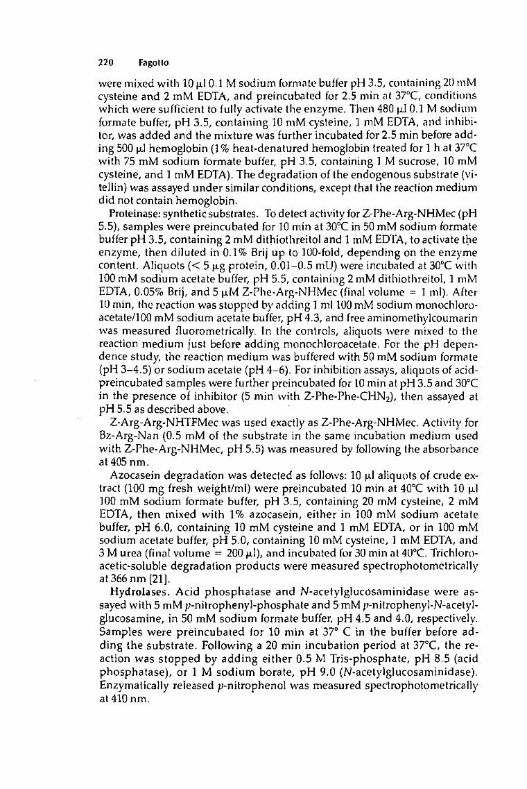
220 Fagotto
were mixed with lOjxlO.l M sodium formate buffer pH 3.5, containing 2OmM cysteine and 2 mM EDTA, and preincubated for 2.5 min at 370C, conditions which were sufficient to fully activate the enzyme. Then 480 u.1 0.1 M sodium formate buffer, pH 3.5, containing 10 mM cysteine, 1 mM EDTA, and inhibitor, was added and the mixture was further incubated for 2.5 min before adding 500 jxl hemoglobin (1% heat-denatured hemoglobin treated for 1 h at 370C with 75 mM sodium formate buffer, pH 3.5, containing 1 M sucrose, 10 mM cysteine, and 1 mM EDTA). The degradation of the endogenous substrate (vi-tellin) was assayed under similar conditions, except that the reaction medium did not contain hemoglobin.
Proteinase: synthetic substrates. To detect activity for Z-Phe-Arg-NHMec (pH 5.5), samples were preincubated for 10 min at 3O0C in 50 mM sodium formate buffer pH 3.5, containing 2 mM dithiothreitol and 1 mM EDTA, to activate the enzyme, then diluted in 0.1% Brij up to 100-fold, depending on the enzyme content. Aliquots (< 5 jxg protein, 0.01-0.5 mU) were incubated at 3O0C with 100 mM sodium acetate buffer, pH 5.5, containing 2mM dithiothreitol, 1 mM EDTA, 0.05% Brij, and 5 u.M Z-Phe-Arg-NHMec (final volume = 1 ml). After 10 min, the reaction was stopped by adding 1 ml 100 mM sodium monochloro-acetate/100 mM sodium acetate buffer, pH 4.3, and free aminomethylcoumarin was measured fluorometrically. In the controls, aliquots were mixed to the reaction medium just before adding monochloroacetate. For the pH dependence study, the reaction medium was buffered with 50 mM sodium formate (pH 3-4.5) or sodium acetate (pH 4-6). For inhibition assays, aliquots of acid-preincubated samples were further preincubated for 10 min at pH 3.5 and 3O0C in the presence of inhibitor (5 min with Z-Phe-Phe-CHN2), then assayed at pH 5.5 as described above.
Z-Arg-Arg-NHTFMec was used exactly as Z-Phe-Arg-NHMec. Activity for Bz-Arg-Nan (0.5 mM of the substrate in the same incubation medium used with Z-Phe-Arg-NHMec, pH 5.5) was measured by following the absorbance at 405 nm.
Azocasein degradation was detected as follows: 10 u.1 aliquots of crude extract (100 mg fresh weight/ml) were preincubated 10 min at 4O0C with 10 u,l 100 mM sodium formate buffer, pH 3.5, containing 20 mM cysteine, 2 mM EDTA, then mixed with 1% azocasein, either in 100 mM sodium acetate buffer, pH 6.0, containing 10 mM cysteine and 1 mM EDTA, or in 100 mM sodium acetate buffer, pH 5.0, containing 10 mM cysteine, 1 mM EDTA, and 3 M urea (final volume = 20OpLl), and incubated for 30 min at 4O0C. Trichloro-acetic-soluble degradation products were measured spectrophotometrically at 366 nm [21].
Hydrolases. Acid phosphatase and N-acetylglucosaminidase were assayed with 5 mM /;-nitrophenyI-phosphate and 5 mM /i-nitrophenyl-N-acetyl-glucosamine, in 50 mM sodium formate buffer, pH 4.5 and 4.0, respectively. Samples were preincubated for 10 min at 37° C in the buffer before adding the substrate. Following a 20 min incubation period at 37°C, the reaction was stopped by adding either 0.5 M Tris-phosphate, pH 8.5 (acid phosphatase), or 1 M sodium borate, pH 9.0 (N-acetylglucosaminidase). Enzymatically released /;-nitrophenoI was measured spectrophotometrically at410nm.
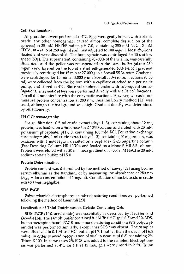
Tick Egg Acid Proteinase 221
Cell Fractionations
All procedures were performed at 40C. Eggs were gently broken with a plastic pestle (any other homogenizer caused almost complete destruction of the spheres) in 25 mM HEPES buffer, pH 7.0, containing 250 mM NaCl, 2 mM EDTA, at a ratio of 250 mg/ml and then adjusted to 100 mg/ml. Most chorions floated and were discarded. The homogenate was centrifuged for 15 s at low speed (50g). The supernatant, containing 70-80% of the vitellin, was carefully discarded, and the pellet was resuspended in the same buffer (about 250 mg/ml) and layered on the top of a 9 ml self-generated 60% Percoli gradient previously centrifuged for 15 min at 27,000 g in a Sorvall SS 34 rotor. Gradients were centrifuged for 15 min at 3,000 g in a Sorvall HB-4 rotor. Fractions (0.33 ml) were collected from the bottom with a capillary attached to a peristaltic pump, and stored at 4°C. Since yolk spheres broke with subsequent centri-fugations, enzymatic assays were performed directly with the Percoli fractions. Percoli did not interfere with the enzymatic reactions. However, we could not measure protein concentration at 280 nm, thus the Lowry method [22] was used, although the background was high. Gradient density was determined by refractometry.
FPLC Chromatography
For gel filtration, 0.5 ml crude extract (days 1-3), containing about 12 mg protein, was loaded on a Superose 6 HR 10/30 column and eluted with 20 mM potassium phosphate, pH 6.8, containing 100 mM KCL For cation-exchange chromatography, 1 ml crude extract (days 1-3), containing 50 mg protein, was oxidized with 1 mM HgCl2, desalted on a Sephadex G-25 Superfine column (Fast Desalting Column HR 10/10), and loaded on a Mono S HR 5/5 column. Proteins were eluted with a 20 ml linear gradient of 0-500 mM NaCl in 20 mM sodium acetate buffer, pH 5.0
Protein Determination
Protein content was determined by the method of Lowry [22] using bovine serum albumin as the standard, or by measuring the absorbance at 280 nm (^280 = for a concentration of 1 mg/ml). Contribution of nucleic acids in crude extracts was negligible.
SDS-PAGE
Polyacrylamide electrophoresis under denaturing conditions was performed following the method of Laemmli [23].
Localization of Thiol-Proteinases on Gelatin-Containing Gels
SDS-PAGE (10% acrylamide) was essentially as described by Heussen and Dowdle [24]. The sample buffer contained 0.1 M Tris-HCl (pH 6.8) and 2% SDS, but no mercaptoethanol. PAGE under nondenaturing conditions (8% Polyacrylamide) was performed similarly, except that SDS was absent. The samples were dissolved in 0.1 M Tris-HCl buffer, pH 7.1 (rather than the usual pH 6.8 value, in order to avoid precipitation of vitellin near its pi 6.8) containing 2% Triton X-100. In some cases 2% SDS was added to the samples. Electrophoresis was performed at 4°C for 4 h at 15 mA, gels were rinsed in 2.5% Triton

222 Fagolto
X-10Ü for 1 h at room temperature, transferred to 0.1 M sodium acetate buffer, pH 3.3, containing 1 mM dithiothreitol, and incubated at 370C for 3-4 h. At the end of the incubation period, gels were stained with Coomassie blue and destained in acetic acid/methanol.
Histochemistry
Eggs were dechorionated and fixed in 1% glutaraldehyde, 0.1 M sodium cacodylate, pH 7.4, 8% sucrose, rinsed in cacodylate buffer, and embedded without dehydratation in glycolmethacrylate (Historesin). Phosphatase reaction was performed on 3 p.m sections with naphthol-AS-BI-phosphate and hexa-zotized pararosaniline [25], except that 5% dimethyl sulfoxide was added to the reaction medium.
U
Electron Microscopy
Fractions from the Percoli gradients were fixed in 1% glutaraldehyde, 0.1 M sodium cacodylate, pH 7.4, containing 89c sucrose, rinsed in cacodylate buffer, postfixed in OsO4, dehydrated, and embedded in Spurr's resin. Thin sections were stained with uranyl acetate and lead citrate.
RESULTS Detection of an Acid Proteinase in Eggs
In the early developmental stages of O. incubata proteolytic activity toward vitellin was negligible under neutral or weakly acidic conditions. In contrast, when extracts were incubated at low pH (maximum pH 3.3), vitellin was hydrolyzed very efficiently into small peptides, as determined by proteinase assay (Fig. 1), SDS-PAGE (not shown), and gel filtration chromatography (Fig. 2). The extracts were similarly active against hemoglobin, in the presence of activators and inhibitors (see below). Thus I preferred to detect the proteinase with the latter, exogenous substrate.
The proteolytic activity is of maternal origin, since it was detected in mature oocytes and in freshly laid eggs. Proteolytic activity has been followed at the embryonic (days 1-10), larval (days 11-15), and nymphal stages of O. moubata (Fig. 3A). During embryonic development and the first days of larval life, the level was almost constant; following day 12 it decreased and reached a minimum at day 16. Only weak proteolysis could be detected in young first instar nymph.
These results were compared with two lysosomal hydrolases, acid phosphatase and N-acetylglucosaminidase, which were also present in the early embryo. However, the levels of these enzymes increased during development, from 1.6 to 2.7 nmol/min/mg for phosphatase, and from 1 to 6 nmol/min/mg for N-acetylglucosaminidase (Fig. 3C,D).
Subcellular Localization of the Proteolytic Activity
Yolk spheres were isolated from early embryos (days 1-3) using slow cen-trifugation followed by purification on a Percoli gradient. In spite of the mild conditions used, extensive breakage occurred during isolation. Typical yields were about 10%, as estimated by the amount of sedimented vitellin by meas-

Tick Egg Acid Proleinase 223
A: act iv i ty against endogenous substrate (vitel l in)
2.0 3.0 4.0 5 0 6 0 7.0 pH
B: act iv i ty against hemoglob in
100-1
a''
2.0 3.0 4.0 5.0 6-0 7,0 pH
C: act iv i ty against Z-Phe-Arg-NHMec
100-
~ 50 i 5° Qi
0 2.0 3.0 4.0 5.0 6.0 7.0
PH
Fig. 1. pH Dependence of the egg proteinase activity. Filled symbols: no preincubation; open symbols: preincubation at pH 3.5 for 5 min at 370C (A + B) or for 10 min at 3O0C (C). Circles = sodium formate (pH 2-4.5); squares = sodium acetate (pH 4-6); triangles = HEPES-NaOH (pH 6-7). Crude extracts (days 1-3) were assayed in the presence of a reducing agent (cysteine or dithiothreitol) as described in Materials and Methods. Activities are expressed as percentage of the maximal activity wi th each substrate.

O OO
EQH W O - '
eax Z C l -
SQ* 19 -
ea>i oft- -O -
O
>
< i . —
O
- T '
O O CJ
C
i "" J Ô
J.-à
"""O I P-'' j
A '9
P Ò •
fiO -C
— re
2 ;
«s i- .Q
* 3 « C QJ —
is < « w QJ
JS"S
x U QJ —
S * ai m
"Si e „ .2 re
"'S U i r ^-:.— CO
QJ 'S 60 c
y E S: m ^ > O
2.Ä re re
Q- D OJ u
t o
Qj Qj re
^i & sis' E u >
£ - t/1 t/l ™ -D -D > e re
™ br % o> K o 1« u. *-
2 S s o 3*5 • .E £ • U OJ
• o E .
: s. a? - f i ™
I * S "HO-SS O _L re QJ i: ^ QJ "O ^ ' Q J = > . = f i S "ôî - * 2 -_= .> re 2 « w C ~
.E e g. § H ^ ° §" O C . . * -Ì 2 U °
• s tt ~ -S a re o u
w e 3
O I S Q-Ï"? » O C eu ___- 'i3 W < S -D
r - o •= s

Tick Egg Acid Proteinase 225
N1
ir
-C O)
"cu 5
Dl E
E
3-
Dl <D
5 JT t/1 O)
O )
E H
Ol 'ci)
5
Ol E
E
A: PROTEINASE
BrACIDPHOSPHATASE
0 H — • — i — • — i — • — i — • — i — ' — i — • — i — ' — i — • — i — • — i — • — i
O 2 4 6- 8 10 12 1-4 16 18 20 days
C: N-ACETYLGLUCOSAMINIDASE 7-
6-
5-
4-
3-
2"
1 •
O 2 4 6 8 10 12 14 16 18 20 days
Fig. 3. Changes in hydrolytic activities during O. moubata development. Acid proteinase was assayed at pH 5.5 using Z-Phe-Arg-NHMec, in the presence of dithiothreitol (A, open circles) and at pH 3.5 using denatured hemoglobin, without reducing agent (A: filled circles), acid phosphatase at pH 4.5 wi th p-nitrophenyl-phosphate (B) and N-acetylglucosaminidase at pH 4.0 with p-nitrophenyl-N-acetylglucosamine (C) as described in Materials and Methods. The activities, expressed as mU/mg of fresh weight (see Table 2 for unit definit ion), are corrected to compensate for a progressive weight loss of eggs during development. E: embryo, L: larva, N I : first nymphal stage. Error bars indicate standard deviations.

226 Fagotto
1.12-
1 10-
£ i 08-f 1 05-S i o « :
1.02-
1.00-
ArGRADIENT
0 boltom
1 0 2 0 3 0
Iracitons YS LV
top
SN
6-
5-
E *• u> 3 .
2-
1 '
o-
ß: PROTEIN
I
\ /° \ f \^^J
0 bottom
10 20 (raclions
30 IOD
20i
lO-
CÌ: ACID PROTEINASE
r • • * * • * •» • *<
0 DOlIO^
10 2 0 3 0 IfACiO-1S
d ° 1 D: ACID PHOSPHATASE
3 0 , ErN-ACETYLGLUCOSAMINIDASE
DO:ÏO '•act o"f
Fig. 4. Localization of acid hydrolases on Percoli density gradient. Freshly laid eggs (day 1) were gently broken in isotonic buffer. The pellet of a first short centrifugation was applied to a self-generated 60% Percoli gradient. Acid proteinase was assayed at pH 5.5 using Z-Phe-Arg-NHMec. See Materials and Methods for other details. YS: yolk spheres fractions; LV: light vesicles fractions; SN: supernatant.
uring absorbance at 400 nm [18]. About 80% of the vitellin was in the supernatant of the first centrifugation and the rest was recovered on the top of the Percoli gradient.
All sedimentable activity, detected both with the fluorogenic substrate Z-Phe-Arg-NHMec (Fig. 4C) and hemoglobin (not shown), was localized in a single peak near the bottom of the gradient. The content was identified as yolk spheres, through the dark brownish color, as well as by examination of the fractions with a light microscope.
Fractions were examined for purity using electron microscopy; they appeared to exclusively contain yolk spheres of different sizes (10-50 ^m) (Fig. 5). Few unidentified contaminants were found, sometimes some pieces of unit membrane (probably plasma membrane) were still bound to the yolk spheres. The yolk spheres were intact, because O. moubata vitellin is soluble and damaged sphere would not sediment.
High activities of acid phosphatase and N-acetylglucosaminidase were also
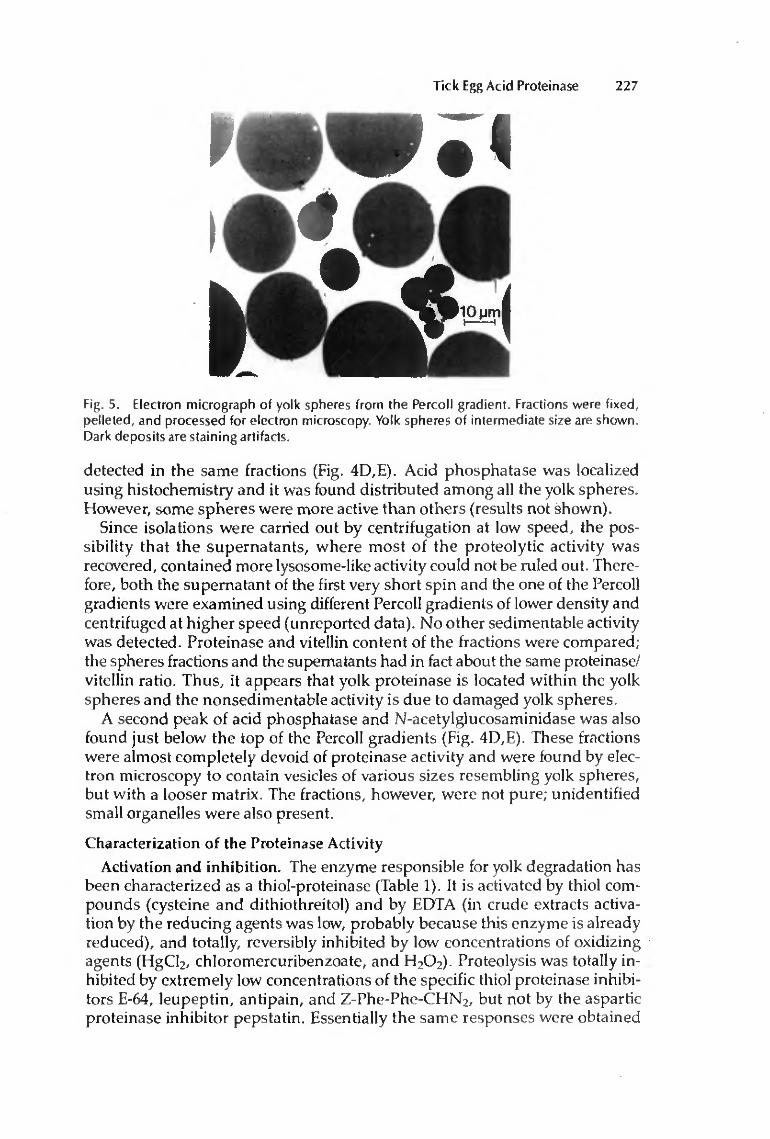
Tick Egg Acid Proteinase 227
Fig. 5. Electron micrograph of yolk spheres from the Percoli gradient. Fractions were fixed, pelleted, and processed for electron microscopy. Yolk spheres of intermediate size are shown. Dark deposits are staining artifacts.
detected in the same fractions (Fig. 4D,E). Acid phosphatase was localized using histochemistry and it was found distributed among all the yolk spheres. However, some spheres were more active than others (results not shown).
Since isolations were carried out by centrifugation at low speed, the possibility that the supernatants, where most of the proteolytic activity was recovered, contained more lysosome-like activity could not be ruled out. Therefore, both the supernatant of the first very short spin and the one of the Percoli gradients were examined using different Percoli gradients of lower density and centrifuged at higher speed (unreported data). No other sedimentable activity was detected. Proteinase and vitellin content of the fractions were compared; the spheres fractions and the supernatants had in fact about the same proteinase/ vitellin ratio. Thus, it appears that yolk proteinase is located within the yolk spheres and the nonsedimentable activity is due to damaged yolk spheres.
A second peak of acid phosphatase and N-acetylglucosaminidase was also found just below the top of the Percoli gradients (Fig. 4D,E). These fractions were almost completely devoid of proteinase activity and were found by electron microscopy to contain vesicles of various sizes resembling yolk spheres, but with a looser matrix. The fractions, however, were not pure; unidentified small organelles were also present.
Characterization of the Proteinase Activity Activation and inhibition. The enzyme responsible for yolk degradation has
been characterized as a thiol-proteinase (Table 1). It is activated by thiol compounds (cysteine and dithiothreitol) and by EDTA (in crude extracts activation by the reducing agents was low, probably because this enzyme is already reduced), and totally, reversibly inhibited by low concentrations of oxidizing agents (HgCl2, chloromercuribenzoate, and H2O2). Proteolysis was totally inhibited by extremely low concentrations of the specific thiol proteinase inhibitors E-64, leupeptin, antipain, and Z-Phe-Phe-CHN2, but not by the aspartic proteinase inhibitor pepstatin. Essentially the same responses were obtained

228 Fagotto
TABLE 1. Characterization of the Cathepsin L-Like Acid Proteinase of O. moubata Eggs
Relative activity
Z-Phe-Arg-NHMec* Vitellin1,
Concentration* CE Mono S CE
Activator Control1'
{reducing agent + EDTA)
Reducing agent EDTA No activator
h ibi tor1' Control1' E-64 Lcupeptin Anlipain Z-Phe-Phe-CHN2
Pepstatin PMSF HgCI2
HgCI2
H,0 2
2 m M / 1 0 m M d
I m M 2 m M / 1 0 m M d
I m M
1 fxM IM-M 2 M-M
l ( i M 10 M-M
1 mM 0.1 m M 1 mM 2 mM
100
70-90 55 40
100 0 0 1 3
90 75 5 0 0
100
70 5 5
100 0 0 1 3
105 70 0 0
—
100
95 80 75
100 0 0
— 10 85
— —
0 —
•'Crude extracts (CE) (days 1-3) and the activity peak from cation exchange chromatography (Mono S) were assayed using the synthetic substrate Z-Phe-Arg-NHMec; samples were first preincubatcd for 10 min at pH 3.5 and 300C for acid activation of the enzyme, then for a further 10 min at the same pH in the presence of various activators and inhibitors (5 min with Z-Phe-PhC-CHN2), and finally for 10 min wi th the substrate at pH 5.5 and 3O0C. ''CE were assayed for activity against endogenous substrate (vitellin); samples were preincubatcd for 2.5 min to activate the enzyme, then for a further 2.5 min in the presence of activators and inhibitors, and finally incubated for 30 min at pH 3.5 and 37°C. 1In crude extracts, 100% of activity in controls corresponds to a specific activity against Z-Phe-Arg-NHMec of about 400U/u-mol of active site. d2mM dithiothreitol wi th Z-Phe-Arg-NHMec and 10 mM cysteine wi th vitellin. ' Inhibit ion tests were performed in the presence of 1 mM EDTA and 2 mM dithiothreitol (Z-Phe-Arg-NHMec) or 10 mM cysteine (endogenous substrate), except when oxidizing agents (HgCI2
and H2O2) were used. In these cases the activity was compared wi th controls without reducing agent. E-64, lcupeptin, antipain, Z-Phe-Phe-CHN, pepstatin, and PMSF were dissolved in di-methylsulfoxide. The inhibitory effect of the solvent was negligible with the concentrations used. 'The final concentrations in the incubation medium are given.
toward activators and inhibitors using the synthetic substrate Z-Phe-Arg-NHMec at pH 5.5, the endogenous substrate (vitellin) at pH 3.5 (Table 1), hemoglobin at pH 3.5 and 5.5, or azocasein (pH 5-6) (not shown).
pH dependence and substrate specificity. Crude extracts were very active against endogenous and exogenous proteins at pH 3-4 (Fig. 1 A,B and Table 2). On the other hand, all substrates tested (endogenous substrate, hemoglobin, Z-Phe-Arg-NHMec, azocasein, Bz-Arg-Nan) were hardly hydrolyzed under less acidic conditions (pH 5-6). However, a large increase in activity was observed if the samples were first preincubated at low pH. In this case, hemoglobin and Z-Phe-Arg-NHMec were efficiently hydrolyzed over a broad pH range, while vitellin was still best degraded at low pH (Fig. 1).
At pH 3.5, activation was almost complete after 2 min at 370C, and after 10 min at 300C. Identical activation rates at pH 3.5 were observed when activity

Tick EgK Acid Proteinase 229
TABLE 2. Specific Activities of Egg Acid Proteinase with Different Substrates*
Substrate Concentration J*I_ Activity
(U/u-moi)-1
Z-Phe-Arg-NHMec Bz^Arg-Nan Z-Arg-Arg-NHTFMec Hemoglobin Endogenous substrate (vitellin) Azocasein Azocasein + 3 M urea
5u.M 5 m M 5 j tM 0.57r 0.1 ?c
\9c 17c
5.5 5.5 5.5 3.5 3.5 6.0 5,0
400 2.5 0
1000 330
15 27
"Crude extracts of early stages (days 1-3) were preincubated at pH 3.5 to activate the enzyme and then assayed as described in Materials and Methods. A reducing agent (dithiothreitol or cysteine) was present in all assays. '"•Activities are expressed in U/fxmol of active site. One unit corresponds Io 1 jimol of synthetic substrate (Z-Phe-Arg-NHMec, Bz-Arg-Nana, and Z-Arg-Arg-NHTFMec) degraded per min at 300C or 1 mg of protein degraded per min at 37°C (hemoglobin, vitellin) or at 403C (azocasein). Activities against hemoglobin and vitellin were measured bv determination of the tyrosine residues recovered in the trichloroacetic acid supernatant (see Materials and Methods). Corresponding activities as mg of protein degraded have been calculated from the known tyrosine content and molecular weight data. For vitellin an A1r of 600,000 and a relative content of 50 tyrosine residues per 1000 amino acids are given [18], or about 270 tyrosine residues per molecule of vitellin. Active site concentration was estimated bv titration with E-64.
at pH 5.5 was detected with either hemoglobin or Z-Phe-Arg-NHMec. Vitellin and hemoglobin degradations were usually measured at pH 3.5 and at 37°C. Since activation was almost immediate under these conditions, no preincubation was required. However, preincubation was necessary in inhibition assays to ensure that enzyme activity, but not activation, was inhibited. Samples were routinely preincubated at pH 3.5 when activity was measured at higher pHs.
Vitellin degradation was maximal at an endogenous substrate concentration of about 1 mg/ml (Table 2). At higher concentrations, the activity decreased, probably because of substrate or product inhibition (unreported data). Hemoglobin degradation was always detected in the presence of excess exogenous substrate (5 mg/ml). Interference from endogenous substrate could not be precisely quantified, but was estimated to be rather low under the conditions used (endogenous protein concentration = 0.02-0.5 mg/ml).
Z-Phe-Arg-NHMec, known to be a good synthetic substrate for the two lysosomal enzymes cathepsin B and L, was hydrolyzed very efficiently; however negligible activity was found with Bz-Arg-Nan and Z-Arg-Arg-NHTFMec, which are split by cathepsin B but not cathepsin L [21] (Table 2). Azocasein degradation was detected both at pH 6.0 and in the presence of 3 M urea at pH 5.0, to compare with previously published data [21 ]. In the presence of urea, the activity was doubled (Table 2). Thus the enzyme behaves like cathepsin L but not cathepsin B, which is strongly inhibited under such conditions.
Both activities against Z-Phe-Arg-NHMec and hemoglobin were localized in yolk spheres, comigrated in ion-exchange chromatography (Fig. 6), and their levels during development followed a similar pattern (Fig. 3A), strongly suggesting that both substrates are degraded by the same enzyme.
Active-Site Titration. Since E-64 is a very strong and specific irreversible inhibitor of cysteine proteinases, it allows active-site titrations [21]. An attempt

23 U Fagotto
fractions
Fig. 6. Cation-exchange chromatography of a crude egg extract (days 1-3) on mono 5 FPLC column. O Proteinase activity against Z-Phe-Arg-NHMec {pH 5.5); # proteinase activity against hemoglobin {3.5) (arbitrary units); protein elution profile (A1Hn); NaCI concentration as percentage of 1 M.
to determine the proteinase active molar concentration was undertaken. Usage of crude extracts with high concentrations of endogenous substrate did not give satisfactory results during short time incubations. However, preincubation of samples for 5 h at 37°C and pH 3.5 (under which conditions most of the vitellin was degraded) resulted in a linear response and enzyme concentration was estimated at about 5 nmol/g of fresh weight.
Chromatographic and electrophoretic behaviour. Acid proteinase, assayed with both hemoglobin and Z-Phe-Arg-NHMec, bound to a cation-exchange column (Mono S) at pH 5-6. When eluted at pH 5.0, both activities were recovered in a single peak at 340 mM NaCl (Fig. 6). When chromatographed at pH 5.5, both activities still coeluted, at 290 mM NaCl (unreported data), further supporting the assumption that they are due to a single enzyme. When assayed at pH 5.5, the chromatographed enzyme was latent and required activation at lower pH. Sensitivity of the active enzyme toward activators and inhibitors was similar to crude extract enzyme (Table 1), and the same substrate was found, since Z-Arg-Arg-NHTFMec was not hydrolyzed.
Gel filtration (Superose 6) separated the activity into two peaks (Fig. 2A), one with a low Mx of about 30,000-40,000 and the other with a very high M1
(400,000-600,000). The lower peak probably corresponds to free proteinase, while recovery of large amounts of activity in the higher peak suggests that proteinase binds to vitellin. When samples were preincubated at pH 3.5 before chromatography, some activity showed a higher apparent Mx (Fig. 2B,C), probably because proteinase was bound to vitellin oligomers, which appeared at these conditions.
The gelatinolytic activity was localized on PAGE under nondenaturing conditions (Fig. 7a), after incubating the gels at pH 3.3. A strong spot of gelatin digestion was found associated with the two close bands of vitellin (monomer and dimer) that had just entered the separating gel, and a weaker band was
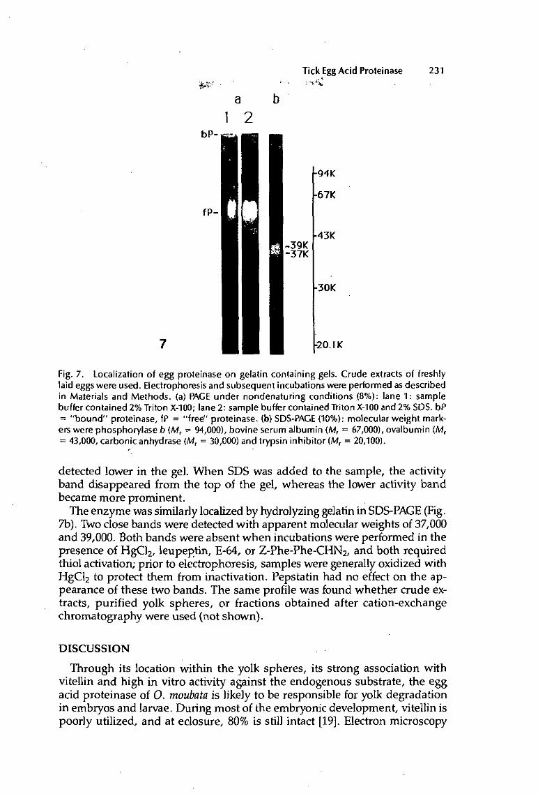
Tick Egg Acid Proteinase 231
a b
1 2
fP-
J-39K
94K
67K
43K
[-3OK
•20. IK
Fig. 7. Localization of egg proteinase on gelatin containing gels. Crude extracts of freshly laid eggs were used. Electrophoresis and subsequent incubations were performed as described in Materials and Methods, (a) PAGE under nondenaturing conditions (8%): lane 1 : sample buffer contained 2% Triton X-100; lane 2: sample buffer contained Triton X-100 and 2% SDS. bP = " b o u n d " proteinase, fP = " f ree" proteinase, (b) SDS-PAGE (10%): molecular weight markers were Phosphorylase b (M1 = 94,000), bovine serum albumin (MT = 67,000), ovalbumin (Mf
= 43,000, carbonic anhydrase (M r = 30,000) and trypsin inhibitor {Mr = 20,100). r
detected lower in the gel. When SDS was added to the sample, the activity band disappeared from the top of the gel, whereas the lower activity band became more prominent.
The enzyme was similarly localized by hydrolyzing gelatin in SDS-PAGE (Fig. 7b). Two close bands were detected with apparent molecular weights of 37,000 and 39,000. Both bands were absent when incubations were performed in the presence of HgCl2, leupeptin, E-64, or Z-Phe-Phe-CHN2, and both required thiol activation; prior to electrophoresis, samples were generally oxidized with HgCl2 to protect them from inactivation. Pepstatin had no effect on the appearance of these two bands. The same profile was found whether crude extracts, purified yolk spheres, or fractions obtained after cation-exchange chromatography were used (not shown).
DISCUSSION
Through its location within the yolk spheres, its strong association with vitellin and high in vitro activity against the endogenous substrate, the egg acid proteinase of O. moubata is likely to be responsible for yolk degradation in embryos and larvae. During most of the embryonic development, vitellin is poorly utilized, and at eclosure, 80% is still intact [19]. Electron microscopy

232 Fagotto
studies showed that only a restricted number of yolk spheres are individually digested (unpublished observations). Egg acid proteinase is stored within the quiescent yolk spheres, and for the most part it is probably used only at the end of embryonic development, when yolk degradation is significantly increased. Decrease in proteolytic activity occurs near the end of the intense phase of yolk degradation and during the subsequent nymphal stage very slow utilization of vitellin is correlated with a low level of proteinase. It is not known whether the residual activity in nymphs is still of maternal origin. However, new synthesis of lysosomal enzymes in larval and nymphal tissues is probable. Likewise, it is not possible to follow the fate of the maternally inherited acid phosphatase and /V-acetylglucosaminidae, since they cannot be distinguished from the newly synthesized ones.
Neutral proteinase has been detected in crude extracts, after day 4 (unpublished observations). This enzyme specifically hydrolyzes one subunit of vitellin into two large fragments. Its molecular properties, cellular localization, and function have not been elucidated and it is not know whether it participates in yolk degradation.
Low speed centrifugation in Percoli gradients allowed to achieve a proper purification of yolk spheres with minimal disruption. Nevertheless most of the yolk spheres content (about 90%) was recovered in the supernatants, making quantitative studies unfeasible. The results reported here, however, showed that all proteinase activity is probably associated with the yolk, which also contains other lysosomal hydrolases.
For acid phosphatase histochemistry, the lead-glycerophosphate method [25] was ineffective on the preparations, and naphthol-AS-BI-phosphate only reacted when dimethyl sulfoxide was added to the medium. Accessibility to the enzyme was probably reduced, because of the extremely dense yolk matrix, which might contribute to earlier negative results [26].
Ineffectiveness of pepstatin clearly indicates that cathepsin D-like enzymes do not contribute to yolk degradation in O. moubata. Likewise, EDTA-sensitive metalloproteinases are not involved (Table 1). Proteolytic activity is modulated with inhibitors and activators specific for cysteine proteinases. Classical lyso-somes contain three main thiol-endopeptidases: cathepsins B, H, and L. Cathepsin H has only a weak endopeptidase activity and does not split Z-Phe-Arg-NHMec [21]. Therefore it does not have the properties of the enzyme studied. No specific substrate or inhibitor is known for cathepsin L. It can be distinguished, however, from cathepsin B [21] as follows: (1) azocasein degradation by cathepsin L is enhanced in the presence of urea, whereas cathepsin B is strongly inhibited, (2) low concentrations of Z-Phe-Phe-CHN2 inhibits cathepsin L faster [27], (3) cathepsin L has a higher affinity for Z-Phe-Arg-NHJVIec, but is unable to hydrolyze Bz-Arg-Nan or Z-Arg-Arg-NHTFMec, which are readily degraded by cathepsin B, and (4) cathepsin L does not readily hydrolyze synthetic substrates, but it is a very powerful endopeptidase when assayed with proteins. When these criteria were examined, egg acid proteinase of O. moubata appeared to be a cathepsin L-like enzyme. The estimated enzyme concentration (5 nmol/g) is high compared to vertebrate tissues [28].
Specific activities for different synthetic and protein substrates are given in Table 2. Values found for the hydrolysis of synthetic substrates and azocasein

Tick Egg Acid Proteinase 233
may be underestimated, because of endogenous proteins; crude extracts act as inhibitors under mildly acidic conditions (unpublished observations). The rate of protein hydrolysis depends on substrate denaturation. Hemoglobin is a relatively small protein which has been fully denatured prior to incubation, and which can thus be easily degraded. In contrast, vitellin is a large protein and acidic conditions are probably not sufficient for complete dissociation and denaturation. Thus, only a small part of this protein may be accessible. In addition, sugar and lipid molecules that are associated with vitellin could also hinder proteolysis. Therefore, the activity measured with an exogenous substrate does not necessarily reflect that measured with the natural substrate. Moreover, proteinase may behave quite differently in vivo, since in the yolk spheres both vitellin and enzyme are highly concentrated (more than 200 mg/ml and about 5 |xM, respectively).
Gel filtration chromatography (Fig. 2) and PAGE under nondenaturing conditions (Fig. 7a), demonstrated that most of the proteinase activity is tightly bound to vitellin. This binding was not affected by high salt concentrations (up to 400 mM NaCI on gel filtration, unpublished) or neutral detergents (Triton X-100, native PAGE), whereas SDS readily dissociated this complex (Fig. 7a). Strong association may play a role in regulating proteinase activity. New evidence indicates that the proteinase is present in eggs as a proenzyme, which is processed during acid treatment [29].
Activity on gelatin/SDS-PAGE appeared as two bands of MT 37,000 and 39,000. This is in agreement with the apparent Mx of the "free" proteinase estimated from gel filtration (Fig. 2). This molecular weight is similar to many other cysteine proteinases, including those of arthropod eggs (see below), but classical cathepsin L isolated from vertebrates is smaller. However, 39,000-41,000 D precursor forms of cathepsins B and L have been recently described in mammals [30,31]. It is not known whether the two bands represent various forms of the same enzyme or different enzymes. It is highly probably that we are dealing with a single enzyme, because both bands were equally sensitive to activators and inhibitors, including Z-Phe-Phe-CHN2- Furthermore, both were coeluted on cation exchange chromatography, and are processed during acid treatment [29].
Comparison with other species is somewhat hampered because different substrates and pH were used. However, in the eggs of Bombyx mori [13,15], Drosophila melanogaster [7], Artemia salme [10,12] and O. moubata, there is a predominant acid cysteine proteinase, inhibited by thiol reagents, Ieupeptin, and E-64. Pepstatin was an ineffective inhibitor, indicating the absence of cathepsin D-like enzymes. The embryonic enzymes efficiently hydrolyze vitellin as well as exogenous proteins. They generally bind to high M1. protein, probably vitellin itself, and have an M1. of 40,000-68,000. Artemia [12] proteinase hydrolyzes Pro-Phe-Arg-Nan more efficiently than Z-Arg-Arg-MeNap, which is a specific cathepsin B substrate. Since Pro-Phe-Arg-Nan closely resembles Z-Phe-Arg-NHMec, it also can be assumed that Artemia enyzme is a cathepsin L-like enzyme. Activation was reported in Drosphiïa [7], where in early stages the activity is strongly enhanced by trypsin treatment. In Artemia [12] all assays were carried out at pH 3.5, thus latency under mild acidic conditions may have been overlooked. In Bombyx [13] no hydrolysis of different
234 Fagotto
synthetic substrates at pH 5-6 could be shown. The authors suggested that the enzyme was different from other known cysteine proteinases. However no pretreatment at low pH was attempted. One can hypothesize that in all these species, as well as many others [13], cathepsin L is present in the eggs and is responsible for yolk digestion.
Another similarity between all these species is the presence of a neutral proteinase [7,14-16]. Such a neutral proteinase has also been detected in O. moubata embryos. These proteinases have specificity toward special yolk protein [15] or may act on a single subunit of vitellin [14]. These enzymes perhaps activate yolk degradation, by splitting yolk proteins into fiagments which are then accessible for hydrolysis by endo- and exopeptidases. Alternately, they may down-regulate degradation by producing vitellin fragments with inhibitory properties [34,35].
Yolk spheres of O. moubata eggs function as lysosomal-like organelles, containing all the machinery they need to degrade the huge amount of vitellin they have accumulated during vitellogenesis.
LITERATURE CITED
Ì. Wallace RA: Vitellogenesis and oocvle growth in nonmammalian vertebrates, !n: Developmental Biologv: A Comprehensive Synthesis. Browder VVL, ed. Plenum Press, New York, voli, pp 127-166(1985).
2. Pasteeis J]: Les corps multivésiculaires de l'oeuf de Bnrnca candida (Mollusque bivalve) étudiés au microscope électronique. Activité phosphatasique et accumulation de rouge neutre. J Embryol Exp Morphol 16, 301 (1966).
3. Krischer KN, Chambers EL: Proteolytic enzvmes in sea urchin eggs: Characterization and activity before and after fertilization. J Cell Physiol 76, 23(1970).
4. Schuel H, Wilson WL, Wilson JR, Bressler RS: Heterogenous distribution of "lysosomal" hydrolases in yolk platelets isolated from unfertilized sea urchin eggs by zonal centrifuga-tion. Dev Biol 46, 404 (1975).
5. Vernier J-M, Sire M-F: Plaquettes vitellines et activité hydrolasique acide au cours du développement embryonnaire de la Truite arc-en-ciel. Etude ultrastructurale et biochimique. Biol Cell 29, 99 (1977).
6. Wall DA, Meleka I: An unusual iysosome compartment involved in vitellogenin endocytosis by Xetwpits oocytes. J Cell Biol JOJ11651 (1985).
7. Medina M, Leon P, Vallejo CG: Drosophila cathepsin B-Iike proteinase: a suggested role in yolk degradation. Arch Biochem Biophys 263(2), 355 (1988).
8. Armant DR, Carson DD, Decker GL, WeIpIy]K, Lennarz WJ: Characterization of yolk platelets isolated from developing embryos of Arlxicia puncttilnta. Dev Biol 113, 342 (1986).
9. Decroly M, Goldfinger M, Six-Tondeur N: Biochemical characterization of lysosomes in unfertilized eggs of Xenopus laevis. Biochim Biophys Acta 557, 567 (1979).
10. Perona R, Vallejo CG: Acid hydrolases during Artemia development: A role in yolk degradation. Comp Biochem Physiol 81B(A), 993 (1985).
11. Pasteels J]: Yolk and lysosomes. In: Lysosomes in Biology and Pathology. Dingle JT, ed. North-Holland, Amsterdam, vol 3, pp 216-233 (1973).
12. Perona R, Vallejo CG: The lysosomal proteinase of Artemia. Purification and characterization. EurJ Biochem 124, 357(1982).
13. Kageyama T, Takahashi SY, Takahashi K: Occurrence of thiol proteinases in the eggs of the silkworm, Bomln/x mori. J. Biochem 90, 665 (1981).
14. Ezquieta B, Vallejo CG: The trypsin-like proteinase of Artemia: Yolk localization and developmental activation. Comp Biochem Physiol 82B, 731 (1985).
15. Indrasith LS, Sasaki T, Ya mash ita O: A unique protease responsible for selective degradation of a yolk protein in Bomln/x mori. Purification, characterization and cleavage profile. J Biol Chem 263 (2), 1045 (1988).

Tick Egg Acid Proteinase 235
16. Yamashita O, Indrasith LS: Metabolic fates of yolk proteins during embryogenesis in Arthropods. Dev Growth Differ 30(4), 337(1988).
17. Fagotto F, Hess E, Aeschlimann A: The early development of the argasid tick, Orniihodoros moubato. Entomol Gen 13,1 (1988).
18. Chinzei Y, Chino H, Takahashi K: Purification and properties of vitellogenin and vitellin from a tick, Ornitlwdoros moubata.J Comp Physiol 152,13 (1983).
19. Chinzei Y, Yano I: Vitellin is a nutrient reserve during starvation in the nymphal stage of a tick. Experientia 41,948 (1985).
20. Mednis A, Remold HG: A sensitive fluorometric assay for the determination of cathepsin D. Anal Biochem 49,134 (1972).
21. Barrett AJ, Kirschke H: Cathepsin B, cathepsin H, cathepsin L. Methods Enzymol 80, 535(1981).
22. Lowry OH, Rosebrough NJ, Farr AL, Randall RJ: Protein measurement with the FoHn phenol reagent. J Biol Chem 193, 265 (1951).
23. Laemmli UK: Cleavage of structural proteins during assembly of the head of bacteriophage T4. Nature (London) 227, 680(1970).
24. Heussen C, Dowdle EB: Electrophoretic analysis of plasminogen activators in Polyacrylamide gels containing SDS and copolymerized substrates. Anal Biochem 102,196(1980).
25. Bancroft JD: Enzyme histochemistry. In: Theory and Practice of Histological Techniques, Bancroft JD, Stevens A, eds. Churchill Livingstone, Edinburgh, pp 379-405 (1982).
26. Lemanski LF, Aldoroty R: Role of acid phosphatase in the breakdown of yolk platelets in developing amphibian embryos. J Morphol 153, 419 (1977).
27. Kirschke H, Shaw E: Rapid inactivation of cathepsin L by Z-Phe-Phe-CHN2 and Z-Phe-Ala-CHN2. Biochem Biophys Res Commun 101, 454 (1981).
28. Mason RW, Taylor MAJ, Etherington DJ: The purification and properties of cathepsin L from rabbit liver. Biochem J 217, 209 (1984).
29. Fagotto F: YoIk degradation in tick eggs. II Evidence that cathespin L-like proteinase is stored as a latent, acid-activable proenzyme. Arch Insect Biochem Physiol. 14, 237 (1990).
30. Nishimura Y, Kawabata T, Kato K: Identification of latent procathepsin B and L in microsomal luman: characterization of enzymatic activation and proteolytic processing in vitro. Arch Biochem Biophys 261(1), 64 (1988).
31. Mort JS, Recklies AD: Interrelationship of active and latent secreted human cathepsin B precursors. Biochem J 233, 57(1986).
32. Kuk-Meiri S, Lichtenstein A, Shulov A, Pener MP: Cathepsin-Hke proteolytic activity in the developing eggs of the African migratory locust {Locusta migratoria migratorioides R&F). Comp Biochem Physiol 18, 783 (1966). .
33. Yokota Y, Kato KH: Degradation of yolk proteins in sea urchin eggs and embryos. Cell Differ 23,191(1988).
34. Salisbury N, Calaprice N, Triplett EL: Amphibian embryo protease inhibitors. VI. Maternal origin and identity with lipovitellin heavy subunit. Cell Differ 9, 219 (1980).
35. Ezquieta B, Vallejo CG: Lipovitellin inhibition of Artemia trypsin-Üke proteinase: A role for storage protein in regulating proteinase activity during development. Arch Biochem Biophys 250(2),410(1986).

Il

Archives of Insect Biochemistry and Physiology 14:237-252 (1990)
YoIk Degradation in Tick Eggs: IL Evidence That Cathepsin L-Like Proteinase Is Stored as a Latent, Acid-Activable Proenzyme François Fagotto institut de Zoologie Université de Neuchâtel, Neuchâtel, Switzerland
Cathepsin L—tike proteinase found in the eggs of the tick Ornithodoros moubata is latent during embryogenesis, but can be activated by acid treatment. In crude extracts as well as in partially purified fractions, activation requires reducing conditions and is inhibited by leupeptin, which indicates that it is mediated by a thiol proteinase, probably by the cathepsin L itself. Latency disappears in vivo at the time of the acute phase of yolk digestion, which lakes place during late embryonic development and larval life. When egg cathepsin L is localized through its gelatinolytic activity on SDS-PACE, the activated enzyme migrates as lower Mr bands than the latent form. Disappearance of the higher Mr bands corresponding to the latent form is directly related to appearance of the lower Mr bands characteristic of the active one; transition from one paticrn to the other and enzymatic activation are in perfect agreement with regard Io kinetics and sensitivity to inhibitors. The same pattern change occurs in vivo, parallel to latency removal and intense yolk degradation. These results strongly suggest that egg cathepsin L is stored in the yolk as a proenzyme which is activated by partial proteolysis at low pH.
Key words: Ornithodoros moubata, embryogenesis procathepsin L, precursor processing, vilellin degradation, acidification
INTRODUCTION
Yolk spheres (granules, or platelets) are large, lysosome-like, maternally inherited organelles which are quiescent during at least part of the embryonic development. Since the early investigations of Pasteels [1], who first postulated the lysosomal nature of the yolk platelets and their "dormancy," few attempts have been made to understand the cellular mechanisms that govern
Acknowledgments: I express my gratitude to Dr. P. Schürmann for critical discussions and valuable advice and to Dr. R Lösel for careful reading of the manuscript. Part of these results have been already published in an abstract form 136]. This work is part of the Ph.D. thesis of the author.
Received December22,1989; accepted May 25,1990.
Address reprint requests to François Fagotto, Institut de Zoologie, Université de Neuchâtel, Chantemerle 22, CH-2000 Neuchâtel.
«y 1990WiIeY-LiSSJnC.

238 Fagotto
their degradation. The studies published to date have focused either on the modifications the yolk undergoes, both ultrastructurally [2-6] and biochemically [2,5,7-9], or on the identification of putative enzymes such as specific proteinases [10-12]. However, these reports are primarily descriptive and almost no evidence about the underlying mechanisms was presented. Vitellin fragments have been reported to act as proteinase inhibitors in amphibian eggs [13,14] and Artemia eggs [15], but their involvement in the regulation of digestive activities has not been demonstrated. Yokota and Kato [5] and Scott et al. [16] mentioned that acidic pH may be involved in yolk degradation in sea urchins, and yolk degradation is inhibited by lysosomotropic agents in Artemia [V] and Drosophila [11] eggs. In vitro activation through tryptic cleavage has been reported for proteinases in Drosophila eggs [11].
Similar studies have been undertaken with the eggs of the African soft tick Ornithodoros mouhata: the main yolk protein has been characterized [18], its degradation quantified [19]. This laboratory recently reported the occurrence of a cathepsin L-like proteinase in the yolk spheres [20], which degraded vitellin very efficiently (pH optimum 3.5). The enzyme was present in large amounts from the beginning and during most of the embryonic development, but subsequently diminished rapidly in the larva, during the period of intense yolk degradation. The proteinase was latent when assayed under mildly acidic conditions (pH 4.5-6), but was highly activated by preincubation at lower pH(pH3-4).
Since latency could be crucial in the regulatory process, the characteristics of the in vitro acid activation have been examined and compared with what happens in vivo, during the embryonic and larval development. The activated enzyme displayed increased electrophoretic mobility/apparently due to partial proteolysis under acidic conditions. The in vitro acid activation closely mimics in vivo activation and processing, which occur at the end of the embryonic development, during the more intense phase of yolk digestion. For the first time evidence is presented that can account for both "dormancy" and activation of the yolk spheres.
MATERIALS AND METHODS Chemicals
Z-Phe-Arg-NHMecHCl,* E-64, leupeptin, pepstatin A, and bovine serum albumin were purchased from Sigma Chemical Company (St. Louis, MO, USA); dithiothreitol and p-chloromercuribenzoate were obtained from Fluka Chemie AG (Buchs, Switzerland); and gelatin came from Difco Laboratories (Detroit, Ml, USA). Z-Phe-Phe-CHN2 was purchased from Bachern AG (Dub-endorf, Switzerland); molecular weight markers for electrophoresis and all
'Abbreviations used: E-64 = N-|N-(DL-3-transcarboxyiran-carbonyl)-L-leucyliagmatine; FPLC = Fast performance liquid chromatography; leupeptin = N-acetylleucyl-leucyl-arginat; pepstatin A = isovaleryl-L-valyl-L-valyl-(3S,4S)-4-amino-3-hydroxy-6-methylhepiantoyl L-alanyl-(3S,4S)-4-amino-3-hydroxy-6-methylheptanoicacid; SDS = sodium dodecyl sulfate; Z-Phe-Arg-NHMec = benzoyloxycarbonyl-phenylalanyl-arginyl-7-amido-4-methylcoumarin; Z-Phe-Phe-CHN2 = benzoyloxycarbonyl-phenylalanyl-phenylalanyl-diazomethylketone.

Tick Egg Procathepsin L Activation 239
FPLC material came from Pharmacia Fine Chemicals AB (Uppsala, Sweden). All other chemicals were of analytical grade.
Preparation of Crude Homogenates
Mated females of O. moubata were artificially fed on swine's blood and allowed to oviposit at 29°C. The eggs were incubated at the same temperature. Under these conditions, larvae hatched after IO days and molted into nymphs at day 15. The eggs were routinely homogenized at 100 mg fresh weigh/ml in 10 mM Tris-HCi, pH 7.2, and centrifuged for 15 min at 40,000¾ and 40C. The pellet was discarded and the supernant was frozen and stored at - 180C. Protein content of the supernatant was about 25 mg/ml.
To detect both latent and active proteinase during development, 10 embryos (days 1 and 7), larvae (days 11 and 13), or young nymphs (day 16) were broken up in 100 ^l 10 mM sodium acetate buffer, pH 5.5. Protein concentration was about 12 mg/ml in homogenates of embryonic stages. Since active proteinase is unstable, it was assayed as soon as possible after homogenization. Thus centrifugation was omitted, and aliquots were immediately diluted and treated as follows: 10-fold dilution in 400 mM sodium acetate buffer, pH 5.5, containing 4 mM EDTA, 4 mM dithiothreitol, with a 1 min incubation at 300C (untreated), or 10-fold dilution in 100 mM sodium formate buffer, pH3.5, containing 2 mM EDTA, 1 mM dithiothreitol, with a 10 min incubation at 3O0C (acid pretreated). All samples were subsequently assayed against Z-Phe-Arg-NHMecat pH 5.5. At least three different batches of each stage were tested both for active proteinase (untreated) and for total, i.e., active plus latent proteinase (acid pretreated). For electrophoretic analysis, 10 embryos, larvae or nymphs, were homogenized at selected Stages in either 50 fil 10 mM sodium acetate buffer, pH 6.0, containing 2 mM EDTA and 1 mM HgCl2, or 50 uJ 10 mM Tris-HCI buffer, pH 6.8, containing 10 p,M leupeptin, and stored frozen. Thawed samples were mixed with five times their volume of cold sample buffer, centrifuged at 10,00O1If for 2 min at room temperature, and 7.5 fxl aliquots of the supernatant, containing about 30 pig protein, were loaded on to the gel.
Preparation of Yolk Spheres
YoIk spheres from early stages (day 1) were purified on a Percoli gradient [20]. All steps were performed at 40C. About 100 mg freshly laid eggs (25 mg protein) were mildly homogenized in 1 ml 25 mM Hepes-NaOH buffer, pH 7.0, containing 250 mM NaCI and 1 mM EDTA. Most chorions floated and were discarded. After a 30 s centrifugation at 5Og, the supernatant was discarded and the pellet was resuspended in 1 ml of the same buffer. The suspension was layered on a 9 ml 60% Percoli gradient, buffered with 25 mM Hepes-NaOH, pH 7.0, containing 250 mM NaCl and 1 mM EDTA, and previously centrifuged for 20 min at 15,000^ in a Sorvall SS-34 rotor. The gradient was centrifuged for 15 min at 5,000# in a Sorvall HB-4 rotor; 0.33 ml fractions were collected from the bottom with a capillary connected to a peristaltic pump and were stored frozen until used. Purified, intact yolk spheres were recovered in 3-4 fractions near the bottom of the gradient (density = 1.12, determined by refractometry). The yield was about 10% as estimated by the relative amount of sedimented vitellini (measured spectrophotometrically at
240 Fagotto
400 nm [18]). The two most concentrateci fractions (3-5 mg protein/ml) were stored at -180C.
Cation Exchange Chromatography
A Mono S HR 5/5 column on a FPLC system was used as advised by the supplier. A 0.5 ml crude homogenate (days 1-3), containing 12 mg protein, was oxidized by addition of 1 mM HgCl2, desalted on a Sephadex G25 superfine column (Fast Desalting Column HR10/10), and immediately loaded on to the Mono S column. Oxidation was needed to prevent slow, progressive transition from the inactive to the active, unstable form at pH 5.0. Proteins were eluted with a 20 ml linear gradient of 0-500 mM NaCl in 20 mM sodium acetate buffer, pH 5.0, and the fractions (0.5 ml) were assayed for Z-Phe-Arg-NHMec hydrolysis. The activity eluted essentially as a single peak at 340 mM. The three most active fractions were stored frozen until further use. Specific activity of these fractions (30-70 mU/mg protein) increased 4-9-fold compared to the crude homogenate (8 mU/mg protein). Protein content was 0.3-0.6 mg/ml.
Acid Pretreatment of the Samples
In order to study the in vitro proteinase activation, samples were prein-cubated under various conditions and subsequently assayed with Z-Phe-Arg-NHMec or analyzed by gelatin/SDS-PAGE. Acid activation was achieved by incubating the samples (crude homogcnntes, purified yolk spheres, or fractions from ion exchange chromatography) for 10 min at 3O0C with an equal volume of 0.1 M sodium formate, pH 3.5. In control experiments, samples were incubated with an equal volume of 0.1 M Tris-HCl, pH 6.8; no activation could be detected enzymatically under these neutral conditions. Unless mentioned, 2 mM dithiothreitol and 1 mM EDTA were present in all experiments. When required, reversible proteinase inhibitors (0.5-20 uJvl leupeptin, 10 fj.M pepstatin A, 0.5 mM HgCl2, or 0.5 mM r>-chloromercuribenzoate) were added, either before or at the end of the preincubation. When HgCl2 or /J-chloromer-curibenzoatc was used, dithiothreitol was omitted. For electrophoretic studies, addition of 5-20 u-M leupeptin to the samples did not interfere with the activity after electrophoresis. When the effect of leupeptin on the acid activation process was studied enzymatically, samples were preincubated for 10 min at pH 3.5 or 3.75 with 0.5 JJLM inhibitor. The final concentration of leupeptin in the enzyme reaction medium was 0.1-0.2 nM. Control activity was largely preserved (>90%) when the same amount of inhibitor was added at the end of the acid activation period. Higher concentrations could not be used, since activity was then strongly inhibited in controls.
For enzyme assays, preincubated samples were diluted up to 100-fold with 0.1% Brij, depending on the enzyme content, and aliquots were assayed as described below. For analysis on gelatin/SDS-PAGE, 10 u,l preincubated samples were mixed with 20-50 ^l cold sample buffer and kept on ice; 5-10 \L\ aliquots, containing 1-30 jxg protein (0.05-0.25 mU proteinase activity with Z-Phe-Arg-NHMec), were loaded on the gels.

Tick Egg Procathepsin L Activation 241
Proteinase Assay
Cathepsin L-Iike activity was assayed at pH 5.5 by using the synthetic fluorogenic substrate Z-Phe-Arg-NHMec [21]. Aliquots [10-20 u,l] containing about 0.1-0.5 mU proteinase activity (<5 ^g protein) were mixed with 100 mM sodium acetate buffer, pH 5.5, containing 0.05% Brij, 2 mM dithiothreitol, 1 mM EDTA, and 5 jxM Z-Phe-Arg-NHMec (final volume = 1 ml), and incubated at 300C for 10 min. The reaction was stopped by addition of 1 ml 100 mM sodium monochIoroacetate/100 mM sodium acetate pH 4.3 and free amino-methylcou marin was measured fluorometrically. All assays were performed at least in triplicate.
Gelatin/SDS-PAGE
Gelatin/SDS-PAGE was essentially prepared as described by Heussen and Dowdle [22]. Separating gels (10% acrylamide) contained 0.1% copolymerized gelatin. Two systems of gel electrophoresis were used. The first one was the alkaline SDS-PAGE of Laemmli [23] (Tris-HCl, pH 8.8 in separating gels, Tris-HCl, pH 6.8, in concentrating gels and Tris-Glycine, pH 8.3 in the reservoir buffer). The sample buffer was 0.1M Tris-HCl pH 6.8, 10% (v/v) glycerol, 2% (w/v) SDS, 0.005% (w/v) Bromophenol blue. Electrophoresis was carried out at 40C for 4 h at 15 m A/gel. The second system was a neutral system normally used for native gels [26]. The separating gel was buffered with Tris-HCl, pH 7.5, the concentrating gel contained Tris-phosphate, pH 5.5, and the reservoir buffer Tris-barbital, pH 7.0, and SDS was0.1% (w/v). The sample buffer was 0.1 M Tris-phosphate, pH 5.5, 10% (v/v) glycerol, 2% (w/v) SDS, 0.005% (w/v) Bromophenol blue. Electrophoresis was performed at 40C and 8 mA overnight.
To reveal the gelatinolytic activity, the gels were rinsed 1 h in 2.5%' Triton X-100 and then incubated for 4-5 h at 37°C in 0.1M sodium acetate pH 3.4, with 1 mM dithiothreitol. The gels were stained with Coomassie blue and destained in 7% acetic acid/5% methanol. Molecular weight markers were Phosphorylase b (Mr = 97,400), bovine serum albumin (Mr = 66,200), ovalbumin (Mr = 42,670), carbonic anhydrase (Mr = 31,000), and trypsin inhibitor (Mr - 20,100).
Protein Determination
Protein concentration was determined at 280 nm (A2Ho = 1-0 for a concentration of 1 mg/ml). Contribution of nucleic acids was negligible. With Percoli fractions A2ßo could not be used; thus the method of Lowry [25] was applied, using bovine serum albumin as the standard.
RESULTS Activation of Latent Cathepsin L by Acid Treatment
Cathepsin L-like activity in tick eggs was almost undetectable when assayed at pH 5.5, but activation was achieved by pretreatment of samples under more acidic conditions. Proteinase latency was observed in crude homogenates as well as in fractions from cation exchange chromatography and in purified yolk spheres. Activation was strongly pH dependent (Fig. 1), occurring much faster

242 Fagotto
Fig. 1. pH dependence of the egg proteinase activation. Aliquots of crude homogenate {days 1-3), incubated at 300C in 50 mM sodium formate (pH 2.5-4.5) or 50 mM sodium acetate (pH 4.5-5.5) buffers, containing 4 mMdithiothreitoland2 mM EDTA, for periods ranging from 0 to 60 min, were tested for Z-Phe-Arg-NHMec hydrolysis at pH 5.5, as described in Materials and Methods. The activities are given as % of the maximal activity after activation at pH3.5 (BmU/mg protein). No activation at all could be detected at pH 4.5 to 5.5, even after 1 h. ---o---:pH 2.5;
• :pH3.0; • :pH 3.5; —o—:pH 4.0.
at low pH (pH 3.5 or loss). Whereas no activation could be detected after 2 h at 3O0C at pH 4.5 or higher, the enzyme was almost instantaneously fully activated at pH 2.5. However, such a low pH destabilized the enzyme.
The activation process has been partially characterized both in crude extracts and ion chromatography fractions: it was thiol-dcpendent, totally but reversibly blocked by oxidizing agents (HgCl2 and p - chloromercuribenzoate). Pepstatin, a specific inhibitor of aspartic proteinases, had no inhibitor effect at all. Proteinase activation was strongly reduced, though not completely blocked, in the presence of 0.5 |xM leupeptin, an inhibitor of cysteine and some serine proteinases. Leupeptin was more efficient at pH 3.75 than pH 3.5, probably because the lower pH activation was too fast.
Stability of Latent and Active Calhepsin L
The latent form of the enzyme was stable at neutral pH. Extractions were routinely performed at pH 7.2 without any loss of activity. The enzyme was even stable for several hours at pH 8 and 250C, whereas most lysosomal thiol-proteinases are rapidly inactivated above pH 7 [21]. Also crude extracts could be repeatedly frozen and thawed without loss of activity.
Once activated, the enzyme was stable provided it was kept under acidic conditions (pH 3-3.5). However, if it was incubated, even only for a few minutes, at neutral or even weakly acidic pH, a rapid loss of activity was observed (Fig. 2). The rate of inactivation gradually increased with pH, and all activity was lost at pH 7.0. Inactivation at neutral pH was irreversible: a subsequent second incubation at pH 3.5 did not restore the activity. On the other hand HgCI2 prevented inactivation.

Tick Egg Procathepsin L Activation 243
100-
>
Fig. 2. Stability of the activated proteinase as function of pH. Aliquots of crude homogenate (days 1-3) were preincubated for 10 min at 300C in 50 mM sodium formate pH 3.5 in the presence of 2 mM dithiothreitol and 1 mM EDTA in order to activate the proteinase. The samples were further diluted and incubated for 20 min at different pH ranging from 3.5 to 7.5 and finally tested at pH 5.5 for activity against Z-Phe-Arg-NHMec. Buffers used were: o : 60 m M sodium formale (pH 3.5); o : 60 mM sodium acetate (pH 4-6); • : 60 mM sodium citrate-phosphate (pH 3.5-6.5); • : 60 mM Hepes-NaOH (pH 6.5-7.5). Each point is the mean value of at least two replicates.
In Vivo Activation of the Proteinase
Proteinase latency during the embryonic and larval development was investigated. In the embryos, no proteinase activity was detected in the absence of prior acid activation. Just after hatching, on day 11, some weak activity was detected without acid preincubation, while about 98% was still latent. In the following days latency gradually disappeared, and in young nymphs the enzyme was fully active. However, as previously reported [20], activity at this stage was less than 10% of the total activity initially present in eggs.
Cathepsin L Processing During Acid Treatment
Crude homogenates, preincubated under various conditions that either promoted or prevented activation of the proteinase, were analyzed for gelatinolytic activity in gels (Fig. 3, Table 1). Besides the classical SDS-PAGE at alkaline pH, SDS-PAGE with a neutral buffer system was employed. The band patterns obtained with both systems were similar. For clarity activity bands have been numbered for each gel and the banding patterns of both gels are summarized in Table 1. Identical band patterns were obtained with purified yolk spheres as well as fractions from cation exchange chromatography (not shown).
When samples were preincubated at neutral pH with dithiothreitol, conditions that do not remove latency (Fig. 3A, lane 3), the two activity bands (1A/B, MrA-t-N = 39,000/37,000, A = alcaline and N = neutral gels) previously detected [20] and two lower, very faint bands (2A/B, MrA = 34,000/32,000, MrN = 35,000/ 33,000) were found. Under the same conditions, if Ieupeptin was added either before or at the end of the preincubation, bands 2A/B appeared much

244 Fagotto
N5 >
B
Fig. 3. Precursor processing by acid treatment. Before electrophoresis aliquots of a day 3 crude homogenate were preincubated for 10 min at 300C under the following conditions. A: Alkaline SDS-PAGE. Lane 1: pH 6.8, 20 »iM leupeptin; lane 2: pH 6.8, 0.5 mM HgCI2; lane 3: pH 6.8; lane 4: pH 3.5,20 j i M leupeptin; lane 5: pH3.5,20 y.M leupeptin added at the end of preincubation. B: Neutral SDS-PAGE. Une 1 : pH 6.8, 0.5 mM HgCI2, 20 yM leupeptin; lane 2: pH 6.8,0.5 mM HgCI2; lane 3, pH 6.8, 20JJLM leupeptin; lane4: pH 6.8, 20 j iM leupeptin added at the end of preincubation; lane 5: pH 3.5; lane 6: pH 3.5, 20 M-M leupeptin; lane 7: pH 3.5, 20 p.M leupeptin added at the end of preincubation. About 15 ^g protein was loaded in each well. See Materials and Methods for further details.
stronger (Fig. 3A, lane 1, Fig. 3B, lanes 3 and 4). Oxidation of the samples by HgCb (Fig. 3A, lane 2) or p-chloromercuribenzoate (not shown) led to some intensification of the higher Mr bands 1A/B, but caused the complete absence of bands 2AJB, even if leupeptin was present (Fig. 3B, lane 2). When the samples were pretreated under conditions that activated the enzyme (10 min incubation at pH 3.5 and 3O0C in the presence of a reducing agent) the pattern 1A/B, 2A/B was no longer observed, but new bands 3 and 4A/B/C appeared. Band 3 (MrA = 35,000, MrN = 32,000) was clearly more intense than the original bands 1A/B, and the intensity was unaffected by leupeptin (Fig. 3A, lanes

39 37 34 32 34(a) 35(b) 32(a) 33(b) 30(a) 31(b) 28 24
Native proenzyme
Large subunits of proenzyme
Intermediate form
Mature enzyme
t
Tick Egg Procathepsin L Activation 245
TABLE 1. Egg Cafhepsin L Processing: Summary of the Different Molecular Forms*
Mr x 1,000
Neutral Alcalin Band buffer system buffer system Putative forms
IA 39 IB 37 2A 35 2B 33 3 33(a)
32(b) 4A 30(a)
29(b) 4B 28(a)
27(b) 4C 25 5 24
*Mr are rounded off to 1,000. Some differences are therefore somewhat exaggerated (such as between forms (a) and (b)). (a): Mr obtained after the shortest preincubation time or when leupeptin was present during preincubation; (b): Mr obtained after longer preincubations. t: Band 5 was observed only in vivo, during larval life.
4 and 5, Fig. 3B, lanes 5 and 7). The two lower bands (4A/B, MrA = 33,000/ 31,000, MrN = 29,000/27,000), that were normally hard to distinguish appeared very intense on addition of leupeptin at the end of the preincubation (Fig. 3A, lane 5, Fig. 3B, lane 7). In the latter case a fourth, lower band 4C (MrA - 28,000,MrN - 25,000) was also detected.
Activity bands were characterized by examining the activity in gel strips in the presence of various inhibitors. All bands were absent when 1 jxM leupeptin, Z-Phe-Phe-CHN2, or E-64 was present, but pepstatin had no effect, confirming that all activity was due to a cysteine proteinase.
In conclusion the latent enzyme migrated with a typical pattern 1A/B,2A/B, and the active one was found in lower bands 3,4A/B/C.
Kinetics and Inhibition of the Processing
The disappearance of the bands corresponding to the latent form (1 A/B,2A/B) and the appearance of the bands assigned to the active form (3,4A/B/C) were studied. Examination of bands 2 and 4 (Fig. 4B) required the addition of leupeptin to the samples, probably to stabilize the catalytic site of the proteinase. On the other hand bands 2A/B masked band 3, which therefore had to be studied separately, in the absence of leupeptin (Fig. 4A). Band 3 was already present in samples preincubated 1 min at pH 3.5 or 5 min at pH 4.0 (Fig. 4A). Bands 4A/B were first.detected after a 1 min preincubation at pH 3.5, but a 10 min preincubation was needed for full intensity (Fig. 4B). At pH 4.0, bands 4A/B were still undetectable after 30 min (they could be detected after 60 min, not shown). Band 4C appeared still later. The disappearance of bands 1A/B, 2/B, characteristic of the inactive enzyme, was clearly related to the appearance of bands 3 and 4. Thus, the banding pattern changed closely following the kinetics of activation measured enzymatically (Fig. 1).
As seen in Figure 3A and 3B, there was a shift of bands 3 and 4A/B as acid preincubation proceeded. Surprisingly, the direction of the shift was opposite

E S S E
E O
o <y es . r ì= -C OJ o ** •£
O
E L-
C 5 5
o 10
Q - D C OJ (TJ « -k . ID (TJ _ Q
E o
« < • a . . o CU
a> —'
*2
a a a; " u e x O OJ * ;
< -a u. O
•— o
" O <=> OJ 0>
X3 ..
0 C_ C-
<
OJ co u o >
(TJ
•3; c
OJ OC
^ ° P -C
S g c »zi O • -
-o o o Ä S E «j _o a>
£ 3 E
5 e 2P Q . OJ ^
oj &.£ ai a) >.
- "- fi o e
CL OJ
Q C ^ DC E OJ U
O e OJ
m ^ U CL OJ c
S (TJ .E </>
il ™2
O
• r » 1 J C L -
»- £ 0 o
LO 3
AS X < Q . •
II
O) O
n TJ t
cn oj ~ "* "S O «/>
-O -C3 OC C OJ C (B "D Vj ^ * o E 5 fi
ili isi a. a.
Il Q..2.Ô

Tick Egg Procathepsin L Activation 247
depending on the gel system: in alkaline gels it was upward instead of downward as seen on neutral gels. Because the shift could be inhibited by Ieupeptin (see below), it appeared to reveal another processing step.
When activation at low pH was blocked by HgCl2 or p-chloromercuriben-zoate, transition from pattern 1A/B, 2A/B to pattern 3, 4A/B/C was prevented; only the oxidized form of the latent enzyme appeared (bands 1A/B). Leupeptin also partially inhibited this processing at rather high concentrations (Fig. 5). Inhibition was more effective at pH 3.75 than at pH 3.5, because processing was slower. The small shift of bands 3, 4A/B was totally blocked by leupeptin (Fig. 3A, lane 3, Fig. 3B, lane 6, Fig. 5). Pepstatin had no effect at all. These results are in perfect agreement with enzymatic data on latency and activation presented above.
Vitellin Degradation by the Mature Proteinase
When samples were acid pretreated, a new protein band (Mr = around 90,000) appeared soon after activity bands 4A/B (Fig. 4A,B, arrows). The appearance of this band was inhibited by leupeptin (Fig. 5, arrows). Since it is likely to be a proteolytic fragment of vitellin, its detection can be considered as the first sign of proteolytic activity.
In Vivo Proteinase Processing
The change of the banding pattern was followed during the development of the embryo, the larva, and the young nymph (Fig. 6). Specimens of all stages had been crushed in either 0.1 M sodium acetate pH 6.0, 2 mM EDTA, 1 mM HgCl2 or in 0.1 M Tris-HCl pH 6.8, 10 \xM leupeptin. In the first case (oxidizing conditions, not shown) we found the pattern 1A/B from the beginning
- 3
- 4A
- 4B
- 4C
Fig. 5. Inhibit ion by leupeptin of procathepsin L processing. Samples (crude homogenate, day 3) were preincubated in 50 m M sodium formate buffer pH 3.5 or pH 3.75,1 m M EDTA, 2 m M dithiothreitol at 300C for 10 min (pH 3.5) or 20 min (pH 3.75); 20 ^ M leupeptin was added either at the beginning ( + ) or the end ( - ) of preincubation. Aliquots containing about 20 u.g protein were analyzed on neutral SDS-PAGE. Arrow: 90,000 dalton putative vitellin degradation product.
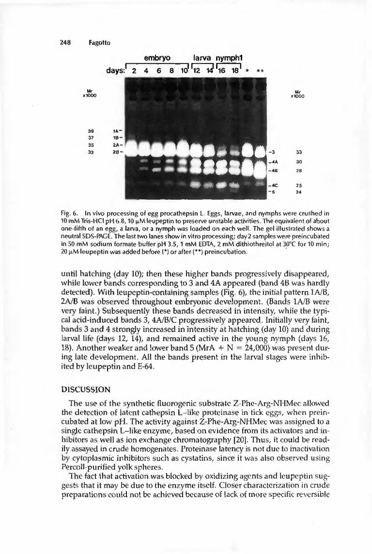
248 Fagotto
embryo larva nympM
days:' 2 4 6 8 101 'l2 W 'l6 18 ' # *
Mr Mr XK)OO XKX)O
39 37
35
33
1A-1B-
2A-
2B- 3
4A
H
4C 5
33
30
28
25 24
Fig. 6. In vivo processing of egg procathepsin L. Eggs, larvae, and nymphs were crushed in 10 m M Tris-HCI pH 6.8,10 p.M leupeptin to preserve unstable activities. The equivalent of about one-fifth of an egg, a larva, or a nymph was loaded on each well. The gel illustrated shows a neutral SDS-PACE. The last two lanes show in vitro processing; day 2 samples were preincubated in 50 m M sodium formate buffer pH 3.5,1 mM EDTA, 2 m M dithiothreitol at 3O0C for 10 min; 20 f iM leupeptin was added before (*) or after (**) preincubation.
until hatching (day 10); then these higher bands progressively disappeared, while lower bands corresponding to 3 and 4A appeared (band 4B was hardly detected). With leupeptin-containing samples (Fig. 6), the initial pattern IAJB, 2A/B was observed throughout embryonic development. (Bands 1A/B were very faint.) Subsequently these bands decreased in intensity, while the typical acid-induced bands 3, 4A/B/C progressively appeared. Initially very faint, bands 3 and 4 strongly increased in intensity at hatching (day 10) and during larval life (days 12, 14), and remained active in the young nymph (days 16, 18). Another weaker and lower band 5 (MrA + N = 24,000) was present during late development. All the bands present in the larval stages were inhibited by leupeptin and E-64.
DISCUSSION
The use of the synthetic fluorogenic substrate Z-Phe-Arg-NHMec allowed the detection of latent cathepsin L-like proteinase in tick eggs, when preincubated at low pH. The activity against Z-Phe-Arg-NHMec was assigned to a single cathepsin L-like enzyme, based on evidence from its activators and inhibitors as well as ion exchange chromatography [20]. Thus, it could be readily assayed in crude homogenates. Proteinase latency is not due to inactivation by cytoplasmic inhibitors such as cystatins, since it was also observed using Percoll-purified yolk spheres.
The fact that activation was blocked by oxidizing agents and leupeptin suggests that it may be due to the enzyme itself. Closer characterization in crude preparations could not be achieved because of lack of more specific reversible

Tick Egg Procathepsin L Activation 249
inhibitors. High concentrations of leupeptin produced only incomplete inhibition, suggesting that the latent form has a much weaker affinity for the inhibitor than the active form. Such a difference has been found for human procathepsin D which, unlike mature cathepsin D, has only poor affinity for pepstatin [26]. In the present case, it cannot be decided whether this difference is due to the conformation of the latent enzyme or to a restricted accessibility to the catalytic site, due to close association with vitellin [20]. The latter suggestion was made to explain the latency of a neutral proteinase of Artemia eggs [15]. McDonald and Kadkhodayan [27] have recently described a latent cathepsin L in guinea pig sperm. Its substrate specificity, sensitivity to inhibitors, as well as activation characteristics were similar to those found for the O. moubata enzyme.
Localization of egg cathepsin L by gelatin SDS-PAGE showed that the enzyme was similarity processed in vitro by acid treatment and in vivo during embryonic and larval development. The different bands could be due to different enzymes, selectively activated or inactivated depending on sample pretreatment. It seems not to be the case, since all bands could be obtained, with the same relative intensity, from the cation exchange fractions in which previous enzymatic studies had detected only cathepsin L-like activity. Furthermore the activity of all these bands was inhibited if specific inhibitors (leupeptin, E-64, Z-Phe-Phe-CHN2) were present in the incubation medium. Finally, disappearance of the latent form and appearance of the active form arc directly related (Fig. 4). It is concluded that the latent form is a precursor of the active enzyme. Low pH leads to partial proteolysis of this proenzyme, probably by an autocatalytic mechanism. A proenzyme has recently been reported for both cathepsin L and B in the human liver [28]. These 39,000 dal-ton precursors are converted to the mature form (Mr = 30,000 and 29,000 respectively) in the lysosomal compartment. Likewise latent, high molecular weight precursors of cathepsin B [29] and cathepsin L [30,31] are excreted by cancer cells. Many other lysosomal enzymes are delivered to the lysosomal or prelysosomal compartment as precursors [26,32,33], Although all these precursors seem to undergo proteolytic processing, data are divergent about the enzymes responsible for this proteolysis. While pro-a-glucosidase processing is blocked in the presence of leupeptin [32], maturation of cathepsins L and B has been reported to be affected by pepstatin [28], by metal chelators, and by leupeptin [34]. On the other hand human procathepsin D (33] and procathepsin L [30] are capable of self-activation, as may be the case for O. moubata cathepsin L.
Based on the data presented here, a working model that can explain the multiplicity of the observed bands is proposed. Since the latent enzyme was efficient in degrading gelatin in the gels, probably some modifications of the catalytic properties had occurred, either due to SDS or to self-activation during incubation of the gels, which was performed at optimal conditions for enzyme activation, pH 3.4. Because the residual activity, recovered after electrophoresis, is dependent on many ill-defined parameters, we could not compare the relative intensity of different bands, but only variations of one band between different samples. The fact that most bands were present as doublets may be explained by the occurrence of two related forms. The different mobil-

25Ü Fagotto
ities could be due to different sugar content or charge, or to partial proteolysis. Since bands 1A/B and 2A/B are interconvertible, depending on the redox conditions, bands 1A/B may correspond to the native enzyme, composed of a large, active subunit (bands 2A/B) and a small, undetectable fragment. Band 3 is found only in acid-pretreated samples, and the activity does not depend on "stabilization" by leupeptin before electrophoresis. Thus it is clearly distinct from band 2A/B, although the respective mobilities are close. Since band 3 rapidly appears during acid treatment, long before bands 4A/B, it may be an intermediate form. Bands 4A/B are the best candidates for the mature, fully active enzyme, as they appear concomitantly with activation measured enzy-matically, and shortly before the 90,000 dalton band, which is probably a product of vitellin degradation. Moreover, leupeptin slowed down both activation and appearance of these bands in a similar way. Band 4C is probably a further processed form of bands 4A/B. The requirement of leupeptin for preservation of band 2 and 4 activities is likely due to poor stability of these forms at neutra] and alkaline pH. Perhaps binding of leupeptin protects the enzyme from denaturation. The discrepancies in molecular weights between the alkaline and neutral gels remain to be solved. Particularly puzzling is the small, but significant upward shift of the mature form observed in alkaline gels, but found to be downwards in neutral gels (Figs. 5,6). Charge modifications such as loss of phosphate residues are one possible explanation.
Occurrence of multiple molecular forms for vertebrate lysosomal hydrolases has been reported [32,33,35]. In some cases, these forms are attributed to different processing states [32,33]. After activation, rat cathepsin L [28] yields two bound subunits. Most proenzymes are processed in several successive steps and at least one, but sometimes several, intermediates can be detected [26|. It seems likely from biosynthetic studies that two different types of cleavage occur: an initial rapid splitting process that yields an intermediate form, which is then processed much more slowly to give the mature enzyme [26]. This could also be the case in yolk spheres, since in these experiments band 3 appears very early and bands 4 much later.
Further studies arc needed to ascertain this process. However, the present results show a direct relationship between the in vitro activation, which requires acidic conditions, and what actually occurs in vivo, where both activation and cleavage of procathepsin L proceed concomitantly with yolk degradation. Since no active form is present at the beginning of embryonic development, but increases progressively during late embryogenesis, the yolk spheres are presumably at neutral pH during early development, and an acidification process takes place later. This problem has been investigated by using the fluorescent pH probe acridine orange and only neutral spheres in early development and an increasing number of acidic spheres as development proceeded were observed (unpublished data, reported in an abstract form [36]). This is in agreement with the data presented herein.
The advantages of storing an inactive procathepsin in yolk platelets are obvious: on the one hand active lysosomal thiol-proteinases, unlike their precursors, arc unstable at neutral pH [21] and could never survive under such conditions. On the other hand the very low pH optimum for procathepsin activation ensures that no uncontrolled digestion occurs within the neutral
Tick Egg Procathepsin L Activation 251
spheres. Furthermore it allows excellent regulation, since maturation rates can be modulated by several orders of magnitude within the physiological pH limits known for the endosomal-lysosomal compartment (pH 6.5 to 4.0) [37-39].
LITERATURE CITED
1. Pasteels JJ: Yolk and lysosomes. In : Lysosomes in Biology and Pathology. Dingle JT, ed., Publishing Co., North-Hol land, Amsterdam, vol. 3, pp 216-233 (1973).
2. Will iams J: YoIk uti l ization. In: The Biochemistry of Animal Development. Weber R, ed. Academic Press, New York, vol. 2, pp 341-377 (1967).
3. Komasaki S, Asashima M: Structural changes of yolk platelets and related organelles dur ing development of the newt embryo. Dev Growth Differ 29, 323 (1987).
4. Vernier J-M, Sire M-F: Plaquettes vitel l ines et activité hydrolastique acide au cours du développement embryonnaire de Ia truite arc-en-ciel. Etude ultrastructurale et biochimique. Biol Cell 29, 99 (1977).
5. Yakota Y, Kato K H : Degradation of yolk proteins in sea urchin eggs and embryos. Cell Differ 23,191 (1988).
6. Armant DR, Carson DD, Decker GL, WeIpIyJK, Lennarz WJ: Characterization of yolk platelets isolated from developing embryos of Arbacia punctulata. Dev Biol 7 J3, 342 (1986).
7. Kari BE, Rottmann WL: Analysis of changes in a yolk glycoprotein complex in the developing sea urchin embryo. Dev Biol 108, 18(1985).
8. Purcell JP, Quinn T M , Kunkel JG, Nord in JH: Correlation of yolk phosphatase expression wi th the programmed proteolysis of vitell in in Blattella germanica dur ing embryonic development. Arch Insect Biochem Physiol 9, 237 (198S).
9. Bownes M, Hames BD: Accumulation and degradation of three major yolk proteins in Droghila melanoxasier. J Exp Zool 200,149 (1977).
10. Yamashita O, lndrasith LS: Metabolic fates o\ volk proteins dur ing embryogenesis in Arthropods. Dev Growth Differ 30, 337 (19HS).
IT. Medina M, Leon.P, Vallejo CG: Droiopliilti cathepsin B-like proteinase: a suggested role in yolk degradation. Arch Biochem Biophys 263, 355(1988).
12. Kageynma T, Takahashi SY, Takahashi K: Occurrence of thiol proteinases on the eggs of the si lkworm, BomtnfX mori. J Biochem 9(J. 665 (1981).
13. Slaugther D, Triplett E: Amphibian embryo protease inhibitor. I; Isolation, purification and characterization. Cell Differ 4, 11 (1975).
14. Salisbury N, Ca !aprico N, Triplett E: Amphibian embryo protease inhibitor. Vl : Maternal origin and identity wi th l ipovitell in heavy subunit. Cell Differ9, 219 (1980).
15. Ezquieta B, Vaile jo CG: Lipovitell in inhibit ion of Artemia Irypsin-like proteinase: a role for a storage protein in regulating proteinase activity during development. Arch Biochem Biophvs 25(),410(1986).
16. Scot! LB, Leahy PS, Decker GL, Lennarz WJ: Loss of yolk platelets and yolk glycoproteins dur ing larval development of the sea urchin embryo. Dev Biol 737, 368 (1990).
17. Ezqutcta B, Vallejo CG: Artemia trypsin-like proteinase: developmental activation is inhibited bva lysosomotropic agent. Biol Cell 58, 227(1986).
IH. Chinzei Y, Chino H, Takahashi K: Purification and properties of vitellogenin and vilel l in from a tick, Onuthodorua mouhntti. J Comp Physiol 152.13 (1983).
19. Chinzei Y, Yano I: Vitell in is the nutrient reserve dur ing starvation in the nymphal stage of a tick. E x p e r i e n t i a l , 948 (1985).
20. Fagotto F: Yolk degradation in tick eggs: occurence of a cathcpsin L-like acid proteinase in yolk spheres. Arch Insect Biochem Physiol. In press.
21. Barrett AJ, Kirschke H: Cathepsin B, cathepsin H, cathepsin L. Methods Enzymol 80. 535 (1981).
22. Heussen C, Dowdle EB: Electrophoretic analysts of plasminogen activators in polycrylamide gels containing SDS and copolymerised substales. Anal Biochem 102,19(1980).
23. Laemmli UK: Cleavage of structural proteins dur ing the assembly of the head of bacteriol-phage T4. Nature (London) 227, 680 (1970).
24. Williams DE, Reisfeld RA: Disc electrophoresis in Polyacrylamide gels: extension to new conditions of p H and buffer. Ann NY Acad Sci 721, 373 (1964).

252 Fagotto
25. L o w r y O H , Rosenbrough NJ, Fair AL , Randall RJ: Protein measurement with the Folin phenol reagent. J Biol Chem m, 265 (1951).
26. Skudlarek MD, Novak EK, Swank RT: Processing of lysosomal enzymes in macrophages and kidney. In : Lysosomes in Biology and Pathology. Dingle JT, Dean RT, SIy W, eds. Elsevier, Amsterdam, vol. 7, pp 17-44 (1984).
27. McDonald JK, Kadkhodayan S; Cathepsin L: a latent proteinase in guinea pig sperm. Bioehem Biophys Res Commun 151, 827 (1988).
28. Nishimura Y, Kawabata T1 Kato K: Identification of latent procathcpsins B and L in microsomal lumen: characterization of enzymatic activation and proteolytic processing in vitro. Arch Bioehem Biophys 261, 64 (1988).
29. Mort JS, Recklies AD: Interrelationship of active and latent secreted human cathepsin B precursors. Bioehem J 233, 57 (1986).
30. GaI S, Gottesman M M : The major excreted protein of transformed fibroblasts is an activable acid-protease. J Biol Chem 267, 1760(1986).
31. Gal S, Gottesman MM: The major excreted protein (MEP) of transformed mouse cells and cathepsin L have similar protease specificity. Bioehem Biophys Res Commun Î39,156 (1986).
32. Tsuji A, Suzuki Y: Biosynthesis of two components of human acid a-glucosidase. Arch Bioehem Biophys 259, 234 (1987).
33. Hasilik A, von Figura K: Processing of lysosomal enzymes in fibroblasts. In: Lysosomes in Biology and Pathology. Dingle JT, Dean RT, Sly W, eds. Elsevier, Amsterdam, vol. 7, pp 3-16 (1984).
34. Hara K, Kominami E, Katunuma'N: Effect of proteinase inhibitors on intracellular processing of cathepsin B, H and L in rat macrophages. FEBS Lett 23I1 229 (1988).
35. Cheng SH, Malcolm S, Pcmble S, Winchester B: Purification and comparison of the structures of human liver acidic a-D-mannosidases A and B. Bioehem J 233, 65(1986).
36. Fagotto F: Evidence that acidification of the yolk spheres promotes yolk digestion in tick eggs through activation of a procathepsin L (abstract). Experientia 46, A82 (1990).
37. Rudnick G: ATP-driven H ' pumping into intracellular organelles. Annu Rev Physiol 4$, 403(1984).
38. Reeves JP: The mechanism of lysosomal acidification. In: Lysosomes in Biology and Pathology. Dingle JT, Dean RT, Sly W.'eds. Elsevier, Amsterdam, vol. 7, pp 175-199 (19M).
39. Yamashim DJ, Maxl'ield FR: Kinetics of endosome acidification in mutant and wild-type Chinese hamster ovary colls. J Cell Biol 105, 2713 (1987).

Ill

YOLK ACIDIFICATION 1
Development, Growth and Differentiation, 000, 1990
Y O L K D E G R A D A T I O N I N T I C K E G G S : I I I . D E V E L O P M E N T A L L Y R E G U L A T E D A C I D I F I C A T I O N O F T H E YOLK SPHERES
François Fagotto, Institut de Zoologie, Université de Neuchâtel, Chantemerle 22, 2000 Neuchâtel.
Key words: Ornithodoros moubata embryogenesis, vitellin, acidic compartment, acridine orange.
SUMMARY
Yolk spheres in tick eggs contain a latent procathepsin L, which is activated in vivo, in parallel with yolk degradation, and in vitro by acid treatment (Fagotto, F., 1990 (b). Arch. Insect Biochem. Physiol., in press). Mature cathepsin L hydrolyzes vitellin at acidic pH (Fagotto, F., 1990 (a). Arch. Insect Biochem. Physiol., in press). Yolk spheres' pH has been estimated using acridine orange. In the early development, all yolk spheres are neutral, then an increasing number acidify, until hatching, where general acidification seems to occur. This fits well with vitellin utilized slowly during embryogenesis, more intensely at hatching (Chinzei and Yano, Experientia 41, 948, 1985), and can be related to sequential degradation of individual spheres during embryonic development, then extensive yolk liquefaction in the larva. Different yolk sphere populations have been separated on Percoli density gradients. In freshly laid eggs, yolk spheres are dense, neutral, undegraded and contain exclusively the precursor of cathepsin L. As development proceeds, yolk spheres are progressively recovered in lower density fractions, displaying acidic interior and cytological signs of degradation. They co-sediment with mature cathepsin L. It is concluded that acidification initiates yolk degradation through procathepsin L activation.
INTRODUCTION
Most eggs accumulate nutrients (yolk proteins, or vitellins (Vns* )) in special, large and dense storage organelles called yolk spheres (YSs) (or granules, or platelets). Yolk precursors (vitellogenins) are taken up by extensive receptor-mediated pinocytosis [21, 27]. They follow a typical endosomal route and are partially processed in a prelysosomal compartment [16] before being concentrated in mature yolk platelets. In many species acid hydrolases have been localized within the yolk [2, 11, 14, 15, 17, 23,25, 26] and are likely to be involved in its subsequent degradation [7, 12, 14, 15, 17, 18, 19, 22, 24, 25, 28].
Since yolk degradation is generally delayed for several hours or days, hydrolytic activities have to be kept momentarily inactivated [17], and subsequently developmentally activated and regulated in a very precise manner. The fact that similar proteinases are likely to be present in the yolk of different species [8], and that yolk proteins have homologies among evolutionarily distant
* ABBREVIATIONS USED: AO, acridine orange; EC, endodermal cell; EM, electron microscopy; FCCP, carbonyl cyanide p-trifluoromethoxyphenylhydrazone; Hepes, N-[2-hydroxyethyi]piperazine-N'-[2-ethanesulfonic acid] ; leupeptin, N-acetylleucyl-leucyl-arginal; PAGE, Polyacrylamide gel electrophoresis; SDS, sodium dodecylsulfate; Vn, vitellin; YS, yolk sphere; Z-Phe-Arg-NHMecttenzoyloxycartonyl-phenylalanyl-arginyl'7-amido4-methylcoumarine.

YOLK ACIDIFICATION 2
species [4] suggests that common mechanisms may act on yolk degradation. In an attempt to clarify this process, a study has been undertaken on the egg of
an African soft tick, Ornithodoros moubata. The O. moubata egg is tightly packed with YSs and displays the centrolecithal structure common to most arthropods [I]. However, unlike insect eggs, it undergoes a total cleavage that leads, at day 2, to the formation of two concentrica! sheets: a central primary endoderm, composed of huge, yolk-filled endodermal cells (ECs) and a surrounding thin, yolk-free ecto-mesoderm, which will form the embryo [10]. The ECs can be easily isolated under a dissecting microscope and even maintained in vitro for some days in a saline buffer solution (unpublished results). Vn, which accounts for 80% of the total protein content in eggs, has been characterized [5] and its utilization quantified [6]; intense degradation only occurs during the larval life, and about 50% is still left undegraded in the young nymph. It will be slowly degraded and used as nutrient until the first blood meal. A latent cathepsin L-like acid proteinase has been detected within the YSs, together with other acid hydrolases [8]. Evidence has been presented that this proteinase is stored as a latent precursor, which is processed both in vitro by acid treatment and in vivo, in parallel with yolk degradation [9].
Since these results suggested a crucial role of acidification, the actual pH of the YSs has been investigated using the fluorescent dye acridine orange (AO). It appears that most YSs are kept neutral during early development, then an increasing number acidify, until a general acidification seems to occur at hatching. Fractionations on Percoli gradients have been performed to separate different populations of YSs and compare their density, internal pH, ultrastructure and enzymatic properties. The results indicate that acidification is clearly related to yolk degradation.
MATERIALS AND METHODS
CHEMICALS
Bovine serum albumin, Z-Phe-Arg-NHMecHCl, leupeptin, monensin, nigericin, FCCP and £-nitrophenyl-N-acetylglucosamine sodium salt were purchased from Sigma Chemical Company (St Louis, MO, USA), dithiohreitol and acridine orange were obtained from Fluka Chemie AG (Buchs, Switzerland), gelatin from Difco Laboratories (Detroit, USA). Percoli and molecular weight markers for electrophoresis were obtained from Pharmacia Fine Chemicals AB (Uppsala, Sweden). AU other chemicals were of analytical grade.
EGG DISSECTION
Mated, fed females were allowed to oviposit at 290C. The eggs were incubated at the same temperature. Under these conditions, they hatched after 10 days. They were dissected at selected stages at room temperature in 25 mM Hepes-NaOH, 50 mM KCl, 200 mM NaCl, pH 7.0. The chorion was broken by suction with a broken Pasteur pipette as described [10], and in late stages extraembryonic membranes were removed using fine tweezers. To get intact ECs, the superficial ectodermal sheet was tom apart and removed together with the germ band. In early stages, ECs readily disaggregated by gentle shaking. From day 6 onwards, however, the yolk-filled primary endoderm was enclosed by a thin external sheet, the future gut epithelium, which had to be broken to get access to the inner ECs. Progressively, most ECs became intimately bound to this epithelium and could no longer be disaggregated. To observe individual YSs, eggs (days 1-10) and young larvae (days 11-12) were opened vigorously, which caused extensive breakage of the ECs and release of the YSs.

YOLK ACIDIFICATION 3
CELL FRACTIONATIONS
All steps were performed at 40C. Self-generated Percoli gradients were prepared as follows: 1 ml 90%, 3 ml 60%, 3 ml 30% and 2 ml 0% Percoli, made isotonic with 25 mM Hepes-NaOH, 50 mM KCl, 200 mM NaCl buffer, pH 7.0, were carefully layered in 10 ml polycarbonate tubes. The tubes were centrifuged 20 min at 27,000xg in a Sorvall SS 34 rotor. Eggs (110-130 mg fresh weight, about 30 mg protein) were very gently homogenized in 1 ml 25 mM NaOH-Hepes buffer, pH 7.0, containing 50 mM KCl and 200 mM NaCl. Most chorions, as well as the large pieces of germ band were discarded, and the suspension was layered on the gradient (YSs readily sink about 1 cm deep). The gradients were centrifuged 20 min at 120xg in a Sorvall HB-4 rotor. 0.5 ml fractions, collected from the bottom with a capillary connected to a peristaltic pump, were either stored at 40C for AO observations and fixation for EM, performed within 1 h after collection, or frozen and stored at -180C until assayed for hydrolases activities. For gelatin/SDS-PAGE analysis, aliquots of each fraction were added with 5 JiM leupeptin immediately after collection to preserve unstable activity [9] and kept frozen. Gradient density was determined by refractometry.
ACRIDINE ORANGE FLUORESCENCE MICROSCOPY
Cells and broken cells (about one third of an egg) were incubated in a 20-50 (il drop of incubation medium (25 mM Hepes-NaOH buffer, pH 7.0, containing 50 mM KCl, 200 mM NaCl and 50-100 \iNi acridine orange) on a glass coverslip, in a small well (5 mm diameter, 0.2 mm depth). With Percoli gradient fractions, 10 \i\ were mixed with 10 fil incubation medium containing 100 (J.M AO. Incubations were carried out for 10 to 30 min at room temperature in the dark, under 100% relative humidity. The well was then carefully closed with a coverslip. Most of the medium was expelled from the small (4-5 |il) chamber obtained, but not the YSs and ECs, because they readily sedimented during incubation. The depth of the chamber was about the size of the ECs, so that fluorescence scattering was minimal and high magnification objectives (40x) could be used. Observations and microphotographs were performed on a Vanox S fluorescence microscope (Olympus) using a violet excitation filter BP 405 (395-415 nm) and a Y 455 barrier filter (> 455 nm). Often direct white light was superimposed to the fluorescent light, since, provided appropriate light intensity and condensor position, more detailed images, with sharper limits could be obtained. Results were similar using 50 ^M or 100 iM AO.
PROTEIN AND VITELLIN DETERMINATION
Protein concentration was measured according to Lowry's method [13], using bovine serum albumin as the standard. Vn content was estimated at 400 nm [5].
HYDROLASES ASSAYS
No particular treatment was needed for complete breakage of the YSs; freeze/thawing (storage) and the use of hypotonic reaction media were sufficient. Cathepsin L was detected at pH 5.5, 30°C, with Z-Phe-Arg-NHMec [3], after preincubation at pH 3.5 under reducing conditions to activate the enzyme [9]. N-acetylglucosaminidase was assayed with 5 mM p-nitrophenyl-N-acetylglucosamine, in 50 mM sodium formate buffer, pH 4.0, at 37°C [8]. In order to compare data from different gradients, enzymatic activities, as well as protein concentration were recalculated to a uniform initial content of 100 mg fresh weight and corrected for weight loss (1 day=100%, 5 days= 95%, 8 days== 85%, 11 days=75%).

YOLK ACIDIFICATION
GELATIN/SDS-PAGE
SDS-PAGE containing 0.1% copolymerized gelatin, using a neutral buffer system (Tris-barbital) was performed as described [9]. 10 \i\ of Percoli fractions containing 5 |iM leupeptin were mixed with 10 JiI of double concentrated sample buffer. After electrophoresis and rinsing in Triton X-100, the gels were incubated at pH 3.4 under reducing conditions, for 4 h at 370C, then stained with Coomassie blue. Molecular weight markers were Phosphorylase b (94,000 kDa), bovine serum albumin (67,000 kDa), ovalbumin (43,000 kDa), carbonic anhydrase (30,000 kDa) and trypsin inhibitor (20,100 kDa).
ELECTRON MICROSCOPY
Fractions from the Percoli gradients were fixed in 1% glutaraldehyde, 0.1 M sodium cacodylate, pH 7.4, 8% sucrose, rinced in cacodylate buffer, embedded in 1.5% agarose, postfixed in 1% Os04, dehydrated and embedded in Spurr's medium. Whole eggs and larvae were fixed in 6.25% glutaraldehyde, 0.1 M sodium/potassium phosphate buffer, pH 7.4, 2% sucrose, rinsed in phosphate buffer, postfixed in 1% Os04, dehydrated and embedded in Spurr's medium. Thin sections were stained with uranyl acetate and lead citrate. Semi-thin sections (0.5 firn) for light microscopy were stained with toluidine blue.
RESULTS
1 BACKGROUND: MODALITIES OF YOLK DEGRADATION
ECs were found nearly unchanged during most of the embryonic development (days 2-9). However, several YSs displayed signs of degradation such as highly irregular shape (Fig. Ia) and inhomogenous matrix (Fig. Ib). Degraded YSs were generally found in the perinuclear region as well as near the border of the cell. Yolk degradation was more intense in the peripheral ECs, especially next to the germ band.
During late embryonic development, the ECs become progressively enclosed in a thin epithelium, which will develop into the larval gut wall [I]. Dissection as well as microscopical observations revealed that the ECs are associated with this epithelium and may contribute to its formation (unpublished data). From day 9, closure of the gut epithelium being almost complete, progressive exocytosis of the yolk was observed (Fig. Id). ECs were depleted of most their yolk content and retracted, leaving a cavity, the gut lumen, filled with liquefied yolk (Fig. Ic, d). Meanwhile, intracellular yolk degradation was intense (Fig. Id). Yolk liquefaction spread over the last two days of embryonic development and all the larval life (days 11-14), and was concomittant with the intense phase of vitellin hydrolysis detected by Chinzei [6].
2 ACRIDINE ORANGE FLUORESCENCE OF ENDODERMAL CELLS
AO was used to evaluate the pH of the YSs. At low concentrations (50-100 (iM) AO has a weak green fluorescence, but it accumulates in acidic compartments, which appear orange to red, due to a shift of the fluorescence.
In whole eggs or isolated endoderms only the fluorescence of the more external YSs was detectable. Therefore endodermal cells were disaggregated and observed individually. Cleavage blastomeres (0-24 h) could not be examined, since they were large and delicate, and invariably broke during dissection. On the other hand, in late embryos, the ECs became tightly bound to the gut epithelium (probably they were inserted into, see above). Day 8 was the latest stage where intact cells could
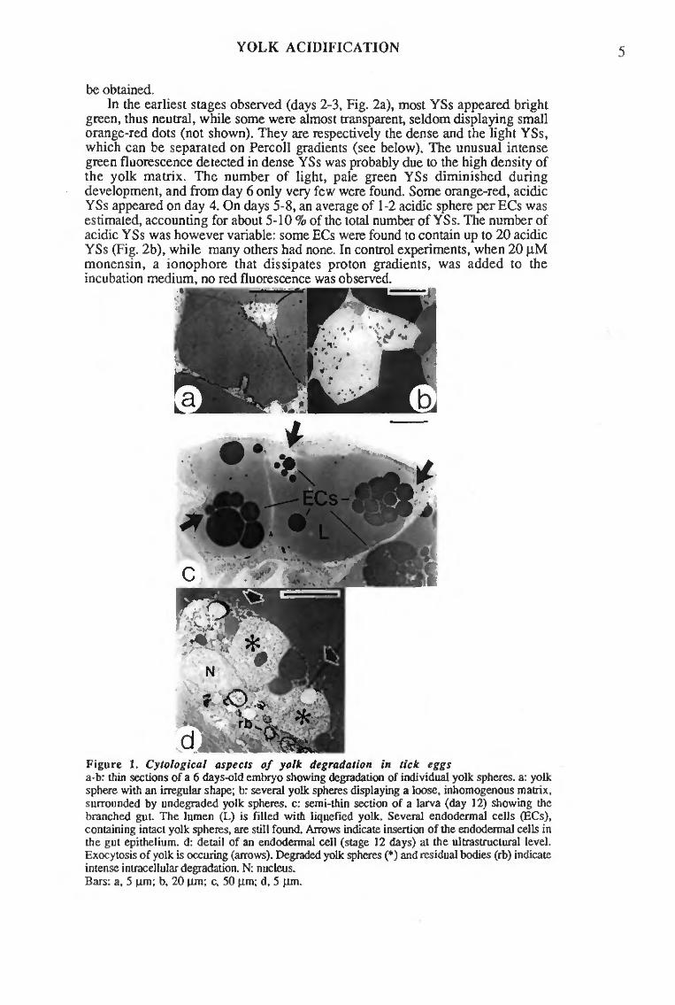
YOLK ACIDIFICATION
be obtained. In the earliest stages observed (days 2-3, Fig. 2a), most YSs appeared bright
green, thus neutral, while some were almost transparent, seldom displaying small orange-red dots (not shown). They are respectively the dense and the light YSs, which can be separated on Percoli gradients (see below). The unusual intense green fluorescence detected in dense YSs was probably due to the high density of the yolk matrix. The number of light, pale green YSs diminished during development, and from day 6 only very few were found. Some orange-red, acidic YSs appeared on day 4. On days 5-8, an average of 1-2 acidic sphere per ECs was estimated, accounting for about 5-10 % of the total number of YSs. The number of acidic YSs was however variable: some ECs were found to contain up to 20 acidic YSs (Fig. 2b), while many others had none. In control experiments, when 20 fiM monensin, a ionophore that dissipates proton gradients, was added to the incubation medium, no red fluorescence was observed.
Figure 1. Cytological aspects of yolk degradation in tick eggs a-b: thin sections of a 6 days-old embryo showing degradation of individual yolk spheres, a: yolk sphere with an irregular shape; b: several yolk spheres displaying a loose, inhomogenous matrix, surrounded by undegraded yolk spheres, c: semi-thin section of a larva (day 12) showing the branched gut. The lumen (L) is filled with liquefied yolk. Several endoderma! cells (ECs), containing intact yolk spheres, are still found. Arrows indicate insertion of the endoderma! cells in the gut epithelium, d: detail of an endodermal cell (stage 12 days) at the ultrastructural level. Exocytosis of yolk is occuring (arrows). Degraded yolk spheres (*) and residual bodies (rb) indicate intense intracellular degradation. N: nucleus. Bars: a, 5 um; b, 20 um; c, 50 um; d, 5 um.
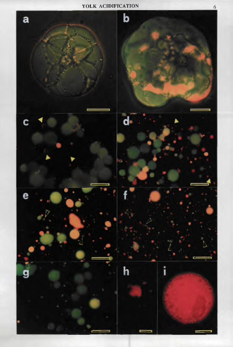
YOLK ACIDIFICATION 6
a
•'v
\
e A *
V % • • * * # 4 ••
•
* - ' ^ w • ^
I i
A
c l .
.'•5u * < *
* 1
1 ^ - "
it- ir • •
M • ' J
• • • • . ' • • * • •
. < % ' . - / ! . • • * .
h
i
•
1 - 1 - .
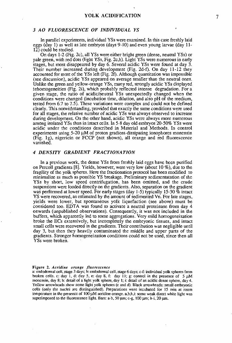
YOLK ACIDIFICATION 7
3 AO FLUORESCENCE OF INDIVIDUAL YS
In parallel experiments, individual YSs were examined. In this case freshly laid eggs (day 1) as well as late embryos (days 9-10) and even young larvae (day 11-12) could be studied.
On days 1-2 (Fig. 2c), all YSs were either bright green (dense, neutral YSs) or pale green, with red dots (light YSs, Fig. 2c,h). Light YSs were numerous in early stages, but most disappeared by day 6. Several acidic YSs were found at day 3. Their number increased during development (Fig. 2d-f). On day 11-12 they accounted for most of the YSs left (Fig. 2f). Although quantitation was impossible (see discussion), acidic YSs appeared on average smaller than the neutral ones. Unlike the green and yellow-orange YSs, many red, strongly acidic YSs displayed inhomogeneities (Fig. 2i), which probably reflected intense degradation. For a given stage, the ratio of acidic/neutral YSs unexpectedly changed when the conditions were changed (incubation time, dilution, and also pH of the medium, tested from 6.7 to 7.5). These variations were complex and could not be defined clearly. This notwithstanding, provided that exactly the same conditions were used for all stages, the relative number of acidic YSs was always observed to increase during development. On the other hand, acidic YSs were always more numerous among isolated YSs than in intact cells. In 5-8 day old embryos 20-50% YSs were acidic under the conditions described in Material and Methods. In control experiments using 5-20 }iM of proton gradient-dissipating ionophores monensin (Fig. Ig), nigericin or FCCP (not shown), all orange and red fluorescence vanished.
4 DENSITY GRADIENT FRACTIONATION
In a previous work, the dense YSs from freshly laid eggs have been purified on Percoli gradients [8]. Yields, however, were very low (about 10 %), due to the fragility of the yolk spheres. Here the fractionation protocol has been modified to minimalize as much as possible YS breakage. Preliminary sedimentatation of the YSs by short, low speed centrifugation, has been omitted, and the crude suspensions were loaded directly on the gradients. Also, separation on the gradient was performed at lower speed. For early stages (day 1-5) typically 15-30 % intact YS were recovered, as estimated by the amount of sedimented Vn. For late stages, yields were lower, but spontaneous yolk liquefaction (see above) must be considered too. EDTA was found to activate a neutral proteinase from day 4 onwards (unpublished observations). Consequently, it was not included in the buffers, which apparently led to some aggregations. Very mild homogeneization broke the ECs extensively, but incompletely the embryonic tissues, and intact small cells were recovered in the gradients. Their contribution was negligible until day 3, but then they heavily contaminated the middle and upper parts of the gradients. Stronger homogeneization conditions could not be used, since then all YSs were broken.
Figure 2. Acridine orange fluorescence a: endodermal cell, slage 3 days; b: endodermal cell, stage 6 days; c-f: individual yolk spheres from broken cells, c: day 1, d: day 5, e: day 8, f: day 11; g: control in the presence of 5 UM monensin, day 8; h: detail of a light yolk sphere, day 1; i: detail of an acidic dense sphere, day 4. Yellow arrowheads show some light yolk spheres (c and d). Black arrowheads: small embryonic cells (only the nuclei are distinguished). Preparations were incubated for 15 min at room temperature in the presence of 100 uM acridine orange. a,b,h,i: some weak direct white light was superimposed to the fluorescence light. Bars: a-b, 50 urn; c-g, 100 urn; h-i, 20 urn.

YOLK ACIDIFICATION
Four representative stages have been examined in some detail: day 1: freshly laid eggs; day 5: middle embryonic development, blastokinesis; day 8: late development, organogenesis, onset of the intense phase of yolk degradation; day 11: young larva, intense yolk degradation. For each stage, the fractions have been examined for internal pH of the YSs (AO fluorescence) and their ultrastructure by EM, assessed for N-acetylglucosaminidase and cathepsin L, and analyzed by gelatin-containing SDS-PAGE to detect precursor and mature forms of cathepsin L through their gelatinolytic activity [9].
4.1 MICROSCOPICAL OBSERVATIONS: AO FLUORESCENCE AND ULTRASTRUCTURE
The gradients from freshly laid eggs, unlike later stages, were devoid of contamination by intact cells. Indeed the large blastomeres, which compose cleaving embryos, all erupted during homogeneization In agreement with previous results [8], two populations of YSs were found. Dense YSs were readily detectable near the bottom of the gradient (peak fraction=6), through the dark brownish color due to Vn. They were almost spherical with a compact, homogenous matrix (Fig. 3a), and all were neutral. The light YSs, previously called light vesicles, appeared as a whitish band just below the top of the gradient (peak fraction= 15). They thus did not appear to contain Vn. They had a much
Figure 3. Ultrastructure of yolk spheres found in Percoli gradients a: undegraded dense yolk sphere, day 1, fractions 4-5; b-d: partially degraded dense yolk spheres, day 8, fractions 11-13; e-f: light spheres, day 1, fractions 14-15. Bars: a, 10 um; b-f, 1 urn.

YOLK ACIDIFICATION 9
looser matrix, often with holes and invaginations (Fig. 3e-f), and displayed the dark green AO fluorescence, with red dots previously observed in intact and broken cells (see above and Fig. 2). These dots seemed to be related to the inhomogeneities of the matrix found by EM. Other organelles like mitochondria poorly penetrated the gradient (fractions>16).
At the stage of 5 days, the bottom of the gradient still contained essentially dense, homogenous, neutral YSs. Small amounts of light YSs were present in low density fractions (fractions 14-17). Many YSs were found in the middle part of the gradient, among the embryonic cells. Most of them were acidic and displayed signs of degradation: their shape was generally irregular, the density and homogeneity of their matrix was variable, and holes and invaginations were observed. In gradients from 8-11 day old eggs/larvae, the light YSs had nearly vanished. The amount of neutral, dense YSs diminished, while few acidic YSs were also found in the same dense fractions. All displayed a dense matrix, but some showed an irregular shape, which is probably the first sign of degradation. On the other hand, most YSs were recovered in the intermediate fractions. Like on day 5 most were acidic and partially degraded (Fig. 3b-d). The peak shifted to lower density (maximum= fraction 11 on day 5,. fraction 13 on day 8 and fraction 14 on day 11), indicating that on average the degradation was more pronounced.
4.2 DETECTION OF HYDROLYTIC ACTIVITIES (Fig. 4)
As already reported [8], dense YSs from freshly laid eggs contained N-acetylglucosaminidase and cathepsin L. Due to extensive breakage of the YSs, a large amount Of both enzymes (and Vn) did not sediment. In the previous study, most of this non-sedimentable activity was discarded with the supernatant of the first short centrifugation. Under those conditions, the top of the Percoli gradient contained few hydrolases activities. A peak of N-acetylglucosaminidase, but not cathepsin L, could be detected just below the supernatant, in the fractions containing the light YSs [8]. Since in the present study the preliminary centrifugation was omitted (see above), all the activity from the broken YSs was recovered on the top of the gradient (fractions >16). Consequently, the lower peak, corresponding to the light YSs, was hardly detected as a shoulder (fractions 14-16).
In later stages, the dense YSs always displayed both proteinase and N-acetylglucosaminidase activities, roughly proportional to the amount of Vn. On the other hand, a broad peak of N-acetylglucosaminidase, but not cathepsin L, appeared in the middle of the gradient. In agreement with data on crude homogenates [8], total cathepsin L activity was stable in embryos, then decreased after hatching (Fig. 4, day 11), while N-acetylglucosaminidase activity increased continuously during development.
4.3 LOCALIZATION OF PRECURSOR AND MATURE CATHEPSIN L
Both the precursor and mature forms of cathepsin L could be detected through their gelatinolytic activity on gelatin-containing SDS-PAGE [9]. The precursor migrated as 37/39 kDa (oxidizing conditions) and 33/35 kDa (reducing conditions) bands. A 33 kDa intermediate form and 25/28/30 kDa bands for the mature enzyme were found. When the Percoli gradients were analyzed (Fig. 5), the dense YSs fractions always contained exclusively the latent form, except for trace amounts of the mature form at day 8. On the other hand, on day 5, some mature form co-sedimented with the degraded YSs, in the middle of the gradient (fractions 10-12), then in fractions of lower densities. As development proceeded, more extensively processed forms were found. Low molecular forms were also detected on day 1 in the upper fractions (fractions 14-16), where the light YSs sedimented.

YOLK ACIDIFICATION 10
1.12-
1.10-
1.08->-CO
c 1.06-Q)
•o 1.04 -
1.02 -
0
• - •
5
\
*
10 15 2 0 fractions
50-1
4 0 -
3 0 -
E
2 0 -
1 0 -
DYS AYS LYS SN
-•—calhepsin L -»-•NAGase ° " " protein (mg/ml)
5 days
10 2 0 fractions fractions
5 0 -
4 0 -
3 0 -
2 0 -
O
O
. ./^Qflîft
8 days
1 \ \ \ k
o-oo
,v 11 days i \
1 0 2 0 f racl ions fractions
Figure 4. Distribution of cathepsin L and N-acetylglucosaminidase in Percoli gradients Fractionation and enzymatic assays were performed as described in Materials and Methods. The distribution of the various yolk sphere populations was determined by acridine orange fluorescence microscopy. DYS= dense yolk spheres, LYS= light yolk spheres (days 1-5), AYS= acidic yolk spheres (days 5-11), SN= supernatant NAGase= N-acetylglucosaminidase.
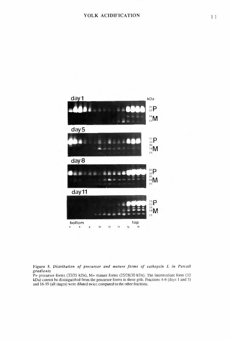
YOLK ACIDIFICATION 1 1
bottom top 4 6 8 10 12 M 16 18
Figure 5. Distribution of precursor and mature forms of cathepsin L in Percoli gradients P= precursor forms (33/35 kDa), M= mature forms (25/28/30 kDa). The intermediate form (33 kDa) cannot be distinguished from the precursor forms in these gels. Fractions 4-6 (days 1 and 5) and 16-19 (all stages) were diluted twice compared to the other fractions.

YOLK ACIDIFICATION 1 2
DISCUSSION
In early embryos the dense YSs are neutral, which is unusual for organelles of the endosomal-lysosomal compartment. As development proceeds, acidification of individual YSs occurs, in agreement with sequential YSs degradation observed at the ultrastructural level. To my knowledge this is the first report of developmentally regulated acidification of organelles.
Unfortunately this model did not allow quantitative studies. Yields of intact ECs after dissection and disaggregation were quite variable, and the more central ones, which resisted better to isolation, especially in later stages, were probably overrepresented. Since YSs degradation appeared more frequent at the periphery of the endoderm, this could explain the fact that fewer acidic YSs were found in intact ECs than among isolated YSs. On the other hand, some regulative mechanism may be suppressed during disruption of the cells, leading to rapid additional acidification. Quantitation was further hampered due to YSs breakage, and because the smallest YSs could not be unequivocally identified by fluorescence microscopy. Also, yolk degradation is a dynamic phenomenon; YSs may well have disappeared during AO incubations.
Cell fractionations have been been recently performed with Anemia and Drosophila embryos [18, 20, 15]. The density of the yolk platelets was found to decrease during development, concomittantly with their degradation. Here similar observations on decreasing density and degradation of the YSs could be correlated with their acidification. In Anemia and Drosophila, YSs containing mitochondria were found in the gradients. Similar images were also found in the present case (not shown), but corresponded in fact to small embryonic cells, hardly recognizable by EM, due to poor preservation of the ultrastructure, but easily identifiable by AO fluorescence microscopy.
The enzymatic and electrophoretic data obtained have to be considered more cautiously, due to contamination of the gradients by intact embryonic cells and extensive breakage of YSs. However mature cathepsin L is localized exclusively in fractions containig acidic YSs, which is in agreement with the in vitro processing of the precursor under acidic conditions [9]. Mature cathepsin L in intermediate fractions of the gradients was detected on gelatin/SDS-PAGE, but not enzymatically; the electrophoretic method is certainly the most sensitive. Moreover, leupeptin, which protects the unstable mature form [9], was present during storage in the first case, but not in the latter one. High N-acetylglucosaminidase activity was detected in these middle fractions, but the respective contributions of the YSs and the embryonic cells are unknown. Similarly the global increase of N-acetylglucosaminidase activity during development (Fig. 3 and ref. 8) may be attributed to growth of the embryonic tissues and/or to some activation of the enzyme contained within the yolk.
Early O.moubata eggs clearly contain two populations of YSs, which differ in density, ultrastructure, AO fluorescence as well as enzymatic content. The light YSs nearly disappear at half development. They thus constitute a particular yolk compartment that probably provides nutrients during early development, while dense spheres are left neutral, undegraded for a much longer period. The storage material of the light YSs, however, is still not defined biochemically. Light YSs resemble ultras true turally the acidic spheres found later, and both contained mature cathepsin L. The red dots, observed in isolated light YSs, but also sometimes in intact ECs, are probable signs of inhomogenous acidification. Likewise acidification (Fig. 2i) and degradation (Fig. 3b-d) of the dense YSs are not homogenous. Thus dense and light YSs may be degraded in a similar manner. Interestingly, a subpopulation of light YSs, enriched in glycosidase activities compared to the denser YSs, has been also reported both in the sea urchin [23] and Xenopus eggs [26].
A model for yolk degradation in O. moubata is proposed (Fig. 6). During embryonic development, a small number of YSs sequentially acidify, leading to maturation, and activation, of some cathepsin L. This accounts for the low rate of Vn degradation during this period [6]. In the larva, the yolk is found liquefied in the gut
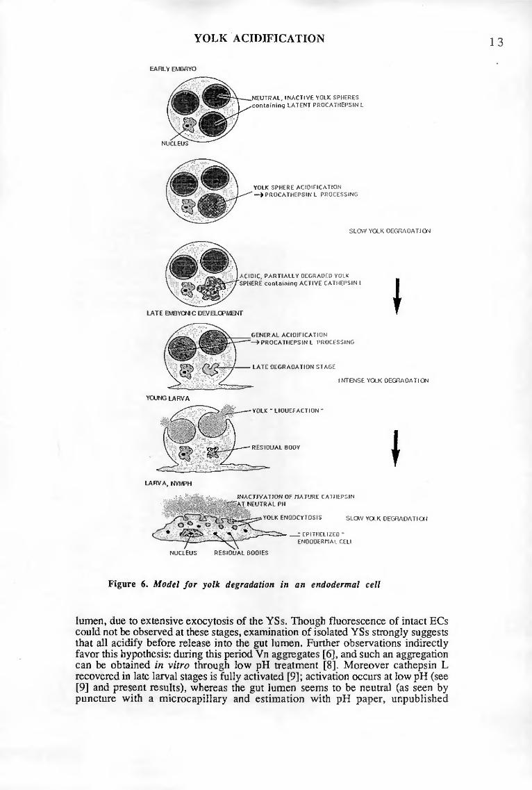
YOLK ACIDIFICATION
EARLY QVBRYO
NUCLEUS
NEUTRAL, INACTIVE YOLK SPHERES con ta i n i ng LATENT PROCATHEPSIN L
YOLK SPHERE ACIDIFICATION —> PROCATHEPSIN L PROCESSING
SLOW YOLK DEGRADATION
ACIDIC, PARTIALLY DEGRADfI) YOLK SPHERE c o n t o i n i n q ACTIVE CATHEPSIN I
I GENERAL ACIDIFICATION —»PROCATHEPSIN L PROCESSING
LATE DEGRADATION STAGE
YOLK "LIQUEFACTION "
INTENSE YOLK DEGRADATION
LARVA, NYMPH
RESIDUAL BODY
INACTIVATION OF MATURE CATHEPSIN
YOLK ENDOCYTOSIS SLOW YOLK DEGRADATION
_:EPITHELI2ED -ENDODERMA! CELI
NUCLEUS RESIDUAL BODIES
Figure 6. Model for yolk degradation in an endodermal cell
lumen, due to extensive exocytosis of the YSs. Though fluorescence of intact ECs could not be observed at these stages, examination of isolated YSs strongly suggests that all acidify before release into the gut lumen. Further observations indirectly favor this hypothesis: during this period Vn aggregates [6], and such an aggregation can be obtained in vitro through low pH treatment [8]. Moreover cathepsin L recovered in late larval stages is fully activated [9]; activation occurs at low pH (see [9] and present results), whereas the gut lumen seems to be neutral (as seen by puncture with a microcapillary and estimation with pH paper, unpublished

YOLK ACIDIFICATION 14
observations). This general acidification is probably responsible for the increased rate of Vn degradation reported by Chinzei [6j. Mature cathepsin L is unstable under neutral conditions [9]. Thus, once delivered together with the yolk into the gut lumen, it is probably rapidly inactivated. This could account for the dramatic decrease in proteinase activity observed during this period [8]. Thereafter Vn is slowly digested [6], through endocytosis by the gut epithelium (Diehl, personal communication).
Acidification has been recently mentioned as a putative mechanism for yolk degradation in the sea urchin [2,24, 28]. It is also implicite in the model proposed for Artemia [20]. In Artemia and Drosophila^ lysosotropic agents inhibit degradation [7, 14]. On the other hand, unlike the other species mentioned, the case of Artemia is rather peculiar: acid hydrolases has not been found in the yolk, but in classical lysosomes [18, 19], which seem to fuse with the YSs to initiate degradation [18, 20]. The situation is quite different in the other examples, including O. moubata^ where yolk platelets already contain all the hydrolytic machinery. Here YSs appear to be autonomous lysosomal-like organelles that may be activated simply through acidification. Acidification implies the presence of a proton-ATPase. This pump may be already inserted in the YS membrane from the beginning, since no patent signs of fusion of other organelles such as lysosomes was detected. In preliminary experiments, YSs acidifie ation was also found to occur in Xenopus embryos (unpublished observations), suggesting that it is a wide-spread mechanism. In O. moubata, pH may still play a further function, that is down-regulation of the digestion through final proteinase inactivation under neutral conditions, to keep nutrients for the next developmental stage.
ACKOWLEDGEMENTS
I thank Christine Kaufmann for assistance in the preparation of figures.
A preliminary report of this work was presented to the Union of the Swiss Societies for Experimental Biology ( Fagotto, F., 1990. Evidence that acidification of the yolk spheres promotes yolk digestion in tick eggs through activation of a procathepsin L. Experientia, 46, A82).
This work is part of the PhD thesis of the author.
LITERATURE CITED
1. Acschlimann, A., 1958. Développement embryonnaire d'Ornithodoros moubata (Murray) et transmission trans-ovarienne de Borrelia duttoni. Acta tropica, 15,15-62.
2. Armant, D.R., Carson, D.D., Decker, G.L., Welply, J.K. and W.J. Lennarz, 1986. Characterization of yolk platelets isolated from developing embryos of Arbacia punctuiata. Develop. Biol. 113, 342-355.
3. Barrett, AJ. and H. Kirschke, 1981. Cathepsin B, cathepsin H, cathepsin L. Methods EnzymoL 80, 535-561.
4. Byrne, B.M., Gruber, M. and G. Ab, 1989. The evolution of egg yolk proteins. Progr. Biophys. molec. Biol, 53, 33-69.
5. Chinzei, Y., Chino, H. and K. Takahashi, 1983. Purification and properties of vitellogenin and vitellin from a tick, Ornithodoros moubata. J.Comp.Physiol., 152, 13-21.
6. Chinzei, Y. and I. Yano, 1985. Vitellin is a nutrient reserve during starvation in the nymphal stage of a tick. Experientia, 41, 948-950.
7. Ezquicta, B. and CG. Vallejo, 1986. Artemia trypsin-like proteinase: developmental activation is inhibited by a lysosomotropic agent. CompBiochem.Physiol., 82B, 731-736.
8. Fagotto, F., 1990a. Yolk degradation in tick eggs: I. Occurence of a cathepsin L-like acid proteinase in yolk spheres, Arch. Insect Biochem. Physiol., in press.
9. Fagotto, F., 1990b. YoIk degradation in tick eggs: II. Evidence that cathepsin L-like proteinase is stored as a latent, acid activatable proenzyme. Arch. Insect Biochem. Physiol., in press.
10. Fagotto, F., Hess, E. and A. Aeschlimann, 1988. The early development of the argasid tick Ornithodoros moubata. Entomol.Gen. 13, 1-8.
11. Krischer, K.N. and EX. Chambers, 1970. Proteolytic enzymes in sea urchin eggs:
YOLK ACIDIFICATION 15
characterization and activity before and after fertilization. J. Ceil. Physiol, 76,23-36. 12. Lemanski, L.F. and R. Aldoroty, 1977. Role of acid phosphatase in the breakdown of
yolk platelets in developing amphibian embryos. J. MorphoL, 153', 419-426. 13. Lowry, O.H., Rosebrough, N.J., Farr, A.L. and RJ. Randall, 1951. Protein
measurement with the folin phenol reagent. J. Biol. Chem., 193, 265-275. 14. Medina, M., Leon, P. and CG. Vallejo, 1988. Drosophila cathepsin B-like proteinase: a
suggested role in yolk degradation. Arch, Biochem. Biophys., 263/2, 355-363. 15. Medina, M. and CG. Vallejo, 1989. The maternal origin of acid hydrolases in
Drosophila and their relation with yolk degradation. Develop. Growth & Differ., 31,241-247. 16. Opresko, L.K. and R.A. Karpf, 1987. Specific proteolysis regulates fusion between
endocytic compartments in Xenopus oocytes. Cell, 51,557-568. 17. Pasteels, JJ., 1973. Yolk and lysosomes. In Lysosomes in Biology and Pathology,
vol. 3 (Ed. J.T. Dingle), pp. 216-233, North-Holland Publishing Co., Amsterdam. 18. Perona, R., Bes, J.-C. and CG. Vallejo, 1988. Degradation of yolk in the brine shrimp
Anemia. Biochemical and morphological studies on the involvement of the lysosomal system. Biol. Cell., 63, 361-366.
19. Perona, R. and CG. Vallejo, 1985. Acid hydrolases during Artemia development: a role in yolk degradation. Comp. Biochem. Physiol., 81£, 993-1000.
20. Perona, R. and CG. Vallejo, 1989. Mechanisms of yolk degradation in Artemia: a morphological study. Comp. Biochem. Physiol., 94A, 231-242.
21. Postlethwait, J.H. and F, Giorgi, 1985. Vitellogenesis in Insects. In Developmental Biology: a Comprehensive Synthesis, vol. 1: Oogenesis (Ed. W.L. Browder), pp. 85-126, Plenum Press, New York.
22. Purcell, J.P., Quinn, T.M., Kunkel. J.G. and J.H. Nordin, 1988. Correlation of yolk phosphatase expression with the programmed proteolysis of vitellin in Blattella germanica during embyonic development. Arch. Insect Biochem. Physiol., 9, 237-251.
23. Schuel, H., Wilson, W.L., Wilson, J.R. and R.S. Bressler, 1975. Heterogenous distribution of "lysosomal" hydrolases in yolk platelets isolated from unfertilized sea urchin eggs by zonal centrifugation. Develop. Biol., 46,404-412.
24. Scott, L.B., Leahy, P.S., Decker, G.L. and WJ. Lennarz, 1990. Loss of yolk platelets and yolk glycoproteins during larval development of the sea urchin embryo. Develop. Biol., 137, 368-377.
25. Vernier, J.-M. and M.-F. Sire, 1977. Plaquettes vitellines et activité hydrolasique acide au cours du développement embryonnaire de la Truite arc-en-ciel. Étude ultrastructurale et biochimique. Biol. Cell, 29, 99-112.
26. Wall, D.A. and I. Meleka, 1985. An unusual lysosome compartment involved in vitellogenin endocytosis by Xenopus oocytes. J. Celt Biol. JOl, 1651-1664.
27. Wallace, R.A., 1985. Vitellogenesis and oocyte growth in nonmammalian vertebrates. In Developmental Biology: a Comprehensive Synthesis, vol. 1: Oogenesis (Ed. W.L. Browder), pp. 127-166, Plenum Press, New York.
28. Yokota, Y. and K.H. Kato, 1988. Degradation of yolk proteins in sea urchin eggs and embryos. Cell Differ., 23, 191-200.

IV

TICK EGG NEUTRAL PROTEINASE 1
IV. LIMITED PROTEOLYSIS OF VITELLIN BY A NEUTRAL PROTEINASE (Unpublished results)
INTRODUCTION
In all arthropod eggs so far examined, both an acid cysteine and a neutral, trypsin-like proteinase are present {Anemia [1,11,12], Locusta [7,13], Bombyx [5,6], Drosophila [9,10]). In Artemia [12] and Drosophila [9,10], the acid proteinase appears to be responsible for vitellin degradation, while the neutral enzyme only partially degrades one specific subunit. In Bombyx [5], the trypsin-like enzyme even does not act on vitellin, but specifically cleaves the egg specific protein (ESP).
During my work on tick egg cathepsin L, a similar neutral activity was also detected. I report here the few data obtained.
MATERIALS AND METHODS
Ornithodoros moubata eggs were obtained from artificially fed females as described [2,3] and were incubated at 290C. One egg or larva (about 100-150 \i% protein) was homogenized in 50 \i\ buffer (25 mM Tris-HCl, pH 7.0, Hepes-NaOH, pH 6.5-8, sodium acetate, pH 4.5-6, or sodium formate, pH 3.5). When required, activators or inhibitors were added. The samples were incubated at 370C for 1 h, then mixed with 50 p.1 SDS-PAGE sample buffer (0.1 M Tris-HCI buffer, pH 6.8, containing 10% glycerol, 2% SDS, 2% mercaptoethanol and 0.005% bromophenol blue), and immediately boiled for 5 min. After centrifugation to eliminate insoluble material, 5 fil aliquots were analyzed by SDS-PAGE (10 % acrylamide) using the Laemmli's buffer system [8]. The gels were stained with Coomassie blue and destained in acetic acid/methanol.
RESULTS AND DISCUSSION
Incubation of crude homogenates under strong acidic conditions (pH 3.5) resulted in vitellin aggregation and extensive degradation [2]. SDS-PAGE analysis revealed several intermediates (not shown). Aggregation and degradation were completely inhibited by 1 p.M leupeptin, but not by pepstatin, confirming the previous enzymatic and chromatographic results.
Analysis of crude homogenates from early embryos (day 1 to 3) incubated at pH 7, revealed not even the slightest modification of the banding pattern (not shown). However, when older stages (day 4 to 14) were assayed (Fig. 1), the larger vitellin subunit (160 kDa) was converted to two fragments (70 and 80 kDa). No other vitellin subunit was affected.
Cleavage of the 160 kDa subunit was slow under normal conditions (lane 2), but was strongly enhanced by 1 mM EDTA (lane 3). 0.1 to 10 mM Ca 2 + and 1 mM Mg 2 + were also activators, but to a lesser extent (lanes 8,9). Total inhibition was achieved by adding 1 mM Hg2 + or 1 |iM leupeptin. PMSF had no effect (lane 7). Optimum pH was in the range of 5.5 to 7.5 (lanes 13-18), proteolysis being negligible at lower pHs (4.5-5) (lanes 11-12).
The absence of inhibition by PMSF and EDTA precludes the involvement of trypsin-like, respectively metallo-proteinases, while the efficiency of H g 2 + and leupeptin suggests that the enzyme may be a thiol-proteinase. However activation by both EDTA and Ca2 + is very puzzling. Clearly, much more work will be needed for proper characterization of this enzyme. A prerequisite step would be the finding of suitable exogenous, preferably synthetic, substrates.

TICK EGG NEUTRAL PROTEINASE
The actual function of this proteinase is also unknown. Subcellular localization, which is a crucial indication, is presently missing. Thus one has to speculate. It is possible that this enzyme has nothing to do with yolk degration; it may be a cytoplasmic enzyme like calpain. Nevertheless the selectivity and efficiency in degrading one vitellin subunit, together with the appearence of the activity at day 4, which corresponds to the onset of yolk spheres acidification [4] and cathepsin L activation [3], favor the hypothesis of a role in yolk degradation. Localization within the yolk would imply that it has already been stored during oogenesis. In this case the enzyme would be latent during the first 4 days. Similar results were obtained from Anemia embryos: the neutral proteinase is latent in dormant embryos, because of inhibition by vitellin, and then activated through vitellin degradation, due to the thiol-cathepsin [I].
The proteolytic fragments of the 160 kDa subunit are not detected in vivo during development. One can suppose either that only small amounts are formed and readily degraded further, or that proteolysis is inhibited in vivo during most of the development.
^ 1 2 3 4 5 6 7 8 D 10 11 12 13 14 15 16 17 18 ^
xl000 XlOOO
130 — 115 — 90 ~ " * t • • • § » # • • • • • • • • • — « o
m m «•* m *m **• ~ ~ 70 60 — 4 m m
50 — tt«
LEGENDS
Before electrophoresis, crude homogenates (day 6) were incubated for 1 h at 370C, under the following conditions: lane 1, no incubation; lane 2, pH 7.0; lane 3, pH 7.0, 1 mM EDTA, lane 4, pH 7.0, 5 mM cysteine, lane 5, pH 7. 0, 5 mM cysteine, 1 mM EDTA; lane 6, pH 7.0, 1 mM EDTA, 1 uM leupeptin; lane 7, pH 7.0, 1 mM EDTA, 1 mM PMSF; lane 8, pH 7.0, 10 mM Ca2+ ' lane 9, pH 7.0, 10 mM Mg2+; lane 10, pH 7.0, 1 mM EDTA, 1 mM Hg2+; lane 11, pH 4.5; lane 12, pH 5.0; lane 13, pH 5.5; lane 14, pH 6.0; lane 15, pH 6.5; lane 16, pH 7.0; lane 17, pH 7.5; lane 18, pH 8.0. Buffers used were: 1-10, Tris-HCl; 11-14, sodium acetate; 15-18, Hepes-NaOH. Mr of the vitellin subunits are indicated on the left side, and those of the two proteolytic fragments on the right side. The M1- of the vitellin subunits does not correspond exactly to the one reported by Chinzei et al (J. Comp. Physiol., 152, 1983, 13-21).

TICK EGG NEUTRAL PROTEINASE 3
LITERATURE CITED
1. Ezquieta, B. and C. G. Vallcjo, 1985. The trypsin-like proteinase of Anemia: Yolk localization and developmental activation. Comp. Biochem. Physiol., 82B1731-736.
2. Fagotto, F., 1990. Yolk degradation in tick eggs: I. Occurence of a cathepsin L-like acid proteinase in yolk spheres. Arch. Insect Biochem. Physiol., 14, 217-235,
3. Fagotto, F., 1990. Yolk degradation in tick eggs: II. Evidence that cathepsin L-like proteinase is stored as a latent, acid activatable proenzyme. Arch. Insect Biochem. Physiol., 14, 237-252.
4. Fagotto, F., 1990. Yolk degradation in tick eggs: III. Developmentally regulated acidification of the yolk spheres. Develop., Growth & Differ., in press.
5. Indrasith, L. S., Sasaki, T. and O. Yamashita, 1988. A unique protease responsible for selective degradation of a yolk protein in Bombyx mori. Purification, characterization and cleavage profile. J. Biol. Chem., 263,1045-1051.
6. Kageyama, T., Takahashi, S. Y. and K. Takahashi, 1981. Occurrence of thiol proteinases in the eggs of the silkworm. Bombyx mori.. J. Biochem., 90,665-671.
7. Kuk-Meiri, S., Lichtenstein, A., Shulov, A. and M. P. Pener, 1966. Cathepsin-like proteolytic activity in the developing eggs of the African migratory locust {Locusta migratoria migratorioides R&F). Comp. Biochem. Physiol, 18, 783-795.
8. Laemmli, U., 1970. Cleavage of structural proteins during assembly of the head of bacteriophage T4. Nature (London), 227,680.
9. Medina, M., Leon, P. and C. G. Vallejo, 1988. Drosophila cathepsin B-like proteinase: a suggested role in yolk degradation. Arch. Biochem. Biophys., 263, 355-363.
10. Medina, M. and C. G. Vallejo, 1989. The maternal origin of acid hydrolases in Drosophila and their relation with yolk degradation. Develop. Growth & Differ., 31,241-247.
11. Perona, R. and C. G. Vallejo, 1982. The lysosomal proteinase of Artemia. Purification and characterization. Eur. J. Biochem., 124,357-362.
12. Perona, R. and C. G. Vallejo, 1985. Acid hydrolases during Artemia development: a role in yolk degradation. Comp. Biochem. Physiol, 81M, 993-1000.
13. Shulov, A., Pener, M. P., Kuk-Meiri, S. and N. Lichtenstein, 1957. Proteolytic enzymes of the eggs of Locusta migratoria migraàoroides. J. Insect Physiol, 1,279-285.

V

GUT MORPHOGENESIS 1
V. FATE OF THE ENDODERMAL CELLS AND GUT MORPHOGENESIS (Unpublished results)
INTRODUCTION
In arthropods the yolk is typically confined to the inner part of the egg (centrolecithal organization) [3,7,9]. This "yolk system" is usually considered not to be involved in the formation of the embryonic tissues, which originate from the peripheral, yolk-free blastoderm. The yolk system has been classically represented as an amorphous mass containing free-swimming cells, called "vitellophages", which seemed responsible for yolk utilization [3]. This image, primarily based on light microscopical observations, has been questioned by electron microscopy studies, which demonstrated that the insect yolk is organized syncytially [8]. In some cases, the syncytium is secondarily fragmented in large "yolk cells" [10].
Unlike fore- and hindguts, whose ectodermal origin has been clearly demonstrated [3,7], the formation of the midgut is not well established [8]. In pterygote insects, many different modalities were reported [7]. Basically, endodermal cell proliferation was observed somewhere deep in the germ band, followed by cell migration and formation of an epithelial sheet. This epithelium progressively enclosed the yolk mass. However, most of the studies reported were exclusively based on light microscopy observations of paraffin sections. In more recent studies on some pterygotes, involvement of yolk cells has been suggested [8]. In apterygote insects, myriapods, crustaceans and arachnids, contribution from large, yolky cells to gut formation is frequent [3]. This has been satisfactorily established in eggs containing moderate amounts of yolk. In heavily yolk-loaded eggs, cellularization of the central part has probably been often overlooked, due to the low resolution of the optical microscopy and unsatisfactory fixation of the yolk. Nevertheless, the yolk system often appears to be more than simply a mass of nutrients, but a true embryonic tissue.
In ticks, the gut is essentially composed of an unistratified digestive epithelium, surrounded by two thin layers of smooth muscle [4]. Tick development resembles the one of insects, and according to Aeschlimann [1], gut formation is similar, i.e. through encircling of the yolk starting from the germ band region. Possible secondary contribution of vitellophages was suggested.
Using the more powerful electron microscopy, the reality of vitellophages in ticks has been recently refuted, the central part of the egg being entirely cellularized, composed of huge, yolk-filled cells, tentatively called primary endodermal cells (endodermal cells I) [6]. Of course questions arise about the fate of these cells as well as about gut morphogenesis. I report here preliminary morphological observations collected during my study on yolk degradation.
MATERIALS AND METHODS
O. moubata eggs (days 5-10) and young larvae (days 11-12) were fixed and processed for TEM (transmission electron microscopy) as described [5,6]. Semi-thin sections for light microscopy were stained with toluidine blue. For SEM (scanning electron microscopy), eggs (days 7-9) were dissected and the central endoderm was isolated, fixed as for TEM, dehydrated, critical point-dried, carbon-and gold-coated and observed in a Philips SEM. For in vitro observations, endodermal cells from 3-6 day -old embryos were isolated and incubated individually, at 290C, in small wells in 25 mM Hepes-NaOH buffer, pH 7.6 or 6.8, containing 200 mM NaCl, 50 mM KCl11 mM CaCl2, or Hank's 199 Medium with glutamine and calcium (Gibco, Paisley, UK), supplemented with 25 mM Hepes-NaOH and \% NaCL Antibiotics (10 U/ml penicillin, 10 U/ml streptomycine, Gibco, Paisley, UK) were added. Observations were performed using an inverted microscope.

GUT MORPHOGENESIS 2
RESULTS AND DISCUSSION
Most endoderma! cells appeared essentially unchanged until day 9, except in the region underlying the germ band. In 5-7 days old embryos, this region was largely yolk-depleted, due to intense yolk degradation in the endodermal cells. In disagreement with earlier descriptions [1], which reported invasion by cells originating from the germ band, this region was exclusively composed of primary endodermal cells; embryonic tissues (ecto- and mesoderm) were found to proliferate on the surface, but did not penetrate the yolk.
During the second half of embryonic development, a thin layer appeared to enclose the yolk system. According to Aeschlimann [1], this layer was composed of migrating endodermal cells (here named secondary endodermal cells, or endodermal cells II), which would form the gut epithelium. These cells were reported to originate from the deep region of the germ band. Since this region seemed to be in fact composed of endodermal cells I (see above), I suggest that endodermal cells II may arise from proliferation of these yolk-depleted cells (Fig. 1 and 2A). This part of the egg would merit further examination, particularly to discover if the endodermal cells actually proliferate once free of yolk. However the occurrence of complex structures, due to extensive yolk degradation, make ultrastructural investigations difficult. Also, the putative secondary endoderm is such a thin layer (often less then 1 |im), that I wonder whether its origin or its fate could be assessed histologically without ambiguity. As seen at the ultrastructural level, contribution of myoblasts, which would constitute the outer muscular layer, was likely (Fig. 3). Moreover, at least some of the numerous overlapping cell processes clearly belonged to yolky, primary endodermal cells (Fig. 3).
An attempt has been made, with the help of C. Kaufmann, to examine the surface of the forming gut by SEM, after removal of the germ band and the ectoderm. In early stages of gut morphogenesis (days 6-8), the dorsal side, still not enclosed by the secondary endoderm, broke, due to lack of adhesion between endodermal cells. Perfectly intact yolk masses could be isolated only from the latest stages (day 9), when closure of the gut wall was almost complete (Fig. 7). The surface appeared to be constituted of flat cells. At their limits small microvilli and cell processes were found, overlapping each other (Fig. 8-10). They were particularly numerous in the ventral region, where the cells were smaller (Fig. 8). The surface progressively flattened towards the dorsal side (Fig. 9,10), where cell limits were often hard to distinguish, even at high magnification. In the most dorsal region, some apparently still uncovered endodermal cells I could be found (Fig. 10). Their limits also displayed microvilli and processes that were entangled with those of the adjacent cells. However the actual characterization of these superficial cells was unclear (endodermal cells II, myoblasts, maybe outer parts of endodermal cells I,...).
While the modalities of the formation of the secondary endoderm still remain speculative, the present observations strongly suggest that endodermal cells I are involved in gut morphogenesis. By TEM, they were found to establish focal contacts, both with other endodermal cells I and with overlying, probably endodermal cells II (Fig. 4). Just before hatching, not only endodermal cells II, but also sometimes endodermal cells I, displayed septate junctions, which are typical structures of invertebrate epithelia [2]. Like the endodermal cells II, the primary endodermal cells were also bordered by a basal lamina. Thus endodermal cells presumably underwent "epithelization" in late embryonic development. This was supported by the fact that, during dissection, they appeared extensively attached to the gut wall. On the last days, gut diverticles appear, due to the lateral pressure of the newly formed dorso-ventral muscles on the gut wall [I]. In fact, each diverticle was found to enclose a small group of endodermal cells I, in such a way that eventually all cells were attached to the gut wall.
From day 9 onwards, closure of the gut being near completion, the yolk was progressively found liquefied in the lumen [Diehl, unpublished results]. In fact, the

GUT MORPHOGENESIS 3
endodermal cells I expelled their yolk sphere content through extensive exocytosis (Fig. 5,6). Different stages of this phenomenon could be recognized (see ref. 5, Fig. 1) and a scheme is presented in Fig. 11: "quiescent" endodermal cell I had a rather regular outline, then the cytoplasm withdrew from the periphery, so that yolk spheres became protruding, and yolk sphere unit membranes were in close contract with the plasma membrane (Fig. 5,6). As endodermal cells I lost their yolk content, they became progressively smaller and flat. In parallel, extensive intracellular yolk sphere degradation was also observed (Fig. 6). All endodermal cells I were always connected to the peripheral gut wall.
When endodermal cells I (3-6 days) were isolated and maintained in vitro, they remained unchanged for some time (1 to 5 days), but then, as could be judged at the low resolution of the inverted microscope used, they underwent the same evolution found in vivo during yolk liquefaction, i.e. first bulging, then progressive loss of the yolk spheres. Finally, they became huge amoeboid cells, filled with many réfringent round organelles, maybe secondary lysosomes. The factors inducing these changes, which could occur soon after isolation or 5 days later are unknown, but apparently endodermal cells I were able to transform themselves into epithelial-like cells in vitro.
Evidently much more work is necessary to elucidate the modalities of gut morphogenesis. The few data available, however, are not in full agreement with the classical description, which led me to propose an alternative model. To my belief, contribution of the central endodermal cells Ï is essential in the present case, unlike what is claimed in pterygote insects.
The central yolky cells of the O. moubata egg have all the feature of typical endodermal cells: central location, high yolk content, primordial role in yolk degradation. In early embryos, these primary endodermal cells constitute a poorly organized mass with no or weak cell to cell adhesions. From day 6 onwards, cell contacts are progressively established, first in the ventral region, where the yolk is already intensively digested, so that cell changes may be made more feasible. "Epithelization" then reach the dorsal side, as well as the egg centre, through formation of diverticules, yielding a convoluted epithelium (Fig. 11). Proliferation of the ventral, yolk-depleted endodermal cells I may provide a source of small cells, which may become the endodermal cells n, and will migrate over the surface of the primary endoderm (Fig. 2A); it cannot be excluded that some other primary endodermal cells, still filled with yolk, may also divide into a small, yolk-free, and a large, yolk-rich cells (Fig. 2B,C), as proposed in some insects [8]. Both such small (type II) and the large (type I) endodermal cells would be inserted into the epithelium. As soon as tightness of the gut has been established, the endodermal cells I begin to pour the yolk into the newly forming gut lumen (Fig. 11). Once the yolk has been expelled, the endodermal cells I, now thin epithelial cells, may participate, together with the small endodermal cells II, in the functions of the gut epithelium (i.e. recapture and digestion of the remaining yolk, then finally blood digestion). In the larva, endocytosis and lysosomal degradation of the liquid yolk probably start while exocytosis by the endodermal cells I has not yet been terminated [Diehl, personal communication]. In the present work, the late larval stages (days 13-14) have not been examined.
This model is of course highly speculative, but fits best the preliminary data obtained. It further reconciles the occurence of central yolk-rich, typically endodermal, cells in early embryos with the formation of a more peripheraly located digestive tissue.

GUT MORPHOGENESIS 4
Figure 1: Schematic tranverse section through a 6 days-old embryo (A) and of a larva (B). A: A thin layer of secondary endodermal cells is enclosing the central core of yolk-filled endoderma! cells I. B. Closure of the gut epithelium (endoderm II) is complete. Some endodermal cells I are still filled with yolk, but most have released their content into the lumen.
Figure 2; Possible modalities of gut morphogenesis: A: Epiboly: Endodermal cells I located on the ventral side (*) digest intensively the yolk, proliferate, and become endodermal cells II, which migrate on the surface of the yolk mass. B: Fragmentation: Peripheral endodermal cells I divide into several smaller cells which will transform into epithelial cells. C: Unequal cleavage: The outer part of an endodermal cell I, which contain few yolk spheres, separates from the inner, yolk-rich part through unequal division. The former become an endodermal cell n.
Figures 3-4: Ultrastructure of the yolk surface in late embryos. Fig. 3; Day 9, endodermal cells I are overlaid by several cell processes, one of which (small arrows) is clearly connected to an endodermal cell I, containing yolk spheres (YS). Another process shows numerous cytoplasmic filaments (large arrow) and probably belong to some myoblast. Fig. 4: Day 8; endodermal cells I (and II ?) establish numerous focal contacts (arrowheads). H, hemocoel; M, myoblast; YS, yolk sphere. Bars: 1 urn.
Figures 5-6: Yolk exocytosis in the larva, day 12. Fig. 5: bulging of yolk spheres (YS). In one case, almost all the cytoplasm has withdrawn, and yolk spheres- and plasma membranes are in close contact (arrow). Fig. 6: yolk sphere (YS) just before exocytosis. Membranes are in close contact (arrow). Intracellular digestion is intense (d). *: liquefied yolk in the gut lumen. Bars: 1 |un.
Figures 7-10: Scanning electron microscopy of the endodermal surface after removal of the epiderm, day 9. Fig. 7: dorsal view, showing ihe diverticules (stars). Arrows: epiderm. Fig. 8-10: cell limits, displaying numerous processes. Fig. 8: ventral; Fig. 9: equatorial; Fig. 10: dorsal; *: possible "naked" endodermal cells I. Bars: 200 m (5), 5 urn (6-8).

GUT MORPHOGENESIS 5
<H
1 >
r .
>
b -m
*
>
rr i
Jl

GUT MORPHOGENESIS 6
11
v 4^1 «amp 8H lIJiiiiL^
n
li»»»«^x
08¾¾^-"-*¾¾
l ' sBi
YS
BSKk^x
4
N «r
rat «L
J
Figure 11: Schematic view of the larval gut. 1-5: the primary endodermal cells are progressively transformed into epithelial cells by releasing, as well as degrading, the yolk. They are inserted into the gut epithelium, composed of small secondary endodermal cells (arrowheads). *, gut lumen, filled with liquid yolk; j , junctional complex (septate junctions); N, nucleus; YS, yolk spheres; d, yolk degradation.

GUT MORPHOGENESIS 7
LITERATURE CITED
1. Aeschlimann, A., 1958. Développement embryonnaire d' Ornithodoros moubata (Murray) et transmission trans-ovarienne de Borrelia duttoni. Acta tropica, 15,15-62.
2. Alberts, B., Bray, D., Lewis, J., Raff, M., Roberts, K. and J. D. Watson, 1983. Molecular biology of the cell. Garland Pubi., Inc., New York.
3. Anderson, D.T., 1973. Embryology and phylogeny in annelids and arthropods. Pregamon Press, Oxford.
4. Coons, L. B., Roseli-Davis, R. and B. I. Tarnowski, 1986. BIoodmeal digestion in ticks. In: Morphology, physiology, and behavioral biology of ticks. (Eds., J. R. Sauer and J. A. Hair), pp. 248-279, Wiley, New York.
5. Fagotto, F., 1990. Yolk degradation in tick eggs: III. DevelopmentalIy regulated acidification of the yolk spheres. Develop., Growth & Differ., in press.
6. Fagotto, F„ Hess, E. and A. Aeschlimann, 1988. The early development of the argasid tick Ornithodoros moubata. Entomol. Gen. 13, 1-8.
7. Haget, A., 1977. L'embryologie des Insectes. In : Traité de Zoologie, vol. 8/5B (Ed. P.-P. Grasse), Masson, Paris.
8. Mori, H., 1983. Origin, development, morphology, functions and phylogeny of the embryonic midgut epithelium in insects. Entomol. Gen., 8, 135-154.
9. Sander, K., Gutzeit, H. O. and H. Jackie, 1985. Insect embryogenesis: morphology, physiology, genetical and molecular apsects. In: Comprehensive insect physiology, biochemistry and pharmacology. Vol. 1: Embryogenesis and reproduction (Eds. G.A. Kerkut and L. I. Gilbert), pp. 319-385, Pergamon Press, Oxford.
10. Takami, T., 1954. Studies on the yolk cell in Bombyx mori. 1. Early division of yolk cells and some related problems. Cytologie, 19,299-305.

VI
Entomol. Gener. 13(1/2):001-008 Stuttgart 1988-05 ISSN: 0171-8177
EGT-Nr 584 1
The early Embryonic Development of the Argasid tick Ornithodorus moubata (Acarina: Ixodoidea: Argasidae)
François Fagotto*, Ernst Hess & André Aeschlimann
Received: 1987-05-06/1987-08-27 Accepted: 1987-09-09
Fagotto, E, Hess, E., & Aeschlimann, A. [Inst. Zool., Univ., CH-20Q0 Neuchatel]: The early Embryonic Development of the tick Ornithodorus moubata (Acarina: Ixodoidea: Argasidae). — Entomol. Gener. 13(1/2): 001-008; Stuttgart 1988. - [Article].
The mode of cleavage of the centrolecithal tick eggs has so far been considered to be superficial. Evidence could be obtained from an electron-microscopic study of Omtthodoros moubata Murray 1877 that cleavage of this species is total. The early development leads to a diploblastic stage composed of a superficial layer of yolk-free micromeres (ecto-mesoderm) and a core of yolk-rich macromeres (primary entoderm).
Key w o r d s : embryonic development, cleavage, Ornithodoros moubata, ticks.
1 Introduction
Arthropod eggs commonly have a centrolecithal structure and a superficial cleavage pattern. This particular pattern, unknown from the other phyla, has been well studied in pterygot insects, where we find the very classical examples such as DrosophiU, Smittia [Sander et al 1985]. Too often, however, superficial cleavage is considered to be the rule in Anhropoda, whereas actually many primitive species of lnsecta, Myriapoda and Chelìcerata exhibit total cleavage [Anderson 1973]. Blastulation of spider and crustacean eggs does not proceed in an orthodox manner either, even though the final result, a blastoderm coating an acellular yolk mass, is considered by many authors to be identical to an insect blastula. In Arachnida and Crustacea, eggs divide totally during the migration of the cleavage nuclei, and intralecithal partitions seem to be aborted only secondarily. Such a mode of cleavage has been called "mixed" [Dawydoff 1928, Fioroni 1970]. One generally admits that superficial and mixed cleavage patterns are characteristic of the more evolved groups of Anhropoda, and that they are related to the high yolk content of their eggs. Studies on the embryology of Acarina are very scarce [Aeschlimann 1958, Anderson 1973, Krantz 1978, Aeschlimann & Hess 1984]. They chiefly ,deal with general morphology, lacking, however, ultra-structural or experimental approaches. The accurate description of the embryonic development of Ornitbodoros moubata Murray 1877, an African soft tick, by Aeschlimann [1958], is still considered as the reference example for embryogenesis of Acarina.
The O. moubata egg is centrolecithal, with a large amount of brown-colored yolk embedded in a thin cytoplasmic layer. According to Aeschlimann's description [1958], cleavage is superficial, the egg preserving a syncytial organisation as the dividing nuclei migrate towards the yolk surface. After 8 mitotic cycles the cleavage nuclei reach the periphery and subsequently cellulation occurs, leading to the formation of the superficial blastoderm. Some nuclei, called
* Part of PhD thesis of the first author
1 Eniomol. Gener. 13 0171-8177/88/0013-0001 $ 2.-© 19B8 E. Schweizerbart'sche Verlagsbuchhandlung, D-7000 Stuttgart 1

002 — F. Fagotto, E. Hess & A. Aeschlimann
viteliophages, leave this layer and penetrate the yolk mass. This description is based on low magnification observations of living embryos, and on light microscopic histological studies of eggs which are less than 1 mm 0 . Under the initiative of Prof. Aeschlimann, the early development of O. moubata is reinvestigated at the ultrastructural level in order to verify the cleavage pattern and to study the viteliophages in a more detailed manner.
2 Material and Methods
The ticks used for this study were reared by membrane feeding on swine blood. The fed and mated 99 were maintained at 27°C. A patch of eggs was fixed every 12 hours, the precise stage of each embryo being determined later according to histological characteristics, mainly from the position of the cleavage nuclei. For fixation, the eggs were immersed in glutaraldehyde [Sabatini et al 1963] containing 4—5% sucrose, and the egg shell was pierced using a tungsten needle. The hypertonic solution compensates the high internal hydrostatic pressure of the embryos and thus prevents their explosion. Immediately after their perforation, the embryos were transferred to fixative at normal osmolarity (2% sucrose), and kept there for 24 h at 4CC, then rinsed in phosphate buffer and postfixed for 2 h with osmium tetroxide [Palade 1952]. After dehydration, the specimens were embedded in Spurr's resin [Spurr 1969]. Thin sections stained with uranyl acetate and lead citrate were observed on a Philips EM210 Transmission Electron Microscope [TEM]. Semi-fine sections were stained with toluidine blue. In order to isolate macromers from more than 2 d old embryos for Scanning Electron Microscopic [SEM] studies, the following method was used: an egg was repeatedlv and violentlv sucked towards the broken opening of a pipette of which the 0 was slightly smaller than the cg£. Thus the shell was slit open and cnuld easily he removed. The embryos were then dissociated on glass slides in 0.2 h phosphate buffer containing 5% sucrose and 40 mg/100 ml EDTAH2. After 30 min, this solution was replaced by an equivalent bufier without EDTA1 containing traces of CaCIi. After 1 h, were most of the isolated cells adhered to the glass where they were fixed as for TEM. Acetone dehydrated specimens were critical-point dried with CO2 and coated with carbon and gold before being observed in a Philips PSEM 500.
3 Results
3.1 Progress of cleavage
From the 8-cell stage on (beginning of the observations) the blastomeres are arranged radially. They are completely separated from each other. After each division the cells become narrower, but their length does not change. Thus, each blastomere has a pyramidal shape and extends from the center to the
Fig 1: Ornithodoros moubata Murray 1877 [Ixodoidea: Argasidae], schematic representation of the total cleavage pattern. — (a) Stage 18 h. The embryo is composed of pyramidal blastomeres, of which the nuclei migrate towards the periphery, (b) Stage 25 h (blastula). The cleavage nuclei have reached the periplasm. (c) Stage 35 h (diploblastic stage). Each pyramidal blastomere has divided into an outer micromere and an inner macromere, resulting in a superficial ectomesoblast surrounding the central primary entoderm. n nucleus, p periplasm, pc perinuclear cytoplasm, r reticuloplasm, v "viteliophages", y yolk.

The early Embryonic Development — 003
periphery (Fig 1 a). During this early phase, the nuclei migrate synchronously toward the surface of the egg reaching the limit of the periplasm after 7—8 mitotic cycles (stage 24 h). At this stage which is defined as the blastula, the pyramidal blastomeres are still arranged radially (Fig lb). During the following division (8th or 9th mitotic cycle), each blastomere divides into an outer micromere and an inner macromere, most of the periplasm being separated from the yolk (Fig Ic). Thus, after 30—35 h of development, the egg is composed of 2 sheets: the central sphere of macromeres containing all the yolk platelets, which is considered as the primary entoderm, and a thin external layer of yolk-free micromeres. Cleavage ends with this diploblastic stage. From this moment on, each embryonic sheet follows its own fate. Some of the macromeres are supposed to build up the embryonic midgut epithelium or secondary entoderm, which appears at day 6, while most of them probably divide only once more and then remain unchanged until hatching. Their fate has not yet been studied in detail.
3.2 Morphology and cytology of blastomeres
The observation of serial sections in LM and TEM reveals that the early embryo is composed of individual cells. These seem to have a simple pyramidal shape under low magnification, but semi-fine sections and TEM observations show that their morphology is highly complicated, characterized by numerous lobes (Fig 2).
Fig 2: Semi-schematic transverse section of an Ornitbodoros moubata Murray 1877 [Ixodoidea: Argasidae], embryo at stage 16 h during late mitosis. One of the 8 dividing blastomeres is drawn in detail. It is composed of numerous lobes, ex extracellular space, f furrow, n nucleus, p periplasm, pc perinuclear cytoplasm, pm plasma membrane, r reticuloplasm, s egg shell, sp spindle, yp yolk platelet surrounded by a unit membrane.
The blastomeres are filled with dense and homogenous yolk platelets (20—50 ^m 0 ) grouped in clusters of various sizes. The yolk mass is so compact and so condensed that the platelets, which are spherical during oogenesis, are now polygonal, touching each other tightly (Fig 2, 4). Yolk-free cytoplasm is restricted to a very small volume. It comprises the perinuclear cytoplasm, the periplasm and the reticuloplasm (Fig 2). The perinuclear cytoplasm represents the major part, and it can be compared with the insect energid. The periplasm has a maximum thickness of 5 (um in the early stages. Subsequently it thickens and raises up to 20 pm at the blastula. Some yolk-free cytoplasm is found ± randomly distributed between the yolk platelets, forming the reticuloplasm. The yolk-free cytoplasm strongly resembles the cytoplasm of the mature ovocyte [Aeschlimann & Hecker 1969], since mainly high quantities of glycogen, lipid droplets, rìbosomes and mitochondria are found (Fig 3). It is divided imo large transparent zones, where glycogen is concentrated and electron denser cytoplasm containing the organelles. It is characterized by numerous microtubules and endoplasmic reticulum. The network of microtubules is continuous with the mitotic apparatuses (spindles and asters). The latter are striking because of their large size (spindles are up to 50 ^m long), and because they are still present during early

004 — F. Fagotto, E. Hess & A. Aeschlimann
interphase. The nuclei have a 0 « 15 firn, and are of a very irregular shape. They are euchromatic and no nucleoli occur earlier than at the blastula stage. Nuclear pores are extremely numerous, especially at the end of cleavage. The plasma membrane presents numerous, sometimes deep invaginations the number of which decreases during development. These pass through the reticuloplasm (Fig 3), but also directly between the yolk platelets (Fig 5). The invaginations are either simple folds separating the lobes of the blastomeres, or they are so called furrows and present a characteristic terminal structure. The furrows always originate from cell-boarding cytoplasm (periplasm or reticuloplasm) (Fig 6), and then penetrate between the yolk platelets in the deeper regions. They show a terminal bud (the base) surrounded by vesiculated cytoplasm (Fig 7). Behind the base, the furrow is closed so that 2 plasma membranes are in intimate contact with each other. Cellular junctions have not b^en observed, even at high magnification, but in their periplasmic region, (he blasj-omeres are attached each other by pushbutton-like structures (Fig 3). In the diploblastic embryo, the macromeres are the only cells containg yolk. They are of irregular shape and have a 0 100—300 ^m (Fig 9). Their polymorphous nucleus of 10/im 0 is embedded in a small area of yolk-free cytoplasm representing the so called vitellophages. Most of the cell volume is occupied by the yolk platelets; glycogen and lipids droplets are, however, more abundant in these cells than in the early polygonal blastomeres, and various inclusions such as dense-cored vesicles appear. The micromers are completely yolk-free, and are cytologically undifferentiated at this stage.
4 Discussion
Cleavage of tick eggs has so far been considered to follow the superficial pattern. The present observations of the early embryo show, however, that the 8 first blastomeres are clearly separated from each other. From this stage on, cleavage of the O. moubata egg is unequivocally total. Perhaps, it is total from the beginning on. (The eggs prior to the 8 cell stage could not be observed, because they did not withstand the dissection process.) The first plane of mitosis probably corresponds to the bilateral plane of the animal, as concluded from the descriptions of perfect longitudinal gyn andromorphous ticks [Stampili 1985, Kostrzewski et al 1986]. An unmistakable sign of the total cleavage pattern can be seen at low magnification during the migration of the nuclei: the "polygonal fields" described by Aeschlimann [1958] represent the periplasmic areas of the pyramidal blastomeres, their margins indicating the cellular limits. This is in contrast to insect eggs following the superficial cleavage pattern. In those the division of the periplasm only occurs when the nuclei have reached the periphery. Aeschlimann [1958] was very close to guess the total cleavage when he wrote: "when the nuclei reach the periphery (of the 'polygonal fields') the 'blastema' (the periplasm) seems to be prepared to receive them".
The reason why previous authors did not recognize the total cleavage pattern, is the complicated structure of the cells and the extreme compactness of the egg. In the LM, it is therefore impossible to identify the cellular limits and the numerous invaginations of the plasmalemma. Even in the TEM, high magnification has to be used to distinguish the plasma membranes and the unit membranes of the yolk platelets (Fig 4, 5). Dissection of the egg results in the partial dissociation of the egg making it possible to distinguish the individual blastomeres, and chemical dissociation is successful in separating the macromeres from the micromeres in diploblastic stages. The numerous invaginations of the plasma membrane of blastomeres represent either the limits of cellular lobes or furrows with a characteristic terminal structure typical for cleavage furrows [Arnold 1976]. Early blastomeres have several furrows the number of which decreases during development. This indicates that these invaginations represent preformed cleavage furrows.

The early Embryonic Development — 005
Fig 3—5: 16 h old Ornithodoros moubata Murray 1877 [Ixodoidea: Argasidae] embryos. — 3 The reticuloplasm is composed of small areas of yolk-free cytoplasm divided in 2 lobes on this picture (arrowhead). g glycogen, Ii lipid droplet, m mitochondrion, y yolk (1020Ox). 4 The yolk platelets, which are surrounded by a unit membrane (arrows), are generally tightly packed. It was not possible to preserve the integrity of the unit membranes, which are disrupted in several places. Dark panicles are artifacts due to staining (83 000 x). 5 Though the high compactness of the yolk, cell membranes (arrowheads) are found in the yolk mass; arrows: unit membrane of the yolk platelet. Dark particles are artifacts (83 000 x).

006 — F Fagotto, E. Hess & A. Aeschlimann
Fig 6—8: 16 h old Ornithodoros moubata Murray 1877 [Ixodoidea: Argasidae], embryos. — 6 Small invagination of the plasmalemma (arrowhead) with a terminal bud (*) which probably represents the beginning of a furrow. The enlarged extracellular space (ex) represents and artifact due to dissection.
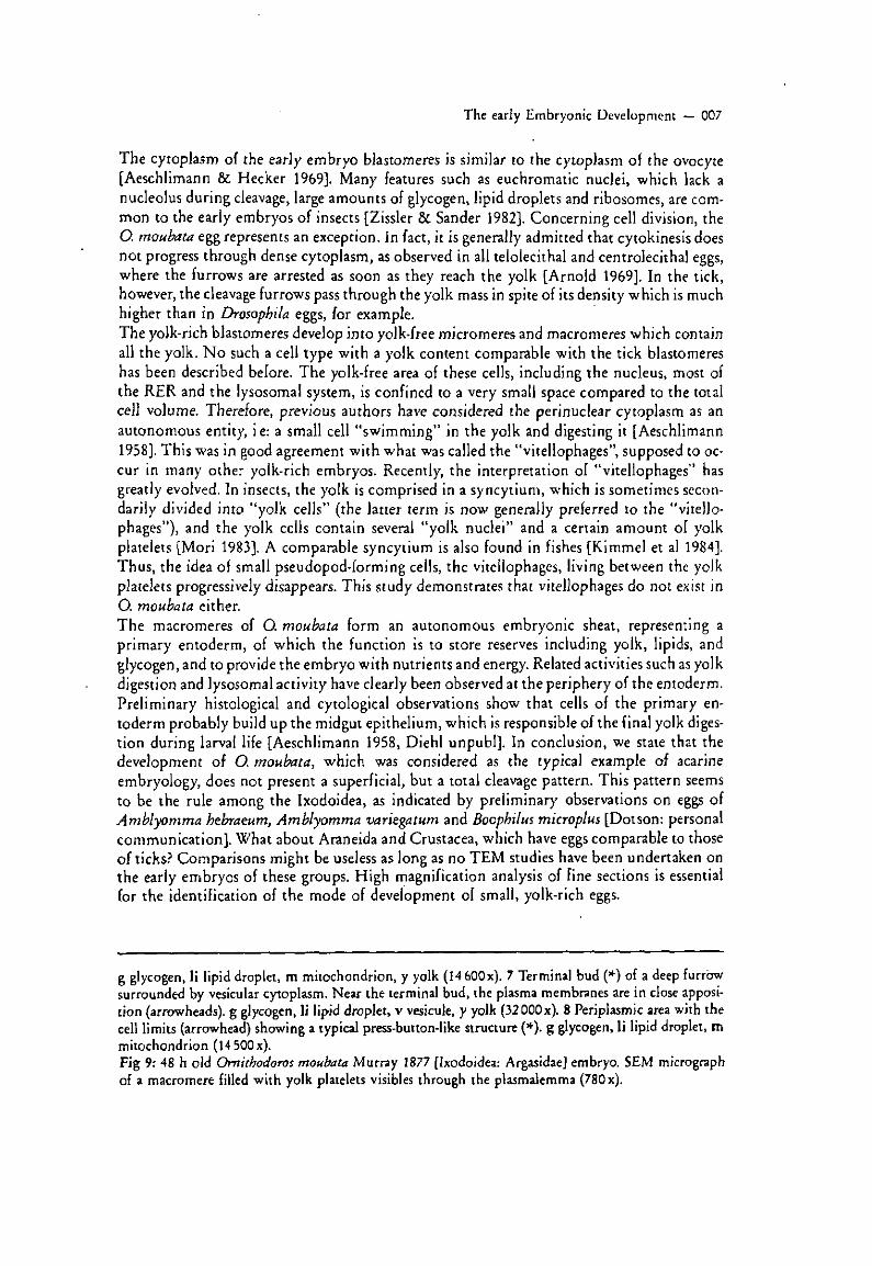
The early Embryonic Development — 007
The cytoplasm of the early embryo blastomeres is similar to the cytoplasm of the ovocyte [Aeschlimann & Hecker 1969]. Many features such as euchromatic nuclei, which lack a nucleolus during cleavage, large amounts of glycogen, lipid droplets and ribosomes, are common to the early embryos of insects [Zissler & Sander 1982]. Concerning cell division, the O. moubata egg represents an exception. In fact, it is generally admitted that cytokinesis does not progress through dense cytoplasm, as observed in all telolecithal and centrolecithal eggs, where the furrows are arrested as soon as they reach the yolk [Arnold 1969], In the tick, however, the cleavage furrows pass through the yolk mass in spite of its density which is much higher than in Drosopbila eggs, for example.
The yolk-rich blastomeres develop into yolk-free micromeres and macromeres which contain all the yolk. N o such a cell type with a yolk content comparable with the tick blastomeres has been described before. The yolk-free area of these cells, including the nucleus, most of the RER and the lysosomal system, is confined to a very small space compared to the total cell volume. Therefore, previous authors have considered the perinuclear cytoplasm as an autonomous entity, ie: a small cell "swimming" in the yolk and digesting it [Aeschlimann 1958], This was in good agreement with what was called the "vitellophages", supposed to occur in many other yolk-rich embryos. Recently, the interpretation of "vitellophages" has greatly evolved. In insects, the yolk is comprised in a syncytium, which is sometimes secondarily divided into "yolk cells" (the latter term is now generally preferred to the "vitellophages"), and the yolk cells contain several "yolk nuclei" and a certain amount of yolk platelets [Mori 1983]. A comparable syncytium is also found in fishes [Kimme! et al 1984]. Thus, the idea of small pseudopod-forming cells, the vitellophages, living between the yolk platelets progressively disappears. This study demonstrates that vitellophages do not exist in O. moubata either.
The macromeres of O. moubata form an autonomous embryonic sheat, representing a primary entoderm, of which the function is to store reserves including yolk, lipids, and glycogen, and to provide the embryo with nutrients and energy. Related activities such as yolk digestion and lysosomal activity have clearly been observed at the periphery of the entoderm. Preliminary histological and cytological observations show that cells of the primary entoderm probably build up the midgut epithelium, which is responsible of the final yolk digestion during larval life [Aeschlimann 1958, Diehl unpubl]. In conclusion, we state that the development of O. moubata, which was considered as the typical example of acarine embryology, does not present a superficial, but a total cleavage pattern. This pattern seems to be the rule among the Ixodoidea, as indicated by preliminary observations on eggs of Amblyomma hebraeum, Amblyomma variegatum and Boopbilus microplus [Dotson: personal communication]. What about Araneida and Crustacea, which have eggs comparable to those of ticks? Comparisons might be useless as long as no TEM studies have been undertaken on the early embryos of these groups. High magnification analysis of fine sections is essential for the identification of the mode of development of small, yolk-rich eggs.
g glycogen, Ii lipid droplet, m mitochondrion, y yolk (14 60Ox). 7 Terminal bud (*) of a deep furrow surrounded by vesicular cytoplasm. Near the terminal bud, the plasma membranes are in close apposition (arrowheads), g glycogen, Ii lipid droplet, v vésicule, y yolk (3200Ox). 8 Periplasmic area with the cell limits (arrowhead) showing a typical press-button-like structure (*). g glycogen, Ii lipid droplet, m mitochondrion (14 50Ox). Fig 9: 48 h old Ornithodoros moubata Murray 1877 [Ixodoidea: Argasidae] embryo. SEM micrograph of a macromere filled with yolk platelets visibles through the plasmalemma (78Ox).

008 — F. Hagotto, E. Hess & A. Aeschlimann
5 References
Aeschlimann, A. [1958]: Développement embryonnaire d'Ornithodorus moubata (Murray) et transmission transovarienne de Borrelia duttonî. — Acta tropica 15(1): 15—62.
Aeschlimann, A., & Hecker, H. [1969]: Vitellogénése et formation cuticulaire chez l'oeuf d'Ornithodorus moubata, Murray. Etude au microscope électronique. — Acarologia 11: 180—191.
Aeschlimann, A., &Hess, E. [1984]: What is our current knowledge of acarine embryology? — In: Griffiths, D. A., & Bowman, CE. : Acarology 6 (1): 90—99. — Ellis Horwood Limited, Chichester.
Anderson, D.T. [1973]: Embryology and phylogeny in annelids and arthropods. — International series of monographs ìn pure and applied biology, zoology division, vol 50. — Perjamon Press, Oxford.
Arnold, J.M. [1969]: Cleavage furrow formation in a telolecithal egg(Lologo peatii). I: Filaments in early furrow formation. - J. Cell. Biol. 41: 894-904.
Arnold, J. M. [1976]: Cytokinesis in animal cells: new answers to old questions. — In: Poste, G., & Nicolson, G. L-: The cell surface in animal embryogenesis and development. Cell surface reviews, vol. 1: 55—80. — North-Holland Publishing Co, Amsterdam - New York-Oxford.
Dawydoff, C. [1928]: Traité d'embryologie comparée des Invertébrés. — Masson. Paris. Fioroni, P. [1970]: Die organogenetische und transitorische Rolle der Vitellophagen in der Darment
wicklung von Galathea (Crustacea, Anomoura). — Z. Morph. Tiere 67: 263—306. Kimmel, C.B., Spray, D.C-, & Bennett, M. V.L. [1984]: Developmental uncoupling between blastoderm
and yolk cell in the embryo of the teleost Fundulus. — Dev. Biol. 102: 483—487. Kostrzewski, M.W., van Niekerk.J.P, & Rechav, Y. [1986]: A case of gynandromorphism in Hyalomma
truncatum (Acari: Ixodidae). — J. Med. Eniomol. 23(1): 116. Krantz, G. W. [1978]: A manual of acarology. (2nd ed). — Oregon Univsersity Book Stores, Inc.. Cor-
vallis. Mori, H. [1983]: Origin, development, morphology and phylogeny of the embryonic midgut epithelium
in insects. — Entomol- Gener. 8(2/3): 135—154. Palade, G. [1952]: A study of fixation for electron microscopy, — J. Exp. Med. 95: 285. Sabatini, D.D., Bentsch, K. G., & Barnet, R.J, [1963]: Cytochemistry and electron microscopy: the
preservation of cellular uhrastructure and enzymatic activity by aldehyde fixation. — J. Cell. Biol. 17: 19-58.
Sander, K., Gutzeit, H. O., & Jackie, JH. [1985]: Insect embryogenesis: morphology, physiology, genetical and molecular aspects. — In: Kerkut, G. A., & Gilbert, L.I. (eds.): Comprehensive insect physiology, biochemistry and pharmacology. Vol I: Embryology and reproduction: 319—385. — Pergamon Press, Oxford-New York.
Spurr, A. R. [1969]: A low-viscosity epoxy resin embedding medium for electron microscopy. — J. Ultrastr. Res. 26: 31-43.
Stampili, N. [1985]: A new case of gynandromorphism in Amblyomma variegatum (Acari, Ixodidae). - Acarologia, 26(1): 03-06.
Zissler, D., & Sander, K. [1982]: The cytoplasmic architecture of the insect egg cell. — In: Kind, R. C , & Akai, H-: Insect ultrastructure 1: 189—216. — Plenum Press, New York-London.
Authors'address —Anschrift der Verfasser: Dr François Fagotto; Dr Ernst Heß & Professor Dr André Aeschlimann, Institut de Zoologie, Université de Neuchâtcl, Chantemerle 22, CH-2000 Neuchâtel; Suisse — Schweiz — Switzerland.
Bunk, B., & Tausch, J.: Moderne Biologie im Unterricht. Bakteriologie mit einfachen Mitteln. [251 Seiten, 46 Abb., 120x 175 mm], — Verlag: Georg Westermann, Braunschweig; ISBN: 3-14-167121-4.
[EGR-Nr 687]. Das Buch enthält allgemeine Anweisungen zur Einrichtung eines mikrobiologischen Arbeitsplatzes, detaillierte Versuchsanweisungen und exemplarische Unterrichtsentwürfe. Die wichtigsten Inhalte der Mikrobiologie sind erfaßt und methodisch gründlich aufgearbeitet. Hinweise auf modernere Hilfsmittel und aktuelle Probleme fehlen. Manfred Noli (Dortmund)