focus on colic - ivis · equine practitioners - focus meeting focus on colic indianapolis, in, usa...
TRANSCRIPT
Close this window to return to IVIS www.ivis.org
Proceedings of the American Association of Equine Practitioners - Focus Meeting
Focus on Colic
Indianapolis, IN, USA – 2011
Next Focus Meetings: July 22-24, 2012 - Focus on Hind Limb Lameness
Oklahoma City, OK, USA
September 6-8, 2012 - Focus on Ophthalmology Raleigh, NC
Reprinted in the IVIS website with the permission of the AAEP http://www.ivis.org
4
Intra-Abdominal Conditions Causing Colic: How They Alter Normal Physiology and Why They Result in Pain Nathaniel A. White, DVM, MS, Diplomate ACVS Author’s address: Marion duPont Scott Equine Medical Center, VMRCVM-Virginia Tech, P.O. Box 1938, Leesburg, VA 20177; e-mail: [email protected]. Take Home Message The clinical signs created by intestinal injury from obstruction, strangulation, enteritis or peritonitis are manifest from intestinal distention, perfusion alterations, entry of bacteria into the systemic circulation, third space accumulation of fluid or blood and release of inflammatory mediators, all of which create local and systemic cellular dysfunction. Though all types of acute abdominal disease can cause similar clinical signs, each disease classification creates specific changes that can help the clinician diagnose the problem and select appropriate treatment. The pathophysiologic events that take place during an acute abdominal crisis include intestinal distension, intestinal ischemia, alterations in tissue perfusion and dysfunction of the intestinal epithelium, serosal mesothelium, smooth muscle and enteric nervous system. The classic pathological degeneration is initiated early in the disease by changes in the microvascular permeability, endothelial cell changes, neuron response, and neutrophil activation.1 Cell injury due to microorganisms invading the mucosa initiates enteritis by both local and systemic inflammatory reactions. Cellular injury was once characterized by the morphologic change it caused. The new paradigm includes cellular responses, which are as much functional as it is structural. The response to a stimulus such as stretch receptors or ischemia is mediated by numerous autocrine and paracrine messengers, which include cytokines, chemokines, prostaglandins, neuropeptides, and proinflammatory substances such as interleukins, tumor necrosis factor (TNFα) complement, histamine, bradykinin, serotonin, and interferon.2 Inflammatory responses to these intercellular messengers are promoted by mucosal cells, fibrocytes, macrophages, mast cells, endothelial cells, neurons, muscle cells and polymorphonuclear cells.2 Though the primary site of intestine injury or dysfunction may create pain and changes in intestinal function, it is clear that there is a systemic cascade of events promoted by inflammatory mediators, which also create clinical signs attributed to an acute abdomen. An understanding of these physiologic and morphologic alterations helps the clinician determine the type of disease, severity, and possible treatments. Simple Colic Little is known about the simple colic (often described as “spasmodic colic” or “gas colic”), which makes up as much as 80-85% of colic in horses. Intestinal function is abnormal either due to adynamic ileus or spasm. Gas accumulation is often detected. The pathophysiologic events are
Published in IVIS with the permission of the AAEP Close this window to return to IVIS
Proceedings of the AAEP Focus Meeting on Colic, Indianapolis, IN, USA - July 24 - 26, 2011
5
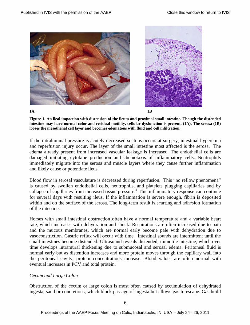
not well understood because research to detect intestinal or systemic responses is difficult in these cases, which readily respond to treatment. It is likely that though the intestine does not appear to be injured to the degree detected with obstruction, strangulation or enteritis, the resulting dysfunction appears to create responses with mediators such as prostaglandins. A wide range of signs including heart rate and dehydration can be seen, but lack of change in the blood or peritoneal fluid values suggest that abnormal cell function rather than cell injury is responsible for the clinical signs seen with this type of colic. Simple Obstruction Small Intestine In the small intestine, obstruction of the intestinal lumen usually is due to intraluminal blockage from a dehydrated food mass or extraluminal pressure from adhesions, thickening of the intestinal wall or infection. The immediate response to a physical obstruction is increased motility of the segment of bowel oral (proximal) to the blockage and relaxation of the intestine aboral (distal) to the blockage. The muscular activity of the intestine is increased around the obstruction, increasing intraluminal pressure in this segment of intestine. Distension of the intestine stretches the wall of the intestine and combines with the reflex muscular spasm to initiate colic. Adynamic ileus may cause functional obstruction due to lack of intestinal movement. Though there is not physical blockage, the lack of intestine movement allows accumulation of fluid and gas creating a similar increase in intraluminal pressure with subsequent changes in the intestinal wall. When the small intestine is obstructed, the enterosystemic circulation of fluid is blocked so that fluid from saliva, stomach fluid, bile, pancreatic fluid, and small intestinal secretion is prevented from passing to the large intestine where it is reabsorbed. Because normal secretion of fluid continues, the small intestine becomes distended oral (proximal) to the blockage. Once the luminal pressure is elevated, the tissue pressure compresses the capillaries and reduces venous drainage. Subsequently, blood flow to the intestine is decreased as the capacitance of the vascular system is decreased. Due to the increased capillary hydrostatic pressure, water from the capillaries, moves from the interstitium into the lymphatic system or the intestinal lumen, or through the serosa into the peritoneal cavity. The increased intraluminal hydrostatic pressure created by the enhanced secretion of fluid then initiates cyclic increases in secretion by continuing to elevate the intraluminal pressure. Because the fluid secreted is isotonic, there is minimal acute change in serum electrolyte values. The consequences of intestinal distension are dehydration from third-space sequestration of the secreted fluid, mucosal injury, serosal injury, abdominal pain, and increased movement of protein across the serosa into the abdominal cavity. The distension eventually reaches a level that inhibits motility in the affected segment of intestine as well as other portions of the intestinal tract. The clinical signs that result are colic, increased heart rate due to pain and decreased circulatory fluid volume, reduced borborygmi, gastric and intestinal fluid sequestration (gastric reflux), and increased protein concentration in peritoneal fluid. An example of this type of disease process is ileal impaction (Fig. 1).
Published in IVIS with the permission of the AAEP Close this window to return to IVIS
Proceedings of the AAEP Focus Meeting on Colic, Indianapolis, IN, USA - July 24 - 26, 2011
6
1A
1A. 1B
Figure 1. An ileal impaction with distension of the ileum and proximal small intestine. Though the distended intestine may have normal color and residual motility, cellular dysfunction is present. (1A). The serosa (1B) looses the mesothelial cell layer and becomes edematous with fluid and cell infiltration.
If the intraluminal pressure is acutely decreased such as occurs at surgery, intestinal hyperemia and reperfusion injury occur. The layer of the small intestine most affected is the serosa. The edema already present from increased vascular leakage is increased. The endothelial cells are damaged initiating cytokine production and chemotaxis of inflammatory cells. Neutrophils immediately migrate into the serosa and muscle layers where they cause further inflammation and likely cause or potentiate ileus.3 Blood flow in serosal vasculature is decreased during reperfusion. This “no reflow phenomena” is caused by swollen endothelial cells, neutrophils, and platelets plugging capillaries and by collapse of capillaries from increased tissue pressure.4 This inflammatory response can continue for several days with resulting ileus. If the inflammation is severe enough, fibrin is deposited within and on the surface of the serosa. The long-term result is scarring and adhesion formation of the intestine. Horses with small intestinal obstruction often have a normal temperature and a variable heart rate, which increases with dehydration and shock. Respirations are often increased due to pain and the mucous membranes, which are normal early become pale with dehydration due to vasoconstriction. Gastric reflux will occur with time. Intestinal sounds are intermittent until the small intestines become distended. Ultrasound reveals distended, immotile intestine, which over time develops intramural thickening due to submucosal and serosal edema. Peritoneal fluid is normal early but as distention increases and more protein moves through the capillary wall into the peritoneal cavity, protein concentrations increase. Blood values are often normal with eventual increases in PCV and total protein. Cecum and Large Colon Obstruction of the cecum or large colon is most often caused by accumulation of dehydrated ingesta, sand or concretions, which block passage of ingesta but allows gas to escape. Gas build
Published in IVIS with the permission of the AAEP Close this window to return to IVIS
Proceedings of the AAEP Focus Meeting on Colic, Indianapolis, IN, USA - July 24 - 26, 2011
7
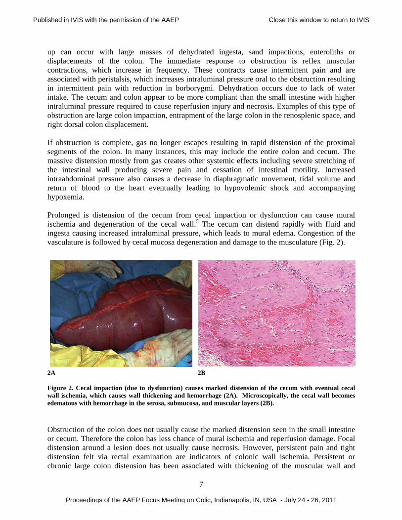
up can occur with large masses of dehydrated ingesta, sand impactions, enteroliths or displacements of the colon. The immediate response to obstruction is reflex muscular contractions, which increase in frequency. These contracts cause intermittent pain and are associated with peristalsis, which increases intraluminal pressure oral to the obstruction resulting in intermittent pain with reduction in borborygmi. Dehydration occurs due to lack of water intake. The cecum and colon appear to be more compliant than the small intestine with higher intraluminal pressure required to cause reperfusion injury and necrosis. Examples of this type of obstruction are large colon impaction, entrapment of the large colon in the renosplenic space, and right dorsal colon displacement. If obstruction is complete, gas no longer escapes resulting in rapid distension of the proximal segments of the colon. In many instances, this may include the entire colon and cecum. The massive distension mostly from gas creates other systemic effects including severe stretching of the intestinal wall producing severe pain and cessation of intestinal motility. Increased intraabdominal pressure also causes a decrease in diaphragmatic movement, tidal volume and return of blood to the heart eventually leading to hypovolemic shock and accompanying hypoxemia. Prolonged is distension of the cecum from cecal impaction or dysfunction can cause mural ischemia and degeneration of the cecal wall.5 The cecum can distend rapidly with fluid and ingesta causing increased intraluminal pressure, which leads to mural edema. Congestion of the vasculature is followed by cecal mucosa degeneration and damage to the musculature (Fig. 2).
2A 2B Figure 2. Cecal impaction (due to dysfunction) causes marked distension of the cecum with eventual cecal wall ischemia, which causes wall thickening and hemorrhage (2A). Microscopically, the cecal wall becomes edematous with hemorrhage in the serosa, submucosa, and muscular layers (2B). Obstruction of the colon does not usually cause the marked distension seen in the small intestine or cecum. Therefore the colon has less chance of mural ischemia and reperfusion damage. Focal distension around a lesion does not usually cause necrosis. However, persistent pain and tight distension felt via rectal examination are indicators of colonic wall ischemia. Persistent or chronic large colon distension has been associated with thickening of the muscular wall and
Published in IVIS with the permission of the AAEP Close this window to return to IVIS
Proceedings of the AAEP Focus Meeting on Colic, Indianapolis, IN, USA - July 24 - 26, 2011
8
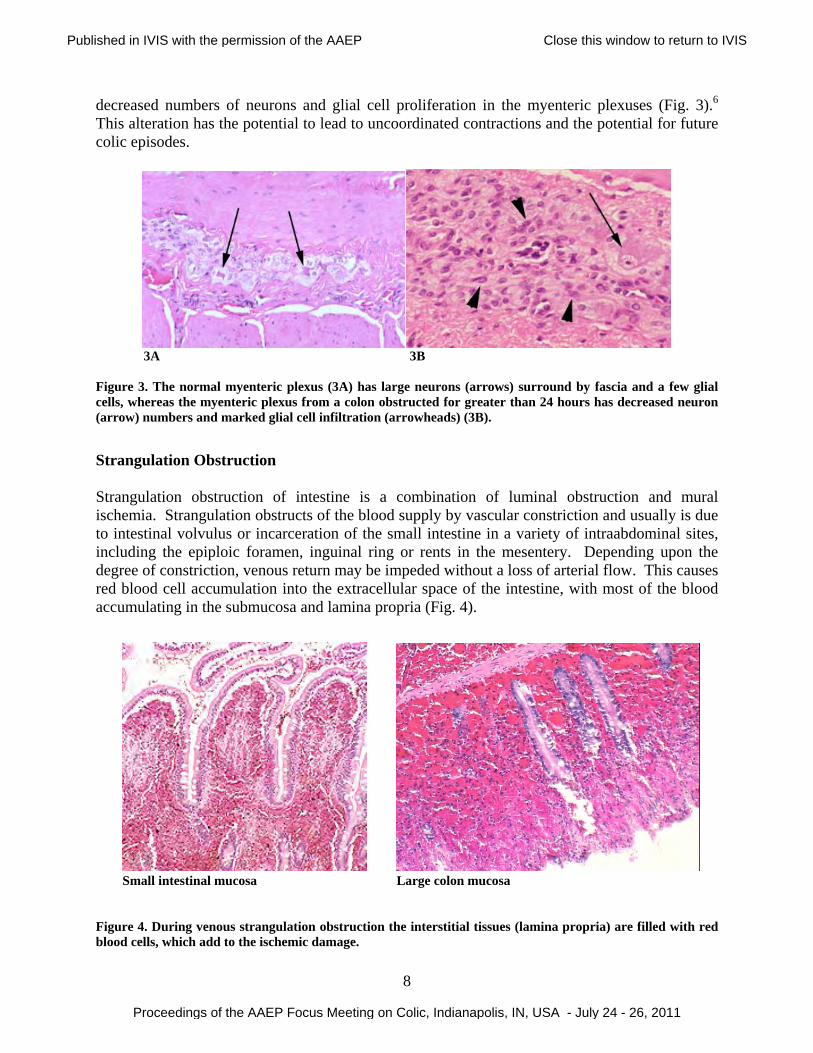
decreased numbers of neurons and glial cell proliferation in the myenteric plexuses (Fig. 3).6 This alteration has the potential to lead to uncoordinated contractions and the potential for future colic episodes.
3A 3B Figure 3. The normal myenteric plexus (3A) has large neurons (arrows) surround by fascia and a few glial cells, whereas the myenteric plexus from a colon obstructed for greater than 24 hours has decreased neuron (arrow) numbers and marked glial cell infiltration (arrowheads) (3B).
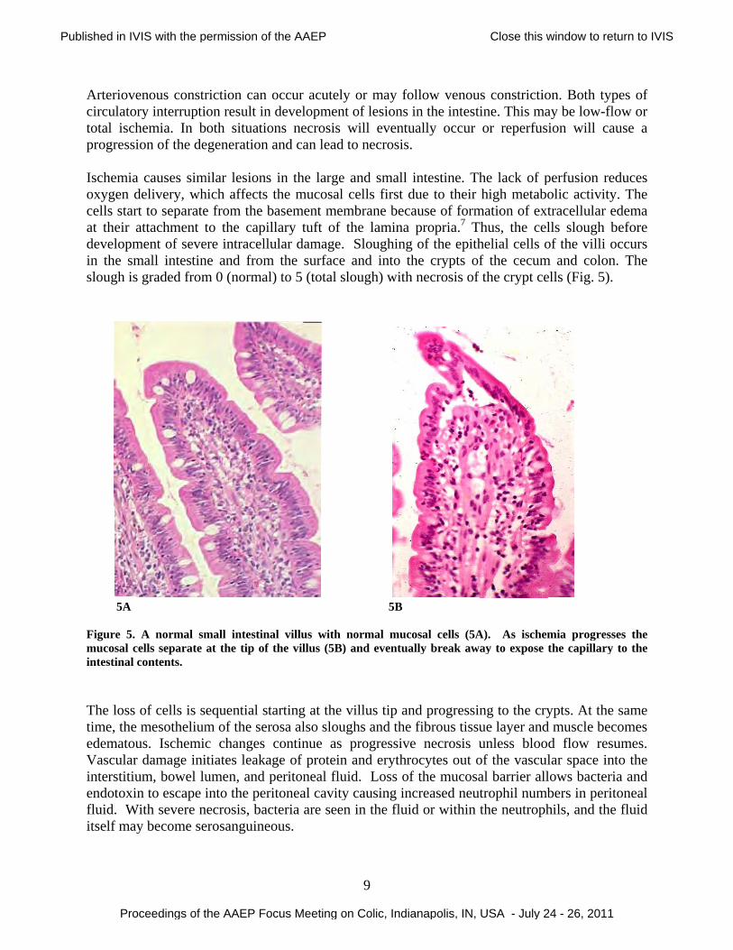
Strangulation Obstruction Strangulation obstruction of intestine is a combination of luminal obstruction and mural ischemia. Strangulation obstructs of the blood supply by vascular constriction and usually is due to intestinal volvulus or incarceration of the small intestine in a variety of intraabdominal sites, including the epiploic foramen, inguinal ring or rents in the mesentery. Depending upon the degree of constriction, venous return may be impeded without a loss of arterial flow. This causes red blood cell accumulation into the extracellular space of the intestine, with most of the blood accumulating in the submucosa and lamina propria (Fig. 4). Small intestinal mucosa Large colon mucosa Figure 4. During venous strangulation obstruction the interstitial tissues (lamina propria) are filled with red blood cells, which add to the ischemic damage.
Published in IVIS with the permission of the AAEP Close this window to return to IVIS
Proceedings of the AAEP Focus Meeting on Colic, Indianapolis, IN, USA - July 24 - 26, 2011
Arteriovecirculatortotal ischprogressi Ischemiaoxygen dcells starat their adevelopmin the smslough is
5A Figure 5. mucosal cintestinal The loss time, the edematouVascularinterstitiuendotoxinfluid. Witself may
enous constrry interruptihemia. In bion of the de
a causes simdelivery, whrt to separateattachment tment of sevemall intestins graded from
A normal smcells separate contents.
of cells is smesothelium
us. Ischemicr damage inium, bowel lun to escape i
With severe ny become se
riction can oon result in
both situationegeneration a
milar lesions hich affects te from the bto the capill
ere intracellune and from m 0 (normal)
mall intestinaat the tip of t
equential stam of the seroc changes citiates leakagumen, and pinto the peri
necrosis, bacterosanguineo
occur acuteldevelopmenns necrosis and can lead
in the largethe mucosalasement melary tuft of
ular damage.the surface
) to 5 (total s
al villus with the villus (5B)
arting at the osa also sloucontinue as ge of proteinperitoneal fluitoneal cavityteria are seeous.
9
y or may font of lesions
will eventud to necrosis.
e and small l cells first dmbrane becathe lamina
. Sloughinge and into thslough) with
5B
normal muc) and eventua
villus tip anughs and the
progressiven and erythruid. Loss ofy causing in
en in the flui
ollow venouin the intesti
ually occur .
intestine. Thdue to their ause of formpropria.7 T
g of the epithhe crypts of
h necrosis of
osal cells (5Aally break aw
nd progressin fibrous tissue necrosis uocytes out of the mucosa
ncreased neutd or within t
s constrictioine. This maor reperfusi
he lack of phigh metabo
mation of exthus, the celhelial cells of the cecum
f the crypt ce
A). As ischemay to expose t
ng to the cryue layer andunless bloodof the vasculal barrier alltrophil numbthe neutroph
on. Both typay be low-floion will cau
perfusion redolic activitytracellular edlls slough bof the villi o
m and colon.ells (Fig. 5).
mia progressethe capillary
ypts. At the d muscle becd flow resular space intlows bacteriabers in peritohils, and the
pes of ow or use a
duces . The dema
before ccurs . The
es the to the
same omes umes. to the a and oneal fluid
Published in IVIS with the permission of the AAEP Close this window to return to IVIS
Proceedings of the AAEP Focus Meeting on Colic, Indianapolis, IN, USA - July 24 - 26, 2011
10
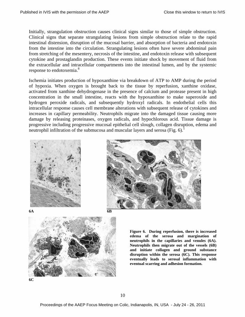
Initially, strangulation obstruction causes clinical signs similar to those of simple obstruction. Clinical signs that separate strangulating lesions from simple obstruction relate to the rapid intestinal distension, disruption of the mucosal barrier, and absorption of bacteria and endotoxin from the intestine into the circulation. Strangulating lesions often have severe abdominal pain from stretching of the mesentery, necrosis of the intestine, and endotoxin release with subsequent cytokine and prostaglandin production. These events initiate shock by movement of fluid from the extracellular and intracellular compartments into the intestinal lumen, and by the systemic response to endotoxemia.8 Ischemia initiates production of hypoxanthine via breakdown of ATP to AMP during the period of hypoxia. When oxygen is brought back to the tissue by reperfusion, xanthine oxidase, activated from xanthine dehydrogenase in the presence of calcium and protease present in high concentration in the small intestine, reacts with the hypoxanthine to make superoxide and hydrogen peroxide radicals, and subsequently hydroxyl radicals. In endothelial cells this intracellular response causes cell membrane alterations with subsequent release of cytokines and increases in capillary permeability. Neutrophils migrate into the damaged tissue causing more damage by releasing proteinases, oxygen radicals, and hypochlorous acid. Tissue damage is progressive including progressive mucosal epithelial cell slough, collagen disruption, edema and neutrophil infiltration of the submucosa and muscular layers and serosa (Fig. 6).1
6A 6B
Figure 6. During reperfusion, there is increased edema of the serosa and margination of neutrophils in the capillaries and venules (6A). Neutrophils then migrate out of the vessels (6B) and initiate collagen and ground substance disruption within the serosa (6C). This response eventually leads to serosal inflammation with eventual scarring and adhesion formation.
6C
Published in IVIS with the permission of the AAEP Close this window to return to IVIS
Proceedings of the AAEP Focus Meeting on Colic, Indianapolis, IN, USA - July 24 - 26, 2011
11
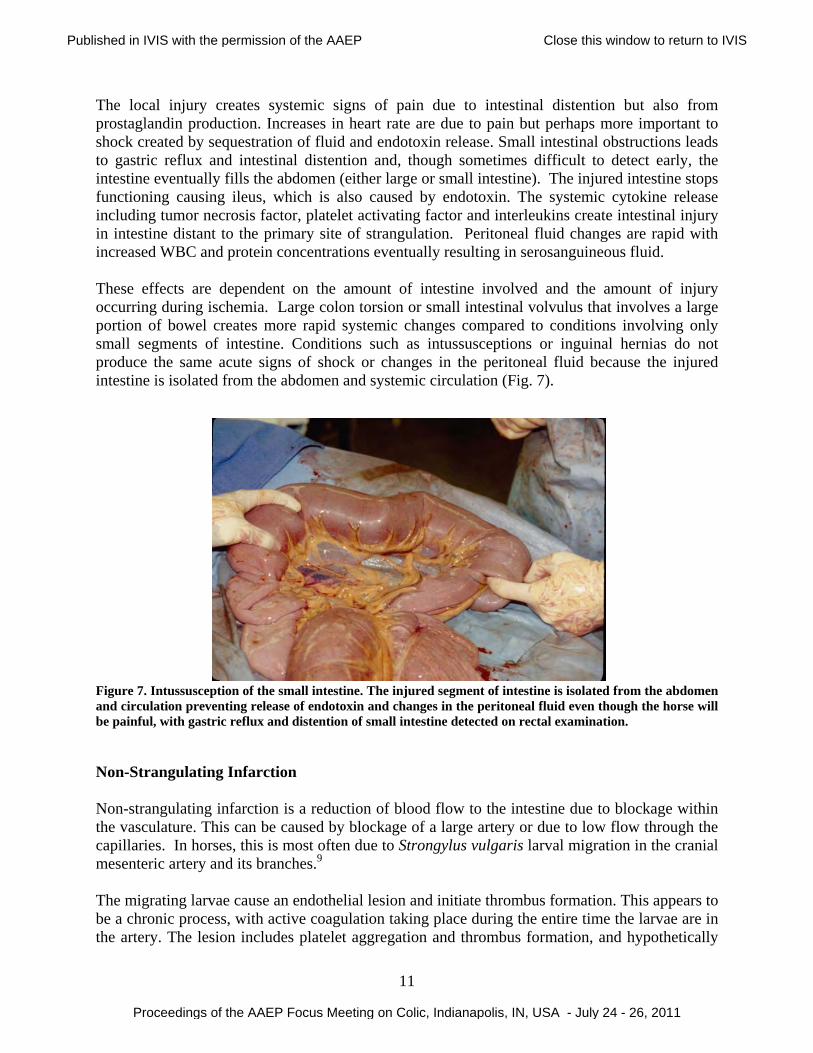
The local injury creates systemic signs of pain due to intestinal distention but also from prostaglandin production. Increases in heart rate are due to pain but perhaps more important to shock created by sequestration of fluid and endotoxin release. Small intestinal obstructions leads to gastric reflux and intestinal distention and, though sometimes difficult to detect early, the intestine eventually fills the abdomen (either large or small intestine). The injured intestine stops functioning causing ileus, which is also caused by endotoxin. The systemic cytokine release including tumor necrosis factor, platelet activating factor and interleukins create intestinal injury in intestine distant to the primary site of strangulation. Peritoneal fluid changes are rapid with increased WBC and protein concentrations eventually resulting in serosanguineous fluid. These effects are dependent on the amount of intestine involved and the amount of injury occurring during ischemia. Large colon torsion or small intestinal volvulus that involves a large portion of bowel creates more rapid systemic changes compared to conditions involving only small segments of intestine. Conditions such as intussusceptions or inguinal hernias do not produce the same acute signs of shock or changes in the peritoneal fluid because the injured intestine is isolated from the abdomen and systemic circulation (Fig. 7). Figure 7. Intussusception of the small intestine. The injured segment of intestine is isolated from the abdomen and circulation preventing release of endotoxin and changes in the peritoneal fluid even though the horse will be painful, with gastric reflux and distention of small intestine detected on rectal examination. Non-Strangulating Infarction Non-strangulating infarction is a reduction of blood flow to the intestine due to blockage within the vasculature. This can be caused by blockage of a large artery or due to low flow through the capillaries. In horses, this is most often due to Strongylus vulgaris larval migration in the cranial mesenteric artery and its branches.9 The migrating larvae cause an endothelial lesion and initiate thrombus formation. This appears to be a chronic process, with active coagulation taking place during the entire time the larvae are in the artery. The lesion includes platelet aggregation and thrombus formation, and hypothetically
Published in IVIS with the permission of the AAEP Close this window to return to IVIS
Proceedings of the AAEP Focus Meeting on Colic, Indianapolis, IN, USA - July 24 - 26, 2011
12
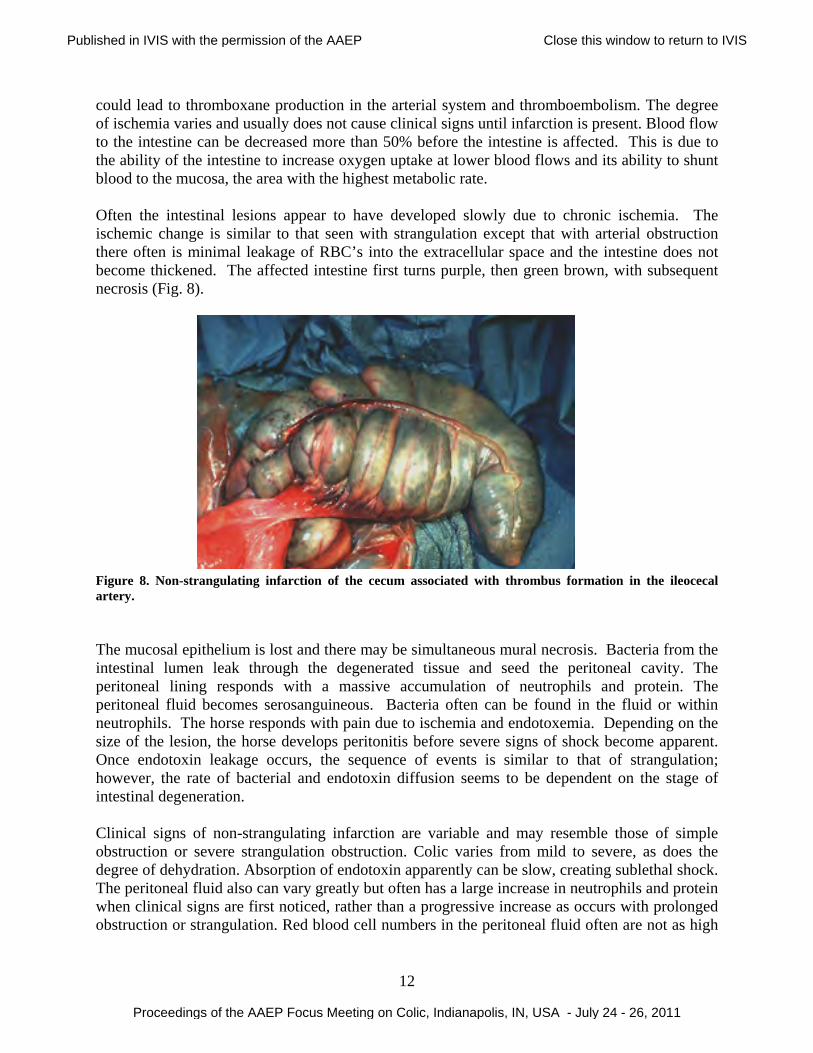
could lead to thromboxane production in the arterial system and thromboembolism. The degree of ischemia varies and usually does not cause clinical signs until infarction is present. Blood flow to the intestine can be decreased more than 50% before the intestine is affected. This is due to the ability of the intestine to increase oxygen uptake at lower blood flows and its ability to shunt blood to the mucosa, the area with the highest metabolic rate. Often the intestinal lesions appear to have developed slowly due to chronic ischemia. The ischemic change is similar to that seen with strangulation except that with arterial obstruction there often is minimal leakage of RBC’s into the extracellular space and the intestine does not become thickened. The affected intestine first turns purple, then green brown, with subsequent necrosis (Fig. 8). Figure 8. Non-strangulating infarction of the cecum associated with thrombus formation in the ileocecal artery. The mucosal epithelium is lost and there may be simultaneous mural necrosis. Bacteria from the intestinal lumen leak through the degenerated tissue and seed the peritoneal cavity. The peritoneal lining responds with a massive accumulation of neutrophils and protein. The peritoneal fluid becomes serosanguineous. Bacteria often can be found in the fluid or within neutrophils. The horse responds with pain due to ischemia and endotoxemia. Depending on the size of the lesion, the horse develops peritonitis before severe signs of shock become apparent. Once endotoxin leakage occurs, the sequence of events is similar to that of strangulation; however, the rate of bacterial and endotoxin diffusion seems to be dependent on the stage of intestinal degeneration. Clinical signs of non-strangulating infarction are variable and may resemble those of simple obstruction or severe strangulation obstruction. Colic varies from mild to severe, as does the degree of dehydration. Absorption of endotoxin apparently can be slow, creating sublethal shock. The peritoneal fluid also can vary greatly but often has a large increase in neutrophils and protein when clinical signs are first noticed, rather than a progressive increase as occurs with prolonged obstruction or strangulation. Red blood cell numbers in the peritoneal fluid often are not as high
Published in IVIS with the permission of the AAEP Close this window to return to IVIS
Proceedings of the AAEP Focus Meeting on Colic, Indianapolis, IN, USA - July 24 - 26, 2011
13
as occurs with strangulating lesions. Fortunately this disease is relatively rare due to several decades of improved parasite control. Enteritis When bacteria cause injury by adhering to the mucosal surface and invading the mucosal cells, the response of the mucosal cells stimulates both the afferent nervous system and an immediate local immunocytes immune. Cytokines, including TNFα, interleukin-1, platelet activating factor and prostaglandins, from local macrophages or injured epithelial cells invaded by the microorganism, serve as the messengers of recognition, which stimulate macrophages and polynucleated cells (both neutrophils and eosinophils) to migrate to the region of invasion.10 Lymphocytes are also activated releasing cytokines including interferon-γ. After an initial delay in mucosal cell apoptosis during bacterial adhesion and invasion, apoptosis is increased, theoretically to increase cell turnover and healing. Enteritis of the colon, colitis, may cause colic, which may be one of the first signs and is transient giving way to depression. Both pain and ileus result from the local inflammatory reaction and potentially from a response by the sympathetic nervous system. Diarrhea is created by hypersecretion due to loss of absorption and increased cyclic AMP activity creating NaCl and water secretion. The resulting volume overload and stimulation of the enteric nervous system decreases transit time, which does not allow reabsorption. The loss or dysfunction of the mucosal barrier results in shock from trans-barrier migration of toxins and bacteria. The large loss of water added to shock causes systemic inflammatory response syndrome (SIRS). Peritoneal fluid is often normal in acute cases of enteritis whereas the circulating WBC concentration is frequently decreased below 3000 cells/µl. Compared to colitis, duodenitis-proximal enteritis (anterior enteritis) creates a different response with acute pain due to gastric distention as well as the intestinal wall inflammation. The pain changes to depression as shock becomes evident. Voluminous gastric reflux is present due to the lack of transit through the duodenum and lack of absorption. Because the injury to the intestine is full thickness, leakage of protein creates increased concentrations in the peritoneal fluid prior to any increase in RBC or WBC. Shock is evident with increases in heart rate and mucous membrane refill time. References 1. Rowe E, White N. Reperfusion Injury in the Equine Intestine. Clinical Techniques in
Equine Practice 2002;1:148-162. 2. Fiocchi C. Cytokines and intestinal inflammation. Transplant Proc 1996;28:2442-2443. 3. Dabareiner RM, White NA, Donaldson LL. Effects of intraluminal distention and
decompression on microvascular permeability and hemodynamics of the equine jejunum. Am J Vet Res 2001;62:225-236.
4. Dabareiner RM, Sullins KE, White NA, et al. Serosal injury in the equine jejunum and ascending colon after ischemia-reperfusion or intraluminal distention and decompression. Vet Surg 2001;30:114-125.
Published in IVIS with the permission of the AAEP Close this window to return to IVIS
Proceedings of the AAEP Focus Meeting on Colic, Indianapolis, IN, USA - July 24 - 26, 2011
14
5. Moore JN, Hardy, J. Diseases of the Cecum In: NA White JM, TS Mair, ed. The Equine Acute Abdomen. Jackson: Teton NewMedia, 2008;618-626.
6. Schusser GE, White NA. Morphologic and quantitative evaluation of the myenteric plexuses and neurons in the large colon of horses. J Am Vet Med Assoc 1997;210:928-934.
7. White NA, Moore JN, Trim CM. Mucosal alterations in experimentally induced small intestinal strangulation obstruction in ponies. Am J Vet Res 1980;41:193-198.
8. Bryant CE, Moore JN. Systemic Inflammatory Response Syndrome: Endotoxemia Reconsidered In: NA White JM, TS Mair, ed. The Equine Acute Abdomen. Jackson: Teton NewMedia, 2008;192-200.
9. White NA, Moore JN, Douglas M. SEM [scanning electron microscopy] study of Strongylus vulgaris larva-induced arteritis in the pony. Equine Veterinary Journal 1983;15:349-353.
10. McKenzie HC. Pathophysiology of enteritis and colitis In: NA White JM, TS Mair, ed. The Equine Acute Abdomen. Jackson: Teton NewMedia, 2008;136-145.
Published in IVIS with the permission of the AAEP Close this window to return to IVIS
Proceedings of the AAEP Focus Meeting on Colic, Indianapolis, IN, USA - July 24 - 26, 2011