focal epithelial hyperplasia: report of a case in an australian aborigine
TRANSCRIPT
Australian Dental Journal, October, I97 I 3 1 5
Focal epithelial hyperplasia: report of a case in an Australian aborigine
K. F. Adkins, M.D.Sc., Ph.D., and A. F. G. Campbell, B.D.Sc., B.Sc., M.B., B.S., F.R.C.S.
Focal epithelial hyperplasia, a rare disease of the oral mucosa which occurs in the form of multiple painless polyps, was introduced into the dental literature in 1965.(1)(2) When a clinical entity is flrst described and the stiological factors are unknown, much impor- tance is often attributed to the geographic distribution of the lesions and the ethnic back- grounds of the patients concerned.
The majority of reports of focal epithelial hyperplasia have described i ts occurrence
almost exclusively in American and South American Indians.") (*) (') Isolated reports added to the literature in the past flve years have documented small numbers of cases i n
Scandinavian girls,@) a Poly- nesian child,'b' a girl in Puerto Rice,(') and two Caucasian women in the United States(E) and Denmark."
This paper reports the occurrence of focal epithelial hyperplasia in a n Australian aborigine.
Received for publication February, 1971. (1) Archard. H. 0.. Heck, J. W., and Stanley, H.
R.-Focal enithelial hyuerplasia : An unusual oral mucnsai lesion found -in Indian children. Oral Surg.: 2 0 : 2. 201-212 (Aug.), 1966.
(2) Witkop, C. J., Jr, and Niswander, J. D.-Focal enithelial 'hspernlasia in Central and South American Indians and Ladinos. Oral Surg.. 2 0 : 2. 213-217 (Aug.), 1965.
( 8 ) Bergenholtz, A,-Multiple polypous hyperplasias of the oral murosa with regression a f te r re- moval of amaleam flllinas. Acta. Odont. Scand., 2 3 : 2. 111-134 (Apr;), 1965.
(')Tan. K. N., Medak, H., and Cohen, L.-Focal epithelial hyperplasia in a Mexican Indian. Arch. Derm.. 1 0 0 : 474-477 (Oct.), 1969.
(5) Ciausen. F. P.-Histopathology of focal epi- thelial hyperplasia. Evidence of viral infec- tion. Tandlaegebladet, 73 : 1013-22 (Nov.),
Case report The patient was a well-nourished Australian
aboriginal woman, 22 years of age. She was in good health at the time of consultation and gave no history of any previous illness.
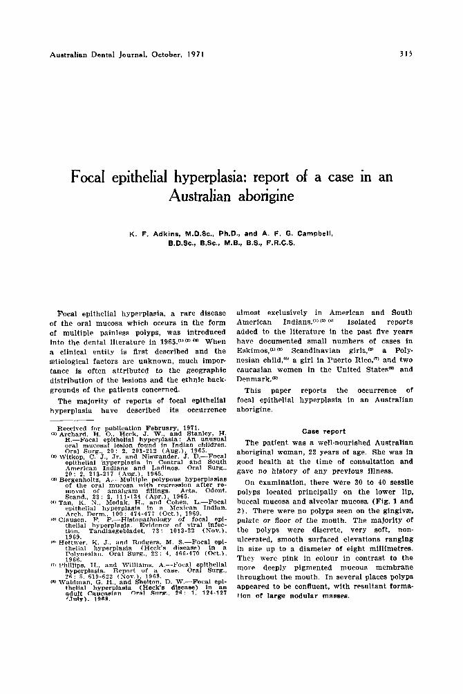
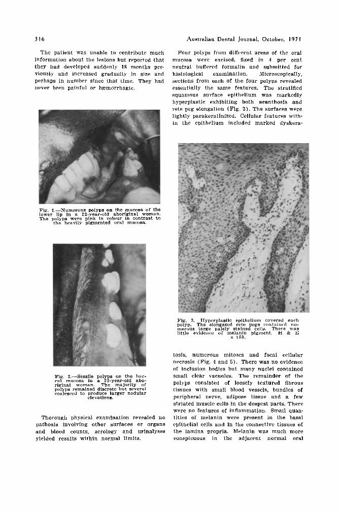
On examination, there were 30 to 40 sessile polyps located principally on the lower lip, buccal mucosa and alveolar mucosa (Fig. 1 and 2 ) . There were no polyps seen on the gingivs, palate or floor of the mouth. The majority of the polyps were discrete, very soft, non-
1969.
Polynesian. Oral Surg.. 2 2 : 4, 466-470 (Oct.).
(71 Phillips, H.. and Williams, A.-Focal epithelial
(8) Waldman. G. H.. and h e l t o n , D. W.-Focal epi-
Hettmey. K, J., and Rodgers, M, S , - F ~ ~ ~ ~ epi- ulcerated, smooth surfaced elevations ranging thelial hyperplasia (Heck's disease) in a in size up to a diameter of eight millimetres. 1966. They were pink in colour in contrast to the
hyperplasia. Report of a case. Oral Surg., more deeply Pigmented mucous membrane 2 6 : 6 619-622 (Xov.) 1968. throughout the mouth. In several places polyps thelial hyperplasia (Heck's disease) in a n appeared to be confluent, with resultant forma- adult Caucaslnn oral S l i m . 2 6 . 1. 124 -127 t,,," large nodular mA88eB, f .TlllV). 1968
316 Australian Dental Journal, October, I97 I
The patient was unable to contribute much Four polyps from different areas of the oral information about the lesions but reported that mucosa were excised, fixed in 4 per cent they liad developed suddenly 18 months pre- neutral buffered formalin and submitted for viously and increased gradually in size and histological examination. Microscopically, perhaps in number since that time. They had sections from each of the four polyps revealed never been painful or haemorrhagic. essentially the same features. The stratified
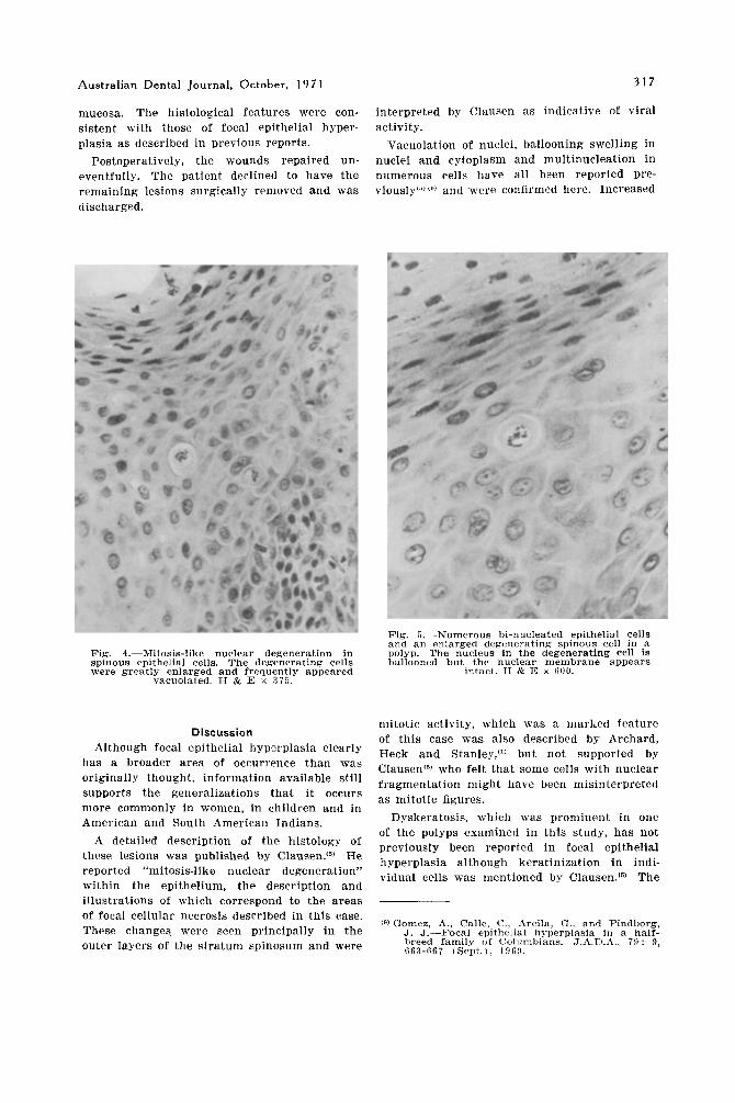
squamous surface epithelium was markedly hyperplastic exhibiting both acanthosis and rete peg elongation (Fig. 3 ) . The surfaces were lightly parakeratinized. Cellular features with- in the epithelium included marked dyskera-
Fig. 1.-Numerous polyps on the mucosa of the lower lip in a 22-year-old aboriginal woman. The polyps were pink In colour in contrast to
the heavily pigmented oral mucosa.
Fig. 3.-Hyperplastic epithelium covered each polyp. The elongated rete pegs contained nu- merous large palely stained cells. There w a s little evidence of melanin pigment. €1 C E
x 150.
Fig. 2.-Sessile polyps on the buc- cal mucosa in a 22-year-old abo- riginal woman. The majority of polyps remained discrete but several coalesced to produce larger nodular
elevations.
Thorough physical examination revealed no pathosis involving other surfaces or organs and blood counts, serology and urinalyses yielded results within normal limits.
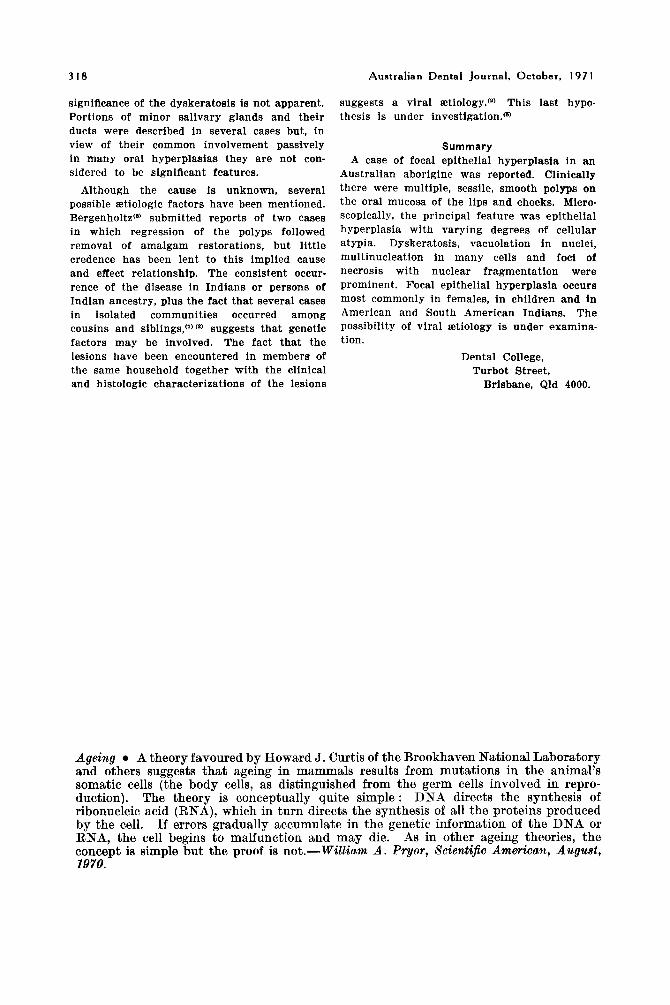
tosis, numerous mitoses and focal cellular necrosis (Fig. 4 and 5) . There was no evidence of inclusion bodies but many nuclei contained small clear vacuoles. The remainder of the polyps consisted of loosely textured fibrous tissues with small blood vessels, bundles of peripheral nerve, adipose tissue and a few striated muscle cells in the deepest parts. There were no features of inflammation. Small quan- tities of melanin were present in the basal epithelial cells and in the connective tissues of the lamina propria. Melanin was much more conspicuous in the adjacent normal oral
Aust ra l ian Dental Journa l , October , 197 I 3 1 7
mucosa. The histological features were con- interpreted by Clausen as indicative of viral sistent with those of focal epithelial hyper- activity. plasia as described in previous reports. Vacuolation of nuclei, ballooning swelling in
Postoperatively, the wounds repaired un- nuclei and cytoplasm and multinucleation in eventfully. The patient declined to have the numerous cells have all been reported pre- remaining lesions surgically removed and was viously(")(") and were confirmed here. Increased discharged.
Fig. 5.-Numerous bi-nucleated epithelial cell8 and a n enlarged degenerating spinous cell in a
Fig. 4.-Mitosis-like nuclear degeneration in polyp. The nucleus in the degenerating cell is spinous epithelial cells. The degenerating cell8 ballooned but the nuclear membrane appears were greatly enlarged and frequently appeared intact. H & E x CiOO.
vacuolated. H & E x 375 .
Discussion Although focal epithelial hyperplasia clearly
has a broader area of occurrence than was originally thought, information available still supports the generalizations that i t occurs more commonly in women, in children and in American and South American Indians.
A detailed description of the histology of these lesions was published by Clau~en. (~) He reported "mitosis-like nuclear degeneration" within the epithelium, the description and
mitotic activity, which was a marked feature of this case was also described by Archard, Heck and Stanley,") but not supported by Clausen'") who felt that some cells with nuclear fragmentation might have been misinterpreted as mitotic figures.
Dyskeratosis, which was prominent in one of the polyps examined in this study, has not previously been reported in focal epithelial hyperplasia although keratinization in indi- vidual cells was mentioned by Clausen.(6) The
illustrations of which correspond to the areas of focal cellular necrosis described in this case.
(9) Gomez, A., Calle, C., Arcila, G, , and Pindborg, These changes were seen principally in the J. J.-Focal epithelial hmerplasia in a half-
breed family of Columbians. J.A.D.A. , 79 : 9, outer layers of the stratum spinosum and were GG3-F67 (Sept.) 196!) .
3 18 Australian Dental Journal, October, I97 I
significance of the dyskeratosis is not apparent. Portions of minor salivary glands and their ducts were described in several cases but, in view of their common involvement passively in many oral hyperplasias they are not con- sidered to be significant features.
Although the cause is unknown, several possible Etiologic factors have been mentioned. Bergenholtz(*) submitted reports of two cases in which regression of the polyps followed removal of amalgam restorations, but little credence has been lent to this implied cause and effect relationship. The consistent occur- rence of the disease in Indians or persons of Indian ancestry, plus the fact that several cases in isolated communities occurred among cousins and siblings,“) (*) suggests that genetic factors may be involved. The fact that the lesions have been encountered in members of the same household together with the clinical and histologic characterizations of the lesions
suggests a viral etiology.(’) This last hypo- thesis is under investigation.”
Summary A case of focal epithelial hyperplasia in a n
Australian aborigine was reported. Clinically there were multiple, sessile, smooth polyps on the oral mucosa of the lips and cheeks. Micro- scopically, the principal feature was epithelial hyperplasia with varying degrees of cellular atypia. Dyskeratosis, vacuolation in nuclei, multinucleation in many cells and foci of necrosis with nuclear fragmentation were prominent. Focal epithelial hyperplasia occurs most commonly in females, in children and in American and South American Indians. The possibility of viral etiology i s under examina- tion.
Dental College, Turbot Street,
Brisbane, Qld 4000.
Ageing A theory favoured by Howard J. Curtis of the Brookhaven National Laboratory and others suggests that ageing in mammals results from mutations in the animal’s somatic cells (the body cells, as distinguished from the germ cells involved in repro- duction). The theory is conceptually quite simple: DNA directs the synthesis of ribonucleic acid (RNA), which in turn directs the synthesis of all the proteins produced by the cell. If errors gradually accumulate in the genetic information of the DNA or RNA, the cell begins to malfunction and may die. As in other ageing theories, the concept is simple but the proof is not.-Willinm, A . Pryor, Scientijio Ammicait, August, 1970.