flow cytometry in translational cancer research: believe in rare!
TRANSCRIPT

Flow cytometry in colon cancer: polychromatic mosaics of TIC and new
targets for tailored therapy
Marica Gemei, PhD

• Flow cytometry in solid cancer analysis
• CD66c as CCSC markers
• Multiparametric FC in CTCs analysis
• Conclusions
• Acknowledgements
Summary
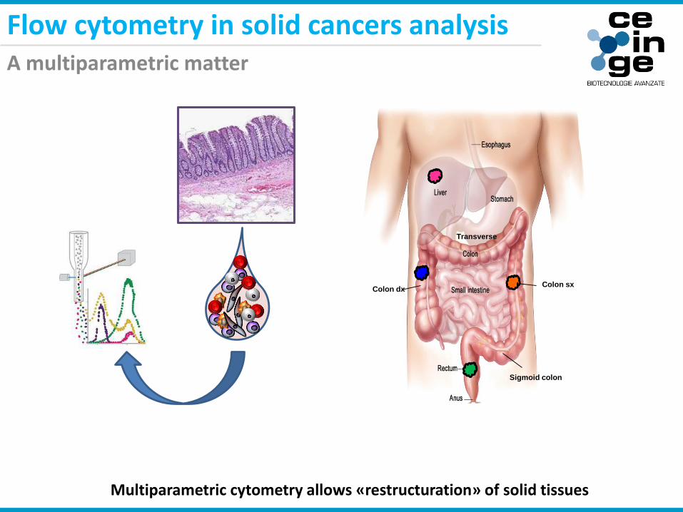
A multiparametric matter
Flow cytometry in solid cancers analysis
Colon dx
Sigmoid colon
Transverse
Colon sx
Multiparametric cytometry allows «restructuration» of solid tissues
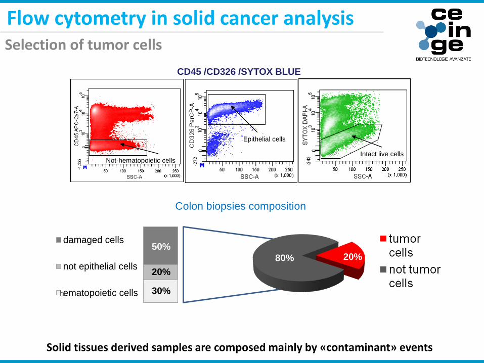
Intact live cellsNot-hematopoietic cells
Epithelial cells
damaged cells
not epithelial cells
ematopoietic cells
80% 20%50%
20%
30%
Colon biopsies composition
h
CD45 /CD326 /SYTOX BLUE
Flow cytometry in solid cancer analysisSelection of tumor cells
Solid tissues derived samples are composed mainly by «contaminant» events

Gemei et al. Unpublished data
Flow cytometry in solid cancer analysisSelection of tumor cells
The use of CD45 together with CD326 allow a better selection of tumor cell population

Migration and Metastatization
CD9 CD49f
CD81
CSC Markers
CD133 CD24
CD29
CD326
CD47
CD90
Injury resistance and malignant behavior
CD227 CD55
CD59
CD66c
CD44
CD26
CD151
CD49b
CD324
CD166
Flow cytometry in solid cancer analysisColon cancer stem cells analysis

Flow cytometry in solid cancer analysisMultiparametric analysis = huge information

CD13
3
CD13
3
CD24
CD24
CD29
CD29
CD44
CD44
CD47
CD47
CD49
b
CD49
b
CD49
f
CD49
f
CD54
CD54
CD55
CD55
CD59
CD59
CD66
b
CD66
b
CD66
c
CD66
c
CD81
CD81
CD16
4
CD16
4
CD16
6
CD16
6
CD22
7
CD22
7
100
1000
10000
100000TUMOR VS NORMAL
CD MARKERS
MFI
CD
133
CD
24
CD
29
CD
44
CD
47
CD
49b
CD
59
CD
66b
CD
66c
CD
81
CD
164
CD
166
CD
227
Me
an
Flu
ore
scen
ce In
ten
sity (
MF
I)
102
103
104
105
CD
55
CD
54
CD
49f
Normal
Cancer
Gemei et al. Unpublished data
Flow cytometry in solid cancer analysisNormal colon and colon cancer surface signature
CD55 and CD66c are strongly upregulated in tumor tissue

TUMORS VS METASTASES
CD24
CD24
CD26
CD26
CD66
c
CD66
c
CD15
1
CD15
1
100
1000
10000
100000
CD MARKERS
MFI
102
104
105
103
Me
an
Flu
ore
sce
nce In
ten
sity (
MF
I)
CD24 CD26 CD66c CD151
P= 0.0099
P= 0.0180
P= 0.0119
P= 0.0395
Metastases
Primary cancer
Gemei et al. Unpublished data
Flow cytometry in solid cancer analysisColon primary cancers and metastases surface signature
CD66c is upregulated in metastases respect to primary tumors

CCSC marker
Migration and invasion
Cell proliferation
Complement defence ?
Flow cytometry in solid cancer analysisCD66c (CEACAM6) correlation with other markers
CD66cbright cells co-expressed other markers involved in CSC behavior
• Flow cytometry in solid cancer analysis
• CD66c as CCSC markers
• Multiparametric FC in CTCs analysis
• Conclusions
• Acknowledgements
Summary
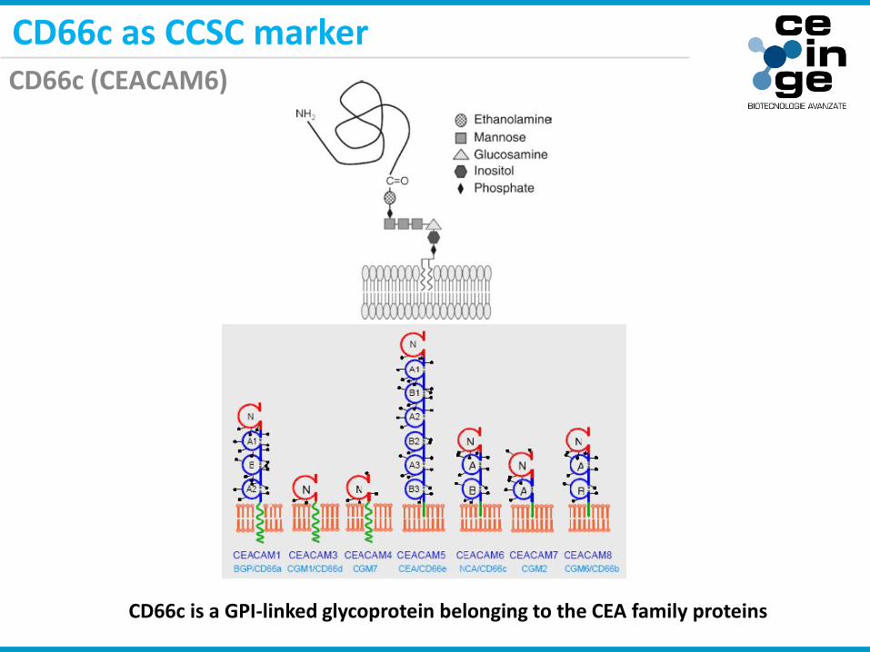
CD66c as CCSC markerCD66c (CEACAM6)
CD66c is a GPI-linked glycoprotein belonging to the CEA family proteins

Normal Tissue Cancer Tissue0
20
40
60
80
100
% C
D66c+
Normal Tissue Cancer Tissue100
1000
10000
100000
CD
66c M
FI
p < 0.0001
p < 0.0001
Gemei et al. Cancer, 2012
CD66c as CCSC markerCD66c expression in normal colon and in colon cancer
CD66c is upregulated in colon cancer tissue

34
0 0 0
8
4
0 014
710
0
5
10
15
20
25
30
35
40
-/+ + ++ +++
nu
mb
er
of
sam
ple
s
CD66c expression score
normal tissue
adenoma
cancer
Normal Adenoma Carcinoma
Gemei et al. Cancer, 2012
CD66c as CCSC markerCD66c expression in normal colon and in colon pathology
CD66c is upregulated in colon cancerogenesis

T1-2 T3-4100
1000
10000
100000
CD
66c
MF
I
p= 0.0073
Tis
N0
M0
T1-2
N0
M0
T3-4
N0
M0
T any
N1-2
M0
T any
N any
M1
Metastasis to
distant organs
M0 M10
10000
20000
30000
40000
CD
66c
MF
I
p= 0.0290
Gemei et al. Cancer, 2012
CD66c as CCSC markerCD66c expression in colon cancer progression
CD66c expression parallels colon cancer progression

CD133+ Normal CD133+ Tumor0
20
40
60
80
100
% C
D66c+
p< 0.0001
Patient 23
(primary tumor)
Patient 23
(normal colon)
CD66C CD66C
normal carcinoma
Gemei et al. Cancer, 2012
CD66c as CCSC markerCD66c expression in normal and cancerous stem cells
CD66c expression parallels colon cancer progression

Adherent Conditions Sphere Conditions
0.1%0.4%
CD133 CD66c
17% 94%
CD133 CD66c
Cell Line Cell Type Origin
GEO Human AdenoCa Primary
CD66c as CCSC markerCD66c expression in CCSCs: colon spheres
CD66c is upregulated in CCSCs enriched colon spheres
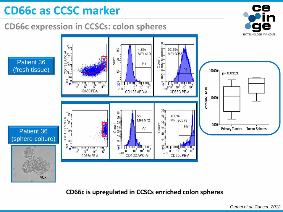
92,6%
MFI 30552
4,8%
MFI 410
5%
MFI 572
100%
MFI 96578
Patient 36
(fresh tissue)
Patient 36
(sphere colture)
Primary Tumors Tumor Spheres1000
10000
100000
CD
66c M
FI
p= 0.0313
40x
Gemei et al. Cancer, 2012
CD66c as CCSC markerCD66c expression in CCSCs: colon spheres
CD66c is upregulated in CCSCs enriched colon spheres
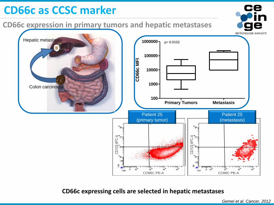
Primary Tumors Metastasis100
1000
10000
100000
1000000
CD
66c M
FI
p= 0.0102
Primary tumor metastasis
Patient 25
(primary tumor)
Patient 25
(metastasis)
Colon carcinoma
Hepatic metastasis
Gemei et al. Cancer, 2012
CD66c as CCSC markerCD66c expression in primary tumors and hepatic metastases
CD66c expressing cells are selected in hepatic metastases

20x
20x
Xenograft
Primary tumor
H&E
H&E
Gemei et al. Cancer, 2012
CD66c as CCSC markerCD66cbright and CD66negative cells tumorigenic potential in vivo
CD66cbright cells are tumorigenic in vivo

77% 24%
SSC
CD
66
c
CD66c siRNA silencing
48h48h
Aspecific siRNA CD66c siRNA0
20
40
60
80
100
%C
D66c+
p=0.0013
Gemei et al. Cancer, 2012
CD66c as CCSC marker: analysis of its functionCD66c silencing in CACO2 cell line

WST-1 colorimetric proliferation assay
24 h 48 h 72 h 96 h 120 h 144 h
0
1
2
3
4Aspecific siRNA
CD66c siRNA
Time
Ce
ll p
rolif
era
tio
n (
OD
45
0-6
50
nm
)
p < 0,0001
Gemei et al. Cancer, 2012
CD66c as CCSC marker: analysis of its functionEffects of CD66c silencing on cell proliferation
CD66c silencing impairs CACO2 cell proliferation

YO-PRO/PI apoptosis-necrosis assay
72h 96h 120h 144h0
10
20
30
Aspecific siRNA
CD66c siRNA
Time
% A
popt
osis
72h 96h 120h 144h0
20
40
60
80
Aspecific siRNA
CD66c siRNA
Time
% N
ecro
sis
p = 0,0013
p = 0,0214
Gemei et al. Cancer, 2012
CD66c as CCSC marker: analysis of its functionEffects of CD66c silencing on cell apoptosis and necrosis
CD66c silencing enhance CACO2 cell death
Aspecific
siRNA
CD66c siRNA
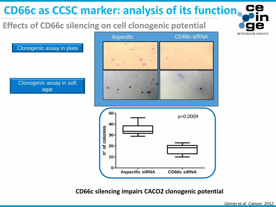
Clonogenic assay in plate
Clonogenic assay in soft
agar
Aspecific siRNA CD66c siRNA0
10
20
30
40
50
n
of
co
lon
ies
p=0.0009
Gemei et al. Cancer, 2012
CD66c as CCSC marker: analysis of its functionEffects of CD66c silencing on cell clonogenic potential
CD66c silencing impairs CACO2 clonogenic potential

CD66c siRNA
treated CACO2
Aspecific siRNA
treated CACO2
55%
0.2%
H&E
H&E
Human Cells
Mouse Cells
Human Cells
Mouse Cells
Gemei et al. Cancer, 2012
CD66c as CCSC marker: analysis of its functionEffects of CD66c silencing on cell tumorigenic potential
CD66c silencing impairs CACO2 tumorigenic potential

Analysis of CD66c associated pathwaysIngenuity pathway analysis

• Multiparametric FC in solid cancer analysis
• CD66c as CCSC markers
• Multiparametric FC in CTCs analysis
• Conclusions
• Acknowledgements
Summary

Primary
tumor
Metastasis
EMT
Blood vessel
Distant
organsCTC
MET
CD26 /CD151 /CD133 /CD44 /CD66c /CD45 /CD326 /SYTO 16
CD326+ CTCs
Multiparametric FC in CTCs analysisColon cancer CTCs phenotype
CD66c along with CD26 could help in the isolation of colon cancer CTCs

0 6 12 18 24 30 36 42
100
80
60
40
20
0
months
Su
rv
iva
l p
ro
ba
bil
ity
(%
)
Number at risk
Group: 0
38 38 35 27 25 9 7 2
Group: 1
34 33 24 15 13 8 5 2
0 6 12 18 24 30 36 42
100
80
60
40
20
0
months
Su
rv
iva
l p
ro
ba
bil
ity
(%
)
Number at risk
Group: 0
46 46 43 31 29 13 10 4
Group: 1
26 25 16 11 9 4 2 0
Three-year progression-free survival rate in the 72 colorectal cancer patients who underwent potentially curative surgery according to (left) pre-and (rightr) post-operative blood levels of CD26+/CD326-cells. In blue: CD26+/CD326-cell-negative patients and in red:CD26+/CD326-cell-positive patients.
Pre-operative CSC Post-operative CSC
Gemei et al. Oncology Letters, 2014
CD326- CTCs
Multiparametric FC in CTCs analysisColon cancer CTCs detection: CD326-/CD26+ cells prognostic impact
CD26+/CD326- CTCs bearing patients have a reduced progression free survival

High pre-operative levels of CD26+/CD326-CTCs correctly predicted tumor relapse in 44.4% of the cases, while 69% of post-operative CD26+/CD326-CTC-positive patients experienced cancer recurrence, with a test accuracy of 88.8%. By contrast, post-operative CD26+/CD326-CTC-negative patients showed an increase in the three-year progression-free survival rate of 86%, along with a reduced risk of tumor relapse of >90%.
Gemei et al. Oncology Letters, 2014
Multiparametric FC in CTCs analysis
Pre operative Post operative
CD26+/CD326- CTCs are predictive of cancer recurrence
Colon cancer CTCs detection: CD326-/CD26+ cells prognostic impact

• Flow cytometry in solid cancer analysis
• CD66c as CCSC markers
• Multiparametric FC in CTCs analysis
• Conclusions
• Acknowledgements
Summary

Conclusion
• A precise gating strategy is necessary to analyze rare populations as CSCs and
CTCs
• Multiparametric flow cytometry allowed the analysis of solid tissues and the
identification of new CSC markers
• Multiparametric flow cytometry can identify CTCs

Acknowledgements
CEINGE-Biotecnologie Avanzate
Prof. Luigi Del Vecchio
Prof. Rosa Di Noto
Dott. Peppino Mirabelli
Dott.ssa Francesca D’Alessio
SUN – Seconda Università di Napoli
Prof. Gennaro Galizia
Dott. Anna Zamboli
Università di Napoli «Federico II»
Prof. Giancarlo Troncone
Dott. Antonino Iaccarino