fibronectin interactions with glycosaminoglycans and ... of fibronectin interactions with...
TRANSCRIPT

Characterization of Fibronectin Interactions with Glycosaminoglycans and Identification of Active Proteolytic Fragments*
(Received for publication, October 31, 1979)
Kenneth M. Yamada,$ Dorothy W. Kennedy,$ Koji Kimata,§ and Robert M. Prattg From the $Laboratory of Molecular Biology, National Cancer Institute, National Institutes of Health, a n d the §Laboratory of Developmental 3Lolom a n d Anomalies. National Institute of Dental Research, National Institutes of Health, Bethesda, ”
Maryland 20205
Fibronectin is a major cell-surface glycoprotein which has been reported to interact with gIycosami- noglycans. A nitrocellulose filter-binding assay was developed to quantitate these interactions at physiolog- ical pH and ionic strength. Fibronectin isolated from chick embryo fibroblasts binds both hyaluronic acid and heparin; heparan sulfate is bound less efficiently, and chondroitin sulfate and glycopeptides are bound minimally. The binding of hyaluronic acid and heparin to fibronectin is saturable and reversible and occurs at separate binding sites. The binding of both molecules to fibronectin is not blocked by EDTA or by other glycosaminoglycans, and is only moderately inhibited by elevated ionic strength. Scatchard analyses reveal nonlinear, high affinity binding to fibronectin with a K D of approximately 10” to lo-’ M for these glycosa- minoglycans. The affinity for heparin was utilized for the isolation of heparin-binding domains of fibronectin on heparia-agarose affinity columns. Heparin-binding proteolytic fragments with apparent molecular weights of 160,000 and 50,000 were isolated following hydrolysis of fibronectin by chymotrypsin or pronase, respec- tively. The possible involvement of such high affinity binding sites of fibronectin in the binding of glycosa- minoglycans to the cell surface or in the organization of extracellular matrices i s discussed.
Cellular fibronectin is a major cell-surface glycoprotein synthesized by a variety of cell types. A related glycoprotein termed plasma fibronectin (cold insohble globulin) is present in blood a t 0.3 mg/ml and is structurally and biologically similar, but not identical, to cellular fibronectin. Both forms of fibronectin appear t,o function as adhesive molecules, af- fecting a wide variety of cellular events (reviewed in Refs. 1- 6).
A series of previous studies established that both forms of fibronectin can interact in some fashion with glycosaminogly- cans. Plasma fibronectin was shown to interact with heparin by precipitation and affinity chromatography studies. Heparin binding was stimulated by calciun~ and apparently abolished by ionic strengths above 0.3 (7). These results suggest that the binding might occur via electrostatic mechanisms. Glyco- saminoglycans can also increase or decrease the extent of binding of plasma fibronectin to collagen (8), and polyamines also inhibit its binding to collagen (9).
With respect to cellular interactions with glycosaminogiy-
* The costs of publication of this article were defrayed in part by
marked “aduertisenent” in accordance with 18 U.S.C. Section 1734 the payment of page charges. This article must therefore be hereby
solely to indicate this fact.
cans plus fibronectin, it has been suggested that cells of the reticuloendothelial system utilize plasma fibronectin for up- take of denatured coIIagen (lo), and that heparin can modu- late this process (11). Cross-linking studies with fibroblastic ceUs suggest that cellular fibronectin is in proximity to sulfated proteoglycans on the cell surface, suggesting that the cellular form of fibronectin also interacts with proteoglycans (12).
All of these previous studies support the concept that glycosaminoglycans can interact with plasma or cellular fibro- nectin. We have investigated the biochemical characteristics and specificity of such interactions with cellular fibronectin. A filter-binding assay was developed to obtain quantitative information; our studies were usually conducted under phys- iological salt conditions in attempts to characterize specific interactions that are independent of simple electrostatic at- traction. We also describe the isolation of active proteolytic fragments of fibronectin that bind to heparin, and provide models for the organization of these sites relative to other recently isolated binding sites of fibronectin.
EXPERIMENTAL PROCEDURES
Materzals-Cellular fibronectin was purified from chick embryo fibroblasts exactly as described previously (13) . The fibronectin was at least 98% pure according to electrophoresis in 4 to 7.5% sodium dodecyl sulfate (SD.5)-polyacrylamide slab gels 113, 14). ‘T-labeled fibronectin was prepared from chick fibroblasts cultured for 24 h in regular culture medium plus 4 pCi/ml of [~-’~C]glycine (54 mCi/ mmol; Amersham).
Highly purified, reference standard grade human umbilical cord hyaluronic acid, bovine lung heparin, sturgeon chondroitin 4-sulfate and chondroitin 6-sulfate, bovine cornea keratan sulfate, and hog mucosal dermatan sulfate were kind gifts from Drs. M. B. Mathews and J. A. Cifonelli, Department of Pediatrics, University of Chicago. Heparin (porcine intestinal mucosa, grade I, 169.7 USP units/mg) was also purchased from Sigma, and shark cartilage chondroitin 6- sulfate was from Miles Biochemicals.
Protease-free bacterial collagenase was from Advance Biofactures Corp. (Form 111, 181 units/pg); u-chymotr.ypsin (49.2 units/mg) and bovine testicular hyaluronidase (11,560 units/mg) were from Worth- ington; pronase (45 units/mg) and Streptomyces hyaluronidase (5,990 units/mg) were from Calbiochem-Behring; and chondroitinase ABC was from Miles Biochemicals. Bovine y-globulins and fibrinogen (90% clottable) were obtained from Miles Biochemicals and fetuin, bovine serum albumin, pepstatin A, N-acetylglucosamine, phenylmethylsul- fonyl fluoride (PMSF), and glucuronic acid were from Sigma. Seph- arose CL4B and Sephadex G-50 and G-IOO were from Pharmacia.
Standard Binding Assay-Purified cellular fibronectin was incu- bated at a final concentration of 275 ,ug/ml (1.25 x 10+ M monomer) with various ”H-labeled, purified glycosaminoglycans in complete Dulbecco’s phosphate-buffered saline (PES) containing an additional 10 mM sodium phosphate, pH 7.4, in polypropylene test tubes at 23°C. The final volume was usually 0.64 mI, although for certain experi- ments with material of high specific activity, the assay was scaled down. After 60 min, the samples were agitated on a Vortex mixer, then vacuum-fdtered through 2.5-cm diameter 0.45-p nitrocellulose fiters (Millipore, type HA). Residual sample was transferred from
6055

6056 Fihronectin-Glycosaminoglycan Interactions
the tubes with two I-ml washes of PBS,' then the filters were rapidly washed with 4 ml of PBS five times at 23°C. The washing procedure required less than 30 s. Preliminary experiments indicated that the recovery of radioactive heparin or hyaluronate bound to fibronectin was not altered if the washing was performed at 4OC, or if serum-free culture medium (Ham's F-10 medium) was substituted for the PBS. Although the fibronectin was routinely added last to reaction mix- tures, the addition of labeled glycosaminoglycans after the fibronectin did not affect the amount of binding even if delayed up to 10 min (the longest period tested).
The filters were placed into 12 ml of Aquasol (New England Nuclear), and agitated until clear, and the radioactivity was deter- mined with a Beckman LS-235 scintillation counter with 349 effi- ciency for tritium, as determined by adding internal standards of 'H- labeled toluene (New England Nuclear).
As will he described below, glycosaminoglycan binding was in- hibited by 4 M NaCl or hyaluronidases in certain cases. This inhibition of binding was not due to artifactual loss of fibronectin from the filters, since recovery of ["Clglycine-labeled fibronectin from filters under these conditions was the same as that of untreated controls
Preparation of Hadioncticre (;lvco.snminogl~cans-Secotldary chick embryo fibroblasts were plated at 2 X 10" cells/100-cm2 plastic tissue culture dish (Falcon) and cultured as described previously in 55 heat-inactivated calf serum plus 10'4 tryptose phosphate in Ham's F-10 medium (13). After 24 h, the medium was changed and 25 pCi/ ml of ~~-ll,f;-'H]glucosamine (43 Ci/mmol; New England Nuclear) was added to the standard culture medium containing 1 mM instead of 5 n m glucose. After further incubation for 18 h, the medium was decanted and the cell layers were washed with 2.5 ml of I'BS at B'2"C: the washes were combined with the medium. Medium fractions and culture dishes containing cell layers were frozen and stored at -20°C until further processing. The medium fractions plus washes were thawed, heat-inactivated for 5 min at 100°C, and cooled. Tris was added to 0.2 M, and the pH was adjusted to 8.0 with HCI. The cell layers were removed from each dish with a Teflon scraper into 2.5 ml of 0.2 M Tris/Cl, pH 8.0, and sonicated with a Kontes sonicator equipped with a microprobe at power setting 8 for 30 s.
Pronase was added to the medium and cell-layer sonicates to 1 mg/ nil. and the solutions were incubated at 5.5"C for 24 h. The pronase digests were cooled to 4°C and trichloroacetic acid was added to a final concentration of 10'3. After 30 min at 4°C. the trichloroacetic acid-insoluble material was removed by centrifugation at 1O.ooO X g for 20 min at 4'C. The trichloroacetic acitl-soluble material was dialyzed extensively at 4OC against distilled H,O, lvophilized. resus- pended in 0.05 M Tris/Cl, pH 7.2, and chromatographed on a IIEAE- cellulose column (2.5 X 10 cm) using a 0 to 1 M NaCl gradient (15, 16). Four labeled peaks were eluted. These were pooled and tentatively indentifieti as labeled glycopeptide, hyaluronic acid, heparan sulfate. and chondroitin sulfate.
The labeled hyaluronic acid was further purified by rechromatog- raphy on a I>EAE-cellulose column eluted with a 0 to 0.5 M NaCl gradient. Similar chromatographic patterns were obtained using either the pronase digestion method or alkali extraction with 0.5 N NaOH at 2%%"C for 24 h (17) . Susceptibility of the components to digestion by various specific enzymes as described previously (15, 1 6 ) was used t o identifv the labeled glycosaminoglycans. Ninety-eight per cent o f the [ 'Hlhyaluronic acid was degraded by Streptomvce.~ hya- luronidase, specific for hyaluronic acid. whereas none of the ['HI- chondroitin sulfate was degraded. Uronlc acid was quantitated by the modified carhazole method (18).
The apparent size of the [ 'Hlhyaluronic acid was determined on a Sepharose CL-4B column (0.9 X 150 cm) in I'BS, calibrated with the following molecular weight standards detected by the phenolsulfuric acid procedure (19): hyaluronic acid (2g'I0.0(H)). derniatan sulfate (45.(MM)). chondroitin &sulfate (29,000). and heparin (14,NK)). The average size of the hyaluronic acid was defined as the apparent nlolecular weight above or below which half of the total radioactivity in the preparation was present.
[ 'H ]Heparin (0.:)32 mCi/mg; New England Nuclear) was applied t o a Sephadex G-100 column (0.9 x 60 cnl) equilibrated in I'BS. The
' The ahbreviations used are: I'BS, Dulhecco's phosphate-buffered saline supplemented with 1 0 mM sodium phosphate. 137 niM NaCI. 2.7 IIIM KCI, 0.9 IIIM Carli, 0.5 nlM MgCli.6H,0. 1.5 mM KHLI'O,, 18.1 n m Na>HI'Ol, pH 7.4; Buffer E, 0.1 M NaC1, 10 mM CaCI,, 50 nlM Tris/CI, pH 7.0; SIH. sodium dodecyl sulfate; I'MSF. phenylmeth- ylsulfonvl fluoride.
(1lH) t 2':').
". ~. . ~ ~ ~ " . . ~. . ~ ~~
high molecular weight peak fractions were pooled as described below;
size, and the remainder was stored at -20°C and used for binding an aliquot was rechromatographed on the same column to calibrate
studies. The column was calibrated with heparin ( M , = 14,000). dextran T10 (5,200), dextran T40 (28,OOO), and sucrose (342). 'H- labeled proteoglycans (AI-Dl fraction) were prepared hy Dr. .John Pennypacker, National Institute of Dental Research, from mouse articular cartilage incubated with [ 'Hlglucosamine exactly as de- scribed (20, e l ) .
Binding Affinities-Increasing amounts of [ 'HJhyaluronic acid (439 cpm/pmol of hyaluronic acid) or [ 'Hlheparin (1,250 cpm/pmol of heparin) were added to the standard assay at final concentrations of 6 to 200 nM or 50 to 940 nM respectively. and routine Scatchard analysis was performed (22). The results were confirmed by using mixtures of reference standard grade unlabeled heparin or hyaluronic acid and labeled material, and the amounts of binding were calculated by accounting for the dilution of the radioactivity by unlabeled material.
Chondroitinase ABC' Digestion of Fihronectln-Fibronectin was labeled with 2 pCi/ml o f I>-[ 1 -"C]glucosamine (66.6 mCi/mmol; New England Nuclear) for 24 h and purified as described ( 1 3 ) . The fihro- nectin was dialyzed against 0.01 M EDTA, 0.01 M Tris/CI, pH 8.0. Since approximately of the radioactivity remained with the dialysis bag, the digestion was performed on the dlalysate plus minced dialysis hag. The reaction mixture contained 2,500 cpm of ["Clfibro- nectin, 0.1 M Tris/Cl, pH 8.0, 0.08 M sodium acetate, 0.01 M EDTA, 0.01 M N-ethylmaleimide, 1 mM I'MSF, 100 pg of bovine seruni albumin, 40 pg of carrier chondroitin (;-sulfate. 6,800 cpm o f [ 'HI- proteoglycan monomer A l - D l (20). and 0.2 unit o f chondroitinase ABC (23) in 1.0 ml. The protease inhibitors were included t o protect tibronectin from traces of protease present in the chondroitinase. The reaction mixture was incubated for 50 min at 37°C with the further addition of chondroitinase ABC after 30 min. 'I'he reaction was stopped by the addition of an equal volume of 8 M guanitlinium chloride. 0 . 0 1 M EIITA. 0.04 M Tris/CI, pH 8.0. l 'he solution was applied to a Sephadex G-50 (superfine) column ( 1 X 30 cnl) equili- brated with 4 M guanidinium chloride, 0.01 M EIITA. 0.02 M Tris/Cl, pH 8.0, and eluted with the same buffer; 0.53-ml fractions were collected and radioactivity was determined with a scintillation spec- trometer.
Nitrous Acid Degradation of Fihronecfin Prepurrrt/on-[ "C](;lu- cosamine-labeled fibronectin (2,500 cpm) was prepared for reaction as described for chondroitinase ABC digests, except that 1 mg of heparin and 200 pg of bovine serum albumin were utilized as carriers. Nitrous acid drgradation was performed as described by Iintlahl et a/. (24) at room temperature in 4 ml of 1.8 M acetic acid and 0.24 M sodium nitrite. The sodium nitrite was omitted in the control. After 8 O min. an equal volume of methanol was added and the contents were evaporated under vacuum. The residue was dissolved in 4 M guanidinium chloride, 0 .01 M EIYrA. 0.02 M Tris/C;I, pH 8.0, anti centrifuged to remove insoluhle material, antl the supernatant was applied to a Sephadex G-60 column as described for the chondroiti- nase ABC digestion. The completeness of the reaction was confirmed by demonstrating the degradation o f carrier heparin. which was detected by the carbazole reaction for uronic acid (18).
Isolatmn of Heparin-hlndmg Fragrnent.\-Hepartn (SignW was coupled to agarose via cyanogen bromide as described (261. A trace amount of proteolytic activity remaining with the heparm was 111-
hibited bv preincubating the heparin-agarose with 2 n1M I'MSF. 5 mM N-ethvlmalelmide, and 5 p~ pepstatin in Buffer E ( 0 . 1 M Na(:l, 1 0 mM CaCl., 50 mM Tris/CI, pH 7.0) for 24 h at 2:l"C. folioweti exhaustive washmg with Buffer E.
For routine analytical studies, the heparin-agarose was poured as 0.5-ml columns in O.Y-cn1 disposable polypropylene columns (Rio- Rad) and washed with 1 0 column volumes of Buffer E . Cellular fibronectin (250 kg) was diluted into 1 ml o f Buffer E antl applied t o the column. l'he eluate was reapplied twice more to ensure maximal binding. The columns were then washed with at least 1 0 volumes o f Buffer E.
Protease digestions of the bound tihronectin were performed di- rectly on the columns hy applying 0.5 ml o f various proteases i n Buffer E. (,-Chymotrypsin was at either 0.5 or 5 pg/ml. anti pronase was at 5 pg/ml. After 1 h at 2:i"C, the columns u'ert. washed with 1 0 volumes of Buffer E containing 2 mM I'MSF. The gel heads were transferred to glass test tubes with 10 mM sodium phosphate. pH 7.0. then setlinlentetl. The pellets were incuhatrd with 0.25 ml offif( SI)S. 6 nlM I'MSF. 30 nlM sodium phosphate, pH 7.0, at I 0 0 " C for three 2- nlin periods alternating with hrief agitation on a Vortex mixer. The

Fibronectin-Glycosaminoglycan Interactions 6057
gel beads were then sedimented and the supernatants were examined of the input radioactivity was recovered on the filters under on polyacrylamide gels after reduction with 0.1 M dithiothreitol (13, the standard incubation conditions. Since "-labeled glyco- 14).
In certain experiments, fibronectin that was bound to heparin- agarose was subjected to sequential digestions with chymotrypsin and pronase. After 1 h in 5 pg/ml of chymotrypsin, the columns were washed with 10 volumes of Buffer E, then incubated an additional 1 h in 5 pg/ml of pronase, washed, and eluted with boiling SDS as described above. In two experiments, the column eluates after 5 pg/ ml of chymotrypsin digestion were reapplied to 0.5-ml gelatin-agarose columns, and washed with Buffer E containing 2 mM PMSF after 10 min; then, the bound fragments were eluted with boiling 2% SDS as described (26).
The recovery of a pronase-generated 50,000-dalton fragment from heparin-agarose columns was determined by two methods. The amounts of protein originally in fibronectin or in the 50,000-dalton fragment were compared in polyacrylamide gels stained with Coo- massie brilliant blue (13, 14) by densitometry with a Joyce-Loebl densitometer. The absorbance was linearly proportional to the amount of protein applied in the ranges utilized.
A second method was to determine the recovery of radioactivity from fibronectin originally labeled for 24 h with ['4C]glycine (an abundant amino acid in fibronectin (13)). The ["Clfibronectin was permitted to bind to I-ml heparin-agarose columns, as described above, and washed with 5 ml of Buffer E, and the beads were transferred to scintillation vials for determination of radioactivity. Parallel columns were subjected to pronase digestion, as described, and washed with 10 ml of Buffer E, and radioactivity was determined. The control column consisted of agarose activated with cyanogen bromide but subjected to coupling conditions in the absence of heparin.
RESULTS
Glycosaminoglycan BindingAssay-Fibronectin is present on the surface of cells a t physiological pH as insoluble, rela- tively immobile aggregates or fibrils (reviewed in Refs. 1, 2, and 5). Although isolated purified cellular fibronectin can be maintained in solution at alkaline pH, it regains its insolubility at neutral pH (13). This property was used to establish a binding assay in which purified fibronectin is neutralized and incubated with various ligands for 1 h in Dulbecco's PBS at pH 7.4 prior to filtration through nitrocellulose filters as described under "Experimental Procedures." Studies with ['4C]glycine-labeled fibronectin indicated that all (101 k 1%)
saminoglycans were not retained by these filters, their binding to fibronectin can be detected by measuring the radioactivity that remains bound to filters in the presence of unlabeled fibronectin.
We fist examined the binding of ["H]hyaluronic acid to fibronectin. The binding was maximal at 200 to 275 pg/ml of fibronectin (Fig. 1). All subsequent studies were consequently performed with 275 pg/ml of fibronectin (1.25 X lo-" M mono- mer, if reduced; the molecule consists primarily of disulfide- bonded dimers and multimers).
The time course of ['Hlhyaluronate binding is shown in Fig. 2. Binding reaches equilibrium in approximately 10 to 15 min; the routine assay involves an incubation of 60 min. The binding is reversible after dilution of reaction mixtures with PBS, and the process requires approximately 2 h (Fig. 3). The presence of unlabeled ligand increases the rate of release of labeled ligand from fibronectin (Fig. 3). Similar binding and reversibility results were obtained with ['Hlheparin, except that heparin binding reached equilibrium within 30 to 60 s (data not shown).
This binding of hyaluronic acid isolated from culture me- dium to fibronectin was compared to the binding of ["HI- hyaluronic acid isolated from the corresponding cell layers following pronase digestion. Cell layer hyaluronate was also bound by fibronectin, although the percentage of total radio- activity bound was lower for cell-associated than for medium hyaluronate from the same cell cultures (36 rt_ 4% versus 58 k 3%, respectively; specific activities were 10,030 versus 2,130 cpm/nmol of uronic acid). We therefore utilized hyaluronic acid purified from culture medium for all subsequent experi- ments.
The specificity of glycosaminoglycan binding to fibronectin was examined with purified glycosaminoglycans isolated from chick embryo fibroblasts. Cultures were labeled for 24 h with ["H]glucosamine, then subjected to pronase digestion and DEAE-cellulose column chromatography as described under "Experimental Procedures." Hyaluronic acid isolated from culture medium was the glycosaminoglycan most effectively
0-4 1 0 0 m 3 0 0 5 0 0 0 5 FIBRONECTIN IpglmlJ
FIG. 1 (left). Binding of hyaluronic acid by fibronectin. 'H- labeled hyaluronic acid was incubated with the indicated concentra- tions of fibronectin in PBS for 60 min a t 23°C; then, the mixtures were collected on nitrocellulose filters and the retained radioactivity was determined as described under "Experimental Procedures." The specific radioactivity of hyaluronic acid was 431 cpm/nmol of uronic acid, and the maximal binding of input radioactivity was 45%. Values represent means 2 S.E. of triplicate samples.
FIG. 2 (center). Time course of hyaluronic acid binding to fibronectin. ['HHJHyaluronic acid (2,000 cpm; 2,130 cpm/nmol of uronic acid) was incubated with 1.25 X IO-'' M fibronectin in 0.64 ml of PBS a t 23°C and collected on nitrocellulose filters at the indicated
10 15 20 60 0 1 2 3 4 TIME (Minutes1 TIME IHoursI
times. FIG. 3 (right). Reversibility of hyaluronic acid binding to
fibronectin. After incubation for 60 min with 2,000 cpm (2,130 cpm/ nmol of uronic acid) of hyaluronic acid plus 1.25 X IO-'' M fibronectin, the mixture was diluted 1:15 or 1:100 with PBS in polyethylene tubes and further incubated at 23°C for the times indicated prior to collec- tion on fdters. M, diluted 1:15 with PBS; A-A, diluted 1:15 with PBS plus the addition of 100 pg/ml of unlabeled hyaluronic acid obtained from Drs. Mathews and Cifonelli; M, diluted 1:100 with PBS; *, binding by a parallel sample diluted 1:15 at the time of initial mixing of hyaluronate and fibronectin, incubated for a total of 5 h prior to filtration.

6058 Fibronectin-Glycosaminoglycan Interactions
bound by fibronectin (Fig. 4). The proportion of original radioactivity bound varied from preparation to preparation depending on the specific radioactivity of labeling of the hyaluronic acid (e.g. 23% in Fig. 4 and up to 70% binding in other experiments).
Heparan sulfate was also bound by fibronectin, but the largest amount bound was only 5% of heparan sulfate input radioactivity; this low extent, of binding does not appear to be due to a low specific activity, since the specific radioactivity of the heparan sulfate used for Fig. 4 was four times that of the hyaluronic acid. Chondroitin sulfate, either from cell monolayers or from medium, bound poorly to fibronectin, and glycopeptides also did not bind (Fig. 4). In experiments de- scribed below, the glycosaminoglycan heparin was also found to bind effectively to fibronectin (up to 85% binding).
Two other procedures were compared to the filter-binding assay for evaluating the binding of chick fibroblast glycosa- minoglycans to fibronectin. Although both yielded results similar to those of the filter-binding experiments, the amounts of binding were less and the background binding was greater in these assays. The fiist assay utilized affinity columns of fibronectin coupled covalently to agarose via cyanogen bro- mide as described (27). Purified ,"H-labeled glycosaminogly- cans that remained bound after extensive washing with PBS were eluted with 8 M urea and quantitated in a scintillation counter. The second assay involved incubating fibronectin with glycosaminoglycans for 1 h as for the filter assay, but the fibronectin was, instead, collected by immunoprecipitation with affinity-purified anti-fibronectin antibodies as described (28). Using these assays and the glycosaminoglycans described in the legend to Fig. 4, we found 5.1 and 1.8% binding of hyaluronic acid by the column and immunoprecipitation tech- niques, respectively, 1.6 and 1.9% binding of heparan sulfate, 0.3 and 0.2% binding of chondroitin sulfate, and 0 and 0%
c e n L ~ Y W Mdum
FIG. 4. Specificity of glycosaminoglycan binding to fibro- nectin. The fraction of total radioactivity added to each assay that was recovered on nitrocellulose filters after a 60-min incubation a t 23°C with 1.25 X lo-" M fibronectin is indicated for "H-labeled glycosaminoglycans or glycopeptides purified as described under "Ex- perimental Procedures"; the sources were chick embryo fibroblast cell monolayers or the media from these cultures labeled with ['Hlglucosamine. The preparations had the following specific activi- ties: chondroitin sulfate (cell layer), 2,080 cpm/nmol; heparan sulfate, 3,620 cpm/nmol; chondroitin sulfate (medium), 3,580 cpm/nmol; hy- aluronic acid, 425 cpm/nmol of uronic acid. The original quantities of radioactively labeled material per assay and the mean background binding in the controls from which fibronectin was omitted were: chondroitin sulfate (cell layer), 884 and 0 cpn; heparan sulfate, 995 and 5 cpm; glycopeptides (cell layer), 1,716 and 9 cpm; chondroitin sulfate (medium), 1,432 and 8 cpm; glycopeptides (medium), 1,258 and 0 cpm; hyaluronic acid, 552 and 0 cpm. Bars indicate S.E. of triplicate samples.
binding of glycopeptides. Because the fiiter-binding assay was more convenient and reliable, and bound more ligand than either of these latter assays, it was employed for all subsequent experiments.
competitive Inhibition Experiments-The binding of ["Hlhyaluronic acid to fibronectin was completely inhibited by the addition of unlabeled hyaluronic acid to reaction mix- tures, but not by the inclusion of unlabeled heparin or chon- droitin sulfate (Fig. 5) or by other glycosaminoglycans (Table I). Half-maximal inhibition of ["Hlhyaluronic acid binding occurred a t approximately 0.25 pg/ml of unlabeled hyaluro- nate (Fig. 5); at this point, 0.1 pg of ["Hlhyaluronate was
' ' I 500 1
d m 1
FIG. 5. Competit ive inhibit ion of ['H]hyaluronic acid bind- ing to fibronectin by unlabeled glycosaminoglycans. The stan- dard 60-min binding assay was performed with 2,OOO cpm of ["HI- hyaluronate at 2,130 cpm/nmol of uronic acid plus 1.25 X 10"' M fibronectin in the presence of the indicated final concentrations of unlabeled hyaluronic acid (HA; M), chondroitin 4-sulfate ( C S M), or heparin ( H , A-A) (unlabeled glycosaminoglycans provided by Drs. Mathews and Cifonelli). The fibronectin was added last to the 0.64-ml reaction mixtures. The closed triangle indicates binding when testicular hyaluronidase (20 pg/ml) was present in the reaction mixtures.
TABLE I Inhibitors of the binding of hyaluronic acid by fibronectin
Binding of ['Hlhyaluronic acid to fibronectin was determined as described under "Experimental Procedures" in the presence of the indicated materials. For the NaCI-containing assays, the PBS was replaced by NaCl plus 20 mM sodium phosphate, pH 7.4; for the EDTA-containing assays, the PBS was prepared without calcium or magnesium. The input ["H]hyaluronic acid was 2,300 cpm with a specific radioactivity of 2,130 cpm/nmol of uronic acid. In the absence of additions, the control bound 61% of input radioactivity. Other glycosaminoglycans are evaluated in Fig. 5. The concentration of fibronectin was 275 pg/ml (1.25 X 10 I' M); in the controls without fibronectin, each protein was also present a t 275 pg/ml.
Condition cprn bound 5 Percentage S.E. of control
Control Heparan sulfate (100 pg/ml) Keratan sulfate (100 pg/ml) Dermatan sulfate (100 pg/ml) Chondroitin sulfate
NaCl (1 M ) NaCl (4 M ) EDTA (10 mM) N-acetylglucosamine (0.1 M) +
curonic acid (0.1 M) Glucosamine (0.1 M ) Collagenase (0.5 mg/ml) Trypsin (0.1 mg/ml) Streptomyces hyaluronidase
(80 units/ml) -Fibronectin
+Fibrinogen +Fetuin +Albumin +y-globulin
(100 M m l )
glu-
1405 f 31 1447 f 13 1264 f 9 1482 * 13
1422 f 49 1447 f 29 796 f 25
1336 f 78 1459 f 31
1318 f 58 1364 f 23
14 f 5
5 f 0.4
5 f l 9 f 0.4 2 f 0.9 5 f 0.4
5 f 2
100 102
104 88.8
99.9
56.7 95.1
103
104
93.8 97.1
1 .0
0.4 0.4 0.4 0.6 0.1 0.4

Fibronectin-Glycosaminoglycan Interactions 6059
bound per reaction, and the total concentration of labeled plus unlabeled hyaluronate was 0.74 pg/ml.
In contrast to the results with [.'H]hyaluronic acid, the binding of ['Hlheparin to fibronectin was inhibited only by unlabeled heparin, with 50% inhibition of binding by 2 ,ug/ml of unlabeled material, but was not inhibited by unlabeled hyaluronic acid or chondroitin sulfate at even 100 pg/ml (Fig. 6). Its binding may be slightly inhibited by dermatan sulfate at 100 ,ug/ml, but not by all other glycosaminoglycans tested including heparan sulfate (Table 11). The binding sites for hyaluronic acid and heparin are therefore saturable and are functionally separable, since there is no cross-competition for binding at each site by other unlabeled glycosaminoglycans.
Binding Affinity-We examined the binding affinities of hyaluronic acid and heparin to fibronectin. These studies were complicated by the broad size ranges of these molecules. For example, the hyaluronic acid chains ranged in molecular weight from 20,000 to at least 500,000 on agarose columns calibrated by glycosaminoglycan standards of known size; the average molecular weight was estimated to be 94,000 (see "Experimental Procedures" for details). Likewise, heparin also has variable chain lengths (Fig. 7); however, since most of the molecules were of a limited size range, we isolated the molec- ular weight range of 12,000 for binding studies (Fig. 7 , Fraction I).
The results of Scatchard analyses are shown in Figs. 8 and 9. The negative reciprocal of the slope is the dissociation constant. For hyaluronic acid, the IC,) f 10" M, although the curve deviates slightly from linearity (Fig. 8). For heparin, the
" 1
0-! 0 10 20 30 40 50 100
rsiml
FIG. 6. Competitive inhibition of I3H]heparin binding to fi- bronectin by unlabeled glycosaminoglycans. The ["HJheparin- binding assay was performed with 19,OOO cpm of ["Hlheparin (0.332 mCi/mg; New England Nuclear) in the presence of the indicated final concentrations of heparin ( H ; ."-.), hyaluronic acid (HA; A-A), or chondroitin sulfate (CS, o"--o).
TABLE I1 Inhibitors ofthe binding of heparin to fibronectin
Binding of heparin Fraction I to fibronectin was determined as described under "Experimental Procedures" in the presence of the indicated materials. Input [%]heparin was 3,760 cpm/assay; 84% of input radioactivity was bound by the control; and background binding to the filter in the absence of fibronectin was 1 2 1 cpm. Other glycosaminoglycans are evaluated in Fig. 6.
Condition cpm bound 2 S.E. Percentage of control
Control 3178 & 43 100 Heparan sulfate (100 pg/ml) 3263 60 103 Keratan sulfate (100 pg/ml) 3254 f 56 102 Dermatan sulfate (100 pg/ml) Chondroitin 6-sulfate (100 pg/ml) EDTA (0.1 M) NaCl(1 M) NaCl ( 2 M) NaCl (4 M) Collagenase (0.5 mg/ml) 3398 -C 210 107
-__
2549 k 53 80.2
3120 +: 51 98.2 613 & 63 19.3 579 k 46 18.2 217 k 22 6.8
3170 k 47 99.7
__l___".~_
28K 14K 5 2K 0.2K
' I i + 2000 --
VI i
l5O0I 1000
500 L l J \\o -i ".-.-.* -... -..* 0 10 20 T 40 0 /:,a I A
Eluuon Volume
FIG. 7. Gel filtration chromatography of original and puri- fied [3H]heparin. Commercial ['Hlheparin (20 pCi) (t".) was chromatographed on a Sephadex G-1M) column as described under "Experimental Procedures." Fractions I and I1 were pooled and Fraction I was rechromatographed on the same column to determine size (m). Arrows indicate thz elution volumes of the molecular weight standards heparin, dextran T40 end T10, and sucrose.
.25
BOUND ipmoll BOUND ( g m d
FIG. 8 (left). Scatchard analysis of the binding of hyaluronic acid to fibronectin. Equilibrium binding between [,'H]hyaluronic acid and 275 pg/ml of cellular fibronectin was determined for 6 to %oO nM hyaluronic acid as described under "Experimental Procedures," and the data were analyzed according to Scatchard ( 2 2 ) . We empha- size that this experiment was performed with hyaluronic acid of a mixture of chain lengths. Estimates of affinity and number of binding sites were therefore calculated using the average molecular weight of 94,000; the binding affinity could differ for differing chain lengths.
FIG. 9 (right). Scatchard analysis of the binding of heparin to fibronectin. Binding of 50 to 940 nM ['Hlheparin (Fraction I ) was determined according to Scatchard (22).
curve is biphasic and the estimated K,, G 10" and 4 X lo-!'. This type of biphasic plot for heparin binding was obtained with three other preparations of fibronectin, as well as in two other experiments in which the concentration of heparin was increased by addition of unlabeled heparin, and the amounts bound were calculated by correcting for the dilution of labeled material. The extrapolated number of binding sites of fibro- nectin for hyaluronic acid was approximately O.l/monomer and for heparin was O.Z/monomer of fibronectin. However, it appears likely that these extrapolations may be in error, since the ligand itself probably has multiple fibronectin binding sites due to the repeating disaccharides; consequently, some glycosaminoglycan molecules may bind more than one fibro- nectin. A direct means of determining the number of binding sites for heparin is described below,
Inhibitors of Binding-Potential inhibitors, in particular high concentrations of salts, were examined for their effects on the binding of hyaluronic acid or heparin to fibronectin. The binding of hyaluronate is not inhibited by 1 M NaCI, and is only inhibited by 438 at 4 M (Table I). Binding is also not inhibited by 10 mM EDTA or by the constituent monosaccha- rides of hyaluronate, N-acetylglucosamine and glucuronic acid. In contrast to certain biological and binding activities of fibronectin, such as hemagglutination, which are inhibited by amino compounds, e.g. glucosamine (9, 29, 30), the binding of hyaluronic acid to fibronectin is not disrupted by this amino

6060 Fibronectin-Glycosaminoglycan Interactions
sugar (Table I). The binding is also not affected by treatment with purified collagenase, but is sensitive to treatment of reaction mixtrues with trypsin or fungal hyaluronidase (Table 1).
If the fibronectin is omitted from reaction mixtures, or if it is repIaced by equal protein concentrations of fibrinogen, fetuin, bovine serum albumin, or y-globulin, there is less than 1% nonspecific binding (Table I).
The binding of heparin to fibronectin is more sensitive to salt inhibition than is the binding of hyaluronate (Table 11). Approximately 80% of the binding was inhibited by 1 M NaCl and over 90% was inhibited by 4 M NaCl (Table 11). However, it is also important to note that binding is highly effective at physiological salt concentrations (standard assay conditions with ionic strength -0.2), in which 84% of the total amount of ["Hlheparin in the reaction mixture is bound to fibronectin.
In addition, we compared the binding of the low molecular weight fraction of commercial ["HJheparin preparations (Fig. 7, Fraction II), and found minimal binding of this fraction to fibronectin (<0.05% of the original radioactivity).
Evaluation for Possible Proteoglycan Contamination- Since proteoglycans are known to bind hyaluronic acid (20), it was important to test for proteoglycan contamination or for covalently bound proteoglycans in our preparations of fibro- nectin. Fibronectin was labeled with ['4C]glucosamine for 24 h as described under "Experimental Procedures," then iso- lated as usual (13). The fibronectin was then subjected to chondroitinase ABC treatment or to nitrous acid degradation as described under "Experimental Procedures" and examined for the release of radioactivity from possible contaminating proteoglycans. Positive controls for the efficacy of hydrolysis consisted of measuring the degradation of added carrier [~'H]glucosamine-IabeIed proteoglycans or of unlabeled hepa- rin.
i
1 0 10 20 30 40 50
0
FRACTION NUMBER
FIG. 10. Digestion of fibronectin preparations by chondroi- tinase ABC. Fibronectin was prepared from chick fibroblasts incu- bated for 24 h with 2 pCi/ml of ['4CJglucosamine and analyzed as
digested for 50 min at 37°C with 0.2 unit/ml of chondroitinase ABC. described under "Experimental Procedures." Top, control; bottom,
M, ['JC]glucosamine-labeled fibronectin preparation; o"--o, "H-labeled cartilage proteoglycan fraction AI-Dl mixed with the ["C]fibronectin to evaluate the efficacy of chondroitinase digestion.
['4C~Glucosamine-labeled fibronectin and ["H]glucosamine- labeled carrier proteoglycans both eluted in the exclusion volume of G-50 columns (Fig. 10). After chondroitinase ABC digestion, there was no detectable change in the ['4C]fibro- nectin elution pattern; 95% of the original radioactivity was recovered in the excluded volume fractions. In contrast, the ["H]proteoglycans were degraded by this enzyme, and the radioactivity chromatographed as a retarded peak (Fig. 10). This retarded ,'H peak was characterized as dimers by paper chromatography. These results indicate that the fibronectin preparation did not contain detectable amounts of chon- droitinase ABC-sensitive proteoglycans of which chondroitin 4-sulfate, chondroitin 6-sulfate, dermatan sulfate, chondroitin, and hyaluronic acid are the glycosaminoglycan constituents.
A mixture of ['4C]glucosamine-labeled fibronectin and hep- arin added as carrier was subjected to nitrous acid degrada- tion. After the reaction, 80% of the starting radioactivity was recovered as a precipitate insoluble in 4 M guanidine. The supernatant was examined for degradation products. By Seph- adex G-50 chromatography similar to that shown in Fig. 10, the radioactivity from control or nitrous acid-treated samples appeared only in the excluded fraction. No retarded radioac- tivity was detected, including in Fractions 35 to 50 in which the degradation products of the carrier heparin were detected by the carbazole reaction. These results suggest that the ["C] fibronectin preparation did not contain glycosaminoglycans or proteoglycans that are sensitive to nitrous acid degradation such as heparin or heparan sulfate.
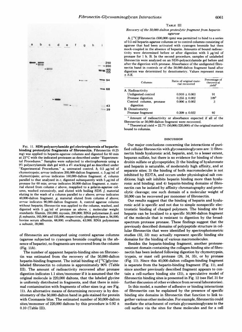
Heparin-binding Fragments-Our demonstration of high affinity glycosaminoglycan sites in fibronectin suggested that. it might be possible to isolate proteolytic fragments still containing these sites. Cellular fibronectin was permitted to bind to heparin-agarose affinity columns, then digested in situ with a-chymotrypsin or pronase as described under "Experi- mental Procedures." The proteolytic fragments that were eluted from the column were compared to those that remained bound after extensive washing. Bound fragments were eluted with bailing 2% SDS; an SDS-polyacrylamide gel analysis is shown in Fig. 11.
After a brief digestion with chymotrypsin, several large fragments remained bound to the heparin affinity column, including a previously isolated (31), large fragment with an apparent molecular weight of 205,000 (Fig. l l b ) . Further digestion resuIted in the generation of a previously isolated component of 160,000 daltons (31) thought to contain a cell- binding site (Fig. l l c ) .
Pronase digestion of the 160,000-dalton fragment generates a fragment of 50,000 daltons (Fig. lld); this fragment is relatively stable, since 2-fold increases or decreases in pronase concentrations or in digestion times had little effect on these results2 We therefore identified this 50,000-dalton fragment as a protease-resistant, heparin-binding domain of fibronectin. This fragment has recently been recovered from heparin affinity columns using elution by high ionic strength, and the fragment will re-bind to heparin affinity columns with pea ter than 9070 efficiency.'
The material that did not bind to the column after chymo- trypsin digestion included a major component of 40,000 dal- tons. This fragment will bind to gelatin (denatured collagen) affinity columns (Fig. l l e ) and it co-migrates electrophoreti- cally with a previously isolated chymotryptic fragment con- taining a collagen-binding site (26).
The material that did not bind to the heparin columns subjected to a subsequent pronase consisted primarily of a fragment of 90,000 daltons (Fig. Ilg). If binding and digestion
' K. M. Yarnada and D. W. Kennedy, unpublished data.
Fibronectin-Glycosaminoglycan Interactions 6061
-CZ _ - .-
--
250 - -200
-E
be - - 94
- 66
43 39
FIG. Il. SDS-polyacrylamide gel electrophoresis of heparin- binding proteolytic fragments of fibronectin. Fibronectin (0.25 mg) was applied to heparin-agarose columns and digested for 60 min at 23’C with the indicated proteases as described under “Experimen- tal Procedures.” Samples were subjected to electrophoresis using a 9% polyacrylamide slab gel with a 4% stacking gel as described under “Experimental Procedures.” a, untreated control. b, 0.5 pg/ml of chymotrypsin; arrouj indicates 205,000-dalton fragment. c, 5 pg/ml of chymotrypsin; arroul indicates 16O,OC!@-dalton fragment. d, column parallel to that analyzed in c, digested subsequently with 5 pg/ml of pronase for 60 min; arrou~ indicates 50,000-dalton fragment. e, mate- rial eluted from column c above, reapplied to a gelatin-agarose col- umn, washed extensively, and eluted with boiling SDS. f, material eluting in the wash of a column parallel to c above; (ITTOW indicates 40,000-dalton fragment. g. material eluted from column d above; arrow indicates 90.000-dalton fragment. h, control agarose column without heparin; tibronectin was applied to the column, washed, and digested with 5 pg/ml of pronase as above. i. molecular weight standards: tilamin, 250,ooO; myosin, 2OO.ooO; RNA polymerase PI and PL’ subunits, 165,000 and 155.000, respectively; phosphorylase a, 94,ooO. bovine serum albumin, 68.oo0, ovalbumin, 43,ooO; RNA polymerase o subunit, 39,000.
of fibronectin are attempted using control agarose columns
(agarose subjected to cyanogen bromide coupling in the ab-
sence of heparin), no fragments are recovered from the column
(Fig. llh). The number of apparent heparin-binding sites on tibronec-
tin was estimated from the recovery of the 50,000-dalton heparin-binding fragment. The initial binding of [‘%]glycine- labeled tibronectin to columns is approximately 90% (Table III). The amount of radioactivity recovered after pronase digestion indicates 1.1 sites/monomer if it is assumed that the original molecule is 220,000 daltons, that the labeled glycine is uniformly distributed in fragments, and that there is mini- mal contamination with fragments of other sizes (e.g. see Fig. ! 1). An alternative means of determining recovery is by den- sitometry of the 50,000-dalton band in gels stained for protein with Coomassie blue. The estimated number of 50,000-dalton sites/monomer of 220,000 daltons by this procedure is 0.92 + 0.10 (Table III).
TABI.E III
Recovery of the SO,WO-dalton proteolytic fragment from heparin- agarose
A. [ WlFibronectin (500,000 cpm) was permitted to bind to a series of 0.5-ml heparin-agarose columns or to control columns consisting of agarose that had been activated with cyanogen bromide but then mock-coupled in the absence of heparin. Amounts of bound radioac- tivity were determined before or after digestion with 5 pg/ml of pronase for 1 h. B. In the second procedure, samples of unlabeled libronectin were analyzed on an SDS-polyacrylamide gel before and after the digestion with pronase. Absorbance of the undigested fibro- nectin band in controls or of the 50.000-dalton fragment band after digestion was determined by densitometry. Values represent mean 2 SE.
Column.. Ratio of original mate- l’erccntage of
rial rerovwed theoretical vi&l” -
A. Radioactivity Undigested control Pronase digestion Control column. pronase
digestion B. Densitometry
Pronase fragment
0.910 + 0.003 91 0.255 k 0.002 109” o.oof5 f 0.002 3
0.209 + 0.022 92
a Amount of radioactivity or absorbance expected if all of the Iibronectin or 50,000-dalton fragment were recovered.
’ Theoretical yield = 22.7% (50.000/220,000) of the original material bound to columns.
DISCUSSION
Our major conclusions concerning the interactions of puri- tied cellular fibronectin with glycosaminoglycans are: 1) fibro- nectin binds hyaluronic acid, heparin, and, to a lesser extent, heparan sulfate, but there is no evidence for binding of chon- droitin sulfate or glycopeptides; 2) the binding of hyaluronate and heparin is saturable, of moderately high affinity, and at separate sites; 3) the binding of both macromolecules is not inhibited by EDTA, and occurs under physiological salt con- ditions; high salt inhibits heparin binding more than hyalu- ronic acid binding; and 4) heparin-binding domains of tibro-
nectin can be isolated by affinity chromatography and prote- olytic cleavage; one such domain of a molecular weight of 50,000 can be recovered per monomer of fibronectin.
Our results suggest that the binding of heparin and hyalu- ronic acid is specific and not due to simple nonspecific elec- trostatic binding of charged polymers. The binding site for heparin can be localized to a specific 50,000-dalton fragment of the molecule that is resistant to digestion by the broad- spectrum protease pronase. These findings suggest that the previously described domains of polypeptide structure in cel- lular fibronectin that were identified by spectrophotometric studies (32, 33) may actually represent specific binding site domains for the binding of various macromolecules.
Besides the heparin-binding fragment, another protease- resistant domain containing the collagen-binding site of fibro- nectin has been isolated following digestion by chymotrypsin, trypsin, or mast cell protease (26, 34, 35), or by pronase (Fig. 11). Since this 40,000-dalton collagen-binding fragment is separate from the heparin-binding fragment (Fig. ll), and since another previously described fragment appears to con- tain a cell-surface binding site (31), a speculative model of fibronectin binding sites is presented in Fig. 12 (see Ref. 31 for further discussion of other evidence from several laboratories).
In this model, a number of adhesive or binding interactions of fibronectin can be explained by the presence of specific binding site domains in fibronectin that function to link to- gether various other molecules. For example, fibronectin could mediate the attachment of certain glycosaminoglycans to the cell surface via the sites for these molecules and for a cell

6062 Fibronectin-Glycosaminoglycan Interactions
$ ‘ CELL, HEPARIN, COLLAGEN S
t 220,ooO 4
I CHYMOTRYPSIN
CELL, HEPARIN, COLLAGEN c I
205,000
I CHYMOTRYPSfN
CELL, HEPARIN , , COLLAGEN !
160,000 4o.ooo
I HEPARIN h
? I 5o.m 9o,m
FIG. 12. Model for functional domains of fibronectin. Three types of binding sites identified in fibronectin, and the sizes of the proteolytic fragments to which these sites have been localized, are indicated for various chymotryptic and pronase fragments of fibro- nectin. The order of the peptides has not been determined.
surface receptor. The presence of all of the indicated sites on each monomer has not been proven, although recent studies of several separate cell- or collagen-binding fragments provide results consistent with this notion (31). In addition, it is of interest that disulfide bonds and oligosaccharide chains have also been shown to be localized to specific regions or domains of the molecule (36, 37).
Glycosaminoglycans are also known to bind to collagen (reviewed in Ref. 38). The binding to collagen appears to be via simple electrostatic interactions. In contrast to our results with fibronectin, a variety of sulfated glycosaminoglycans bind effectively to collagen, whereas hyaluronic acid does not. We find that treatment with even very high concentrations of collagenase does not alter fibronectin binding to heparin or hyaluronic acid (Tables I and 11).
Specific binding of hyaluronic acid by cartilage proteogly- cans has been characterized by Hascall and others (20,39,40; see also 41). By chondroitinase ABC and nitrous acid degra- dation procedures, we do not find evidence for such proteo- glycans in our fibronectin preparations; it is pertinent that uronic acid and N-acetylgalactosamine were also not found in our fibronectin (13). However, it has not been excluded that the binding might be related to that of the cartilage-type “link” protein; t,his possibility is currently under investigation.
It is curious that the binding of heparin and fibronectin shows high and lower affinity components (Fig. 9). It is possible that these two binding affinities reflect the presence of the two different classes of heparin discovered in studies of the anticoagulant activity of heparin (42).
Although fibronectin is a major constituent of cell surfaces, extracellular spaces, and possibly basement membranes (1 ,2 , 43) little is known about how it might interact with glycosa- minoglycans in vivo. Culp and co-workers (44-46) have re- ported that adhesive regions on the undersurface of cultured cells are enriched in fibronectin and in heparan sulfate, chon- droitin sulfate, and hyaluronic acid. Fibronectin exogenously added to cultured cells can be cross-linked to sulfated proteo- glycans by chemical cross-linkers (12), suggesting that they may also bind in uiuo. Glycosaminoglycans or proteoglycans present on fibroblastic cells are known to be released by proteases, but not by high concentrations of salts or EDTA, suggesting that there is a protease-sensitive component to their binding (47, 48). Some of this binding may be via noncovalent interactions, since heparan sulfate on the cell surface can be released from cells by incubating cultures with heparin (49).
In addition, detailed studies of the binding of hyaluronate by cultured cells suggest that cells contain high affinity recep- tors for hyaluronate (50,51) as well as mechanisms for binding heparin and heparan sulfate (52). Hylauronic acid binding is of particular interest, since this molecule may play roles in cellular adhesiveness (53) and in a variety of developmental morphogenetic events (54-56). It will therefore be important to determine whether this binding of glycosaminoglycans to cells is due to the high affinity binding sites of cell-surface fibronectin.
In addition, it is possible that the fibronectin found in extracellular spaces and in or near basement membranes might serve as a cross-linking, structural glycoprotein. Since it can bind to collagen types I, 11, 111, and IV, as well as to certain glycosaminoglycans (reviewed in Refs. 1 and 2), it may also play an important role in the internal organization of these matrices.
Acknowledgments-We thank Dr. Jack Pennypacker for the .’H- labeled proteoglycan standard and for valuable discussions, Ms. Eliz- abeth Lovelace and Ms. Annie Harris for assistance with tissue culture, DE. M. B. Mathews and J. A. Cifonelli for the gifts of reference standard grade glycosaminoglycans, and Drs. Vincent Has- call and Hynda Kleinman for comments on the manuscript.
REFERENCES 1. Yamada, K. M., and Olden, K. (1978) Nature 275, 179-184 2. Vaheri, A,, and Mosher, D. F. (1978) Biochim. Biophys. Acta 516,
3. Grinnell, F. (1978) Int. ReLl. Cytol. 53, 65-144 4. Mosesson, M. (1977) Thromb. Haernostasis 38, 742-750 5. Hynes, R. 0. (1976) Biochim. Biophys. Acta 458, 73-107 6. Yamada, K. M., and Kennedy, D. W . (1979) J. Cell Biol. 80,492-
7. Stathakis, N. E., and Mosesson, M. W. (1977) J. Clin. Invest. 60,
8. Jilek, F., and Hormann, H. (1979) Hoppe-Seyler’s 2. Physiol.
1-25
498
855-865
9. 10.
11.
12. 13.
14. 15.
16.
17.
18. 19. 20.
21.
22. 23.
24.
25.
26.
27. 28. 29.
30. 31. 32.
Chem. 360,597-603 ~~
Vuento, M., and Vaheri, A. (1978) Biochem. J . 175, 333-336 Blumenstock, F. A,, Saba, T. M., Weber, P., and Laffin, R. (1978)
Molnar, J., McLain, S., Allen, C., Laga, H., Gara, A,, and Gelder,
Perkins, M. E., Ji, T. H., and Hynes, R. 0. (1979) Cell 16,941-952 Yamada, K. M., Schlesinger, D. H., Kennedy, D. W., and Pastan,
Laemmli, U. K. (1970) Nature (Lond.) 227,680-685 Pratt, R. M., Goggins, J. F., Wilk, A. L., and King, R. T. G. (1973)
Orkin, R. W., Pratt, R. M., and Martin, G. R. (1976) Deu. Biol.
Anderson, B., Hoffman, P., and Meyer, K. (1965) J. B ~ o l . Chem.
Bitter, T., and Muir, H. M. (1962) Anal. Biochem. 4, 330-334 Ashwell, G. (1966) Methods Enzyrnol8,85-95 Hascall, V. C., and HeinegHrd, D. (1974) J. Biol. Chem. 249,4232-
Kleinman, H. K., Pennypacker, J . P., and Brown, K. S. (1977)
Scatchard, G. (1949) Ann. N. Y . Acad. Sci. 51, 660-672 Yamagata, T., Saito, H., Habuchi, O., and Suzuki, S. (1968) J.
Lindahl, U., Backstrom, G., Jansson, L., and Hallen, A. (1973) J.
Fujikawa, K., Thompson, A. R., Legaz, M. E., Meyer, R. G., and
Hahn, L.-H. E., and Yamada, K. M. (1979) Proc. Natl. Acad. Sci.
Yamada, K. M. (1978) J. Cell Biol. 78, 520-541 Olden, K., and Yamada, K. M. (1977) Cell 11,957-969 Yamada, K. M., Yamada, S. S., and Pastan, I. (1975) Proc. Natl .
Pearlstein, E. (1976) Nature 262, 497-500 Hahn, L.-H. E., and Yamada, K. M. (1979) Cell 18, 1043-1051 Alexander, S. S., Jr., Colonna, G., Yamada, K. M., Pastan, I., and
J. Biol. Chem. 253,4287-4291
F. (1977) Biochim. Biophys. Acta 493, 37-54
I. (1977) Biochemistry 16,5552-5559
Deu. Biol. 32,230-237
50, 82-94
240, 156-167
4241
Growth 41, 171-177
B i d . Chem. 243,1523-1535
Biol. Chem. 248,7234-7241
Davie, E. W . (1973) Biochemistry 12, 4938-4945
U. S. A. 76, 1160-1163
Acad. Sci. U. S. A. 72,3158-3162
Edelhoch, H. (1978) J. Biol. Chem. 253, 5820-5824

Fibronectin-Glycosaminoglycan Interactions 6063
33. Colonna, G., Alexander, S. S., Jr., Yamada, K. M., Pastan, I., and
34. Balian, G., Click, E. M., Crouch, E., Davidson, J. M., and Born-
35. Ruoslahti, E., Hayman, E. G., Kuusela, P., Shively. J . E., and
36. Fukuda, M., and Hakomori, S. (1979) J . B i d . Chem. 254, 5442-
37. Wagner, D. U., and Hynes, R. 0. (1979) J. Biol. C'hem. 254,6746-
38. Lindahl, U., and Hook, M. (1978) Annu. Ret,. Biochem. 47, 385-
39. Hascall, V. C., and Heinegird, D. (1974) J . Biol. Chem. 249,4242-
40. Christner, J . E., Brown, M. L., and Dziewiatkowski, D. D. (1979)
41. Ototani, N., and Yosizawa, Z. (1978) J. Biochem. (Tokyo) 84,
42. Rosenberg, H. D., Armand, G., and Lam, L. H. (1978) Proc. Natl.
43. Bray, B. A. (1978) J. Clin. Znoest. 62, 745-752
Edelhoch, H. (1978) J. Biol. Chem. 253, 7787-7790
stein, P. (1979) J. Bid . Chem. 254, 1429-1432
Engvall, E. (1979) J. B ~ o l . Chem. 254, 6054-6059
5450
6754
417
4249
J. Bid. Chem. 254, 4624-4630
1005- 1008
Acad. Sci. L'. S. A . 75, 3065-3069
44. Culp, L. A,, and Buniel, J . F. (1976) J. Cell. Physiol. 88, 89-106 45. Culp, L. A,, Rollins, B. J., Buniel, J., and Hitri. S. (1978) J. Cell
46. Rollins, B. J.. and Culp, L. A. (1979) Biochemistry 18, 141-148 47. Kraemer, 1'. M. (1971) Biochemistry 10, 1437-1445 48. Vogel, K. G., and Kelley, R. 0. (1977) J . Cell. Physiol. 92, 469-
49. Kraemer, P. M. (1977) Biochem. Biophys. Res. Commun. 78,
50. Underhill, C. B., and Toole, B. P. (1979) J. Cell Bid . 82, 475-484 51. Truppe, W., Basner, R., von Figura, K., and Kresse, H. (1977)
52. Kjellen. L., Oldberg, A,, Rubin, K., and Hook, M. (1977) Biochem.
53. Underhill, C., and Dorfman, A. (1978) Exp. Cell Res. 117, 155-
54. Toole, B. P. (1976) in Neuronal Recognition (Barondes, S. H..
55. Pintar, J . E. (1978) Der. B i d . 67, 444-464 56. Pratt, R. M., Larsen, M. A., and Johnson, M. C. (1975) Del,. Bid .
Biol. 79, 788-801
480
1334-1340
Biochem. Biophys. Res. Commun. 78, 713-719
Biophys. Res. Commun. 74, 126-133
164
ed) pp. 275-329, Plenum Publishing Corp., New York
44, 298-305