fibroadenoma in axillary supernumerary breast: case … · reports of fibroadenoma in supernumerary...
TRANSCRIPT

ABSTRACT
CAS
e R
epo
RTDélio Marques Conde
Renato Zocchio Torresan
Eiji Kashimoto
Luiz Eduardo Campos de Carvalho
Cássio Cardoso Filho
Fibroadenoma in axillary supernumerary breast: case reportHospital Estadual Sumaré, Universidade Estadual de Campinas (Unicamp), Sumaré, São Paulo, Brazil
CONTEXT: Supernumerary breast tissue may be affected by the same diseases and alterations that compromise topical breast tissue. Nevertheless, reports of fibroadenoma in supernumerary breast tissue in the axillae are rare.
OBJECTIVE: To describe a case of fibroadenoma in an axillary supernumerary breast.
DESIGN: Case report.
CASE REPORT: A 39-year-old woman was referred to the gynecology and obstetrics outpatient clinic at Hospital Estadual Sumaré, complaining of bilateral axillary masses. The patient reported cosmetic problems and local pain and discomfort. On physical examination, alterations compatible with bilateral axillary accessory breasts, without palpable nodules, were observed. Supplementary examinations (mammography and ultrasonography) revealed a 1.1 cm mass in the right axillary breast. The patient underwent resection of the supernumer-ary breasts and histopathological examination revealed fibroadenoma of the right axillary breast tissue.
KEY WORDS: Breast diseases. Fibroadenoma. Breast neoplasms. Axilla. Diagnosis.
INTRoDUCTIoNAnomalies associated with breast develop-
ment are not uncommon. Between 1% and 5% of women and men present supernumerary nipples (polythelia) and less often, supernumerary breasts (polymastia).1 These alterations are more common in women and are most frequently located along the mammary line, extending from the axilla to the pubic region. Polymastia is most commonly found in the axillae.1,2 Although controversial, an association between these anomalies and renal malformations has been described.2
Supernumerary breast tissue is subject to the same alterations and diseases, whether benign or malignant, that affect normal breast tissue.1,2 Since publications describing this anomaly are rare in the literature, we decided to report on a case of fibroadenoma in axillary breast tissue.
CASe RepoRTA 39-year-old white female patient was
referred to the gynecology and obstetrics outpatient clinic at Hospital Estadual Sumaré, in October 2003. She complained of masses in the axillary region, bilaterally, which were associated with cosmetic problems, local pain and discomfort, especially during her menstrual periods. She reported having noticed these changes during her first pregnancy 19 years previously, due to increased volume and pain.
Her gynecological and obstetric history was the following: menarche and thelarche began at the age of 12; she had her first child at the age of 20; she had a second pregnancy that was also carried to term; and finally she was sterilized by tubal liga-tion. There was no family history of breast cancer and polymastia.
On physical examination, we observed symmetrical breasts, and no masses or pap-illary discharge. In the axillae, there were subcutaneous masses (right mass measuring 7 cm in diameter, and left mass of 5 cm)
that were painful, without palpable nodules or lymph nodes. Nipples and areola were absent. No similar alterations were found in other parts of her body.
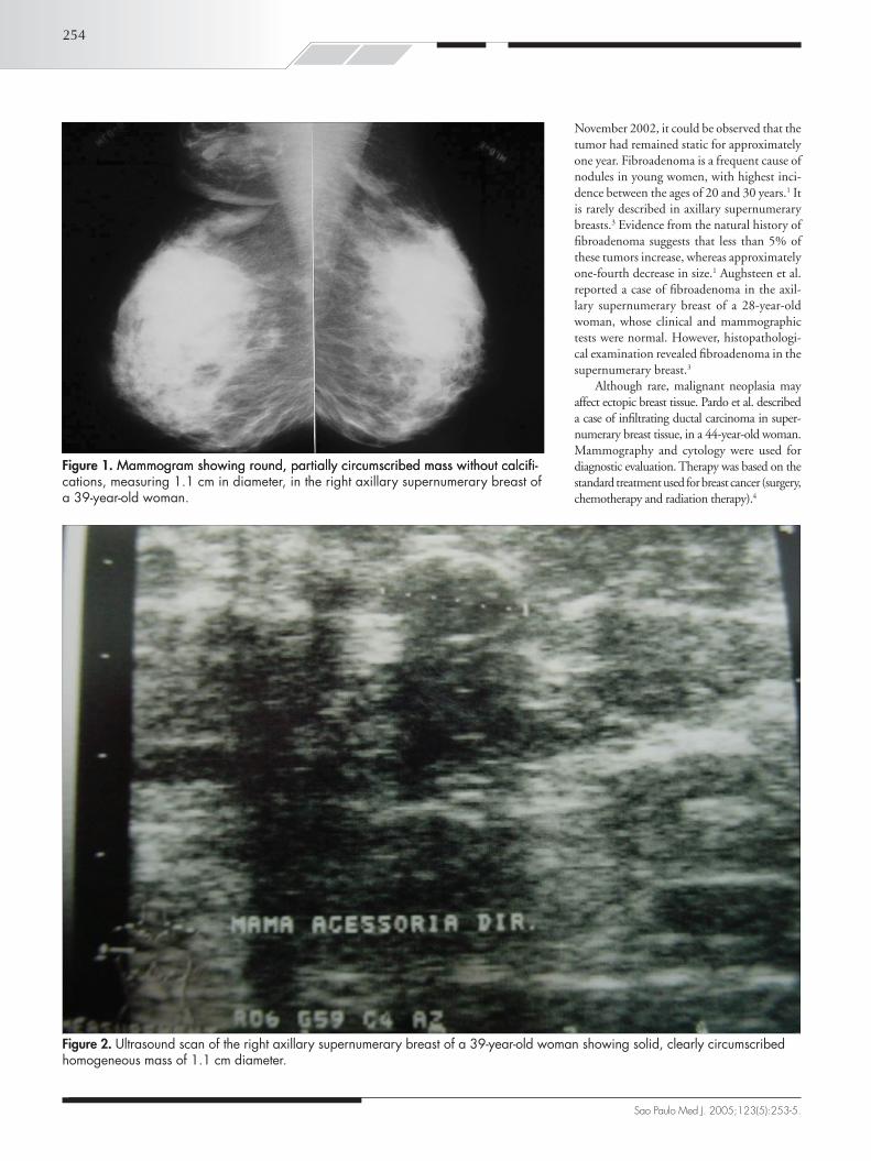
A mammogram that had been made in January 2003 did not show any abnormality that would give rise to suspicion of malignancy in bi-lateral supernumerary breasts. The right axillary breast exhibited a round, partially circumscribed mass, measuring 1.1 cm in diameter (Figure 1). Ultrasonography of the accessory breasts per-formed in November 2002 had revealed a 1.1 cm mass in the right axilla. A new ultrasound scan was performed, which demonstrated no abnormality in the left axillary breast, but showed a clearly circumscribed, homogeneous solid mass, measuring 1.1 cm in diameter in the right axilla (Figure 2), thus maintaining the same characteristics as seen previously. No biopsy or cytological study of the mass was performed. Renal ultrasonography was normal.
The patient underwent surgical resection of the supernumerary breasts in October 2003. Histopathological examination revealed bilat-eral breast tissue, with no abnormality in the left side and fibroadenoma in the right side, measuring 1.2 cm in diameter (Figure 3). The patient is currently undergoing follow-up and remains asymptomatic.
DISCUSSIoNSupernumerary breast tissue is well docu-
mented in the medical literature, and polymas-tia is one of its most common presentations.1,2 However, reports of benign and malignant tumors in supernumerary breasts are rare.3,4
The present case describes fibroadenoma in the axillary breast of a 39-year-old woman. The patient had been presenting complaints of bilateral axillary masses for 19 years. The progression of the fibroadenoma could not be accurately estimated. Nevertheless, on the basis of ultrasonography performed in
Sao Paulo Med J. 2005;123(5):253-5.
254
November 2002, it could be observed that the tumor had remained static for approximately one year. Fibroadenoma is a frequent cause of nodules in young women, with highest inci-dence between the ages of 20 and 30 years.1 It is rarely described in axillary supernumerary breasts.3 Evidence from the natural history of fibroadenoma suggests that less than 5% of these tumors increase, whereas approximately one-fourth decrease in size.1 Aughsteen et al. reported a case of fibroadenoma in the axil-lary supernumerary breast of a 28-year-old woman, whose clinical and mammographic tests were normal. However, histopathologi-cal examination revealed fibroadenoma in the supernumerary breast.3
Although rare, malignant neoplasia may affect ectopic breast tissue. Pardo et al. described a case of infiltrating ductal carcinoma in super-numerary breast tissue, in a 44-year-old woman. Mammography and cytology were used for diagnostic evaluation. Therapy was based on the standard treatment used for breast cancer (surgery, chemotherapy and radiation therapy).4
Figure 1. �ammogram showing round, partially circumscribed mass without calcifi-�ammogram showing round, partially circumscribed mass without calcifi-cations, measuring 1.1 cm in diameter, in the right axillary supernumerary breast of a 39-year-old woman.
Sao Paulo Med J. 2005;123(5):253-5.
Figure 2. Ultrasound scan of the right axillary supernumerary breast of a 39-year-old woman showing solid, clearly circumscribed homogeneous mass of 1.1 cm diameter.

255
Figure 3. Photomicrograph (hematoxylin and eosin; 400 X) showing fibroadenoma of the right axillary supernumerary breast.
ReSUMo
Fibroadenoma de mama supranumerária axilar: relato de caso
CONTEXTO: O tecido mamário supranumerário pode ser acometido pelas mesmas doenças e alterações que comprometem o tecido mamário tópico. Porém, o relato de fibroadenoma em mama acessória axilar é raro.
OBJETIVO: Descrever um caso de fibroadenoma de mama supranumerária axilar.
TIPO DE ESTUDO: Relato de caso.
RELATO DE CASO: �ulher de 39 anos, encaminhada ao Ambulatório de Ginecologia e Obstetrícia do Hospital Estadual Sumaré, com queixa de massa axilar bilateral. Paciente referia problemas cosméticos, dor e desconforto local. No exame físico, observaram-se alterações compatíveis com mamas acessórias axilares bilaterais, sem nódulos palpáveis. Os exames complementares (mamografia e ultra-sonografia) demonstraram nódulo de 1.1 cm em mama axilar direita. Paciente foi submetida à ressecção de mamas supranumerárias, com exame histopatológico revelando fibroadenoma de tecido mamário axilar direito.
PALAVRAS-CHAVE: Doenças mamárias. Fibroadenoma. Neoplasias mamárias. Axila. Diagnóstico.
AUTHoR INFoRMATIoN
Délio Marques Conde, MD, PhD. Department of GynecologyDepartment of Gynecology and Obstetrics, Hospital Estadual Sumaré, Universidade Estadual de Campinas, Sumaré, São Paulo, Brazil.
Renato Zocchio Torresan, MD, PhD. Department of Gynecolo-Department of Gynecolo-gy and Obstetrics, Hospital Estadual Sumaré, Universidade Estadual de Campinas, Sumaré, São Paulo, Brazil.
Eiji Kashimoto, MD. Postgraduate student, Department of Gynecology and Obstetrics, Hospital Estadual Sumaré, Universidade Estadual de Campinas, Sumaré, São Paulo, Brazil.
Luiz Eduardo Campos de Carvalho, MD, MSc. Department of Gynecology and Obstetrics, Hospital Estadual Suma-ré, Universidade Estadual de Campinas, Sumaré, São Paulo, Brazil.
Cássio Cardoso Filho, MD. Postgraduate student, Depart-ment of Gynecology and Obstetrics, Hospital Estadual Sumaré, Universidade Estadual de Campinas, Sumaré, São Paulo, Brazil.
Address for correspondence: Délio �arques Conde
Rua Jasmim, 750 — Bloco 1 — Apto. 74 Chácara PrimaveraCampinas (SP) — Brasil — CEP 13087-460Tel. (+55 19) 3883-8900E-mail: [email protected]
Copyright © 2005, Associação Paulista de �edicina
1. Dixon JM, Mansel RE. ABC of breast diseases. Congenital
problems and aberrations of normal breast development and
involution. BMJ. 1994;309(6957):797-800.
2. Grossl NA. Supernumerary breast tissue: historical perspectives
and clinical features. South Med J. 2000;93(1):29-32.
3. Aughsteen AA, Almasad JK, Al-Muhtaseb MH. Fibroadenoma of the
supernumerary breast of the axilla. Saudi Med J. 2000;21(6):587-9.
4. Pardo M, Silva F, Jiménez P, Karmelic M. Carcinoma mamario en
tejido mamario ectópico. [Mammary carcinoma in ectopic breast
tissue. A case report]. Rev Med Chil. 2001;129(6):663-5.
Sources of funding: Not declared Conflict of interest: Not declaredDate of first submission: �ay 7, 2004Last received: �ay 14, 2004Accepted: July 6, 2005
ReFeReNCeS
Sao Paulo Med J. 2005;123(5):253-5.
Tumors in supernumerary breast tissue should be diagnosed with the same methods applied to normal breast tissue (mammography, ultrasonography, cytology and biopsy), observing specific indications. However, due to its low inci-dence, diagnosis may be delayed or even ignored, thus making treatment more difficult. When tumors or nodules are found along the mam-mary line, the presence of breast tissue should be considered during the investigation.2
This case demonstrates a rare occurrence of fibroadenoma in an axillary supernumerary breast. Although the benign nature and natural history of fibroadenoma are well known, biopsy should be considered for women aged 40 years or older, due to the increased rate of cancer in this age range. Among women of this age, if conservative management is chosen, periodic clinical and mammographic control is required, following negative cytological tests.
The need for careful investigation of supernumerary breast tissue should be em-phasized, because it may be affected by benign and malignant diseases.