farmer’s lung
TRANSCRIPT

Moustapha Mounib
Consultant Chest Diseases
Military Medical Academy

•Introduction
•Definition
•Aetiology
•Pathophysiology
•Clinical picture
•Smoking
•Investigations
•Diagnostic criteria
•Differential diagnosis
•Treatment
•Prognosis

Introduction

One of the first written description of hypersensitivity pneumonitis or extrensic allergic alveolitis was in 1713 by Ramazzini, who observed that ‘ minute worms ‘ contained in grain cause a syndrome of dyspnea and cachexia associated with a shortened life span.

Definition

It is a group of lung diseases caused by inhalation of organic antigen to which the individual has been previously sensitized to. It is often divided into ‘ acute ‘ and ‘ chronic ‘ forms based on the time course of presentation.

Acute form often follows a short period of exposure to a high concentration of antigen, and is usually reversible.

Chronic form typically follows a period of chronic exposure to a low antigen dose and is less reversible.

These two presentations may overlap and ‘ subacute ‘ form of the disease is recognized.

Aetiology

The disease is usually named colourfullyafter the environment in which it occurs (e.g. farmer’s lung and bird fancier’s lung ) and has been reported in over 30 different occupations and environment.
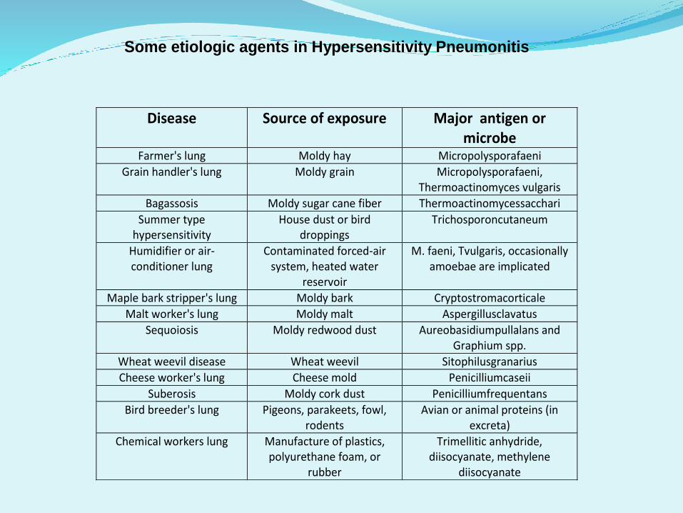
Some etiologic agents in Hypersensitivity Pneumonitis
Major antigen or microbe
Source of exposure Disease
Micropolysporafaeni Moldy hay Farmer's lung
Micropolysporafaeni, Thermoactinomyces vulgaris
Moldy grain Grain handler's lung
Thermoactinomycessacchari Moldy sugar cane fiber Bagassosis
Trichosporoncutaneum House dust or bird droppings
Summer type hypersensitivity
M. faeni, Tvulgaris, occasionally amoebae are implicated
Contaminated forced-air system, heated water
reservoir
Humidifier or air-conditioner lung
Cryptostromacorticale Moldy bark Maple bark stripper's lung
Aspergillusclavatus Moldy malt Malt worker's lung
Aureobasidiumpullalans and Graphium spp.
Moldy redwood dust Sequoiosis
Sitophilusgranarius Wheat weevil Wheat weevil disease
Penicilliumcaseii Cheese mold Cheese worker's lung
Penicilliumfrequentans Moldy cork dust Suberosis
Avian or animal proteins (in excreta)
Pigeons, parakeets, fowl, rodents
Bird breeder's lung
Trimellitic anhydride, diisocyanate, methylene
diisocyanate
Manufacture of plastics, polyurethane foam, or
rubber
Chemical workers lung

Pathophysiology

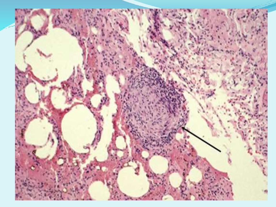
The pathogenesis is not fully understood, and may involve T-cell mediated immunity and granulomaformation ( type IV hypersensitivity ) and/or antibody-antigen immune complex formation ( type III hypersensitivity ).

It is not an atopic disease, and is not characterized by a rise in tissue eosinophilsor Ig E ( type I hypersensitivity ); this may in part be due to the small particle size of offending antigens which tend to be deposited more distally in the air spaces than the larger particles associated with asthma.

Clinical picture

•Breathlessness, dry cough, and systemic symptoms ( fever, chills, arthralgia, myalgia, headache ) occur 4-8 hours after exposure to antigen.
•Examination : crackles and squeaks on auscultation, fever.
•In the absence of ongoing exposure, symptoms settle spontaneously within 1-3 days.
•Episodes may be recurrent.

•Progressive exertional breathlessness, dry cough, sometimes systemic symptoms ( weight loss ) over course of months-years.
•May be history of acute episodes.
•Examination: crackles and squeaks on auscultation, clubbing rare, may be features of corpulmonale.

Smoking seems to protect towards hypersensitivity pneumonitis, although the disease has been described in a small number of smokers. The reason behind this protection might be the downregulation of the immune system by tobacco smoke and nicotine.

Investigations

Imaging

Chest X ray
Diffuse small (1-3 mm) nodules or infiltrates, sometimes ground glass change, apical sparing.
Normal in up to 20% of cases.

High resolution CT
Patchy ground glass change and poorly defined nodules.
Areas of increased lucency ( enhanced in expiratory HRCT ) occur due to air trapping from bronchiolar involvement.

Both chest X ray and HRCT appearances may quickly normalize following removal from antigen exposure.

Chest X ray
Typically upper and mid zone reticulation.
High resolution CT
Diffuse well defined centrilobular nodules, ground glass change, increased lucency from air trapping. May mimic UIP.


•Typically restrictive pattern with reduced gas transfer and lung volumes.
•Mild obstruction is also sometimes observed.
•Hypoxia may occur.
•Inhalation antigen challenge may be unpleasant, and it is not recommended routinely.

Acute form associated with neutrophilia but not eosinophilia.
Inflammatory markers are often increased.

Are neither specific nor sensitive. In fact, 10% of asymptomatic farmers and 40% of pigeon breaders have precipitating antibodies to causative antigens, but no clinical evidence of disease.

A lymphocytic alveolitis characterizes the BAL fluid of patients. In fact, a BAL lymphocytic count of less than 30% makes the diagnosis unlikely, except in smokers and more chronic forms in which lymphocytosis is less prominent.
However, a BAL lymphocytosis is not specific because it may be present in many other conditions, including sarcoidosis, chronic beryllium disease, and several autoimmune lung diseases.

Findings on transbronchial biopsy are non specific and non diagnostic in 50% of patients.
Proceeding to surgical lung biopsy may be necessary when faced with diagnostic uncertainty, because features of the disease overlap with many other inflammatory lung diseases.

•Exposure to a known offending antigen.
•Symptoms occurring 4-8 hours after exposure.
•Positive precipitating antibodies to the offending antigen.
•Inspiratory crackles on physical examination.
•Recurrent episodes of symptoms.
•Weight loss.

•Atypical pneumonia.
•Idiopathic interstitial pneumonia ( particularly UIP and COP )
•Sarcoidosis.
•Vasculitis.
•Occupational asthma.
•Drug induced lung disease ( including pesticides).
•Organic Dust Toxic Syndrome ( follow very high levels of exposure to agricultural dusts, symptoms transient, benign course ).

Treatment

The only treatment for allergic diseases is to avoid exposure to the offending allergen.
Respiratory protection can be used to minimize the exposure as much as possible.

Systemic glucocorticosteroids are usually required to treat severely symptomatic patients, although there is no formal evidence that such treatment is associated with long term abatement of symptoms or radiologic or pulmonary function tests abnormalities.

The usual treatment is prednisone or prednisolone, 40 to 60 mg a day for 2 weeks, followed by a gradual decrease over 2 to 4 weeks.

Prognosis

The natural history of the disease is variable and probably depends on the type and duration of antigen exposure and the host immune response.
Acute form generally resolves within several weeks with corticosteroid therapy and removal from antigen exposure.

Continued symptoms and progressive lung impairment have been reported after recurrent acute attacks and even after a single acute attack. Additionally, progressive persistent airway hyperresponsiveness and emphysema may impact long term recovery.
