extracellular vesicles: potential roles in regenerative ... · extracellular vesicles: potential...
TRANSCRIPT
REVIEW ARTICLEpublished: 03 December 2014
doi: 10.3389/fimmu.2014.00608
Extracellular vesicles: potential roles in regenerativemedicineOlivier G. De Jong1, Bas W. M. Van Balkom1,2*, Raymond M. Schiffelers3, Carlijn V. C. Bouten2 andMarianne C. Verhaar 1
1 Department of Nephrology and Hypertension, University Medical Center Utrecht, Utrecht, Netherlands2 Department of Biomedical Engineering, Eindhoven University of Technology, Eindhoven, Netherlands3 Department of Clinical Chemistry and Hematology, University Medical Center Utrecht, Utrecht, Netherlands
Edited by:Marcella Franquesa, ErasmusMedisch Centrum, Netherlands
Reviewed by:Rae Ritchie, Bioscience Vaccines Inc.,USAMiroslaw Kornek, University ofSaarland, Germany
*Correspondence:Bas W. M. Van Balkom, Departmentof Nephrology and Hypertension,UMC Utrecht, Heidelberglaan 100,G.02.402, Utrecht 3584 CX,Netherlandse-mail: [email protected]
Extracellular vesicles (EV) consist of exosomes, which are released upon fusion of themultivesicular body with the cell membrane, and microvesicles, which are released directlyfrom the cell membrane. EV can mediate cell–cell communication and are involved in manyprocesses, including immune signaling, angiogenesis, stress response, senescence, pro-liferation, and cell differentiation.The vast amount of processes that EV are involved in andthe versatility of manner in which they can influence the behavior of recipient cells makeEV an interesting source for both therapeutic and diagnostic applications. Successes inthe fields of tumor biology and immunology sparked the exploration of the potential ofEV in the field of regenerative medicine. Indeed, EV are involved in restoring tissue andorgan damage, and may partially explain the paracrine effects observed in stem cell-basedtherapeutic approaches. The function and content of EV may also harbor information thatcan be used in tissue engineering, in which paracrine signaling is employed to modulatecell recruitment, differentiation, and proliferation. In this review, we discuss the functionand role of EV in regenerative medicine and elaborate on potential applications in tissueengineering.
Keywords: regenerative medicine, tissue engineering, extracellular vesicles, exosomes, microvesicles
INTRODUCTIONRegenerative medicine aims at the functional restoration of a dam-aged, malfunctioning, or missing tissue. There are three mainapproaches in regenerative medicine. The first approach is cell-based therapies, where cells are administered to restore a tis-sue either directly or through paracrine functions. The secondapproach is often referred to as classical tissue engineering, andconsists of the combined use of cells and a bio-degradable scaffoldto form a tissue. Lastly, much progress has been made in material-based approaches, which rely on bio-degradable materials, oftenfunctionalized with cellular functions.
The first development in replacing malfunctioning tissues wasby transplanting organs, tissues, or cells. Over the course of the lastcentury vast improvements were made in the field of transplanta-tion, starting with bone and cornea transplants at the beginningof the twentieth century, followed by the first kidney transplanta-tion in the 1950s (1–3). As transplantation techniques for otherorgans developed over the following decades, the limiting factor forthese procedures shifted from technical limitations to the supply ofsuitable organs and tissues. Besides shortage in supply, organ andtissue transplantation have another major drawback: the risk ofimmune rejection and the required chronic immunosuppressiontreatment.
In response to these issues, research focused on strategiesthat allow functional restoration of damaged tissues by cell-free approaches or approaches using autologous cell and tissuesources. Embracing the rapid developments in technology and
our understanding of biological processes, the field of regener-ative medicine is focusing on a wide array of techniques andapproaches to restore tissue function. Suitable approaches dependon the function and environment of the newly generated tis-sue. For instance, in the replacement of insulin-producing cells inpatients with type-1 diabetes, there is little need for load-bearingstructures, but rather for structures mimicking the extracellularmatrix (ECM) like hydrogels, to retain and stimulate insulin-producing cells (4). Heart valve replacements on the other handrequire materials that are able to withstand large forces in addi-tion to high flexibility (5), but due to their direct contact witha patients’ circulation also require the use of materials with highhemocompatibility and low immunogenicity. Utilizing autologousstem-, progenitor-, and tissue-specific cells to restore damaged tis-sues may bypass the problem of immunogenic responses againstthese implants. Following recent insights that the structural con-tribution of stem cells to regenerated tissues is limited, and thatrather the stimulation of local healing processes plays an impor-tant role (6–9), research has increasingly focused on the paracrinehypothesis, investigating the stimulating factors released by thesestem- and progenitor cells, including growth factors, cytokines,and extracellular vesicles (EV). At the same time, major break-throughs in the field of EV have uncovered roles for EV in manyprocesses including angiogenesis, regulation of immune responses,and ECM remodeling (10–13), which may be of specific interestfor tissue engineering. Here, we review the recent developmentsin regenerative medicine and EV research, and discuss potential
www.frontiersin.org December 2014 | Volume 5 | Article 608 | 1

De Jong et al. Extracellular vesicles in regenerative medicine
therapeutic applications of EV in restoring function in damagedtissues.
REGENERATIVE MEDICINE: CELL THERAPIESOne of the earliest applications of cell therapy was the administra-tion of cells for the reconstitution of blood or bone marrow (14,15). As a result of developments during the last decades, includingimproved techniques in both transient and permanent regulationof gene expression, methods of cell isolation and propagation,and improved protocols to regulate differentiation of cells, celltherapies currently play a prominent role in the field of regen-erative medicine (16). Cell therapies can directly aid repair byforming new functional tissues, or support tissue repair throughparacrine mechanisms, for instance by secreting growth factors,immunomodulatory molecules, and EV. Examples of direct tis-sue formation by cell therapy are the use of autologous epithelialcells to repair cornea injuries (17), expansion, and transplanta-tion of chondrocytes in cartilage repair (18), or the adminis-tration of endothelial colony-forming cells (ECFC) in a murinehind limb ischemia model to increase neovascularization (19).In these studies cell populations were isolated, expanded ex vivo,and re-introduced at the site of injury to generate new, functionaltissues. The ex vivo expansion step allows the use of only limitedamounts of tissue and the proper characterization of isolated cells.Adverse effects as dedifferentiation and induction of senescenceare great challenges adhered to this approach (20). For instance,in vitro passaging of mesenchymal stem cells (MSC) results incell enlargement, differentiation, and decrease in proliferationwithin 10 passages (21), and causes a strong response to micro-environment stiffness, affecting cell morphology, and function(22). Progenitor cells from aged or diseased donors show decreasedproliferation, prevalence, as well as functionality (23–25). Despitethese challenges, promising results have been achieved, for instancein treatment of patients with severe autoimmune diseases withhematopoetic stem cell transplantation (26).
It has become increasingly apparent that a more supportingrole, employed by secretion products of stem and progenitorcells is responsible for many of the observed effects of stem celltherapies (6–9). These paracrine factors secreted by stem- andprogenitor cells, like growth factors and cytokines, are of majorinterest to discover new therapeutics that stimulate local tissueregeneration for the use in tissue engineering as well [reviewed inRef. (27, 28)].
TISSUE-ENGINEERING: (BIO-)ENGINEERED SUPPORTRepair of damaged tissue requires not only the presence ofcells capable of restoring the damaged structure, but requires amicroenvironment that promotes appropriate tissue regenerationas well. In addition, cells need to be guided to form a structure ofthe appropriate size and shape, and in many cases (for instance inbone or cartilage repair, as well as in cardiovascular substitutes),require structural support. In a healthy tissue, the ECM plays a keyrole in guiding and regulating these processes, whereas in damagedtissue, the ECM is often absent, damaged, or functionally impaired.To address this problem and allow in situ regeneration, structuresthat (temporarily) provide the requirements for cell retention andtissue regeneration are employed and are referred to as scaffolds.
Scaffolds can either be of natural origin, such as decellular-ized ECM or modified elastin- or collagen gels, or of syntheticorigin, such as synthetic hydrogels or porous polymer scaffolds.Using decellularized ECM from xenogenic or allogenic donorsprovides scaffolds that are most similar to the natural extracel-lular environment. Use of decellularized matrices is a promisingtechnique, which yields biocompatible scaffolds with appropri-ate physical and biological properties. Many ECM components,as well as growth factors, are often conserved and can aid inproper regeneration of functional tissues (29). To decrease therisk of immune responses against antigens in these scaffolds, aswell as the potential transfer of pathogens, a combination ofenzymatic, physical, and chemical treatments is used to removecellular components from the tissue (29). Decellularized matriceshave been used for tissue engineering of several tissues, includ-ing heart valves (30), vascular grafts (31), and trachea (32).However, use of decellularized matrices has several disadvan-tages. Acquiring and isolating of appropriate tissues, followed bydecellularization protocols, can be a relatively time-consumingand expensive procedure, and incomplete decellularization orantigen removal can result in immune reactions against grafts(33). Cell seeding of decellularized matrices can be technicallychallenging due to structural dimensions and porosity. Further-more, control over the exact content of the matrices is lim-ited due to donor variation, and despite pretreatment still thereexists the risk of transfer of pathogens. In order to create scaf-folds in a safe, reproducible, affordable, and controlled man-ner, extensive research is ongoing on the production of artificialporous scaffolds, exploring various production techniques andmaterials (34).
Artificial porous scaffolds should meet specific requirementsto allow homing of appropriate cell populations. Ideally, a syn-thetic scaffold temporarily provides the required support andmicro-environment, is bio-degradable and eventually replaced byautologous ECM. For cells to be able to migrate or be seededin the scaffold and allow an environment with proper supply ofnutrients, a porous structure is required (35). There are severaltechniques to generate porous scaffolds, including solvent cast-ing, forming emulsions before polymerization, gas foaming, aswell as binding of polymeric fibers by chemical treatment or heat-ing (36–39). Using these techniques in generating scaffolds withconsistent porosity in complex shapes, containing areas of vary-ing thickness and materials, is technically challenging. Currently,the most commonly used technique in generating porous syn-thetic scaffold is electrospinning, which allows the generation ofconstructs with complex geometry, consisting of combinations offiber types in both mixed and layered patterns (40). Bio-degradablepolymers used in electrospinning include poly(ε-caprolactone)(PCL), poly(glycolic acid) (PGA), poly(hydroxy alkanoate) (PHA),and poly(lactic acid) (PLA). Mixing fiber types in specific pat-terns allows modulation of degradability, strength, and biologicalactivity of scaffolds (41).
Electrospun scaffolds can be pre-seeded with autologous cells,which may be re-programmed, differentiated, and expandedin vitro, and can then be directly implanted or incubated in abioreactor until the electrospun meshwork is fully degraded andreplaced with ECM (5). Alternatively, scaffolds can be implanted
Frontiers in Immunology | Immunotherapies and Vaccines December 2014 | Volume 5 | Article 608 | 2
De Jong et al. Extracellular vesicles in regenerative medicine
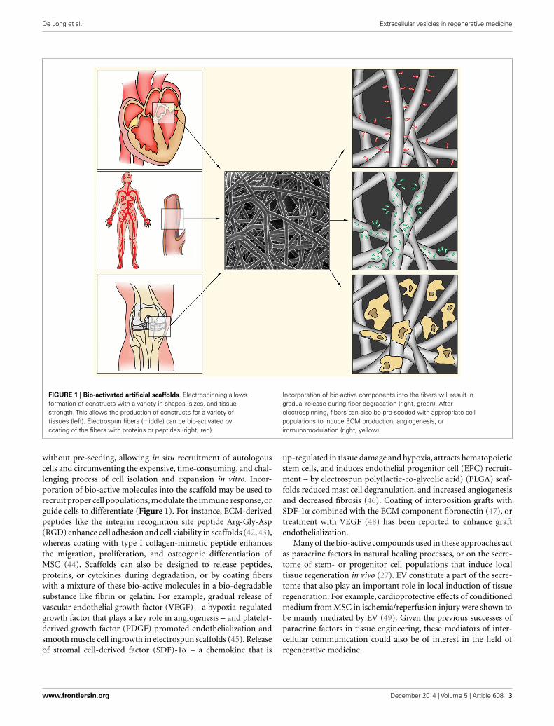
FIGURE 1 | Bio-activated artificial scaffolds. Electrospinning allowsformation of constructs with a variety in shapes, sizes, and tissuestrength. This allows the production of constructs for a variety oftissues (left). Electrospun fibers (middle) can be bio-activated bycoating of the fibers with proteins or peptides (right, red).
Incorporation of bio-active components into the fibers will result ingradual release during fiber degradation (right, green). Afterelectrospinning, fibers can also be pre-seeded with appropriate cellpopulations to induce ECM production, angiogenesis, orimmunomodulation (right, yellow).
without pre-seeding, allowing in situ recruitment of autologouscells and circumventing the expensive, time-consuming, and chal-lenging process of cell isolation and expansion in vitro. Incor-poration of bio-active molecules into the scaffold may be used torecruit proper cell populations, modulate the immune response, orguide cells to differentiate (Figure 1). For instance, ECM-derivedpeptides like the integrin recognition site peptide Arg-Gly-Asp(RGD) enhance cell adhesion and cell viability in scaffolds (42, 43),whereas coating with type I collagen-mimetic peptide enhancesthe migration, proliferation, and osteogenic differentiation ofMSC (44). Scaffolds can also be designed to release peptides,proteins, or cytokines during degradation, or by coating fiberswith a mixture of these bio-active molecules in a bio-degradablesubstance like fibrin or gelatin. For example, gradual release ofvascular endothelial growth factor (VEGF) – a hypoxia-regulatedgrowth factor that plays a key role in angiogenesis – and platelet-derived growth factor (PDGF) promoted endothelialization andsmooth muscle cell ingrowth in electrospun scaffolds (45). Releaseof stromal cell-derived factor (SDF)-1α – a chemokine that is
up-regulated in tissue damage and hypoxia, attracts hematopoieticstem cells, and induces endothelial progenitor cell (EPC) recruit-ment – by electrospun poly(lactic-co-glycolic acid) (PLGA) scaf-folds reduced mast cell degranulation, and increased angiogenesisand decreased fibrosis (46). Coating of interposition grafts withSDF-1α combined with the ECM component fibronectin (47), ortreatment with VEGF (48) has been reported to enhance graftendothelialization.
Many of the bio-active compounds used in these approaches actas paracrine factors in natural healing processes, or on the secre-tome of stem- or progenitor cell populations that induce localtissue regeneration in vivo (27). EV constitute a part of the secre-tome that also play an important role in local induction of tissueregeneration. For example, cardioprotective effects of conditionedmedium from MSC in ischemia/reperfusion injury were shown tobe mainly mediated by EV (49). Given the previous successes ofparacrine factors in tissue engineering, these mediators of inter-cellular communication could also be of interest in the field ofregenerative medicine.
www.frontiersin.org December 2014 | Volume 5 | Article 608 | 3
De Jong et al. Extracellular vesicles in regenerative medicine
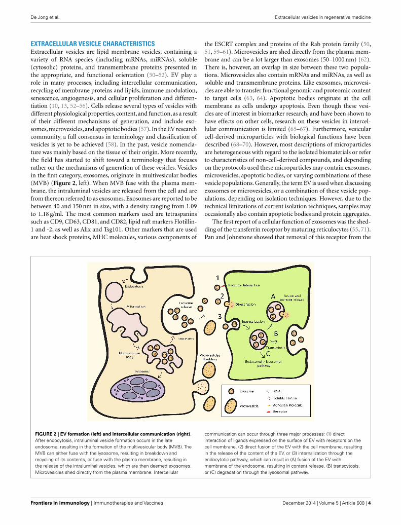
EXTRACELLULAR VESICLE CHARACTERISTICSExtracellular vesicles are lipid membrane vesicles, containing avariety of RNA species (including mRNAs, miRNAs), soluble(cytosolic) proteins, and transmembrane proteins presented inthe appropriate, and functional orientation (50–52). EV play arole in many processes, including intercellular communication,recycling of membrane proteins and lipids, immune modulation,senescence, angiogenesis, and cellular proliferation and differen-tiation (10, 13, 52–56). Cells release several types of vesicles withdifferent physiological properties, content, and function, as a resultof their different mechanisms of generation, and include exo-somes, microvesicles, and apoptotic bodies (57). In the EV researchcommunity, a full consensus in terminology and classification ofvesicles is yet to be achieved (58). In the past, vesicle nomencla-ture was mainly based on the tissue of their origin. More recently,the field has started to shift toward a terminology that focusesrather on the mechanisms of generation of these vesicles. Vesiclesin the first category, exosomes, originate in multivesicular bodies(MVB) (Figure 2, left). When MVB fuse with the plasma mem-brane, the intraluminal vesicles are released from the cell and arefrom thereon referred to as exosomes. Exosomes are reported to bebetween 40 and 150 nm in size, with a density ranging from 1.09to 1.18 g/ml. The most common markers used are tetraspaninssuch as CD9, CD63, CD81, and CD82, lipid raft markers Flotillin-1 and -2, as well as Alix and Tsg101. Other markers that are usedare heat shock proteins, MHC molecules, various components of
the ESCRT complex and proteins of the Rab protein family (50,51, 59–61). Microvesicles are shed directly from the plasma mem-brane and can be a lot larger than exosomes (50–1000 nm) (62).There is, however, an overlap in size between these two popula-tions. Microvesicles also contain mRNAs and miRNAs, as well assoluble and transmembrane proteins. Like exosomes, microvesi-cles are able to transfer functional genomic and proteomic contentto target cells (63, 64). Apoptotic bodies originate at the cellmembrane as cells undergo apoptosis. Even though these vesi-cles are of interest in biomarker research, and have been shown tohave effects on other cells, research on these vesicles in intercel-lular communication is limited (65–67). Furthermore, vesicularcell-derived microparticles with biological functions have beendescribed (68–70). However, most descriptions of microparticlesare heterogeneous with regard to the isolated biomaterials or referto characteristics of non-cell-derived compounds, and dependingon the protocols used these microparticles may contain exosomes,microvesicles, apoptotic bodies, or varying combinations of thesevesicle populations. Generally, the term EV is used when discussingexosomes or microvesicles, or a combination of these vesicle pop-ulations, depending on isolation techniques. However, due to thetechnical limitations of current isolation techniques, samples mayoccasionally also contain apoptotic bodies and protein aggregates.
The first report of a cellular function of exosomes was the shed-ding of the transferrin receptor by maturing reticulocytes (55, 71).Pan and Johnstone showed that removal of this receptor from the
FIGURE 2 | EV formation (left) and intercellular communication (right).After endocytosis, intraluminal vesicle formation occurs in the lateendosome, resulting in the formation of the multivesicular body (MVB). TheMVB can either fuse with the lysosome, resulting in breakdown andrecycling of its contents, or fuse with the plasma membrane, resulting inthe release of the intraluminal vesicles, which are then deemed exosomes.Microvesicles shed directly from the plasma membrane. Intercellular
communication can occur through three major processes: (1) directinteraction of ligands expressed on the surface of EV with receptors on thecell membrane, (2) direct fusion of the EV with the cell membrane, resultingin the release of the content of the EV, or (3) internalization through theendocytotic pathway, which can result in (A) fusion of the EV withmembrane of the endosome, resulting in content release, (B) transcytosis,or (C) degradation through the lysosomal pathway.
Frontiers in Immunology | Immunotherapies and Vaccines December 2014 | Volume 5 | Article 608 | 4
De Jong et al. Extracellular vesicles in regenerative medicine
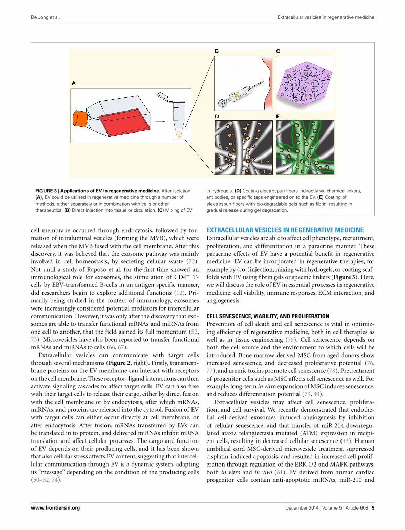
FIGURE 3 | Applications of EV in regenerative medicine. After isolation(A), EV could be utilized in regenerative medicine through a number ofmethods, either separately or in combination with cells or othertherapeutics. (B) Direct injection into tissue or circulation. (C) Mixing of EV
in hydrogels. (D) Coating electrospun fibers indirectly via chemical linkers,antibodies, or specific tags engineered on to the EV. (E) Coating ofelectrospun fibers with bio-degradable gels such as fibrin, resulting ingradual release during gel degradation.
cell membrane occurred through endocytosis, followed by for-mation of intraluminal vesicles (forming the MVB), which werereleased when the MVB fused with the cell membrane. After thisdiscovery, it was believed that the exosome pathway was mainlyinvolved in cell homeostasis, by secreting cellular waste (72).Not until a study of Raposo et al. for the first time showed animmunological role for exosomes, the stimulation of CD4+ T-cells by EBV-transformed B-cells in an antigen specific manner,did researchers begin to explore additional functions (12). Pri-marily being studied in the context of immunology, exosomeswere increasingly considered potential mediators for intercellularcommunication. However, it was only after the discovery that exo-somes are able to transfer functional mRNAs and miRNAs fromone cell to another, that the field gained its full momentum (52,73). Microvesicles have also been reported to transfer functionalmRNAs and miRNAs to cells (66, 67).
Extracellular vesicles can communicate with target cellsthrough several mechanisms (Figure 2, right). Firstly, transmem-brane proteins on the EV membrane can interact with receptorson the cell membrane. These receptor–ligand interactions can thenactivate signaling cascades to affect target cells. EV can also fusewith their target cells to release their cargo, either by direct fusionwith the cell membrane or by endocytosis, after which mRNAs,miRNAs, and proteins are released into the cytosol. Fusion of EVwith target cells can either occur directly at cell membrane, orafter endocytosis. After fusion, mRNAs transferred by EVs canbe translated in to protein, and delivered miRNAs inhibit mRNAtranslation and affect cellular processes. The cargo and functionof EV depends on their producing cells, and it has been shownthat also cellular stress affects EV content, suggesting that intercel-lular communication through EV is a dynamic system, adaptingits “message” depending on the condition of the producing cells(50–52, 74).
EXTRACELLULAR VESICLES IN REGENERATIVE MEDICINEExtracellular vesicles are able to affect cell phenotype, recruitment,proliferation, and differentiation in a paracrine manner. Theseparacrine effects of EV have a potential benefit in regenerativemedicine. EV can be incorporated in regenerative therapies, forexample by (co-)injection, mixing with hydrogels, or coating scaf-folds with EV using fibrin gels or specific linkers (Figure 3). Here,we will discuss the role of EV in essential processes in regenerativemedicine: cell viability, immune responses, ECM interaction, andangiogenesis.
CELL SENESCENCE, VIABILITY, AND PROLIFERATIONPrevention of cell death and cell senescence is vital in optimiz-ing efficiency of regenerative medicine, both in cell therapies aswell as in tissue engineering (75). Cell senescence depends onboth the cell source and the environment to which cells will beintroduced. Bone marrow-derived MSC from aged donors showincreased senescence, and decreased proliferative potential (76,77), and uremic toxins promote cell senescence (78). Pretreatmentof progenitor cells such as MSC affects cell senescence as well. Forexample, long-term in vitro expansion of MSC induces senescence,and reduces differentiation potential (79, 80).
Extracellular vesicles may affect cell senescence, prolifera-tion, and cell survival. We recently demonstrated that endothe-lial cell-derived exosomes induced angiogenesis by inhibitionof cellular senescence, and that transfer of miR-214 downregu-lated ataxia telangiectasia mutated (ATM) expression in recipi-ent cells, resulting in decreased cellular senescence (13). Humanumbilical cord MSC-derived microvesicle treatment suppressedcisplatin-induced apoptosis, and resulted in increased cell prolif-eration through regulation of the ERK 1/2 and MAPK pathways,both in vitro and in vivo (81). EV derived from human cardiacprogenitor cells contain anti-apoptotic miRNAs, miR-210 and
www.frontiersin.org December 2014 | Volume 5 | Article 608 | 5
De Jong et al. Extracellular vesicles in regenerative medicine
miR-132, and treatment with these EV in a myocardial infarc-tion resulted in decreased cardiomyocyte apoptosis (82). Similarly,bone marrow MSC-derived exosomes were able to decrease apop-tosis and increase cell proliferation in an acute kidney injurymodel, and the authors hypothesized that this was the resultof exosome-mediated RNA transfer (83). Similar results wereobtained by Bruno et al., who showed that administration ofMSC-derived microvesicles decreased apoptosis in an acute kid-ney injury model and in vitro in cisplatin treated human epithelialcells, through up-regulation of anti-apoptotic genes and down-regulation of several apoptotic genes (84). Further in vitro studiesshowed that cardiomyocyte protection by MSC is partially medi-ated by transfer of miR-221 in microvesicles, resulting in reducedcaspase activity after ischemic injury (85). Certain EV have alsobeen shown to increase cell proliferation. Tumor-derived EV werereported to induce proliferation in a variety of tissues (86–88).MSC-derived EV have also been found to increase proliferation:bone marrow MSC-derived exosomes induced proximal tubularepithelial cell proliferation in an acute kidney injury model (89),and umbilical cord MSC-derived exosomes increased in vitro skincell proliferation as well as migration after heat-stress, throughWnt signaling by trafficking of Wnt4 (90). Interestingly, Zhanget al. also observed that treatment with these vesicles in a ratskin burn model resulted in accelerated epithelialization (90).Exosomes derived from tubular epithelial cells stimulated withhypoxia activated fibroblasts through TGF-β1 signaling, resultingin increased fibroblast proliferation, which could aid in accelera-tion of tissue repair (91). These studies indicate that EV play a rolein local tissue repair through regulation of cell proliferation.
The capacity of EV to regulate cell senescence, apoptosis, andproliferation, parameters that greatly affect tissue engineering andcell therapy outcome, suggest therapeutic potential in regenerativemedicine. Indeed, MSC-derived vesicles show positive effects ontissue repair through various pathways, even reducing apoptosis asa result of ischemic injury (92). This is of interest, since ischemiain larger tissue-engineered constructs is a substantial issue (93).
ANGIOGENESISTissue engineering of large tissues requires proper vasculariza-tion for sufficient supply of nutrients and oxygen, and drainingof cellular waste. Since tissue-engineered constructs thicker than100–200 µm already run in to problems in respect to oxygena-tion, nutrient supply, and removal of waste products, controlledvascularization of neo-tissue is vital (93). Strategies to inducevascularization include addition of endothelial (progenitor) cells,engineering vasculature, as well as the use of paracrine factors(93–95). Several studies on cancer-derived EV demonstrated theirrole in tumor angiogenesis through a variety of pathways, includ-ing cell cycle-related mRNAs, several major intracellular kinasepathways, transfer of miRNAs, and by carrying pro-angiogeniccytokines (96–100). EV from endothelial cells have also beendemonstrated to induce an angiogenic program in target endothe-lial cells in vitro and in vivo both through Notch-dependent tip-cellformation and induction of a pro-angiogenic program in par-allel to miR-214-dependent repression of senescence (13, 101).EV from other cell types have been demonstrated to stimulatein vitro and in vivo vessel formation by endothelial cells as well.
For example, adipose MSC-derived EV, which could be increasedin function and number by PDGF stimulation (102), as well asbone marrow MSC-derived EV, promoted angiogenesis in a ratmyocardial infarction model (103). In the latter model, hypoxicstimulation of the EV-producing cells was required to obtain func-tional EV. Similar effects of hypoxia were observed in microvesiclesfrom human umbilical cord MSC, which promote angiogenesisin vitro as well as in vivo in a rat hindlimb ischemia model (103,104). These findings underline the importance of culturing con-ditions of their producing cells on EV content (74). Cantaluppiet al. showed that EPC-derived microvesicles increase endothe-lial cell proliferation, migration, and vessel formation in vitroby transfer of pro-angiogenic miRNAs, miR-126 and miR-296.These EPC microvesicles also increased vascularization of isletendothelium and β-cells transplanted in SCID mice (105) and,in a SCID mouse hind limb ischemia model increased capillarydensity, enhanced limb perfusion, and reduced injury after 7 days(106). A study by Sahoo et al. in 2011 showed that exosomes iso-lated from CD34+ mononuclear cells increased endothelial cellviability, proliferation and tube formation in vitro, and stimulatedangiogenesis in vivo in both matrigel plug- and corneal assays, andthat the pro-angiogenic effect of these cells was mainly throughthese EV (107).
Overall, different types of EV appear to be able to induce angio-genesis through a variety of pathways, and through transfer ofmRNA,miRNAs,and proteins,underlining their potential in tissueengineering.
EXTRACELLULAR MATRIX INTERACTIONSThe ECM plays a major role in tissue engineering, providing shapeand strength to the newly formed tissue as well as a site for interac-tions with and guidance of cells. Both ECM architecture and mole-cular composition are determinants for cell recruitment, retention,and differentiation, and thus the final local cell phenotype. Intissue engineering strategies using bio-degradable scaffolds, theload-bearing and cell retaining function of the scaffold will haveto be fulfilled by the locally produced ECM after the scaffold isdegraded. EV are able to influence ECM composition throughdirect ECM interactions, or by interacting with ECM-producingcells.
Extracellular vesicles express adhesion molecules, includingmembers of the immunoglobin superfamily and integrins. Exo-somes derived from B-cells, endothelial cells, and dendritic cells,express ICAM-1 (74, 108, 109), and endothelial cell-derivedexosomes express CD44, CD166, PECAM, and B-CAM (74).Reticulocyte-derived exosomes have been shown to bind tofibronectin via integrin α4β1 (110). B-cell-derived exosomes con-tain β1 and β2 integrins, which were able to bind to collagen-1, fibronectin, and TNF-α activated fibroblasts (108). Exosomesderived from dendritic cells have also been reported to containintegrins (111). These studies show that EV may not only bind toand interact with cells, but also bind to various ECM components.It has been suggested that EV could adhere to the ECM to forma gradient or potential reservoir that could be released in case ofinflammation or ECM degradation (108).
Besides molecules responsible for ECM interaction, EV havealso been shown to express ECM-remodeling proteins, like matrix
Frontiers in Immunology | Immunotherapies and Vaccines December 2014 | Volume 5 | Article 608 | 6
De Jong et al. Extracellular vesicles in regenerative medicine
metalloproteinases (MMPs), which can degrade collagens, elastin,fibronectin, and laminin. These processes are important in ECMre-structuring, as well as cytokine release, angiogenesis, and cellmigration (112, 113). For example, human fibrosarcoma andmelanoma cell-derived exosomes contain both full length andproteolytically processed MMP14, shown to be enzymaticallyactive since these exosomes activated pro-MMP2 resulting inthe degradation of both collagen-1 and gelatin (114). Cardiomy-ocyte progenitor-derived exosomes expressed enzymatically activeMMP2, as well as MMP-activator EMMPRIN (115). EMMPRINhas also been found on CD8+ T-cell microparticles, which havebeen shown to induce fibrolytic activation in hepatic stellate cells(70). Madin-Darby canine kidney cells (MDCK) that have under-gone epithelial to mesenchymal transition (EMT) showed anincrease in MMP1, -14, and -19 expression in their exosomes, aswell as several integrins (116). Additionally, EV can also stimulateMMP production in target cells. Keratinocyte-like cells are able tostimulate MMP1 expression in dermal fibroblasts through transferof several 14-3-3 isoforms by EV (117). Furthermore, monocyteand T-cell-derived microparticles are able to induce production ofMMP-1, MMP-3, MMP-9, and MMP-13 in fibroblasts (68). Thus,EV can influence MMP abundance and activity on several levels.
Extracellular vesicles also have the ability to contribute to ECMstrength. Members of the lysyl oxidase family crosslink collagensand elastin, increasing ECM load-bearing properties. Lysyl oxi-dase treatment of tissue-engineered cartilage constructs resultsin increased stiffness and enhanced cartilage integration, andlysyl oxidase-like 2 induces angiogenic sprouting through inter-acting with collagen-4 in the basal membrane (118, 119). Lysyloxidase was shown to be enriched in exosomes derived fromhypoxic glyoma cells (98) and lysyl oxidase-like 2 in endothe-lial cells (74). Interestingly, exosomes from hypoxic endothelialcells also showed increased abundances of the ECM componentsfibronectin, collagen-4 and -12 subunits, and perlecan, suggestinga hypoxia-mediated role in focal ECM modification by exosomes(74). EV are also able to affect local ECM production. Borges et al.found that upon hypoxic stimulation, epithelial cells stimulatefibroblasts through exosome-mediated TGF-β1 signaling, result-ing in increased collagen-1 production (91) and suggesting anexosome-mediated response resulting in local tissue repair. Theeffects of EV on both ECM production and remodeling could beof use in the steering of in situ ECM formation.
IMMUNOMODULATIONModulating immune responses is vital in tissue engineering. Thetype and severity of the immune response against an implantdepends on several factors including injury from surgery, the(bio)materials used, location of the graft, and the condition ofthe patient (120). An excessive or inappropriate immune responsecould result in damage, encapsulation or rejection of a tissue-engineered construct. On the other hand, immune responses arepotent triggers for regenerative processes, including cell recruit-ment, proliferation, and angiogenesis, which are key to the successof in situ tissue engineering (121).
When transplanting a tissue-engineered construct, the innateimmune response consists of the acute and the chronic phase.The acute immune response is an immediate reaction against
foreign structures, such as certain (bio)materials. An influx ofneutrophils and macrophages induces the release of inflammatorycytokines, which results in local inflammation and the recruit-ment of additional immune cells. Cross-talk between macrophagesand T-cells, as well as environmental cues, regulate a shiftin macrophage sub-types in to either M1 (inflammatory), orthe M2 (anti-inflammatory, regenerative) subtype (122). M1macrophages promote recruitment of inflammatory immune cells,and release ECM-degrading proteins to allow quick migrationthrough inflamed tissues. As the subtype of macrophages shiftsto M2, pro-inflammatory cytokine release is inhibited, angiogenicstimulation is increased, and local fibroblasts are activated in orderto produce and restore the ECM. Long-term inflammation resultsin a foreign body response (FBR) in which case a foreign tissueis encapsulated by a fibrous, barely vascularized connective scar-like tissue (123). An antibody-mediated immune response againstallografts or tissues seeded with non-autologous cells could resultin rejection of a graft. These findings underline the importanceof tuning the immune response in tissue engineering: sufficientto induce vascularization, cell recruitment, and ECM production,while preventing fibrosis, tissue damage, and FBR.
The modulatory role of EV in innate immune responses couldprove beneficial in tissue engineering. MSC-derived exosomesinduced an M2-like phenotype in monocytes in vitro, resulting inpolarization of activated CD4 T-cells to regulatory T-cells (124).Additionally, tumor-derived exosomes have been shown to inducea shift toward an activated M2 phenotype (125), as well as anM1 phenotype (126). Furthermore, EV can play a role in thesuppression of allograft rejection. Autologous regulatory T-cell-derived exosomes postponed allograft rejection in a rat kidneytransplantation model (92). Immature dendritic cell-derived exo-somes induced allograft tolerance in a cardiac allograft mousemodel (127), as well as in a rat intestinal transplantation model(128) by increasing regulatory T-cell populations.
Mesenchymal stem cells themselves have been a tool of inter-est for their immunosuppressive capacities, inhibiting B- andT-cells, natural killer cells, macrophages, and dendritic cells (129–131). Accordingly, MSC-derived exosomes promote secretion ofanti-inflammatory cytokines, and contain an array of tolerogenicmolecules (132), and administration of MSC-derived exosomesin a myocardial ischemia/reperfusion injury model showed asignificant reduction of local and systemic inflammation after24 h (133). In a renal ischemia-reperfusion model in rats, MSC-derived microvesicles administered to the caudal vein inhibitedinflammation as well as renal fibrosis (134). Indeed, a system-atic literature study of MSC-derived EV revealed that modulationof EV responses, as well as repair of organ injury and suppres-sion of tumor growth in preclinical studies, shows therapeuticpotential (135).
The potential immunomodulatory role of EV may be relevantfor regenerative medicine by steering vascularization, cell recruit-ment, and ECM formation, as well as the prevention of tissuedamage, and FBR.
EXTRACELLULAR VESICLES POTENTIALAll in all, EV show great potential for a role in regenerative medi-cine because of their role in cell recruitment, differentiation, and
www.frontiersin.org December 2014 | Volume 5 | Article 608 | 7
De Jong et al. Extracellular vesicles in regenerative medicine
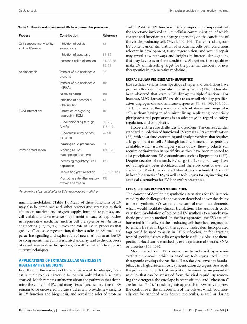
Table 1 | Functional relevance of EV in regenerative processes.
Process Contribution Reference
Cell senescence, viability
and proliferation
Inhibition of cellular
senescence
13
Inhibition of apoptosis 81–85
Increased cell proliferation 81, 83, 86,
89–91
Angiogenesis Transfer of pro-angiogenic
proteins
96
Transfer of pro-angiogenic
miRNAs
105
Notch signaling 101
Inhibition of endothelial
senescence
13
ECM interactions Formation of signaling
reservoir in ECM
108
ECM remodeling through
MMPs
68, 70,
115–117
ECM crosslinking by lysyl
oxidases
74, 98
Inducing ECM production 91
Immunomodulation Steering M1-M2
macrophage phenotype
124–126
Increasing regulatory T-cell
population
128
Decreasing graft rejection 85, 127, 128
Promoting anti-inflammatory
cytokine secretion
132
An overview of potential roles of EV in regenerative medicine.
immunomodulation (Table 1). Many of these functions of EVmay also be combined with other regenerative strategies as theireffects on nutrient and oxygen supply, immune responses, andcell viability and senescence may benefit efficacy of approachesin regenerative medicine, such as cell therapies or in situ tissueengineering (27, 75, 93). Given the role of EV in processes thatgreatly affect tissue regeneration, further studies in EV-mediatedparacrine signaling and exploration of new methods to utilize EVor components thereof is warranted and may lead to the discoveryof novel regenerative therapeutics, as well as methods to improvecurrent techniques.
APPLICATIONS OF EXTRACELLULAR VESICLES INREGENERATIVE MEDICINEEven though, the existence of EV was discovered decades ago, inter-est in their role as paracrine factor was only relatively recentlysparked. Much remains unknown about the pathways that deter-mine the content of EV, and many tissue-specific functions of EVremain to be uncovered. Future studies will provide new insightsin EV function and biogenesis, and reveal the roles of proteins
and miRNAs in EV function. EV are important components ofthe secretome involved in intercellular communication, of whichcontent and function can change depending on the conditions ofthe vesicle producing cells (74, 91, 102–104). Therefore, changes inEV content upon stimulation of producing cells with conditionsrelevant in development, tissue regeneration, and wound repairmay reveal new pathways and insights in intercellular signalingthat play key roles in these conditions. Altogether, these qualitiesmake EV an interesting target for the potential discovery of newtherapeutics in regenerative medicine.
EXTRACELLULAR VESICLES AS THERAPEUTICSExtracellular vesicles from specific cell types and conditions havepositive effects on regeneration in many tissues (136). It has alsobeen observed that certain EV display multiple functions. Forinstance, MSC-derived EV are able to steer cell viability, prolifer-ation, angiogenesis, and immune responses (81–83, 103, 104, 124,132). Harnessing the paracrine effects of stem- and progenitorcells without having to administer living, replicating, potentiallypluripotent cell populations is an advantage in regard to safety,regulation, and complexity.
However, there are challenges to overcome. The current goldenstandard in isolation of functional EV remains ultracentrifugation(58), which is a time-consuming and costly procedure that requiresa large amount of cells. Although faster commercial reagents areavailable, which isolate higher yields of EV, these products stillrequire optimization in specificity as they have been reported toalso precipitate non-EV contaminants such as lipoproteins (137).Despite decades of research, EV cargo trafficking pathways havenot completely been elucidated, and therefore control over thecontent of EV,and unspecific additional effects, is limited. Researchin both biogenesis of EV, as well as techniques for engineering forartificial alternatives for EV is therefore warranted.
EXTRACELLULAR VESICLES MODIFICATIONThe concept of developing synthetic alternatives for EV is moti-vated by the challenges that have been described above: the abilityto form synthetic EVs would allow control over these elements,which would facilitate clinical translation. The approach couldvary from modulation of biological EV synthesis to a purely syn-thetic production method. In the first approach, the EVs are stillharvested from cells, but the producing cells have been engineeredto enrich EVs with tags or therapeutic molecules. Incorporatedtags could be used to assist in EV purification, or for targetingtoward specific tissues, cells, or synthetic scaffolds. Also, the thera-peutic payload can be enriched by overexpression of specific RNAsor proteins (138, 139).
More control over EV content can be achieved by a semi-synthetic approach, which is based on techniques used in thetherapeutic enveloped virus-field. Here, the viral envelope is solu-bilized in a high critical micelle concentration detergent. As a resultthe proteins and lipids that are part of the envelope are present inmicelles that can be separated from the viral capsid. By remov-ing the detergent, the envelope is reconstituted, and “virosomes”are formed (140). Translating this approach to EVs may improvethe control over the composition of the bilayer, which addition-ally can be enriched with desired molecules, as well as during
Frontiers in Immunology | Immunotherapies and Vaccines December 2014 | Volume 5 | Article 608 | 8
De Jong et al. Extracellular vesicles in regenerative medicine
the reconstitution step, offering full control over the encapsulated(therapeutic) compounds in the aqueous core. At the same time,the naturally encapsulated molecules are removed.
SYNTHETIC EXTRACELLULAR VESICLESThe semi-synthetic approach still relies on the biological pro-duction of vesicles. The power of synthetic strategies lies in thescalability of the process. The minimal EV mimic is already onthe market and is known as liposomes (141). Liposomes consistof a phospholipid bilayer around an aqueous core, and have beeninvestigated as therapeutic delivery systems over the last 40 years.Therapeutic liposomes tend to be around 100 nm in size and havea lipid composition that allows them to circulate for prolongedperiods in the blood stream. Generally, therapeutic liposomes areprepared in batches that vary between liters to hundreds of litersin size, with a colloidal stability of several years, even in solution.Still the translation of liposome technology to mimic EVs hassome obstacles to overcome. For instance, the lipid and proteincomposition of EV, which may be important for their cellular inter-actions, is often complex, and the current production process ofliposomes involves simple synthetic lipid mixtures without othercomponents within the bilayer. However, liposomes have been suc-cessfully equipped with targeting ligands (such as antibodies) anda variety of therapeutic payloads including biologicals (142). Thesecharacteristics are several orders of magnitude away from the cur-rent state of the art in the EV field, but do illustrate the potentialvalue of synthetic EV.
CONCLUSIONOver the past decades, it has been shown that EV play a regu-latory role, and have modulatory potential, in many biologicalprocesses. EV show great potential for therapeutics, biomarkerresearch, and even alternatives to stem-cell-based therapies whichrely on paracrine effects. These new approaches have great poten-tial for the support of endogenous repair, including enhancementsof existing regenerative medicine approaches. This potential mer-its further research in the potential of EV, as well the study of newtechniques to produce and utilize engineered EV.
ACKNOWLEDGMENTSThis work was made possible by the multidisciplinary collabo-ration on Regenerative Medicine between the University MedicalCenter Utrecht and Eindhoven University of Technology (Mari-anne C. Verhaar and Carlijn V. C. Bouten), Olivier G. De Jong andBas W. M. Van Balkom are supported by The Netherlands Institutefor Regenerative Medicine (NIRM, grant No. FES0908), RaymondM. Schiffelers is supported by ERC Starting Grant MINDS (No.260627) and Marianne C.Verhaar is supported by The NetherlandsOrganization for Scientific Research (Vidi grant 016.096.359).
REFERENCES1. Harrison JH, Merrill JP, Murray JE. Renal homotransplantation in identical
twins. Surg Forum (1956) 6:432–6.2. Lexer E. The use of free osteoplasty together with trials on arthrodesis and joint
transplantation. Archiv fur klin Chirurgie. 1908;86(4):939-954. Clin OrthopRelat Res (2008) 466(8):1771–6. doi:10.1007/s11999-008-0314-4
3. Zirm EK. Eine erfolgreiche totale Keratoplastik (A successful total kerato-plasty). 1906. Refract Corneal Surg (1989) 5(4):258–61.
4. Drury JL, Mooney DJ. Hydrogels for tissue engineering: scaffold designvariables and applications. Biomaterials (2003) 24(24):4337–51. doi:10.1016/S0142-9612(03)00340-5
5. Mol A, Driessen NJ, Rutten MC, Hoerstrup SP, Bouten CV, Baaijens FP.Tissue engineering of human heart valve leaflets: a novel bioreactor for astrain-based conditioning approach. Ann Biomed Eng (2005) 33(12):1778–88.doi:10.1007/s10439-005-8025-4
6. Wagers AJ, Sherwood RI, Christensen JL, Weissman IL. Little evidence fordevelopmental plasticity of adult hematopoietic stem cells. Science (2002)297(5590):2256–9. doi:10.1126/science.1074807
7. Murry CE, Soonpaa MH, Reinecke H, Nakajima H, Nakajima HO, RubartM, et al. Haematopoietic stem cells do not transdifferentiate into cardiacmyocytes in myocardial infarcts. Nature (2004) 428(6983):664–8. doi:10.1038/nature02446
8. Den Haan MC, Grauss RW, Smits AM, Winter EM, van Tuyn J, PijnappelsDA, et al. Cardiomyogenic differentiation-independent improvement of car-diac function by human cardiomyocyte progenitor cell injection in ischaemicmouse hearts. J Cell Mol Med (2012) 16(7):1508–21. doi:10.1111/j.1582-4934.2011.01468.x
9. Van Koppen A, Joles JA, Bongartz LG, van den Brandt J, Reichardt HM,Goldschmeding R, et al. Healthy bone marrow cells reduce progression ofkidney failure better than CKD bone marrow cells in rats with establishedchronic kidney disease. Cell Transplant (2012) 21(10):2299–312. doi:10.3727/096368912X636795
10. Janowska-Wieczorek A, Wysoczynski M, Kijowski J, Marquez-Curtis L,Machalinski B, Ratajczak J, et al. Microvesicles derived from activated plateletsinduce metastasis and angiogenesis in lung cancer. Int J Cancer (2005)113(5):752–60. doi:10.1002/ijc.20657
11. Mu W, Rana S, Zoller M. Host matrix modulation by tumor exosomes pro-motes motility and invasiveness. Neoplasia (2013) 15(8):875–87. doi:10.1593/neo.13786
12. Raposo G, Nijman HW, Stoorvogel W, Liejendekker R, Harding CV, MeliefCJ, et al. B lymphocytes secrete antigen-presenting vesicles. J Exp Med (1996)183(3):1161–72. doi:10.1084/jem.183.3.1161
13. Van Balkom BW, de Jong OG, Smits M, Brummelman J, den Ouden K, de BreePM, et al. Endothelial cells require miR-214 to secrete exosomes that suppresssenescence and induce angiogenesis in human and mouse endothelial cells.Blood (2013) 121(19):3997–4006. doi:10.1182/blood-2013-02-478925
14. Thomas ED, Lochte HL Jr, Lu WC, Ferrebee JW. Intravenous infusion of bonemarrow in patients receiving radiation and chemotherapy. N Engl J Med (1957)257(11):491–6. doi:10.1056/NEJM195709122571102
15. Blundell J. Experiments on the transfusion of blood by the syringe. Med ChirTrans (1818) 9(Pt 1):56–92.
16. Slaper-Cortenbach IC. Current regulations for the production of multipotentmesenchymal stromal cells for clinical application. Transfus Med Hemother(2008) 35(4):295–8. doi:10.1159/000144043
17. Nishida K, Yamato M, Hayashida Y, Watanabe K, Yamamoto K, Adachi E,et al. Corneal reconstruction with tissue-engineered cell sheets composed ofautologous oral mucosal epithelium. N Engl J Med (2004) 351(12):1187–96.doi:10.1056/NEJMoa040455
18. Bartlett W, Skinner JA, Gooding CR, Carrington RW, Flanagan AM, BriggsTW, et al. Autologous chondrocyte implantation versus matrix-induced autol-ogous chondrocyte implantation for osteochondral defects of the knee: aprospective, randomised study. J Bone Joint Surg Br (2005) 87(5):640–5.doi:10.1302/0301-620X.87B5.15905
19. Schwarz TM, Leicht SF, Radic T, Rodriguez-Araboalaza I, Hermann PC, BergerF, et al. Vascular incorporation of endothelial colony-forming cells is essentialfor functional recovery of murine ischemic tissue following cell therapy. Arte-rioscler Thromb Vasc Biol (2012) 32(2):e13–21. doi:10.1161/ATVBAHA.111.239822
20. Beltrami AP, Cesselli D, Beltrami CA. Stem cell senescence and regenerativeparadigms. Clin Pharmacol Ther (2012) 91(1):21–9. doi:10.1038/clpt.2011.262
21. Siddappa R, Licht R, van Blitterswijk C, de Boer J. Donor variation andloss of multipotency during in vitro expansion of human mesenchymalstem cells for bone tissue engineering. J Orthop Res (2007) 25(8):1029–41.doi:10.1002/jor.20402
22. Engler AJ, Sen S, Sweeney HL, Discher DE. Matrix elasticity directs stem celllineage specification. Cell (2006) 126(4):677–89. doi:10.1016/j.cell.2006.06.044
www.frontiersin.org December 2014 | Volume 5 | Article 608 | 9

De Jong et al. Extracellular vesicles in regenerative medicine
23. Jie KE, Zaikova MA, Bergevoet MW,Westerweel PE, Rastmanesh M, BlankestijnPJ, et al. Progenitor cells and vascular function are impaired in patientswith chronic kidney disease. Nephrol Dial Transplant (2010) 25(6):1875–82.doi:10.1093/ndt/gfp749
24. Scruggs BA, Semon JA, Zhang X, Zhang S, Bowles AC, Pandey AC, et al. Age ofthe donor reduces the ability of human adipose-derived stem cells to alleviatesymptoms in the experimental autoimmune encephalomyelitis mouse model.Stem Cells Transl Med (2013) 2(10):797–807. doi:10.5966/sctm.2013-0026
25. Williamson KA, Hamilton A, Reynolds JA, Sipos P, Crocker I, Stringer SE,et al. Age-related impairment of endothelial progenitor cell migration corre-lates with structural alterations of heparan sulfate proteoglycans. Aging Cell(2013) 12(1):139–47. doi:10.1111/acel.12031
26. Hugle T, Daikeler T. Stem cell transplantation for autoimmune diseases.Haematologica (2010) 95(2):185–8. doi:10.3324/haematol.2009.017038
27. Muylaert DE, Fledderus JO, Bouten CV, Dankers PY, Verhaar MC. Combiningtissue repair and tissue engineering; bioactivating implantable cell-free vascularscaffolds. Heart (2014) 100(23):1825–30. doi:10.1136/heartjnl-2014-306092
28. Vanden Berg-Foels WS. In situ tissue regeneration: chemoattractants forendogenous stem cell recruitment. Tissue Eng Part B Rev (2014) 20(1):28–39.doi:10.1089/ten.teb.2013.0100
29. Badylak SF. Xenogeneic extracellular matrix as a scaffold for tissue reconstruc-tion. Transpl Immunol (2004) 12(3–4):367–77. doi:10.1016/j.trim.2003.12.016
30. Knight RL, Wilcox HE, Korossis SA, Fisher J, Ingham E. The use of acellularmatrices for the tissue engineering of cardiac valves. Proc Inst Mech Eng H(2008) 222(1):129–43. doi:10.1243/09544119JEIM230
31. Borschel GH, Huang YC, Calve S, Arruda EM, Lynch JB, Dow DE, et al. Tissueengineering of recellularized small-diameter vascular grafts. Tissue Eng (2005)11(5–6):778–86. doi:10.1089/ten.2005.11.778
32. Baiguera S, Jungebluth P, Burns A, Mavilia C, Haag J, De Coppi P, et al. Tis-sue engineered human tracheas for in vivo implantation. Biomaterials (2010)31(34):8931–8. doi:10.1016/j.biomaterials.2010.08.005
33. Zheng MH, Chen J, Kirilak Y, Willers C, Xu J, Wood D. Porcine small intestinesubmucosa (SIS) is not an acellular collagenous matrix and contains porcineDNA: possible implications in human implantation. J Biomed Mater Res B ApplBiomater (2005) 73(1):61–7. doi:10.1002/jbm.b.30170
34. Ratner BD, Bryant SJ. Biomaterials: where we have been and where we aregoing. Annu Rev Biomed Eng (2004) 6:41–75. doi:10.1146/annurev.bioeng.6.040803.140027
35. Mendelson K, Schoen FJ. Heart valve tissue engineering: concepts, approaches,progress, and challenges. Ann Biomed Eng (2006) 34(12):1799–819. doi:10.1007/s10439-006-9163-z
36. Mikos AG, Lyman MD, Freed LE, Langer R. Wetting of poly(L-lactic acid) andpoly(DL-lactic-co-glycolic acid) foams for tissue culture. Biomaterials (1994)15(1):55–8. doi:10.1016/0142-9612(94)90197-X
37. Whang K, Thomas CH, Healy KE, Nuber G. A novel method to fabricate bioab-sorbable scaffolds. Polymer (1995) 36(4):837–42. doi:10.1016/0032-3861(95)93115-3
38. Mooney DJ, Baldwin DF, Suh NP, Vacanti JP, Langer R. Novel approach tofabricate porous sponges of poly(D,L-lactic-co-glycolic acid) without the useof organic solvents. Biomaterials (1996) 17(14):1417–22. doi:10.1016/0142-9612(96)87284-X
39. Mooney DJ, Mazzoni CL, Breuer C, McNamara K, Hern D,Vacanti JP, et al. Sta-bilized polyglycolic acid fibre-based tubes for tissue engineering. Biomaterials(1996) 17(2):115–24. doi:10.1016/0142-9612(96)85756-5
40. Stankus JJ, Guan J, Wagner WR. Fabrication of biodegradable elastomericscaffolds with sub-micron morphologies. J Biomed Mater Res A (2004)70(4):603–14. doi:10.1002/jbm.a.30122
41. Li M, Mondrinos MJ, Chen X, Gandhi MR, Ko FK, Lelkes PI. Co-electrospunpoly(lactide-co-glycolide), gelatin, and elastin blends for tissue engineeringscaffolds. J Biomed Mater Res A (2006) 79(4):963–73. doi:10.1002/jbm.a.30833
42. Choi WS, Bae JW, Lim HR, Joung YK, Park JC, Kwon IK, et al. RGDpeptide-immobilized electrospun matrix of polyurethane for enhancedendothelial cell affinity. Biomed Mater (2008) 3(4):044104. doi:10.1088/1748-6041/3/4/044104
43. Yu J, Lee AR, Lin WH, Lin CW, Wu YK, Tsai WB. Electrospun PLGA fibersincorporated with functionalized biomolecules for cardiac tissue engineering.Tissue Eng Part A (2014) 20(13–14):1896–907. doi:10.1089/ten.tea.2013.0008
44. Kolambkar YM, Bajin M, Wojtowicz A, Hutmacher DW, Garcia AJ, Guld-berg RE. Nanofiber orientation and surface functionalization modulate human
mesenchymal stem cell behavior in vitro. Tissue Eng Part A (2014) 20(1–2):398–409. doi:10.1089/ten.tea.2012.0426
45. Han F, Jia X, Dai D, Yang X, Zhao J, Zhao Y, et al. Performance of a multi-layered small-diameter vascular scaffold dual-loaded with VEGF and PDGF.Biomaterials (2013) 34(30):7302–13. doi:10.1016/j.biomaterials.2013.06.006
46. Thevenot PT, Nair AM, Shen J, Lotfi P, Ko CY, Tang L. The effect of incorpo-ration of SDF-1alpha into PLGA scaffolds on stem cell recruitment and theinflammatory response. Biomaterials (2010) 31(14):3997–4008. doi:10.1016/j.biomaterials.2010.01.144
47. De Visscher G, Mesure L, Meuris B, Ivanova A, Flameng W. Improved endothe-lialization and reduced thrombosis by coating a synthetic vascular graft withfibronectin and stem cell homing factor SDF-1alpha. Acta Biomater (2012)8(3):1330–8. doi:10.1016/j.actbio.2011.09.016
48. Tsai TN, Kirton JP, Campagnolo P, Zhang L, Xiao Q, Zhang Z, et al. Contribu-tion of stem cells to neointimal formation of decellularized vessel grafts in anovel mouse model. Am J Pathol (2012) 181(1):362–73. doi:10.1016/j.ajpath.2012.03.021
49. Lai RC, Arslan F, Lee MM, Sze NS, Choo A, Chen TS, et al. Exosome secretedby MSC reduces myocardial ischemia/reperfusion injury. Stem Cell Res (2010)4(3):214–22. doi:10.1016/j.scr.2009.12.003
50. Stoorvogel W, Kleijmeer MJ, Geuze HJ, Raposo G. The biogenesis and func-tions of exosomes. Traffic (2002) 3(5):321–30. doi:10.1034/j.1600-0854.2002.30502.x
51. Thery C, Zitvogel L, Amigorena S. Exosomes: composition, biogenesis andfunction. Nat Rev Immunol (2002) 2(8):569–79. doi:10.1038/nri855
52. Valadi H, Ekstrom K, Bossios A, Sjostrand M, Lee JJ, Lotvall JO. Exosome-mediated transfer of mRNAs and microRNAs is a novel mechanism of geneticexchange between cells. Nat Cell Biol (2007) 9(6):654–9. doi:10.1038/ncb1596
53. Bobrie A, Colombo M, Raposo G, Thery C. Exosome secretion: molecularmechanisms and roles in immune responses. Traffic (2011) 12(12):1659–68.doi:10.1111/j.1600-0854.2011.01225.x
54. Gutzeit C, Nagy N, Gentile M, Lyberg K, Gumz J, Vallhov H, et al. Exosomesderived from Burkitt’s lymphoma cell lines induce proliferation,differentiation,and class-switch recombination in B cells. J Immunol (2014) 192(12):5852–62.doi:10.4049/jimmunol.1302068
55. Pan BT, Johnstone RM. Fate of the transferrin receptor during maturation ofsheep reticulocytes in vitro: selective externalization of the receptor. Cell (1983)33(3):967–78. doi:10.1016/0092-8674(83)90040-5
56. Xu D, Tahara H. The role of exosomes and microRNAs in senescence and aging.Adv Drug Deliv Rev (2013) 65(3):368–75. doi:10.1016/j.addr.2012.07.010
57. El Andaloussi S, Lakhal S, Mager I, Wood MJ. Exosomes for targeted siRNAdelivery across biological barriers. Adv Drug Deliv Rev (2013) 65(3):391–7.doi:10.1016/j.addr.2012.08.008
58. Gould SJ, Raposo G. As we wait: coping with an imperfect nomenclature forextracellular vesicles. J Extracell Vesicles (2013) 2:20389. doi:10.3402/jev.v2i0.20389
59. van Balkom BW, Pisitkun T, Verhaar MC, Knepper MA. Exosomes and the kid-ney: prospects for diagnosis and therapy of renal diseases. Kidney Int (2011)80(11):1138–45. doi:10.1038/ki.2011.292
60. Escola JM, Kleijmeer MJ, Stoorvogel W, Griffith JM, Yoshie O, Geuze HJ. Selec-tive enrichment of tetraspan proteins on the internal vesicles of multivesicularendosomes and on exosomes secreted by human B-lymphocytes. J Biol Chem(1998) 273(32):20121–7. doi:10.1074/jbc.273.32.20121
61. Vader P, Breakefield XO, Wood MJ. Extracellular vesicles: emerging targets forcancer therapy. Trends Mol Med (2014) 20(7):385–93. doi:10.1016/j.molmed.2014.03.002
62. Cocucci E, Racchetti G, Meldolesi J. Shedding microvesicles: artefacts no more.Trends Cell Biol (2009) 19(2):43–51. doi:10.1016/j.tcb.2008.11.003
63. Baj-Krzyworzeka M, Szatanek R, Weglarczyk K, Baran J, Urbanowicz B,Branski P, et al. Tumour-derived microvesicles carry several surface deter-minants and mRNA of tumour cells and transfer some of these deter-minants to monocytes. Cancer Immunol Immunother (2006) 55(7):808–18.doi:10.1007/s00262-005-0075-9
64. Ratajczak J, Wysoczynski M, Hayek F, Janowska-Wieczorek A, Ratajczak MZ.Membrane-derived microvesicles: important and underappreciated mediatorsof cell-to-cell communication. Leukemia (2006) 20(9):1487–95. doi:10.1038/sj.leu.2404296
65. Huber J, Vales A, Mitulovic G, Blumer M, Schmid R, Witztum JL,et al. Oxidized membrane vesicles and blebs from apoptotic cells contain
Frontiers in Immunology | Immunotherapies and Vaccines December 2014 | Volume 5 | Article 608 | 10
De Jong et al. Extracellular vesicles in regenerative medicine
biologically active oxidized phospholipids that induce monocyte-endothelialinteractions. Arterioscler Thromb Vasc Biol (2002) 22(1):101–7. doi:10.1161/hq0102.101525
66. Loyer X, Vion AC, Tedgui A, Boulanger CM. Microvesicles as cell-cell messen-gers in cardiovascular diseases. Circ Res (2014) 114(2):345–53. doi:10.1161/CIRCRESAHA.113.300858
67. Zernecke A, Bidzhekov K, Noels H, Shagdarsuren E, Gan L, Denecke B,et al. Delivery of microRNA-126 by apoptotic bodies induces CXCL12-dependent vascular protection. Sci Signal (2009) 2(100):ra81. doi:10.1126/scisignal.2000610
68. Distler JH, Jungel A, Huber LC, Seemayer CA, Reich CF III, Gay RE, et al. Theinduction of matrix metalloproteinase and cytokine expression in synovialfibroblasts stimulated with immune cell microparticles. Proc Natl Acad Sci U SA (2005) 102(8):2892–7. doi:10.1073/pnas.0409781102
69. Julich H, Willms A, Lukacs-Kornek V, Kornek M. Extracellular vesicle profilingand their use as potential disease specific biomarker. Front Immunol (2014)5:413. doi:10.3389/fimmu.2014.00413
70. Kornek M, Popov Y, Libermann TA, Afdhal NH, Schuppan D. Human T cellmicroparticles circulate in blood of hepatitis patients and induce fibrolytic acti-vation of hepatic stellate cells. Hepatology (2011) 53(1):230–42. doi:10.1002/hep.23999
71. Harding C, Heuser J, Stahl P. Receptor-mediated endocytosis of transferrinand recycling of the transferrin receptor in rat reticulocytes. J Cell Biol (1983)97(2):329–39. doi:10.1083/jcb.97.2.329
72. Johnstone RM, Mathew A, Mason AB, Teng K. Exosome formation during mat-uration of mammalian and avian reticulocytes: evidence that exosome release isa major route for externalization of obsolete membrane proteins. J Cell Physiol(1991) 147(1):27–36. doi:10.1002/jcp.1041470105
73. Pegtel DM, Cosmopoulos K, Thorley-Lawson DA, van Eijndhoven MA, Hop-mans ES, Lindenberg JL, et al. Functional delivery of viral miRNAs via exo-somes. Proc Natl Acad Sci U S A (2010) 107(14):6328–33. doi:10.1073/pnas.0914843107
74. De Jong OG, Verhaar MC, Chen Y, Vader P, Gremmels H, Posthuma G,et al. Cellular stress conditions are reflected in the protein and RNA con-tent of endothelial cell-derived exosomes. J Extracell Vesicles (2012) 1:18396.doi:10.3402/jev.v1i0.18396
75. Huu AL, Paul A, Prakash S, Shum-Tim D. Route of delivery, cell retention, andefficiency of polymeric microcapsules in cellular cardiomyoplasty. Methods MolBiol (2013) 1036:121–35. doi:10.1007/978-1-62703-511-8_11
76. Gruber HE, Somayaji S, Riley F, Hoelscher GL, Norton HJ, Ingram J, et al.Human adipose-derived mesenchymal stem cells: serial passaging, doublingtime and cell senescence. Biotech Histochem (2012) 87(4):303–11. doi:10.3109/10520295.2011.649785
77. Stolzing A, Jones E, McGonagle D, Scutt A. Age-related changes in human bonemarrow-derived mesenchymal stem cells: consequences for cell therapies. MechAgeing Dev (2008) 129(3):163–73. doi:10.1016/j.mad.2007.12.002
78. Adijiang A, Higuchi Y, Nishijima F, Shimizu H, Niwa T. Indoxyl sulfate, a uremictoxin, promotes cell senescence in aorta of hypertensive rats. Biochem BiophysRes Commun (2010) 399(4):637–41. doi:10.1016/j.bbrc.2010.07.130
79. Bonab MM, Alimoghaddam K, Talebian F, Ghaffari SH, Ghavamzadeh A,Nikbin B. Aging of mesenchymal stem cell in vitro. BMC Cell Biol (2006) 7:14.doi:10.1186/1471-2121-7-14
80. Vacanti V, Kong E, Suzuki G, Sato K, Canty JM, Lee T. Phenotypic changesof adult porcine mesenchymal stem cells induced by prolonged passaging inculture. J Cell Physiol (2005) 205(2):194–201. doi:10.1002/jcp.20376
81. Zhou Y, Xu H, Xu W, Wang B, Wu H, Tao Y, et al. Exosomes released by humanumbilical cord mesenchymal stem cells protect against cisplatin-induced renaloxidative stress and apoptosis in vivo and in vitro. Stem Cell Res Ther (2013)4(2):34. doi:10.1186/scrt194
82. Barile L, Lionetti V, Cervio E, Matteucci M, Gherghiceanu M, Popescu LM,et al. Extracellular vesicles from human cardiac progenitor cells inhibit car-diomyocyte apoptosis and improve cardiac function after myocardial infarc-tion. Cardiovasc Res (2014) 103(4):530–41. doi:10.1093/cvr/cvu167
83. Reis LA, Borges FT, Simoes MJ, Borges AA, Sinigaglia-Coimbra R, Schor N.Bone marrow-derived mesenchymal stem cells repaired but did not preventgentamicin-induced acute kidney injury through paracrine effects in rats. PLoSOne (2012) 7(9):e44092. doi:10.1371/journal.pone.0044092
84. Bruno S, Grange C, Collino F, Deregibus MC, Cantaluppi V, Biancone L,et al. Microvesicles derived from mesenchymal stem cells enhance survival
in a lethal model of acute kidney injury. PLoS One (2012) 7(3):e33115.doi:10.1371/journal.pone.0033115
85. Yu B, Gong M, Wang Y, Millard RW, Pasha Z, Yang Y, et al. Cardiomyocyteprotection by GATA-4 gene engineered mesenchymal stem cells is partiallymediated by translocation of miR-221 in microvesicles. PLoS One (2013)8(8):e73304. doi:10.1371/journal.pone.0073304
86. Inder KL, Ruelcke JE, Petelin L, Moon H, Choi E, Rae J, et al. Cavin-1/PTRF alters prostate cancer cell-derived extracellular vesicle content andinternalization to attenuate extracellular vesicle-mediated osteoclastogenesisand osteoblast proliferation. J Extracell Vesicles (2014) 3:23784. doi:10.3402/jev.v3.23784
87. Xu Y, Luo F, Liu Y, Shi L, Lu X, Xu W, et al. Exosomal miR-21 derivedfrom arsenite-transformed human bronchial epithelial cells promotes cellproliferation associated with arsenite carcinogenesis. Arch Toxicol (2014).doi:10.1007/s00204-014-1291-x
88. Yang L, Wu XH, Wang D, Luo CL, Chen LX. Bladder cancer cell-derived exo-somes inhibit tumor cell apoptosis and induce cell proliferation in vitro. MolMed Rep (2013) 8(4):1272–8. doi:10.3892/mmr.2013.1634
89. Tomasoni S, Longaretti L, Rota C, Morigi M, Conti S, Gotti E, et al. Trans-fer of growth factor receptor mRNA via exosomes unravels the regenera-tive effect of mesenchymal stem cells. Stem Cells Dev (2013) 22(5):772–80.doi:10.1089/scd.2012.0266
90. Zhang B, Wang M, Gong A, Zhang X, Wu X, Zhu Y, et al. HucMSC-exosomemediated-Wnt4 signaling is required for cutaneous wound healing. Stem Cells(2014). doi:10.1002/stem.1771
91. Borges FT, Melo SA, Ozdemir BC, Kato N, Revuelta I, Miller CA, et al. TGF-beta1-containing exosomes from injured epithelial cells activate fibroblasts toinitiate tissue regenerative responses and fibrosis. J Am Soc Nephrol (2013)24(3):385–92. doi:10.1681/ASN.2012101031
92. Yu X, Huang C, Song B, Xiao Y, Fang M, Feng J, et al. CD4+CD25+ regulatoryT cells-derived exosomes prolonged kidney allograft survival in a rat model.Cell Immunol (2013) 285(1–2):62–8. doi:10.1016/j.cellimm.2013.06.010
93. Jain RK, Au P, Tam J, Duda DG, Fukumura D. Engineering vascularized tissue.Nat Biotechnol (2005) 23(7):821–3. doi:10.1038/nbt0705-821
94. Du C, Narayanan K, Leong MF, Wan AC. Induced pluripotent stem cell-derived hepatocytes and endothelial cells in multi-component hydrogel fibersfor liver tissue engineering. Biomaterials (2014) 35(23):6006–14. doi:10.1016/j.biomaterials.2014.04.011
95. Glynn JJ, Hinds MT. Endothelial outgrowth cells: function and performance invascular grafts. Tissue Eng Part B Rev (2013) 20(4):294–303. doi:10.1089/ten.teb.2013.0285
96. Ekstrom EJ, Bergenfelz C, von Bulow V, Serifler F, Carlemalm E, Jonsson G, et al.WNT5A induces release of exosomes containing pro-angiogenic and immuno-suppressive factors from malignant melanoma cells. Mol Cancer (2014) 13:88.doi:10.1186/1476-4598-13-88
97. Hong BS, Cho JH, Kim H, Choi EJ, Rho S, Kim J, et al. Colorectal can-cer cell-derived microvesicles are enriched in cell cycle-related mRNAs thatpromote proliferation of endothelial cells. BMC Genomics (2009) 10:556.doi:10.1186/1471-2164-10-556
98. Kucharzewska P, Christianson HC, Welch JE, Svensson KJ, Fredlund E,Ringner M, et al. Exosomes reflect the hypoxic status of glioma cells andmediate hypoxia-dependent activation of vascular cells during tumor devel-opment. Proc Natl Acad Sci U S A (2013) 110(18):7312–7. doi:10.1073/pnas.1220998110
99. Skog J, Wurdinger T, van Rijn S, Meijer DH, Gainche L, Sena-Esteves M, et al.Glioblastoma microvesicles transport RNA and proteins that promote tumourgrowth and provide diagnostic biomarkers. Nat Cell Biol (2008) 10(12):1470–6.doi:10.1038/ncb1800
100. Tadokoro H, Umezu T, Ohyashiki K, Hirano T, Ohyashiki JH. Exosomes derivedfrom hypoxic leukemia cells enhance tube formation in endothelial cells. J BiolChem (2013) 288(48):34343–51. doi:10.1074/jbc.M113.480822
101. Sheldon H, Heikamp E, Turley H, Dragovic R, Thomas P, Oon CE, et al. Newmechanism for Notch signaling to endothelium at a distance by Delta-like4 incorporation into exosomes. Blood (2010) 116(13):2385–94. doi:10.1182/blood-2009-08-239228
102. Lopatina T, Bruno S, Tetta C, Kalinina N, Porta M, Camussi G. Platelet-derivedgrowth factor regulates the secretion of extracellular vesicles by adipose mes-enchymal stem cells and enhances their angiogenic potential. Cell CommunSignal (2014) 12:26. doi:10.1186/1478-811X-12-26
www.frontiersin.org December 2014 | Volume 5 | Article 608 | 11

De Jong et al. Extracellular vesicles in regenerative medicine
103. Bian S, Zhang L, Duan L, Wang X, Min Y, Yu H. Extracellular vesicles derivedfrom human bone marrow mesenchymal stem cells promote angiogenesis ina rat myocardial infarction model. J Mol Med (Berl) (2014) 92(4):387–97.doi:10.1007/s00109-013-1110-5
104. Zhang HC, Liu XB, Huang S, Bi XY, Wang HX, Xie LX, et al. Microvesiclesderived from human umbilical cord mesenchymal stem cells stimulated byhypoxia promote angiogenesis both in vitro and in vivo. Stem Cells Dev (2012)21(18):3289–97. doi:10.1089/scd.2012.0095
105. Cantaluppi V, Biancone L, Figliolini F, Beltramo S, Medica D, Deregibus MC,et al. Microvesicles derived from endothelial progenitor cells enhance neoan-giogenesis of human pancreatic islets. Cell Transplant (2012) 21(6):1305–20.doi:10.3727/096368911X627534
106. Ranghino A, Cantaluppi V, Grange C, Vitillo L, Fop F, Biancone L, et al.Endothelial progenitor cell-derived microvesicles improve neovascularizationin a murine model of hindlimb ischemia. Int J Immunopathol Pharmacol(2012) 25(1):75–85.
107. Sahoo S, Klychko E, Thorne T, Misener S, Schultz KM, Millay M, et al. Exo-somes from human CD34(+) stem cells mediate their proangiogenic paracrineactivity. Circ Res (2011) 109(7):724–8. doi:10.1161/CIRCRESAHA.111.253286
108. Clayton A, Turkes A, Dewitt S, Steadman R, Mason MD, Hallett MB. Adhesionand signaling by B cell-derived exosomes: the role of integrins. FASEB J (2004)18(9):977–9. doi:10.1096/fj.03-1094fje
109. Segura E, Nicco C, Lombard B, Veron P, Raposo G, Batteux F, et al. ICAM-1on exosomes from mature dendritic cells is critical for efficient naive T-cellpriming. Blood (2005) 106(1):216–23. doi:10.1182/blood-2005-01-0220
110. Rieu S, Geminard C, Rabesandratana H, Sainte-Marie J, Vidal M. Exo-somes released during reticulocyte maturation bind to fibronectin via integrinalpha4beta1. Eur J Biochem (2000) 267(2):583–90. doi:10.1046/j.1432-1327.2000.01036.x
111. Thery C, Regnault A, Garin J, Wolfers J, Zitvogel L, Ricciardi-Castagnoli P, et al.Molecular characterization of dendritic cell-derived exosomes. Selective accu-mulation of the heat shock protein hsc73. J Cell Biol (1999) 147(3):599–610.doi:10.1083/jcb.147.3.599
112. Chen Q, Jin M, Yang F, Zhu J, Xiao Q, Zhang L. Matrix metalloproteinases:inflammatory regulators of cell behaviors in vascular formation and remodel-ing. Mediators Inflamm (2013) 2013:928315. doi:10.1155/2013/928315
113. Nissinen L, Kahari VM. Matrix metalloproteinases in inflammation. BiochimBiophys Acta (2014) 1840(8):2571–80. doi:10.1016/j.bbagen.2014.03.007
114. Hakulinen J, Sankkila L, Sugiyama N, Lehti K, Keski-Oja J. Secretion ofactive membrane type 1 matrix metalloproteinase (MMP-14) into extracel-lular space in microvesicular exosomes. J Cell Biochem (2008) 105(5):1211–8.doi:10.1002/jcb.21923
115. Vrijsen KR, Sluijter JP, Schuchardt MW, van Balkom BW, Noort WA,Chamuleau SA, et al. Cardiomyocyte progenitor cell-derived exosomes stim-ulate migration of endothelial cells. J Cell Mol Med (2010) 14(5):1064–70.doi:10.1111/j.1582-4934.2010.01081.x
116. Tauro BJ, Mathias RA, Greening DW, Gopal SK, Ji H, Kapp EA, et al. OncogenicH-ras reprograms Madin-Darby canine kidney (MDCK) cell-derived exoso-mal proteins following epithelial-mesenchymal transition. Mol Cell Proteomics(2013) 12(8):2148–59. doi:10.1074/mcp.M112.027086
117. Medina A, Ghahary A. Transdifferentiated circulating monocytes release exo-somes containing 14-3-3 proteins with matrix metalloproteinase-1 stimulat-ing effect for dermal fibroblasts. Wound Repair Regen (2010) 18(2):245–53.doi:10.1111/j.1524-475X.2010.00580.x
118. Athens AA, Makris EA, Hu JC. Induced collagen cross-links enhance cartilageintegration. PLoS One (2013) 8(4):e60719. doi:10.1371/journal.pone.0060719
119. Bignon M, Pichol-Thievend C, Hardouin J, Malbouyres M, Brechot N, NasciuttiL, et al. Lysyl oxidase-like protein-2 regulates sprouting angiogenesis and typeIV collagen assembly in the endothelial basement membrane. Blood (2011)118(14):3979–89. doi:10.1182/blood-2010-10-313296
120. Hanson S, D’Souza RN, Hematti P. Biomaterial-mesenchymal stem cell con-structs for immunomodulation in composite tissue engineering. Tissue EngPart A (2014) 20(15–16):2162–8. doi:10.1089/ten.tea.2013.0359
121. Artlett CM. Inflammasomes in wound healing and fibrosis. J Pathol (2012)229(2):157–67. doi:10.1002/path.4116
122. Galli SJ, Borregaard N, Wynn TA. Phenotypic and functional plasticity of cellsof innate immunity: macrophages, mast cells and neutrophils. Nat Immunol(2011) 12(11):1035–44. doi:10.1038/ni.2109
123. Brown BN, Ratner BD, Goodman SB, Amar S, Badylak SF. Macrophagepolarization: an opportunity for improved outcomes in biomaterials andregenerative medicine. Biomaterials (2012) 33(15):3792–802. doi:10.1016/j.biomaterials.2012.02.034
124. Zhang B, Yin Y, Lai RC, Tan SS, Choo AB, Lim SK. Mesenchymal stemcells secrete immunologically active exosomes. Stem Cells Dev (2014)23(11):1233–44. doi:10.1089/scd.2013.0479
125. Soki FN, Koh AJ, Jones JD, Kim YW, Dai J, Keller ET, et al. Polariza-tion of prostate cancer-associated macrophages is induced by milk fatglobule-EGF factor 8 (MFG-E8)-mediated efferocytosis. J Biol Chem (2008)289(35):24560–72. doi:10.1074/jbc.M114.571620
126. Jang JY, Lee JK, Jeon YK, Kim CW. Exosome derived from epigallocatechin gal-late treated breast cancer cells suppresses tumor growth by inhibiting tumor-associated macrophage infiltration and M2 polarization. BMC Cancer (2013)13:421. doi:10.1186/1471-2407-13-421
127. Li X, Li JJ, Yang JY, Wang DS, Zhao W, Song WJ, et al. Tolerance inductionby exosomes from immature dendritic cells and rapamycin in a mouse car-diac allograft model. PLoS One (2013) 7(8):e44045. doi:10.1371/journal.pone.0044045
128. Yang X, Meng S, Jiang H, Zhu C, Wu W. Exosomes derived from immaturebone marrow dendritic cells induce tolerogenicity of intestinal transplantationin rats. J Surg Res (2011) 171(2):826–32. doi:10.1016/j.jss.2010.05.021
129. Corcione A, Benvenuto F, Ferretti E, Giunti D, Cappiello V, Cazzanti F, et al.Human mesenchymal stem cells modulate B-cell functions. Blood (2006)107(1):367–72. doi:10.1182/blood-2005-07-2657
130. Shi M, Liu ZW, Wang FS. Immunomodulatory properties and therapeuticapplication of mesenchymal stem cells. Clin Exp Immunol (2011) 164(1):1–8.doi:10.1111/j.1365-2249.2011.04327.x
131. Xue Q, Luan XY, Gu YZ, Wu HY, Zhang GB, Yu GH, et al. The negative co-signaling molecule b7-h4 is expressed by human bone marrow-derived mes-enchymal stem cells and mediates its T-cell modulatory activity. Stem Cells Dev(2010) 19(1):27–38. doi:10.1089/scd.2009.0076
132. Mokarizadeh A, Delirezh N, Morshedi A, Mosayebi G, Farshid AA, Mar-dani K. Microvesicles derived from mesenchymal stem cells: potent organellesfor induction of tolerogenic signaling. Immunol Lett (2012) 147(1–2):47–54.doi:10.1016/j.imlet.2012.06.001
133. Arslan F, Lai RC, Smeets MB,Akeroyd L, Choo A,Aguor EN, et al. Mesenchymalstem cell-derived exosomes increase ATP levels, decrease oxidative stress andactivate PI3K/Akt pathway to enhance myocardial viability and prevent adverseremodeling after myocardial ischemia/reperfusion injury. Stem Cell Res (2013)10(3):301–12. doi:10.1016/j.scr.2013.01.002
134. Zou X, Zhang G, Cheng Z, Yin D, Du T, Ju G, et al. Microvesicles derived fromhuman Wharton’s Jelly mesenchymal stromal cells ameliorate renal ischemia-reperfusion injury in rats by suppressing CX3CL1. Stem Cell Res Ther (2014)5(2):40. doi:10.1186/scrt428
135. Akyurekli C, Le Y, Richardson RB, Fergusson D, Tay J, Allan DS. A systematicreview of preclinical studies on the therapeutic potential of mesenchymal stro-mal cell-derived microvesicles. Stem Cell Rev Rep (2014). doi:10.1007/s12015-014-9545-9
136. Lamichhane TN, Sokic S, Schardt JS, Raiker RS, Lin JW, Jay SM. Emergingroles for extracellular vesicles in tissue engineering and regenerative medicine.Tissue Eng Part B Rev (2014). doi:10.1089/ten.teb.2014.0300
137. Witwer KW, Buzas EI, Bemis LT, Bora A, Lasser C, Lotvall J, et al. Standardiza-tion of sample collection, isolation and analysis methods in extracellular vesicleresearch. J Extracell Vesicles (2013) 2:20360. doi:10.3402/jev.v2i0.20360
138. Li W, Mu D, Tian F, Hu Y, Jiang T, Han Y, et al. Exosomes derived from Rab27a-overexpressing tumor cells elicit efficient induction of antitumor immunity.Mol Med Rep (2013) 8(6):1876–82. doi:10.3892/mmr.2013.1738
139. Squadrito ML, Baer C, Burdet F, Maderna C, Gilfillan GD, Lyle R, et al. Endoge-nous RNAs modulate microRNA sorting to exosomes and transfer to acceptorcells. Cell Rep (2014) 8(5):1432–46. doi:10.1016/j.celrep.2014.07.035
140. Daemen T, de Mare A, Bungener L, de Jonge J, Huckriede A, Wilschut J. Viro-somes for antigen and DNA delivery. Adv Drug Deliv Rev (2005) 57(3):451–63.doi:10.1016/j.addr.2004.09.005
141. van der Meel R, Fens MH, Vader P, van Solinge WW, Eniola-Adefeso O,Schiffelers RM. Extracellular vesicles as drug delivery systems: lessonsfrom the liposome field. J Control Release (2014) 195:72–85. doi:10.1016/j.jconrel.2014.07.049
Frontiers in Immunology | Immunotherapies and Vaccines December 2014 | Volume 5 | Article 608 | 12
De Jong et al. Extracellular vesicles in regenerative medicine
142. van der Meel R, Vehmeijer LJ, Kok RJ, Storm G, van Gaal EV. Ligand-targetedparticulate nanomedicines undergoing clinical evaluation: current status. AdvDrug Deliv Rev (2013) 65(10):1284–98. doi:10.1016/j.addr.2013.08.012
Conflict of Interest Statement: Raymond M. Schiffelers is the CSO of Excytex, acompany developing tools for extracellular vesicle research. The other co-authorsdeclare that the research was conducted in the absence of any commercial or financialrelationships that could be construed as a potential conflict of interest.
Received: 16 September 2014; accepted: 12 November 2014; published online: 03December 2014.
Citation: De Jong OG, Van Balkom BWM, Schiffelers RM, Bouten CVC and Ver-haar MC (2014) Extracellular vesicles: potential roles in regenerative medicine. Front.Immunol. 5:608. doi: 10.3389/fimmu.2014.00608This article was submitted to Immunotherapies and Vaccines, a section of the journalFrontiers in Immunology.Copyright © 2014 De Jong, Van Balkom, Schiffelers, Bouten and Verhaar. This is anopen-access article distributed under the terms of the Creative Commons AttributionLicense (CC BY). The use, distribution or reproduction in other forums is permitted,provided the original author(s) or licensor are credited and that the original publica-tion in this journal is cited, in accordance with accepted academic practice. No use,distribution or reproduction is permitted which does not comply with these terms.
www.frontiersin.org December 2014 | Volume 5 | Article 608 | 13