external carotid artery, its branches and anastomosesanat.lf1.cuni.cz/souhrny/ofa_a5.pdf ·...
TRANSCRIPT
External carotidartery, its branches
and anastomosesTOPOGRAPHIC
REGIONS and SPACES
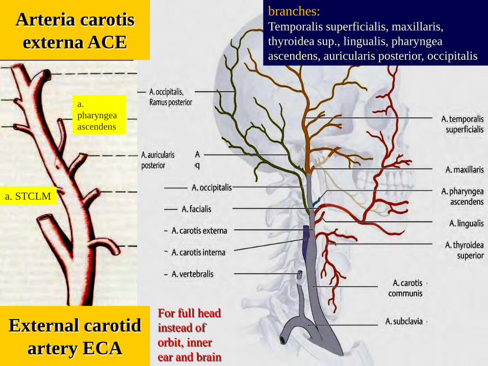
Arteria carotisexterna ACE
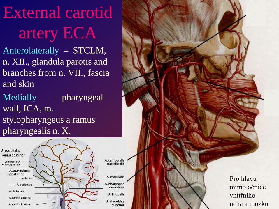
External carotidartery ECA
a. STCLM
a. pharyngeaascendens
branches:Temporalis superficialis, maxillaris, thyroidea sup., lingualis, pharyngeaascendens, auricularis posterior, occipitalis
For full headinstead oforbit, innerear and brain
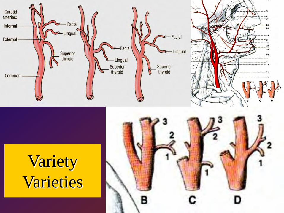
Variety Varieties
External carotid artery ECA
Anterolaterally – STCLM, n. XII., glandula parotis and branches from n. VII., fascia and skinMedially – pharyngeal wall, ICA, m. stylopharyngeus a ramus pharyngealis n. X.
Pro hlavu mimo očnice vnitřního ucha a mozku

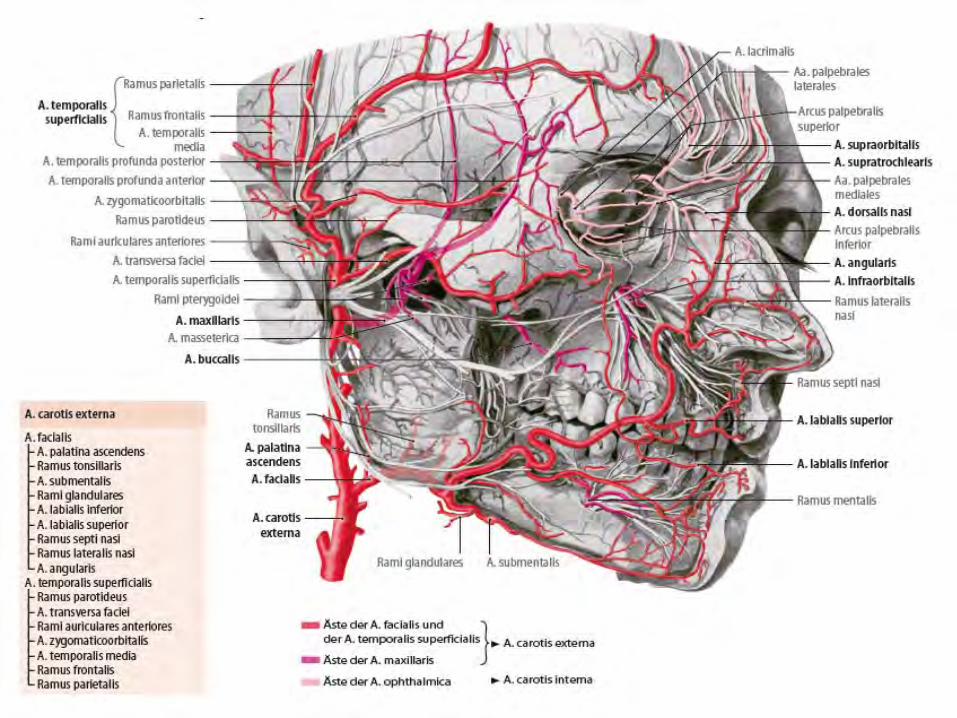
Superficial temporal artery
for gl. parotis, TMJ, m. orbicularis oculi, m. temporalis;
• rr. glandulares• a. transversa faciei
(pro mimické svaly)• rr. auriculares
anteriores(capsule TMJ)
• a. zygomaticoorbitalis• a. temporalis media• r. frontalis• r. parietalis
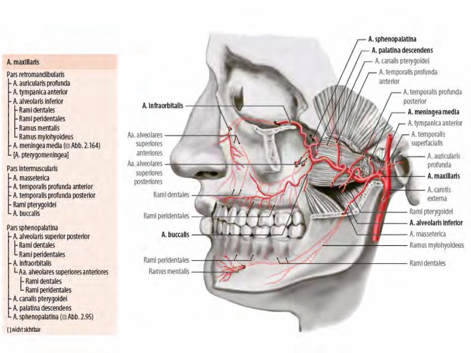
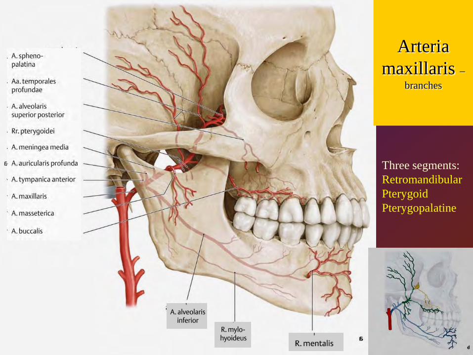
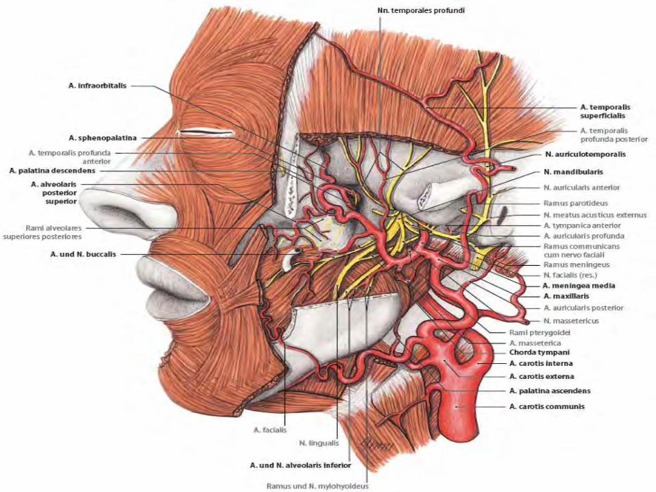
Arteria maxillaris –
branches
Three segments:RetromandibularPterygoidPterygopalatine
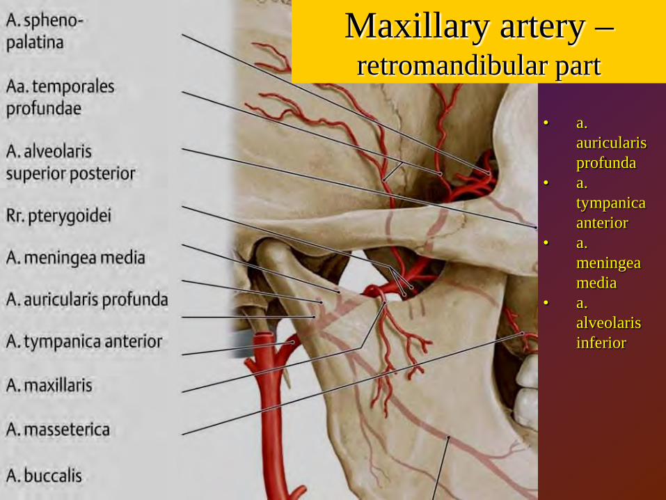
• a. auricularis profunda
• a. tympanica anterior
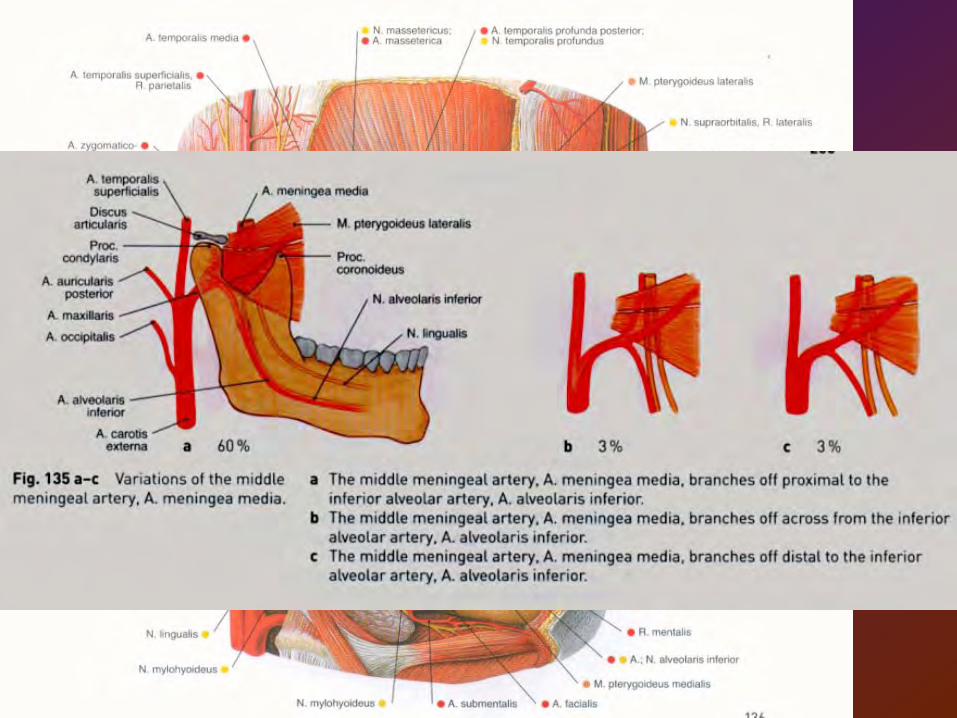
• a. meningea media
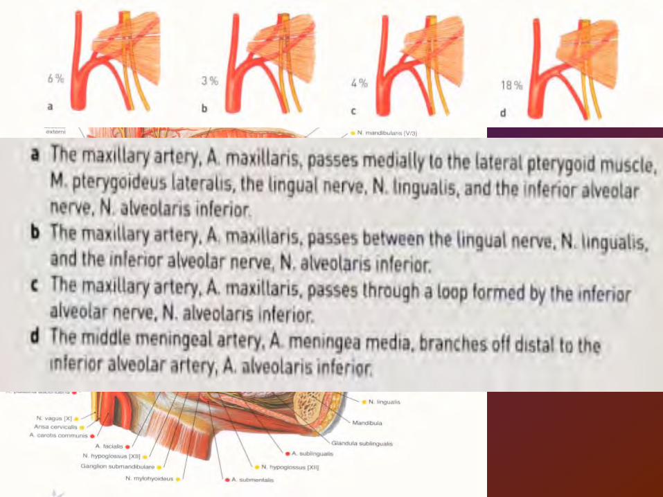
• a. alveolaris inferior
Maxillary artery –retromandibular part
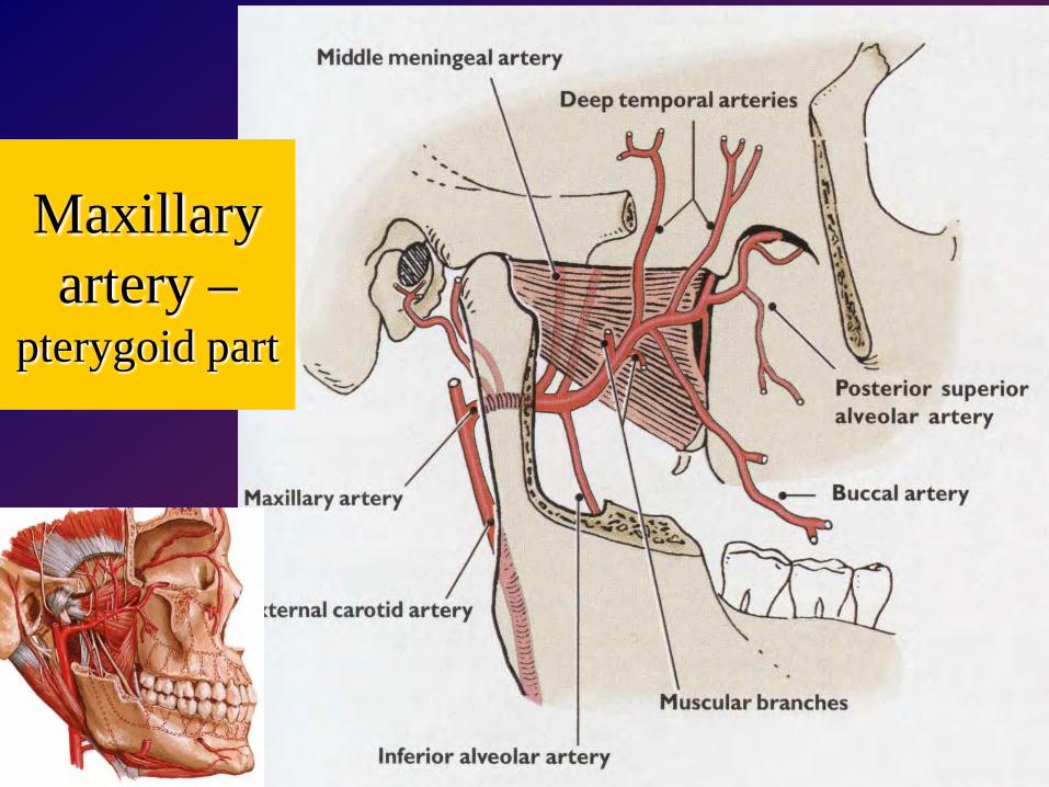
Maxillaryartery –
pterygoid part
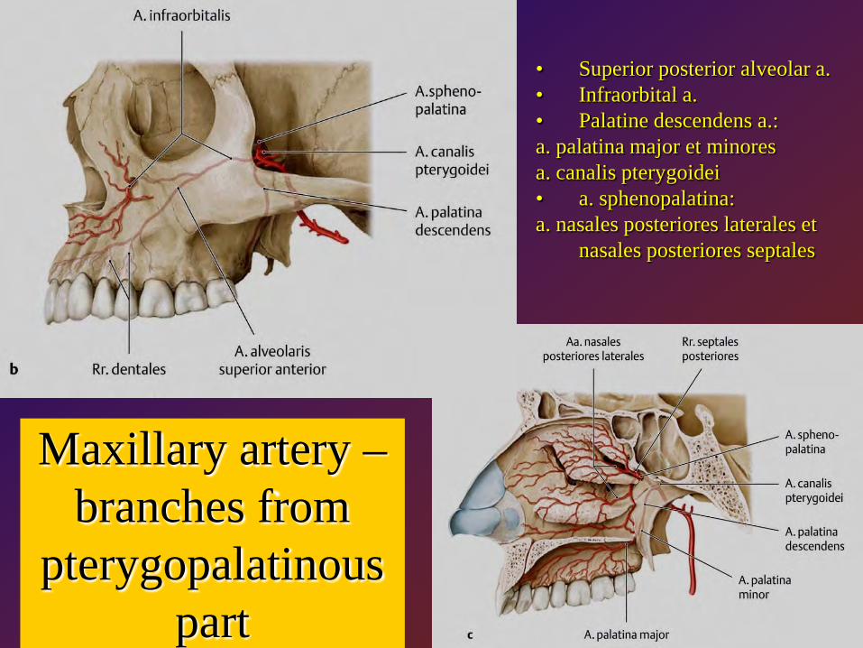
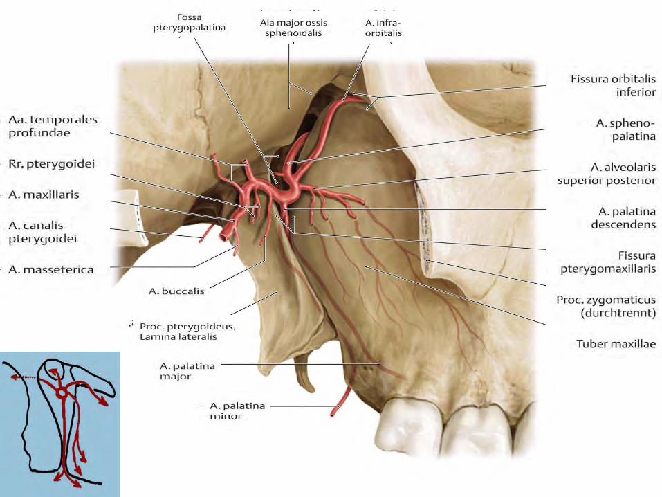
Maxillary artery –branches from
pterygopalatinouspart
• Superior posterior alveolar a. • Infraorbital a. • Palatine descendens a.: a. palatina major et minoresa. canalis pterygoidei• a. sphenopalatina: a. nasales posteriores laterales et
nasales posteriores septales
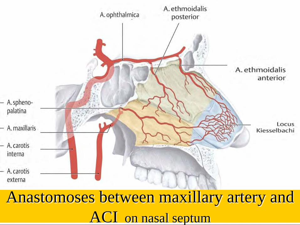
Anastomoses between maxillary artery andACI on nasal septum
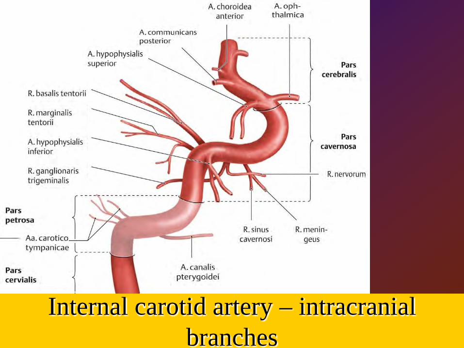
Internal carotid artery – intracranialbranches
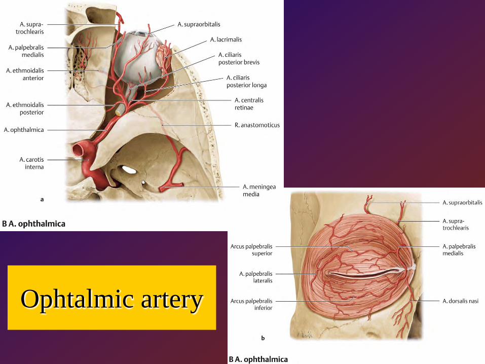
Ophtalmic artery
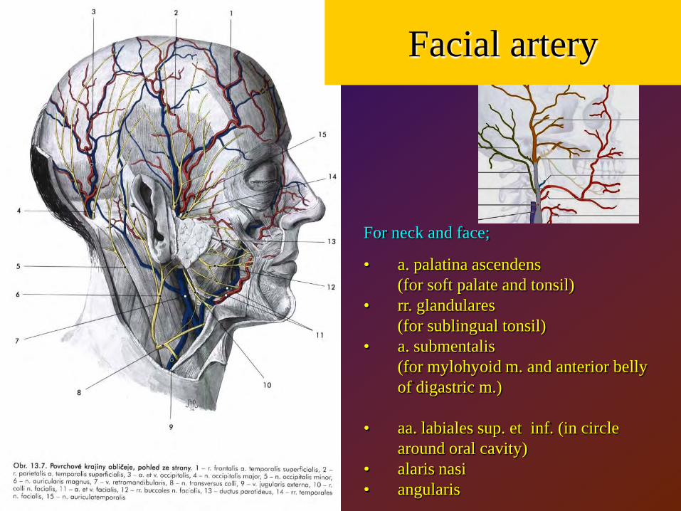
Facial artery
For neck and face;
• a. palatina ascendens(for soft palate and tonsil)
• rr. glandulares(for sublingual tonsil)
• a. submentalis(for mylohyoid m. and anterior belly of digastric m.)
• aa. labiales sup. et inf. (in circlearound oral cavity)
• alaris nasi• angularis
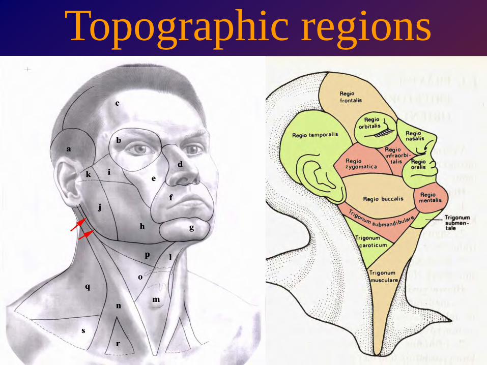
Topographic regions
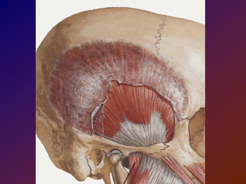
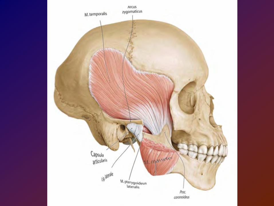
Temporal space (fossa)
lies between skin and the superficial temporal fascia (superficial part of he space)
lies between superficial temporal fascia and(squamous part of the temporal bone)
1
23
4
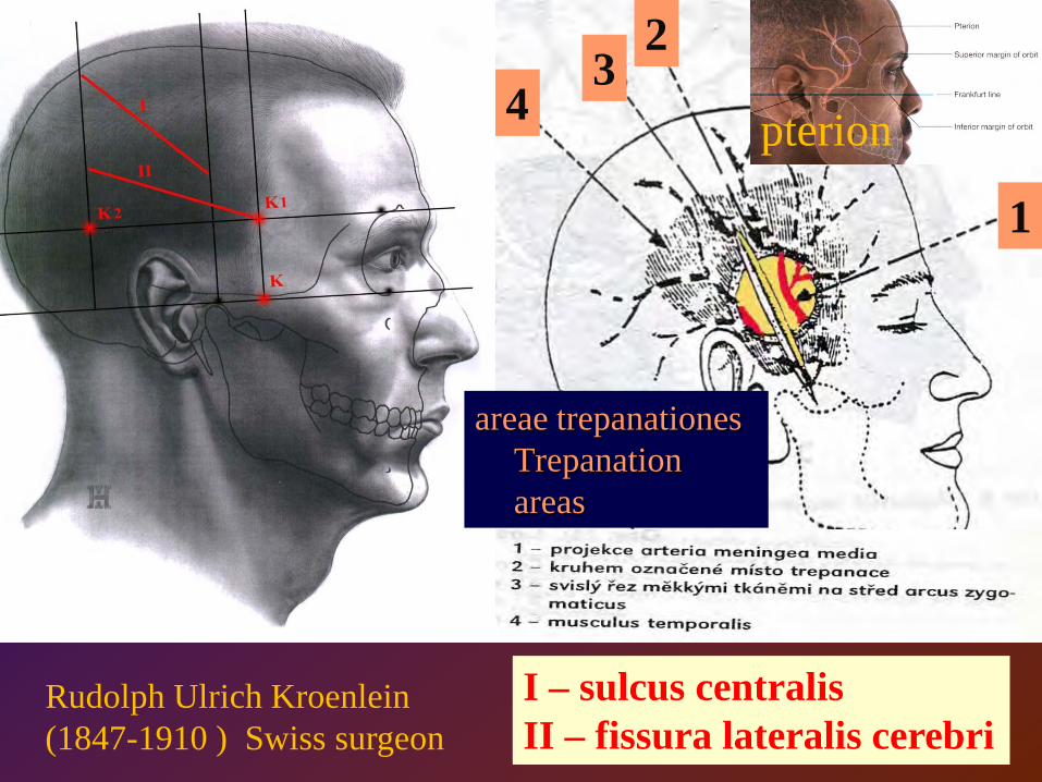
I – sulcus centralisII – fissura lateralis cerebri
Rudolph Ulrich Kroenlein (1847-1910 ) Swiss surgeon
pterion
areae trepanationesTrepanationareas
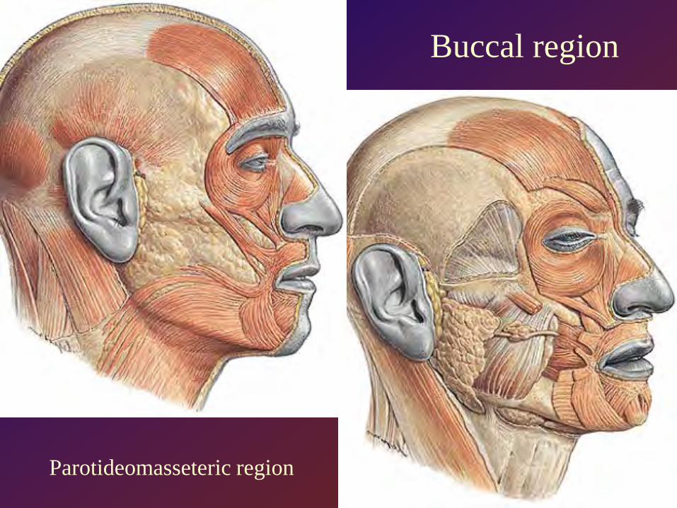
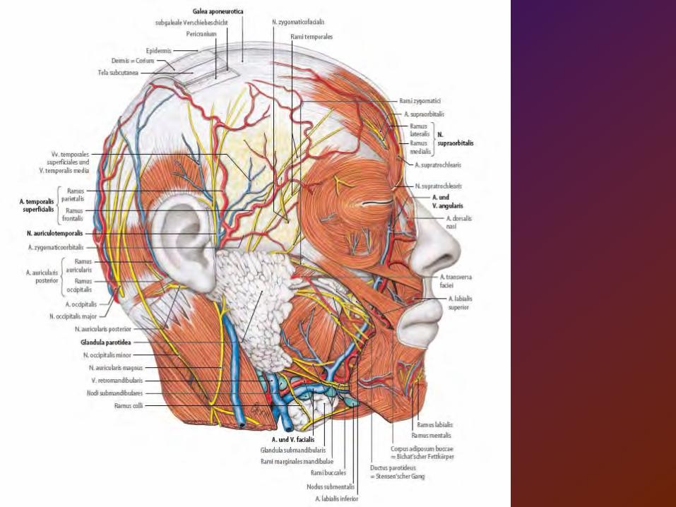
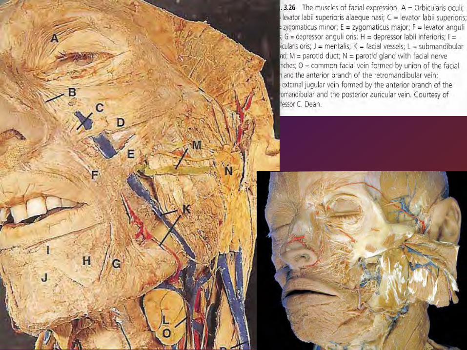
Buccal region
Parotideomasseteric region
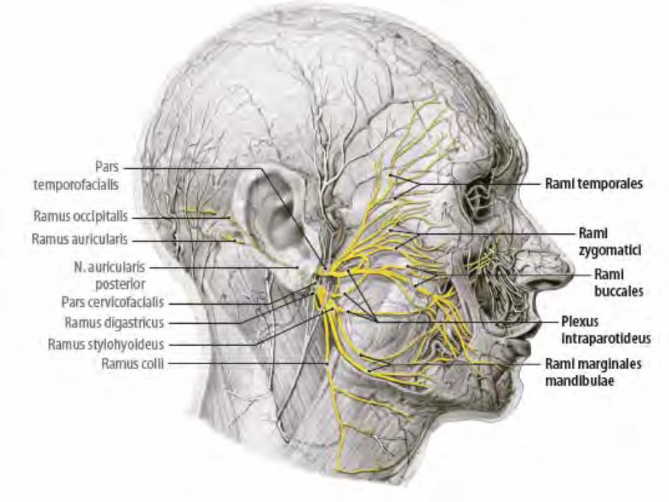
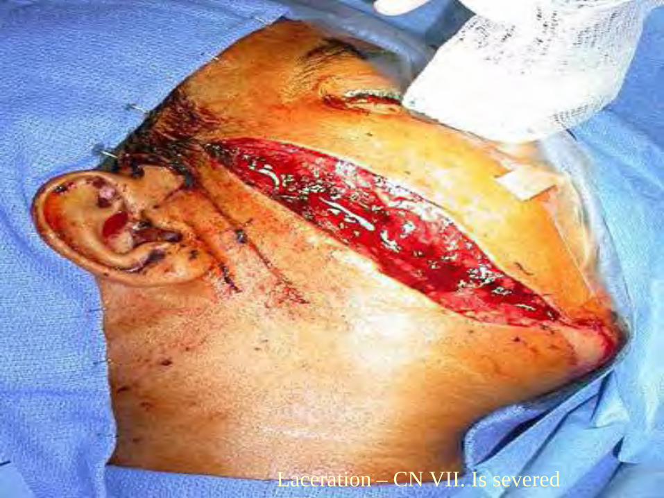
Laceration – CN VII. Is severed
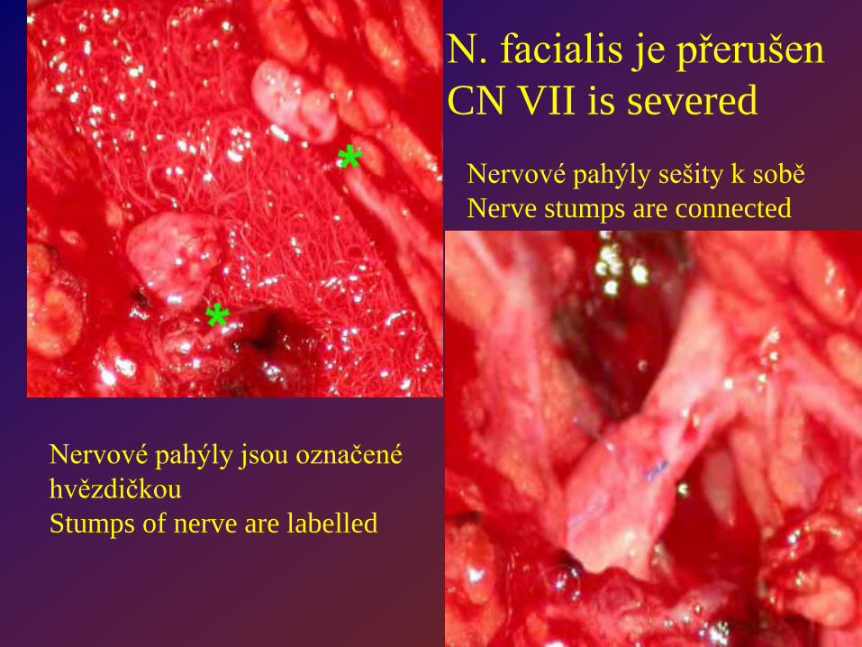
Nervové pahýly jsou označené hvězdičkouStumps of nerve are labelled
Nervové pahýly sešity k soběNerve stumps are connected
N. facialis je přerušen CN VII is severed
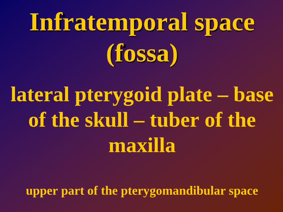
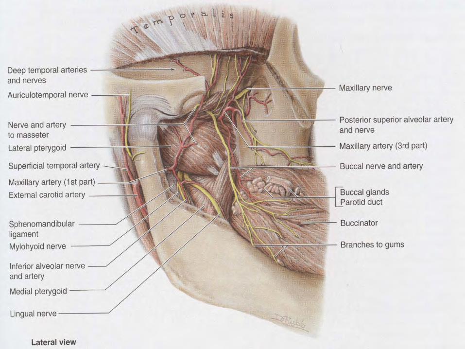
Infratemporal space (fossa)
lateral pterygoid plate – base of the skull – tuber of the
maxilla
upper part of the pterygomandibular space
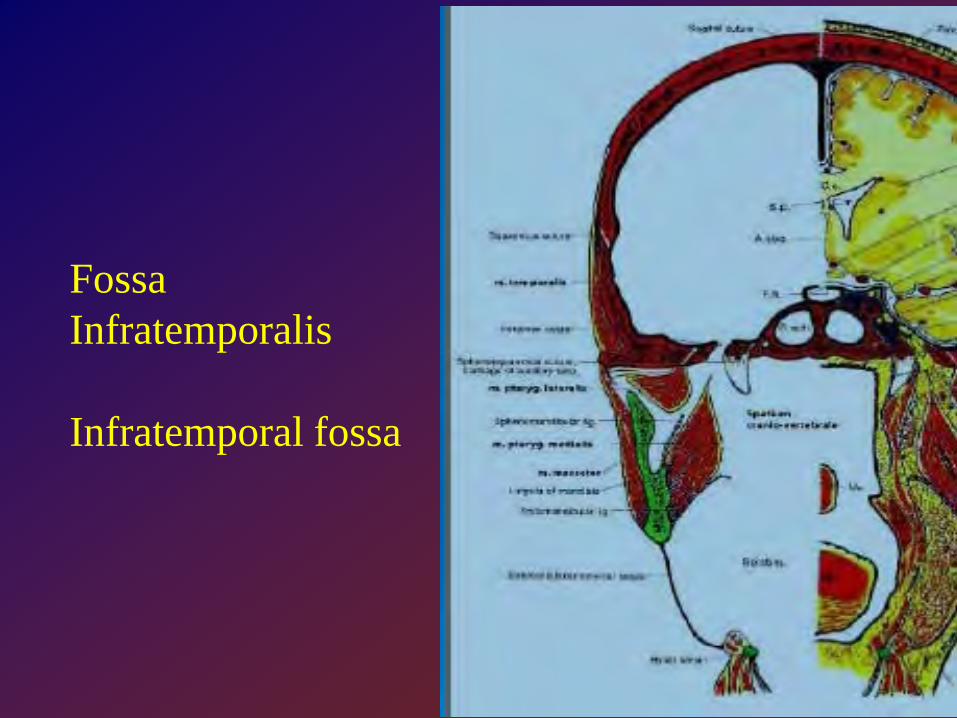
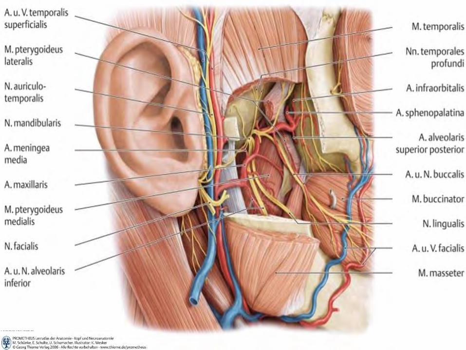
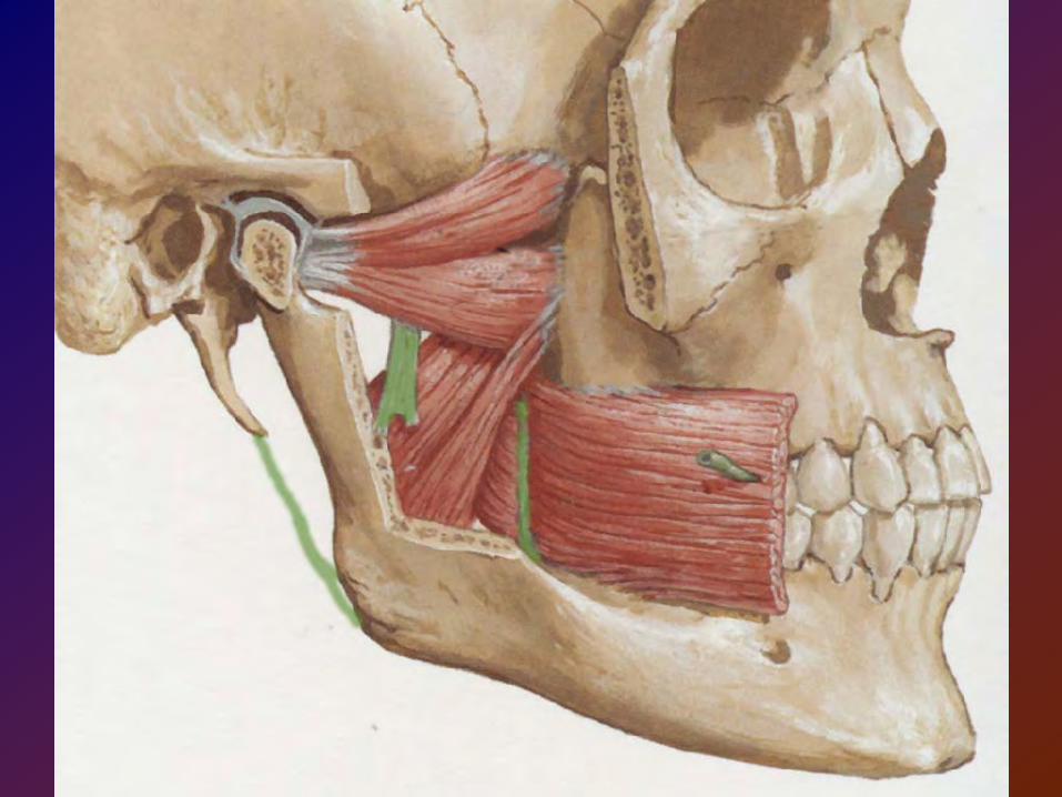
FossaInfratemporalis
Infratemporal fossa
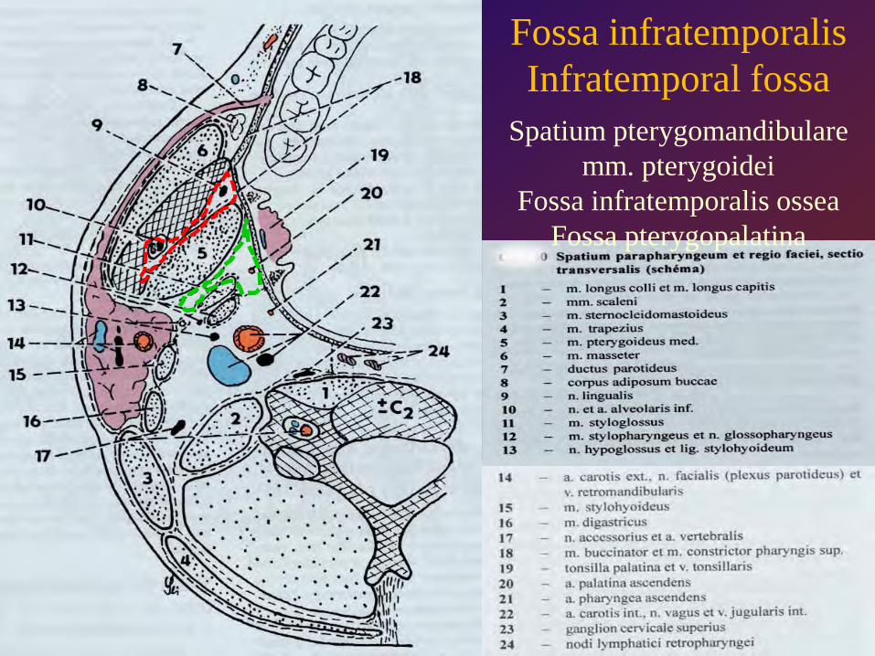
Fossa infratemporalisInfratemporal fossa
Spatium pterygomandibularemm. pterygoidei
Fossa infratemporalis osseaFossa pterygopalatina
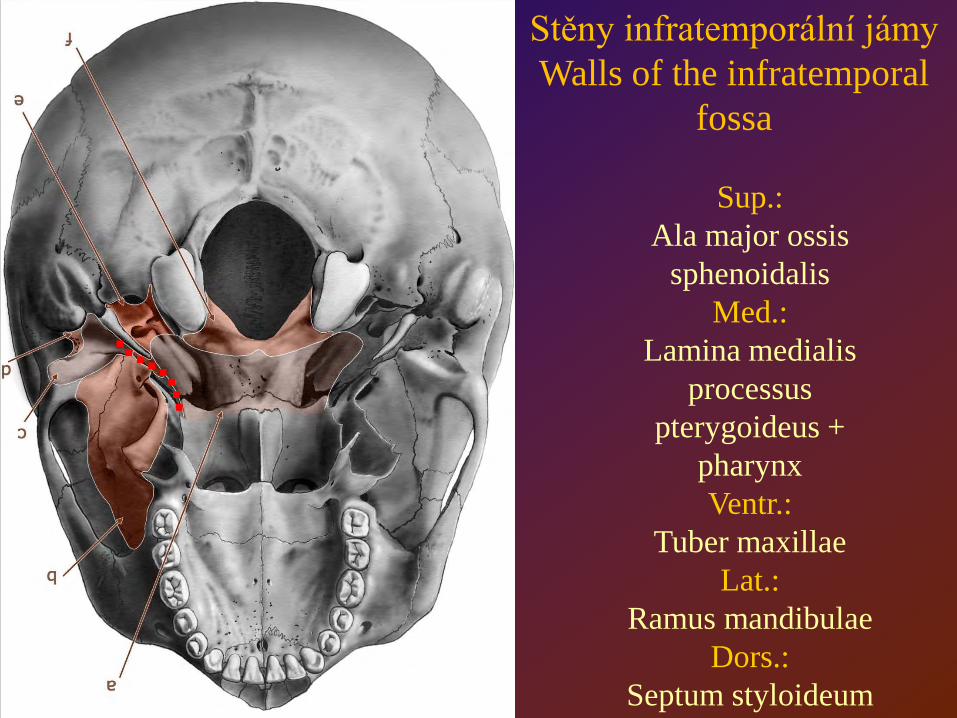
Sup.:Ala major ossis
sphenoidalisMed.:
Lamina medialisprocessus
pterygoideus + pharynxVentr.:
Tuber maxillaeLat.:
Ramus mandibulaeDors.:
Septum styloideum
Stěny infratemporální jámyWalls of the infratemporal
fossa
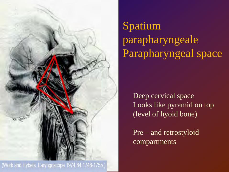
Spatium parapharyngealeParapharyngeal space
Deep cervical spaceLooks like pyramid on top (level of hyoid bone)
Pre – and retrostyloid compartments
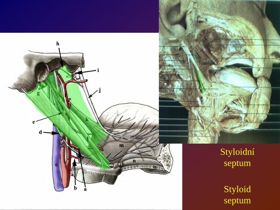
Styloidní septum
Styloid septum
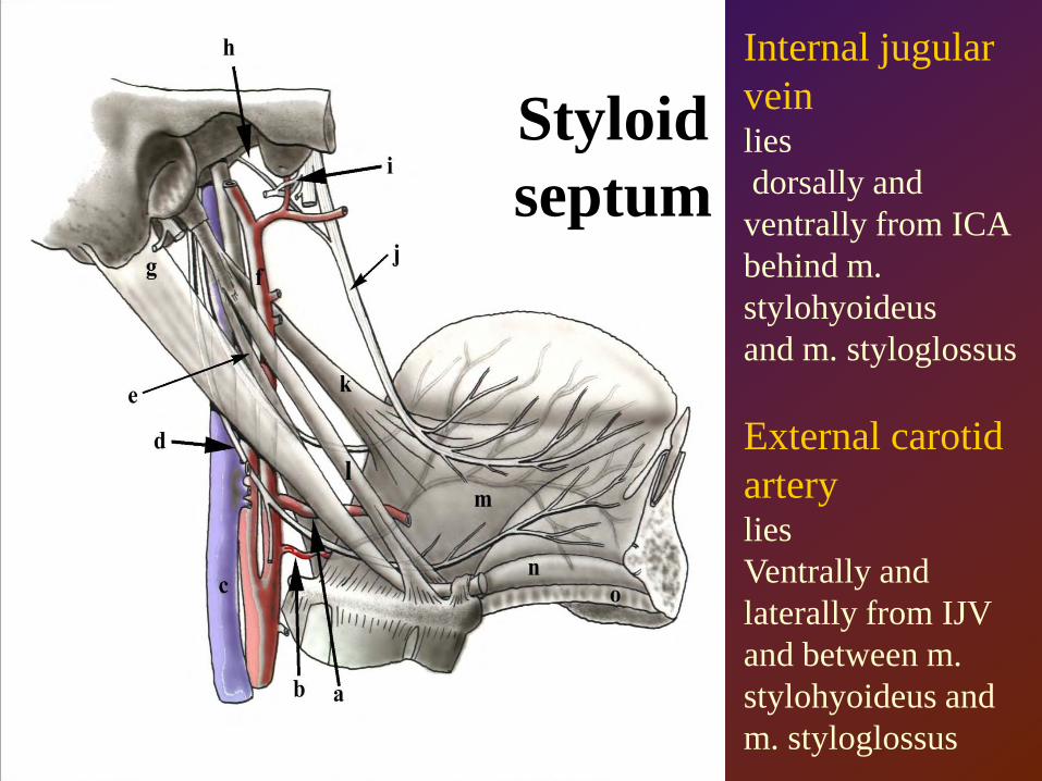
Styloidseptum
Internal jugular veinliesdorsally and ventrally from ICA behind m. stylohyoideus and m. styloglossus
External carotid arterylies Ventrally and laterally from IJV and between m. stylohyoideus and m. styloglossus
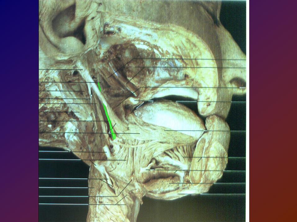
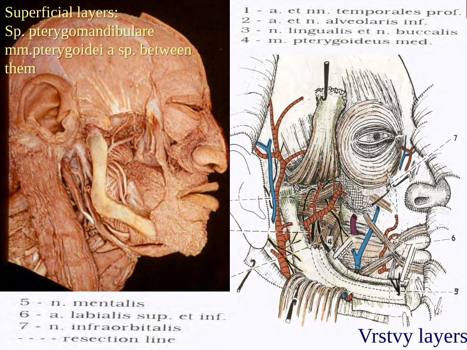
Superficial layers:Sp. pterygomandibularemm.pterygoidei a sp. betweenthem
Vrstvy layers
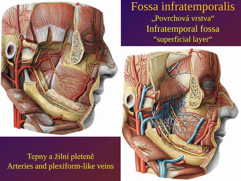
Fossa infratemporalis„Povrchová vrstva“
Infratemporal fossa“superficial layer“
Tepny a žilní pleteněArteries and plexiform-like veins
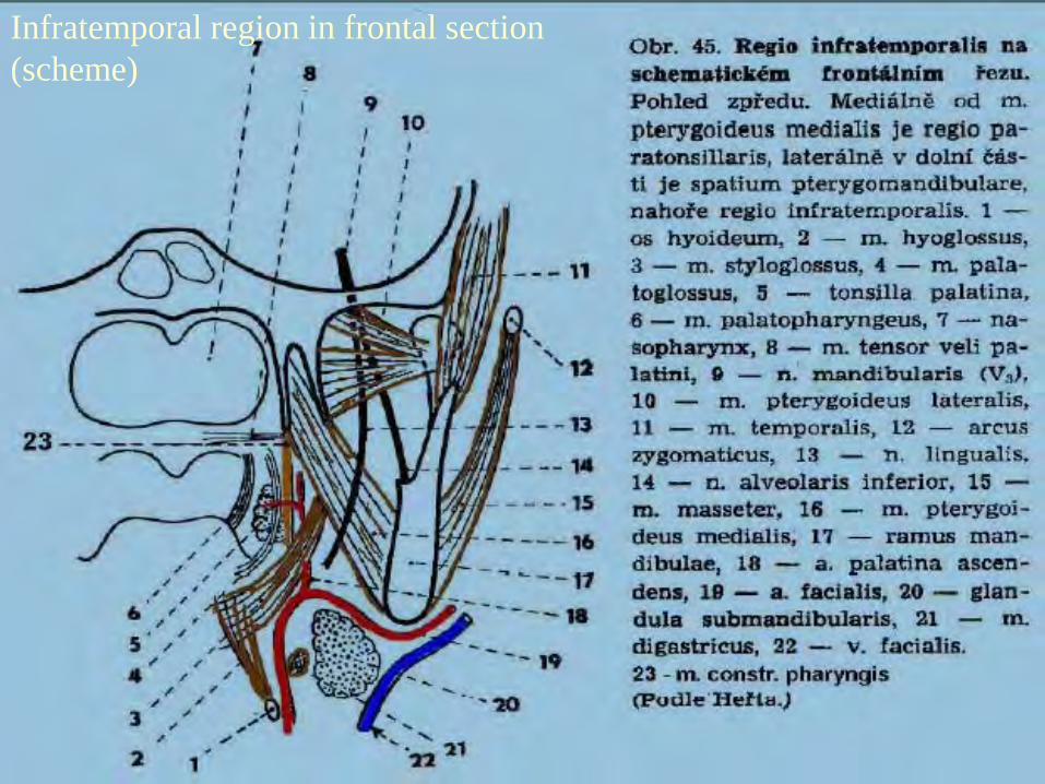
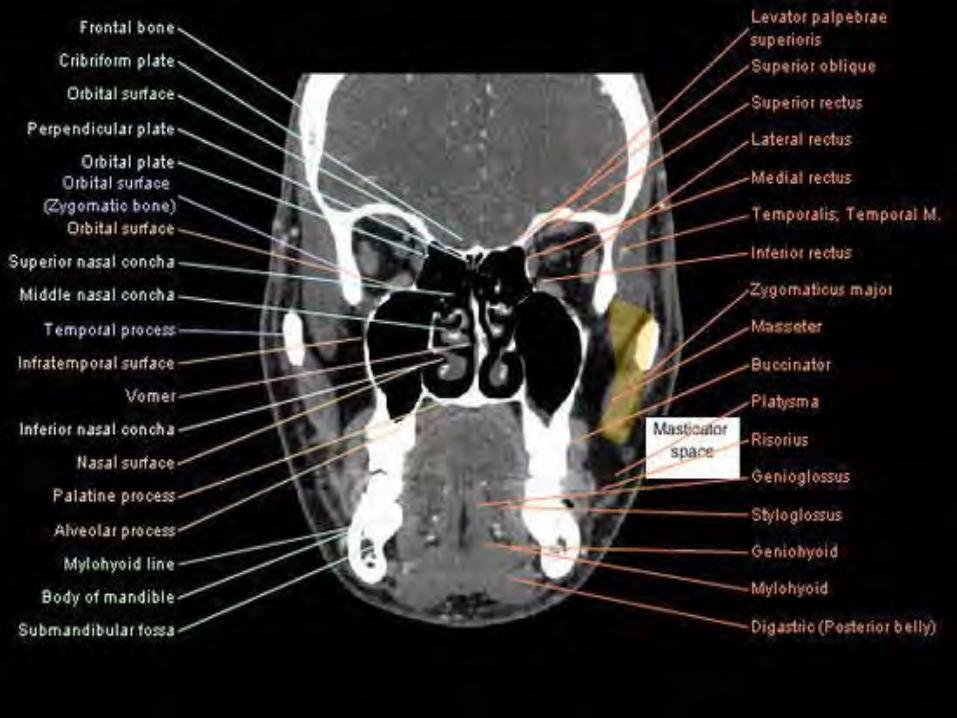
Infratemporal region in frontal section (scheme)
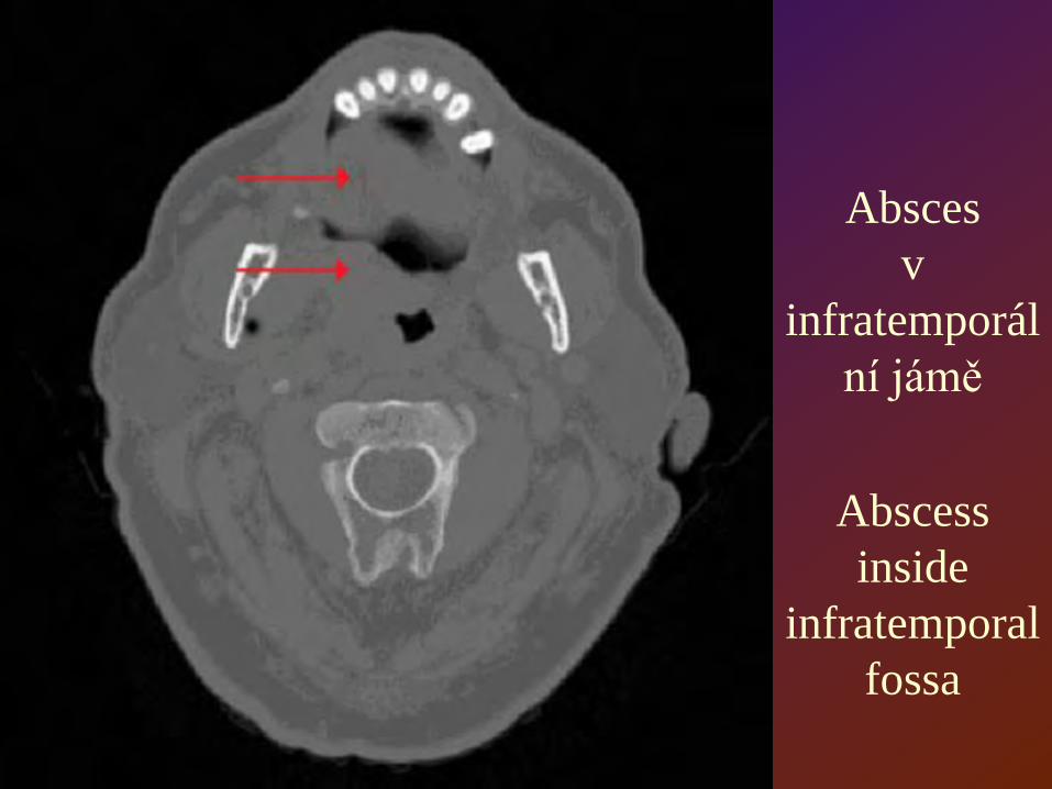
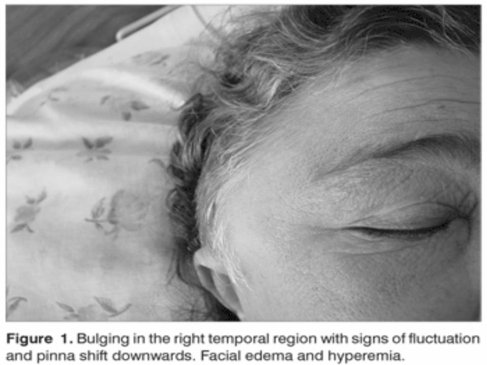
Absces v
infratemporální jámě
Abscess inside
infratemporal fossa
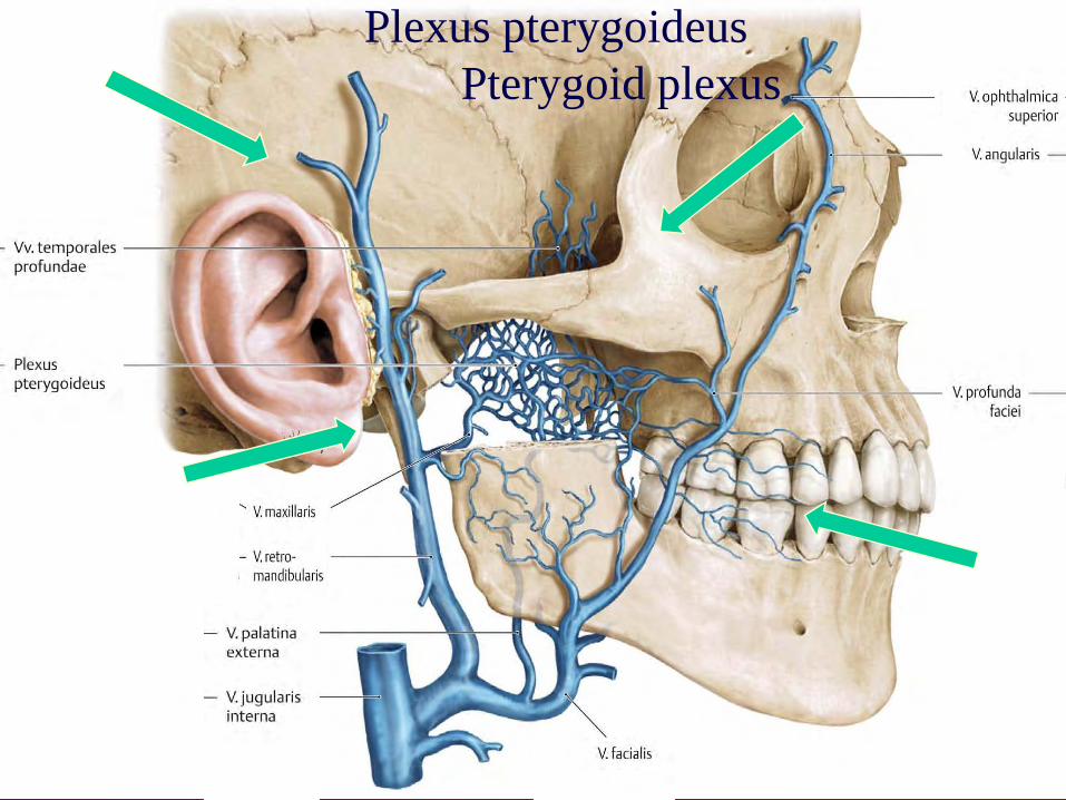
Plexus pterygoideusPterygoid plexus
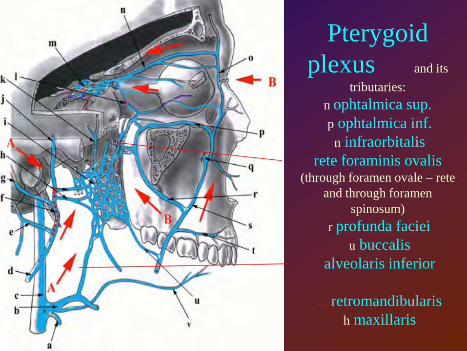
Pterygoid plexus and its
tributaries:n ophtalmica sup. p ophtalmica inf.
n infraorbitalis rete foraminis ovalis
(through foramen ovale – rete and through foramen
spinosum) r profunda faciei
u buccalisalveolaris inferior
... retromandibularis h maxillaris
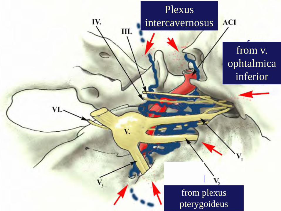
Plexus intercavernosus
from v. ophtalmica
inferior
from plexus pterygoideus
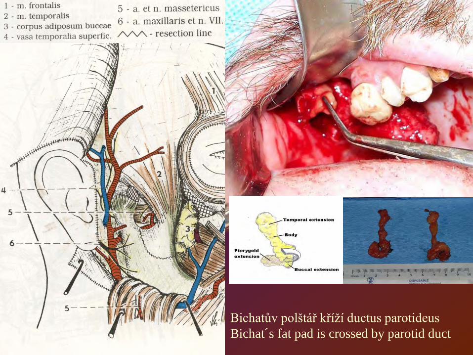
Bichatův polštář kříží ductus parotideusBichat´s fat pad is crossed by parotid duct
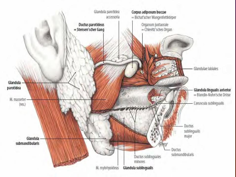
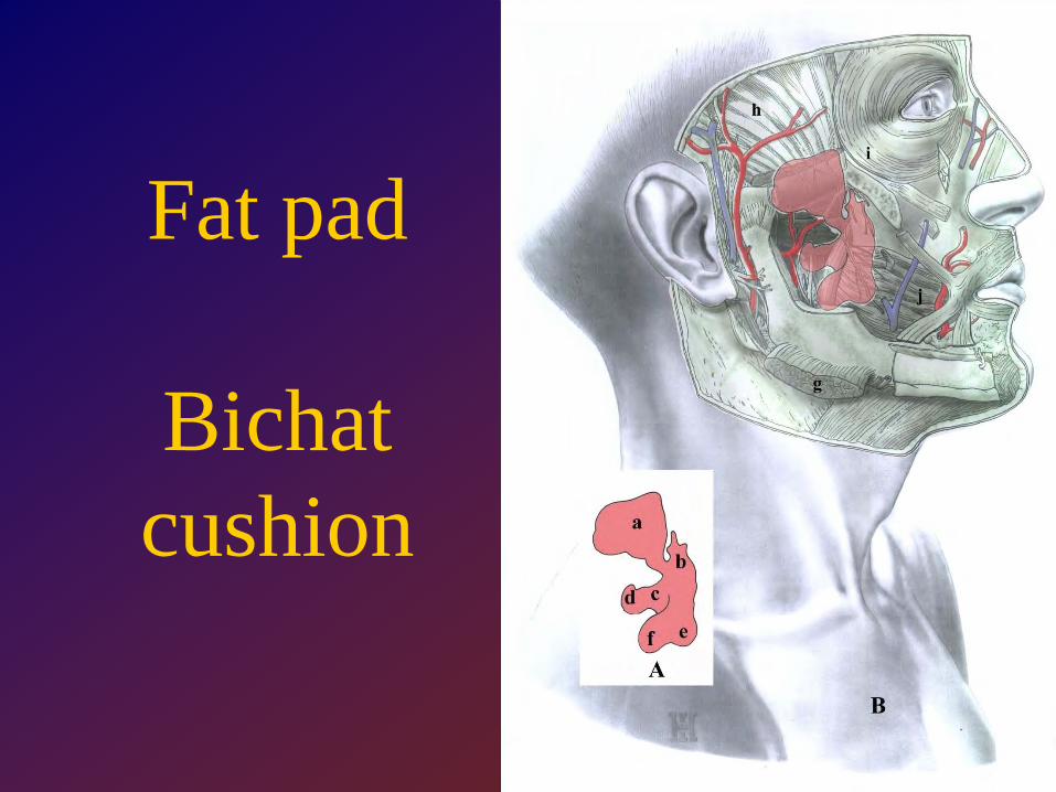
Fat pad
Bichat cushion
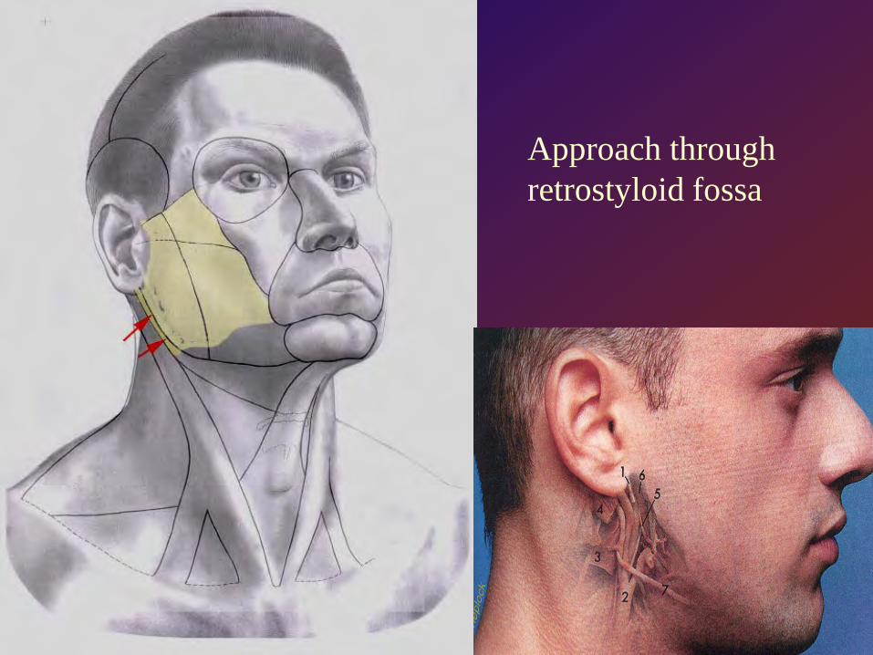
Approach through retrostyloid fossa
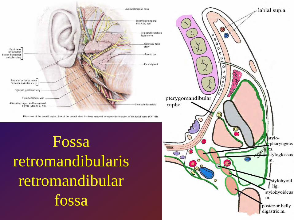
Fossa retromandibularisretromandibular
fossa
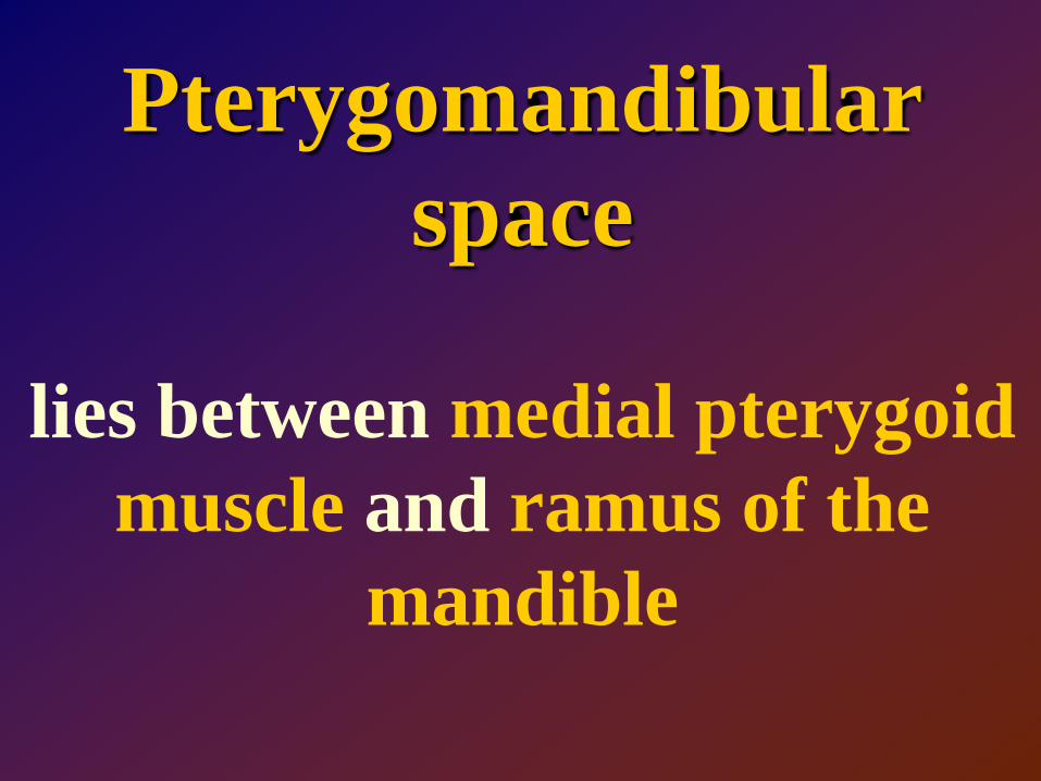
Pterygomandibular space
lies between medial pterygoid muscle and ramus of the
mandible
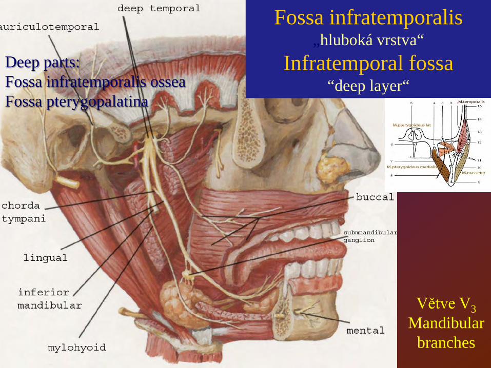
Fossa infratemporalis„hluboká vrstva“
Infratemporal fossa“deep layer“
Větve V3Mandibular
branches
Deep parts:Fossa infratemporalis osseaFossa pterygopalatina
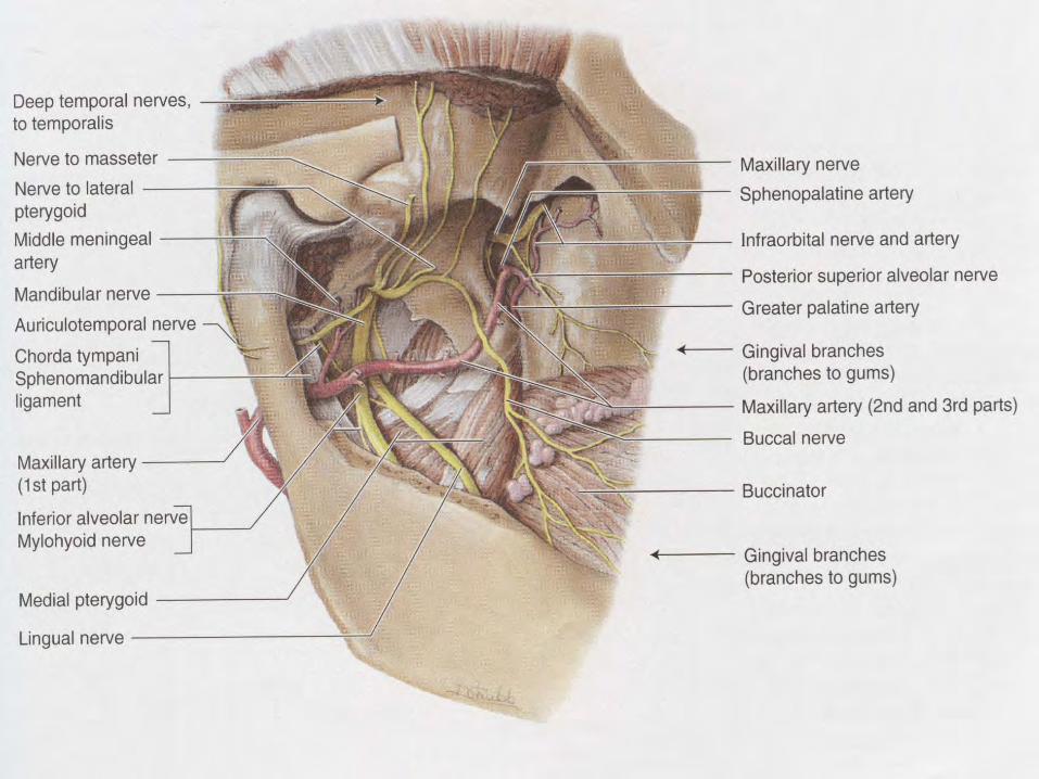

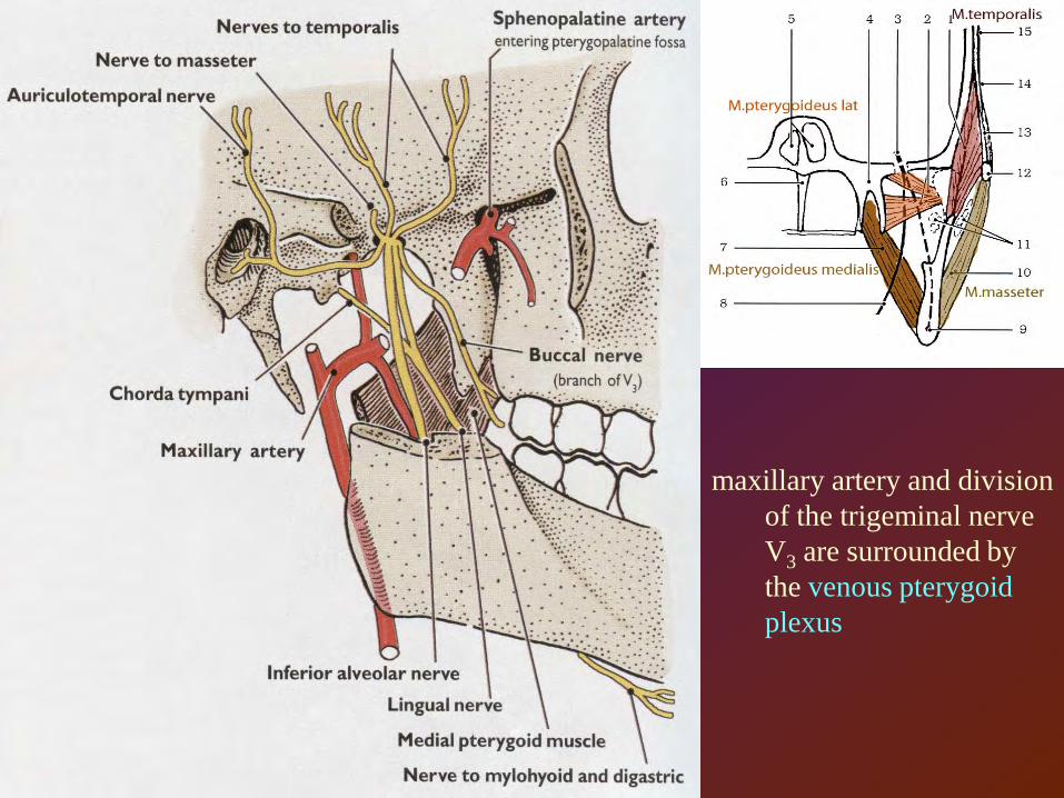
maxillary artery and division of the trigeminal nerve V3 are surrounded by the venous pterygoid plexus
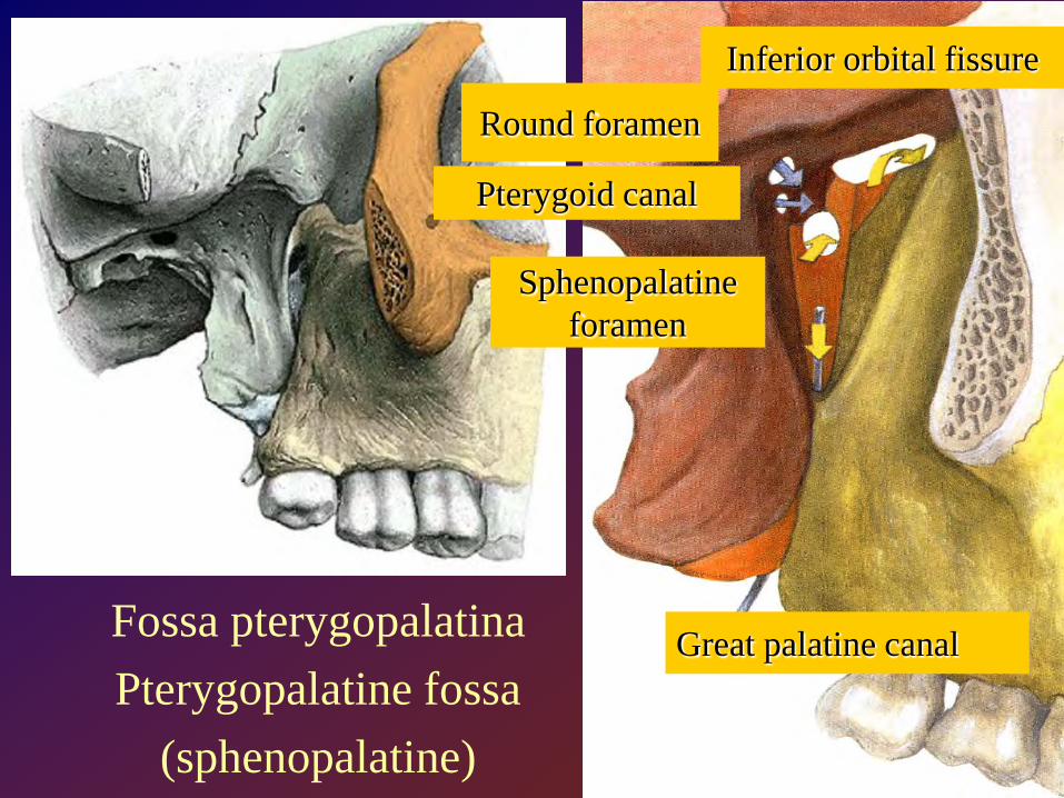
Fossa pterygopalatinaPterygopalatine fossa
(sphenopalatine)
Pterygoid canal
Great palatine canal
Sphenopalatineforamen
Inferior orbital fissure
Round foramen
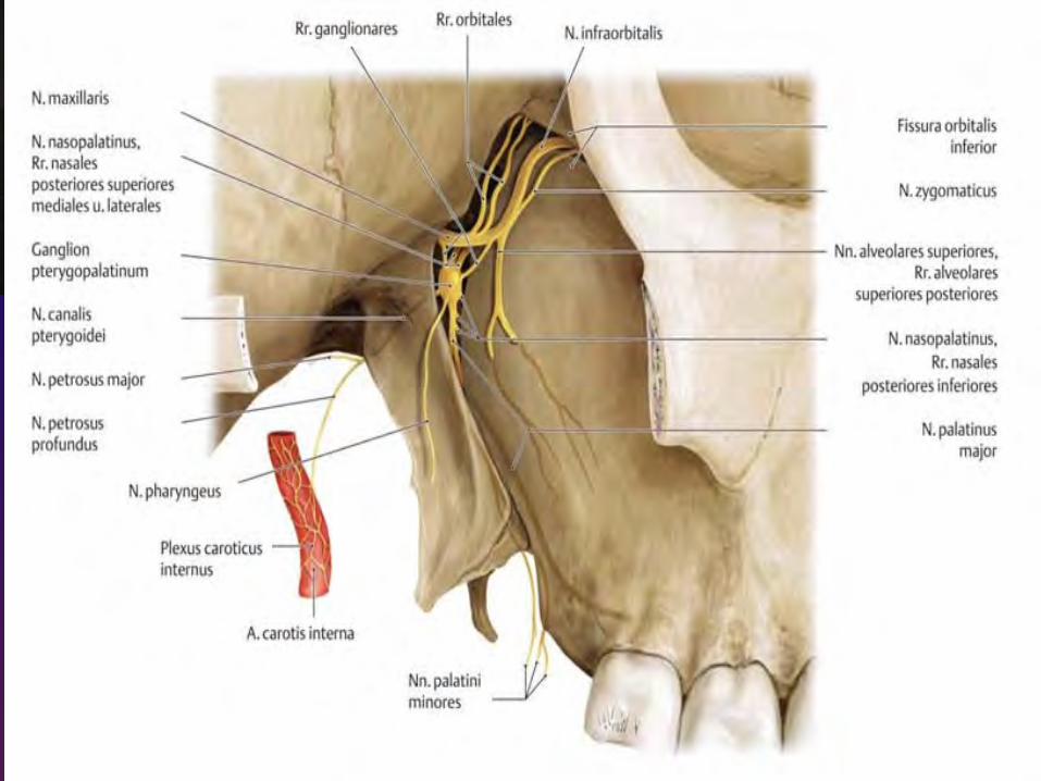
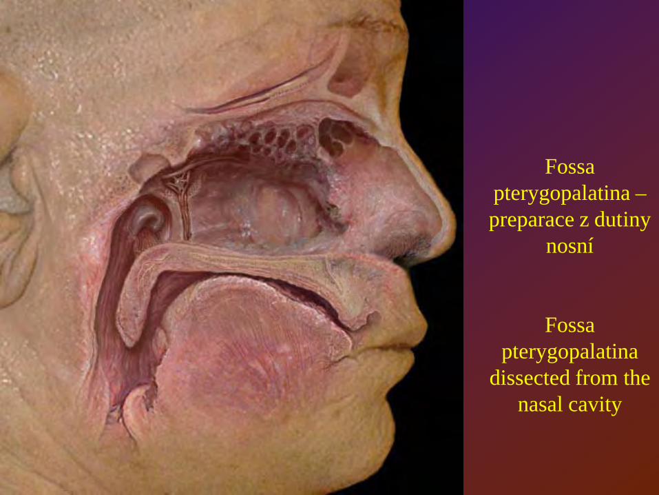
Fossa pterygopalatina –preparace z dutiny
nosní
Fossa pterygopalatina
dissected from the nasal cavity
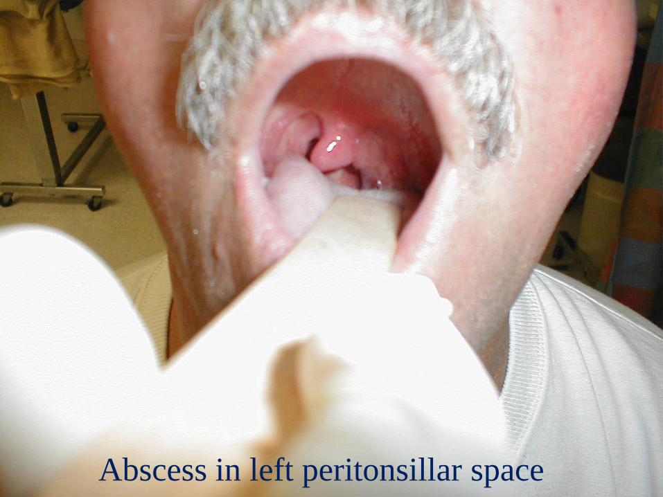
Abscess in left peritonsillar space
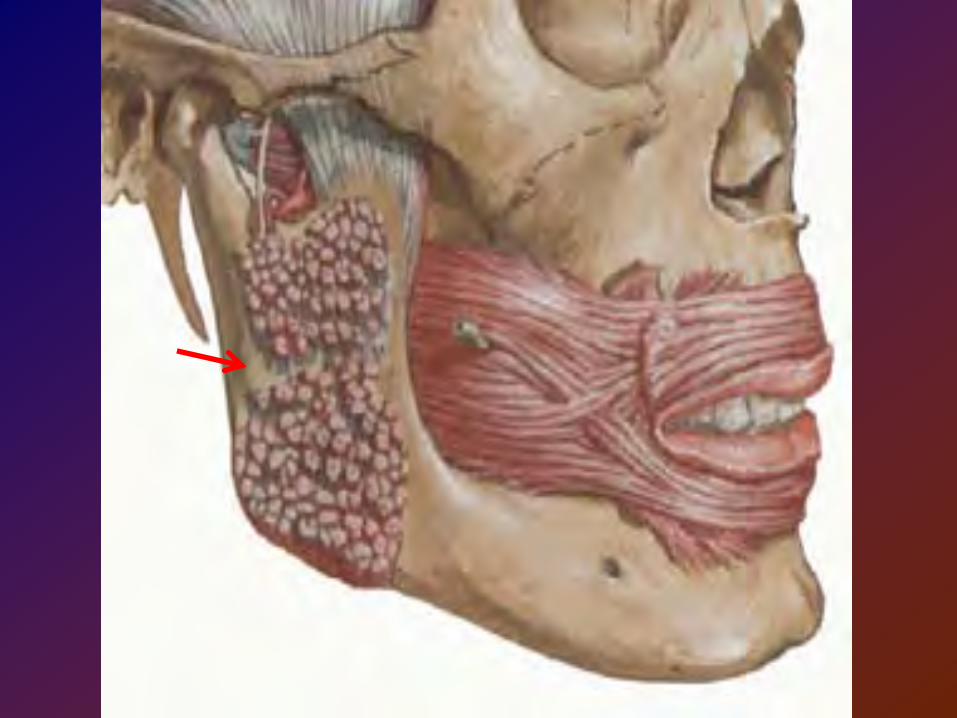
Submasseteric space(massetericomandibular)
lies between masseter andramus of the mandible
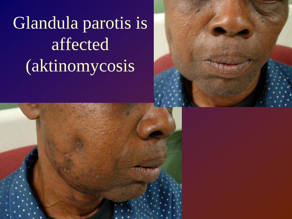
Glandula parotis is affected
(aktinomycosis
Sublingual space
lies in the floor of the mouth between mylohyoid muscle and the oral mucosa
Pus from this space can be accumulated inside canine fossa between levator labii superioris and zygomaticus muscles (facial expression muscle
group)
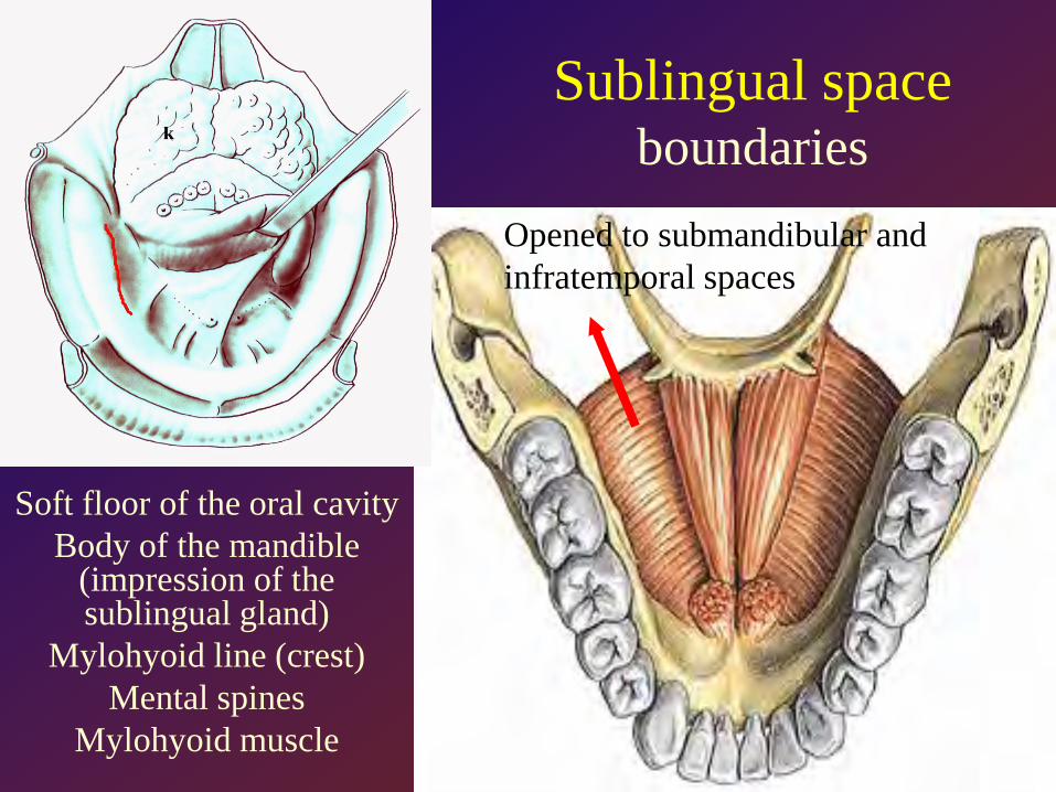
Soft floor of the oral cavityBody of the mandible
(impression of the sublingual gland)
Mylohyoid line (crest)Mental spines
Mylohyoid muscle
Sublingual spaceboundaries
Opened to submandibular and infratemporal spaces
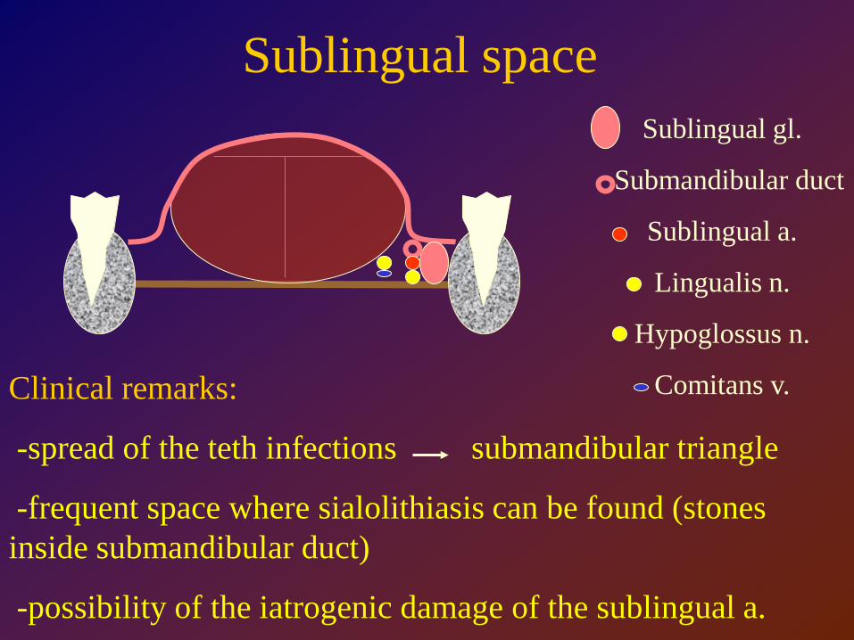
Sublingual space
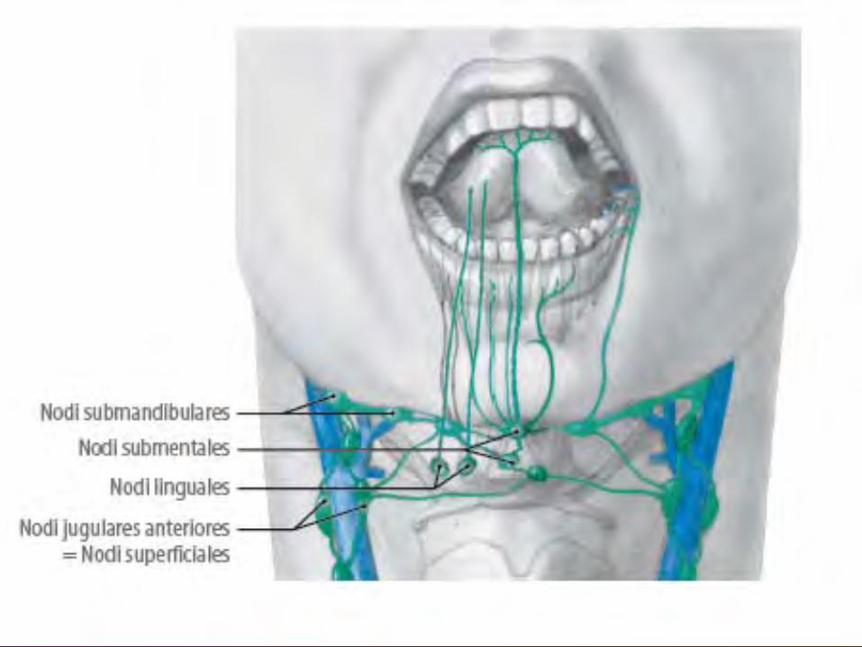
Clinical remarks:
-spread of the teth infections submandibular triangle
-frequent space where sialolithiasis can be found (stones inside submandibular duct)
-possibility of the iatrogenic damage of the sublingual a.
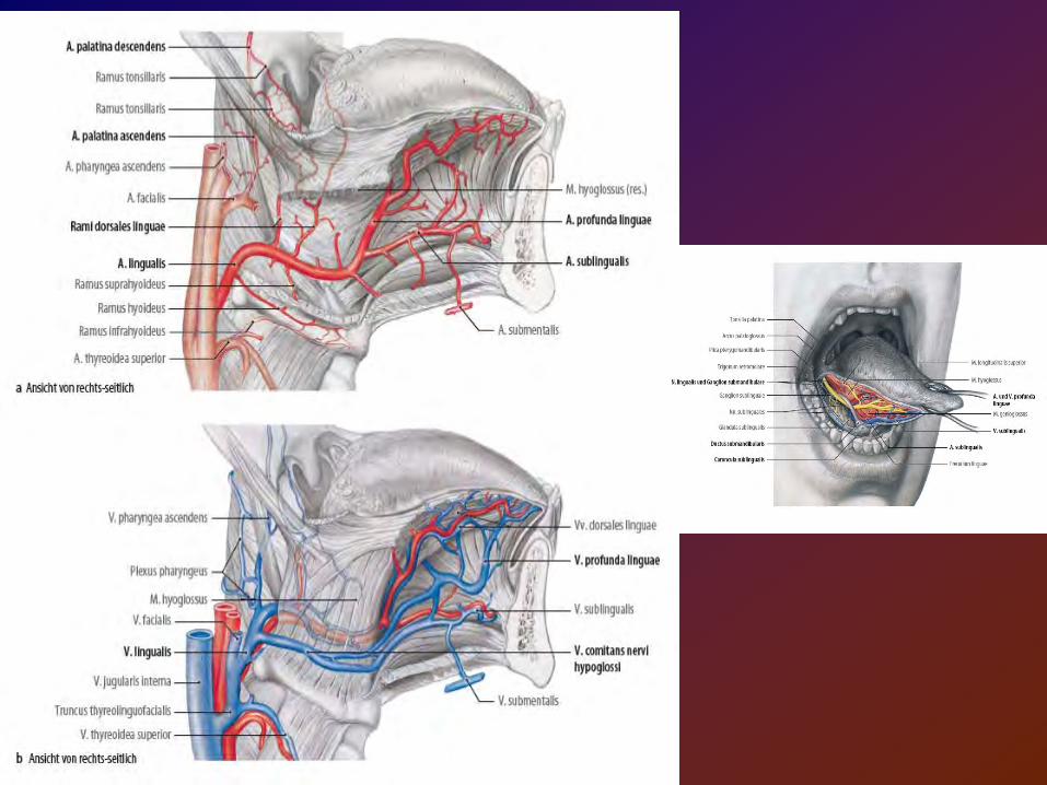
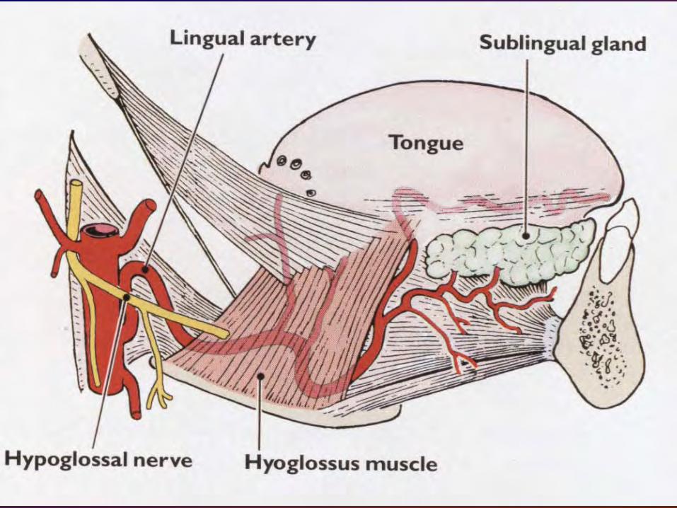
Sublingual gl.
Submandibular duct
Sublingual a.
Lingualis n.
Hypoglossus n.
Comitans v.
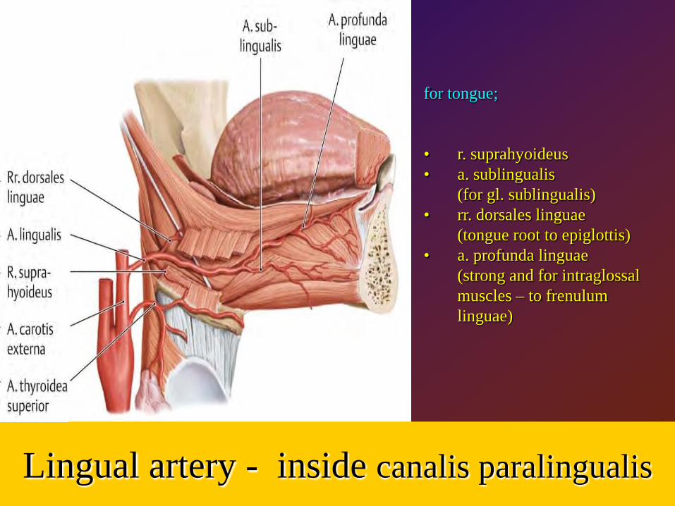
Lingual artery - inside canalis paralingualis
for tongue;
• r. suprahyoideus• a. sublingualis
(for gl. sublingualis)• rr. dorsales linguae
(tongue root to epiglottis)• a. profunda linguae
(strong and for intraglossalmuscles – to frenulumlinguae)
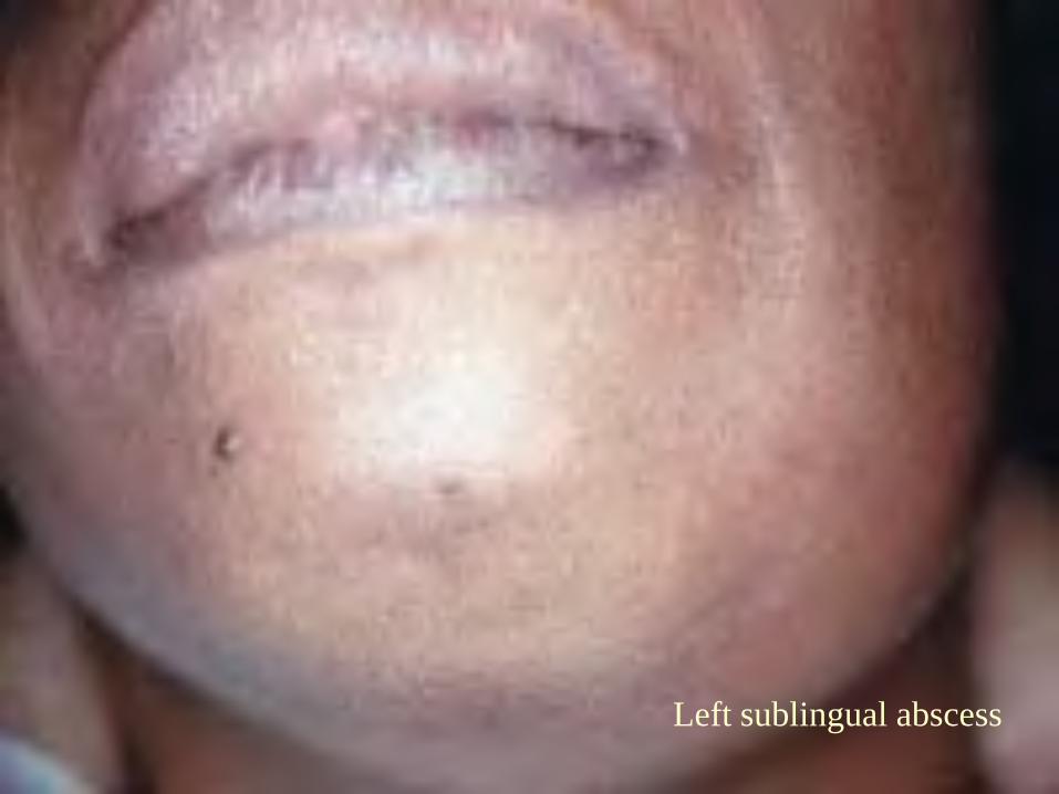
Left sublingual abscess
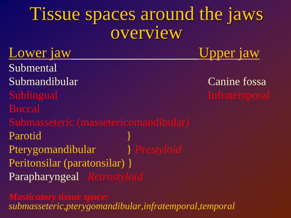
Tissue spaces around the jawsoverview
Lower jaw Upper jawSubmentalSubmandibular Canine fossaSublingual InfratemporalBuccalSubmasseteric (massetericomandibular)Parotid }Pterygomandibular } PrestyloidPeritonsilar (paratonsilar) }Parapharyngeal Retrostyloid
Masticatory tissue space:submasseteric,pterygomandibular,infratemporal,temporal