expression differentiated functions in hepatoma cell ... · tatwas assayed as described (1), in...
TRANSCRIPT
Proc. Nat. Acad. Sci. USAVol. 68, No. 12, pp. 3026-3030, December 1971
Expression of Differentiated Functions in Hepatoma Cell Hybrids: Reappearanceof Tyrosine Aminotransferase Inducibility after the Loss of Chromosomes*
(dexamethasone/actinomycin D/chromosome numbers/logarithmic growth phase)
MARY C. WEISS AND MICHELE CHAPLAIN
Centre de G~n~tique Mol6culaire, C.N.R.S., 91, Gif-sur-Yvette, France
Communicated by Boris Ephrussi, September 9, 1971
ABSTRACT Hybrids from a cross of rat hepatoma cellswith diploid epithelial cells from rat liver have been studiedwith respect to karyotype and expression of two functionslimited to the hepatoma parent: high level of the enzymetyrosine aminotransferase (EC 2.6.1.5; L-tyrosine: 2-oxo-glutarate aminotransferase) and its inducibility withsteroid hormones. The hybrids that contain the completechromosomal complements from both parents show lowenzyme activity and no inducibility. One hybrid clone,and all of its derivatives, which have lost 30-40% of thechromosomes initially present, show enzyme inducibility.Induction of tyrosine aminotransferase in the hepatomaand hybrid cells responds similarly to inhibition by cyclo-heximide and actinomycin D, and to steroid concentration.The enzymes from induced and noninduced hepatomacells and from induced hybrid cells are similar in heatsensitivity and intracellular distribution; those from non-induced hybrid and diploid rat epithelial cells are different.
In the first paper of this series (1), it was shown that somatichybrids between mouse fibroblasts and well differentiatedrat hepatoma cells fail to exhibit high baseline levels andsteroid-mediated induction of the enzyme tyrosine amino-transferase (EC 2.6.1.5; L-tyrosine:2-oxoglutarate amino-transferase; TAT). Both a high baseline and inducibility ofTAT are characteristics of hepatic differentiation and areunique to the hepatoma parent. These results are similar tothose reported for hybrids from several other crosses betweencells exhibiting one or more specialized functions and fibro-blasts (2, 3).The study of the expression of TAT in somatic hybrids
has been extended to hybrid cells from a new cross: rat hepa-toma cells x epithelial cells derived from rat liver. Althoughthese hybrid cells have properties similar to those previouslyreported, the use of selective conditions has permitted theisolation of numerous hybrid subclones that have undergoneextensive chromosome losses, and several of these, in spiteof a low basal level of TAT, have been found to show enzymeinduction.
MATERIALS AND METHODS
Cultures were maintained as described (1). Chemicals werepurchased from Sigma or Calbiochem; actinomycin D wasobtained from Merck Co.
Fu5-5 is a subclone of the line Fu5 (1), derived from theReuber H35 (4) rat hepatoma line H4IIEC3 of Pitot et al.
Abbreviation: TAT, tyrosine aminotransferase (L-tyrosine: 2-oxoglutarate aminotransferase).* This is the third paper of a series: "Expression of Differ-entiated Functions in Hepatoma Cell Hybrids". The first paperis ref. 1.
(5). BRL-1 is a subclone of line BRL-1D1 (generously sup-plied by Dr. Hayden Coon), one of the many diploid epi-thelial lines established by direct cloning from the liver of ayoung Buffalo rat (6). Although several of these lines havebeen reported to express some differentiated liver functions(6), none has been detected in BRL-1; these include aldolaseB (EC 4.1.2.7) and alcohol dehydrogenase (EC 1.1.1.1) (22)as well as TAT. Whether BRL-1 cells are "dedifferentiated"hepatocytes or from some other epithelial component of theliver is unknown.Normal chromosomes of the rat can be divided into several
groups: large submetacentric, small metacentric, and telocen-tric; 2 long satellited chromosomes have been grouped amongthe telocentrics. One marker, a large metacentric chromosome,is found in all cells of the parental hepatoma line.
Cell fusion was obtained by treatment of a mixture of equalnumbers of parental cells with 300 hemagglutinating units ofUV-inactivated Sendai virus. The cell suspension was platedat low density in normal medium. Colonies of large cells wereisolated and subcultured; karyotypic analysis showed thatone of these colonies was hybrid.
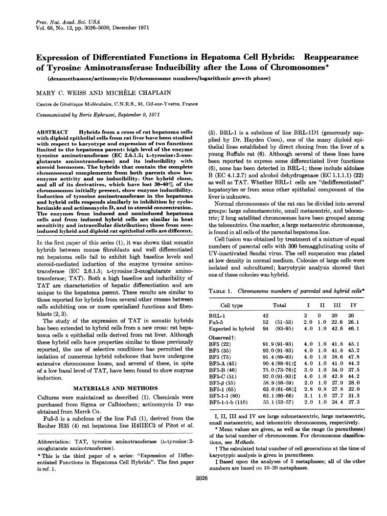
TABLE 1. Chromosome numbers of parental and hybrid cells*
Cell type Total I II III IV
BRL-1 42 2 0 20 20Fu5-5 52 (51-53) 2.0 1.0 22.6 26.1Expected in hybrid 94 (93-95) 4.0 1.0 42.6 46.1
Observedt:BF5 (22) 91.9(91-93) 4.0 1.0 41.8 45.1BF5 (35) 92.0(91-93) 4.0 1.0 41.8 45.2BF5 (75) 91.4(89-93) 4.0 1.0 38.6 47.8BF5-A(45) 90.4(88-91)4 4.0 1.0 41.0 44.2BF5-B (46) 75.0 (73-76)4 3.0 1.0 34.0 37.5BF5-C (51) 92.0(91-93)4 4.0 1.0 42.8 44.2BF5-,8 (55) 58.9(58-59) 2.0 1.0 27.9 28.0BF5-1 (65) 63.0 (61-68)4 2.8 0.8 27.8 32.0BF5-1-1 (80) 63.1(60-66) 3.1 1.0 27.7 31.3BFS-1-1-b (110) 55.1(52-57) 2.0 1.0 24.4 27.3
I, II, III and IV are large submetacentric, large metacentric,small metacentric, and telocentric chromosomes, respectively.
* Mean values are given, as well as the range (in parentheses)of the total number of chromosomes. For chromosome classifica-tions, see Methods.
t The calculated total number of cell generations at the time ofkaryotypic analysis is given in parentheses.
t Based upon the analyses of 5 metaphases; all of the othernumbers are based on 10-20 metaphases.
3026
Differentiated Functions in Hepatoma Hybrids 3027
TABLE 2. Activity and inducibility of tyrosine aminotransferase during the growth cycle
Specific activity of tyrosine aminotransferase*
Assay It Assay IItNo. of cells per10-cm dish (X 106) 5 hr 24 hr 5 hr 24 hr
Cell type Inoculum Assay It Assay IIt C§ I§ C I C I C I
BRL-1 0.4 1.8 2.1 0.2 0.3 -s 0.2 - 0.7 0.6BF5-C 0.2 1.2 2.7 0.4 0.4 0.4 0.4 0.4 0.6 0.4 0.5BF5-,B 0.1 0.75 2.3 0.6 0.7 0.2 0.3 0.3 0.9 0.2 0.3BF5-1-1 0.2 4.1 17.2 0.5 4.3 0.6 3.9 0.4 5.7 0.4 1.3BF5-1-1-b 0.2 4.0 12.0 1.1 1.6 9.2 0.2 3.0 - 8.4FuS-5 0.2 2.9 10.4 16.9 144.5 21.0 583.0 41.6 171.0 38.5 224.0
* One unit of TAT activity equals 1 Amol of p-hydroxyphenylpyruvate formed per min at 37°C. Specific activity is expressed as milli-units per mg of protein.
t Assay I: cultures in early logarithmic growth phase, 3-4 days after inoculation.t Assay II: cultures in late logarithmic or stationary phase, 6-7 days after inoculation.§ C: control or noninduced; I: induced (1 /AM dexamethasone).¶ -, Not assayed.
Cells were induced with 1 1AM dexamethasone, added tofresh growth medium, containing the standard 5% fetal calfserum. At the start of each experiment, cultures were re-newed with fresh growth or induction medium; in most ex-periments, control and induced cultures were harvestedsimultaneously in order to correct for possible effects ofmedium renewal on TAT activity.
Cultures were collected, enzyme extracts were prepared,protein was determined, and TAT specific activity was de-fined as described (1). For the separation of soluble andmitochondrial TAT activity, cells were homogenized in thestandard TAT buffer (7), centrifuged, and washed twice(12,000 X g for 20 min); all supernatants were retained andpooled to be used as the soluble extract. Mitochondria werebroken by freezing and thawing three times in alcohol- dryice; both mitochondrial and crude soluble extracts werethen centrifuged for 1 hr at 20,000 X g.TAT was assayed as described (1), in duplicate, with du-
plicate blanks. Control assays have shown that the simul-taneous presence of all three components of the reaction(tyrosine, 2-oxoglutarate and pyridoxal-5'-phosphate) isrequired to obtain activity in this assay, and that for extractscontaining very little enzyme, activity is a linear function ofprotein concentration, up to 0.4 mg/ml of protein.
Inhibition of protein synthesis by cycloheximide was de-termined by treating a series of cultures with 0.02 gCi/mlof 1-['4C]leucine (C.E.A., Saclay; sp act 127.2 Ci/mol), bothin the presence and absence of 0.1 mM cycloheximide. Atvarious times, the cells were collected and washed (as forthe TAT assay), the pellets were treated as described byMcCormick and Penman (8) and counted in a PackardTriCarb scintillation counter as described by Attardi et al. (9).
RESULTSA single hybrid clone, designated BF5, was isolated from thecross of BRI-1 x Fu5-5. It can be seen (Table 1) that thehybrid cells retained, during 4 months (75 generations) ofcontinuous culture, almost exactly the expected number ofchromosomes. Assays of TAT in the hybrid cells showed thatthe enzyme behaves in these hybrids as in those reported (1):a low basal level (<1.0) and no inducibility. The TAT activity
in the hybrids is similar to that in BRL-1 cells, and less than1/20th of that of noninduced hepatoma (Fu5-5) cells.
It is known that modifications in the morphology andgrowth properties of hybrid cells may occur as the result ofloss of chromosomes (10, 11). The BF5 cells, like BRI-, showcontact inhibition of growth. By contrast, the hepatoma cellsgrow in multiple layers. In order to select for variant hy-brids that show hepatoma-like growth, confluent culturesof BF5 were maintained for 1-3 months with frequent re-newals of medium. After several weeks, foci (colonies) ofmultilayered growth were observed. 14 colonies of variantmorphology, from five different culture bottles, were isolated,and in some cases, recloned. All had reduced chromosomenumbers: instead of the 91-93 chromosomes of BF5, thesevariants contained 60-80 chromosomes.Simple cloning of BF5 was undertaken in parallel, in an
attempt to isolate similar but spontaneously occurringvariants with reduced chromosome numbers; among threesubclones (BF5-A, -B, and -C) thus isolated, none showedclearly multilayered growth, and only one showed a reductionin chromosome number (Table 1). By contrast, two of thevariants, BF5-,B and BF5-1, contained chromosome numbersonly slightly higher than in Fu5-5; Table 1 shows that thedistributions of numbers in each of the chromosome groupsis similar to that of Fu5-5. Also included in Table 1 are twosubclones (BF5-1-1 and BF5-1-1-b) isolated from BF5-1; eachnumber or letter in the strain designation indicates a succes-sive clonal isolation.
All variants and subelones were tested for TAT activityand inducibility. All had only very small amounts of activity,but one of them, variant BF5-1 (and all of its descendents),showed increased activity in the presence of dexamethasone.This observation suggested the interesting possibility thatBF5-1 re-expresses hepatoma genes involved in TAT in-duction as a consequence of loss of certain BRL1 chromo-somes. However, preliminary experiments showed that TATinduction in BF5-1 is transitory: a significant increase occursafter 5 hr of incubation with dexamethasone, but is followedby a rapid decline, whereas hepatoma cells retain high activityafter at least 24 hr in dexamethasone; Moreover, the max-imum "induced" TAT activity in these hybrid cells is less
Proc. Nat. Acad. Sci. USA 68 (1971)
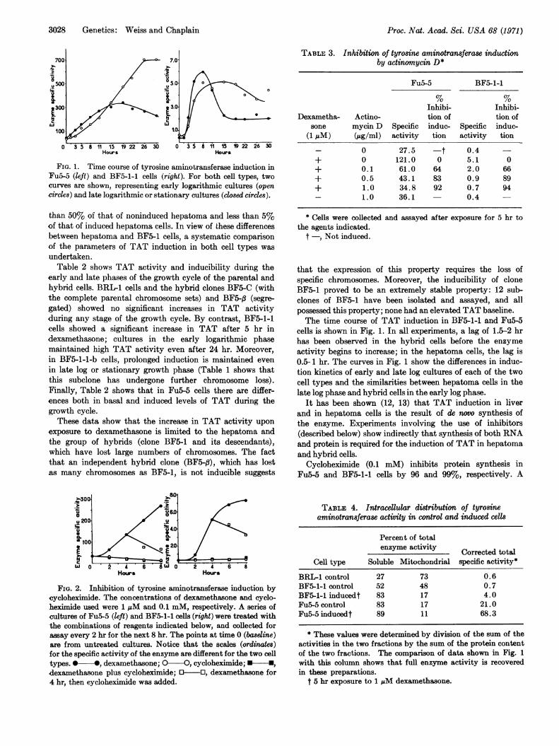
3028 Genetics: Weiss and Chaplain
Hours Hours
FIG. 1. Time course of tyrosine aminotransferase induction inFu5-5 (left) and BF5-1-1 cells (right). For both cell types, two
curves are shown, representing early logarithmic cultures (opencircles) and late logarithmic or stationary cultures (closed circles).
than 50% of that of noninduced hepatoma and less than 5%of that of induced hepatoma cells. In view of these differencesbetween hepatoma and BF5-1 cells, a systematic comparisonof the parameters of TAT induction in both cell types was
undertaken.Table 2 shows TAT activity and inducibility during the
early and late phases of the growth cycle of the parental andhybrid cells. BRL-1 cells and the hybrid clones BF5-C (withthe complete parental chromosome sets) and BF5-q (segre-gated) showed no significant increases in TAT activityduring any stage of the growth cycle. By contrast, BF5-1-1cells showed a significant increase in TAT after 5 hr indexamethasone; cultures in the early logarithmic phasemaintained high TAT activity even after 24 hr. Moreover,in BF5-1-1-b cells, prolonged induction is maintained even
in late log or stationary growth phase (Table 1 shows thatthis subelone has undergone further chromosome loss).Finally, Table 2 shows that in Fu5-5 cells there are differ-ences both in basal and induced levels of TAT during thegrowth cycle.
These data show that the increase in TAT activity upon
exposure to dexamethasone is limited to the hepatoma andthe group of hybrids (clone BF5-1 and its descendants),which have lost large numbers of chromosomes. The factthat an independent hybrid clone (BF5-P3), which has lostas many chromosomes as BF5-1, is not inducible suggests
Hours Hours
FIG. 2. Inhibition of tyrosine aminotransferase induction bycycloheximide. The concentrations of dexamethasone and cyclo-heximide used were 1 AM and 0.1 mM, respectively. A series ofcultures of Fu5-5 (left) and BF5-1-1 cells (right) were treated withthe combinations of reagents indicated below, and collected forassay every 2 hr for the next 8 hr. The points at time 0 (basleine)are from untreated cultures. Notice that the scales (ordinaes)for the specific activity of the enzyme are different for the two celltypes. 0 *, dexamethasone; O-O, cycloheximide; -,
dexamethasone plus cycloheximide; O-O, dexamethasone for4 hr, then cycloheximide was added.
Proc. Nat. Acad. Sci. USA 68 (1971)
TABLE 3. Inhibition of tyrosine aminotransferase inductionby actinomycin D*
Fu5-5 BF5-1-1
Inhibi- Inhibi-Dexametha- Actino- tion of tion of
sone mycin D Specific induc- Specific induc-(1 AM) (jug/ml) activity tion activity tion
- 0 27.5 -t 0.4+ 0 121.0 0 5.1 0+ 0.1 61.0 64 2.0 66+ 0.5 43.1 83 0.9 89+ 1.0 34.8 92 0.7 94- 1.0 36.1 0.4 -
* Cells were collected and assayed after exposure for 5 hr tothe agents indicated.
t -, Not induced.
that the expression of this property requires the loss ofspecific chromosomes. Moreover, the inducibility of cloneBF5-1 proved to be an extremely stable property: 12 sub-clones of BF5-1 have been isolated and assayed, and allpossessed this property; none had an elevated TAT baseline.The time course of TAT induction in BF5-1-1 and Fu5-5
cells is shown in Fig. 1. In all experiments, a lag of 1.5-2 hrhas been observed in the hybrid cells before the enzymeactivity begins to increase; in the hepatoma cells, the lag is0.5-1 hr. The curves in Fig. 1 show the differences in induc-tion kinetics of early and late log cultures of each of the twocell types and the similarities between hepatoma cells in thelate log phase and hybrid cells in the early log phase.
It has been shown (12, 13) that TAT induction in liverand in hepatoma cells is the result of de novo synthesis ofthe enzyme. Experiments involving the use of inhibitors(described below) show indirectly that synthesis of both RNAand protein is required for the induction of TAT in hepatomaand hybrid cells.
Cycloheximide (0.1 mM) inhibits protein synthesis inFu5-5 and BF5-1-1 cells by 96 and 99%, respectively. A
TABLE 4. Intracelular distribution of tyrosineaminotransferase activity in control and induced cells
Percent of totalenzyme activity Corrected total
Cell type Soluble Mitochondrial specific activity*BRL-1 control 27 73 0.6BF5-1-1 control 52 48 0.7BF5-1-1 inducedt 83 17 4.0Fu5-5 control 83 17 21.0Fu5-5inducedt 89 11 68.3
* These values were determined by division of the sum of theactivities in the two fractions by the sum of the protein contentof the two fractions. The comparison of data shown in Fig. 1with this column shows that full enzyme activity is recoveredin these preparations.
t 5 hr exposure to 1 gM dexamethasone.
Differentiated Functions in Hepatoma Hybrids 3029
series of parallel cultures were treated with combinationsof dexamethasone and cycloheximide, as indicated in Fig. 2.It can be seen that for both cell types, cycloheximide com-pletely inhibits enzyme induction. Moreover, the additionof cycloheximide to induced cultures blocks further induction;the rate of decline, a rough measure of the rate of enzymedegradation, is similar for both Fu5-5 and BF5-1-1 (thecalculated half life of TAT is about 4-6 hr in both cases).Note, however, that cycloheximide causes a rapid declineof the noninduced enzyme only in the hepatoma cells.Table 3 shows the effect of the simultaneous addition of
dexamethasone and actinomycin D on TAT induction inFu5-5 and BF5-1-1 cells. All concentrations of actinomycin Dtested inhibited induction; the degree of inhibition causedby each concentration was similar for the two cell types.The spectrum of dexamethasone concentrations eliciting
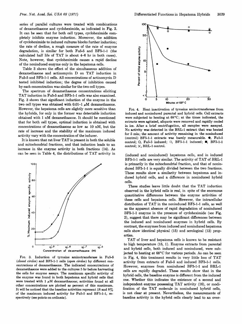
TAT induction in Fu5-5 and BF5-1-1 cells was also examined.Fig. 3 shows that significant induction of the enzyme in thetwo cell types was obtained with 0.01-1 pM dexamethasone.However, the hepatoma cells are slightly more sensitive thanthe hybrids, for only in the former was detectable inductionobtained with 1 nM dexamethasone. It should be mentionedthat for both cell types, optimal induction is obtained withconcentrations of dexamethasone as low as 10 nM, but therate of increase and the stability of the maximum inducedactivity vary with the concentration of the inducer.
It is known that rat liver TAT is present in both the solubleand mitochondrial fractions, and that induction leads to anincrease in the enzyme activity in both fractions (14). Ascan be seen in Table 4, the distributions of TAT activity in
100X
U 80-
U
* 60-
NC
U40X
EXx 20
0 10-9 1o_8 10B-7 10' 6
Concentration of dexamethasone (M)
FIG. 3. Induction of tyrosine aminotransferase in Fu5-5(closed circles) and BF5-1-1 cells (open crcles) by different con-centrations of dexamethasone. The indicated concentrations ofdexamethasone were added to the cultures 5 hr before harvestingthe cells for enzyme assays. The maximum specific activity ofthe enzyme was found in both hepatoma and hybrid cells thatwere treated with 1 pM dexamethasone; activities found at allother concentrations are plotted as percent of this maximum.It will be noticed that the baseline activities represent 18 and 8%of the maximum induced activity for Fu5-5 and BF5-1-1, re-spectively (see points on ordinate).
00E
<0Ic 5
0 5 10 20 30Minutes at 68° C
FIG. 4. Heat inactivation of tyrosine aminotransferase frominduced and noninduced parental and hybrid cells. Cell extractswere subjected to heating at 680C; at the times indicated, theextracts were agitated, aliquots were removed and rapidly cooledin ice. After a brief centrifugation, all samples were assayed.No activity was detected in the BRL-1 extract that was heatedfor 5 min; the amount of activity remaining in the noninduced(control) BF5-1-1 extracts was barely measurable. 0, Fu5-5control; 0, Fu5-5 induced; El, BF5-1-1 induced; U, BF5-1-1control; X, BRL-1 control.
(induced and noninduced) hepatoma cells, and in inducedBF5-1-1 cells are very similar. The activity of TAT of BRI1is primarily in the mitochondrial fraction, and that of nonin-duced BF5-1-1 is equally divided between the two fractions.These results show a similarity between hepatoma and in-duced hybrid cells, and a difference in noninduced hybridcells.These studies leave little doubt that the TAT induction
observed in the hybrid cells is real, in spite of the enormousquantitative differences between the enzyme activities ofthese cells and hepatoma cells. However, the intracellulardistribution of TAT in the noninduced BF5-1-1 cells, as wellas the apparent absence of rapid degradation of noninducedBF5-1-1 enzyme in the presence of cycloheximide (see Fig.2), suggest that there may be significant differences betweenthe induced and noninduced enzymes in hybrid cells. Bycontrast, the enzymes from induced and noninduced hepatomacells show identical physical (15) and serological (13) prop-erties.TAT of liver and hepatoma cells is known to be resistant
to high temperature (15, 1). Enzyme extracts from parentaland hybrid cells, both induced and noninduced, were sub-jected to heating at 680C for various periods. As can be seenin Fig. 4, this treatment results in very little loss of TATactivity from extracts of Fu5-5 and induced BF5-1-1 cells.However, enzymes from noninduced BF5-1-1 and BRIL1cells are rapidly degraded. These results show that in thehybrid cells, the baseline enzyme is different from the inducedone. Whether this indicates the existence of a second andindependent enzyme possessing TAT activity (16), or modi-fication of the TAT molecule in noninduced hybrid cells,is not clear at present. Nevertheless, the measurements ofbaseline activity in the hybrid cells clearly lead to an over-
Proc. Nat. Acad. Sci. USA 68 (1971)
3030 Genetics: Weiss and Chaplain
estimation of the activity of heat-stable TAT, which isinducible with dexamethasone.
DISCUSSION
In most studies of intraspecific somatic hybrids, chromosomeloss has been found to be slight (3), except in the cases ofvery prolonged cultivation of L-cell hybrids (17), and selec-tion in vivo of malignant segregants of A9-Ehrlich hybrids(11). In the present study, the use of selective conditions haspermitted the isolation of hybrid subclones containing only4-10 more chromosomes than the hepatoma parent.The properties of TAT in the BF5 hybrid with complete
sets of parental chromosomes are similar to those of theenzyme in rat hepatoma (Fu5) x mouse fibroblast (3T3)hybrids (1), and show that extinction of TAT and its induci-bility in the interspecific hybrids is not merely a consequenceof species differences. However, unlike 3T3, the BRILparental cells used in these studies are: (a) diploid (ratherthan sub-tetraploid like cells of most permanent mouse lines)and contributed to the hybrid a balanced diploid set ofchromosomes; (b) epithelial liver cells and are thereforeontogenetically less far removed from the hepatoma partnerthan fibroblasts. Nevertheless, the BRL-1 component sup-presses hepatoma TAT as effectively as a fibroblast. In astudy of heterokaryons formed by fusion of TAT induciblehepatoma cells with cells of a line similar in origin to BRL-1,Thompson and Gelehrter (18) have reported similar findings.Moreover, analysis of aldolase B activity in both 3T3 x Fu5and BRI-1 x Fu5-5 hybrids has shown that this hepatomafunction is similarly abolished in the two kinds of hybrids (22).The properties of the segregated hybrid subclones that are
inducible show a clear separation between two properties:high baseline of TAT and its inducibility. The model ofTomkins et al. of TAT induction proposes the existence of twogenes controlling these properties (19); our study of TAT inhepatoma x fibroblast hybrids supported this suggestion (1).The observation that TAT induction can take place in theabsence of appreciable "baseline" activity further supportsthe hypothesis that a specific gene mediates enzyme induction,and that its action is independent of the high TAT baseline.Perhaps the most interesting conclusion to be drawn from
our studies is that the extinction of a differentiated functionrequires the continuous presence of specific chromosomes, andwhen these are lost, the function reappears. This leads to theinference that the potentiality to perform this function canbe inherited in the absence of its expression. Earlier, Klebeet al. (20) arrived at similar conclusions in their study showingre-expression of kidney esterase-2 in somatic hybrids afterthe loss of specific chromosomes of the nondifferentiatedparent. These two observations suggest that re-expression ofdifferentiated functions after loss of specific chromosomesmay be a general phenomenon; if so, it should be possible toobtain as many classes of hybrids as there are (unlinked)genes that affect, directly or indirectly, the expression ofdifferentiated functions within a given cell type.
The inducible hybrids possess little or no baseline activityof (heat stable) TAT; this is most unusual in mammaliansystems. In spite of the described similarity of the parametersof TAT induction in hepatoma and segregated hybrid cells,there exist some differences. In particular, the phenomenonof actinomycin D "super-induction", well established forHTC (Hepatoma Tissue Culture) cells (15, 21) and observedalso in Fu5 (1) and Fu5-5 cells, has never been observed inthe hybrid cells. One interpretation of this difference is thatin the hepatoma and hybrid cells, the factors that limit thecomplex series of molecular events culminating in inducedTAT synthesis, are present in different proportions.
We thank Melle Anne Debon for excellent technical assistance,Drs. Drew Schwartz and Boris Ephrussi for stimulating discus-sions, and Dr. Ephrussi for critical reading of the manuscript.It is a pleasure to acknowledge the participation of Dr. JerrySchneider in the early stages of this work. This work was con-ducted with the aid of a grant to Dr. Ephrussi from the D6l6ga-tion G6n6rale A la Recherche Scientifique et Technique.
1. Schneider, J. A., and M. C. Weiss, Proc. Nat. Acad. Sci.USA, 68, 127 (1971).
2. Davidson, R. L., B. Ephrussi, and K. Yamamoto, Proc.Nat. Acad. Sci. USA, 56, 1437 (1966).
3. Ephrussi, B., Hybridization of Somatic Cells (PrincetonUniversity Press, in press).
4. Reuber, M. D., J. Nat. Cancer Inst.; 26, 891 (1961).5. Pitot, H. C., C. Peraino, P. A. Morse, and V. R. Potter,
Nat. Cancer Inst., Monogr., 13, 229 (1964).6. Coon, H. G., J. Cell Biol., 39, 29a (1968); Coon, H. G.,
Carnegie Inst. Washington, Yearb., 67, 419 (1969).7. Gelehrter, T. D., and G. M. Tomkins, Proc. Nat. Acad. Sci.
USA, 64, 723 (1969).8. McCormick, W., and S. Penman, J. Mol. Biol., 39, 315
(1969).9. Attardi, G., H. Parnas, M.-I. Hwang, and B. Attardi,
J. Mol. Biol., 20, 145 (1966).10. Weiss, M. C., G. J. Todaro, and H. Green, J. Cell. Physiol.,
71, 105 (1968).11. Harris, H., 0. J. Miller, G. Klein, P. Worst, and T. Tachi-
bana, Nature, 223, 363 (1969).12. Kenney, F. T., J. Biol. Chem., 237, 3495 (1962).13. Granner, D. K., S. Hayashi, E. B. Thompson, and G. M.
Tomkins, J. Mol. Biol., 35, 291 (1968).14. Fellman, J. H., P. J. Vanbellinghen, R. T. Jones, and R. D.
Koler, Biochemistry, 8, 615 (1969).15. Thompson, E. B., G. M. Tomkins, and J. F. Curran, Proc.
Nat. Acad. Sci. USA, 56, 296 (1966).16. Miller, J. E., and G. Litwack, J.- Biol. Chem., 246, 3234
(1971).17. Engel, E., B. J. McGee, and H. Harris, Nature, 223, 152
(1969).18. Thompson, E. B., and T. D. Gelehrter, Proc. Nat. Acad. Sci.
USA, 68, 2589 (1971).19. Tomkins, G. M., T. D. Gelehrter, D. Granner, D. Martin
Jr., H. Samuels, and E. B. Thompson. Science, 166, 1474(1969).
20. Klebe, R. J., T. Chen, and F. R. Ruddle, Proc. Nat. Acad.Sci. USA, 66, 1220 (1970).
21. Thompson, E. B., D. K. Granner, and G. M. Tomkins, J.Mol. Biol., 54, 159 (1970).
22. Bertolotti, R., and M. C. Weiss, J. Cell Physiol, in press.
Proc. Nat. Acad. Sci. USA 68 (1971)