expression analysis of brain-derived neurotrophic factor (bdnf) mrna isoforms after chronic and...
TRANSCRIPT

www.elsevier.com/locate/brainres
Brain Research 1000 (2004) 148–155
Research report
Expression analysis of brain-derived neurotrophic factor (BDNF) mRNA
isoforms after chronic and acute antidepressant treatment
Mario Altieria,*, Francesca Marinib, Roberto Arbana, Giovanni Vitullia, Birger O. Janssona,1
aCentre of Excellence for Drug Discovery, Psychiatry, GlaxoSmithKline Research Centre, via Fleming 4, I-37135 Verona, ItalybDepartment of Medicine and Public Health, Section of Pharmacology, School of Medicine, University of Verona,
Policlinico Borgo Roma, I-37134 Verona, Italy
Accepted 18 December 2003
Abstract
The neurotrophin brain-derived neurotrophic factor (BDNF) is considered to be a key factor for neuronal survival, differentiation and
plasticity. According to a proposed hypothetical model BDNF expression might play a central role in the pathogenesis of depression. The
BDNF gene is rather complex in its structure and it can express four different mRNA isoforms by alternative splicing, each producing the
same protein. This might reflect fine tuning of gene regulation by different signalling networks. Since the BDNF gene has been reported to be
upregulated by antidepressants, the expression of the four BDNF mRNA isoforms was measured by real-time quantitative RT-PCR in rat
hippocampi after chronic and acute treatment with the antidepressant drug fluoxetine and GR205171, a selective NK-1 receptor antagonist
with anxiolytic-like properties. The aim of this study was to test the hypothesis of differential regulation of the mRNA isoforms by those
compounds. Our results indicate that the expression of BDNF mRNA isoforms is not affected by chronic or acute treatment with fluoxetine or
GR205171.
D 2004 Elsevier B.V. All rights reserved.
Theme: Development and regeneration
Topic: Neurotrophic factors: expression and regulation
Keywords: Gene expression; Alternative splicing; Hippocampus; Depression; Fluoxetine; Rat
1. Introduction in combination with other genetic and environmental factors
Brain-derived neurotrophic factor (BDNF) belongs to the
family of neurotrophins [2], secreted polypeptides which
regulate neuronal survival [8] differentiation and plasticity
[22]. The regulation of BDNF has been proposed to be
involved in the pathogenesis of depression. According to
this model [6], chronic stress leads to a long-term decreased
expression of BDNF, which makes hippocampal neurons
(CA3 pyramidal cells, CA1 pyramidal cells and dentate
gyrus granule cells) more vulnerable to several types of
neuronal insult with consequent atrophy or damage of these
cellular types. In the long term this pathological mechanism,
0006-8993/$ - see front matter D 2004 Elsevier B.V. All rights reserved.
doi:10.1016/j.brainres.2003.12.028
* Corresponding author. Tel.: +39-45-921-8548; fax: +39-45-921-
8047.
E-mail address: [email protected] (M. Altieri).1 Present address: Affibody AB, Box 20137, SE-161 02 Bromma,
Sweden.
could result in the development of depressive disorders [6].
Chronic (but not acute) treatment with major classes of
antidepressants has been reported to increase the expression
of the rat BDNF gene in CA3 and dentate gyrus, [14,15] and
to change the BDNF immunoreactivity in rat hippocampus
in a dose dependent manner [24], thus directly linking
BDNF to the mechanism of antidepressant action. An
increased expression of BDNF could reverse the proposed
pathological mechanism by improving the survival of hip-
pocampal neurons [6] and restoring hippocampal neuro-
genesis [11].
The rat BDNF gene comprises five exons: I, II, III, IV
and V (Fig. 1).
Exons I, II, III and IV are non-coding (exon I contains an
extra in-frame initiation codon) and the whole protein open
reading frame (ORF) is encoded within exon V. Four
different mRNA isoforms can be expressed by splicing each
of the non-coding exons to exon V [23].

Fig. 1. The structure of the rat BDNF gene with its mRNA transcripts. The gene structure is shown at the top, with boxes representing the exons and lines
representing the introns. Broken lines represent introns larger than 10 kb. A full box represents the region coding for the mature protein, while a hatched box
represents the region coding for the prepro-BDNF protein. The position of the CRE-like element is indicated. The eight transcripts are shown at the bottom,
with the alternatively spliced gene regions represented as in the gene structure. Adapted from reference [23].
M. Altieri et al. / Brain Research 1000 (2004) 148–155 149
These transcripts are expressed by four independent
promoters each controlled by a different signalling network
[23]. The exon III-specific mRNA isoform is the major
Ca2 + inducible form in cortical neurons and it is regulated
by the transcription factor CREB. (A CRE-like element is
present in the exon III promoter) [21]. Each non-coding
exon has one promoter located in its 5V flanking region [21].In addition, each transcript can either have a longer or a
shorter 3V-UTR (untranslated region), which is also encoded
in exon V [23]. Therefore, each mRNA isoform can either
be long (4.4 kb) or short (1.8 kb), using two alternative
polyadenylation sites located within the untranslated region
[23].
With this peculiar structure the BDNF gene can express
up to eight different types of mRNA molecules, all
producing the same protein. It is likely that this transcrip-
tional complexity allows a very precise regulation poten-
tially responding to signalling cascades in different cell
types.
We hypothesised that antidepressants might differentially
regulate the expression of the four BDNF promoter-specific
mRNA transcripts. Exon I mRNA transcript was indeed
reported to be significantly increased in the rat hippocam-
pus by chronic treatment with the antidepressant drug
tranylcypromine while exon II transcript was not affected
[18]. However, extensive studies on how antidepressants
might specifically regulate the expression of BDNF mRNA
isoforms were not yet available in the literature. Very
recently, during the preparation of this manuscript, a work
on the effect of antidepressants and electroconvulsive
seizure on the rat BDNF mRNA isoforms was published
[5].
With the aim of testing our hypothesis, we therefore
measured the expression of the four BDNF promoter-spe-
cific mRNA transcripts in rat hippocampi after acute and
chronic treatment with fluoxetine, a selective serotonin
reuptake inhibitor (SSRI), and GR205171, a selective NK-
1 receptor antagonist. [7,12] Fluoxetine is a benchmark
SSRI, widely prescribed as antidepressant, reported to
upregulate BDNF mRNA and to increase neurogenesis in
rat hippocampus [11,15]. NK-1 receptor antagonists have
been recently proposed as potential therapeutic agents in the
treatment of anxiety and depression [10] and have been
hypothesised to stimulate hippocampal neurogenesis [17].
In this work, we used real-time quantitative RT-PCR, which
is considered one of the most precise and sensitive methods
for the quantification of gene expression [20].
2. Materials and methods
2.1. Animals and drug treatments:
Male Sprague–Dawley rats (200–250 g, Charles Riv-
er, Italy) were housed on a 12-h light/dark cycle (light at
6:00 am), with free access to food and water. The animals
were divided into six experimental groups (n = 4): three
groups received a chronic drug treatment (21 days) while
the other three received an acute treatment (1 day). Rats were
orally administered fluoxetine (5mg/kg once a day),
GR205171 (5mg/kg twice a day) or vehicle (sterile water).
Plasma samples from parallel groups (n= 3) were taken on
days 1, 9 and 21, respectively, for pharmacokinetic monitor-
ing. Peak levels (Cmax) were measured 2–5 hours after
M. Altieri et al. / Brain Research 1000 (2004) 148–155150
administration. Fluoxetine and GR205171 minimum levels
(Cmin) were measured respectively 24 and 8 h after adminis-
tration. Norfluoxetine (N-demethylated fluoxetine metabo-
lite) levels were measured on day 21 only. Drug levels were
measured using high-performance liquid chromatography
coupled to mass-spectrometry (HPLC-MS). The GR205171
dose was increased to 10mg/kg twice a day on day 15 in order
to keep the plasma level of the drug constant throughout the
treatment, since a tendency to decrease was observed on day
9. The animals were sacrificed by decapitation 24 h after the
last drug administration. Hippocampi were dissected from the
left side of brains immediately after sacrifice, quickly frozen
in dry ice and stored at � 80 jC.This work was performed under a Project License
obtained according to Italian law (art. 7, Legislative Decree
No. 116, 27 January 1992), which acknowledged the
European Directive 86/609/EEC, and was fully compliant
with GlaxoSmithKline policy on the care and use of
laboratory animals and related codes of practice.
2.2. RNA isolation
Frozen brain tissues were homogenised in tubes contain-
ing beads (Lysing Matrix D, Q-Biogene USA) and RLT
buffer (Qiagen) supplemented with 1% (v/v) h-mercaptoe-
thanol, using a Ribolyser (Hybaid) apparatus (20 s at speed
6). After the homogenisation was complete, the tubes were
centrifuged in a microfuge (14,000� g, 3 min) at room
temperature and the clear tissue lysate was collected. Total
RNAwas then isolated using the RNeasy Midi Kit (Qiagen)
by following the manufacturer’s protocol. Potential trace
amounts of residual genomic DNA were removed by diges-
tion with RNase-free DNase (Qiagen) included in the
purification method according to the Rneasy Midi/Maxi
Handbook (Qiagen). Purified total RNA was eluted in
RNase-free water and stored at � 80 jC. RNA concentra-
tion was measured in a GeneQuant II spectrophotometer
(Pharmacia) at 260 nm, and the quality of samples was
determined by analysis on 1.2% agarose electrophoresis gels
Table. 1
List of primers
Rat target gene GenBank Accession No. Primers Sequence
h-actin V01217 Forward TGAACC
Reverse CTCATA
GAPDH AF106860 Forward CAAGGT
Reverse GGGCCA
BDNF exon I X67106 Forward GCTGGT
Reverse CCAGGT
BDNF exon II X67106 Forward GGCTGG
Reverse CCGGTG
BDNF exon III X67107 Forward CACTGA
Reverse TGTACT
BDNF exon IV X67107 Forward GGCGCA
Reverse TCAGGG
(containing 0.22 M formaldehyde), after staining with
SYBR Gold (Molecular Probes).
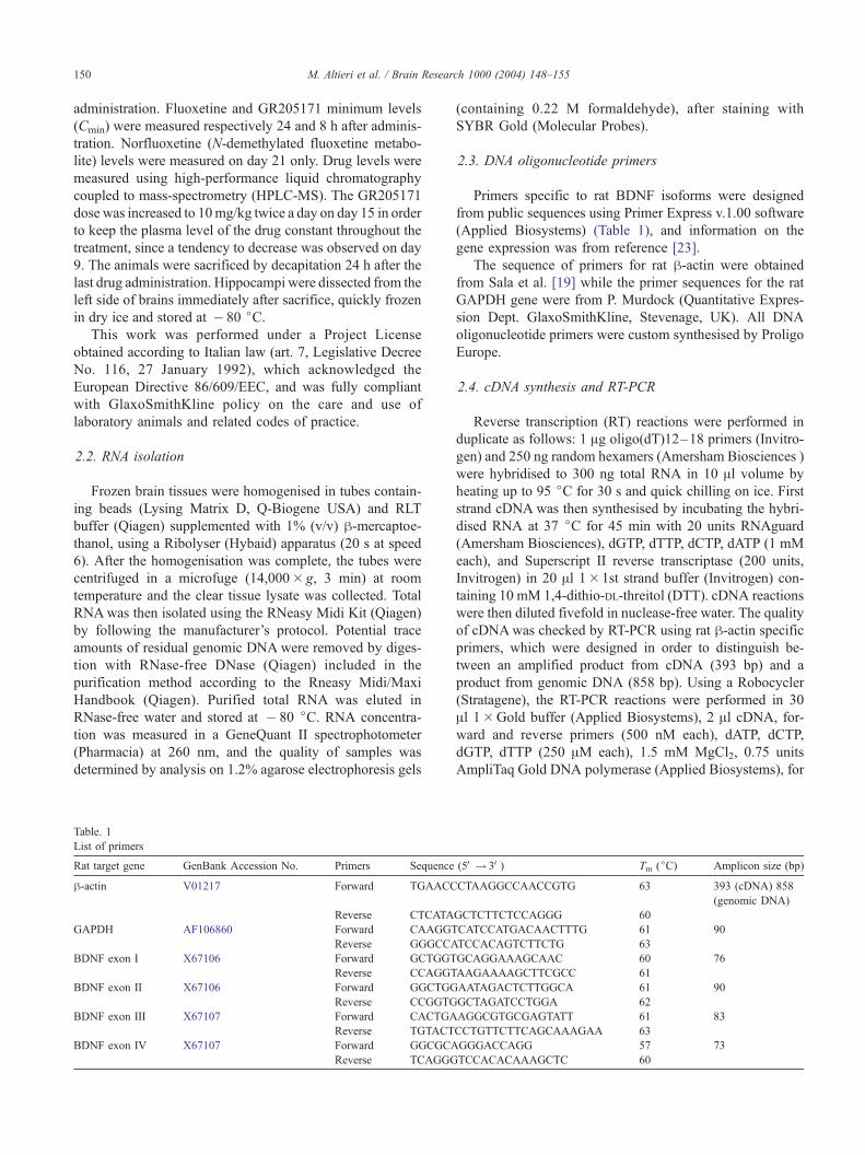
2.3. DNA oligonucleotide primers
Primers specific to rat BDNF isoforms were designed
from public sequences using Primer Express v.1.00 software
(Applied Biosystems) (Table 1), and information on the
gene expression was from reference [23].
The sequence of primers for rat h-actin were obtained
from Sala et al. [19] while the primer sequences for the rat
GAPDH gene were from P. Murdock (Quantitative Expres-
sion Dept. GlaxoSmithKline, Stevenage, UK). All DNA
oligonucleotide primers were custom synthesised by Proligo
Europe.
2.4. cDNA synthesis and RT-PCR
Reverse transcription (RT) reactions were performed in
duplicate as follows: 1 Ag oligo(dT)12–18 primers (Invitro-
gen) and 250 ng random hexamers (Amersham Biosciences )
were hybridised to 300 ng total RNA in 10 Al volume by
heating up to 95 jC for 30 s and quick chilling on ice. First
strand cDNA was then synthesised by incubating the hybri-
dised RNA at 37 jC for 45 min with 20 units RNAguard
(Amersham Biosciences), dGTP, dTTP, dCTP, dATP (1 mM
each), and Superscript II reverse transcriptase (200 units,
Invitrogen) in 20 Al 1�1st strand buffer (Invitrogen) con-
taining 10 mM 1,4-dithio-DL-threitol (DTT). cDNA reactions
were then diluted fivefold in nuclease-free water. The quality
of cDNAwas checked by RT-PCR using rat h-actin specific
primers, which were designed in order to distinguish be-
tween an amplified product from cDNA (393 bp) and a
product from genomic DNA (858 bp). Using a Robocycler
(Stratagene), the RT-PCR reactions were performed in 30
Al 1�Gold buffer (Applied Biosystems), 2 Al cDNA, for-ward and reverse primers (500 nM each), dATP, dCTP,
dGTP, dTTP (250 AM each), 1.5 mM MgCl2, 0.75 units
AmpliTaq Gold DNA polymerase (Applied Biosystems), for
(5V! 3V) Tm (jC) Amplicon size (bp)
CTAAGGCCAACCGTG 63 393 (cDNA) 858
(genomic DNA)
GCTCTTCTCCAGGG 60
CATCCATGACAACTTTG 61 90
TCCACAGTCTTCTG 63
GCAGGAAAGCAAC 60 76
AAGAAAAGCTTCGCC 61
AATAGACTCTTGGCA 61 90
GCTAGATCCTGGA 62
AGGCGTGCGAGTATT 61 83
CCTGTTCTTCAGCAAAGAA 63
GGGACCAGG 57 73
TCCACACAAAGCTC 60

Table 2
Drug levels in rat plasma
Day 1 Day 9 Day 21 Ki
Fluoxetine 2.00
Cmax 97.48 224.64 297.57
Cmin 27.85 20.52 3.66
Norfluoxetine 1.9
Cmax 637.4
Cmin 228.7
GR205171 0.32
Cmax 95.12 70.20 107.07
Cmin 83.08 35.86 31.06
Values (nM) are mean from three animals. Ki values (nM) for fluoxetine, its
N-demethylated metabolite norfluoxetine and GR205171 were derived from
the literature [7,16].
M. Altieri et al. / Brain Research 1000 (2004) 148–155 151
40 cycles (94 jC 1 min, 55 jC 1 min,72 jC 1 min) after an
initial step at 94 jC, 12 min to activate the DNA polymerase.
Only the former small amplicon was detected in all samples
on 4% Nu-Sieve (FMC) agarose gel electrophoresis, but not
the latter larger amplicon, demonstrating no contamination
of genomic DNA (data not shown).
2.5. Real-time quantitative RT-PCR
The amplification reactions were performed in triplicate in
30 Al 1� SYBR Green PCR master mix (Applied Biosys-
tems), 5 Al cDNA, forward and reverse primers (300 nM
each) using an ABI PRISM 7700 Sequence Detector (Ap-
plied Biosystems). The cycling parameters were: 95 jC 15 s,
60 jC 1min, 40 cycles, after one initial step at 95 jC, 10 min,
which was set to activate the AmpliTaq Gold polymerase.
Fig. 2. The expression of BDNF mRNA isoforms reported for each animal. Bars re
hippocampal total RNA sample, each measured in triplicate). Error bars represent t
acute fluoxetine. FlxCh: chronic fluoxetine. GR205171Ac: acute GR205171. GR
Ct (cycle threshold) values were calculated by the SDS
software v1.9 (Applied Biosystems) from fluorescence read-
ings, and these values were converted into copy number per
ng total RNA input, by using a standard curve of serially
diluted amounts of rat genomic DNA (Clontech). Primer
specificity was checked by analysing PCR products from rat
genomic DNA using 4% Nu-Sieve agarose gel electropho-
resis, and the dissociation curve method according to the
Applied Biosystems protocol.
2.6. Data analysis
The average copy number per ng total RNA input was
calculated for each sample (2 RTs per sample, 3 real-time
quantitative RT-PCR reactions per RT). An analysis of
covariance (ANCOVA) was performed on these data. The
expression data for the housekeeping gene GAPDH (refer-
ence gene) were used as covariate under the hypothesis that
it was not affected by drug treatments, in order to remove
the effects due to RNA sample quality from the analysis.
This method has been described in Ref. [1].
3. Results
3.1. Drug plasma levels in animals
Fluoxetine, its N-demethylated metabolite norfluoxetine
and GR205171 resulted to keep their plasma concentration
in treated animals above their Ki throughout the treatment,
as shown in Table 2.
present an average value from 6 datapoints (2 reverse transcriptions of each
he standard deviation. CtrlAc: acute control. CtrlCh: chronic control. FlxAc:
205171Ch: chronic GR205171.

Table 3
Results from ANCOVA analysis
BDNF mRNA isoform SS DF MS F p
Exon I GAPDH 0.029 1 0.029 5.048 0.0382
Treatment 0.103 5 0.021 3.541 0.0224
Error 0.099 17 0.006
Exon II GAPDH 0.000 1 0.000 0.027 0.8715
Treatment 0.055 5 0.011 2.937 0.0433
Error 0.063 17 0.004
Exon III GAPDH 0.083 1 0.083 18.707 0.0005
Treatment 0.018 5 0.004 0.809 0.5589
Error 0.075 17 0.004
Exon IV GAPDH 0.044 1 0.044 9.527 0.0067
Treatment 0.045 5 0.009 1.947 0.1391
Error 0.079 17 0.005
M. Altieri et al. / Brain Research 1000 (2004) 148–155152
3.2. Expression of BDNF mRNA isoforms in rat hippocampi
The expression of BDNF mRNA isoforms is shown in
Fig. 2. We found that exon III mRNAwas the most abundant
form in the rat hippocampus, followed by exon II, exon IV
and exon I (25.2%, 13.7% and 3.7% of exon III, respective-
ly). This expression profile is in good agreement with
Fig. 3. Fold changes from controls (bar centres) and their respective 95% confid
GAPDH), following acute (a) and chronic treatment (b). Control baseline value =
previously published data [23]. The expression of the rat
gene glyceraldehyde-3-phosphate dehydrogenase (GAPDH)
was measured in the same samples as an internal reference in
order to normalise the data for RNA quantity and quality.
3.3. Statistical analysis of data
The gene expression data were statistically evaluated by
analysis of covariance (ANCOVA), considering the expres-
sion of GAPDH as a covariate, under the hypothesis that it
was not affected by the treatments. The covariance efficien-
cy factor (CEF) for groups was determined to be 0.992, thus
proving independence from treatments. The results from
ANCOVA are shown on Table 3.
Post-hoc analyses (Dunnett’s test, p < 0.05) were per-
formed by comparing acute treatment groups to the acute
control group and chronic treatment groups to the chronic
control group (Fig. 3).
In our experimental conditions chronic treatment with
either fluoxetine or GR205171 did not show any statisti-
cally significant effect on the expression level of the four
BDNF mRNA transcripts (Table 4). Acute treatment of
ence intervals (bar edges), for the BDNF mRNA isoforms (normalized to
1.00.

Table 4
Fold changes with their p-values (Dunnett’s test)
Treatment BDNF mRNA
isoform
Fold change
from control
p-value
Acute fluoxetine Exon I 1.199 0.471
Exon II 1.144 0.542
Exon III 1.035 0.998
Exon IV 0.733a 0.046
Acute GR205171 Exon I 0.764 0.159
Exon II 0.913 0.827
Exon III 0.890 0.730
Exon IV 0.803 0.222
Chronic fluoxetine Exon I 1.068 0.977
Exon II 0.986 1.000
Exon III 1.155 0.564
Exon IV 1.018 1.000
Chronic GR205171 Exon I 0.852 0.582
Exon II 0.953 0.983
Exon III 1.024 1.000
Exon IV 1.048 0.991
a P < 0.05.
M. Altieri et al. / Brain Research 1000 (2004) 148–155 153
the two antidepressants also had no effect, except for a
downregulation of exon IV mRNA by fluoxetine (� 27%,
p = 0.046).
4. Discussion
In this work, the expression of four BDNF mRNA iso-
forms was analysed after chronic and acute treatment with
fluoxetine and GR205171, a specific NK-1 receptor antago-
nist. An expression analysis of the BDNF mRNA isoforms
after treatment with a NK-1 receptor antagonist has never
been reported before. The data indicate that neither chronic
nor acute treatment with these two drugs affected the expres-
sion of BDNF mRNA isoforms in the rat hippocampus. Only
acute fluoxetine had a small but significant negative effect on
the expression of exon IV-specific isoform, but it is not
known if this effect has any biological relevance.
Our investigation did not confirm a previous study,
published by Nibuya et al. [15], who reported an increased
BDNF mRNA expression in the rat hippocampus after
chronic treatment with fluoxetine. We cannot explain the
reason for this different result, but similar discrepancies have
already been observed by the authors of three other pub-
lished studies. Chronic treatment with fluoxetine decreased
BDNF mRNA expression in the rat hippocampus in one case
[13], while it had no significant effect on the expression of
BDNF mRNA in the mouse hippocampus [3]. Finally, in the
very recently published new work, chronic fluoxetine treat-
ment had no significant effect on the expression of the BDNF
mRNA isoforms in the rat hippocampal regions (dentate
gyrus, CA1 and CA3) [5], in full agreement with our work.
Possible methodological or biological factors should be
taken into account in an effort to explain these discordant
results. Technical differences in animal treatment (oral
administration used in this study, minipumps [13], or i.p.
administration [3,5,15]) could result in different drug phar-
macokinetics and therefore alter the exposure to drugs.
Chronic treatment with the antidepressant venlafaxine has
been recently reported to increase BDNF immunostaining at
a lower dose, but to decrease it at a higher dose in rat
hippocampus [24]. Moreover, different animal treatments
could also introduce different level of stress. Chronic
treatment with i.p. injection, for example, could be more
stressful than oral administration. It could be hypothesised
that the association of the treatment factors with different
techniques used to measure gene expression could lead to
very different results. Four different methodologies have
been used to measure gene expression in these studies: real-
time quantitative RT-PCR, used only in our study, Northern
blot [15], RNase protection assay [3] and in situ hybrid-
isation [5,13,15]. Real-time quantitative RT-PCR has the
advantage to be the most sensitive and reliable technique
used in gene quantitation [20] but, as Northern blot and
RNase protection assay, it requires the isolation of RNA
from a whole tissue area, even though it can be very small.
These techniques might fail to detect regional changes in
gene expression if the mRNA under study is diluted in the
RNA from other cellular types. In situ hybridisation, which
works directly on thin tissue slices without the need for
RNA isolation, is more appropriate for the detection of
regional changes in gene expression, even though its many
laborious steps can introduce a high level of variability.
Chronic treatment with fluoxetine increased the expression
of the BDNF gene in all hippocampal subfields (dentate
gyrus, CA3 and CA1) in the study by Nibuya et al. [15], but
only in one subfield (dentate gyrus) in another recently
published work [4], as shown in both studies by in situ
hybridisation. Differences in the treatment protocol have
been taken into consideration by the authors of the latter
study in an effort to explain this discrepancy [4]. Since
Nibuya et al. [15] used both in situ hybridisation and
Northern blot in whole hippocampus, our work should have
necessarily confirmed their results. In addition, since we
used real-time quantitative RT-PCR and we measured the
individual expression of the BDNF mRNA transcripts,
rather than the totality of the mRNA species, our assay
was much more sensitive than Northern blot. However, our
conditions might have induced an upregulation of BDNF
mRNA only in one hippocampal subfield and the real-time
quantitative RT-PCR might have failed to detect this change
in total hippocampal RNA. This possibility might explain
why we could not confirm the observed upregulation of the
BDNF gene after chronic treatment with fluoxetine [15],
and why this finding was not confirmed in the mouse
hippocampus using RNase protection assay [3]. This expla-
nation, however, remains theoretical and highly speculative.
Acute treatment with the antidepressant paroxetine or tra-
nylcypromine was reported to downregulate exon IV BDNF
mRNA specifically in the rat dentate gyrus [9]. In our study
we have also observed a small but statistically significant
downregulation of exon IV BDNF mRNA after acute

M. Altieri et al. / Brain Research 1000 (2004) 148–155154
fluoxetine treatment. Our result was quantitatively similar to
the reported work [9]. Furthermore, in the latest published
study on the effect of antidepressants on the rat BDNF
mRNA isoforms, it was observed, using in situ hybrid-
isation, a small decrease of exon IV BDNF mRNA in the
hippocampal CA1 subfield after acute treatment with fluox-
etine [5]. Although the biological meaning of this effect is
unclear, this would indicate that subtle regional changes in
the BDNF gene expression could have indeed been detected
in our assay. RNase protection assay did not detect any
change in the BDNF mRNA expression in the mouse
hippocampus after chronic treatment with fluoxetine, while
it could measure an upregulation of BDNF mRNA expres-
sion after chronic treatment with desipramine [3]. Therefore
it is unlikely that we could not detect any effect on BDNF
gene expression after chronic treatment with fluoxetine
because of our assay technique.
Finally, a recent work has shown that the time between the
last drug administration and the sacrifice can affect the
expression of the BDNF gene. It was found that chronic
treatment with antidepressants (fluoxetine included) de-
creased the BDNF mRNA expression in the rat hippocampus
at a post drug time of 4 h, while it increased the BDNF
mRNA expression at a post drug time of 24 h [4]. Therefore
this post drug time has become an additional variable to take
into account in order to explain the discrepancies of these
studies. In their very recent work [5], Dias et al. have
considered the post drug time as a possible reason to explain
why they could not confirm the work published by Nibuya et
al. [15]. They did not find any significant effect after chronic
fluoxetine treatment on the expression of the BDNF mRNA
isoforms in the rat hippocampus 2 hours after the last drug
administration [5], while Nibuya et al. [15], measured the
BDNF gene expression in the rat hippocampus 18 h after the
last drug administration. Doses and administration route
(i.p.) were the same in both studies. In our work, however,
we could not detect any significant effect on the expression
of the BDNF mRNA isoforms in the rat hippocampus 24
h after the end of chronic treatment with fluoxetine, given at
the same dose as in the quoted two studies but through an oral
administration route. Therefore, in our case it is unlikely that
the post drug time could account for the different result. In
conclusion, further studies are needed to develop a better
understanding of how BDNF might be involved in the
mechanism of antidepressant action. The effect of different
antidepressant classes on the regulation of BDNF mRNA
isoforms, how dose and post administration time influence
the drug effect on the regulation of the BDNF gene expres-
sion, and how this regulation is related to the antidepressant
mode of action need to be investigated more deeply.
Acknowledgements
The authors wish to thank Matteo Sartori, Lucia Carboni,
(GlaxoSmithKline, Italy) Paolo Repeto and Brian C. Bond
(Biostatistics and Data Sciences, GlaxoSmithKline, UK) for
their expert assistance; Joseph M. Rimland and Enrico
Domenici (GlaxoSmithKline, Italy) for critically reviewing
the manuscript. Enrico Domenici’s support for this work is
also acknowledged.
References
[1] B.C. Bond, D.J. Virley, N.J. Cairns, A.J. Hunter, G.B.T. Moore,
S.J. Moss, A.W. Mudge, F.S. Walsh, E. Jazin, P. Preece, The
quantification of gene expression in an animal model of brain
ischaemia using TaqMan real-time RT-PCR, Mol. Brain Res. 106
(2002) 101–116.
[2] M. Bothwell, Functional interactions of neurotrophins and neurotro-
phin receptors, Annu. Rev. Neurosci. 18 (1995) 223–253.
[3] A.C. Conti, J.F. Cryan, A. Dalvi, I. Lucki, J.A. Blendy, cAMP re-
sponse element-binding protein is essential for the upregulation of
brain-derived neurotrophic factor transcription, but not the behavioral
or endocrine responses to antidepressant drugs, J. Neurosci. 22 (2002)
3262–3268.
[4] A.L. Coppell, Q. Pei, T.S.C. Zetterstrom, Bi-phasic change in BDNF
gene expression following antidepressant drug treatment, Neurophar-
macology 44 (2003) 903–910.
[5] B.G. Dias, S.B. Banerjee, R.S. Duman, V.A. Vaidya, Differential
regulation of brain derived neurotrophic factor transcripts by antide-
pressant treatments in the adult rat brain, Neuropharmacology 45
(2003) 553–563.
[6] R.S. Duman, G.R. Heninger, E.J. Nestler, A molecular and cellular
theory of depression, Arch. Gen. Psychiatry 54 (1997) 597–606.
[7] C.J. Gardner, D.R. Armour, D.T. Beattie, J.D. Gale, A.B. Hawcock,
G.J. Kilpatrick, D.J. Twissel, P. Ward, GR205171: a novel antagonist
with high affinity for the tachykinin NK-1 receptor, and potent broad-
spectrum anti-emetic activity, Regul. Pept. 65 (1996) 45–53.
[8] A. Ghosh, J. Carnahan, M.E. Greenberg, Requirement for BDNF in
activity-dependent survival of cortical neurons, Science 263 (1994)
1618–1623.
[9] A.A. Khundakar, T.S.C. Zetterstrom, Differential expression of BDNF
exons in rat brain after systemic administration of paroxetine and
tranylcypromine, Br. J. Pharmacol. 137 (2002) 122 (Proc. suppl.).
[10] M.S. Kramer, N. Cutler, J. Feighner, R. Shrivastava, J. Carman, J.J.
Sramek, S.A. Reines, G. Liu, D. Snavely, E. Wyatt-Knowles, J.J. Hale,
S.G. Mills, M. MacCoss, C.J. Swain, T. Harrison, R.G. Hill, F. Hefti,
E.M. Scolnick, M.A. Cascieri, G.G. Chicchi, S. Sadowski, A.R. Wil-
liams, L. Hewson, D. Smith, E.J. Carlson, R.J. Hargreaves, N.M.J.
Rupniak, Distinct mechanism for antidepressant activity by blockade
of central substance p receptors, Science 281 (1998) 1640–1645.
[11] J.E. Malberg, A.J. Eisch, E.J. Nestler, R.S. Duman, Chronic antide-
pressant treatment increases neurogenesis in adult rat hippocampus,
J. Neurosci. 20 (2000) 9104–9110.
[12] M.J. Millan, F. Lejeune, G. De Nanteuil, A. Gobert, Selective block-
ade of neurokinin (NK)1 receptors facilitates the activity of adrenergic
pathways projecting to frontal cortex and dorsal hippocampus in rats,
J. Neurochem. 76 (2001) 1949–1954.
[13] X. Miro, S. Perez-Torres, F. Artigas, P. Puigdomenech, J.M. Palacios,
G. Mengod, Regulation of cAMP phosphodiesterase mRNAs expres-
sion in rat brain by acute and chronic fluoxetine treatment. An in situ
hybridization study, Neuropharmacology 43 (2002) 1148–1157.
[14] M. Nibuya, S. Morinobu, R.S. Duman, Regulation of BDNF and trkB
mRNA in rat brain by chronic electroconvulsive seizure and antide-
pressant drug treatments, J. Neurosci. 15 (1995) 7539–7547.
[15] M. Nibuya, E.J. Nestler, R.S. Duman, Chronic antidepressant ad-
ministration increases the expression of cAMP response element
binding protein (CREB) in rat hippocampus, J. Neurosci. 16 (1996)
2365–2372.

M. Altieri et al. / Brain Research 1000 (2004) 148–155 155
[16] M.J. Owens, W.N. Morgan, S.J. Plott, C.B. Nemeroff, Neurotransmit-
ter receptor and transporter binding profile of antidepressants and
their metabolites, J. Pharmacol. Exp. Ther. 283 (1997) 1305–1322.
[17] N.M. Rupniak, Elucidating the antidepressant actions of substance P
(NK1 receptor) antagonists, Curr. Opin. Investig. Drugs 3 (2002)
257–261.
[18] A.A. Russo-Neustadt, R.C. Beard, Y.M. Huang, C.W. Cotman, Phys-
ical activity and antidepressant treatment potentiate the expression of
specific brain-derived neurotrophic factor transcripts in the rat hippo-
campus, Neuroscience 101 (2000) 305–312.
[19] C.F. Sala, E. Formenti, G.C. Terstappen, A. Caricasole, Identification,
gene structure, and expression of human frizzled-3 (FZD3), Biochem.
Biophys. Res. Commun. 273 (2000) 27–34.
[20] T.D. Schmittgen, B.A. Zakrajsek, A.G. Mills, V. Gorn, M.J. Singer,
M.W. Reed, Quantitative reverse transcription-polymerase chain re-
action to study mRNA decay: comparison of endpoint and real-time
methods, Anal. Biochem. 285 (2000) 194–204.
[21] X. Tao, S. Finkbeiner, D.B. Arnold, A.J. Shaywitz, M.E. Greenberg,
Ca2 + influx regulates BDNF transcription by a CREB family tran-
scription factor-dependent mechanism, Neuron 20 (1998) 709–726.
[22] H. Thoenen, Neurotrophins and neuronal plasticity, Science 270
(1995) 593–598.
[23] T. Timmusk, K. Palm, M. Metsis, T. Reintam, V. Paalme, M. Saarma,
H. Persson, Multiple promoters direct tissue-specific expression of the
rat BDNF gene, Neuron 10 (1993) 475–489.
[24] H. Xu, J.S. Richardson, X.M. Li, Dose-related effects of chronic anti-
depressants on neuroprotective proteins BDNF, Bcl-2 and Cu/Zn-SOD
in rat hippocampus, Neuropsychopharmacology 28 (2003) 53–62.