exercise prevents diet-induced cellular senescence in...
TRANSCRIPT
Marissa J. Schafer,1,2 Thomas A. White,1 Glenda Evans,1 Jason M. Tonne,3
Grace C. Verzosa,4 Michael B. Stout,1,5 Daniel L. Mazula,1 Allyson K. Palmer,1
Darren J. Baker,1,6 Michael D. Jensen,7 Michael S. Torbenson,8 Jordan D. Miller,1,4
Yasuhiro Ikeda,3 Tamara Tchkonia,1 Jan M. van Deursen,1,9 James L. Kirkland,1,5
and Nathan K. LeBrasseur1,2
Exercise Prevents Diet-InducedCellular Senescence in Adipose TissueDiabetes 2016;65:1606–1615 | DOI: 10.2337/db15-0291
Considerable evidence implicates cellular senescencein the biology of aging and chronic disease. Diet andexercise are determinants of healthy aging; however,the extent to which they affect the behavior and accre-tion of senescent cells within distinct tissues is notclear. Here we tested the hypothesis that exercise pre-vents premature senescent cell accumulation and sys-temic metabolic dysfunction induced by a fast-fooddiet (FFD). Using transgenic mice that express EGFPin response to activation of the senescence-associatedp16INK4a promoter, we demonstrate that FFD consump-tion causes deleterious changes in body weight and com-position as well as in measures of physical, cardiac,and metabolic health. The harmful effects of the FFDwere associated with dramatic increases in severalmarkers of senescence, including p16, EGFP, senescence-associated b-galactosidase, and the senescence-associated secretory phenotype (SASP) specificallyin visceral adipose tissue. We show that exercise pre-vents the accumulation of senescent cells and the ex-pression of the SASP while nullifying the damagingeffects of the FFD on parameters of health. We alsodemonstrate that exercise initiated after long-termFFD feeding reduces senescent phenotype markers invisceral adipose tissue while attenuating physical impair-ments, suggesting that exercise may provide restorativebenefit by mitigating accrued senescent burden. Thesefindings highlight a novel mechanism by which exercisemediates its beneficial effects and reinforces the effect ofmodifiable lifestyle choices on health span.
Unhealthy diets and sedentary lifestyles are factorsfueling the obesity epidemic, wherein ;35% of middle-aged Americans are obese (1). Heavily implicated in thispublic health issue is routine consumption of calorie-dense,nutrient-poor fast foods and sugar-sweetened beverages,akin to a fast-food diet (FFD) (2). Nutrient excess leadingto metabolic dysfunction increases the risk for and acceler-ates the onset of numerous age-related conditions, includingdiabetes, cardiovascular disease, Alzheimer disease, andcancer (3,4). Fat mass distribution further influenceschronic disease risk, with visceral adiposity serving as astronger predictor of all-cause mortality relative to sub-cutaneous adiposity (5). In contrast, exercise positivelyaffects body composition, enhances physical fitness, andis protective against numerous age-related diseases (6).Despite the widely recognized effects of diet and exerciseon health span, the fundamental mechanisms by whichthey influence the biology of aging and chronic diseaseremain elusive.
Cellular senescence is a state of stable growth arresttriggered by telomere erosion, DNA lesions, reactive oxygenspecies, and other mitogenic and metabolic stressors. It ismediated by the inhibition of cell cycle progression throughp16INK4a/retinoblastoma protein and/or the activation ofcell cycle arrest through p53/p21. Characteristic gene ex-pression signature and morphological shifts define thetransition into a senescence state, but the functional roleof senescent cells within a given tissue milieu is highly de-pendent on cell type, concentration, and context (7). Multiple
1Robert and Arlene Kogod Center on Aging, Mayo Clinic, Rochester, MN2Department of Physical Medicine and Rehabilitation, Mayo Clinic, Rochester, MN3Department of Molecular Medicine, Mayo Clinic, Rochester, MN4Department of Surgery, Mayo Clinic, Rochester, MN5Department of Internal Medicine, Mayo Clinic, Rochester, MN6Department of Pediatric and Adolescent Medicine, Mayo Clinic, Rochester, MN7Division of Endocrinology, Department of Medicine, Mayo Clinic, Rochester, MN8Department of Laboratory Medicine and Pathology, Mayo Clinic, Rochester, MN9Department of Biochemistry and Molecular Biology, Mayo Clinic, Rochester, MN
Corresponding author: Nathan K. LeBrasseur, [email protected].
Received 4 March 2015 and accepted 29 February 2016.
This article contains Supplementary Data online at http://diabetes.diabetesjournals.org/lookup/suppl/doi:10.2337/db15-0291/-/DC1.
© 2016 by the American Diabetes Association. Readers may use this article aslong as the work is properly cited, the use is educational and not for profit, andthe work is not altered.
1606 Diabetes Volume 65, June 2016
OBESITY
STUDIES
lines of evidence implicate cellular senescence in the bi-ology of aging and the genesis of age-related conditions(8,9). In particular, biomarkers of senescent cells, includ-ing p16 and senescence-associated b-galactosidase (SA-b-gal) levels, increase in multiple tissues with advancingage and in the context of chronic disease (10).
Senescent cells actively secrete a broad repertoire ofcytokines, chemokines, matrix-remodeling proteases, andgrowth factors, collectively referred to as the senescence-associated secretory phenotype (SASP) (11). Despite theircell-autonomous role in the prevention of malignanttransformation, through the SASP, senescent cells damageneighboring cells, paradoxically fuel the aberrant growthand invasion of malignant cells, and promote inflammation(7,8). Senescent cells and the SASP are thus believed todrive degenerative, hyperproliferative, and inflammatoryconditions of aging (12). This premise is further sup-ported by studies demonstrating that targeted deletionof senescent cells expressing p16INK4a delays the onsetof several age-related phenotypes, including thymic invo-lution (13), and, in a mouse model of accelerated aging,cataracts, lordokyphosis, and diminished exercise capacity(14). More recently, senolytics, the term given to pharma-cological agents selected for their ability to kill senescentcells or inhibit the SASP, have shown therapeutic benefiton parameters of physical health and function when admin-istered to chronologically aged, progeroid, and/or irradiatedmice (15,16).
Whether and how lifestyle choices in middle ageinfluence the premature genesis of proaging senescentphenotypes in distinct tissues remains unclear. Accord-ingly, we sought to determine the extent to which nutrientexcess and exercise affect the onset and progression ofcellular senescence and the SASP using adult transgenicmice that express a construct harboring EGFP in responseto the senescence-sensitive promoter, p16INK4a.
RESEARCH DESIGN AND METHODS
Mice and Experimental InterventionsMice harboring the p16INK4a-EGFP transgenic construct(14) were generated on a genetically heterogeneous back-ground (four-strain cross, as previously detailed [17]). Forthe prevention study, 8-month-old male mice were dividedinto four groups of comparable mean body weights. Thegroups were randomly assigned to one of the following 16-week interventions: normal diet (ND) (13% energy as fat;PicoLab Rodent Diet 20 [5053]; LabDiet, St. Louis, MO),FFD (40% energy as fat [milk fat, 12% saturated] with0.2% cholesterol; Western Diet [5342]; TestDiet, St. Louis,MO), and high-fructose corn syrup in the drinking water(42 g/L; see [18]), ND plus exercise, or FFD plus exercise.
For the treatment study, 5- to 6-month-old male micewere provided ND or FFD for 16 weeks. FFD mice werethen randomized to sedentary or exercise groups based onbody weight, for a total of three groups, which weremonitored for an additional 14 weeks. Thus, all mice inthe prevention and treatment studies were ;1 year old at
necropsy. All mice were individually housed in ventilatedcages and provided food and water ad libitum. Exercisedmice were provided wireless running wheels, and exercisebehavior was monitored using Wheel Manager Data Acqui-sition Software (Med Associates, St. Albans, VT). Experi-ments were performed under protocols approved by theMayo Clinic Institutional Animal Care and Use Committee.
Body Composition and Health Span MeasuresBody weight and food intake were measured weekly. Bodycomposition (total body lean and fat mass) was assessedmonthly in unanesthetized mice by quantitative MRI(EchoMRI-100; Houston, TX), as previously described (19).At the end of the study, subcutaneous and visceral fat inthe lumbar region was quantified in anesthetized miceusing microcomputed tomography (vivaCT 40; ScancoMedical, Wayne, PA). As a measure of physical function,exercise capacity was determined on a motorized tread-mill (Columbus Instruments, Columbus, OH), as previ-ously described (20). Cardiac function in mice under lightisoflurane anesthesia was assessed by echocardiographyusing the Vevo 2100 system (FUJIFILM VisualSonics,Inc., Toronto, Ontario, Canada), as recently described (21).For metabolic function, glucose and insulin concentrationsand glucose tolerance after a 6-h fast were assessed, aspreviously described (22).
Tissue AssessmentsIndividual tissues were harvested, weighed, and processedfor downstream analyses. Portions of individual adiposetissue depots were fixed in PBS containing 2.0% formal-dehyde and 0.2% glutaraldehyde for cell size determinationand SA-b-gal activity. The sizes of adipocytes in fat tissuewere determined using Metamorph software (MolecularDevices, Sunnyvale, CA). Liver tissue was fixed in 10%formalin, dehydrated, and embedded in paraffin. Liversections were stained with hematoxylin and eosin foroverall morphology. A pathologist who was not aware oftreatment assignments gave grades of 0, 1, 2, 3, and 4 tosections in which 0, 1–4, 5–30, 31–60, and 61–100% of he-patocytes, respectively, had lipid macrovesicles. Grades of 0, 1,2, and 3 were assigned to liver sections with 0, 1–30, 31–60,and 61–100% of hepatocytes containing lipid microvesicles.Liver ceramides were quantified using ultraperformance liquidchromatography/tandem mass spectrometry, as recently de-scribed (23). Pancreata were embedded and frozen in OptimalCutting Temperature Compound (Sakura Finetek USA, Inc.,Torrance, CA). Immunostaining of cryosections and quantifi-cation of insulin-positive mass was performed, as previouslydescribed (24).
Markers of Cellular Senescence and the SASPFor transcriptional analysis, TRIzol-based extraction wasused to isolate RNA from whole mouse tissues, whichwere subjected to nanodrop concentration and purityanalysis before cDNA synthesis. TaqMan quantitativePCR (qPCR) assays (Life Technologies, Carlsbad, CA) wereused for detection of p16 (Mm00494449_m1), monocyte
diabetes.diabetesjournals.org Schafer and Associates 1607

chemoattractant protein 1 (Mcp1; Mm00441243_g1), andinsulin-like growth factor 1 (Igf1; Mm00439561_m1).PrimeTime 59 nuclease qPCR assays (Integrated DNA Tech-nologies, Coralville, IA) were used for detection of p21(Mm.PT.56a.17125846), p53 (Mm.PT.56a.44013092), in-terleukin 6 (Il6; Mm.PT.56a.10005566), plasminogen activa-tor 1 (Pai1; Mm.PT.58.6413525), matrix metalloprotease 3(Mmp3; Mm.PT.58.9719290), CD68 (Mm.PT.58.32698807),and Tbp (Mm.PT.39a.22214839). A SYBR qPCR assay (In-tegrated DNA Technologies) was used for detection ofEGFP (forward 59-CAA CTA CAA CAG CCA CAA CG-39;reverse 59-GGT CAC GAA CTC CAG CAG-39). Adipose tissuedepots were stained for SA-b-gal activity, as previously de-scribed (25).
Statistical AnalysisSignificant differences between groups for the dependentvariables of diet (ND and FFD) and behavior (sedentaryand exercise) were tested using one- or two-way ANOVA.The Tukey multiple comparisons test was used for posthoc analyses for between-group comparisons. Analyseswere conducted using GraphPad Prism Statistical Soft-ware Version 6.0 (GraphPad Software, Inc., San Diego, CA).
RESULTS
Exercise Prevents Multiple Indices of Diet-InducedMetabolic DysfunctionTo investigate the potential role of cellular senescence indiet-induced dyshomeostasis, which may be attenuated byexercise, we provided 8-month-old male mice harboring
an EGFP transgene driven by the p16INK4a promoter withan ND or a high-fat diet enriched with saturated fat,cholesterol, and high fructose corn syrup, equivalent toan FFD, for 4 months. Subsets of ND- and FFD-fed micewere provided with running wheels. Mice provided withthe FFD consumed more total calories within the firstweeks of the study, but within 1 month, total calorie in-take was not different among any of the groups (Supple-mentary Fig. 1A). Increased energy intake in exercisingFFD mice corresponded to elevated average daily runningdistances, albeit nonstatistically significant, within thefirst 2 months of the study (Supplementary Fig. 1B).
We assessed whether clinically relevant health indices,including body weight, adipose mass, physical activity,circulating insulin and glucose concentrations, cardiacfunction, and liver health were altered by diet and exercise.At study onset, average body weight and fat mass wereequivalent among ND- and FFD-fed mice, but after 4months, FFD-fed mice weighed 31% more and accumulatedtwice the total fat of ND-fed mice (Fig. 1A and B). Differ-ences between ND- and FFD-fed mice in subcutaneousand visceral fat were evident by weight (SupplementaryFig. 2A) and in microcomputed tomography scans of thelumbar region (Fig. 1C). The visceral fat of FFD-fed micewas composed of significantly larger adipocytes and agreater percentage of large adipocytes than that of ND-fed mice (Supplementary Fig. 2B). Exercise blunted theND- and, more dramatically, the FFD-induced accretionof body weight and fat mass (Fig. 1A–C and Supplementary
Figure 1—Nutrient excess and exercise exert opposing effects on body composition and physical endurance. Compared with mice fed anND, mice fed an FFD for 16 weeks exhibited marked gains in body weight (A) and fat mass as determined by quantitative MRI (B). C: FFD-induced obesity was further evident in volumes of visceral (pink) and subcutaneous (gray) fat in the lumbar region of mice as assessed bycomputed tomography. D: Exercised mice fed the ND or FFD exhibited significantly greater distances run to exhaustion on a treadmill thansedentary mice fed either diet. For all analyses, n = 6–7 mice/group. *P < 0.05, **P < 0.01, ***P < 0.001.
1608 Senescence Prevention Through Exercise Diabetes Volume 65, June 2016
Fig. 2A). In fact, the visceral and subcutaneous fat weightsof exercised FFD-fed mice were not statistically differentfrom those of ND-fed mice (Supplementary Fig. 2A). Exer-cise also prevented the FFD-induced hypertrophy of adipo-cytes (Supplementary Fig. 2B).
Assessment of physical function using a treadmill testrevealed that the FFD diet alone caused a modest butnonsignificant decrease in physical performance, whereasexercised mice fed the ND and FFD both ran a signifi-cantly greater distance to exhaustion than sedentarypeers (Fig. 1D). Exercised mice also had positive cardiacadaptations relative to sedentary peers, including increasedheart weight–to–body weight ratios, indicative of physio-logical hypertrophy (Supplementary Fig. 3A), and improvedejection fractions measured by echocardiography (Supple-mentary Fig. 3B). Sedentary mice fed the FFD exhibitedpoorer values for both of these parameters of cardiachealth (Supplementary Fig. 3A and B) and a deleteriousincrease in the left ventricular end diastolic dimensionrelative to mice fed the ND and exercised FFD-fed peers(Supplementary Fig. 3C).
With respect to metabolic function, no differences infasting glucose were observed between groups (data not
shown); however, sedentary mice fed the FFD had dra-matically increased insulin concentrations. This hallmarkof diet-induced insulin resistance was robustly reduced byexercise (Fig. 2A). Correspondingly, we observed grosslyenlarged b-cell masses and insulin-positive areas in thepancreata of sedentary but not exercised FFD-fed mice(Fig. 2B and C). Further evidence of preserved insulinaction in exercised mice fed the FFD was apparent in aglucose tolerance test, in which they were indistinguish-able from mice fed the ND (Fig. 2D). In contrast, seden-tary mice fed the FFD had more pronounced excursionsand impaired clearance of circulating glucose.
Nutrient excess can lead to adipocyte dysfunction,reflected in impaired triglyceride deposition, increasedlipolysis and lipotoxicity, or the accumulation of lipidsin peripheral tissues (26). As expected, liver weights ofsedentary FFD-fed mice were significantly greater thanND-fed mice (Supplementary Fig. 4A). Also significantlyelevated in the livers of sedentary mice were longer chainceramides, C16 and C24:1, and liver triglycerides, whichare associated with insulin resistance (27) (SupplementaryFig. 4B and C). These markers of hepatic lipotoxicity wereprevented by exercise (Supplementary Fig. 4B and C), as
Figure 2—Diet-induced deterioration of metabolic health is attenuated by exercise. A: Compared with sedentary and exercised mice fedthe ND, sedentary FFD-fed mice exhibited significantly higher circulating insulin concentrations. B and C: Correspondingly, cross sectionsof the pancreata of sedentary FFD-fed mice exhibited markedly greater b-cell masses and insulin-positive areas (representative images, B).A–C: Remarkably, these features of insulin resistance in FFD-fed mice were abrogated by exercise. D: Compromised and improved insulinactions in sedentary and exercised FFD-fed groups, respectively, were apparent in a glucose tolerance test. Namely, the higher peakand greater excursions in glucose concentrations observed in sedentary FFD-fed mice relative to ND-fed mice were erased by exercise. Forall analyses, n = 6–7 mice/group. *P < 0.05, **P < 0.01, ***P < 0.001.
diabetes.diabetesjournals.org Schafer and Associates 1609
were select FFD-induced gross morphological changes(Supplementary Fig. 4D). Compared with ND-fed mice,livers of sedentary and exercised FFD-fed mice demon-strated increases in the percentage of hepatocytes withlipid macrovesicles in their cytoplasm, an early event inthe pathogenesis of steatosis (Supplementary Fig. 4E).Most of the hepatocytes in the livers of sedentary micefed the FFD also had highly abundant lipid microvesicles,a feature associated with mitochondrial injury or dysfunc-tion (28). This consequence of the FFD was abrogated byexercise (Supplementary Fig. 4F). Collectively, these dataunderscore the salutary influence of exercise on severalparameters of health and its ability to prevent multipleharmful effects of nutrient excess.
The Detrimental Effects of Nutrient Excess onSenescent Cell Burden and the SASP in Visceral FatAre Prevented by ExerciseAlthough accumulation of senescent cells occurs withadvancing age, prematurely elevated senescent cell burdenmay be both a cause and consequence of metabolic dysfunc-tion (9,29). We hypothesized that routine FFD consumptionin middle age promotes accretion of senescent cells, andaccordingly, we probed expression of senescent biomarkersp16, p21, and p53 within discrete tissues. Comparedwith ND-fed mice, the visceral fat of sedentary FFD-fedmice contained significantly higher mRNA levels of p16(Fig. 3A) and p53 (Fig. 3B) as well as p21 (Fig. 3C), itsdownstream target, indicating pronounced activation ofsenescence effectors. Exercise completely blocked FFD-induced increases in p53 and p21 (Fig. 3B and C). Theexpression of p16 in subcutaneous adipose tissue, liver,skeletal muscle, pancreas, kidney, heart (left ventricle),and aorta was not altered in response to diet or exercise(Fig. 3A). Similar outcomes were observed for the expres-sion of p53, which, in addition, was higher in the subcuta-neous fat of FFD-fed mice relative to that of exercisedND-fed mice (Fig. 3B). Expression of p21 was significantlyelevated in subcutaneous fat and liver of sedentary FFD-fedmice, an effect that was prevented by exercise (Fig. 3C).These findings suggest that nutrient excess in middle ageactivates expression of prosenescence markers in visceraladipose tissue and that this effect is robustly attenuatedby exercise. To a lesser degree, senescent signaling mayalso occur in tissues other than visceral fat and may beprevented by exercise. However, this likely involves mech-anisms other than p16, such as the p53/p21 pathway.
Elevated expression of senescence markers distinctlywithin visceral adipose led us to further investigate theinfluence of nutrient excess and exercise on additionalindicators of cellular senescence and the SASP in thistissue. Quantification of EGFP expression confirmed thatFFD-feeding greatly enhanced p16INK4a promoter activity,which was strongly prevented by exercise (Fig. 4A). Tovalidate expression-based data, we stained visceraladipose tissue for the classic biomarker of senescence,SA-b-gal. In sedentary and exercised mice fed the ND, ;2%
of cells stained were positive for SA-b-gal (Fig. 4B and C).In comparison, more than 12% of cells in sedentary micefed the FFD stained positively. Strikingly, exercise nulli-fied this effect of the FFD, and as a result, the percentageof cells positive for SA-b-gal in exercised FFD-fed micewas identical to that of ND-fed middle-aged mice (Fig.4B and C).
Senescent cells partly disrupt a tissue’s structure andfunction and affect the systemic environment through thefactors they secrete. Indeed, FFD-fed mice demonstratedsignificant increases in the expression of proinflammatorySASP markers, including Il6, Pai1, and Mcp1 (Fig. 3D). Wealso observed significantly increased expression of Mmp3,a matrix remodeling protein and SASP component, withinvisceral fat after FFD-feeding. No significant differenceswere found in Igf1 (Fig. 4D). With the exception of Pai1,exercise prevented the induction of the SASP by the FFD.SASP signaling may instigate senescence in a paracrinemanner while recruiting inflammatory cells (30), ultimately
Figure 3—The effects of diet and exercise on senescence markersin multiple tissues. To determine the extent to which nutrient excessand exercise affected cellular senescence in middle-aged mice, wecompared the expression of p16 (A), p53 (B), and p21 (C ) by qPCRin multiple tissues, including visceral (Vis) fat, subcutaneous (SQ)fat, liver, gastrocnemius (gastroc), pancreas (panc), kidney, heart,and aorta. For all analyses, n = 6–7 mice/group. *P < 0.05, **P <0.01, ***P < 0.001.
1610 Senescence Prevention Through Exercise Diabetes Volume 65, June 2016
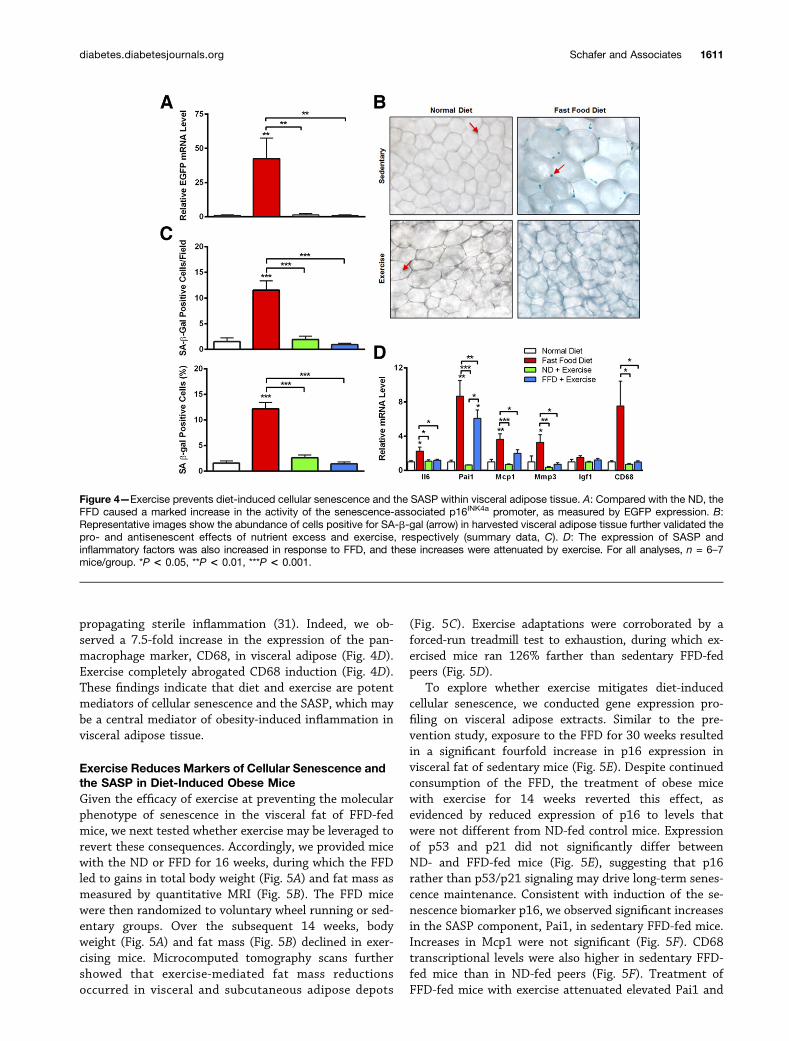
propagating sterile inflammation (31). Indeed, we ob-served a 7.5-fold increase in the expression of the pan-macrophage marker, CD68, in visceral adipose (Fig. 4D).Exercise completely abrogated CD68 induction (Fig. 4D).These findings indicate that diet and exercise are potentmediators of cellular senescence and the SASP, which maybe a central mediator of obesity-induced inflammation invisceral adipose tissue.
Exercise Reduces Markers of Cellular Senescence andthe SASP in Diet-Induced Obese MiceGiven the efficacy of exercise at preventing the molecularphenotype of senescence in the visceral fat of FFD-fedmice, we next tested whether exercise may be leveraged torevert these consequences. Accordingly, we provided micewith the ND or FFD for 16 weeks, during which the FFDled to gains in total body weight (Fig. 5A) and fat mass asmeasured by quantitative MRI (Fig. 5B). The FFD micewere then randomized to voluntary wheel running or sed-entary groups. Over the subsequent 14 weeks, bodyweight (Fig. 5A) and fat mass (Fig. 5B) declined in exer-cising mice. Microcomputed tomography scans furthershowed that exercise-mediated fat mass reductionsoccurred in visceral and subcutaneous adipose depots
(Fig. 5C). Exercise adaptations were corroborated by aforced-run treadmill test to exhaustion, during which ex-ercised mice ran 126% farther than sedentary FFD-fedpeers (Fig. 5D).
To explore whether exercise mitigates diet-inducedcellular senescence, we conducted gene expression pro-filing on visceral adipose extracts. Similar to the pre-vention study, exposure to the FFD for 30 weeks resultedin a significant fourfold increase in p16 expression invisceral fat of sedentary mice (Fig. 5E). Despite continuedconsumption of the FFD, the treatment of obese micewith exercise for 14 weeks reverted this effect, asevidenced by reduced expression of p16 to levels thatwere not different from ND-fed control mice. Expressionof p53 and p21 did not significantly differ betweenND- and FFD-fed mice (Fig. 5E), suggesting that p16rather than p53/p21 signaling may drive long-term senes-cence maintenance. Consistent with induction of the se-nescence biomarker p16, we observed significant increasesin the SASP component, Pai1, in sedentary FFD-fed mice.Increases in Mcp1 were not significant (Fig. 5F). CD68transcriptional levels were also higher in sedentary FFD-fed mice than in ND-fed peers (Fig. 5F). Treatment ofFFD-fed mice with exercise attenuated elevated Pai1 and
Figure 4—Exercise prevents diet-induced cellular senescence and the SASP within visceral adipose tissue. A: Compared with the ND, theFFD caused a marked increase in the activity of the senescence-associated p16INK4a promoter, as measured by EGFP expression. B:Representative images show the abundance of cells positive for SA-b-gal (arrow) in harvested visceral adipose tissue further validated thepro- and antisenescent effects of nutrient excess and exercise, respectively (summary data, C). D: The expression of SASP andinflammatory factors was also increased in response to FFD, and these increases were attenuated by exercise. For all analyses, n = 6–7mice/group. *P < 0.05, **P < 0.01, ***P < 0.001.
diabetes.diabetesjournals.org Schafer and Associates 1611
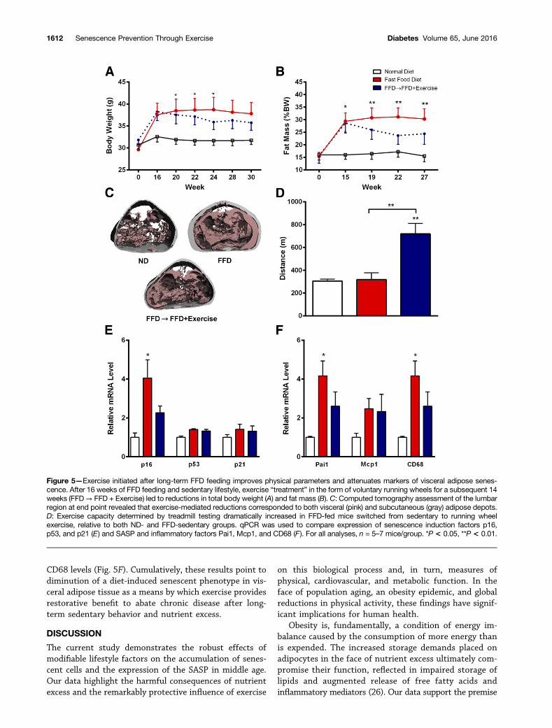
CD68 levels (Fig. 5F). Cumulatively, these results point todiminution of a diet-induced senescent phenotype in vis-ceral adipose tissue as a means by which exercise providesrestorative benefit to abate chronic disease after long-term sedentary behavior and nutrient excess.
DISCUSSION
The current study demonstrates the robust effects ofmodifiable lifestyle factors on the accumulation of senes-cent cells and the expression of the SASP in middle age.Our data highlight the harmful consequences of nutrientexcess and the remarkably protective influence of exercise
on this biological process and, in turn, measures ofphysical, cardiovascular, and metabolic function. In theface of population aging, an obesity epidemic, and globalreductions in physical activity, these findings have signif-icant implications for human health.
Obesity is, fundamentally, a condition of energy im-balance caused by the consumption of more energy thanis expended. The increased storage demands placed onadipocytes in the face of nutrient excess ultimately com-promise their function, reflected in impaired storage oflipids and augmented release of free fatty acids andinflammatory mediators (26). Our data support the premise
Figure 5—Exercise initiated after long-term FFD feeding improves physical parameters and attenuates markers of visceral adipose senes-cence. After 16 weeks of FFD feeding and sedentary lifestyle, exercise “treatment” in the form of voluntary running wheels for a subsequent 14weeks (FFD→ FFD + Exercise) led to reductions in total body weight (A) and fat mass (B). C: Computed tomography assessment of the lumbarregion at end point revealed that exercise-mediated reductions corresponded to both visceral (pink) and subcutaneous (gray) adipose depots.D: Exercise capacity determined by treadmill testing dramatically increased in FFD-fed mice switched from sedentary to running wheelexercise, relative to both ND- and FFD-sedentary groups. qPCR was used to compare expression of senescence induction factors p16,p53, and p21 (E) and SASP and inflammatory factors Pai1, Mcp1, and CD68 (F). For all analyses, n = 5–7 mice/group. *P < 0.05, **P < 0.01.
1612 Senescence Prevention Through Exercise Diabetes Volume 65, June 2016
that visceral adipose tissue dysfunction and its sequelaeare partly mediated through cellular senescence (12). Thestromal vascular fraction of adipose tissue is rich in pro-genitor cells, or preadipocytes, that are prone to senes-cence and exhibit a proinflammatory profile (12,16,29).We show that nutrient excess markedly increased theexpression of p16 and other markers of senescence, in-cluding p53 and p21 and the activity of SA-b-gal. Thesechanges were associated with induction of proinflammatorycytokines, chemokines, and matrix remodeling proteins (e.g.,Il6, Mcp1, Pai1, and Mmp3, respectively), collectively re-ferred to as the SASP.
SASP factors mechanistically contribute to metabolicdisease. Knockout of Pai1 abrogates insulin resistance andobesity brought on by high-fat feeding (32). Similarly, block-ade of adipose Mcp1 signaling exerts anti-inflammatory ef-fects (33), and ablation of the Mcp1 receptor (C-C motifchemokine receptor 2 [Ccr22/2]) increases adiponectinlevels and improves glucose homeostasis after high-fatfeeding relative to Ccr2+/+ controls matched for adiposity(34). SASP signaling has been further implicated as ameans of senescence transmission to neighboring cells(30), suggesting that the SASP may be responsible for in-duction and amplification of inflammation arising fromnutrient excess. We demonstrate that FFD feeding stronglyinduces visceral adipose expression of Pai1, Mcp1, andCD68 coincident with increases in p16 expression and cellspositive for SA-b-gal, which is prevented by exercise. Sim-ilarly, we show that treatment of obese mice with voluntaryexercise is able to revert aspects of this molecular pheno-type. Our data and prior evidence support the premise thatsenescent cells may be a primary source of obesity-associatedinflammation, which is central to the pathogenesis of type2 diabetes and its complications (9) and highlights thepotential of exercise as an effective intervention.
In agreement with our findings, Minamino et al. (35)reported that senescent cells accumulate in the adiposetissue of younger mice with ectopic expression of agoutipeptide, which leads to excessive nutrient intake, obesity,and diabetes. However, a more recent study failed to showthat high-fat feeding accelerates age-related p16INK4a ex-pression as quantified by whole-body luciferase imagingor mRNA abundance in isolated livers or spleens (36). It isplausible that imaging was not adequately sensitive to de-tect diet-induced changes in the abundance of p16INK4a-positive senescent cells in vivo. Furthermore, nutrient excessmay more potently induce the accumulation of p16INK4a
and/or p53-positive senescent cells in adipose tissue com-pared with other organs. Our results show that a senescencephenotype is readily activated specifically within visceral fatand, to a lesser degree within subcutaneous fat, in middleage by the FFD. Because visceral adipose is a stronger driverof metabolic-induced morbidity, relative to other depots(5), senescent signaling may mediate this organ’s uniquerole in sensing and negatively affecting the body in responseto nutrient excess, particularly regarding the inflammatorycomponent of obesity-induced dyshomeostasis. The causal
role of senescent cells and the SASP in the genesis ofobesity-associated conditions and the therapeutic efficacyof their removal, therefore, requires further examination.
The tissue specificity and temporal induction of cellularsenescence warrants further consideration. Other groupshave demonstrated increased senescence markers in vari-able tissues in response to nutrient excess, includingelevated aortic p16 levels after 20 weeks of high-fat feedingin 4-week-old C57BL/6 J mice (37), elevated hepatic p16and p21 levels after 13 weeks of high-fat feeding in 5-week-old rats (38), and elevated pancreatic p38 and SA-b-gallevels after 12 months of high-fat feeding in 4-week-oldC57BL/6 J mice (39). We conducted analyses in 12-month-old mice administered dietary and/or exercise interventionfor the previous 16 or 30 weeks, and our experimentsused a diet that was high in sugar and fat. We observedrobust induction of p16 and p53 in visceral adipose andinduction of p21 in visceral and subcutaneous adiposeand liver. Expression of these markers also appeared toincrease in other tissues, including the pancreas, but didnot reach statistical significance. Given the differences inage, genetic background, and diet composition betweenour study and the noted reports, that others have identi-fied senescence signatures in tissues that were not prom-inent in our exploration is not surprising. However, thecomposite results unanimously show that nutrient excessleads to induction of senescence in multiple tissues re-sponsible for coordinating metabolic health and cumula-tively highlight the need for additional work to tease outthe time course of this progression in uniform contexts.
The beneficial effects of exercise on health span areirrefutable; however, the biological mechanisms throughwhich they act are not completely understood. Our findingsconfirm that exercise can positively affect multiple param-eters of physical, cardiac, and metabolic health in middle-aged mice and, importantly, overcome the damaging effectsof nutrient excess. Moreover, using established biomarkers,we show for the first time that exercise prevents andreduces indicators of cellular senescence in visceral adiposetissue induced by an FFD. This is significant given theconsiderable evidence implicating senescent cells and theSASP in the biology of chronic diseases (8) and the bene-ficial effects of their removal on several parameters ofhealth, at least in a model of accelerated aging (14). Thereare three possibilities by which exercise prevented the diet-induced accumulation of senescent cells:
First, the increased energy demands and use of dietarymacronutrients during bouts of exercise may have limitedthe metabolic and replicative stresses experienced by cellsin adipose tissue and, consequently, their transition into asenescent state. Smaller adipocytes and fat depots as wellas lower liver weights and abundance of liver ceramides inexercised FFD-fed mice, compared with their sedentarypeers, may reflect this.
Second, it is plausible that exercise may have augmentedthe clearance of senescent cells. Their fate is highly variable.In benign melanocytic nevi, senescent cells can persist for
diabetes.diabetesjournals.org Schafer and Associates 1613
decades (40), whereas senescent liver carcinoma cellsacutely activate the innate immune system to mediate theirclearance and limit tumor growth (41). This possibility issupported by our data showing exercise reduces p16 ex-pression in mice even after obesity and its consequenceshave been established.
Third, exercise could have invoked protective responsesagainst triggers of cellular senescence in the context ofnutrient excess, because exercise counters DNA damage(42), telomere erosion (43), oxidative stress (44), proteinaggregation (45), and mitochondrial dysfunction (46) inmultiple cell types. We speculate that exercise may haveinduced such defense systems to prevent adipose tissuecells from senescing. Indeed, additional work is needed tobetter understand how and when to leverage exercise toaffect the accumulation, behavior, and persistence of se-nescent cells in the context of nutrient excess.
Adipose tissue is a critically important tissue in organis-mal health and aging. In contrast to the associations betweenobesity and phenotypes suggestive of accelerated aging,reductions in fat mass through calorie restriction (47),surgery (48), mutations in the insulin-signaling pathway(49), or exercise, as shown here, enhance health span invarious organisms. Our findings lend support to the con-cept that the SASP is a major determinant of the secretoryprofile, or endocrine function, of adipose tissue. In agingand obesity, adipose tissue is a primary source of inflam-matory mediators implicated in the genesis of diabetesand other chronic diseases (12). We previously demon-strated that the expression of components of the SASP,including Pai1 and Il6, were distinctly higher in p16INK4a-positive senescent cells than nonsenescent cells residingin adipose tissue (9). In the current study, our resultsshow that exercise prevents the SASP within visceral ad-ipose tissue, and remarkably, reduces aspects of the SASPwhen initiated after its accumulation. We propose this isan unappreciated mechanism through which physical ac-tivity interventions may affect health span, particularly inthose who are overweight or obese. Of note, the associa-tion between obesity-associated subclinical inflammationand the genesis of type 2 diabetes has been reported to bestronger in women than in men (50). Additional work isneeded to determine the extent to which senescent cellburden and the SASP may account for this sex differenceand whether exercise is as protective in females as weobserved in males. Therapeutic approaches to suppressthe SASP, such as exercise, may offer a means to negatethe deleterious systemic effects of senescent cells in thegenesis of obesity- and age-related chronic diseases.
In sum, our data highlight a novel and significantmechanism by which exercise positively affects organis-mal health. Given the considerable evidence that cellularsenescence is a fundamental mechanism of aging and thegenesis of chronic diseases, our findings reinforce thenotion that lifestyle choices are powerful determinants ofhealth span. Additional studies are necessary to determinethe mechanisms by which exercise prevents and reverses
cellular senescence and the SASP and at what ages and inwhat disease states it is most effective.
Acknowledgments. The authors greatly appreciate the technicalexpertise and support of Tamara Pirtskhalava, Kurt Johnson, Nathan W. Werneburg,Carolyn M. Roos, Anthony J. Croatt, Xuan-Mai Persson, and all of the MayoClinic.Funding. This work was supported by the Glenn Foundation for MedicalResearch (D.J.B., J.M.v.D., J.L.K., N.K.L.), National Institutes of Health, NationalInstitute on Aging grant AG-041122 (J.M.v.D., J.L.K., N.K.L.), the Pritzker Foundation(N.K.L.), a generous gift from Robert and Arlene Kogod, and by the Metabolic StudiesCore of the Minnesota Obesity Center (DK-50456).Duality of Interest. Mayo Clinic, A.K.P., D.J.B., T.T., J.M.v.D., J.L.K., andN.K.L. have a financial interest related to this research with intellectual propertylicensed to a commercial entity. This research has been reviewed by the MayoClinic Conflict of Interest Review Board and was conducted in compliance withMayo Clinic Conflict of Interest policies. No other potential conflicts of interestrelevant to this article were reported.Author Contributions. M.J.S. helped collect and analyze data, designedand implemented follow-up experiments, and drafted and revised the manu-script. T.A.W. helped design the study, collected and analyzed data, and draftedthe manuscript. G.E., J.M.T., G.C.V., M.B.S., D.L.M., A.K.P., M.S.T., and Y.I.collected and analyzed data. D.J.B. and J.M.v.D. helped design the studyand provided study resources. M.D.J., J.D.M., and T.T. helped design the study andcollected and analyzed data. J.L.K. helped design study, interpreted data, anddrafted the manuscript. N.K.L. designed the study, interpreted data, and draftedand revised the manuscript. N.K.L. is the guarantor of this work and, as such, hadfull access to all the data in the study and takes responsibility for the integrity of thedata and the accuracy of the data analysis.
References1. Ogden CL, Carroll MD, Kit BK, Flegal KM. Prevalence of childhood and adultobesity in the United States, 2011-2012. JAMA 2014;311:806–8142. Pereira MA, Kartashov AI, Ebbeling CB, et al. Fast-food habits, weight gain,and insulin resistance (the CARDIA study): 15-year prospective analysis. Lancet2005;365:36–423. Must A, Spadano J, Coakley EH, Field AE, Colditz G, Dietz WH. The diseaseburden associated with overweight and obesity. JAMA 1999;282:1523–15294. Whitmer RA, Gunderson EP, Barrett-Connor E, Quesenberry CP Jr, Yaffe K.Obesity in middle age and future risk of dementia: a 27 year longitudinal pop-ulation based study. BMJ 2005;330:13605. Fox CS, Massaro JM, Hoffmann U, et al. Abdominal visceral and sub-cutaneous adipose tissue compartments: association with metabolic risk factorsin the Framingham Heart Study. Circulation 2007;116:39–486. Blair SN, Kohl HW 3rd, Barlow CE, Paffenbarger RS Jr, Gibbons LW, MaceraCA. Changes in physical fitness and all-cause mortality. A prospective study ofhealthy and unhealthy men. JAMA 1995;273:1093–10987. Campisi J. Senescent cells, tumor suppression, and organismal aging: goodcitizens, bad neighbors. Cell 2005;120:513–5228. Tchkonia T, Zhu Y, van Deursen J, Campisi J, Kirkland JL. Cellular se-nescence and the senescent secretory phenotype: therapeutic opportunities. JClin Invest 2013;123:966–9729. Palmer AK, Tchkonia T, LeBrasseur NK, Chini EN, Xu M, Kirkland JL. Cellularsenescence in type 2 diabetes: a therapeutic opportunity. Diabetes 2015;64:2289–229810. Collado M, Blasco MA, Serrano M. Cellular senescence in cancer and aging.Cell 2007;130:223–23311. Coppé JP, Desprez PY, Krtolica A, Campisi J. The senescence-associated se-cretory phenotype: the dark side of tumor suppression. Annu Rev Pathol 2010;5:99–11812. Tchkonia T, Morbeck DE, Von Zglinicki T, et al. Fat tissue, aging, andcellular senescence. Aging Cell 2010;9:667–684
1614 Senescence Prevention Through Exercise Diabetes Volume 65, June 2016
13. Liu Y, Johnson SM, Fedoriw Y, et al. Expression of p16(INK4a) preventscancer and promotes aging in lymphocytes. Blood 2011;117:3257–326714. Baker DJ, Wijshake T, Tchkonia T, et al. Clearance of p16Ink4a-positivesenescent cells delays ageing-associated disorders. Nature 2011;479:232–23615. Zhu Y, Tchkonia T, Pirtskhalava T, et al. The Achilles’ heel of senescentcells: from transcriptome to senolytic drugs. Aging Cell 2015;14:644–65816. Xu M, Tchkonia T, Ding H, et al. JAK inhibition alleviates the cellular se-nescence-associated secretory phenotype and frailty in old age. Proc Natl AcadSci U S A 2015;112:E6301–E631017. Miller RA, Austad S, Burke D, et al. Exotic mice as models for aging re-search: polemic and prospectus. Neurobiol Aging 1999;20:217–23118. Charlton M, Krishnan A, Viker K, et al. Fast food diet mouse: novel smallanimal model of NASH with ballooning, progressive fibrosis, and high physio-logical fidelity to the human condition. Am J Physiol Gastrointest Liver Physiol2011;301:G825–G83419. Akasaki Y, Ouchi N, Izumiya Y, Bernardo BL, Lebrasseur NK, Walsh K.Glycolytic fast-twitch muscle fiber restoration counters adverse age-relatedchanges in body composition and metabolism. Aging Cell 2014; 13:80–9120. LeBrasseur NK, Schelhorn TM, Bernardo BL, Cosgrove PG, Loria PM, BrownTA. Myostatin inhibition enhances the effects of exercise on performance andmetabolic outcomes in aged mice. J Gerontol A Biol Sci Med Sci 2009;64:940–94821. Roos CM, Hagler M, Zhang B, Oehler EA, Arghami A, Miller JD. Transcriptionaland phenotypic changes in aorta and aortic valve with aging and MnSOD deficiencyin mice. Am J Physiol Heart Circ Physiol 2013;305:H1428–H143922. Bernardo BL, Wachtmann TS, Cosgrove PG, et al. Postnatal PPARdeltaactivation and myostatin inhibition exert distinct yet complimentary effects on themetabolic profile of obese insulin-resistant mice. PLoS One 2010;5:e1130723. Chow LS, Mashek DG, Austin E, et al. Training status diverges musclediacylglycerol accumulation during free fatty acid elevation. Am J Physiol En-docrinol Metab 2014;307:E124–E13124. Tonne JM, Sakuma T, Deeds MC, et al. Global gene expression profiling ofpancreatic islets in mice during streptozotocin-induced b-cell damage andpancreatic Glp-1 gene therapy. Dis Model Mech 2013;6:1236–124525. Villaret A, Galitzky J, Decaunes P, et al. Adipose tissue endothelial cells fromobese human subjects: differences among depots in angiogenic, metabolic, and in-flammatory gene expression and cellular senescence. Diabetes 2010;59:2755–276326. Guilherme A, Virbasius JV, Puri V, Czech MP. Adipocyte dysfunctions linkingobesity to insulin resistance and type 2 diabetes. Nat Rev Mol Cell Biol 2008;9:367–37727. Haus JM, Kashyap SR, Kasumov T, et al. Plasma ceramides are elevated inobese subjects with type 2 diabetes and correlate with the severity of insulinresistance. Diabetes 2009;58:337–34328. Fromenty B, Pessayre D. Inhibition of mitochondrial beta-oxidation as amechanism of hepatotoxicity. Pharmacol Ther 1995;67:101–15429. Escande C, Nin V, Pirtskhalava T, et al. Deleted in Breast Cancer 1 regulatescellular senescence during obesity. Aging Cell 2014;13:951–95330. Acosta JC, Banito A, Wuestefeld T, et al. A complex secretory programorchestrated by the inflammasome controls paracrine senescence. Nat Cell Biol2013;15:978–99031. Freund A, Orjalo AV, Desprez PY, Campisi J. Inflammatory networks duringcellular senescence: causes and consequences. Trends Mol Med 2010;16:238–246
32. Ma LJ, Mao SL, Taylor KL, et al. Prevention of obesity and insulin resistancein mice lacking plasminogen activator inhibitor 1. Diabetes 2004;53:336–34633. Yu R, Kim CS, Kwon BS, Kawada T. Mesenteric adipose tissue-derivedmonocyte chemoattractant protein-1 plays a crucial role in adipose tissuemacrophage migration and activation in obese mice. Obesity (Silver Spring) 2006;14:1353–136234. Weisberg SP, Hunter D, Huber R, et al. CCR2 modulates inflammatory andmetabolic effects of high-fat feeding. J Clin Invest 2006;116:115–12435. Minamino T, Orimo M, Shimizu I, et al. A crucial role for adipose tissue p53in the regulation of insulin resistance. Nat Med 2009;15:1082–108736. Sorrentino JA, Krishnamurthy J, Tilley S, Alb JG Jr, Burd CE, Sharpless NE.p16INK4a reporter mice reveal age-promoting effects of environmental toxicants.J Clin Invest 2014;124:169–17337. Wang CY, Kim HH, Hiroi Y, et al. Obesity increases vascular senescence andsusceptibility to ischemic injury through chronic activation of Akt and mTOR. SciSignal 2009;2:ra1138. Zhang X, Zhou D, Strakovsky R, Zhang Y, Pan YX. Hepatic cellular senes-cence pathway genes are induced through histone modifications in a diet-induced obese rat model. Am J Physiol Gastrointest Liver Physiol 2012;302:G558–G56439. Sone H, Kagawa Y. Pancreatic beta cell senescence contributes to thepathogenesis of type 2 diabetes in high-fat diet-induced diabetic mice. Dia-betologia 2005;48:58–6740. Michaloglou C, Vredeveld LC, Soengas MS, et al. BRAFE600-associatedsenescence-like cell cycle arrest of human naevi. Nature 2005;436:720–72441. Xue W, Zender L, Miething C, et al. Senescence and tumour clearance istriggered by p53 restoration in murine liver carcinomas. Nature 2007;445:656–66042. Radák Z, Naito H, Kaneko T, et al. Exercise training decreases DNA damageand increases DNA repair and resistance against oxidative stress of proteins inaged rat skeletal muscle. Pflugers Arch 2002;445:273–27843. Werner C, Hanhoun M, Widmann T, et al. Effects of physical exercise onmyocardial telomere-regulating proteins, survival pathways, and apoptosis. J AmColl Cardiol 2008;52:470–48244. Ji LL. Exercise-induced modulation of antioxidant defense. Ann N Y Acad Sci2002;959:82–9245. He C, Bassik MC, Moresi V, et al. Exercise-induced BCL2-regulated auto-phagy is required for muscle glucose homeostasis. Nature 2012;481:511–51546. Safdar A, Bourgeois JM, Ogborn DI, et al. Endurance exercise rescuesprogeroid aging and induces systemic mitochondrial rejuvenation in mtDNAmutator mice. Proc Natl Acad Sci U S A 2011;108:4135–414047. Masoro EJ. Caloric restriction and aging: an update. Exp Gerontol 2000;35:299–30548. Muzumdar R, Allison DB, Huffman DM, et al. Visceral adipose tissuemodulates mammalian longevity. Aging Cell 2008;7:438–44049. Blüher M, Kahn BB, Kahn CR. Extended longevity in mice lacking the insulinreceptor in adipose tissue. Science 2003;299:572–57450. Thorand B, Baumert J, Kolb H, et al. Sex differences in the prediction of type2 diabetes by inflammatory markers: results from the MONICA/KORA Augsburgcase-cohort study, 1984-2002. Diabetes Care 2007;30:854–860
diabetes.diabetesjournals.org Schafer and Associates 1615