exercise 6-d staining of microorganisms ... 6-d staining of microorganisms endospore stains, capsule...
TRANSCRIPT
Exercise 6-D
STAINING OF MICROORGANISMS ENDOSPORE STAINS, CAPSULE STAINS
& FLAGELLA Introduction Endospore stains, capsule stains, and flagellar stains are staining techniques that allow for the differentiation of specific bacterial structures found either inside or outside of cells. Such stains are sometimes referred to as special stains or structural stains. Bacterial endospores are dormant structures produced by a variety of bacterial species and which are highly resistant to heat, desiccation, and toxic chemicals. Endospores resist staining, but once stained are highly resistant to being decolorized and counterstained (much like acid-fast cells). A single bacterium can contain only one endospore, the shape and location of which is usually characteristic of the species. The degree to which an endospore changes the shape of the sporangium (spore-containing cell) is also a feature useful in identification. Not all cells present within a population will contain endospores, and older cells will often degenerate leaving their spores (now called exospores) behind in the environment. Many prokaryotic cells can produce a layer of gelatinous or slimy material external to their cell wall referred to as a glycocalyx. If this material is highly organized into a compact structure it is called a capsule and can be rendered visible with a capsule stain. If it is less well organized and tends to spread into the surrounding medium it is called a slime layer and is less readily stained and observed. Capsules may be made up of a variety of materials (polysaccharides, glycoproteins or polypeptides) but in general appear to be water soluble, and are unstable when subjected to heat. The ability of bacteria to produce capsules is genetically determined; however, individual cells may or may not have a visible glycocalyx depending upon their age and the availability of necessary nutrients. Capsule stains are useful in determining the virulence of pathogens, but are not overly useful for culture identification. Bacterial flagella are normally too small to be observed with the light microscope; however, certain staining techniques can render them visible. The procedures used in flagella staining involve covering the surface of the flagella with a mordant, thus increasing their diameter. Although we will not make flagellar stain preparations, we will observe some prepared slides of flagellar stains. Methods: A. Endospore Stain The presence of endospores may be demonstrated using either of the two procedures (methods)
outlined below. In general the production of endospores by a bacterial culture requires time, so the best spore samples will be obtained from cultures that are several days old. Many genera of bacteria including Bacillus, Lysinibacillus, Paenibacillus, Brevibacillus and Clostridium are known to produce endospores; however, even cultures that are genetically capable of forming endospores may not contain these structures if growing conditions do not stimulate spore formation.
Note – Endospores are often visible in Gram stain preparations. Under these circumstances,
mature endospores will typically appear as white or light colored areas within darker colored cells; immature or germinating endospores may appear dark.
The Dorner Method: 1. Put five drops of water in a clean test tube, and then add enough bacteria to make a heavy
suspension of the organisms provided (use several loopfuls of bacteria). 2. Add eight drops of carbol-fuchsin to the suspension, and place the tube in a boiling water
bath allowing the stain to act for at least 10 minutes. 3. Transfer several loopfuls of the suspension to a clean glass slide and immediately add a
small amount of nigrosin (one or two loopfuls). Mix the nigrosin with the cell suspension and then spread the mixture over the slide to make a thin, uniform smear.
4. Allow the smear to air dry, and it is ready to be examined under the oil immersion lens. If
properly prepared, a Dorner method endospore stain will leave vegetative cells white or pale pink against a dark background, while the spores will appear a bright reddish-pink (fuchsia).
The Malachite Green Stain or Schaeffer-Fulton Method: 1. At one end of a clean glass slide prepare a small smear containing one or more types of
Bacillus cells. At the other end of the slide prepare a smear containing cells from the morphological unknown culture assigned. Air dry and heat-fix these smears.
2. Place the slide on a stain rack, cover each smear with a small section of paper towel (do not
allow the paper towel to hang over the edge of the slide), and then flood the surface with malachite green.
3. Use a clothespin to carefully lift the wet preparation and pass it over the flame of a Bunsen
burner until the paper towel is steaming and the stain is bubbling (this doesn’t take long). Do not set the clothespin on fire.
4. Return the slide to the stain rack and allow it to cool off for about two minutes, then add
several drops of water to rewet the paper. Alternate method (steps 3-4) Place the slide on a rack above a beaker of boiling water, and
allow the stain to act for at least ten minutes. Do not allow the smear dry out during the staining process (add drops of deionized water to replace that evaporating from the slide surface when the paper texture becomes visible and the preparation takes on a metallic sheen).
5. Use a pair of forceps to lift off the paper towel, and place it in the waste beaker provided. 6. Rinse the smears thoroughly with clean tap water. It is important to remove all the excess
malachite green stain from the smear before applying the counterstain. 7. Counterstain the smears with safranin by flooding the slide surface (smear side up) and
allowing the stain to act for 60 seconds. 8. Rinse the smears with water, remove excess water from the bottom of the slide and allow the
smears to air dry (place near the base of a lit Bunsen burner).
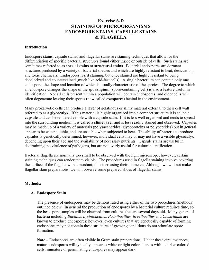
9. Observe your preparations using the oil immersion lens (focus with 10X first) and record your results. Endospores stained with malachite green will appear blue-green or turquoise in color while the vegetative cells will be stained pink with safranin.
Note - It is also possible to stain bacterial endospores using the acid-fast staining technique described in the preceding section on differential stains. If the acid-fast technique is used, the endospores will stain red and the vegetative cells will stain blue. Acid-fast cells stained with a malachite green stain will usually appear green or turquoise.
Fig. 6.3 - Bacterial Endospores (central and terminal) B. Capsule Stain Because capsules tend to be soluble in water, the procedures used to stain them avoid the use of
water or involve water only after the outline of the capsule has been indicated by a negative stain. Several different methods may be used to demonstrate the presence of capsules.
Nigrosin Capsule stain (a combination direct and indirect stain): 1. Using aseptic technique, make a thin smear containing a loopful of nigrosin and a mixture of
the capsule-forming bacteria provided (Klebsiella, Streptococcus, Azotobacter, etc.) at one end of a clean glass slide. Be careful not to add too much cellular material.
2. At the other end of the slide make a similar smear using your morphological unknown
culture. Do not mix cells from one smear into the other. 3. Thoroughly air-dry the smears, and then fix the cells to the glass with alcohol (flood the slide
surface with 95% ethanol or 99% isopropanol, and allow this to act for at least 2 minutes). 4. Dump off the residual alcohol, cover the smears with crystal violet, and stain for 2 minutes. 5. After staining with crystal violet, gently rinse the smears by running a stream of water over
your fingertips and onto the slide. Be careful not to dislodge the nigrosin layer. 6. Allow the slide to air dry and it is ready to be examined. Focus with your 10X objective first
and look for regions showing cells against a uniform (not cracked or broken) dark background. Then switch to 100X to complete observations and record results.
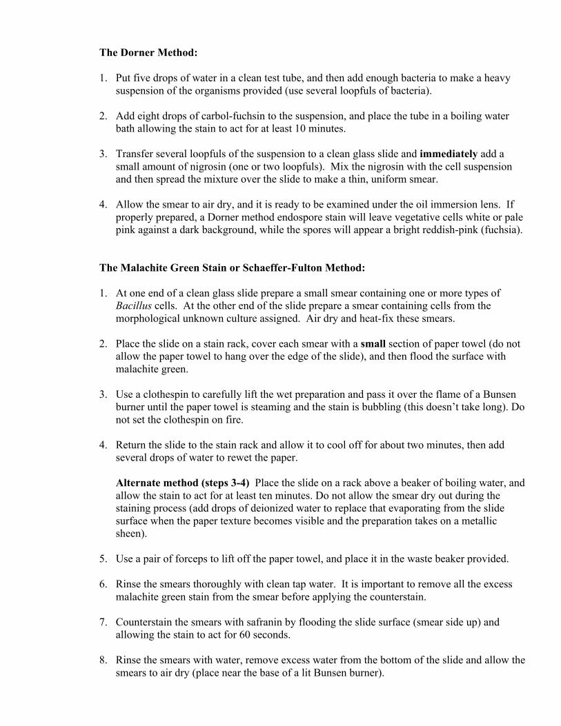
In a nigrosin capsule stain the capsules will appear white against a dark purple-gray background, and the cells will be stained violet. Capsules cannot be observed if you are viewing purple stained cells against a white background.
Fig. 6.4 - Bacterial Capsules in a nigrosin capsule stain Congo red Capsule Stain (a combination of direct and indirect stains): 1. Using aseptic technique, make a thin smear containing a loopful of Congo red and a mixture
of the capsule-forming bacteria provided (Klebsiella, Streptococcus, Azotobacter, etc.) at one end of a clean glass slide. Be careful not to add too much cellular material.
2. At the other end of the slide make a similar smear using your morphological unknown
culture. Do not mix cells from one smear into the other. 3. Thoroughly air-dry the smears, and then fix the cells to the glass with acid-alcohol (flood the
slide surface and allow this to act for at least 2 minutes). Acid-alcohol will turn the congo red dark and usually blue in color.
4. Dump off the residual acid-alcohol and cover each smear with crystal violet. Allow this to
remain on the glass for 2 minutes. Note – Do not rinse the slide with water between the application of the acid-alcohol and the crystal violet.
5. After staining with crystal violet, gently rinse the smears by running a stream of water over
your fingers and onto the slide. Be careful not to dislodge the Congo red layer. 6. Allow the slide to air dry and it is ready to be examined under 100x magnification (focus
with 10x first). In a Congo red capsule stain the capsules will appear white against a dark purple-brown background and the cells will be stained violet.
Procedure: 1. Prepare endospore stains as described above. Observe and be able to recognize cells stained with
both Dorner method and Malachite green (Schaeffer-Fulton method) endospore stains. Note the locations of endospores (central or terminal), their shape (spherical or ellipsoidal), and if or not sporangia are swollen.
2. On the worksheet provided, record representations of bacterial endospores stained with the
Dorner method and with Malachite Green. Label the endospores present indicating whether they are central or terminal, ellipsoidal or spherical, and if or not the sporangia are swollen. Be sure to include and label exospores as well.
3. Make a capsule stain using the nigrosin or Congo red staining methods described above.
Illustrate your capsule stain on the worksheet provided, and label it to clearly differentiate capsules and cells. Note that beginning microbiology students often confuse cells within capsules for endospores within vegetative cells. The proper labeling of accurate illustrations will help minimize such errors.
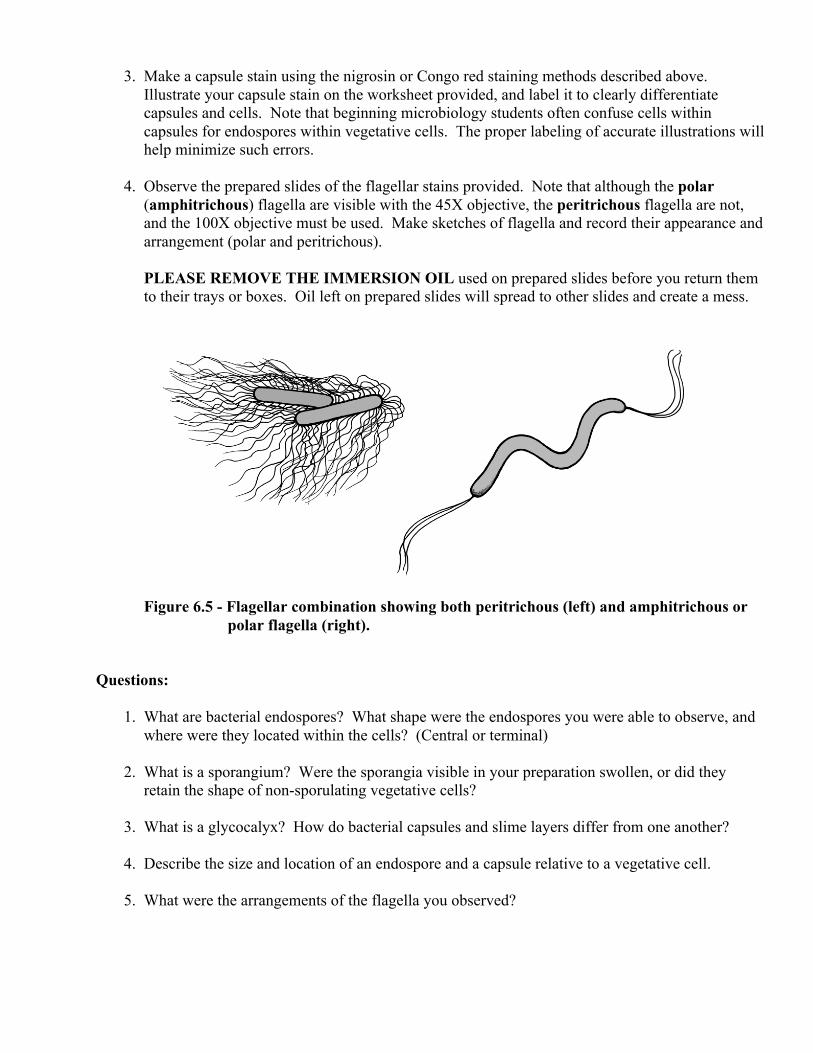
4. Observe the prepared slides of the flagellar stains provided. Note that although the polar
(amphitrichous) flagella are visible with the 45X objective, the peritrichous flagella are not, and the 100X objective must be used. Make sketches of flagella and record their appearance and arrangement (polar and peritrichous).
PLEASE REMOVE THE IMMERSION OIL used on prepared slides before you return them
to their trays or boxes. Oil left on prepared slides will spread to other slides and create a mess.
Figure 6.5 - Flagellar combination showing both peritrichous (left) and amphitrichous or
polar flagella (right). Questions: 1. What are bacterial endospores? What shape were the endospores you were able to observe, and
where were they located within the cells? (Central or terminal) 2. What is a sporangium? Were the sporangia visible in your preparation swollen, or did they
retain the shape of non-sporulating vegetative cells? 3. What is a glycocalyx? How do bacterial capsules and slime layers differ from one another? 4. Describe the size and location of an endospore and a capsule relative to a vegetative cell. 5. What were the arrangements of the flagella you observed?
Name________________________________ Lab Section_____________
WORKSHEET Exercise 6D
Staining of Microorganisms: Endospore Stains, Capsule Stains & Flagella Goals: __________________________________________________________________________ ________________________________________________________________________________ ________________________________________________________________________________ Materials & Methods: 1. Endospore Stain Malachite Green (Schaeffer-Fulton) Method: Age of Unknown: ____________ Reagents used: __________________________________ __________________________________________________________________________ 2. Capsule Stain Age of Unknown: ____________ Reagents used: __________________________________ ___________________________________________________________________________ Data & Results: 1. Endospore Stain Malachite Green Method A) Endospore-positive control
Specimen: Total Magnification: Length: units x µm/unit = µm Width: units x µm/unit = µm Notes:
B) Morphological unknown
Endospore Stain (Malachite Green Method) Data Summary:
Specimen Data Result
2. Capsule Stain A) Capsule-positive control
Specimen: Total Magnification: Length: units x µm/unit = µm Width: units x µm/unit = µm Notes:
Specimen: Total Magnification: Length: units x µm/unit = µm Width: units x µm/unit = µm Notes:
B) Morphological unknown
Capsule Stain Data Summary:
Specimen Data Result
Conclusions: Based on your results, what can you conclude about your Morphological Unknown? ____________ ________________________________________________________________________________ ________________________________________________________________________________ Does the cell morphology of your Morphological Unknown match what you saw for Exercise 6A, 6B, and 6C? ___________ Additional Comments: ____________________________________________________________ _______________________________________________________________________________ _______________________________________________________________________________
Specimen: Total Magnification: Length: units x µm/unit = µm Width: units x µm/unit = µm Notes:
Observations of Flagellar Stains (prepared slides) Conclusions: What is the flagellar arrangement for each specimen observed? _____________________________ ________________________________________________________________________________ ________________________________________________________________________________
Specimen: Flagellar Arragement, Left Smear Total Magnification: Length: units x µm/unit = µm Width: units x µm/unit = µm Notes:
Specimen: Flagellar Arragement, Right Smear Total Magnification: Length: units x µm/unit = µm Width: units x µm/unit = µm Notes: