evolutionary biology and biodiversity · lhs, and acts on the ovarian follicle cells to produce mih...
TRANSCRIPT
61
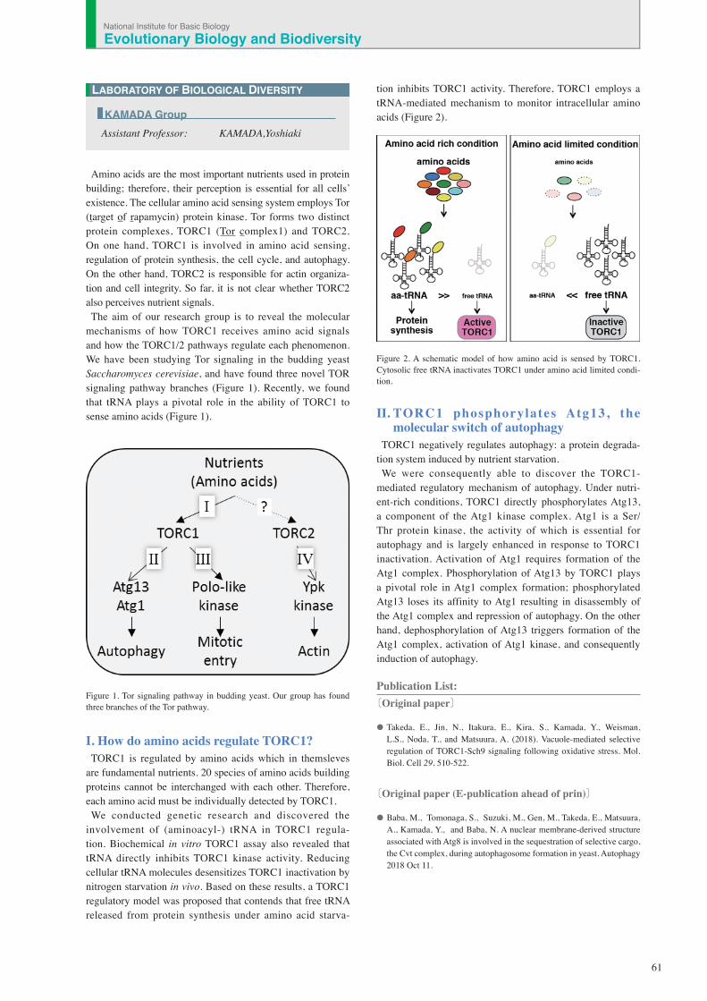
Amino acids are the most important nutrients used in protein building; therefore, their perception is essential for all cells’ existence. The cellular amino acid sensing system employs Tor (target of rapamycin) protein kinase. Tor forms two distinct protein complexes, TORC1 (Tor complex1) and TORC2. On one hand, TORC1 is involved in amino acid sensing, regulation of protein synthesis, the cell cycle, and autophagy. On the other hand, TORC2 is responsible for actin organiza-tion and cell integrity. So far, it is not clear whether TORC2 also perceives nutrient signals.The aim of our research group is to reveal the molecular
mechanisms of how TORC1 receives amino acid signals and how the TORC1/2 pathways regulate each phenomenon. We have been studying Tor signaling in the budding yeast Saccharomyces cerevisiae, and have found three novel TOR signaling pathway branches (Figure 1). Recently, we found that tRNA plays a pivotal role in the ability of TORC1 to sense amino acids (Figure 1).
Figure 1. Tor signaling pathway in budding yeast. Our group has found three branches of the Tor pathway.
I. How do amino acids regulate TORC1? TORC1 is regulated by amino acids which in themsleves
are fundamental nutrients. 20 species of amino acids building proteins cannot be interchanged with each other. Therefore, each amino acid must be individually detected by TORC1. We conducted genetic research and discovered the
involvement of (aminoacyl-) tRNA in TORC1 regula-tion. Biochemical in vitro TORC1 assay also revealed that tRNA directly inhibits TORC1 kinase activity. Reducing cellular tRNA molecules desensitizes TORC1 inactivation by nitrogen starvation in vivo. Based on these results, a TORC1 regulatory model was proposed that contends that free tRNA released from protein synthesis under amino acid starva-
tion inhibits TORC1 activity. Therefore, TORC1 employs a tRNA-mediated mechanism to monitor intracellular amino acids (Figure 2).
Figure 2. A schematic model of how amino acid is sensed by TORC1. Cytosolic free tRNA inactivates TORC1 under amino acid limited condi-tion.
II. TORC1 phosphorylates Atg13, the molecular switch of autophagy
TORC1 negatively regulates autophagy: a protein degrada-tion system induced by nutrient starvation. We were consequently able to discover the TORC1-
mediated regulatory mechanism of autophagy. Under nutri-ent-rich conditions, TORC1 directly phosphorylates Atg13, a component of the Atg1 kinase complex. Atg1 is a Ser/Thr protein kinase, the activity of which is essential for autophagy and is largely enhanced in response to TORC1 inactivation. Activation of Atg1 requires formation of the Atg1 complex. Phosphorylation of Atg13 by TORC1 plays a pivotal role in Atg1 complex formation; phosphorylated Atg13 loses its affinity to Atg1 resulting in disassembly of the Atg1 complex and repression of autophagy. On the other hand, dephosphorylation of Atg13 triggers formation of the Atg1 complex, activation of Atg1 kinase, and consequently induction of autophagy.
Publication List:〔Original paper〕
• Takeda, E., Jin, N., Itakura, E., Kira, S., Kamada, Y., Weisman, L.S., Noda, T., and Matsuura, A. (2018). Vacuole-mediated selective regulation of TORC1-Sch9 signaling following oxidative stress. Mol. Biol. Cell 29, 510-522.
〔Original paper (E-publication ahead of prin)〕
• Baba, M., Tomonaga, S., Suzuki, M., Gen, M., Takeda, E., Matsuura, A., Kamada, Y., and Baba, N. A nuclear membrane-derived structure associated with Atg8 is involved in the sequestration of selective cargo, the Cvt complex, during autophagosome formation in yeast. Autophagy 2018 Oct 11.
LABORATORY OF BIOLOGICAL DIVERSITY
Assistant Professor: KAMADA,Yoshiaki
KAMADA Group
National Institute for Basic BiologyEvolutionary Biology and Biodiversity
62
LABORATORY OF BIOLOGICAL DIVERSITY
Assistant Professor: OHNO, Kaoru
OHNO Group
The aim of this laboratory is to research reproductive hormones in invertebrates, especially in echinoderms, and to analyze the mechanisms by which they work. The compari-sons of such molecules and mechanisms in various species are expected to provide insights into the evolution of repro-ductive hormone systems.
I. Gonadotropins in the starfish, Patiria pectinifera
Gonadotropins play important regulatory roles in reproduc-tion in both vertebrates and invertebrates. The vertebrate gonadotropins, LH and FSH, are structurally and function-ally conserved across various species, whereas no such molecule has been identified in invertebrates. The insect parsin hormones are assumed to be the physiological coun-terpart of LH and FSH in mammals. Some gonadotropic hormones (e.g. the mosquito’s egg development neurosecre-tory hormone, the sea hare’s egg-laying hormone, and the terrestrial isopod’s androgenic gland hormone) have been found in invertebrate species. More recently, an insulin-like peptide was reported to be responsible for the regulation of egg maturation in the mosquito, Aedes aegypti, thus demon-strating the involvement of insulin signaling in egg matura-tion among invertebrates.The gonad-stimulating substance (GSS) of an echinoderm,
the starfish, was the very first gonadotropin to be identified in invertebrates. GSS mediates oocyte maturation in starfish by acting on the ovary to produce the maturation-inducing hormone (MIH), 1-methyladenine, which in turn induces the maturation of the oocytes. In this sense, GSS is functionally identical to vertebrate LH, especially piscine and amphibian LHs, and acts on the ovarian follicle cells to produce MIH to induce the final maturation or meiotic resumption of the oocyte. Considering the functional similarity that GSS shares with vertebrate LH, it is very important from an evolutionary point of view to know the chemical and molecular struc-ture of GSS. We cloned the gene encoding the amino acid sequence of purified GSS from radial nerves of the starfish, Pateria pectinifera. Interestingly, phylogenetic analyses revealed that it belonged to the insulin/insulin-like growth factor (IGF)/relaxin superfamily and, more precisely, to the subclass of relaxin peptides (Figure 1).
II. Search for reproductive hormones in echinoderms
In a collaborative effort with Prof. Yoshikuni’s Laboratory at Kyushu Univ., we are searching for reproductive hormones in echinoderms, including starfishes, brittle stars, sea urchins, sea cucumbers, and crinoids. The collaborat-ing parties have been able to purify physiological materi-als which induce egg maturation from nerve extracts and analyze them with a protein sequencer and a tandem mass spectrometer in the analytical center of our institute. One of
them, named cubifrin, which is an IWMGY-amide peptide, is in the sea cucumber Aposticopus japonicus. The others are in preparation for publication. We have identified many neuropeptides from our EST
analysis of nerve tissues and many from RNA-seq and WGS data of the NCBI database. In particular, relaxin like peptide precursor genes and insulin/IGF like peptide precursor genes were identified from many species. We are producing these neuropeptides by biological methods (e.g. bacterial systems and yeast systems) to provide to collaborating researchers for biological assays.
III. Search for the lost mutants of female hormones E2 in Oryzias latipes
Sequence analysis by capillary sequencer was carried out from the tilling library of O. latipes in order to analyze the function of fish female reproductive hormone E2 as a part of our collaborative research. Upon the discovery of aroma-tase mutants, a detailed analysis was carried out using these strains.
Publication List:〔Original paper〕
• Nakamoto, M., Shibata, Y., Ohno, K.. Usami, T., Kamei, Y., Taniguchi, Y., Todo, T., Sakamoto, T., Young, G., Swanson, P., Naruse, K., and Nagahama, Y. (2018). Ovarian aromatase loss-of-function mutant medaka undergo ovary degeneration and partial female-to-male sex reversal after puberty. Mol. Cell Endocrinol. 460, 104-122.
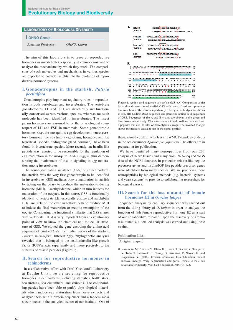
Figure 1. Amino acid sequence of starfish GSS. (A) Comparison of the heterodimeric structure of starfish GSS with those of various representa-tive members of the insulin superfamily. The cysteine bridges are shown in red. (B) Coding DNA sequence and predicted amino acid sequences of GSS. Sequences of the A and B chains are shown in the green and blue boxes, respectively. Characters shown in red boldface indicate basic dipeptides that are the sites of proteolytic cleavage. The inverted triangle shows the deduced cleavage site of the signal peptide.
Evolutionary Biology and BiodiversityNational Institute for Basic Biology
63
While genomic structures (as well as their genetic informa-tion) appear to stably transmit into daughter cells during cell division, and also into the next generation, they can actually vary genetically and/or epigenetically. Such variability has a large impact on gene expression and evolution. To under-stand these genome dynamics in eukaryotes, especially in plants, we are analyzing the flower pigmentation of morning glories including Ipomoea nil (Japanese morning glory), I. purpurea (the common morning glory), and I. tricolor.
I. Flower pigmentation patternsThe wild type morning glories produce flowers with uni-
formly pigmented corolla. However a number of mutants displaying particular pigmentation patterns have been col-lected for this study. Because flower pigmentation patterns are easily observable, the molecular mechanisms underlying these phenomena provide useful model systems for investi-gating genome variability. The recessive mutations, duskish of I. nil and pearly-v of
I. tricolor, confer variegated flowers. They are caused by a stable insertion of a transposable element into a gene for flower pigmentation. Furthermore, epigenetic mechanisms are thought to regulate thispigmentation (Figure 1). We are currently analyzing the detailed molecular mechanisms of these mutations.
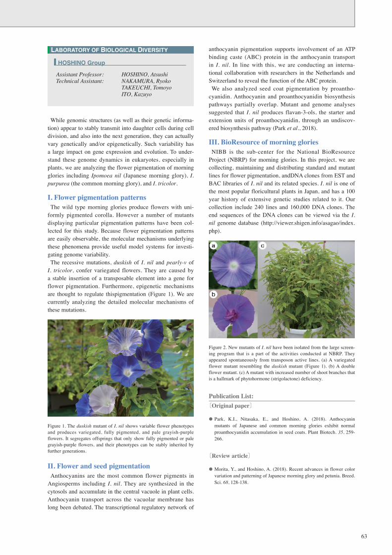
Figure 1. The duskish mutant of I. nil shows variable flower phenotypes and produces variegated, fully pigmented, and pale grayish-purple flowers. It segregates offsprings that only show fully pigmented or pale grayish-purple flowers, and their phenotypes can be stably inherited by further generations.
II. Flower and seed pigmentationAnthocyanins are the most common flower pigments in
Angiosperms including I. nil. They are synthesized in the cytosols and accumulate in the central vacuole in plant cells. Anthocyanin transport across the vacuolar membrane has long been debated. The transcriptional regulatory network of
anthocyanin pigmentation supports involvement of an ATP binding caste (ABC) protein in the anthocyanin transport in I. nil. In line with this, we are conducting an interna-tional collaboration with researchers in the Netherlands and Switzerland to reveal the function of the ABC protein.We also analyzed seed coat pigmentation by proantho-
cyanidin. Anthocyanin and proanthocyanidin biosynthesis pathways partially overlap. Mutant and genome analyses suggested that I. nil produces flavan-3-ols, the starter and extension units of proanthocyanidin, through an undiscov-ered biosynthesis pathway (Park et al., 2018).
III. BioResource of morning glories NIBB is the sub-center for the National BioResource
Project (NBRP) for morning glories. In this project, we are collecting, maintaining and distributing standard and mutant lines for flower pigmentation, andDNA clones from EST and BAC libraries of I. nil and its related species. I. nil is one of the most popular floricultural plants in Japan, and has a 100 year history of extensive genetic studies related to it. Our collection include 240 lines and 160,000 DNA clones. The end sequences of the DNA clones can be viewed via the I. nil genome database (http://viewer.shigen.info/asagao/index.php).
Figure 2. New mutants of I. nil have been isolated from the large screen-ing program that is a part of the activities conducted at NBRP. They appeared spontaneously from transposon active lines. (a) A variegated flower mutant resembling the duskish mutant (Figure 1). (b) A double flower mutant. (c) A mutant with increased number of shoot branches that is a hallmark of phytohormone (strigolactone) deficiency.
Publication List:〔Original paper〕
• Park, K.I., Nitasaka, E., and Hoshino, A. (2018). Anthocyanin mutants of Japanese and common morning glories exhibit normal proanthocyanidin accumulation in seed coats. Plant Biotech. 35, 259-266.
〔Review article〕
• Morita, Y., and Hoshino, A. (2018). Recent advances in flower color variation and patterning of Japanese morning glory and petunia. Breed. Sci. 68, 128-138.
LABORATORY OF BIOLOGICAL DIVERSITY
Assistant Professor: HOSHINO, AtsushiTechnical Assistant: NAKAMURA, Ryoko TAKEUCHI, Tomoyo ITO, Kazuyo
HOSHINO Group
64
Although transposons occupying a large portion of the genome in various plants were once thought to be junk DNA, they play an important role in genome reorganization and evolution. Active DNA transposons are important tools for gene functional analysis. The endogenous non-autono-mous transposon, nDart1, in rice (Oryza sativa L.) is said to generate various transposon-insertion mutants because nDart1 elements tend to insert into genic regions under natural growth conditions. The transpositions of nDart1 were promoted by an active autonomous element, aDart1-27, on chromosome 6. By using the endogenous nDart1/aDart1-27 system in rice, a large-scale nDart-inserted mutant popula-tion was easily generated under normal field conditions, and the resulting tagged lines were free of somaclonal variation. The nDart1/aDart1-27 system was introduced into a rice variety, Koshihikari, named MK-1. 3000 MK-1 plants were grown in field conditions (IPSR, Okayama Univ.). All plants’ genomes were isolated for identifying the insertion sites of nDart1.
I. Large grain (Lgg) mutation in riceSeed size and number were controlled by various genes
in the plants. It was reported that expression changes in high contribution genes for seed size, number and panicle shape resulted in a decrease of the total yield. A strategy for boosting rice yield based on molecular biology is to stack the finely tuned gene expressions. The Lgg mutant which was isolated from MK-1 plants bore slightly larger grains (Figure 1) as a dominant inheritance. Transposon-display identified the insertion site of nDart1 in the Lgg mutant.
Figure 1. Phenotype of Large grain (Lgg). Harvested panicle and seeds.
II. Analysis of Lgg mutantsThe identified LGG gene shows similarity to RNA binding
proteins. In the Lgg mutants, insertion of nDart1 and genomic deletion was confirmed in a 5’ untransrated region of the LGG gene. It was estimated that genomic deletion in Lgg mutants derived from microhomology in both the
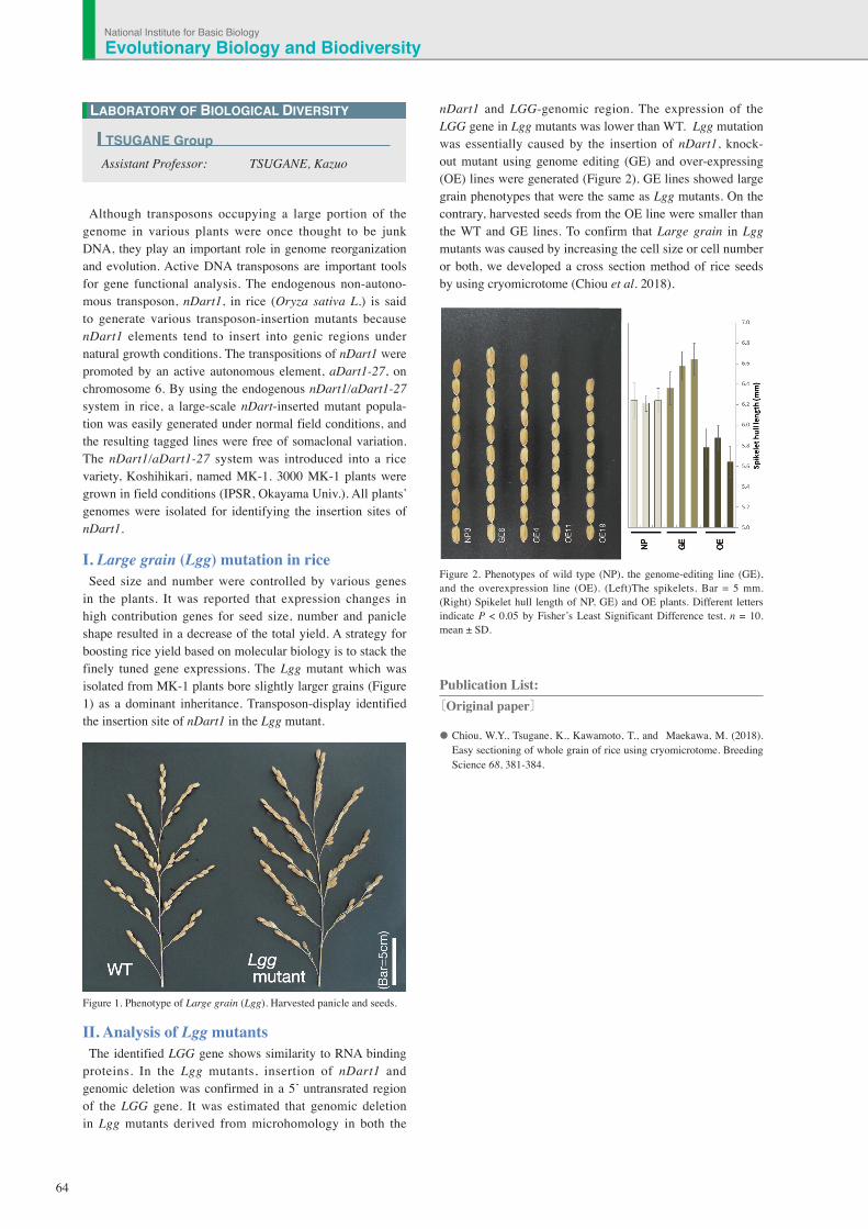
nDart1 and LGG-genomic region. The expression of the LGG gene in Lgg mutants was lower than WT. Lgg mutation was essentially caused by the insertion of nDart1, knock-out mutant using genome editing (GE) and over-expressing (OE) lines were generated (Figure 2). GE lines showed large grain phenotypes that were the same as Lgg mutants. On the contrary, harvested seeds from the OE line were smaller than the WT and GE lines. To confirm that Large grain in Lgg mutants was caused by increasing the cell size or cell number or both, we developed a cross section method of rice seeds by using cryomicrotome (Chiou et al. 2018).
Figure 2. Phenotypes of wild type (NP), the genome-editing line (GE), and the overexpression line (OE). (Left)The spikelets. Bar = 5 mm. (Right) Spikelet hull length of NP, GE) and OE plants. Different letters indicate P < 0.05 by Fisher’s Least Significant Difference test. n = 10. mean ± SD.
Publication List: 〔Original paper〕
• Chiou, W.Y., Tsugane, K., Kawamoto, T., and Maekawa, M. (2018). Easy sectioning of whole grain of rice using cryomicrotome. Breeding Science 68, 381-384.
LABORATORY OF BIOLOGICAL DIVERSITY
Assistant Professor: TSUGANE, Kazuo
TSUGANE Group
Evolutionary Biology and BiodiversityNational Institute for Basic Biology
65
Chromosome condensation is a basic cellular process that ensures the faithful segregation of chromosomes in both mitosis and meiosis. This process is required not only for decreasing chromome arm length, but also for resolving entanglements between sister-chromatids that are created during DNA replication. Any abnormality in this process leads to segregation errors or aneuploidy, which results in cell death. Chromosome condensation is mainly achieved by condensin, a hetero-pentameric protein complex, widely conserved across a variety of organisms ranging from yeast to humans. Despite its conservation and importance in chro-mosome dynamics, it is not fully understood how condensin works. Recent studies have revealed that condensin functions are not restricted to chromosome condensation and segrega-tion during cell divisions, and is required for diverse DNA metabolism such as genome stability, transcriptional regula-tion, and cell differentiation.Our aim is to understand the mechanism and regula-
tion of chromosome condensation. To this end, we have been studying the role of condensin in the budding yeast Saccharomyces cerevisiae. Microscopic observation has indi-cated the nucleolar localization of condensin. Consistent with this, the ribosomal RNA gene (rDNA) repeat is the most con-densed region in the genome during mitosis. We have found that condensin specifically binds to the RFB site located within the rDNA repeat. To date, the best characterized con-densin binding region is the rDNA repeat on the right arm of chromosome XII in budding yeast. We further discovered the multiple protein network required to recruit condensin to the RFB site.
I. Dynamic relocalization of condensin during meiosis
Our genetic screening indicated that two proteins, Csm1 and Lrs4, were required for condensin recruitment to the RFB site. Physical interactions between Csm1/Lrs4 and subunits of condensin are important for the recruitment of condensin to the RFB site. These proteins are known as components of the monopolin complex required for faithful segregation of homologous chromosomes during meiotic division I. During meiosis I, the monopolin complex re-localizes from rDNA repeat to the centromere and acts to ensure sister-chromatid co-orientation. Re-localization of Csm1/Lrs4 proteins suggested that re-localization of con-densin from rDNA repeat to centromere had occurred. As expected, chromatin-IP experiments indicated that condensin re-localizes to the centromere during meiosis I. Condensin might clamp sister-chromatids together during meiosis I.
II. Condensin-dependent chromatin foldingThe RFB site, which consists of a ~150bp DNA sequence,
functions as a cis-element for the recruitment of condensin
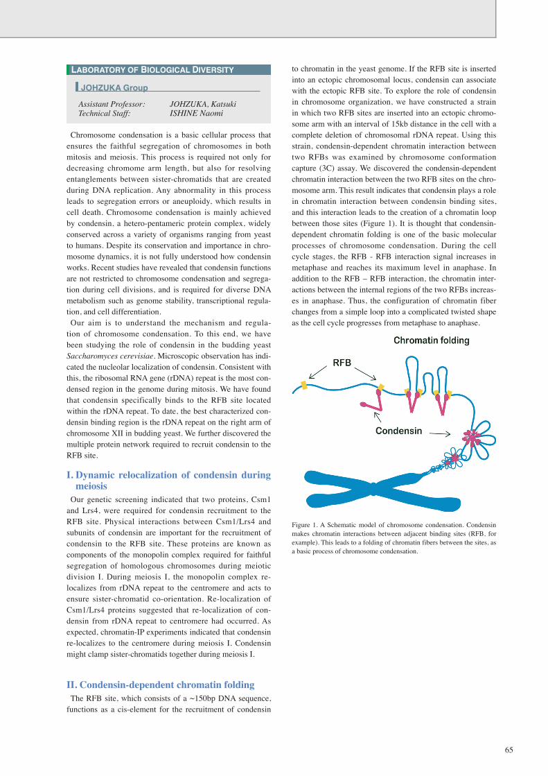
to chromatin in the yeast genome. If the RFB site is inserted into an ectopic chromosomal locus, condensin can associate with the ectopic RFB site. To explore the role of condensin in chromosome organization, we have constructed a strain in which two RFB sites are inserted into an ectopic chromo-some arm with an interval of 15kb distance in the cell with a complete deletion of chromosomal rDNA repeat. Using this strain, condensin-dependent chromatin interaction between two RFBs was examined by chromosome conformation capture (3C) assay. We discovered the condensin-dependent chromatin interaction between the two RFB sites on the chro-mosome arm. This result indicates that condensin plays a role in chromatin interaction between condensin binding sites, and this interaction leads to the creation of a chromatin loop between those sites (Figure 1). It is thought that condensin-dependent chromatin folding is one of the basic molecular processes of chromosome condensation. During the cell cycle stages, the RFB - RFB interaction signal increases in metaphase and reaches its maximum level in anaphase. In addition to the RFB – RFB interaction, the chromatin inter-actions between the internal regions of the two RFBs increas-es in anaphase. Thus, the configuration of chromatin fiber changes from a simple loop into a complicated twisted shape as the cell cycle progresses from metaphase to anaphase.
Figure 1. A Schematic model of chromosome condensation. Condensin makes chromatin interactions between adjacent binding sites (RFB, for example). This leads to a folding of chromatin fibers between the sites, as a basic process of chromosome condensation.
LABORATORY OF BIOLOGICAL DIVERSITY
Assistant Professor: JOHZUKA, KatsukiTechnical Staff: ISHINE Naomi
JOHZUKA Group
66
Organogenesis is accomplished by a series of deforma-tions of the planar cell sheet into a three-dimensional shape during embryogenesis. This drastic structural change is the integrated result of individual cell behaviors in response to spatio-temporally controlled mechanisms. To better understand the programs underlying organ forma-
tion, it is necessary to quantitatively analyze individual cells’ morphology and dynamics. However, it is difficult to do so due to the massive images generated by 4D microscopy and their ambiguity. To unveil organogenesis from the point of view of distinct
cell behaviors, we are developing application software that is capable of describing cell dynamics from 4D time-lapse imaging data sets by employing image processing tech-niques.
I. 4D cell segmentation/tracking systemEpithelial morphogenesis in developing embryos is consid-
ered to be an important model for collective cell migrations. Drastic cell rearrangements lead to drastic structural changes in building elaborate organs such as the tubular network of Drosophila trachea. We are developing a software pipeline, which automatically recognizes individual cell shapes out of 3D space and tracks them through time. This system extracts cell boundaries and reconstitutes cell shapes as a skeleton-ized chain of voxels spanning 3D space. This abstract form of cell visualization makes it possible to describe morpho-metric quantities and kinetics of cells in single-cell resolu-tion (Figure 1). These morphometric quantities allow us to perform comparative studies on shapes and behaviors more precisely among several experimental conditions to gain a better understanding of the genetic programs underlying organogenesis. We are now applying this system to several experimental models to determine the practicality of the system (Shinoda et al.).
II. Image processing pipeline for 3D cell culture
To elucidate the relationship between mechanical forces and epithelial deformation, we developed an image process-ing pipeline for segmentation of nucleus within 3D culture of MDCK cells. This pipeline automated a segmentation/quantification process of a large number of images acquired by several experimental conditions for subsequent statistical analysis (Nishimura et al.).
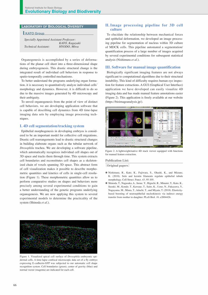
III. Software for manual image quantification Biologically significant imaging features are not always
significant to computational algorithms due to their structural instability. This kind of difficulty requires human eye inspec-tion for feature extractions. A GUI (Graphical User Interface) application we have developed can easily visualize 4D imaging data and has made manual feature annotations easier (Figure 2). This application is freely available at our website (https://bioimageanalysis.jp/).
Figure 2. A lightweight/native 4D stack viewer equipped with functions for manual feature extraction.
Publication List:〔Original papers〕
• Nishimura, R., Kato, K., Fujiwara, S., Ohashi, K., and Mizuno, K. (2018). Solo and keratin filaments regulate epithelial tubule morphology. Cell Struct. Funct. 43, 95-105.
• Shinoda, T,, Nagasaka, A., Inoue, Y., Higuchi, R., Minami, Y,. Kato, K., Suzuki, M., Kondo, T., Kawaue, T., Saito, K., Ueno, N., Fukazawa, Y., Nagayama, M., Miura, T., Adachi, T., and Miyata, T. (2018). Elasticity-based boosting of neuroepithelial nucleokinesis via indirect energy transfer from mother to daughter. PLoS Biol. 16. e2004426.
LABORATORY OF BIOLOGICAL DIVERSITY
Specially Appointed Assistant Professor: KATO, KagayakiTechnical Assistant: HYODO, Miwa
KATO Group
Figure 1. Visualized apical cell surface of Drosophila embryonic epi-dermal cells. A time-lapse confocal microscopic data set of a fly embryo expressing E-cadherin-GFP was subjected to our automatic cell shape recognition system. Cell boundaries (green), center of gravity (blue) and normal vector (magenta) are indicated for each cell.
Evolutionary Biology and BiodiversityNational Institute for Basic Biology
67
Image processing methods significantly contribute to the visualization of biomedical targets acquired from a variety of imaging techniques, which include wide-field optical and electron microscopy, X-ray computed tomography, magnetic resonance imaging and mammography. However, quantitative interpretation of the wide range of complicated biomedical images poses many challenges for research. To counter these, we have developed new computational methods based on math-ematical morphology for quantitative image analysis. One of the most important purposes of image processing is to derive meaningful information, which is expressed as image structural properties. To this end, mathematical morphology is a nonlinear image processing method based on set theory, which is useful for the extraction of the structural properties from an image. It can be used as a fundamental tool to analyze biomedical images.
Novel contrast enhancement method based on mathematical morphology for medical diagnosisImage processing is a crucial step in the analysis of medical
imaging data. As such, it is fundamental to a wide range of biomedical imaging and clinical research fields. Image pro-cessing derives structural features, which are then numeri-cally quantified by image analysis. Contrast enhancement of structural details of lesion regions plays an especially impor-tant role in diagnostic imaging. It improves image quality and aids in clinical diagnosis. By using contrast enhancement methods, more accurate medical diagnoses can be expected. However, conventional image enhancement techniques also emphasize the noise and structure of various normal tissues other than regions containing legions.In this study, a contrast enhancement approach based on a
new type of mathematical morphology was introduced. This method emphasizes only the structure of the lesion while suppressing the emphasis of noise and normal anatomical structure.Mathematical morphology is a methodology for extracting
shape and size information from an image. It involves con-figuration of a set of nonlinear operators that act on images by using structuring elements (SE). The SE, which indicates the shape characteristics in an image, is generally a small and simple binary image. The two basic morphological operators are dilation and erosion from which many operations can be derived. However, since the size of lesions vary, in many cases it is not possible to process them with fixed-size SE.This proposed method is based on a morphological subtrac-
tion method. In this new type of morphological enhance-ment method, h-maxima transform is applied to the original medical image. The unwanted structures that surround the target are suppressed in the process of target enhancement. Furthermore, this new method has no restrictions on the size
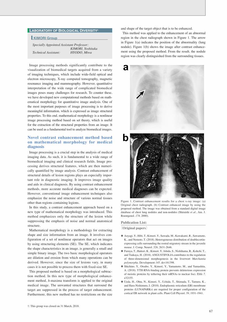
and shape of the target object that is to be enhanced.This method was applied to the enhancement of an abnormal
region in the chest radiograph shown in Figure 1. The arrow in Figure 1(a) indicates the position of the abnormality (lung nodule). Figure 1(b) shows the image after contrast enhance-ment using the proposed method. From the result, the nodule region was clearly distinguished from the surrounding tissues.
Figure 1. Contrast enhancement results for a chest x-ray image: (a) Original chest radiograph. (b) Contrast enhanced image by using the proposed method. The image was obtained from a standard digital image database of chest lung nodules and non-nodules (Shiraishi et al., Am. J. Roentgenol. 174, 2000).
Publication List:〔Original papers〕
• Aoyagi, Y., Hibi, T., Kimori, Y., Sawada, M., Kawakami, R., Sawamoto, K., and Nemoto, T. (2018). Heterogeneous distribution of doublecortin-expressing cells surrounding the rostral migratory stream in the juvenile mouse. J. Comp. Neurol. 526, 2631-2646.
• Furuya, T., Hattori, K., Kimori, Y., Ishida, S., Nishihama, R., Kohchi, T., and Tsukaya, H. (2018). ANGUSTIFOLIA contributes to the regulation of three-dimensional morphogenesis in the liverwort Marchantia polymorpha. Development 145. dev161398.
• Shichino, Y., Otsubo, Y., Kimori, Y., Yamamoto, M., and Yamashita, A. (2018). YTH-RNA-binding protein prevents deleterious expression of meiotic proteins by tethering their mRNAs to nuclear foci. Elife 7. e32155.
• Ueda, H., Ohta, N., Kimori, Y., Uchida, T., Shimada, T., Tamura, K., and Hara-Nishimura, I. (2018). Endoplasmic reticulum (ER) membrane proteins (LUNAPARKs) are required for proper configuration of the cortical ER network in plant cells. Plant Cell Physiol. 59, 1931-1941.
LABORATORY OF BIOLOGICAL DIVERSITY†
Specially Appointed Assistant Professor: KIMORI, YoshitakaTechnical Assistant: HYODO, Miwa
KIMORI Group
†: This group was closed on 31 March, 2018.