evolution of hiv-1 in northern tanzania: a … · using a peptide-binding enzyme immunoassay...
TRANSCRIPT
International Community Health Department of General Practice and Community Medicine
Faculty of Medicine, University of Oslo, Norway
EVOLUTION OF HIV-1 IN NORTHERN TANZANIA: A RETROSPECTIVE STUDY
THESIS
By
Balthazar Melchior Rugaitika Nyombi
Thesis submitted as part of fulfillment of Master of Philosophy
Degree in International Community Health
Supervisor: Gunnar Bjune, Professor
Co-supervisors: Stig Jeansson, Professor
Holm-Hansen Carol, PhD
Barongo Longin, Mmed
Nkya Watoky, Professor
June 2002

2
Dedications This work is dedicated to my father, the late Omulangira Melchior Rugaitika. To my dear mother Ma Asteria, my beloved grand mother Omukaile Ma Yozefina, my brothers and sisters. For your love and prayers.
3
TABLE OF CONTENTS
Dedications ..................................................................................................................... 2 Acknowledgements......................................................................................................... 4 Abstract ........................................................................................................................... 5 Abbreviations.................................................................................................................. 6 Introduction..................................................................................................................... 7 Global situation of HIV/AIDS........................................................................................ 9 Current situation of HIV/AIDS in Tanzania................................................................. 10 General description of a retrovirus ............................................................................... 12 Replication cycle of HIV .............................................................................................. 13 Structure of HIV-1 genome .......................................................................................... 14 HIV-1 group M genetic variability ............................................................................... 16 The global distribution of HIV-1 subtypes ................................................................... 17 Molecular epidemiology of HIV-1 in Tanzania............................................................ 18 Subtyping techniques .................................................................................................... 19
Peptide-binding enzyme immunoassay..................................................................... 19 Polymerase chain reaction and DNA sequencing..................................................... 20 Heteroduplex mobility assay..................................................................................... 22
Peptide-binding enzyme immunoassay in HIV-1 subtyping ........................................ 23 Rationale for using PEIA.............................................................................................. 25 Aim of the study............................................................................................................ 26 Research hypothesis...................................................................................................... 27 Main objective of the study........................................................................................... 28 Specific objectives ........................................................................................................ 28 Materials and methods .................................................................................................. 28
Study site................................................................................................................... 28 Study design.............................................................................................................. 30 Peptides ..................................................................................................................... 30 Serum specimens ...................................................................................................... 31 Serum sample selection............................................................................................. 32 Serum sample controls.............................................................................................. 32 Sample analysis......................................................................................................... 32 V3 PEIA.................................................................................................................... 33 V3 peptide antigen limiting dilution assay ............................................................... 34 gp41 PEIA................................................................................................................. 34
Results........................................................................................................................... 35 Discussion and conclusion............................................................................................ 40 Appendix....................................................................................................................... 45 References..................................................................................................................... 46

4
Acknowledgements I praise God for His blessings and guidance throughout the period of this work.
I wish to thank the Section for International Health, Department of General Practice and Community Medicine at the University of Oslo for providing me with the opportunity to persue the degree of Master of Philosophy in International Community Health. In addition, I am grateful to Statens Lånekasse for financial support. Special thanks are extended to the Ministry of Health and Kilimanjaro Christian Medical Centre (KCMC) in Tanzania for granting me permission to undertake the studies in Norway. I thank Prof. Gunnar Bjune, my supervisor for his academic guidance, critics and support. Special thanks are extended to the Section for International Health for financial support to cover expenses related to fieldwork and for making this work possible. Appreciation and special thanks to my friend and co-supervisor Dr. Carol Holm-Hansen
for tireless encouragement and making it possible for me to persue further studies. I am
also grateful for her professional guidance, support and enthusiasm.
Appreciation is also extended to Prof. Stig Jeansson my co-supervisor for continual
assistance, dedication, guidance, helpful critics and material support for this work. Prof.
Stig and Dr. Carol, without you this thesis would not have become a reality.
Furthermore, I would like to thank Dr. Pontiano Kaleebu of UVRI, Entebbe, Uganda for
the invitation and training in peptide ELISA at his institute. Thank you for the guidance
and advice. I wish to acknowledge the Centralized Facility for AIDS Reagents, supported
by the EU Programme EVA (contract BMH4 97/2515) and the UK Medical Research
Council for peptide donation. I also thank Dr. Chou Pau of CDC, Atlanta for donating the
gp41 peptides. Without their donations this work would not have been possible.
I would like to thank KCMC and the Research Laboratory for allowing me to use the
banked sera and laboratory facilities for analysis. Appreciation is extended to Prof. Nkya
and Dr. Barongo, my co-supervisors, for their encouragement and advice during the
sample analysis.
To my dearest wife Evelyn, and children Lacatus, Henrick and Vanessa. I thank them for
their patience, understanding and prayers.
To my fellow classmates, thank you for being my jovial family in Oslo.

5
Abstract
Development of an effective vaccine against HIV has faced great challenges due to the
great genetic diversity of the virus. However, in different geographical areas candidate
vaccines based on the prevalent HIV-1 subtype(s) are now entering clinical trials.
Genotypic and serological assays have been used in HIV-1 subtyping. The peptide-
binding enzyme immunoassay has been a useful tool for subtyping HIV-1. This assay in
combination with an antigen limiting dilution assay was established at the Kilimanjaro
Christian Medical Centre (KCMC) in Moshi, Tanzania. HIV-1 positive serum samples
stored at KCMC were characterized using these assays to determine the trend since the
start of the epidemic. In addition, the distribution of HIV-1 subtypes currently circulating
in northern Tanzania was determined in order to provide important information that
might be of relevance to future vaccine studies and trials in Tanzania.
Frozen HIV-1 positive serum samples from individuals diagnosed at KCMC between
1985 and 2001 were used in this study. Synthetic peptides representing HIV-1 subtypes
A, B, C, D and E derived from consensus gp120 V3 sequences were used in an indirect
peptide-binding enzyme immunoassay and an antigen limiting dilution assay. The gp41
peptide D was used to increase the specificity by discriminating subtype D from non-D
viruses.
Two hundred and twenty six samples were analyzed; 196 (87%) samples were
successfully subtyped while 30 (13%) could not be typed using these methods. In 1985
the prevalent subtypes were A (52%) and D (48%). Subtype C started to circulate in the
late 1980s and in 2001 was the most prevalent circulating subtype in Tanzania. The
currently circulating subtypes are A (32%), C (51%) and D (17%).
HIV-1 vaccines entering clinical trials in Tanzania should be based on the predominant
circulating subtype(s).
6
Abbreviations
AIDS acquired immunodeficiency syndrome CRF circulating recombinant forms CCR5 CC chemokine receptor-5 CD4 cluster of differentiation 4 CD8 cluster of differentiation 8 CXCR4 CXC chemokine receptor-4 DNA deoxyribonucleic acid ELISA enzyme linked immunosorbent assay env envelope gag group associated genes gp glycoprotein HIV-1, -2 human immunodeficiency virus type 1, -2 HMA heteroduplex mobility assay HTA heteroduplex tracking assay IDR immunodominant region KCMC Kilimanjaro Christian Medical Centre LTR long terminal repeat MHC major histocompatibility complex MOH Ministry of Health NACP National AIDS Control Programme nef negative factor NSI non-syncytium inducing PCR polymerase chain reaction PEIA peptide enzyme immunoassay pol polymerases rev retroviral regulatory protein RNA ribonucleic acid mRNA messenger RNA SD standard deviation SSA sub-Saharan Africa SI syncytium inducing tat transcriptional transactivator UNAIDS Joint United Nations Programme on HIV/AIDS vif viral infectivity factor vpu viral protein U Wb Western blot WHO World Health Organization
7
Introduction The human immunodeficiency virus (HIV) has brought about a global epidemic far more
extensive than what was predicted even a decade ago. Since the epidemic began, HIV has
now infected more than 60 million individuals. By the end of 2001, an estimated 40
million people were living with HIV worldwide (1). HIV is the etiology of the slow
progressing and serious acquired immunodeficiency syndrome (AIDS). There are two
HIV types, HIV-1 and HIV-2, of which the first one is responsible for the pandemic.
HIV-1 infections are characterized by a brief acute phase with high viraemia followed by
a chronic phase characterized by the depletion of CD4+ T-lymphocytes in the peripheral
circulation to less than 20% of normal within 8 – 10 years. Opportunistic infections and
certain rare cancers occur due to the immunosuppresion. AIDS is the leading cause of
death in Sub-Saharan African (SSA) and the fourth biggest killer worldwide. The
epidemic has a profound impact on social, political, cultural and economic development.
Millions of people in SSA carry the virus but do not know they are infected. Many more
know little or nothing about how to protect themselves against HIV. Developing
countries in SSA cannot care for their HIV infected people and preventive efforts are
very difficult. In light of the current HIV/AIDS situation it is clear that preventive
methods that are inexpensive, accessible and effective are urgently needed. Hence, a HIV
vaccine may be one of the best preventive methods for most of the developing countries
such as Tanzania. The heterogeneity of HIV found in different parts of the world
indicates that the strains initially used to develop vaccine candidates were not
representative of the global epidemic. The genetic variability of HIV is considered to be
one of the major obstacles in the design and development of effective HIV vaccines.
8
Kaleebu and colleagues in Uganda developed an algorithmic method to subtype HIV-1
using a peptide-binding enzyme immunoassay (PEIA), the heteroduplex mobility assay
(HMA) and DNA sequencing. Serological analysis using a V3 peptide antigen limiting
dilution assay and a gp41 PEIA correctly identified 78% of characterized HIV-1 samples
(2). We decided to use the serological method of Kaleebu in the present study.
Our study provides information on currently circulating HIV-1 subtypes as well as of
subtypes that occurred from 1985 to 2001 in northern Tanzania. This information is of
value in the development of a HIV vaccine for Tanzania. The efficacy of vaccine
candidates may be strain or subtype specific. It is therefore important to know the
distribution of the HIV-1 subtypes in order to evaluate the effectiveness of the proposed
vaccine candidates and to target future vaccine development and laboratory testing. The
public health implications of such findings include prevention and diagnostic strategies.
Until now the information available on the molecular epidemiology of HIV-1 in Tanzania
is based on limited data generated from small hospital-based descriptive studies done ten
years after the first case of HIV was reported (Table 1). The present study indicates that
HIV-1 subtypes A and D were the first in northern Tanzania. Furthermore, it is shown
that subtype C was introduced in northern Tanzania in late 80s and now is the most
prevalent HIV-1 subtype circulating in the region. The present study demonstrates that
subtypes A, C and D are the currently circulating HIV-1 subtypes in northern Tanzania. It
is important that vaccine candidates in Tanzania should be based on these HIV-1
subtypes.
The purpose of this study was to establish the PEIA and use it to track the evolution and
distribution of HIV-1 subtypes in northern Tanzania for the period from 1985 to 2001.
9
Global situation of HIV/AIDS HIV was first recognized in 1981 among homosexual men in USA. These men had
infections seldom observed among immunocompetent individuals. Later it was
discovered that these individuals had a severe immunodeficiency. HIV infection was not
only confined to homosexual men, new cases in women were reported and heterosexual
HIV transmission was described (3). It was noted that exchange of body fluids from an
infected individual to an uninfected individual was the mode of transmission. This was
reported for heterosexual transmission (3) as well as for intravenous drug users (4),
prenatal infections and breastfeeding (5), transfusions, organ transplants and
inseminations (6). Globally, the leading mode of transmission is heterosexual. The
challenge caused by HIV varies geographically depending on how far and fast the virus is
spreading. According to the UNAIDS and WHO estimates for the year 2001, the number
of people living with HIV/AIDS is 40 million and about 5 million people were newly
infected with 14,000 new infections everyday. About 85% of the new infections were in
adults (15-49 years old) and 50% of these were among women. As a result we are
witnessing a rise in the number of children born with HIV. The total number of deaths
due to AIDS in the year 2001 was estimated to be 3 million worldwide (1).
Countries in SSA are experiencing a severe impact of the HIV epidemic. Regional
HIV/AIDS statistics by the end of 2001 show that the total number of people living with
HIV/AIDS is estimated to be 28 million which is 70% of total world’s people living with
HIV/AIDS. The total number of people newly infected with HIV during 2001 in SSA
was estimated to be 3.4 million, which is 68% of the world’s newly infected people in the
10
same year (1). Eastern Europe and central Asian countries have also shown a rapid
increase of the HIV epidemic. UNAIDS and WHO estimates put the number of adults
and children living with HIV/AIDS in these countries at 1 million by the end of the year
2001 as compared to 700,000 in the previous year.
The HIV epidemic in some SSA countries seems to be stabilizing. This may be due to the
fact that epidemic has been going on for so long. The infection has already affected many
people in the sexually active population leaving a smaller pool of people still able to
acquire the infection. The other factor is the success of preventive programs in some of
the African countries, notably Uganda, which has reduced national infection rates. The
epidemic in Uganda has not only stabilized but, more importantly, both the prevalence
and incidence rates are decreasing.
Current situation of HIV/AIDS in Tanzania The first AIDS case was reported in the Kagera region in northwestern Tanzania in 1983
(7). This started as a disease of businessmen and women who were crossing the
Tanzania/Uganda border. They often wore shirts with a picture of an eagle by the name
of “Juliana”. Hence, the sick persons were said to have acquired “Juliana” disease.
Three cases were reported in 1983 and by the end of 1985 eight of the twenty regions of
Tanzania had reported 404 AIDS cases to the Ministry of Health (MOH). By the end of
1986 the number reached 1,525 and all the regions in the country had reported AIDS
cases (8, 9). In 1988 the Ministry of Health established the National AIDS Control
Programme (NACP) to coordinate the AIDS control activities and monitor the HIV/AIDS
11
epidemic in Tanzania. Ten years after the first case was reported (8, 9) the total number
of notified cases had reached 73,572. The cumulative number of AIDS cases reported to
the NACP from 20 regions of the Tanzanian mainland by the end of 2000 was 130,386
with 11,673 newly reported cases (9). However, according to the UNAIDS and WHO
estimates, Tanzania had 1.3 million people living with HIV/AIDS by the end of 1999.
The AIDS deaths during 1999 were estimated to be 140,000. The estimated cumulative
number of orphans since the beginning of the epidemic was 1.1 million with two thirds of
these orphans still living (10).
In the 2000 report by the NACP, the overall prevalence among blood donors was 9.9%
countrywide. Furthermore, reports from voluntary counseling and HIV testing centers
showed that 6,539 clients were counseled, 3,338 agreed to be tested and out of them
59.5% tested HIV positive. Heterosexual HIV transmission accounts for 77.2%, mother
to child 3.4% and blood transfusion 0.4% of all HIV transmissions in Tanzania.
Information on the mode of transmission for 19% was not available. Data derived from
28 sentinel surveillance sites show that the countrywide HIV prevalence among pregnant
women for the year 2000 ranged from 4.2% in one site in Mwanza region to 32.1% in
one site in Iringa region (9). It is clear that the number of HIV/AIDS cases in Tanzania is
on the rise resulting in early mortality, loss of manpower and breadwinners, increase in
the number of orphans and social instability. Tanzania being a developing country cannot
care for her HIV infected patients and it is very difficult to establish effective preventive
measures.
It is clear that preventive methods that are inexpensive, accessible and effective are
urgently needed in response to the current HIV/AIDS situation. Hence, an HIV vaccine
12
may be one of the best preventive methods for most developing countries including
Tanzania.
General description of a retrovirus
HIV is a RNA virus that belongs to the family of retroviruses, genus Lentiviridae (11).
The central nucleocapsid core of the virion contains two copies of the RNA genome, the
enzymes reverse transcriptase and integrase, and core proteins. Surrounding the core is
protein capsid surrounded by a phospholipid envelope with embedded surface
glycoproteins (figure 1).

13
Figure 1. Model of HIV-1 virion.
Source: http//www.nature.com/reviews/immunol - April 2002 volume 2.
Replication cycle of HIV HIV replicates within a host cell using RNA as a template to make DNA. HIV infection
begins when the HIV particle encounters a cell with the surface receptor molecule CD4.
The virus particle uses gp120 to attach itself on the cell membrane and enters the CD4
positive cell with the help of the chemokine receptors CXCR4 or CCR5 (12). CXCR4 is a
co-receptor for the T-cell tropic syncytium inducing (SI) HIV strains while the CCR5 is
strictly for macrophage tropic non-syncytium inducing (NSI) HIV strains. Recently, it
14
has been shown that some HIV-1 variants can infect CD8 cells rather than CD4 cells.
(13) (14).
Within the cell, the virus particle releases its two RNA molecules and the enzyme reverse
transcriptase converts the viral RNA into DNA. This HIV DNA then moves into the cell
nucleus where, with the help of the enzyme integrase, it is inserted into the host cell
DNA. Once in the cell’s chromosomes, HIV DNA is called proviral DNA. The viral
mRNA is transcribed from the HIV proviral DNA by the host cell RNA polymerase and
translated into several large polyproteins. Copies of RNA and newly created polyproteins
move closer to the cell membrane. New virions begin to form and bulge on the side of the
of the host cell. Then the virions separate completely from host cell in the budding stage.
During the budding from the host cell membrane, the viral proteinases become active
resulting in cleavage of various subunits and generation of the mature form of HIV (15)
(12).
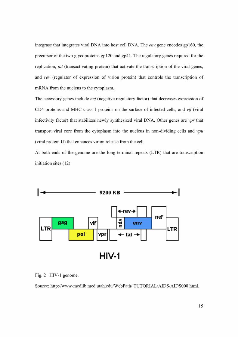
Structure of HIV-1 genome The HIV genome consists of two identical molecules of single stranded, positive polarity
RNA. Each RNA strand is approximately 9200 bases long. Like all retroviruses, the HIV
genome (figure 2) consists of structural genes gag, pol and env, which are in the order 5’-
gag-pol-env-3’ and encode the structural proteins. In addition the HIV genome has six
regulatory genes including tat and rev for the replication of the virus and nef, vif, vpr and
vpu, which are accessory genes, not involved in replication. The gag gene encodes the
internal core proteins p7, p17 and p24. The pol gene encodes reverse transcriptase, which
transcribes RNA genome into DNA, protease that cleaves precursor polypeptides and
15
integrase that integrates viral DNA into host cell DNA. The env gene encodes gp160, the
precursor of the two glycoproteins gp120 and gp41. The regulatory genes required for the
replication, tat (transactivating protein) that activate the transcription of the viral genes,
and rev (regulator of expression of virion protein) that controls the transcription of
mRNA from the nucleus to the cytoplasm.
The accessory genes include nef (negative regulatory factor) that decreases expression of
CD4 proteins and MHC class 1 proteins on the surface of infected cells, and vif (viral
infectivity factor) that stabilizes newly synthesized viral DNA. Other genes are vpr that
transport viral core from the cytoplasm into the nucleus in non-dividing cells and vpu
(viral protein U) that enhances virion release from the cell.
At both ends of the genome are the long terminal repeats (LTR) that are transcription
initiation sites (12)
Fig. 2 HIV-1 genome.
Source: http://www-medlib.med.utah.edu/WebPath/ TUTORIAL/AIDS/AIDS008.html.
16
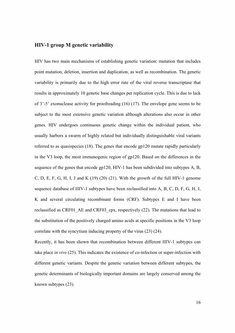
HIV-1 group M genetic variability HIV has two main mechanisms of establishing genetic variation: mutation that includes
point mutation, deletion, insertion and duplication, as well as recombination. The genetic
variability is primarily due to the high error rate of the viral reverse transcriptase that
results in approximately 10 genetic base changes per replication cycle. This is due to lack
of 3’-5’ exonuclease activity for proofreading (16) (17). The envelope gene seems to be
subject to the most extensive genetic variation although alterations also occur in other
genes. HIV undergoes continuous genetic change within the individual patient, who
usually harbors a swarm of highly related but individually distinguishable viral variants
referred to as quasispecies (18). The genes that encode gp120 mutate rapidly particularly
in the V3 loop, the most immunogenic region of gp120. Based on the differences in the
sequence of the genes that encode gp120, HIV-1 has been subdivided into subtypes A, B,
C, D, E, F, G, H, I, J and K (19) (20) (21). With the growth of the full HIV-1 genome
sequence database of HIV-1 subtypes have been reclassified into A, B, C, D, F, G, H, J,
K and several circulating recombinant forms (CRF). Subtypes E and I have been
reclassified as CRF01_AE and CRF03_cpx, respectively (22). The mutations that lead to
the substitution of the positively charged amino acids at specific positions in the V3 loop
correlate with the syncytium inducing property of the virus (23) (24).
Recently, it has been shown that recombination between different HIV-1 subtypes can
take place in vivo (25). This indicates the existence of co-infection or super-infection with
different genetic variants. Despite the genetic variation between different subtypes, the
genetic determinants of biologically important domains are largely conserved among the
known subtypes (23).
17
The global distribution of HIV-1 subtypes HIV-1 subtypes are unevenly distributed in different geographical locations. Almost all
subtypes are present in SSA where the HIV-1 epidemic is believed to be of long duration.
Subtypes A, B, C, D, F, G, H, CRF01_AE and group O are found in west and central
Africa. Subtype C is prevalent in the horn of Africa including Ethiopia, in addition to the
south and southwest of Africa.
In Asia subtypes B, C and CRF01_AE are predominant, subtype C is predominant in
India while CRF01_AE predominates in Thailand.
In North America subtype B is predominant in addition to the identification of subtypes
C, D, CRF01-AE, CRF02-AG and group O. Subtypes that are found in south America
include B, F, C and CRF12-BF. In Europe subtype B accounts for the majority of HIV-1
infections with non-B subtypes being detected in several parts of the continent. Group O
has been reported in France and Spain. Subtype F has been reported in Romania and
subtype G in Russia. HIV-1 infections in Australia have been reported to be of subtype B.
However, due to the increased extent of travel and mobility of people from one region to
another, HIV subtypes are no longer limited to a few geographical areas but can be found
throughout the majority of countries (19) (20) (21) (22).
18
Molecular epidemiology of HIV-1 in Tanzania Studies conducted in Tanzania on the molecular epidemiology of HIV-1 subtypes showed
a predominant occurrence of subtype A, C and D. Subtype C is predominant in Mbeya in
the southwestern part of Tanzania bordering Zambia as well as with other southern
African countries. Hoelscher and colleagues (26) studied HIV-1 V3 serotypes of patients
in Mbeya town and four subtypes were identified: A (29%), C (55%), D (15%) and G
(1%). Subtypes A and D have been documented in Dar-es-Salaam and Kilimanjaro. A
study on 14 virus isolates by Holm-Hansen and colleagues (27) in Dar-es-Salaam
indicated divergent and clustered sequences of HIV-1 subtypes A, C and D. A study by
Blackard and colleagues (25) on the diversity of HIV-1 LTR following mother-to-child
transmission in 19 mother-infant pairs, detected subtypes A, C, D and intersubtype
recombinants. In a recent study by Kiwelu and colleagues (28) in Kilimanjaro, northern
Tanzania, on the determination of nucleotide sequences of the gp120 V3 from isolates
circulating in the region, subtypes A, C, D and B-like sequences were identified (28).
In summary, the available information on the molecular epidemology of HIV-1 in
Tanzania is based on limited data generated from a small number of hospital-based
studies. All these studies were done ten years after the first HIV/AIDS case was reported
in Tanzania. Table 1 summarizes the previous studies done on the molecular
epidemiology of HIV-1 in Tanzania.
19
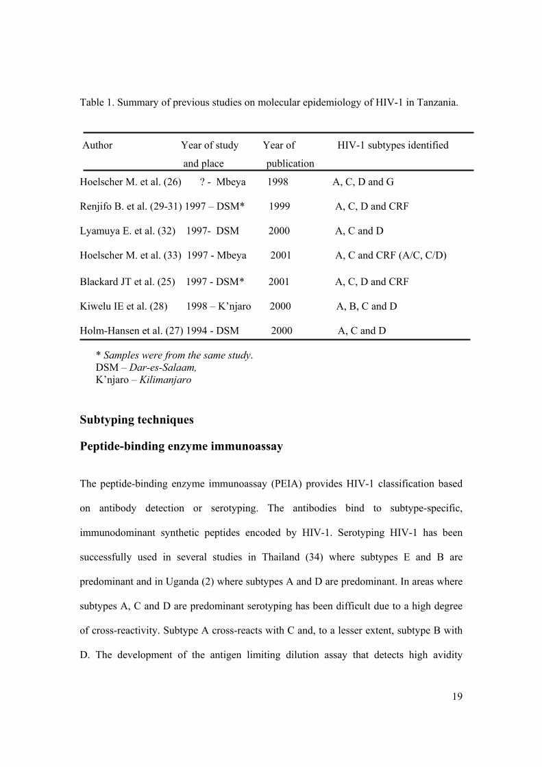
Table 1. Summary of previous studies on molecular epidemiology of HIV-1 in Tanzania.
Author Year of study Year of HIV-1 subtypes identified
and place publication
Hoelscher M. et al. (26) ? - Mbeya 1998 A, C, D and G Renjifo B. et al. (29-31) 1997 – DSM* 1999 A, C, D and CRF Lyamuya E. et al. (32) 1997- DSM 2000 A, C and D Hoelscher M. et al. (33) 1997 - Mbeya 2001 A, C and CRF (A/C, C/D) Blackard JT et al. (25) 1997 - DSM * 2001 A, C, D and CRF Kiwelu IE et al. (28) 1998 – K’njaro 2000 A, B, C and D Holm-Hansen et al. (27) 1994 - DSM 2000 A, C and D
* Samples were from the same study. DSM – Dar-es-Salaam, K’njaro – Kilimanjaro
Subtyping techniques Peptide-binding enzyme immunoassay The peptide-binding enzyme immunoassay (PEIA) provides HIV-1 classification based
on antibody detection or serotyping. The antibodies bind to subtype-specific,
immunodominant synthetic peptides encoded by HIV-1. Serotyping HIV-1 has been
successfully used in several studies in Thailand (34) where subtypes E and B are
predominant and in Uganda (2) where subtypes A and D are predominant. In areas where
subtypes A, C and D are predominant serotyping has been difficult due to a high degree
of cross-reactivity. Subtype A cross-reacts with C and, to a lesser extent, subtype B with
D. The development of the antigen limiting dilution assay that detects high avidity
20
antibodies improved the specificity of the V3 PEIA (35). The antigen limiting dilution
assay is characterized by subjecting a single dilution of the serum sample to increasing
dilutions of antigen. A 17-amino acid immunodominant region (IDR) of the envelope
gp41 from ELI, an HIV-1 isolate from Zaire discriminates subtype D from non-D viruses
(2). PEIA requires less rigorous sample processing, is practical for large scale screening
of samples in the field environment, is easy to perform and requires less sophisticated
equipment compared to molecular techniques. PEIA is therefore a useful tool in
molecular epidemiological studies.
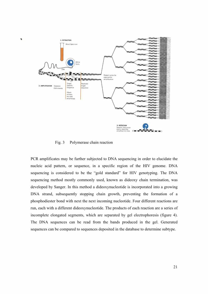
Polymerase chain reaction and DNA sequencing The polymerase chain reaction (PCR) method is based on a primer-initiated bi-directional
DNA synthesis of a region of nucleic acid. This method is the basis of genotypic analysis
and enables the detection of limited copy numbers of DNA. DNA can be either
synthesized from plasma RNA or proviral DNA extracted from the mononuclear cells.
PCR on RNA templates (reverse transcriptase PCR or RT–PCR) is preceded by a reverse
transcription step to produce cDNA. On several cycles DNA strands can be amplified
exponentially on subjection to specific primers and polymerase enzyme (figure 3). Sets of
HIV-1 subtype specific primers have been used in PCRs to differentiate distinct
genotypes (36).
21
Fig. 3 Polymerase chain reaction
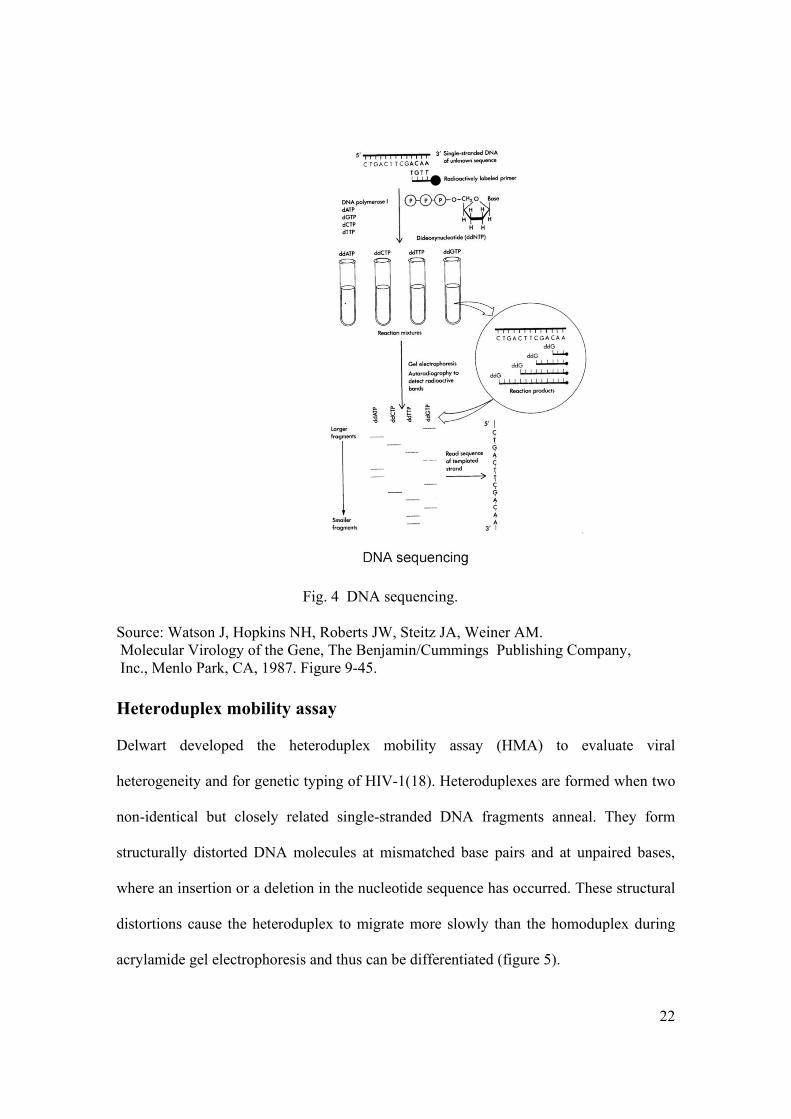
PCR amplificates may be further subjected to DNA sequencing in order to elucidate the
nucleic acid pattern, or sequence, in a specific region of the HIV genome. DNA
sequencing is considered to be the “gold standard” for HIV genotyping. The DNA
sequencing method mostly commonly used, known as dideoxy chain termination, was
developed by Sanger. In this method a dideoxynucleotide is incorporated into a growing
DNA strand, subsequently stopping chain growth, preventing the formation of a
phosphodiester bond with next the next incoming nucleotide. Four different reactions are
run, each with a different dideoxynucleotide. The products of each reaction are a series of
incomplete elongated segments, which are separated by gel electrophoresis (figure 4).
The DNA sequences can be read from the bands produced in the gel. Generated
sequences can be compared to sequences deposited in the database to determine subtype.
22
Fig. 4 DNA sequencing.
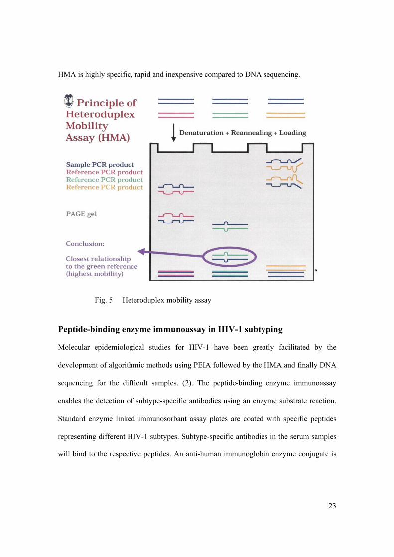
Source: Watson J, Hopkins NH, Roberts JW, Steitz JA, Weiner AM. Molecular Virology of the Gene, The Benjamin/Cummings Publishing Company, Inc., Menlo Park, CA, 1987. Figure 9-45. Heteroduplex mobility assay Delwart developed the heteroduplex mobility assay (HMA) to evaluate viral
heterogeneity and for genetic typing of HIV-1(18). Heteroduplexes are formed when two
non-identical but closely related single-stranded DNA fragments anneal. They form
structurally distorted DNA molecules at mismatched base pairs and at unpaired bases,
where an insertion or a deletion in the nucleotide sequence has occurred. These structural
distortions cause the heteroduplex to migrate more slowly than the homoduplex during
acrylamide gel electrophoresis and thus can be differentiated (figure 5).
23
HMA is highly specific, rapid and inexpensive compared to DNA sequencing.
Fig. 5 Heteroduplex mobility assay
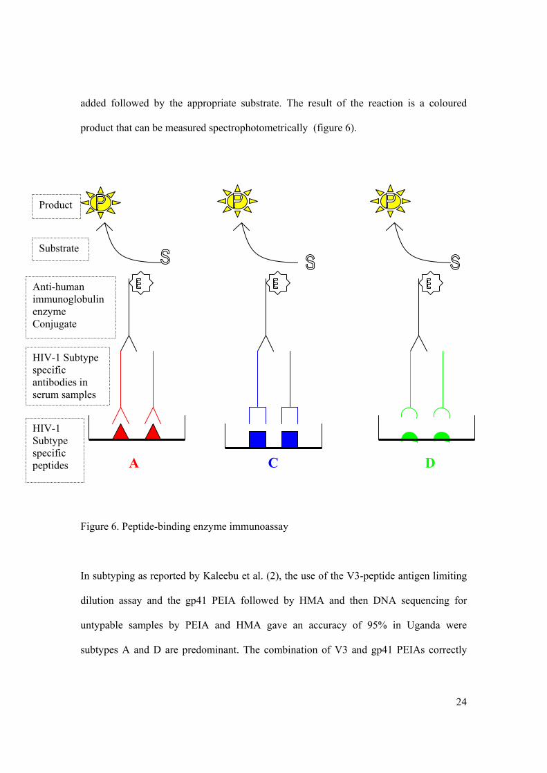
Peptide-binding enzyme immunoassay in HIV-1 subtyping Molecular epidemiological studies for HIV-1 have been greatly facilitated by the
development of algorithmic methods using PEIA followed by the HMA and finally DNA
sequencing for the difficult samples. (2). The peptide-binding enzyme immunoassay
enables the detection of subtype-specific antibodies using an enzyme substrate reaction.
Standard enzyme linked immunosorbant assay plates are coated with specific peptides
representing different HIV-1 subtypes. Subtype-specific antibodies in the serum samples
will bind to the respective peptides. An anti-human immunoglobin enzyme conjugate is
24
added followed by the appropriate substrate. The result of the reaction is a coloured
product that can be measured spectrophotometrically (figure 6).
A C D
Figure 6. Peptide-binding enzyme immunoassay
In subtyping as reported by Kaleebu et al. (2), the use of the V3-peptide antigen limiting
dilution assay and the gp41 PEIA followed by HMA and then DNA sequencing for
untypable samples by PEIA and HMA gave an accuracy of 95% in Uganda were
subtypes A and D are predominant. The combination of V3 and gp41 PEIAs correctly
Product
Substrate
Anti-human immunoglobulin enzyme Conjugate
HIV-1 Subtype specific antibodies in serum samples
HIV-1 Subtype specific peptides
25
typed 78% of the samples. This is an acceptable level of accuracy for a large-scale
epidemiological study.
The total cost for consumables was reduced when subtyping was based on an algorithm
in which all the samples were screened by PEIA followed by HMA and finally DNA
sequencing on untypable samples. The UNAIDS Network for HIV Isolation and
Characterization has reported that subtype A and D are difficult to differentiate by V3
PEIA alone (35) (37). In addition, HIV-1 subtypes A and C have numerous common
amino acids in V3 region and are also difficult or impossible to distinguish by V3 PEIA
alone.
Rationale for using PEIA HIV-1 subtyping can be done by molecular biological or serological techniques. PEIA is
a serological method. Many groups have reported a good correlation between the results
of HIV-1 subtyping using serological and molecular techniques (2) (35-38). Synthetic
peptides representing dominant antigenic epitopes in the V3-loop of the envelope
glycoproteins gp120 and gp41 are commonly used in PEIA. The amino acid sequences of
the peptides are based on the HIV-1 subtype consensus sequences for the V3-loop and
gp41. Synthetic peptides are used as antigens in serotyping of HIV-1. Studies using PEIA
in Thailand and Uganda correctly predicted 80% to 95% of HIV-1 genotypes (36) (2).
This is sufficient for population-based epidemiological purposes. Genetic subtyping using
differential PCR (36), DNA sequencing and HMA provides a more reliable and
comprehensive approach to HIV-1 subtyping. These methods are costly, time and labour
intensive, technically difficult and require sophisticated equipment not available in field
26
settings. HMA needs a number of reference standards for each subtype used in the assay.
This makes genetic subtyping unsuitable for population-based studies with large numbers
of samples. Genetic subtyping is too expensive to be performed in developing countries
such as Tanzania.
Serological subtyping can supplement genetic subtyping, enable the use of banked sera
and allow large numbers of samples to be studied. The method is simple, economical,
less technically rigorous, there is no need for sophisticated equipment, it is easy to obtain
small volumes of serum for analysis and it can be done in the field settings where large-
scale vaccine trials are likely to occur. Therefore, we decided to use this methodology for
subtyping HIV-1 in northern Tanzania where previous studies have shown subtypes A, C
and D to be predominant.
Aim of the study Tanzania is one of the SSA countries that have been most affected by the HIV-1
epidemic. Intervention measures have reduced HIV-1 infection rates in some areas in
Tanzania. A decline in the prevalence of HIV-1 from 13.7% in 1996 to 7% in 1999
among pregnant women attending antenatal clinic in Bukoba urban in Kagera region was
reported (8). An effective vaccine against HIV infection may provide one way to prevent
the fast increase of the HIV/AIDS epidemic. The genetic variability of HIV is one of the
major obstacles for the development of effective HIV vaccines.
In July 2000, during the AIDS Conference in Durban, South Africa, it was announced
that the first HIV-1 vaccine candidates designed for Africa would be entering Phase I
clinical trials in Kenya and South Africa. In a collaboration between British and Kenyan
27
scientists, a vaccine based on HIV-1 subtype A has being developed. In South Africa, a
candidate vaccine designed using an attenuated form of Venezuelan equine encephalitis
virus with genes from HIV-1 subtype C isolates will be entering clinical trials in the near
future. The International AIDS Vaccine Initiative (IAVI) together with U.S. National
Institute of Allergy and Infectious Diseases and the South African AIDS Vaccine
Initiative are funding the development of these vaccines (39).
The northern part of Tanzania is famous for its national parks and tourist industry. In
addition, the famous trunk road from Kenya to southern Africa passes through the region.
For Tanzania, being geographically situated in the middle of the two vaccine trial
countries, it is important to know which HIV-1 subtypes are circulating in the event
vaccine studies will be conducted in this region. The efficacy of these candidate vaccines
may be strain- or subtype-specific. It is therefore important to know the distribution of
the HIV-1 subtypes in order to determine the relevance of proposed vaccine candidates
and to add valuable information regarding the composition of future vaccines suitable for
Tanzania.
Research hypothesis In northern Tanzania, the epidemic has been growing since 1983 and there is a possibility
that the patterns of circulating HIV-1 subtypes have changed over time.
28
Main objective of the study The objective of the present study was to determine the evolution of HIV-1 subtypes in
northern Tanzania for the period from 1985 to 2001.
Specific objectives • To establish the peptide ELISA at Kilimanjaro Christian Medical Centre (KCMC).
• To evaluate changes in the pattern of HIV-1 subtypes in northern Tanzania from 1985
to 2001.
• To determine the present circulating HIV-1 subtypes in northern Tanzania.
• To generate information useful for future HIV-1 vaccine studies.
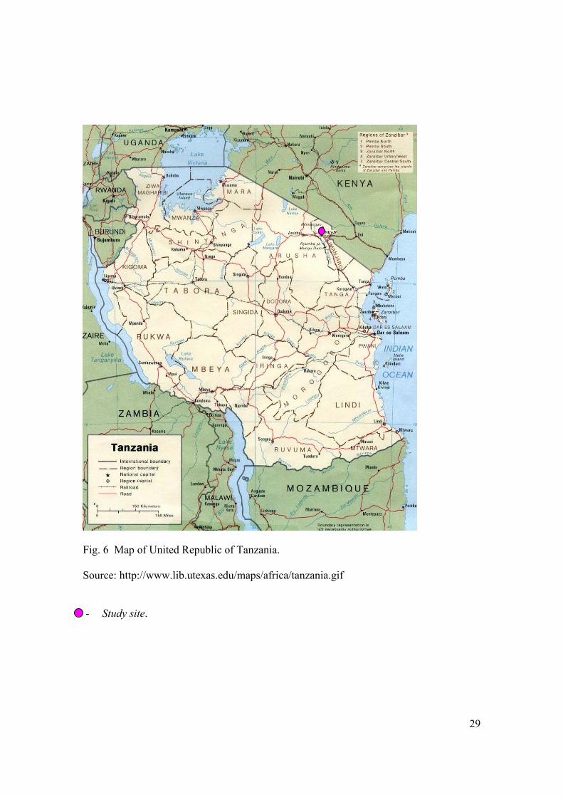
Materials and methods Study site This study was conducted at the Kilimanjaro Christian Medical Centre (KCMC) in
Moshi, Tanzania, and was designed to study changes in the circulating subtypes, genetic
diversity and distribution pattern of HIV-1 subtypes in northern Tanzania for the period
from 1985 to 2001. KCMC is a referral and university hospital for the northern zone of
Tanzania, which covers Tanga, Kilimanjaro, Arusha, Singida and Dodoma administrative
regions. The five regions have an estimated population of 7.2 million with Tanga,
Kilimanjaro, Arusha, Singida and Dodoma having populations of 1.7, 1.9, 2.0, 1.0 and
1.6 million, respectively (Planning Commission, 1999). These regions border with Kenya
in the north, the Indian Ocean on the east, Mara, Shinyanga and Tabora regions on the
west and Pwani, Morogoro and Iringa regions in the south (figure 7).
29
Fig. 6 Map of United Republic of Tanzania.
Source: http://www.lib.utexas.edu/maps/africa/tanzania.gif
- Study site.
30
Study design
A retrospective study was conducted on HIV-1 positive frozen serum samples from
patients who attended the outpatient clinics and/or were admitted to KCMC in the period
from 1985 to 2001.
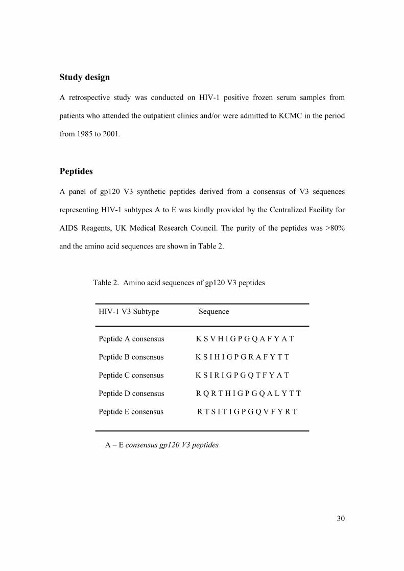
Peptides
A panel of gp120 V3 synthetic peptides derived from a consensus of V3 sequences
representing HIV-1 subtypes A to E was kindly provided by the Centralized Facility for
AIDS Reagents, UK Medical Research Council. The purity of the peptides was >80%
and the amino acid sequences are shown in Table 2.
Table 2. Amino acid sequences of gp120 V3 peptides
HIV-1 V3 Subtype Sequence Peptide A consensus K S V H I G P G Q A F Y A T Peptide B consensus K S I H I G P G R A F Y T T Peptide C consensus K S I R I G P G Q T F Y A T Peptide D consensus R Q R T H I G P G Q A L Y T T Peptide E consensus R T S I T I G P G Q V F Y R T
A – E consensus gp120 V3 peptides
31
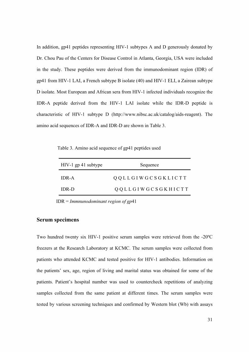
In addition, gp41 peptides representing HIV-1 subtypes A and D generously donated by
Dr. Chou Pau of the Centers for Disease Control in Atlanta, Georgia, USA were included
in the study. These peptides were derived from the immunodominant region (IDR) of
gp41 from HIV-1 LAI, a French subtype B isolate (40) and HIV-1 ELI, a Zairean subtype
D isolate. Most European and African sera from HIV-1 infected individuals recognize the
IDR-A peptide derived from the HIV-1 LAI isolate while the IDR-D peptide is
characteristic of HIV-1 subtype D (http://www.nibsc.ac.uk/catalog/aids-reagent). The
amino acid sequences of IDR-A and IDR-D are shown in Table 3.
Table 3. Amino acid sequence of gp41 peptides used
HIV-1 gp 41 subtype Sequence IDR-A Q Q L L G I W G C S G K L I C T T IDR-D Q Q L L G I W G C S G K H I C T T
IDR = Immnunodominant region of gp41 Serum specimens
Two hundred twenty six HIV-1 positive serum samples were retrieved from the -20ºC
freezers at the Research Laboratory at KCMC. The serum samples were collected from
patients who attended KCMC and tested positive for HIV-1 antibodies. Information on
the patients’ sex, age, region of living and marital status was obtained for some of the
patients. Patient’s hospital number was used to countercheck repetitions of analyzing
samples collected from the same patient at different times. The serum samples were
tested by various screening techniques and confirmed by Western blot (Wb) with assays
32
provided by various manufactures during the period from 1985 to 2001. The sera were
immediately frozen at -20ºC after Wb confirmation and never subjected to thawing and
freezing.
Serum sample selection
HIV-1 positive serum samples were sorted into four groups representing time of
collection. The four groups included all samples collected in 1985/86, 1990/91, 1995/96
and 2000/01. Serum samples were thawed and an aliquot of 100 µL was used in the
analysis. The groups comprised 25, 74, 29 and 98 samples for the groups representing
1985/86, 1990/91, 1995/96 and 2000/01, respectively.
Serum sample controls
Seronegative control samples that were collected during the same period were included in
each run to determine cut-off (CO) and to control the sensitivity and specificity of the
PEIA. A panel of seropositive control serum samples from previously genetically
characterized HIV-1 subtypes kindly donated by the Uganda Virus Research Institute in
Entebbe was included in each test run to ensure the validity of our results.
Sample analysis
All 226 serum samples were subjected to indirect PEIA against gp120 V3 peptides A to E
and gp41 A and D peptides. The sera that showed high antibody binding with gp120 V3
D peptide and reactive to gp41 D peptide were confirmed as subtype D. The rest were
33
subjected to the antigen limiting dilution assay using gp120 V3 peptides. The peptide that
showed high antibody binding at the highest antigen dilution determined the subtype for
the serum sample.
V3 PEIA
Reagents were prepared according to the protocol given in the appendix. Serum samples
were tested for antibody binding to V3 by an indirect PEIA as previously described (35).
The supplied freeze-dried peptides were reconstituted using double distilled water to a
concentration of 1000 µg/mL. The peptide solution was kept frozen at -20ºC in aliquots
of 50 µL in glass vials with teflon coated tops and thawed only once before use. The V3
peptides were coated directly on ELISA microtitre plates (Nunc immunosorb, 439454
F96 Cert. Maxisorb) with 100 µL in each well at a concentration of 5 µg/mL in 20 mM
carbonate buffer (pH 9.6) for 48 hours at room temperature. The plates were washed
twice with wash buffer, blocked with blocking buffer and then washed six times using
wash buffer. Coated plates were kept at 4ºC until use. Serum samples were diluted to
1:100 in serum diluent buffer and 100 µL of the diluted samples were incubated with the
immobilized peptides at 37ºC for 90 minutes. The plates were washed six times with
wash buffer and then incubated with rabbit anti-human immunoglobin peroxidase
conjugate (Sigma-Aldrich, Saint Louis, Missouri, USA) at a dilution of 1:6,000 for 1
hour at 37ºC to react with the bound antibody. The excess conjugate was removed by
washing six times using wash buffer. The colour was developed by incubating the plates
with ortho-phenylenediamine dihydrochloride (OPD) substrate (Sigma-Aldrich, Saint
Louis, Missouri, USA) for 20 minutes at room temperature in the dark. The colour
34
development was stopped by addition of 1 M sulphuric acid and the optical densities
(OD) were measured spectrophotometrically at 492 nm. Twelve HIV-1 negative sera
were included for each run of the peptide on each ELISA plate. The OD of the negative
controls was used for the calculation of the cut-off value. The cut-off value was
determined by calculating [(mean OD of the negative samples + 3SD) X 2]. Furthermore,
the ratio of the antibody bound to the peptide to the cut-off value was calculated
(OD/CO). Serum samples yielding a ratio greater than one were subjected to antigen
limiting dilution assay.
V3 peptide antigen limiting dilution assay The V3 peptide was coated on the same type of microtitre ELISA as previously described
plates at a concentration of 1, 0.1 and 0.01 µg/mL in 20 mM carbonate buffer (pH 9.6).
Each well was coated with 100 µL of each peptide dilution. The sera were used at a
single dilution of 1:100 and 100 µL was added to each well. The rest of the PEIA was
performed exactly as the indirect PEIA described above. Antibody-binding ratio
(OD/CO) was calculated for each peptide at each dilution and the highest antibody-
binding ratio at the highest antigen dilution indicated the subtype of the particular serum
specimen.
gp41 PEIA
Each individual peptide was dissolved in dimethyl sulfoxide (DMSO) at 5 mg/mL and
aliquoted into 50 µL volumes in glass vials with teflon coated cap liners and stored at
35
-20ºC until use. The microtitre ELISA plates were coated with peptide at a concentration
of 2.5 µg/mL in freshly prepared cold 0.1M carbonate-bicarbonate buffer (pH 9.6). In
each well 100 µL of the peptide was added and incubated overnight at 4ºC. The plates
were washed with wash buffer 2 times, air-dried at 37ºC for 15 minutes and stored at -
20ºC until use. The PEIA was performed exactly as in indirect PEIA except that a 1:200
serum dilution was used.
Results Of the 226 serum samples that were analyzed, 196 (87%) samples could be subtyped and
30 (13%) could not be subtyped using PEIA and the antigen limiting dilution assay. One
hundred and eighteen (52%) samples were from males, 98 (43%) were from females and
for 10 (5%) the gender was not available. The minimum age was 1 year and maximum
age was 66 years with a mean age of 34 years. Ages were grouped into 15-year intervals.
Children under 15 years were 15 (7%), 16 to 30 years were 64 (28%), 31 to 45 years were
93 (41%), 46 to 66 years were 32 (14%) and 22 (10%) were of unknown age. Most of the
patients, 105 (47%), were inhabitants of the Kilimanjaro region followed by
neighbouring regions of Arusha 24 (11%), Tanga 7 (3%) and Dar es Salaam 6 (3%). Two
patients (1%) came from Mwanza region and the remaining 2% came from Pwani, Mara,
Mbeya, Rukwa and Kagera regions with one patient each. The regions from which 77
(34%) patients came from were not known. HIV-1 subtypes A, C and D were the
subtypes that were found to circulate in northern Tanzania.
During the initial screening by the indirect PEIA there was an equivocal antibody binding
by some serum samples to some V3 peptides. This cross-reactivity was shown by subtype
36
A cross-reacting with C, D and E peptides while subtype C cross-reacted with subtypes
E, A and D peptides. Subtype D cross-reacted with A, B and C peptides. All the serum
samples were subjected to gp41 A and D peptides and 68 samples did not react with
either of the two peptides. Out of 62 samples that were typed as subtype D, 50 serum
samples reacted with gp41 D peptides. The 12 samples that did not react with the gp41 D
peptide had a high antibody-binding ratio to V3 peptide D in the indirect PEIA despite 8
(13%) and 4 (6%) samples showing cross-reaction with peptide B and C respectively. Of
the 62 typed D sera, 48 (77%) were in agreement with both V3 indirect PEIA and gp41
D. Of the remaining 2 (4%) samples, one showed to cross-reacted with A peptide and the
other was non-reactive by indirect PEIA. One hundred and eight samples reacted with the
gp41 A peptide and 54 of these samples had a high antibody-binding ratio to V3 peptide
C by both antigen limiting dilution assay and indirect PEIA. Hence, they were typed as
HIV-1 subtype C. The remaining 54 samples were typed as HIV-1 subtype A. The gp41
peptide non-reacting 68 samples were subjected to antigen limiting dilution assay and
indirect PEIA whereby 30 samples were untypable. The 38 serum samples that were
typed, 12 samples were typed as HIV-1 subtype D, 11 samples as HIV-1 subtype A and
15 samples as HIV-1 subtype C. Distribution of these subtypes with time is summarized
in Table 4.
37
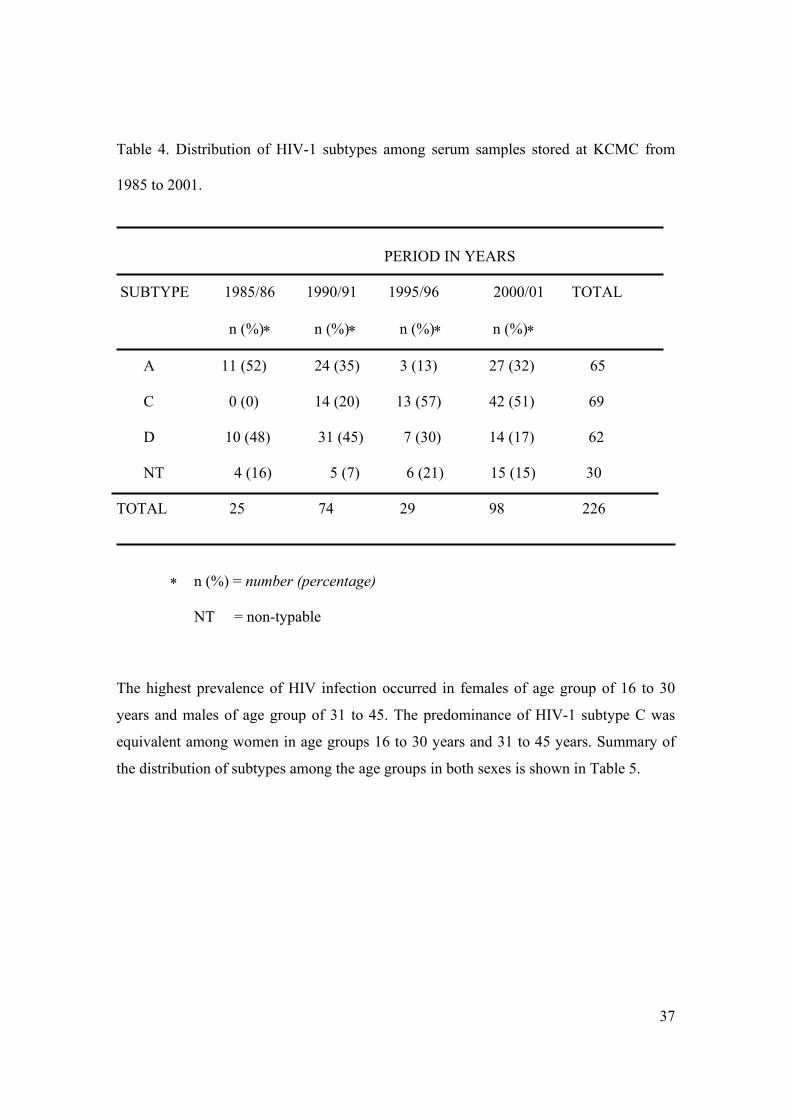
Table 4. Distribution of HIV-1 subtypes among serum samples stored at KCMC from
1985 to 2001.
PERIOD IN YEARS
SUBTYPE 1985/86 1990/91 1995/96 2000/01 TOTAL
n (%)∗ n (%)∗ n (%)∗ n (%)∗
A 11 (52) 24 (35) 3 (13) 27 (32) 65
C 0 (0) 14 (20) 13 (57) 42 (51) 69
D 10 (48) 31 (45) 7 (30) 14 (17) 62
NT 4 (16) 5 (7) 6 (21) 15 (15) 30
TOTAL 25 74 29 98 226
∗ n (%) = number (percentage)
NT = non-typable
The highest prevalence of HIV infection occurred in females of age group of 16 to 30
years and males of age group of 31 to 45. The predominance of HIV-1 subtype C was
equivalent among women in age groups 16 to 30 years and 31 to 45 years. Summary of
the distribution of subtypes among the age groups in both sexes is shown in Table 5.
38
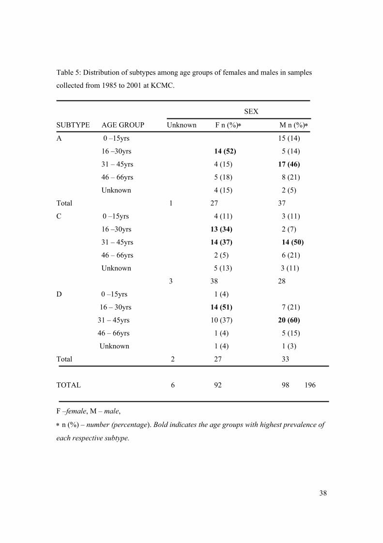
Table 5: Distribution of subtypes among age groups of females and males in samples
collected from 1985 to 2001 at KCMC.
SEX
SUBTYPE AGE GROUP Unknown F n (%)∗ M n (%)∗
A 0 –15yrs 15 (14)
16 –30yrs 14 (52) 5 (14)
31 – 45yrs 4 (15) 17 (46)
46 – 66yrs 5 (18) 8 (21)
Unknown 4 (15) 2 (5)
Total 1 27 37
C 0 –15yrs 4 (11) 3 (11)
16 –30yrs 13 (34) 2 (7)
31 – 45yrs 14 (37) 14 (50)
46 – 66yrs 2 (5) 6 (21)
Unknown 5 (13) 3 (11)
3 38 28
D 0 –15yrs 1 (4)
16 – 30yrs 14 (51) 7 (21)
31 – 45yrs 10 (37) 20 (60)
46 – 66yrs 1 (4) 5 (15)
Unknown 1 (4) 1 (3)
Total 2 27 33
TOTAL 6 92 98 196
F –female, M – male,
∗ n (%) – number (percentage). Bold indicates the age groups with highest prevalence of
each respective subtype.
39
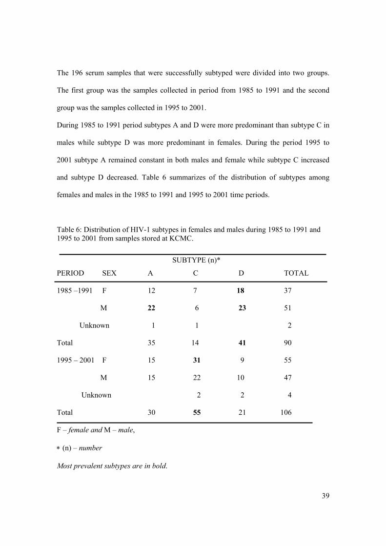
The 196 serum samples that were successfully subtyped were divided into two groups.
The first group was the samples collected in period from 1985 to 1991 and the second
group was the samples collected in 1995 to 2001.
During 1985 to 1991 period subtypes A and D were more predominant than subtype C in
males while subtype D was more predominant in females. During the period 1995 to
2001 subtype A remained constant in both males and female while subtype C increased
and subtype D decreased. Table 6 summarizes of the distribution of subtypes among
females and males in the 1985 to 1991 and 1995 to 2001 time periods.
Table 6: Distribution of HIV-1 subtypes in females and males during 1985 to 1991 and 1995 to 2001 from samples stored at KCMC.
SUBTYPE (n)*
PERIOD SEX A C D TOTAL
1985 –1991 F 12 7 18 37
M 22 6 23 51
Unknown 1 1 2
Total 35 14 41 90
1995 – 2001 F 15 31 9 55
M 15 22 10 47
Unknown 2 2 4
Total 30 55 21 106
F – female and M – male,
∗ (n) – number
Most prevalent subtypes are in bold.
40
Discussion and conclusion
Epidemiological studies on the genetic diversity of HIV-1, particularly the current
circulating subtypes in different parts of the world, yield information of importance for
vaccine development. Establishing cost-effective methodologies in countries with limited
resources is especially important in the HIV-1 endemic areas where HIV-1 diversity is
greatest and HIV-1 vaccine studies are urgently needed. Different HIV-1 subtypes occur
worldwide with different patterns of geographical distribution. Serological methods using
synthetic peptides from consensus sequences of HIV-1 genomes especially from the
gp120 V3 loop and gp41 have been used for subtyping HIV-1 (2)(35)(37). These
methods are of great importance in monitoring the spread of HIV-1 subtypes. They are
inexpensive, do not need highly trained personnel and enable large-scale screening for
epidemiological studies. In contrast to the previous serological studies on HIV-1
subtyping, our study was done in an epidemic area where multiple subtypes are co-
circulating with a recent increase in the prevalence of subtype C.
Our study aimed at presenting the trend in incidences of HIV-1 subtypes starting from the
early years of the epidemic using an inexpensive method that can be applied in a country
with limited resources. Furthermore, we aimed at presenting the current circulating
subtypes in Tanzania, a country that has one of the high prevalence of HIV-1 infections
within the SSA. The samples analyzed in this study included serum specimens that were
drawn from patients who attended KCMC in four periods of 1985/86, 1990/91, 1995/96
and 2000/01 and were confirmed to be HIV-1 seropositive. The results show that HIV-1
subtypes A and D were the first to circulate and in equal proportions during the start of
the epidemic. Our study has shown that HIV-1 subtypes A, C and D are currently co-
41
circulating in northern Tanzania. HIV-1 subtype C was first identified in the 1990/91
group of serum samples. This suggests that subtype C was probably introduced into the
population during late 1980s. Our study also shows that subtype C is increasing in
prevalence as the time goes on. Currently subtype C is circulating at a higher proportion
followed by subtype A that has remained constant while subtype D is decreasing over
time (Table 1). In other parts of Tanzania such as southwest in the Mbeya region, subtype
C was reported to circulate at a higher proportion and has remained stable since 1989
(33). In Uganda, where the epidemic started more or less at the same time as Tanzania, a
stable predominance of subtypes A and D with only limited distribution of subtype C has
been reported (41). This is also the situation in Kenya where subtypes A and D
predominate with limited proportions of subtype C and G (42). This suggests that subtype
C may have spread through southwest Tanzania to the northern region, possibly through
the traders between bordering Zambia and Malawi where subtype C predominates. The
increase in subtype C and decrease in subtype D HIV-1 in Tanzania raises research
questions that need to be addressed.
The distribution of HIV-1 subtypes did not differ significantly between the sexes within a
given time frame (Table 6). From the serum samples included in our study, it is not
possible to determine whether a specific subtype was most prevalent within a given age
group. The serum samples included in our study represent all serum samples stored at the
Research Laboratory at KCMC for each given time period. Therefore, we can conclude
that HIV infection is most prevalent among women aged 16 to 30 years and among men
aged 31 to 45 years in this study (Table 5). It is well known that HIV infection is most
prevalent among sexually active persons and that men with HIV infection are on average
42
older than women in SSA. However, it is of interest to note that the prevalence of HIV-1
subtype C is equivalent among women in age groups 16 to 30 and 31 to 45 years. This
may reflect the recent increase of HIV-1 subtype C infections as previously discussed or
a possibility of slow disease progression among persons with HIV-1 subtype C
infections. In order to make a statement regarding the distribution of HIV-1 subtypes
between different age groups, we would have to include equal numbers of samples from
each age group. We successfully established PEIA at KCMC and used this assay for our
sample analysis. Our results are in agreement with the previous studies done in Tanzania.
Studies conducted in Dar-es-Salaam have shown the same distribution of HIV-1 subtypes
during of the 1990s (25)(27)(29-32). However, a study done in Kilimanjaro did not
indicate the same distribution and showed the presence of subtype B-like viruses(28).
Our study used a different methodology with a sample size five times greater than that
used in the previous Kilimanjaro study. We could not detect subtype B. These data
warrant further studies in this population as subtype B has not been documented
elsewhere in Tanzania to date.
Serotyping using PEIA in combination with the antigen limiting dilution assay using
envelope peptides provides a good tool for determining the prevalence of different HIV-1
subtypes. The peptides that were used in our study were identical to those used by
Kaleebu in Uganda and Cheingsong-Popov on samples from Uganda and Rwanda
(2)(35). Tanzanian sera showed cross-reactions especially between A, C and E, and also
between D and B in accordance to previous reports (2)(35). Most of the serum samples
that showed a high antibody-binding ratio among the cross-reacting sera on indirect PEIA
also showed high antibody-binding on the antigen limiting dilution assay on low antigen
43
concentrations. Our approach of combining the V3 and gp41 PEIAs and the antigen
limiting dilution assay enabled us to determine the subtypes co-circulating in northern
Tanzania. Further studies are needed especially to determine a suitable consensus peptide
for the Tanzanian HIV-1 subtype A.
The antigen limiting dilution assay in combination with the gp41 PEIA discriminates
between cross-reacting subtypes. Subtype C could be distinguished from cross-reacting
subtypes such as A by the use of the antigen limiting dilution assay in contrast to
previous reports (35)(37). As the samples included in our study were not genetically
characterized, there is a need to evaluate the antigen limiting dilution assay for
discriminating genetically typed subtypes A and C. Such studies are imperative for the
use of the antigen limiting dilution assay in Tanzania where HIV-1 subtypes A and C are
co-circulating. In addition, we could not detect the CRF reported in previous studies done
in Tanzania (Table 1). This is one of the limitations of PEIA in HIV-1 subtyping.
In our study sample we could not subtype 30 (13%) sera by a combination of PEIA and
the antigen limiting dilution assay. It is conceivable that some serum samples had
antibodies concentration below the level of detection by these assays. This was also
demonstrated in Uganda (2) where 18% were untypable. These data warrant further
studies in which samples are subjected to methods such as HMA and DNA sequencing in
addition to PEIA. While inexpensive methods can be used to determine the HIV-1
subtypes in countries with limited resources, there is a need for genetic methods such as
HMA and DNA sequencing to describe the genetic diversity in detail. Furthermore,
international collaboration is needed in which groups with adequate resources, both
44
financial and technical, can perform analyses that are not possible in countries where
laboratories have limited facilities and funding.
The peptide-binding enzyme immunoassay used in this study is a great aid in determining
the prevalence of circulating HIV-1 subtypes within a given region. In addition, this
method is suitable for use in developing countries at referral laboratories. It is important
to know what subtypes are circulating in any given region with respect to the
administration of an effective vaccine against HIV-1. The data generated in this study
may be helpful should Tanzania become involved in vaccine trials in the near future.

45
Appendix Preparation of buffers: 1. 20 mM carbonate buffer, pH 9.6 Anhydrous Na2CO3 M.W = 105.99 Dissolve 2.12 gm in about 800 ml of distilled water. Initial pH is about 11, adjust
to 9.6 by drop wise addition of concentrated hydrochloric acid and make to one litre with distilled water.
2. 0.1 M Phosphate citric acid buffer, pH 5.0. Dissolve 6.66 gm of citric acid and 9.46 gm of disodium monohydrogen
phosphate in about 800 ml of distilled water. Adjust pH and make up to one litre with distilled water.
3. 10X - Phosphate buffered saline (PBS), pH 7.4 Dissolve 80 gm of NaCl, 2 gm of KCl, 11.5 gm of disodium monohydrogen
phosphate and 2 gm of potassium dihydrogen phosphate in about 900 mL of distilled water. Adjust pH and make to one litre with distilled water.
4. PBS for use. Dilute the 10X PBS to 1 in 10 with distilled water. Check pH. 5. Washing buffer Add 500 µL of Tween 20 in one litre of PBS for use. 6. Blocking buffer (100 mL preparation) Dissolve 5 gm of powdered skimmed milk in 90 mL of PBS for use and add 10
mL of heat-inactivated newborn calf serum. 7. Serum diluting buffer (100 mL preparation) Add 100 µL of Tween 20 into 99.9 mL of blocking buffer. 8. Stop solution 1 M sulphuric acid.
46
References 1. UNAIDS/WHO. Global HIV/AIDS update. December 2001.
2. Kaleebu P, Yirrell D, French N, Lyagoba F, Rutebemberwa A, Cheingsong-Popov R,
et al. An improved algorithm for determining HIV type 1 subtypes in a primary
laboratory in Uganda. AIDS Res Hum Retroviruses 2000;16(7):621-5.
3. Harris C, Small CB, Klein RS, Friedland GH, Moll B, Emeson EE, et al.
Immunodeficiency in female sexual partners of men with the acquired
immunodeficiency syndrome. N Engl J Med 1983;308(20):1181-4.
4. Mansur. An Outlook of Acquired Pneumocyst carinii pneumonia. N Eng J Med
1981(305):1431.
5. Colebunders R, Kapita B, Nekwei W, Bahwe Y, Lebughe I, Oxtoby M, et al.
Breastfeeding and transmission of HIV. Lancet 1988;2(8626-8627):1487.
6. Stewart GJ, Tyler JP, Cunningham AL, Barr JA, Driscoll GL, Gold J, et al.
Transmission of human T-cell lymphotropic virus type III (HTLV-III) by artificial
insemination by donor. Lancet 1985;2(8455):581-5.
7. Mhalu F, Bredberg-Raden U, Mbena E, Pallangyo K, Kiango J, Mbise R, et al.
Prevalence of HIV infection in healthy subjects and groups of patients in Tanzania.
AIDS1987;1(4):217-21.
8. NACP-MOH Tanzania. National AIDS Control Programme HIV/AIDS/STD
Surveillance; 1999 September 2000. Report No.: 14.
9. NACP-MOH Tanzania. National AIDS Control Programme HIV/AIDS/STD
Surveillance; 2000. Report No.: 15.
47
9. UNAIDS/WHO. AIDS Epidemiogical Fact Sheet on HIV/AIDS and Sexually
Transmitted Infections, 2000 Update; 2000 June 2000.
10. Chiu IM, Yaniv A, Dahlberg JE, Gazit A, Skuntz SF, Tronick SR, et al. Nucleotide
sequence evidence for relationship of AIDS retrovirus to lentiviruses. Nature
1985;317(6035):366-8.
11. Jawetz L. Medical Microbiology & Immunology: Examination & Board Review.
Sixth ed: Lange/McGraw-Hill; 2000.
12. Saha K. Isolation of primary HIV-1 that target CD+8 T lymphocytes using CD8 as a
receptor. Nat Med 2001;7(1):65-72.
13. Saha K, Zhang J, Zerhouni B. Evidence of productively infected CD8+ T cells in
patients with AIDS: Implications for HIV-1 pathogenesis. J Acquir Immune Defic
Syndr 2001;26(3):199-207.
15. Vaishnav YN, Wong-Staal F. The biochemistry of AIDS. Annu Rev Biochem
1991;60:577-630.
16. Preston. Fidelity of HIV-1 reverse transcriptase. Science 1988;242 (4882):
1168 - 71.
17. Bebenek K, Abbotts J, Roberts JD, Wilson SH, Kunkel TA. Specificity and
mechanism of error-prone replication by human immunodeficiency virus-1 reverse
transcriptase. J Biol Chem 1989;264(28):16948-56.
18. Delwart EL, Shpaer EG, Louwagie J, McCutchan FE, Grez M, Rubsamen-Waigmann
H, et al. Genetic relationships determined by a DNA heteroduplex mobility assay:
analysis of HIV-1 env genes. Science 1993;262(5137):1257-61.
48
19. http://hiv-web.lanl.gov/content/hiv-db/HelpDocs/subtypes-more.html.
20. Robertson DL, Anderson JP, Bradac JA, Carr JK, Foley B, Funkhouser RK, et al.
HIV-1 nomenclature proposal. Science 2000;288(5463):55-6.
21. Triques K, Bourgeois A, Vidal N, Mpoudi-Ngole E, Mulanga-Kabeya C, Nzilambi N,
et al. Near-full-length genome sequencing of divergent African HIV type 1 subtype F
viruses leads to the identification of a new HIV type 1 subtype designated K. AIDS
Res Hum Retroviruses 2000;16(2):139-51.
22. McCutchan FE. Understanding the genetic diversity of HIV-1. AIDS 2000;14 Suppl
3:S31-44.
23. Wolf D. Syncytium-inducing and non-inducing capacity of human immunodeficiency
virus type 1 subtypes other than B phenotypic and genotypic characteristics. WHO
Network for HIV Isolation and Characterization. AIDS Res Hum Retroviruses
1994;10(11):1387-400.
24. Holm-Hansen C, Baan E, Asjo B, Pascu FR, Goudsmit J, De Jong JJ. Determinants
for the syncytium-inducing phenotype of HIV-1 subtype F isolates are located in the
V3 region. AIDS Res Hum Retroviruses 2000;16(9):867-70.
25. Blackard JT, Renjifo B, Chaplin B, Msamanga G, Fawzi W, Essex M. Diversity of the
HIV-1 long terminal repeat following mother-to-child transmission. Virology
2000;274(2):402-11.
26. Hoelscher M, Hanker S, Barin F, Cheingsong-Popov R, Dietrich U, Jordan-Harder B,
et al. HIV type 1 V3 serotyping of Tanzanian samples: probable reasons for
mismatching with genetic subtyping. AIDS Res Hum Retroviruses 1998;14(2):139-
49.
49
27 Holm-Hansen C, Stern B, Rustad S, Shao J, Asjo B. V3 sequence analysis and
biological characterization of HIV-1 isolates from asymptomatic and early
symptomatic Tanzanian individuals. APMIS 2000;108(9):608-16.
28. Kiwelu IE, Nakkestad HL, Shao J, Sommerfelt MA. Evidence of subtype B-like
sequences in the V3 loop region of human immunodeficiency virus type 1 in
Kilimanjaro, Tanzania. AIDS Res Hum Retroviruses 2000;16(12):1191-5.
29. Renjifo B, Chaplin B, Mwakagile D, Shah P, Vannberg F, Msamanga G, et al.
Epidemic expansion of HIV type 1 subtype C and recombinant genotypes in
Tanzania. AIDS Res Hum Retroviruses 1998;14(7):635-8.
30. Renjifo B, Gilbert P, Chaplin B, Vannberg F, Mwakagile D, Msamanga G, et al.
Emerging recombinant human immunodeficiency viruses: uneven representation of
the envelope V3 region. AIDS 1999;13(13):1613-21.
31. Renjifo B, Fawzi W, Mwakagile D, Hunter D, Msamanga G, Spiegelman D, et al.
Differences in perinatal transmission among human immunodeficiency virus type 1
genotypes. J Hum Virol 2001;4(1):16-25.
32. Lyamuya E, Olausson-Hansson E, Albert J, Mhalu F, Biberfeld G. Evaluation of a
prototype Amplicor PCR assay for detection of human immunodeficiency virus type
1 DNA in blood samples from Tanzanian adults infected with HIV-1 subtypes A, C
and D. J Clin Virol 2000;17(1):57-63.
33. Hoelscher M, Kim B, Maboko L, Mhalu F, von Sonnenburg F, Birx DL, et al. High
proportion of unrelated HIV-1 intersubtype recombinants in the Mbeya region of
southwest Tanzania. AIDS 2001;15(12):1461-70.
50
34. Pau CP, Lee-Thomas S, Auwanit W, George JR, Ou CY, Parekh BS, et al. Highly
specific V3 peptide enzyme immunoassay for serotyping HIV-1 specimens from
Thailand. AIDS 1993;7(3):337-40.
35. Cheingsong-Popov R, Lister S, Callow D, Kaleebu P, Beddows S, Weber J.
Serotyping HIV type 1 by antibody binding to the V3 loop: relationship to viral
genotype. WHO Network for HIV Isolation and Characterization. AIDS Res Hum
Retroviruses 1994;10(11):1379-86.
36. Gaywee J, Artenstein AW, VanCott TC, Trichavaroj R, Sukchamnong A, Amlee P,
et al. Correlation of genetic and serologic approaches to HIV-1 subtyping in
Thailand. J Acquir Immune Defic Syndr Hum Retrovirol 1996;13(4):392-6.
37. Cheingsong-Popov R, Osmanov S, Pau CP, Schochetman G, Barin F, Holmes H, et
al. Serotyping of HIV type 1 infections: definition, relationship to viral genetic
subtypes, and assay evaluation. UNAIDS Network for HIV-1 Isolation and
Characterization. AIDS Res Hum Retroviruses 1998;14(4):311-8.
38. Cheingsong-Popov R, Williamson C, Lister S, Morris L, van Harmelen J, Bredell H,
et al. Usefulness of HIV-1 V3 serotyping in studying the HIV-1 epidemic in South
Africa. AIDS 1998;12(8):949-50.
39. Connett H. South African village prepares for first HIV vaccine trial. Nat Med
2000;6(11):1199-200.
40. Wain-Hobson. Nucleotide sequence of the AIDS virus, LAV. Cell 1985;40 (1):9-17.
41. Hu DJ, Baggs J, Downing RG, Pieniazek D, Dorn J, Fridlund C, et al.
42. Predominance of HIV-1 subtype A and D infections in Uganda. Emerg Infect Dis
2000;6(6):609-15.

51
42. Neilson JR, John GC, Carr JK, Lewis P, Kreiss JK, Jackson S, et al. Subtypes of
human immunodeficiency virus type 1 and disease stage among women in Nairobi,
Kenya. J Virol 1999;73(5):4393-403.