evaluation of immune responses - iarc publications website · 2016-11-21 · preserving secondary...
TRANSCRIPT
Unit 3 • Chapter 13. Evaluation of immune responses 215
Un
it 3
Ch
ap
ter
13
unit 3.assessing exposure to the environment
chapter 13.
Evaluation of immune responses
Robert Vogt and Paul A. Schulte
Summary
This chapter will present some general background material on the cellular, biochemical, and genetic mechanisms of the immune system, then focus on specific examples that illustrate the promise and pitfalls of using immune biomarkers as tools for molecular epidemiologic research and public health practice. Some of the most exciting frontiers in medical science will be discussed: early detection of cancer through autoimmunity; malignancies that arise from the immune system itself; newborn screening for lethal immune deficiencies and latent autoimmune disorders; and neurodevelopmental disabilities that could result from maternal immune responses, which protect the mother but harm the fetus. The chapter concludes with some thoughts about current challenges and future directions.
Introduction
Over the past 15 years, familiarity with the immune system has increased substantially among public health scientists, as well as the public at large. Since the use of immune biomarkers in molecular epidemiology was first addressed (1), the essential role of the immune system in maintaining health has been brought to public attention by the global HIV epidemic (2), the composite burden of autoimmune diseases (3,4,5), the genetic errors that lead to primary immune deficiencies (6), and the enigmatic relationship between immunity and malignancy (7,8). During this period, our understanding of the cellular and molecular processes that constitute the immune response has also increased in both scope and detail. These advances open new avenues for the use of immune biomarkers
in epidemiologic field studies and public health applications. At the same time, the general principles advocated earlier remain fully relevant today, perhaps even more so, given that the pace of technological development often outstrips our ability to harness it in a meaningful fashion.
This chapter will first update some of the general background material presented before (1) with respect to the cellular, biochemical, and genetic mechanisms of the immune system. Thereafter, the focus will be on specific examples that illustrate the promise and pitfalls of using immune biomarkers as tools for translation research and public health practice. Some of the most exciting frontiers in medical science will be discussed: early detection of cancer through
216
autoimmunity; environmental risk factors for malignancies that arise from the immune system itself; newborn screening for lethal immune deficiencies and latent autoimmune disorders; and neurodevelopmental disabilities that could result from maternal immune responses, which protect the mother but harm the fetus. The chapter concludes with some thoughts about current challenges and future directions.
Immune biomarkers as functional elements and sentinel indicators
The benefits and limitations of using immune markers in epidemiologic studies may be best appreciated by understanding their relationship to the basic biology of the host defence system: a complex network of cells and mediators with recognition and response functions that occur throughout most tissues of higher organisms (1). The primary functions of the host defence system are repairing injured tissue, identifying and removing foreign substances, destroying or containing infectious agents, and, in some cases, eradicating cancer cells.
Innate (non-specific) and acquired (specific) immunity
Host defence functions are carried out through non-specific mechanisms of innate immunity, and through specific mechanisms of acquired (adaptive) immunity, which develop as the organism encounters environmental agents (antigens). The term immune system is used in this chapter to refer to all components of both non-specific innate immunity and antigen-specific acquired immunity, as their components and activities are invariably intertwined (1). Nonetheless, the distinction between markers that are antigen-
specific and those that are not is often important, especially in exposure-related studies. The ability of the immune system to recognize foreign molecules is so discerning that it has even been likened to a self-referential sensory organ (9).
Inflammation
Whether innate or acquired, the result of host defence activity is often inflammation. The cardinal signs of inflamed tissue were described by the ancient Greek physicians Celsus and Galen: calor (heat), dolor (pain), rubor (redness), tumour (swelling), and functio laesa (loss of function) (10). Our current knowledge of the cellular and molecular basis of inflammation is exhaustive, but the complexity of the in situ inflammatory response still lies beyond our complete understanding. Still, the cells and mediators of inflammation provide essential biomarkers for medicine, biomedical research, and, more recently, for epidemiologic studies.
Inflammation is essential for host defence, as it brings cells and mediators to the site of tissue injury and infection, sequestering the insult, destroying infectious agents or the cells they have infected, clearing the debris and promoting repair. However, it is a two-edged sword, and many of the symptoms following injury or infection come not from the insult but from the host response to it. Immunopathology is the study of how the immune system creates as well as prevents disease. From the classic animal models of viral meningitis (11) and tuberculosis (12) to the recent revelation that human cardiovascular disease and diabetes are largely inflammatory pathologies (13), biomarkers have shown that immunity and inflammation are inexorably linked.
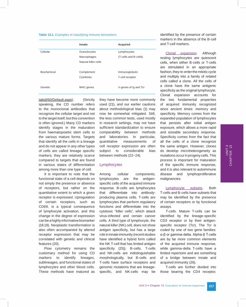
Biological categories of immune biomarkers
The distinction between antigen-specific and non-specific biomarkers is fundamental and unique to the immune system. For convenience, this distinction can be overlaid onto three major types of intrinsic biological markers: cellular, biochemical and genetic (Table 13.1). These three categories are arbitrary and somewhat artificial, since biochemical and genetic markers originate in cells. In fact, many cells of the immune system are defined by the biochemical surface receptors they express or the unique gene rearrangements they contain.
Cellular biomarkers
Cellular immunology was for many years a phenomenologic area of study, often confusing and contradictory. Two technical breakthroughs combined to bring order to this area. Monoclonal antibodies allowed the development of specific probes without a priori knowledge of the properties of the cellular target. Flow cytometry allowed the cell-by-cell detection of these targets using fluorescent-labelled monoclonal antibodies and a dynamic streaming process that could analyse and sort thousands of cells per second (14,15). The sorted cells could then be characterized for their functional activities and other properties, and linked to their respective target. The cellular targets, which are all proteins (or glycoproteins) and are usually cell surface receptors, are most often identified by their cluster of differentiation (CD) number (16). International workshops are held periodically to update CD nomenclature (17); as of 2010, the list was up to CD363 (see http://www.hlda9.org/HLDA9Workshop/
Unit 3 • Chapter 13. Evaluation of immune responses 217
Un
it 3
Ch
ap
ter
13
tabid/60/Default.aspx). (Strictly speaking, the CD number refers to the monoclonal antibodies that recognize the cellular target and not to the target itself, but this convention is often ignored.) Many CD markers identify stages in the maturation from haematopoietic stem cells to the various mature forms. Targets that identify all the cells in a lineage and do not appear in any other types of cells are called lineage specific markers; they are relatively scarce compared to targets that are found in various states of differentiation among more than one type of cell.
It is important to note that the functional state of a cell depends on not simply the presence or absence of receptors, but rather on the quantitative extent to which a given receptor is expressed. Upregulation of certain receptors, such as CD69, is a typical consequence of lymphocyte activation, and this change in the degree of expression can be a highly informative biomarker (18,19). Neoplastic transformation is also often accompanied by altered receptor expression that may be correlated with genetic and clinical features (20).
Flow cytometry remains the customary method for using CD markers to identify lineages, sublineages, and functional states of lymphocytes and other blood cells. These methods have matured as
they have become more commonly used (21), and our earlier cautions about methodological bias (1) may now be somewhat mitigated. Still, the less common tests, used mostly in research settings, may not have sufficient standardization to ensure comparability between methods and laboratories. In particular, quantitative measurements of cell receptor expression are often subject to considerable bias between methods (22–24).
Lymphocytes
Among cellular components, lymphocytes are the antigen-specific cells of the acquired immune response. B-cells are lymphocytes that differentiate into antibody-producing plasma cells. T-cells are lymphocytes that perform regulatory functions and differentiate into the cytotoxic “killer cells”, which attack virus-infected and certain cancer cells. A third type of lymphocyte, the natural killer (NK) cell, does not show antigen specificity, but has a large role in innate immunity (recent studies have identified a hybrid form called the NK T-cell that has limited antigen specificity (25)). B-cells, T-cells and NK-cells are indistinguishable morphologically, but B-cells and T-cells have surface receptors and genomic mutations that are lineage-specific, and NK-cells may be
identified by the presence of certain markers in the absence of the B-cell and T-cell markers.
Clonal expansion. Although resting lymphocytes are quiescent cells, when either B-cells or T-cells are stimulated in an appropriate fashion, they re-enter the mitotic cycle and multiply into a family of related cells called a clone. All the cells of a clone have the same antigenic specificity as the original lymphocyte. Clonal expansion accounts for the two fundamental properties of acquired immunity recognized since ancient times: memory and specificity. Memory comes from the expanded population of lymphocytes that persists after initial antigen exposure, which allows a more rapid and sizeable secondary response. Specificity comes from the fact that all the cells of a clone recognize the same antigen. However, clones do develop microheterogeneity as mutations occur in progeny cells. This process is important for maturation of the specific immune response, and it is also relevant to autoimmune disease and lymphoproliferative malignancies.
Lymphocyte subsets. Both T-cells and B-cells have subsets that may be identified by the presence of certain receptors or by functional assays.
T-cells. Mature T-cells can be identified by the lineage-specific CD3 receptor or by their antigen-specific receptor (Tcr). The Tcr is coded by one of two gene families: α-β or gamma-delta. Alpha-β T-cells are by far more common elements of the acquired immune response, while gamma-delta T-cells have a limited repertoire and are something of a bridge between innate and acquired immunity (26).
T-cells are further divided into those bearing the CD4 receptor,
Table 13.1. Examples of classifying immune biomarkers
Innate Acquired
Cellular Granulocytes Lymphocytes
Macrophages (T-cells and B-cells)
Natural killer cells
Biochemical Complement Immunoglobulin
Cytokines T-cell receptor
Genetic MHC genes V-genes of Ig and Tcr
218
those bearing the CD8 receptor, and a small fraction of those that bear both. Most of the CD4-bearing cells are helper T-cells (Th) that upregulate the immune response. The CD8-bearing T-cells were originally considered to be either cytotoxic (killer) cells (Tc) or suppressor cells that downregulate the immune response. CD8 cytotoxic T-cells are well characterized, but evidence for suppressor activity in this subset was never convincing. In 1995, the real suppressor population was identified among CD4 T-cells as a small proportion that also bears the CD25 receptor and contains a high concentration of the Foxp3 transcription factor (27). These CD4-CD25 T-cells are now called regulatory T-cells (Treg); they are critical for preventing autoimmunity, preserving secondary immunity (immune memory), and protecting pregnancies (28).
Helper T-cells may also be characterized in terms of their regulatory roles. The original paradigm described a TH1 response, which led to delayed hypersensitivity mediated by cellular responses, and a TH2 response, which led to humoral immunity and allergy mediated by antibody production. The association of TH2 responses with both parasitic infections and allergies has been well defined in laboratory and clinical studies (29–31). However, this simple picture has been replaced by a more complex interaction involving the cytokine IL-17, which mediates a third functional type called the TH17 T-cell (32). Research on the TH17 subset has progressed rapidly, and it is now seen as having a central role in immune regulation (33), autoimmunity (34), inflammation (35) and the link between innate and acquired immune activities (36,37). The TH17 pathway may even explain the suspected immunotoxic effects of halogenated aryl hydrocarbons,
such as PCB and dioxins (38,39).The TH1 response is associated
with gamma interferon and tumour necrosis factor (TNF-α), the TH2 response with interleukin-4 and interleukin-13, and the TH17 response with the IL-17 family of six cytokines designated 17A-17F (35). The relative elevation of these cytokines in tissue or serum is generally taken as evidence for the respective type of in vivo response. However, the measurement of these factors (especially in serum) is not standardized, and their use in epidemiologic field studies should be approached cautiously. In particular, artefacts of the immunoassays used to measure cytokines may produce spurious differences (40,41); interestingly, such misleading artefacts may still have biologic and immunologic validity (42). In any case, the remarkable heterogeneity and plasticity of T helper cells (43) can make interpretation of relevant biomarkers enigmatic at best.
One other subclassification of T-cells deserves mention: the distinction between naive and memory T-cells. Naive (virgin) T-cells have not encountered antigen, while memory T-cells arose by clonal expansion caused by antigen-driven activation. The surface receptor CD45 exists in two isoforms: CD45RA is associated with naive T-cells, while CD45RO is associated with memory T-cells. While the two isoforms can be readily distinguished by flow cytometry, the categorization is probably oversimplified, especially for CD8 T-cells. However, the distinction may provide some insight into the pathogenesis of immune-mediated disorders (44) and environmental exposures (45,46).
B-cells. Mature B-cells may be identified by the lineage-specific CD19 receptor and by the presence of their surface immunoglobulin (sIg)
molecules. All of the sIg molecules on a particular B-cell have the same antigen binding site, which gives B-cells their specificity. When B-cells are activated by antigen binding, they proliferate and redifferentiate into antibody-producing cells. The endpoint in this secondary differentiation is the plasma cell, which in essence is a cellular factory for making antibodies. Several other receptors are expressed during the various stages of progression towards plasma cells or diversion to memory cells (47); identification of these has long been a staple of diagnostic pathology for B-cell malignancies (48). B-cells do not have major functional subsets analogous to CD4 and CD8 in T-cells. However, the presence or absence of CD5 (a receptor found on all T-cells) appears to define distinct B-cell populations. CD5 B-cells are associated with chronic humoral responses, mucosal immunity, autoimmunity and possibly with an increased risk of transforming into a B-cell malignancy (49).
Non-lymphoid cells
In addition to lymphocytes, several other types of cells are important participants in immune function; most of them spend at least part of their life cycle in the bloodstream, where they (along with lymphocytes) are collectively referred to as leukocytes or white blood cells (WBC). The most numerous of the bloodstream leukocytes are granulocytes, end-stage cells with short lifetimes whose granules contain pre-formed mediators ready for immediate release. Most of them are neutrophils, which migrate into inflamed tissue where they ingest (phagocytise) and destroy bacteria. Eosinophils and basophils are normally present in much smaller numbers; they are involved
Unit 3 • Chapter 13. Evaluation of immune responses 219
Un
it 3
Ch
ap
ter
13
in allergy and the host response to parasitic infections. Although granulocytes are endstage, they do have some limited ability to modify their functional status. For instance, activated neutrophils upregulate the expression of the CD64 receptor (50), a response that is now used clinically as a sign of occult infection, and activated eosinophils upregulate co-stimulatory and adhesion molecules in response to parasitic infection (51).
Resident cells in the connective tissue underlying the skin, mucosa and internal epithelium are also critical to immune function. Macrophages ingest, process and package antigens for presentation to T-cells, a process mediated by a transient intercellular macromolecular complex recently termed the immune synapse (52,53). Other accessory cells, such as dendritic cells (54), are also involved in antigen presentation. Mast cells contain histamine and other mediators of allergy in pre-formed granules ready for immediate release. They have surface receptors that bind very strongly to IgE antibodies, sensitizing them (and the individual they inhabit) to allergens recognized by the IgE. When allergens interact with their surface-bound IgE, activation and degranulation lead to immediate hypersensitivity (55). Mast cells also mediate signalling between the peripheral nerves and local immune activity, one reason that immediate hypersensitivity responses can be induced rather easily by Pavlovian conditioning (56).
Biochemical biomarkers
Biochemical biomarkers (excluding genomic DNA) include protein and RNA macromolecules as well as smaller molecules, such as steroid hormones and prostaglandins. Technical issues attend all the methods used to measure
these markers, particularly the macromolecules. Proteins are often detected and quantified by antibody-binding methods, which may be subject to cross-reactivities or other interferences that cause spurious results (57). The use of mass spectroscopy for protein analysis has increased, particularly as a biomarker discovery tool (58–60). Some of the initial, promising results obtained this way have turned out to be disappointing (61); a careful approach to method evaluation and study design is required for meaningful results (62). RNA is generally detected and quantified by hybridization reactions, often in an expression microarray with thousands of targets. These methods are also subject to technical vagaries, but some standardization has been
achieved (63). RNA microarrays have shown considerable promise in some clinical applications (64), but again, a careful approach to method evaluation and study design is required for meaningful results.
Antigen-specific biochemical markers
Among the wide range of biochemicals involved with immunity, only antibodies (also called immunoglobulins (Ig)) and T-cell receptors (Tcr) are antigen-specific.
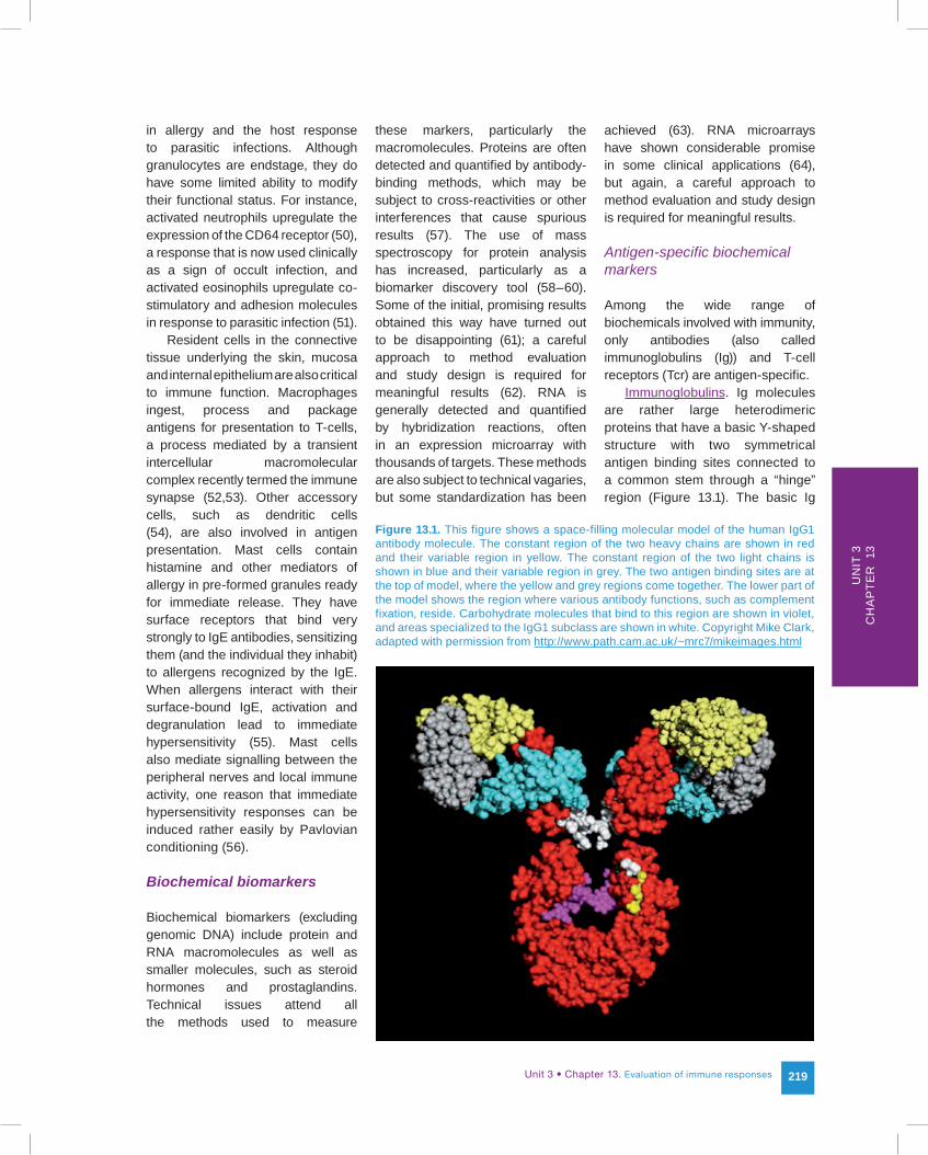
Immunoglobulins. Ig molecules are rather large heterodimeric proteins that have a basic Y-shaped structure with two symmetrical antigen binding sites connected to a common stem through a “hinge” region (Figure 13.1). The basic Ig
Figure 13.1. This figure shows a space-filling molecular model of the human IgG1 antibody molecule. The constant region of the two heavy chains are shown in red and their variable region in yellow. The constant region of the two light chains is shown in blue and their variable region in grey. The two antigen binding sites are at the top of model, where the yellow and grey regions come together. The lower part of the model shows the region where various antibody functions, such as complement fixation, reside. Carbohydrate molecules that bind to this region are shown in violet, and areas specialized to the IgG1 subclass are shown in white. Copyright Mike Clark, adapted with permission from http://www.path.cam.ac.uk/~mrc7/mikeimages.html
220
unit is composed of two identical copies of each of two peptide chains, the light chain and the heavy chain. Light chains come in two varieties, kappa and lambda, coded by different genes from different loci. Heavy chains are coded by only one genetic locus, but somatic recombination within the locus can produce different variants called isotypes through a process called class-switching.
Immunoglobulins have two functions in the immune response: membrane-bound Ig molecules are the antigen-specific receptors on B-cells, and secreted Ig molecules have a variety of effector functions critical to host defence including complement fixation, opsonization (which promotes phagocytosis) and viral inactivation. When antigen binds to Ig receptors on B-cells, they proliferate and secrete antibody of the same specificity as the original Ig receptor. Although the secreted antibody has the same antigen binding site as the B-cell receptor, it may differ in other portions of the molecule, which accounts for the different isotypes: IgM, IgG, IgA, IgD, and IgE. As an effector molecule, IgM is the first isotype to be produced in a primary immune response; it is especially good at binding antigen into complexes and activating complement. IgD functions only as a B-cell receptor and is not normally secreted. IgG is the most common isotype in serum, normally accounting for up to one-third of total serum protein. It exists in four subclasses, which differ in functions such as complement fixation and placental transfer. IgA is responsible for mucosal immunity (65,66) and is secreted from epithelial surfaces in the airways and the gut; a variant form is present in serum (67). IgE is (in humans) uniquely involved in immediate hypersensitivity, best known as the
cause of common allergy and a major factor in the pathogenesis of asthma (68).
In terms of measurement, earlier difficulties in the standardization of assays to measure the major Ig isotypes in serum (1) have been largely resolved (69). However, measurement of isotype subclasses is not as well standardized. IgE concentrations in serum are much lower than the other secreted isotypes and must be measured by more sensitive methods. Measurement of antigen-specific IgE is an important marker for allergy in diagnostic, occupational, and research settings. Most of these assays are well standardized (70), but customized tests for IgE to novel allergens must be carefully characterized to assure sensitivity and specificity.
T-cell receptors (Tcr). Tcr proteins have a molecular structure and antigen specificity analogous to, but somewhat different than, that of antibodies. Tcr molecules are not secreted and have no effector function. There are two major types of Tcr: α-β and gamma-delta. These types may in turn be grouped into families that can be differentiated by monoclonal antibodies (71) or genetic analysis (see below). Like surface receptor antibodies on B-cells, the Tcr receptors on T-cells allow them to respond to antigen binding by proliferating and secreting effector molecules, in this case peptide regulators called lymphokines (13,72). Cytotoxic (killer) T-cells use their Tcr receptors to identify their cellular targets: viral-infected cells or, in some cases, cells that have undergone malignant transformation (73).
Non-specific biochemical markers
The immune system uses a variety of proteins, peptides, and smaller
molecules to effect and regulate host defence.
Lymphokines, cytokines and interleukins. Lymphokines, which are produced by lymphocytes, are a subset of the localized cellular peptide mediators called cytokines elaborated by a variety of cells. The interleukins are cytokines particularly involved with signalling among leukocytes. The first interleukins (IL-1 and IL-2) were labouriously identified as functional activation and growth factors from cell culture supernatants (74). One of the most recent (IL-34) was uncovered by a systematic search of the extracellular proteome expressed by a set of recombinant secreted proteins, using a suite of assays that measured metabolic, growth or transcriptional responses in diverse cell types (75).
All cytokines are localized tissue mediators, and their concentration in peripheral blood is normally extremely low or undetectable. Increased concentrations due to spillover from tissue sites of inflammation may sometimes be detected in serum, but the assays used to measure them are not standardized and are subject to interferences, matrix effects and considerable bias between methods.
Genetic polymorphisms in cytokines and cytokine receptors have been shown to be useful biomarkers of susceptibility for lymphoid malignancies (76), other cancers (77–80), oral diseases (81), allergies (82) and autoimmune disorders (83,84). Soluble cytokine receptors, which are deliberately released from cells by a variety of specific mechanisms (85), are important mediators of inflammation (86). While serum concentrations of soluble receptors and receptor-cytokine complexes are good candidate biomarkers for inflammation-related disorders,
Unit 3 • Chapter 13. Evaluation of immune responses 221
Un
it 3
Ch
ap
ter
13
the assays to measure them are not standardized, and results from different methods may not give concordant results.
Other non-antigenic-specific mediators. In addition to lymphokines, many other non-specific biochemical mediators are used in host defence activities. They often involve inflammation and include acute-phase reactant proteins, such as complement (a family of proteins that react in a cascading fashion) and C-reactive protein, as well as small molecules such as histamine, prostaglandins and endocrine hormones. Although these substances may serve as biomarkers of immune function, their non-specific nature and highly interactive functional pathways often make it difficult to distinguish cause from effect. The importance of neuropeptides and nerve growth factors in the immune response and inflammation has become increasingly apparent (87,88).
N o n - a n t i g e n - s p e c i f i c cell receptors. Besides Ig and Tcr, several other cell surface receptor protein families are critical components of immune function even though they are not antigen-specific. Two of the most important are the receptors coded by the genes of the major histocompatibility complex (MHC), and the Toll-Like Receptors (TLR).
Major histocompatibility complex (MHC) (transplantation) antigens. The MHC receptors come in three primary classes; classes I and II comprise the human leukocyte antigens (HLA). (The class III MHC region contains a diverse set of genes, some of which code for certain immune-related proteins like cytokines and complement components.) Although they were discovered because they caused rejection of organ transplants, their normal biological function involves packaging antigens for presentation
to T-cells. Class I proteins present antigens to CD8 cytotoxic T-cells, and class II proteins present antigens to CD4 helper T-cells. All of the HLA loci are highly polymorphic within a species; the four major loci in humans include over 2500 different alleles, leading to the difficulty in finding matches between organ donors and recipients. Different allotypes may confer relative susceptibility or resistance to autoimmune diseases and certain infectious agents (89). HLA protein allotypes can be identified by reactions with allo-specific antibody reagents, and alloantibodies in previously sensitized individuals can be detected by binding assays (90). Alloreactivity between tissues from two individuals may be detected by mixing lymphocytes from the two sources and measuring functional responses (e.g. proliferation, lymphokine secretion or mRNA production) (91). These types of assays are used clinically, but are highly specialized and should be performed by experienced histocompatibility laboratories. T-cell antigen recognition normally involves presentation by viral-infected epithelial cells or by accessory cells, such as macrophages that have packaged the antigen with HLA proteins. The detection of antigen-specific T-cells using in vitro stimulation assays is greatly enhanced by pre-packaging the antigen with a suitable HLA protein into a complex called a tetramer (92). These assays are highly specialized and used primarily in research settings, although some degree of standardization has been achieved (93). The use of tetramers to identify CD8 cytotoxic T-cells specific for viral or tumour antigens has been quite successful, but identification of antigen-specific CD4 helper T-cells remains problematic (94).
Toll-like receptors (TLRs). This class of receptor, named for its similarity to the Toll receptor of Drosophila, is largely responsible for initiating the inflammatory response to microbes and for the host perception of microbes in general (95). TLRs are evolutionarily ancient proteins, and most mammalian species have about a dozen different types; some shared across species and others unique. TLRs account for much of the protective effects of the host response to infection, such as the induction of lasting specific immunity, as well as its pathological effects (e.g. systemic inflammation and shock) (95). Their discovery illustrates the use of forward genetic methods in identifying genes for biomarkers that are constitutively expressed but conditionally functional (96). Originally considered as effectors of innate immunity, TLRs are now seen to have important roles in acquired immunity as well. They are involved in the class-switching maturation of B-cells, as the immune system transitions from the primary response dominated by IgM to the secondary responses dominated by IgG and IgA (97). They also appear to interact with the superantigen-mediated polyclonal activation of T-cells responsible for toxic shock syndrome (98), an often fatal condition associated with the use of tampons that reached epidemic proportions in the United States around 1980 (99).
TLRs belong to a larger group of molecules called the pattern recognition receptors (PRRs) that recognize conserved molecular motifs from pathogenic microbes: pathogen-associated molecular patterns (PAMP). A PAMP database has been established that contains about 500 patterns, including 177 recognized by TLRs (http://www.imtech.res.in/raghava/prrdb/) (100).
222
The biology of TLRs has just started to impact clinical medicine and public health (101–106), and aberrant variations in TLRs expression caused by genetic polymorphisms, mutations or dysregulation will become increasingly important biomarkers.
Genetic biomarkers
Immunoglobulin and T-cell receptor genes
Immunoglobulin and T-cell receptor genes code for the only antigen-specific proteins in the immune system. The three genes that produce Ig molecules are located on different chromosomes: the kappa light chain gene (chromosome 2), the lambda light chain gene (chromosome 22), and the heavy chain gene (chromosome 14). During differentiation, B-cells determine whether they will use the kappa or lambda gene, and that choice is maintained by all progeny of the clone. The two light chains are never expressed together in the same cell. The T-cell receptor, which has an analogous genetic basis, is coded for by either alpha and beta genes or by gamma and delta genes; like Ig light chains, the two pairs are never expressed in the same cell. The beta and gamma genes are on chromosome 7, while the alpha and delta genes are on chromosome 17.
All seven of the Ig and Tcr genetic loci contain two distinct regions separated by an intervening sequence of nucleotides. One region contains multiple sequences, referred to as V-genes, that code for the N-terminal portion of the peptide which will form the antigen binding site. The other region, referred to as the C-gene, contains sequences that code for the remainder of the peptide chain, which is not
involved in antigen recognition. In a differentiation process unique to lymphocytes, somatic recombination removes the intervening sequence between the V-genes and C-genes to form a new gene that codes for the intact Ig or Tcr protein. This recombination persists as a genetic fingerprint in all the clonal progeny that subsequently arise from the lymphocyte undergoing the original recombination. The details differ for each locus, and Ig heavy chain genes have a special feature of multiple C-genes that sequentially recombine with the chosen V-gene to form different antibody isotypes with the same antigen specificity.
The V-genes that code for antigen binding sites might at first be considered precise markers for antigen specificity; however, immune specificity is a selective process. The antigen binding site in Ig or Tcr molecules is not designed to fit a particular antigen; rather, binding sites are created stochastically, and those that happen to react with antigens are selected for clonal expansion. In fact, each antigen binding site is polyspecific (107), and immune specificity to most antigens depends on the wide repertoire of specificities that reinforces common reactivities and dilutes out the others. The relationship between V-genes and antigen specificity is therefore indirect and ultimately must be determined by antigen binding, not gene sequences.
Because of the unique recombination of V-genes and the selective clonal expansion or diminution of the lymphocytes containing them, the repertoire of V-genes differs between individuals and within individuals over time. They can therefore serve as biomarkers of exposure and effect, as well as biomarkers of susceptibility.
Major histocompatibility complex (MHC) genes
The other major gene family involved in the immune response is the major histocompatibility complex (MHC), which comprises 26 different genes including those that code for the human HLA proteins (see http://www.ebi.ac.uk/imgt/hla/). These genes do not influence antigen specificity directly, but they do have notable effects on antigen-specific immune responses and are often used as biomarkers of susceptibility. In particular, the association of MHC polymorphisms has been a sine qua non for autoimmune disease since it was first uncovered in a mouse model of autoimmune thyroiditis (108,109).
The high degree of genetic polymorphism in the MHC presents difficulties for genetic analysis, which can be done at low resolution for modest costs or higher resolution for higher costs. The technical issues revolve first around the regions and primers selected for PCR amplification. Thereafter, the amplified product can be tested by probes, but they too may cross-react with different alleles. Selected regions associated with the allotypes (which may lie in intron-exon boundaries) can be sequenced, but even sequence-based typing cannot rule out a polymorphism that lies outside the sequenced region. As with HLA protein analysis, these analyses should be done by experienced laboratories, and epidemiologic investigators should understand the limitations of methods used.
Other genes
Many other genes may be biomarkers of susceptibility or effect modifiers for immune and inflammatory pathologies. Some
Unit 3 • Chapter 13. Evaluation of immune responses 223
Un
it 3
Ch
ap
ter
13
of these genes code for other non-specific immune mediators, while others have no direct relationship to the immune system. Polymorphisms in cytokine genes, such as TNF-α and IL-8, have been associated with several clinical endpoints including severity of rheumatoid arthritis (110), incident cardiovascular disease (111), inflammatory bowel disease and cancer (112), type 2 diabetes (113) and thrombotic disease in children (114). They have also been associated with effect modification in chemical exposures (115) and nutritional biomarkers (116). However, such associations may not be apparent when tested in large-scale, longitudinal studies (117).
Genes that have no direct relationship to the immune system, such as those involved with metabolism, can also influence immunity and immunopathology. An association between oxidative metabolites of therapeutic drugs and the autoimmune disease systemic lupus erythematosis (118) was long attributed to a slow-acetylator polymorphism of the arylamine-N-acetyltransferase-2 gene. While subsequent epidemiological studies have cast doubt on the relationship with clinical disease (119,120), a relationship with autoimmunity may still exist. Observations in a mouse model of an association between expression of the aryl hydrocarbon receptor (AHR) and the TH17 T-cell subset (38) suggest that exposure to aryl hydrocarbons, modified by AHR polymorphisms, may be associated with autoimmunity and perhaps with B-cell malignancies (121). Epigenetic changes could also account for differences between individuals in the way their gene–environment interactions lead to acquired susceptibility or resistance for autoimmunity (122) or lymphocyte malignancies (123). Genes, such as the autoimmune
regulator that controls the expression of tissue-specific antigens, may have a profound impact on immune tolerance and autoimmunity (124). Genes concerned with the regulation of cell growth, such as BCL2, are especially important in lymphocytes, given their propensity for clonal expansion, and the non-coding regulatory microRNAs that are involved with cell growth pathways may be more important markers than many coding genes (125).
Special considerations for using immune biomarkers in epidemiologic studies
Immune biomarkers may be used to evaluate populations for disorders of the immune system itself, for immunogenic exposures, or for pathological conditions in other organ systems that provoke changes in immune status.
Disorders of the immune system
Three general types of disorders of the immune system may have adverse health consequences: immune deficiencies, inappropriate immune reactivities, and unregulated proliferation leading to lymphoid malignancies (1).
Immune deficiency disorders
Immune deficiency disorders are those in which the immune system fails to mount adequate protective responses against infection or certain forms of cancer. Deficiencies may be primary (caused by inherited genetic traits or spontaneous mutations) or secondary (caused by exposures or infections, such as HIV). Depending on the nature of the deficiency, the health consequences can range from almost unnoticeable,
such as increases in the incidence of mild infections, to life-threatening, such as overwhelming sepsis. Immune deficiencies may be indicated by low or absent levels of serum immunoglobulins, low or absent numbers of immune cells, or decreased functional responses.
Immune reactive disorders
Immune reactive disorders are due to inappropriate or poorly regulated responses in which the ensuing inflammation damages host tissues. Autoimmune and allergic diseases are the major types of reactive disorders. Depending on their cause and nature, they can range from mild to severe.
Common allergies are caused by inappropriate responses to environmental antigens (usually referred to as allergens) that release histamine and lipid-derived mediators. These allergic reactions are often directed against airborne antigens and often contribute to the pathogenesis of asthma. Their severity ranges from mild localized symptoms, such as rhinitis, to life-threatening systemic anaphylaxis. Depending on the causative antigen, in vitro tests for allergen-specific immunoglobulin E (IgE) serum antibodies are often good markers for exposure to the antigens that evoke allergies (70).
Autoimmune disorders are often debilitating diseases in which the immune system reacts against its own host tissues. Autoimmune reactions can damage the skin, liver, kidneys, various glands, joints and other tissues, leading to diseases such as rheumatoid arthritis, ankylosing spondylitis, systemic lupus erythematosis, thyroiditis, multiple sclerosis, myasthenia gravis and type 1 diabetes. Autoimmune diseases are almost always associated with antibodies that
224
react to self-proteins in particular tissues or cell components, and these autoantibodies can serve as predictive markers (108).
Immune proliferative disorders
Immune proliferative disorders include lymphoma, multiple myeloma, and chronic lymphocytic leukaemia. Like other forms of cancer, they involve the uncontrolled expansion of one family (clone) of cells. Immune proliferative disorders have unique clonal characteristics in both their receptor phenotypes and molecular genotypes, which can often serve as excellent biomarkers.
Immunogenic exposures
An acquired immune response can provide biomarkers of specific exposure to infectious agents or sensitizing chemicals. Antibodies are commonly employed in seroprevalence studies for viruses (126), bacteria (127) and parasites (128). IgE antibodies can reveal exposure to allergy-inducing antigens and their association with asthma. Antibodies to sensitizing chemicals, such as toluene diisocyanate, can serve as markers of exposure and susceptibility in occupational settings (129). Functional assays, such as T-cell proliferation, are more difficult to perform, but can be useful with particular exposures; for example, the lymphoproliferative test for beryllium has been carefully evaluated as a marker for sensitization (130). It should be emphasized that any biomarker of acquired immunity requires sensitization and therefore cannot rule out a non-sensitizing exposure.
Disorders of other organ systems
Infections
Infectious diseases that involve any tissues are likely to cause changes in the host defence system; in fact, many of the symptoms associated with infections are caused not by the infectious agents themselves, but by cellular and molecular activities of the host response. Antibodies and antigen-specific T-cells can provide markers for specific infectious agents, while elevations in acute phase serum proteins, some cytokines, and certain cell surface receptors (50) are non-specific markers that suggest infection.
Malignancies
Some solid tumours that release tumour-specific antigens may elicit immunogenic responses that serve as markers of the malignancy. These markers may manifest as tumour-associated autoantibodies or T-cell responses.
Other conditions
Malnutrition, chronic disease, stress, pregnancy and a variety of other factors can all influence and be influenced by the immune system. Immune markers could be used as indicators of these conditions; conversely, these effects can be confounding variables when immune markers are used in attempts to characterize the host defence system itself (1).
Immune biomarkers in animal models and epidemiologic studies
The type of samples used to test for immune components illustrates a major difference between the use of
immune biomarkers in public health investigations compared to most basic research investigations (1). Animal models in general, and mice in particular, have been the mainstay of basic immunology research. Central lymphoid tissues, such as spleen, are readily harvested from mice; however, useful quantities of peripheral blood are difficult to obtain. Conversely, human studies are often limited to sampling peripheral blood. Although it does provide a convenient source of both cells and mediators, peripheral blood is by no means representative of the immune system as a whole. Host defence activities take place in the central lymphoid tissues (spleen, lymph nodes, epithelial-associated lymphoid tissues) and in interstitial tissue at local sites of injury and infection. Cell traffic and recirculation through the blood is carefully regulated: activated cells and molecules are quickly removed, while some cells and mediators persist outside the bloodstream for days and even years. With these points in mind, epidemiologists should appreciate the challenge of adapting findings from animal models of immune function to the design of epidemiologic studies.
Summary of basic concepts
Almost all markers used as tests of immune status are active participants in protective, regulatory or pathogenic processes of the immune system. This direct biological relevance provides special opportunities to learn about the mechanisms of host injury and response through tests for immune components. However, it can also make interpretation more difficult, since physiologic interactions among markers can mask changes or create internal confounders. Moreover, the continual changes
Unit 3 • Chapter 13. Evaluation of immune responses 225
Un
it 3
Ch
ap
ter
13
that occur as the immune system senses and responds to environmental influences makes the normal ranges of variability for immune constituents very large between individuals, and even within individuals over time. Finally, the immune system of each individual continues to evolve throughout life, its course determined by a combination of inherited traits and acquired exposures. Since human beings are generally outbred and often exposed to a great variety of environmental stimuli, the diversity among individual immune systems is far greater than that among any organ system other than the neurobehavioural. The numerous confounding factors that can influence the immune response must also be taken into account (1).
Examples of using immune biomarkers in epidemiology and public health
Autoantibodies as pre-clinical markers of cancer
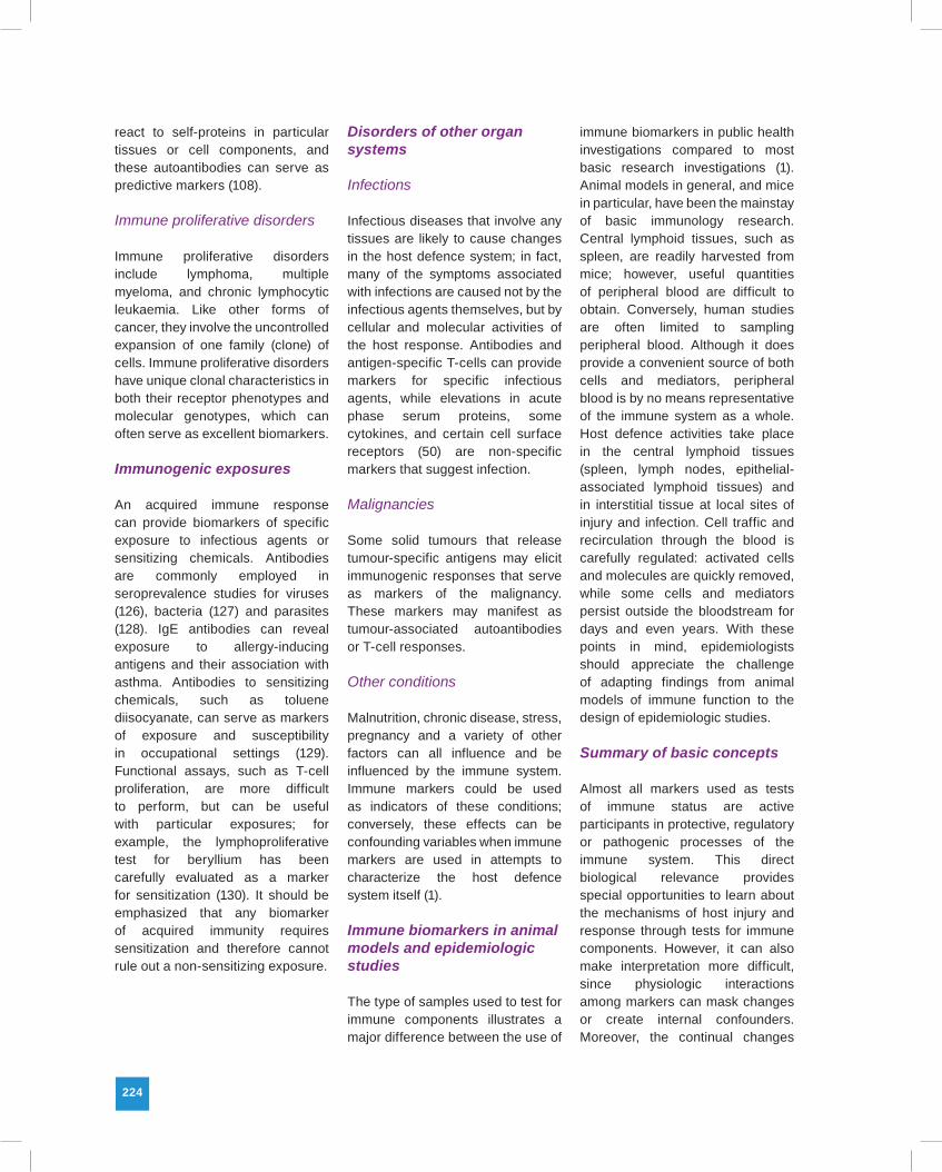
Cancer cells may express proteins that are not expressed in normal tissue or are expressed only at very low levels, and are therefore seen as foreign antigens by the immune system (Figure 13.2) (131). Such tumour-specific antigens (TSAs) were first uncovered in rodent tumours induced by coal tar dyes (132) and were later found in certain naturally-occurring human cancers (133). These discoveries augured two attractive concepts: first, that tumour defence was a primary function of the immune system that might be harnessed therapeutically; second, that testing for TSAs would allow early detection of cancer. For several decades, neither concept
lived up to its presumed promise, but newly-uncovered biomarkers have revitalized efforts aimed at both therapy (131) and early detection. In part, the resurgence involved a more realistic perspective on the use of biomarkers. As studies showed that many so-called TSAs could be detected in persons without cancer, the term has largely been replaced by the more appropriate tumour-associated antigen (TAA).
The first human TAA identified was carcinoembryonic antigen (CEA) produced by colon cancer (133), and it remains useful as a marker for tumour recurrence and progression (134). While serum CEA levels did provide statistically significant predictive value when used in prospective population studies (135), neither the sensitivity nor specificity of the test justified its use in general screening (136). Prostate-specific antigen (PSA) has proven somewhat more serviceable,
Figure 13.2. Three ways for self antigens to become tumour antigens. Peptides from three normal self proteins (yellow, blue, and green) are presented on the cell surface as normal self peptides (yellow, blue, and green) in major histocompatibility complex (MHC) molecules. In cases of mutation (A), failure of the tumour cell to repair DNA damage can result in a mutation (red) in a normal protein and, consequently, presentation of mutated peptides (red) on the surface of tumour cells. Because of a mutation, or factors that regulate its expression, a normal protein (green) can be overexpressed in a tumour cell and its peptides presented on the cell surface at highly abnormal levels (B). In cases of post-translational modification (C), a normal protein can be abnormally processed (spliced, glycosylated, phosphorylated, or lipidated) post-translationally (green stripes), resulting in an abnormal repertoire of peptides on the surface of the tumour cell. Used directly from (131) by permission from the Massachusetts Medical Society.
226
though still problematic, as a screen for prostate cancer (137), but in general blood screening for TAA has not improved the early detection.
The paradigm shift that has occurred over the last several years focused attention not on TAAs themselves, but rather on the immune responses to them. In essence, TAAs can act as autoantigens, producing a weak but detectable antibody or T-cell response. Although this is not a new idea (138,139), modern methods of molecular engineering allow TAA genes to be cloned and transfected, providing a ready supply of antigen to use in high-throughput multiplexed assays with attomolar sensitivity (140,141). Promising results have been obtained for the detection of autoantibodies in lung cancer (142), liver cancer (143), prostate cancer (141) and ovarian cancer (144). Assays for detecting TAA-specific T-cells have been applied mostly to studies of tumour vaccines or immunotherapy (145), but exploratory studies show that such T-cells can be detected in breast cancer patients naive to immunotherapy (146). Immune biomarkers may yet prove to be useful tools in the early detection of cancer.
Genetic and environmental risk factors for B-cell malignancies
While the role of the mammalian immune system in protection against cancer remains enigmatic, it is clear that lymphocytes themselves can lose control of their proliferative potential and expand uncontrollably into lymphoid malignancies. In the Eastern hemisphere, most lymphoid malignancies arise from T-cells, a phenomenon directly related to the endemic presence of human T-cell lymphocytotrophic viruses (HTLV).
In the Western hemisphere, T-cell malignancies are rare, but B-cell malignancies represent a major proportion of cancers not related to the obvious risk factors of smoking and diet.
B-cells arise from haematopoietic stem cells and undergo multiple stages of differentiation, terminating as antibody-secreting plasma cells. Four major classes of cancer arise from these various stages: acute lymphoblastic leukaemia, non-Hodgkin lymphoma, chronic
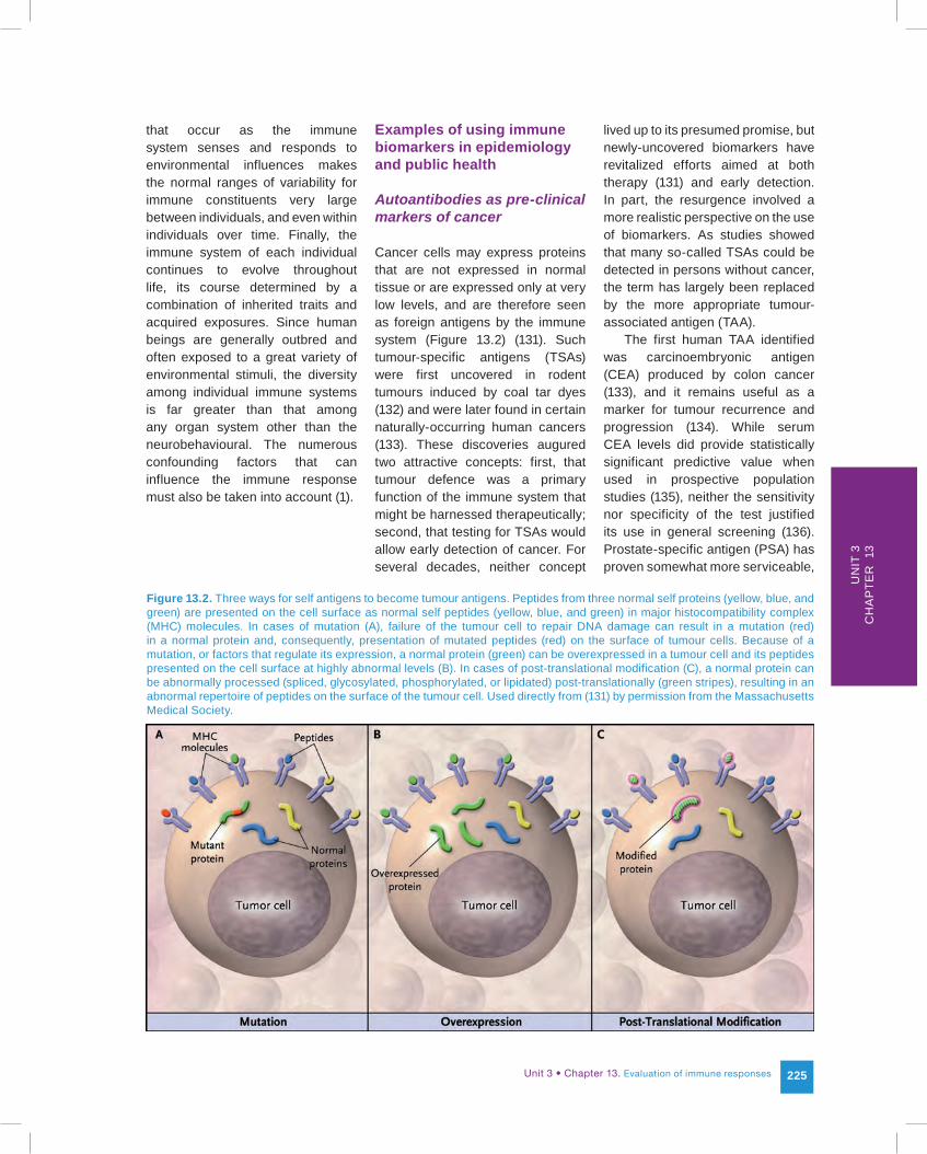
lymphocytic leukaemia, and multiple myeloma/plasmocytoma. B-cell acute lymphoblastic leukaemia, the most common form of childhood leukaemia, originates so early in the B-cell differentiation pathway that it is usually considered a stem cell malignancy. The remaining B-cell malignancies all arise from B-cells in later stages of differentiation (Figure 13.3) (147). The genetic and environmental risk factors for these B-cell malignancies remain surprisingly elusive, but information
Figure 13.3. A Venn Diagram illustrating the hypothetical relationship among the MBL, MGUS, and the malignant B-cell diseases to which they may progress. The diagram is based on the spectrum of progression endpoints for MBL and MGUS and the interrelationships of their respective biomarkers. The overlapping areas indicate some extent of shared biomarkers or clinical endpoints. Since the definition of MBL excludes any haematolymphoid disorder, areas of overlap between MBL and a malignancy is meant to convey shared biomarkers or progression from MBL. MBL and MGUS usually appear independently, but may appear together. Both conditions can remain quiescent over the lifespan of the individuals in whom they are found, or they can progress to clinical disease. CLL is shown as a complete subset of MBL, and MM/WM/SMM as nearly complete subsets of MGUS, under the presumption that all CLL is preceded by MBL and nearly all MM and WM is preceded by MGUS (although the precedent conditions may not be detected before the clinical disease endpoints are diagnosed). SMM frequently, but not always, progresses to MM. MGUS cases may infrequently develop CLL or NHL. At least one case with a combination of MBL and MGUS that developed into WM has been reported. MBL is detectable in a subset of already-diagnosed NHL cases (but to date there have been no reports of MBL developing into NHL). At least one type of NHL (small lymphocytic lymphoma) is considered to be a variant of CLL. AL may be associated with MGUS, SMM, MM, or WM and rarely with PC, CLL, or NHL. MBL progression directly to AL has not been reported to date. Used directly from (147) with permission
AL, immunoglobulin light chain amyloidosis; CLL, chronic lymphocytic leukaemia; MBL, monoclonal B-cell lymphocytosis; MGUS, monoclonal gammopathy of undetermined significance; MM, multiple myeloma; NHL, non-Hodgkin lymphoma; PC, plasmacytoma; SMM, smouldering multiple myeloma; WM, Waldenstrom macroglobulinemia.
Unit 3 • Chapter 13. Evaluation of immune responses 227
Un
it 3
Ch
ap
ter
13
revealed by population studies and immune biomarkers has allowed a much better understanding of their natural history.
The clonal expansion of lymphocytes must be carefully regulated to avoid overwhelming the host. Protective immunization generally involves the controlled proliferation of many different lymphocyte families, leading to a polyclonal response. When proliferation is dominated by a single clone, the result is a monoclonal response. Monoclonal proliferation is the first step in a progression that may lead to a lymphoid malignancy (148,149).
Chronic lymphocytic leukaemia (CLL) is a classic example of a B-cell malignancy arising from monoclonal expansion (150). Cellular, biochemical and genetic biomarkers have all contributed to our increased understanding of the natural history of CLL. The disease process probably begins with chronic immune stimulation by infectious agents, other external antigens, or autoantigens. As normal B-cell clones expand in response to antigen stimulation, the chance of individual cells acquiring genetic defects increases. Some of these defects cause the cell to escape regulatory control of proliferation: the best example to date is the loss of the microRNAs miR-15a and miR-16–1, critical regulatory factors in the bcl-2 pathway for apoptosis (151). Continued expansion of the deregulated clone promotes opportunity for other genetic lesions to accumulate, including epigenetic changes (123). At some point, the damaged clone exhibits phenotypic changes, typically an increased expression of CD5 and decreased expression of CD20. Eventually the clonal proliferation causes clinical disease by accumulating in lymphoid tissues and displacing
normal haematopoietic cells in the bone marrow. While most cases of CLL are sporadic, a familial variant has been recognized for many years (152).
The transition from a pre-clinical B-cell proliferative disorder to CLL has been documented in a succession of studies made possible by the advent of flow cytometry (15). The term monoclonal B-cell lymphocytosis (MBL) is now used to describe the pre-clinical state (153). One of the first systematic studies of MBL originated from environmental public health studies in which 13 individuals with MBL were detected (prevalence of 0.9% among participants age 40 or above) (154). These individuals were followed for up to 12 years, along with other study participants who had high B-cell counts without MBL. The majority of MBL cases remained stable or died of unrelated causes, but progression to a B-cell malignancy was observed in two of the 13: one case of CLL and one case of Waldenstrom macroglobulinemia, a related disease. Interestingly, the high B-cell counts in individuals who did not develop MBL regressed to normal over the follow-up period.
The other seminal MBL study involved familial CLL, where 18% of first-degree family members without CLL were found to have MBL (155). This striking increase over the general population prevalence suggests the familial risk for CLL is reflected in the risk for MBL. Since these studies, other population surveys have shown that the prevalence of MBL increases with age, approaching 3–5% in older, otherwise healthy, adults (156,157).
Even with the power of multiparameter flow cytometry, the detection of MBL is not trivial, since it depends on subjective assessment and sequential selection (“gating”) strategies that
isolate B-cell subsets with distinct phenotypic characteristics. Once a distinct population has been identified, MBL can be identified by light chain restriction. Antibodies may have either kappa or lambda light chains, but all the cells of a particular clone must make the antibodies with the same light chain. A phenotypic cluster that shows only one type of light chain may therefore be considered monoclonal. The complexity of cell preparation, flow cytometry and data analysis make standardization of methods to detect clonality critical for epidemiological assessment. Fortunately, the raw data from flow cytometric analysis can be captured and re-analysed using different gating strategies, allowing retrospective analysis of existing data (158).
Long-term studies of MBL and CLL using established and newly-uncovered biomarkers, such as microRNAs, will be required to sort out environmental risk factors, innate susceptibility and biomarkers of progression.
Newborn screening for immune disorders
Newborn infants that appear healthy may actually have serious latent disorders that will cause future disease, disabilities or even premature death. Newborn bloodspot screening (NBS) is designed to identify such infants quickly so that medical intervention can begin before they fall victim to such disorders. A small amount of blood from a heel stick is collected on filter paper to form a dried blood spot (DBS). DBS samples are sent to central laboratories where they are analysed by various methods to detect biomarkers of latent disorders (159). The first conditions detected by NBS were metabolic or endocrine disorders, and they remain the
228
dominant type screened for in current NBS programs. However, interest in screening for other types of disorders is growing rapidly (160), and the idea of screening for risk factors of future disorders, in addition to screening for established (though occult) conditions, has been gaining attention. Two immune disorders typify these two trends: severe combined immune deficiency (SCID), and type 1 (juvenile) diabetes.
Severe combined immune deficiency
Severe combined immune deficiency (SCID) is a lethal congenital failure of immune development (161). It is often called “bubble boy disease” because of early attempts to prevent infection through sequestering the child from the natural environment. Because of the persistence of placentally-transferred maternal antibodies, SCID remains concealed for several weeks after birth, but without a functional immune system, babies soon become infected and typically die in infancy. A series of landmark studies have shown that newborns with SCID can be rescued before they become symptomatic by transplanting bone marrow progenitor cells (162). However, such rescue is difficult or impossible after SCID babies become infected. SCID thus meets the ideal criteria for NBS: a lethal disorder with a latent onset that can be prevented by medical intervention. The birth prevalence of SCID is not certain, but is estimated to be in the range of 1 to 4 per 100 000.
The process of finding an immune biomarker for SCID that can be measured on a newborn DBS illustrates how far-reaching our knowledge of the immune system and our abilities to probe it have come. First came the understanding
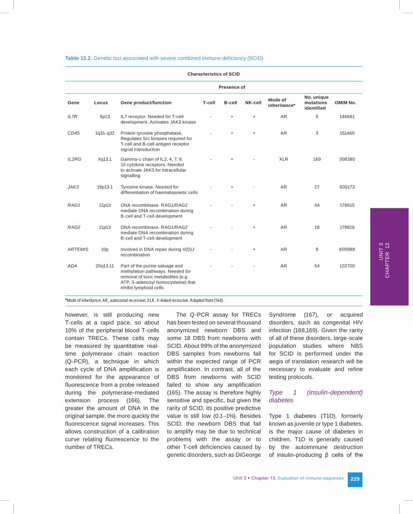
that, although SCID is expressed as a combined deficiency involving both humoral (B-cell) and cell-mediated (T-cell) immunity, it is actually a defect in T-cell development. B-cell counts in SCID babies are normal or even elevated, and they are fully functional. However, without functional T-cells to provide help, even humoral responses are deficient, giving the phenotype of a combined immune deficiency. The second realization was that mutations at any one of several unrelated genetic loci could result in failure of T-cell development: SCID was a single gene defect in each individual case, but with multiple genetic causes overall (Table 13.2). Other loci in which mutations could cause SCID may yet be uncovered (163). Moreover, the mutations at these various loci are widely
scattered throughout the exons, so screening by conventional genetic tests is not feasible.
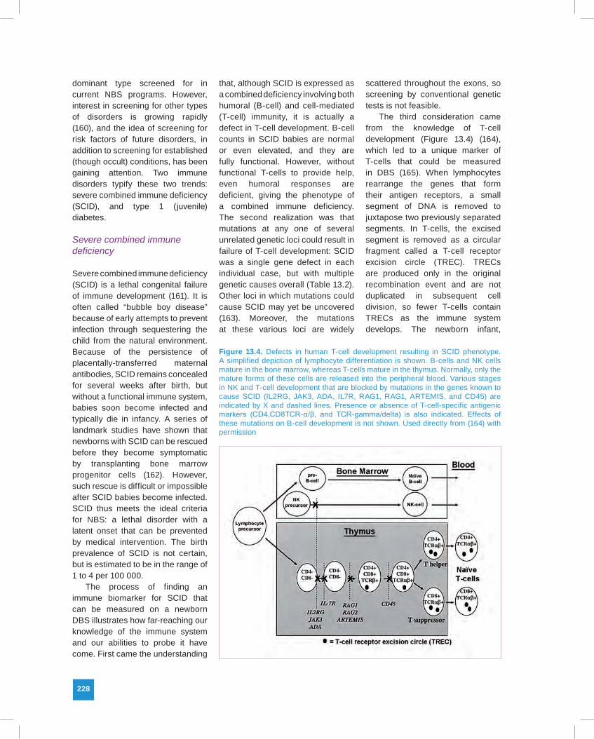
The third consideration came from the knowledge of T-cell development (Figure 13.4) (164), which led to a unique marker of T-cells that could be measured in DBS (165). When lymphocytes rearrange the genes that form their antigen receptors, a small segment of DNA is removed to juxtapose two previously separated segments. In T-cells, the excised segment is removed as a circular fragment called a T-cell receptor excision circle (TREC). TRECs are produced only in the original recombination event and are not duplicated in subsequent cell division, so fewer T-cells contain TRECs as the immune system develops. The newborn infant,
Figure 13.4. Defects in human T-cell development resulting in SCID phenotype. A simplified depiction of lymphocyte differentiation is shown. B-cells and NK cells mature in the bone marrow, whereas T-cells mature in the thymus. Normally, only the mature forms of these cells are released into the peripheral blood. Various stages in NK and T-cell development that are blocked by mutations in the genes known to cause SCID (IL2RG, JAK3, ADA, IL7R, RAG1, RAG1, ARTEMIS, and CD45) are indicated by X and dashed lines. Presence or absence of T-cell-specific antigenic markers (CD4,CD8TCR-α/β, and TCR-gamma/delta) is also indicated. Effects of these mutations on B-cell development is not shown. Used directly from (164) with permission
Unit 3 • Chapter 13. Evaluation of immune responses 229
Un
it 3
Ch
ap
ter
13
Table 13.2. Genetic loci associated with severe combined immune deficiency (SCID)
Characteristics of SCID
Presence of
Gene Locus Gene product/function T-cell B-cell NK-cell Mode of inheritance*
No. unique mutations identified
OMIM No.
IL7R 5p13 IL7 receptor. Needed for T-cell development. Activates JAK3 kinase
- + + AR 5 146661
CD45 1q31-q32 Protein tyrosine phosphatase. Regulates Src kinases required for T-cell and B-cell antigen receptor signal transduction
- + + AR 3 151460
IL2RG Xq13.1 Gamma-c chain of IL2, 4, 7, 9, 15 cytokine receptors. Needed to activate JAK3 for intracellular signalling
- + - XLR 169 308380
JAK3 19p13.1 Tyrosine kinase. Needed for differentiation of haematopoietic cells
- + - AR 27 600173
RAG1 11p13 DNA recombinase. RAG1/RAG2 mediate DNA recombination during B-cell and T-cell development
- - + AR 44 179615
RAG2 11p13 DNA recombinase. RAG1/RAG2 mediate DNA recombination during B-cell and T-cell development
- - + AR 18 179616
ARTEMIS 10p Involved in DNA repair during V(D)J recombination
- - + AR 9 605988
ADA 20q13.11 Part of the purine salvage and methylation pathways. Needed for removal of toxic metabolites (e.g. ATP, S-adenosyl homocysteine) that inhibit lymphoid cells
- - - AR 54 102700
*Mode of inheritance: AR, autosomal recessive; XLR, X-linked recessive. Adapted from (164).
however, is still producing new T-cells at a rapid pace, so about 10% of the peripheral blood T-cells contain TRECs. These cells may be measured by quantitative real-time polymerase chain reaction (Q-PCR), a technique in which each cycle of DNA amplification is monitored for the appearance of fluorescence from a probe released during the polymerase-mediated extension process (166). The greater the amount of DNA in the original sample, the more quickly the fluorescence signal increases. This allows construction of a calibration curve relating fluorescence to the number of TRECs.
The Q-PCR assay for TRECs has been tested on several thousand anonymized newborn DBS and some 18 DBS from newborns with SCID. About 99% of the anonymized DBS samples from newborns fall within the expected range of PCR amplification. In contrast, all of the DBS from newborns with SCID failed to show any amplification (165). The assay is therefore highly sensitive and specific, but given the rarity of SCID, its positive predictive value is still low (0.1–1%). Besides SCID, the newborn DBS that fail to amplify may be due to technical problems with the assay or to other T-cell deficiencies caused by genetic disorders, such as DiGeorge
Syndrome (167), or acquired disorders, such as congenital HIV infection (168,169). Given the rarity of all of these disorders, large-scale population studies where NBS for SCID is performed under the aegis of translation research will be necessary to evaluate and refine testing protocols.
Type 1 (insulin-dependent) diabetes
Type 1 diabetes (T1D), formerly known as juvenile or type 1 diabetes, is the major cause of diabetes in children. T1D is generally caused by the autoimmune destruction of insulin-producing β cells of the
230
pancreas (170). The autoimmune pathogenesis of T1D was revealed by two biomarkers, one genetic and one acquired. The genetic biomarker is linkage with certain alleles of the MHC genes that code for the human leukocyte antigens (HLA), a risk locus shared by all autoimmune disorders. About half of the attributable risk for T1D is genetic, and about half of that risk is contained in the HLA genes. The genetic risk for T1D is associated particularly with the class II MHC genes that code for the HLA-D antigens (171). Some alleles confer susceptibility, while others confer resistance. Interestingly, resistance is dominant, which allows more cost-effective screening approaches that identify protective alleles and eliminate them from further testing.
The acquired biomarker for T1D is a group of autoantibodies that react with pancreatic islet cell antigens (172). Autoantibodies are the other essential biomarker of autoimmune disorders. In the rheumatic disorders, such as systemic lupus erythematosis, they are an obvious part of the pathogenic process; in organ-specific disorders, such as T1D, they are thought to be largely paraphenomena, but still serve as useful markers. Originally discovered by immunofluorescence microscopy using pancreas tissue to visualize antibody binding to islet cells (173), most testing today is done biochemically using purified islet cell antigens produced by cloned genes. Autoantibodies to three major islet cell antigens have been the important determinants of T1D risk in epidemiologic and natural history studies, but antibodies to other islet cell antigens have been reported on the basis of distinct tissue binding patterns (172).
A series of prospective studies by research centres around the world has established a consensus
paradigm (Figure 13.5) for the progression from innate risk to islet cell autoimmunity and ultimately to T1D. The major remaining puzzle is the role of environmental exposures in triggering or advancing the autoimmune process (174). The candidates for such exposures include bacterial and viral infections (particularly enteroviruses and rhinoviruses), food antigens, xenobiotic chemicals, allergens, ultraviolet light, and the immunomodulatory effects of stress. Clearly, the identification of environmental risk factors would open new possibilities for prevention and intervention.
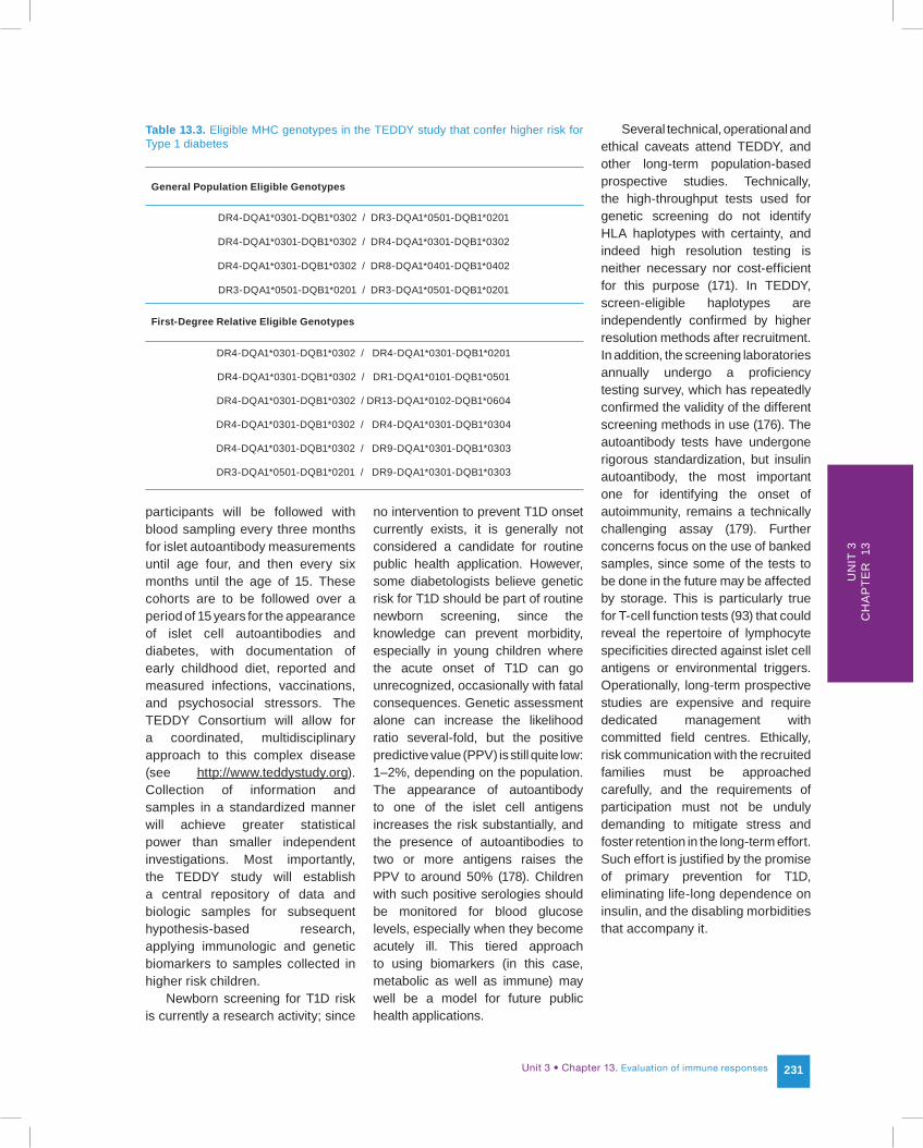
With this goal in mind, a prospective multisite natural history study has been initiated to address comprehensively the role of environmental exposures in T1D. Called TEDDY (The Environmental Determinants of Diabetes in the Young), this study is recruiting infants at higher genetic risk for T1D (as well as controls without higher genetic risk) and assembling them into a
long-term study cohort (175,176). To maximize the proportion of recruited children who will develop T1D, the highest genetic risk, defined as one of four MHC class II haplotypes (Table 13.3), is required for eligibility in the general population. However, since familial risk contributes independently, six additional MHC haplotypes are eligible in families where a first-degree relative of the prospective recruit already has T1D (Table 13.3).
Because risk from environmental exposures may begin very early, perhaps even in utero (177), TEDDY collects the first samples to look for environmental factors at three months of age. With such a short window to identify and recruit participants, TEDDY investigators seek informed consent for the initial genetic screen from the parents of newborns, making it a research application of newborn screening. By the close of the screening phase, some 300 000 newborns will have been screened, and about 8000 higher-risk infants enrolled. The
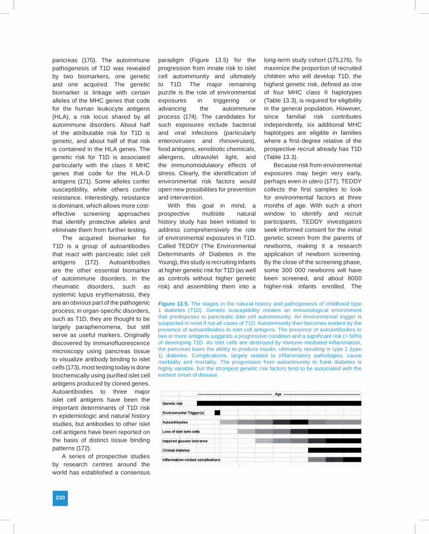
Figure 13.5. The stages in the natural history and pathogenesis of childhood type 1 diabetes (T1D). Genetic susceptibility creates an immunological environment that predisposes to pancreatic islet cell autoimmunity. An environmental trigger is suspected in most if not all cases of T1D. Autoimmunity then becomes evident by the presence of autoantibodies to islet cell antigens. The presence of autoantibodies to two or more antigens suggests a progressive condition and a significant risk (> 50%) of developing T1D. As islet cells are destroyed by immune-mediated inflammation, the pancreas loses the ability to produce insulin, ultimately resulting in type 1 (type 1) diabetes. Complications, largely related to inflammatory pathologies, cause morbidity and mortality. The progression from autoimmunity to frank diabetes is highly variable, but the strongest genetic risk factors tend to be associated with the earliest onset of disease
Unit 3 • Chapter 13. Evaluation of immune responses 231
Un
it 3
Ch
ap
ter
13
Table 13.3. Eligible MHC genotypes in the TEDDY study that confer higher risk for Type 1 diabetes
General Population Eligible Genotypes
DR4-DQA1*0301-DQB1*0302 / DR3-DQA1*0501-DQB1*0201
DR4-DQA1*0301-DQB1*0302 / DR4-DQA1*0301-DQB1*0302
DR4-DQA1*0301-DQB1*0302 / DR8-DQA1*0401-DQB1*0402
DR3-DQA1*0501-DQB1*0201 / DR3-DQA1*0501-DQB1*0201
First-Degree Relative Eligible Genotypes
DR4-DQA1*0301-DQB1*0302 / DR4-DQA1*0301-DQB1*0201
DR4-DQA1*0301-DQB1*0302 / DR1-DQA1*0101-DQB1*0501
DR4-DQA1*0301-DQB1*0302 / DR13-DQA1*0102-DQB1*0604
DR4-DQA1*0301-DQB1*0302 / DR4-DQA1*0301-DQB1*0304
DR4-DQA1*0301-DQB1*0302 / DR9-DQA1*0301-DQB1*0303
DR3-DQA1*0501-DQB1*0201 / DR9-DQA1*0301-DQB1*0303
participants will be followed with blood sampling every three months for islet autoantibody measurements until age four, and then every six months until the age of 15. These cohorts are to be followed over a period of 15 years for the appearance of islet cell autoantibodies and diabetes, with documentation of early childhood diet, reported and measured infections, vaccinations, and psychosocial stressors. The TEDDY Consortium will allow for a coordinated, multidisciplinary approach to this complex disease (see http://www.teddystudy.org). Collection of information and samples in a standardized manner will achieve greater statistical power than smaller independent investigations. Most importantly, the TEDDY study will establish a central repository of data and biologic samples for subsequent hypothesis-based research, applying immunologic and genetic biomarkers to samples collected in higher risk children.
Newborn screening for T1D risk is currently a research activity; since
no intervention to prevent T1D onset currently exists, it is generally not considered a candidate for routine public health application. However, some diabetologists believe genetic risk for T1D should be part of routine newborn screening, since the knowledge can prevent morbidity, especially in young children where the acute onset of T1D can go unrecognized, occasionally with fatal consequences. Genetic assessment alone can increase the likelihood ratio several-fold, but the positive predictive value (PPV) is still quite low: 1–2%, depending on the population. The appearance of autoantibody to one of the islet cell antigens increases the risk substantially, and the presence of autoantibodies to two or more antigens raises the PPV to around 50% (178). Children with such positive serologies should be monitored for blood glucose levels, especially when they become acutely ill. This tiered approach to using biomarkers (in this case, metabolic as well as immune) may well be a model for future public health applications.
Several technical, operational and ethical caveats attend TEDDY, and other long-term population-based prospective studies. Technically, the high-throughput tests used for genetic screening do not identify HLA haplotypes with certainty, and indeed high resolution testing is neither necessary nor cost-efficient for this purpose (171). In TEDDY, screen-eligible haplotypes are independently confirmed by higher resolution methods after recruitment. In addition, the screening laboratories annually undergo a proficiency testing survey, which has repeatedly confirmed the validity of the different screening methods in use (176). The autoantibody tests have undergone rigorous standardization, but insulin autoantibody, the most important one for identifying the onset of autoimmunity, remains a technically challenging assay (179). Further concerns focus on the use of banked samples, since some of the tests to be done in the future may be affected by storage. This is particularly true for T-cell function tests (93) that could reveal the repertoire of lymphocyte specificities directed against islet cell antigens or environmental triggers. Operationally, long-term prospective studies are expensive and require dedicated management with committed field centres. Ethically, risk communication with the recruited families must be approached carefully, and the requirements of participation must not be unduly demanding to mitigate stress and foster retention in the long-term effort. Such effort is justified by the promise of primary prevention for T1D, eliminating life-long dependence on insulin, and the disabling morbidities that accompany it.
232
Immune biomarkers ofneurodevelopmental disorders
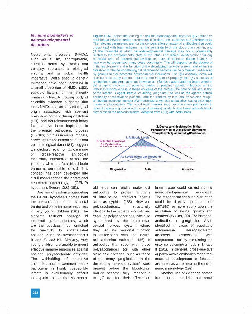
Neuromental disorders (NMDs), such as autism, schizophrenia, attention deficit syndromes and epilepsy, represent a biological enigma and a public health imperative. While specific genetic mutations have been identified in a small proportion of NMDs (180), etiologic factors for the majority remain unclear. A growing body of scientific evidence suggests that many NMDs have an early etiological origin associated with aberrant brain development during gestation (181), and neuroimmunomodulatory factors have been implicated in the prenatal pathogenic process (182,183). Studies in animal models, as well as limited human studies and epidemiological data (184), suggest an etiologic role for autoimmune or cross-reactive antibodies maternally transferred across the placenta when the fetal blood-brain barrier is permeable to IgG. This concept has been developed into a full model termed the gestational neuroimmunopathology (GENIP) hypothesis (Figure 13.6) (181).
One line of evidence supporting the GENIP hypothesis comes from the consideration of the placental barrier and of the immune responses in very young children (181). The placenta restricts passage of maternal IgG2 antibodies, which are the subclass most enriched for reactivity to encapsulated bacteria, such as meningococcus B and E. coli K1. Similarly, very young children are unable to mount effective immune responses against bacterial polysaccharide antigens. The withholding of protective antibodies against common deadly pathogens in highly susceptible infants is evolutionarily difficult to explain, since the six-month-
old fetus can readily make IgG antibodies to protein antigens of intrauterine infectious agents such as syphilis (185). However, polysaccharides, structurally identical to the bacterial α-2,8-linked capsular polysaccharides, are also synthesized by the mammalian central nervous system, where they regulate neuronal function in association with the neural cell adhesion molecule (186). If antibodies that react with these polysaccharides (or with other sialic acid epitopes, such as those of the many gangliosides in the developing nervous system) were present before the blood-brain barrier became fully impervious to IgG transfer, their effects on
brain tissue could disrupt normal neurodevelopmental processes. The mechanism for such disruption could be directly upon neurons (187,188), or more subtly upon the regulation of axonal growth and connectivity (189,190). For instance, antibodies to ganglioside GM1, identified in cases of paediatric autoimmune neuropsychiatric disorders associated with streptococci, act by stimulating the enzyme calcium/calmodulin kinase II (191). In general, cross-reactive or polyreactive antibodies that effect neuronal development or function are seen as an emerging theme in neuroimmunology (192).
Another line of evidence comes from animal models that show
Figure 13.6. Factors influencing the risk that transplacental maternal IgG antibodies could cause developmental neuromental disorders, such as autism and schizophrenia. The relevant parameters are: (1) the concentration of maternal antibodies that could cross-react with brain antigens, (2) the permeability of the blood-brain barrier, and (3) the threshold at which neurodevelopmental damage may occur, presumably related to the developmental state of the fetus. The clinical manifestations for any particular type of neuromental dysfunction may be detected during infancy, or may only be recognized many years postnatally. This will depend on the degree of initial involvement in the function of the developing nervous system, and when the threshold for the neuropathological disorders to become clinically manifest, is lowered by genetic and/or postnatal environmental influences. The IgG antibody levels will also be affected by immune factors in the mother or progeny: the IgG subclass of antibodies to antigens common between an infectious agent and the brain; whether the antigens involved are polysaccharides or proteins; genetic influences on the immune responsiveness to these antigens of the mother; the time of her acquisition of the infectious agent, before, or during, pregnancy, as well as the agent’s natural chronicity or reactivation potential, and the transfer by feto-fetal transfusion of IgG/antibodies from one member of a monozygotic twin pair to the other, due to a common chorionic placentation. The blood-brain barriers may become more permissive in case of trauma (e.g. a prolonged vaginal delivery), in which case lower antibody levels may cross to the nervous system. Adapted from (181) with permission
Unit 3 • Chapter 13. Evaluation of immune responses 233
Un
it 3
Ch
ap
ter
13
antibodies can influence behaviour if they reach the brain. Mice that were given autoantibodies associated with systemic lupus erythematosis developed behavioural anomalies, if, and only if, they also were given pharmacologic agents that opened the blood-brain barrier (193). Infant monkeys gestationally exposed to IgG antibodies purified from the serum of mothers with autistic children demonstrated the stereotypies characteristic of autistic behaviour (194).
A third line of evidence for GENIP is the identification of antibodies that react with brain tissue antigens in children with neurodevelopmental disorders (195,196). These studies have used research assays that are not widely employed or validated by inter-laboratory studies, but the methods are generally sound, and the differences with control samples are often striking. Although these studies cannot prove causality, they suggest an association that could at least provide useful markers for stratification within the complex spectrum of neurobehavioural diseases.
The key to providing direct evidence for the GENIP hypothesis is the newborn baby, whose blood contains large amounts of placentally-transferred IgG from the mother, as well as the low levels of antibodies made in utero by the fetus. Samples from newborns can therefore reveal the spectrum of antibody reactivities to which the developing brain has been exposed. Since blood samples from virtually every newborn in the
United States are routinely collected as DBS for newborn screening, the samples required to test the GENIP hypothesis are readily available. Highly-multiplexed suspension arrays have been developed for detecting a wide variety of potentially relevant antibodies in DBS, and they are currently being applied to epidemiologic studies of autism, epilepsy, and other neuromental disorders. Perhaps the major concern in such studies lies not in the arcane realm of immune biomarkers, but rather with the classic epidemiologic dilemma of establishing a consistent case definition for these complex neurodevelopmental conditions.
Future opportunities and challenges
The continual advances of biomedical research in immunology offer an ever-widening opportunity to employ immune biomarkers in epidemiologic studies. In the foreseeable future biomarkers will lead to an increased understanding of the relationship between innate and acquired immunity; the influences of microRNAs on immune cell differentiation and function; the role of epigenetic mechanisms; the regulation of immune responses by the TH1-TH2-TH17 network; the environmental factors that trigger allergy, autoimmunity and immune malignancy; and the interaction of the neurobehavioural and immune systems in neuromental disorders. Also envisioned is a more rapid pace of translation from basic and
clinical research to epidemiologic field studies and public health applications, such as newborn screening and early cancer detection.
The translational process will have to confront challenges in the standardization of laboratory measurements, the establishment of biologic validity and the meaningful interpretation of results. A multidisciplinary approach that engages epidemiologists, laboratory scientists and clinicians offers the best chance for useful field studies and public health applications. All the scientists involved in such efforts should have some sense of the way immune markers are measured, of their functional role in health and disease, and of the statistical methods that will be used to analyse the data. While these aphorisms can be applied to any type of biomarker, the self-referential sensory nature of the immune system, its wide range of effector functions, and the narrow margin between protection and pathology make them especially pertinent to the use of immune biomarkers. When properly met, the challenges will open new routes to scientific discovery, disease prevention and public health practice.
Disclaimer: The findings and conclusions in this chapter are those of the author and do not necessarily represent the views of the Centers for Disease Control and Prevention.
234
References
1. Vogt RF, Schulte PA. Immune markers in epidemiologic field studies. In: Schulte PA, Perera FP, editors. Molecular epidemiology: principles and practices. New York (NY): Academic Press; 1993. p. 407–442.
2. Allen MA, Liang TS, La Salvia T et al. (2005). Assessing the attitudes, knowledge, and awareness of HIV vaccine research among adults in the United States. J Acquir Immune Defic Syndr, 40:617–624.doi:10.1097/01.qai.0000174655.63653.38 PMID:16284540
3. Jacobson DL, Gange SJ, Rose NR, Graham NM (1997). Epidemiology and estimated population burden of selected autoimmune diseases in the United States. Clin Immunol Immunopathol, 84:223–243.doi:10.1006/clin.1997.4412 PMID:9281381
4. Rose NR (2006). The significance of autoimmunity in myocarditis. Ernst Schering Res Found Workshop, 55:141–1 5 4 . d o i : 1 0 .1 0 0 7 / 3 - 5 4 0 - 3 0 8 2 2 - 9 _ 9 PMID:16331858
5. Eaton WW, Rose NR, Kalaydjian A et al. (2007). Epidemiology of autoimmune diseases in Denmark. J Autoimmun, 29:1–9.doi: 10.1016/j.jaut.2007.05.002 PMID:17582741
6. Lindegren ML, Kobrynski L, Rasmussen SA et al. (2004). Applying public health strategies to primary immunodeficiency diseases: a potential approach to genetic disorders. MMWR Recomm Rep, 53 RR-1;1–29. PMID: 14724556
7. Beyer M, Schultze JL (2008). Immunoregulatory T cells: role and potential as a target in malignancy. Curr Oncol Rep, 10:130–136.doi:10.1007/s11912-008-0021-z PMID:18377826
8. Shim YK, Silver SR, Caporaso NE et al. (2007). B cells behaving badly. Br J Haematol, 139:658–662.doi:10.1111/j.1365-2141.2007.06842.x PMID:18021079
9. Davis MM, Krogsgaard M, Huse M et al. (2007). T cells as a self-referential, sensory organ. Annu Rev Immunol, 25:681 –695.doi:10.1146/annurev.immunol.24.021605. 090600 PMID:17291190
10. Scott A, Khan KM, Cook JL, Duronio V (2004). What is “inflammation”? Are we ready to move beyond Celsus? Br J Sports Med, 38:248–249.doi:10.1136/bjsm.2003.011221 PMID:15155418
11. Cole GA, Nathanson N, Prendergast RA (1972). Requirement for theta-bearing cells in lymphocytic choriomeningitis virus-induced central nervous system disease. Nature, 238:335–337.doi:10.1038/238335a0 PMID:4561841
12. Dannenberg AM. Pathogenesis of human pulmonary tuberculosis: insights from the rabbit model. Washington (DC): ASM Press; 2006.
13. Fisman EZ, Adler Y, Tenenbaum A (2008). Biomarkers in cardiovascular diabetology: interleukins and matrixins. Adv Cardiol, 45:44–64.doi:10.1159/000115187 PMID:182 30955
14. Tung JW, Heydari K, Tirouvanziam R et al. (2007). Modern flow cytometry: a practical approach. Clin Lab Med, 27: 453–468, v.doi:10.1016/j.cll.2007.05.001 PMID: 17658402
15. Shapiro HM (2007). Cytometry in monoclonal B-cell lymphocytosis and chronic lymphocytic leukaemia–the Hunting of the Snark? Br J Haematol, 139:772–773.do i :10.1111/ j .1365 -2141. 20 07.0 6855.x PMID:18021090
16. Ellmark P, Woolfson A, Belov L, Christopherson RI (2008). The applicability of a cluster of differentiation monoclonal antibody microarray to the diagnosis of human disease. Methods Mol Biol, 439:199–209.doi:10.1007/978 -1-59745 -188 -8_14 PMID:18370105
17. Zola H, Swart B, Banham A et al. (2007). CD molecules 2006–human cell differentiation molecules. J Immunol Methods, 319:1–5.doi:10.1016/j.jim.2006.11.001 PMID:17174972
18. Beeler A, Zaccaria L, Kawabata T et al. (2008). CD69 upregulation on T cells as an in vitro marker for delayed-type drug hypersensitivity. Allergy, 63:181–188. PMID: 18005225
19. Lindsey WB, Lowdell MW, Marti GE et al. (2007). CD69 expression as an index of T-cell function: assay standardization, validation and use in monitoring immune recovery. Cytotherapy, 9:123–132.doi:10.1080/146532 40601182838 PMID:17453964
20. Wiestner A, Rosenwald A, Barry TS et al. (2003). ZAP-70 expression identifies a chronic lymphocytic leukemia subtype with unmutated immunoglobulin genes, inferior clinical outcome, and distinct gene expression profile. Blood, 101:4944–4951.doi:10.1182/blood-2002-10-3306 PMID:12595313
21. Clinical and Laboratory Standards Institute. Enumeration of immunologically defined cell populations by flow cytometry; approved guideline. 2nd ed. CLSI document H42–A2 [ISBN 1–56238–000–0]. Wayne (PA): Clinical and Laboratory Standards Institute; 2007.
22. Wang L, Abbasi F, Gaigalas AK et al. (2007). Discrepancy in measuring CD4 expression on T-lymphocytes using fluorescein conjugates in comparison with unimolar CD4-phycoerythrin conjugates. Cytometry B Clin Cytom, 72:442–449. PMID: 17474131
23. Marti GE, Vogt RF Jr, Stetler-Stevenson M (2003). Clinical quantitative flow cytometry: “Identifying the optimal methods for clinical quantitative flow cytometry”. Cytometry B Clin Cytom, 55:59.doi:10.1002/cyto.b.10053 PMID:12949961
24. Clinical and Laboratory Standards Institute. Fluorescence calibration and quantitative measurement of fluorescence intensity. NCCLS document I/LA24-A [ISBN 1–56238–000–0]. Wayne (PA): Clinical and Laboratory Standards Institute, 2004.
25. Elewaut D, Kronenberg M (2000). Molecular biology of NK T cell specificity and development. Semin Immunol, 12:561–568.doi:10.1006/smim.2000.0275 PMID:11145862
26. Hedges JF, Lubick KJ, Jutila MA (2005). Gamma delta T cells respond directly to pathogen-associated molecular patterns. J Immunol, 174:6045–6053. PMID:15879098
27. Tang Q, Bluestone JA (2008). The Foxp3+ regulatory T cell: a jack of all trades, master of regulation. Nat Immunol, 9:239–244.doi: 10.1038/ni1572 PMID:18285775
28. Fehervari Z, Sakaguchi S (2006). Peacekeepers of the immune system. Sci Am, 295:56–63. PMID:16989481
29. Roumier T, Capron M, Dombrowicz D, Faveeuw C (2008). Pathogen induced regulatory cell populations preventing allergy through the Th1/Th2 paradigm point of view. Immunol Res, 40:1–17.doi:10.1007/s12026-007-0058-3 PMID:18193360
30. Levy DA (2004). Parasites and allergy: from IgE to Th1/Th2 and beyond. Clin Rev Allergy Immunol, 26:1–4.doi:10.1385/CRIAI:26:1:1 PMID:14755070
31. Kawamoto N, Kaneko H, Takemura M et al. (2006). Age-related changes in intracellular cytokine profiles and Th2 dominance in allergic children. Pediatr Allergy Immunol, 17:125–133.doi:10.1111/j.1399-3038.2005. 00363.x PMID:16618362
32. Steinman L (2007). A brief history of T(H)17, the first major revision in the T(H)1/T(H)2 hypothesis of T cell-mediated tissue damage. Nat Med, 13:139–145.doi:10.1038/nm1551 PMID:17290272
Unit 3 • Chapter 13. Evaluation of immune responses 235
Un
it 3
Ch
ap
ter
13
33. Kaiko GE, Horvat JC, Beagley KW, Hansbro PM (2008). Immunological decision-making: how does the immune system decide to mount a helper T-cell response? Immunology, 123:326–338.doi:10.1111/j.1365-2567.2007.02719.x PMID:17983439
34. Weaver CT, Murphy KM (2007). The central role of the Th17 lineage in regulating the inflammatory/autoimmune axis. Semin Immunol, 19:351–352.doi:10.1016/j.smim.2008.01.001 PMID:18276155
35. Korn T, Oukka M, Kuchroo V, Bettelli E (2007). Th17 cells: effector T cells with inflammatory properties. Semin Immunol, 19:362–371.doi:10.1016/j.smim.2007.10.007 PMID:18035554
36. Stockinger B, Veldhoen M, Martin B (2007). Th17 T cells: linking innate and adaptive immunity. Semin Immunol, 19:353 –361.doi:10.1016/j.smim.2007.10.008 PMID: 18023589
37. Yu JJ, Gaffen SL (2008). Interleukin-17: a novel inflammatory cytokine that bridges innate and adaptive immunity. Front Biosci, 13:170–177.doi:10.2741/2667 PMID:17981535
38. Veldhoen M, Hirota K, Westendorf AM et al. (2008). The aryl hydrocarbon receptor links TH17-cell-mediated autoimmunity to environmental toxins. Nature, 453:106–109.doi:10.1038/nature06881 PMID:18362914
39. Stevens EA, Bradfield CA (2008). Immunology: T cells hang in the balance. Nature, 453:46–47.doi:10.1038/453046a PMID:18451850
40. Wilson SB, Kent SC, Patton KT et al. (1998). Extreme Th1 bias of invariant Valpha24JalphaQ T cells in type 1 diabetes. Nature, 391:177–181.doi:10.1038/34419 PMID:9428763
41. Wilson SB, Kent SC, Patton KT et al. (1999). Extreme Th1 bias of invariant V24JQ T cells in type 1 diabetes [Erratum]. Nature, 399:84 doi:10.1038/20007.
42. Redondo MJ, Gottlieb PA, Motheral T et al. (1999). Heterophile anti-mouse immunoglobulin antibodies may interfere with cytokine measurements in patients with HLA alleles protective for type 1A diabetes. Diabetes, 48:2166–2170.doi:10.2337/diabetes.48.11.2166 PMID:10535450
43. Zhu J, Paul WE (2010). Heterogeneity and plasticity of T helper cells. Cell Res, 20:4–12.doi:10.1038/cr.2009.138 PMID:20010916
44. Neidhart M, Fehr K, Pataki F, Michel BA (1996). The levels of memory (CD45RA-, RO+) CD4+ and CD8+ peripheral blood T-lymphocytes correlate with IgM rheumatoid factors in rheumatoid arthritis. Rheumatol Int, 15:201–209.doi:10.1007/BF00290522 PMID: 8717104
45. Yamaoka M, Kusunoki Y, Kasagi F et al. (2004). Decreases in percentages of naïve CD4 and CD8 T cells and increases in percentages of memory CD8 T-cell subsets in the peripheral blood lymphocyte populations of A-bomb survivors. Radiat Res, 161:290–298.doi:10.1667/RR3143 PMID:14982485
46. Pawlik I, Mackiewicz U, Lacki JK et al. (1997). The differences in the expression of CD45 isoforms on peripheral blood lymphocytes derived from patients with seasonal or perennial atopic allergy. Tohoku J Exp Med, 182:1–8.doi:10.1620/tjem.182.1 PMID:9241767
47. Fairfax KA, Kallies A, Nutt SL, Tarlinton DM (2008). Plasma cell development: from B-cell subsets to long-term survival niches. Semin Immunol, 20:49–58.doi:10.1016/j.smim.2007.12.002 PMID:18222702
48. Stetler-Stevenson M, Braylan RC (2001). Flow cytometric analysis of lymphomas and lymphoproliferative disorders. Semin Hematol, 38:111–123.doi:10.1053/shem.2001.21923 PMID:11309693
49. Youinou P, Pers JO, Jamin C, Lydyard PM (2000). CD5-positive B cells at the crossroads of B cell malignancy and nonorgan-specific autoimmunity. Pathol Biol (Paris), 48:574–576. PMID:10965537
50. Davis BH, Olsen SH, Ahmad E, Bigelow NC (2006). Neutrophil CD64 is an improved indicator of infection or sepsis in emergency department patients. Arch Pathol Lab Med, 130:654–661. PMID:16683883
51. Silveira-Lemos D, Teixeira-Carvalho A, Martins-Filho OA et al. (2006). High expression of co-stimulatory and adhesion molecules are observed on eosinophils during human Schistosoma mansoni infection. Mem Inst Oswaldo Cruz, 101 Suppl 1;345–351.doi:10.1590/S0074-02762006000900056 PMID:17308795
52. Davis DM (2006). Intrigue at the immune synapse. Sci Am, 294:48–55.doi:10.1038/scientificamerican0206-48 PMID:16478026
53. Wan S, Flower DR, Coveney PV (2008). Toward an atomistic understanding of the immune synapse: large-scale molecular dynamics simulation of a membrane-embedded TCR-pMHC-CD4 complex. Mol Immunol, 45:1221–1230.doi:10.1016/j.molimm.2007.09.022 PMID:17980430
54. Steinman RM (2007). Dendritic cells: understanding immunogenicity. Eur J Immunol, 37 Suppl 1;S53–S60.doi:10.1002/eji.200737400 PMID:17972346
55. Rivera J, Olivera A (2008). A current understanding of Fc epsilon RI-dependent mast cell activation. Curr Allergy Asthma Rep, 8:14–20.doi:10.1007/s11882-008-0004-z PMID:18377769
56. MacQueen G, Marshall J, Perdue M et al. (1989). Pavlovian conditioning of rat mucosal mast cells to secrete rat mast cell protease II. Science, 243:83–85.doi:10.1126/science.2911721 PMID:2911721
57. Clinical and Laboratory Standards Institute. Clinical evaluation of immunoassays; approved guideline. 2nd ed. CLSI document I/LA21–A2. Wayne (PA): Clinical and Laboratory Standards Institute; 2008.
58. Becker AM, Das S, Xia Z et al. (2008). Serum inflammatory protein profiles in patients with chronic rhinosinusitis undergoing sinus surgery: a preliminary analysis. Am J Rhinol, 22:139–143.doi:10.2500/ajr.2008.22.3151 PMID:18416969
59. Liu W, Li X, Ding F, Li Y (2008). Using SELDI-TOF MS to identify serum biomarkers of rheumatoid arthritis. Scand J Rheumatol, 37:94–102.doi:10.1080/03009740701747152 PMID:18415765
60. Langbein S (2008). Identification of disease biomarkers by profiling of serum proteins using SELDI-TOF mass spectrometry. Methods Mol Biol, 439:191–197.doi:10.1007/978-1-59745-188-8_13 PMID:18370104
61. Jacobs IJ, Menon U (2004). Progress and challenges in screening for early detection of ovarian cancer. Mol Cell Proteomics, 3:355–366.doi:10.1074/mcp.R400006-MCP200 PMID:14764655
62. McGuire JN, Overgaard J, Pociot F (2008). Mass spectrometry is only one piece of the puzzle in clinical proteomics. Brief Funct Genomic Proteomic, 7:74–83.doi:10.1093/bfgp/eln005 PMID:18308835
63. Clinical and Laboratory Standards Institute. Diagnostic nucleic acid microarrays; approved guideline. CLSI document MM12-A (ISBN 1–56238–608–5). Wayne (PA): Clinical and Laboratory Standards Institute; 2006.
64. Alizadeh AA, Eisen MB, Davis RE et al. (2000). Distinct types of diffuse large B-cell lymphoma identified by gene expression profiling. Nature, 403:503–511.doi:10.1038/35000501 PMID:10676951
65. Craig SW, Cebra JJ (2008). Peyer’s patches: an enriched source of precursors for IgA-producing immunocytes in the rabbit. 1971. J Immunol, 180:1295–1307. PMID:18209023
66. Ogra PL, Welliver RC Sr (2008). Effects of early environment on mucosal immunologic homeostasis, subsequent immune responses and disease outcome. Nestle Nutr Workshop Ser Pediatr Program, 61:145–181.doi:10.1159/000113492 PMID:18196951
67. Otten MA, van Egmond M (2004). The Fc receptor for IgA (FcalphaRI, CD89). Immunol Lett, 92:23–31.doi:10.1016/j.imlet.2003.11.018 PMID:15081523
68. Gould HJ, Sutton BJ (2008). IgE in allergy and asthma today. Nat Rev Immunol, 8:205–217.doi:10.1038/nri2273 PMID:18301424
69. Ledue TB, Johnson AM, Cohen LA, Ritchie RF (1998). Evaluation of proficiency survey results for serum immunoglobulins following the introduction of a new international reference material for human serum proteins. Clin Chem, 44:878–879. PMID:9554502
70. Clinical and Laboratory Standards Institute. Analytical performance characteristics and clinical utility of immunological assays for human immunoglobulin E (IgE) antibodies and defined allergen specificities; approved guideline. 2nd ed. CLSI document I/LA20–A2 [ISBN 1–56238–000–0]. Wayne (PA): Clinical and Laboratory Standards Institute, 2008.
236
71. McLean-Tooke A, Barge D, Spickett GP, Gennery AR (2008). T cell receptor Vbeta repertoire of T lymphocytes and T regulatory cells by flow cytometric analysis in healthy children. Clin Exp Immunol, 151:190–198.doi:10.1111/j.1365-2249.2007.03536.x PMID: 17983445
72. Perl A, Gergely P Jr, Puskas F, Banki K (2002). Metabolic switches of T-cell activation and apoptosis. Antioxid Redox Signal, 4:427–443.doi:10.1089/15230860260196227 PMID: 12215210
73. Ng LG, Mrass P, Kinjyo I et al. (2008). Two-photon imaging of effector T-cell behavior: lessons from a tumor model. Immunol Rev, 221:147–162.doi:10.1111/j.1600-065X.2008. 00596.x PMID:18275480
74. Smith KA, Lachman LB, Oppenheim JJ, Favata MF (1980). The functional relationship of the interleukins. J Exp Med, 151:1551–1556.doi:10.1084/jem.151.6.1551 PMID:6770028
75. Lin H, Lee E, Hestir K et al. (2008). Discovery of a cytokine and its receptor by functional screening of the extracellular proteome. Science, 320:807–811.doi:10.1126/science.1154370 PMID:18467591
76. Wang SS, Purdue MP, Cerhan JR et al. (2009). Common gene variants in the tumor necrosis factor (TNF) and TNF receptor superfamilies and NF-kB transcription factors and non-Hodgkin lymphoma risk. PLoS One, 4:e5360.doi:10.1371/journal.pone.0005360 PMID:19390683
77. Gunter MJ, Canzian F, Landi S et al. (2006). Inflammation-related gene polymorphisms and colorectal adenoma. Cancer Epidemiol Biomarkers Prev, 15:1126–1131.doi:10.1158/1055-9965.EPI-06-0042 PMID:16775170
78. Kong F, Liu J, Liu Y et al. (2010). Association of interleukin-10 gene polymorphisms with breast cancer in a Chinese population. J Exp Clin Cancer Res, 29:72.doi:10.1186/1756-9966 -29-72 PMID:20553628
79. Sugimoto M, Yamaoka Y, Furuta T (2010). Influence of interleukin polymorphisms on development of gastric cancer and peptic ulcer. World J Gastroenterol, 16:1188–1200.doi:10.3748/wjg.v16.i10.1188 PMID:20222161
80. Han W, Kang SY, Kang D et al. (2010). Multiplex genotyping of 1107 SNPs from 232 candidate genes identified an association between IL1A polymorphism and breast cancer risk. Oncol Rep, 23:763–769. PMID:20127018
81. Dutra WO, Moreira PR, Souza PE et al. (2009). Implications of cytokine gene polymorphisms on the orchestration of the immune response: lessons learned from oral diseases. Cytokine Growth Factor Rev, 20:223–232.doi:10.1016/j.cytogfr.2009.05.005 PMID:19502097
82. Ryan JJ, Kashyap M, Bailey D et al. (2007). Mast cell homeostasis: a fundamental aspect of allergic disease. Crit Rev Immunol, 27:15–32. PMID:17430094
83. Aguillón JC, Cruzat A, Aravena O et al. (2006). Could single-nucleotide polymorphisms (SNPs) affecting the tumour necrosis factor promoter be considered as part of rheumatoid arthritis evolution? Immunobiology, 211:75–84.doi:10.1016/j.imbio.2005.09.005 PMID:16446172
84. Svejgaard A (2008). The immunogenetics of multiple sclerosis. Immunogenetics, 60:275–286.doi:10.1007/s00251-008-0295-1 PMID:18461312
85. Levine SJ (2008). Molecular mechanisms of soluble cytokine receptor generation. J Biol Chem, 283:14177–14181.doi:10.1074/jbc R700052200 PMID:18385130
86. Mantovani A, Garlanda C, Locati M et al. (2007). Regulatory pathways in inflammation. Autoimmun Rev, 7:8–11.doi:10.1016/j.autrev. 2007.03.002 PMID:17967718
87. Pavlovic S, Daniltchenko M, Tobin DJ et al. (2008). Further exploring the brain-skin connection: stress worsens dermatitis via substance P-dependent neurogenic inflammation in mice. J Invest Dermatol, 128:434– 446.doi:10.1038/sj. j id.5701079 PMID:17914449
88. Elenkov IJ (2008). Neurohormonal-cytokine interactions: implications for inflammation, common human diseases and well-being. Neurochem Int, 52:40–51.doi:10.1016/j.neuint.2007.06.037 PMID:1771 6784
89. Leslie S, Donnelly P, McVean G (2008). A statistical method for predicting classical HLA alleles from SNP data. Am J Hum Genet, 82:48–56.doi:10.1016/j.ajhg.2007.09.001 PMID:18179884
90. Clinical and Laboratory Standards Institute. Detection of HLA-specific alloantibody by flow cytometry and solid phase assays; approved guideline. CLSI document I/LA29-A. Wayne (PA): Clinical and Laboratory Standards Institute; 2008.
91. Stordeur P (2007). Assays for alloreactive responses by PCR. Methods Mol Biol, 407:209–224.doi:10.1007/978-1-59745-536-7_15 PMID:18453258
92. Altman JD, Davis MM. MHC-peptide tetramers to visualize antigen-specific T cells. Curr Protoc Immunol 2003;17(Unit 17.3).
93. Clinical and Laboratory Standards Institute. Performance of single cell immune response assays; approved guideline. CLSI document I/LA26-A [ISBN 1–56238–546–1]. Wayne (PA): Clinical and Laboratory Standards Institute; 2004.
94. Vollers SS, Stern LJ (2008). Class II major histocompatibility complex tetramer staining: progress, problems, and prospects. Immunology, 123:305–313.doi:10.1111/j.1365-2567.2007.02801.x PMID:18251991
95. Hoebe K, Jiang Z, Tabeta K et al. (2006). Genetic analysis of innate immunity. Adv Immunol, 91:175–226.doi:10.1016/S0065-2776(06)91005-0 PMID:16938541
96. Beutler B (2005). The Toll-like receptors: analysis by forward genetic methods. Immunogenetics, 57:385–392.doi:10.1007/s00251-005-0011-3 PMID:16001129
97. Han JH, Akira S, Calame K et al. (2007). Class switch recombination and somatic hypermutation in early mouse B cells are mediated by B cell and Toll-like receptors. Immunity, 27:64–75.doi:10.1016/j.immuni.2007.05.018 PMID:17658280
98. Dalpke AH, Heeg K (2003). Synergistic and antagonistic interactions between LPS and superantigens. J Endotoxin Res, 9:51–54. PMID:12691619
99. Reingold AL. Epidemiology of toxic-shock syndrome, United States, 1960–1984. MMWR CDC Surveill Summ 1984;33(3):19SS-22SS.
100. Lata S, Raghava GP (2008). PRRDB: a comprehensive database of pattern-recognition receptors and their ligands. BMC Genomics, 9:180.doi:10.1186/1471-2164-9-180 PMID:18423032
101. Sheu JJ, Chang LT, Chiang CH et al. (2008). Prognostic value of activated toll-like receptor-4 in monocytes following acute myocardial infarction. Int Heart J, 49:1–11.doi:10.1536/ihj.49.1 PMID:18360060
102. Senthilselvan A, Rennie D, Chénard L et al. (2008). Association of polymorphisms of toll-like receptor 4 with a reduced prevalence of hay fever and atopy. Ann Allergy Asthma Immunol, 100:463–468.doi:10.1016/S1081-1206(10)60472-3 PMID:18517079
103. Ramanathan M Jr, Lee WK, Spannhake EW, Lane AP (2008). Th2 cytokines associated with chronic rhinosinusitis with polyps down-regulate the antimicrobial immune function of human sinonasal epithelial cells. Am J Rhinol, 22:115–121.doi:10.2500/ajr.2008.22.3136 PMID:18416964
104. Tversky JR, Le TV, Bieneman AP et al. (2008). Human blood dendritic cells from allergic subjects have impaired capacity to produce interferon-alpha via Toll-like receptor 9. Clin Exp Allergy, 38:781–788.doi:10.1111/j.1365-2222.2008.02954.x PMID:18318750
105. Mrabet-Dahbi S, Dalpke AH, Niebuhr M et al. (2008). The Toll-like receptor 2 R753Q mutation modifies cytokine production and Toll-like receptor expression in atopic dermatitis. J Allergy Clin Immunol, 121:1013–1019.doi:10.1016/j.jaci.2007.11.029 PMID:18234309
106. Zhao J, Kim KD, Yang X et al. (2008). Hyper innate responses in neonates lead to increased morbidity and mortality after infection. Proc Natl Acad Sci USA, 105:7528–7 5 3 3 . d o i :10 .10 7 3 / p n a s . 0 8 0 015 210 5 PMID:18490660
107. Wucherpfennig KW, Allen PM, Celada F et al. (2007). Polyspecificity of T cell and B cell receptor recognition. Semin Immunol, 19:216–224.doi:10.1016/j.smim.2007.02.012 PMID:17398114
108. Rose NR (2007). Prediction and prevention of autoimmune disease: a personal perspective. Ann N Y Acad Sci, 1109:117–128.doi:10.1196/annals.1398.014 PMID:17785297
Unit 3 • Chapter 13. Evaluation of immune responses 237
Un
it 3
Ch
ap
ter
13
109. Vladutiu AO, Rose NR (1971). Autoimmune murine thyroiditis relation to histocompatibility (H-2) type. Science, 174:1137–1139.doi:10.1126/science.174.4014. 1137 PMID:5133731
110. Nemec P, Pavkova-Goldbergova M, Stouracova M et al. (2008). Polymorphism in the tumor necrosis factor-alpha gene promoter is associated with severity of rheumatoid arthritis in the Czech population. Clin Rheumatol, 27:59–65.doi:10.1007/s10067-007-0653-7 PMID:17562093
111. Bis JC, Heckbert SR, Smith NL et al. (2008). Variation in inflammation-related genes and risk of incident nonfatal myocardial infarction or ischemic stroke. Atherosclerosis, 198:166–173.doi:10.1016/j.atherosclerosis.2007.09.031 PMID:17981284
112. Garrity-Park MM, Loftus EV Jr, Bryant SC et al. (2008). Tumor necrosis factor-alpha polymorphisms in ulcerative colitis-associated colorectal cancer. Am J Gastroenterol, 103:407–415.doi:10.1111/j.1572-0241.2007. 01572.x PMID:18289203
113. Susa S, Daimon M, Sakabe J et al. (2008). A functional polymorphism of the TNF-alpha gene that is associated with type 2 DM. Biochem Biophys Res Commun, 369:943–947.doi:10.1016/j.bbrc.2008.02.121 PMID:18328809
114. Unal S, Gumruk F, Aytac S et al. (2008). Interleukin-6 (IL-6), tumor necrosis factor-alpha (TNF-alpha) levels and IL-6, TNF-polymorphisms in children with thrombosis. J Pediatr Hematol Oncol, 30:26–31.doi:10.1097/MPH.0b013e31815b1a89 PMID:18176176
115. Lv L, Kerzic P, Lin G et al. (2007). The TNF-alpha 238A polymorphism is associated with susceptibility to persistent bone marrow dysplasia following chronic exposure to benzene. Leuk Res, 31:1479–1485.doi:10.1016/ j . leukres.2007.01.014 PMID:17367855
116. Fontaine-Bisson B, Wolever TM, Chiasson JL et al. (2007). Genetic polymorphisms of tumor necrosis factor-alpha modify the association between dietary polyunsaturated fatty acids and fasting HDL-cholesterol and apo A-I concentrations. Am J Clin Nutr, 86:768–774. PMID:17823444
117. Gallicchio L, Chang H, Christo DK et al. (2008). Single nucleotide polymorphisms in inflammation-related genes and mortality in a community-based cohort in Washington County, Maryland. Am J Epidemiol, 167:807–813.doi:10.1093/aje/kwm378 PMID:18263601
118. Rubin RL (2005). Drug-induced lupus. Toxicology, 209:135–147.doi:10.1016/j.tox. 2004.12.025 PMID:15767026
119. Mongey AB, Sim E, Risch A, Hess E (1999). Acetylation status is associated with serological changes but not clinically significant disease in patients receiving procainamide. J Rheumatol, 26:1721–1726. PMID:10451068
120. Zschieschang P, Hiepe F, Gromnica-Ihle E et al. (2002). Lack of association between arylamine N-acetyltransferase 2 (NAT2) polymorphism and systemic lupus erythematosus. Pharmacogenetics, 12:559–563.doi:10.1097/00008571-200210000 -00008 PMID:12360107
121. Vogt RF, Shim YK, Middleton DC et al. (2007). Monoclonal B-cell lymphocytosis as a biomarker in environmental health studies. Br J Haematol, 139:690–700.doi:10.1111/j.1365-2141.2007.06861.x PMID:18021083
122. Richardson B (2007). Primer: epigenetics of autoimmunity. Nat Clin Pract Rheumatol, 3:521– 527.do i :10.1038 /ncprheum0573 PMID:17762851
123. Plass C, Byrd JC, Raval A et al. (2007). Molecular profiling of chronic lymphocytic leukaemia: genetics meets epigenetics to identify predisposing genes. Br J Haematol, 139:744–752.doi:10.1111/j.1365-2141.2007. 06875.x PMID:17961188
124. Ruan QG, Tung K, Eisenman D et al. (2007). The autoimmune regulator directly controls the expression of genes critical for thymic epithelial function. J Immunol, 178: 7173–7180. PMID:17513766
125. Fabbri M, Garzon R, Andreeff M et al. (2008). MicroRNAs and noncoding RNAs in hematological malignancies: molecular, clinical and therapeutic implications. Leukemia, 22:1095–1105.doi:10.1038/leu. 2008.30 PMID:18323801
126. Meyer TE, Bull LM, Cain Holmes K et al. (2007). West Nile virus infection among the homeless, Houston, Texas. Emerg Infect Dis, 13:1500–1503. PMID:18257995
127. Huang DB, Brown EL, DuPont HL et al. (2008). Seroprevalence of the enteroaggregative Escherichia coli virulence factor dispersin among USA travellers to Cuernavaca, Mexico: a pilot study. J Med Microbiol, 57:476–479.doi:10.1099/jmm.0. 47495-0 PMID:18349368
128. Nissapatorn V, Lim YA, Jamaiah I et al. (2005). Parasitic infections in Malaysia: changing and challenges. Southeast Asian J Trop Med Public Health, 36 Suppl 4;50–59. PMID:16438180
129. Vogt RF, Whitfield WE, Jackson RL, Sampson EJ. Biomarkers of acquired immunity as indicators of prior environmental exposures. In: Mendelsohn ML, Mohr LC, Peeters JP, editors. Biomarkers: medical and workplace applications. Washington (DC): John Henry Press; 1998. p. 301–10.
130. Middleton DC, Fink J, Kowalski PJ et al. (2008). Optimizing BeLPT criteria for beryllium sensitization. Am J Ind Med, 51:166–172.doi:10.1002/ajim.20548 PMID:18181198
131. Finn OJ (2008). Cancer immunology. N Engl J Med, 358:2704–2715.doi:10.1056/NEJMra072739 PMID:18565863
132. Baldwin RW (1955). Immunity to methylcholanthrene-induced tumours in inbred rats following atrophy and regression of the implanted tumours. Br J Cancer, 9:652–657.doi:10.1038/bjc.1955.70 PMID:13304228
133. Gold P, Freedman SO (1965). Specific carcinoembryonic antigens of the human digestive system. J Exp Med, 122:467–481.doi:10.1084/jem.122.3.467 PMID:4953873
134. Jochmans I, Topal B, D’Hoore A et al. (2008). Yield of routine imaging after curative colorectal cancer treatment. Acta Chir Belg, 108:88–92. PMID:18411580
135. Cullen KJ, Stevens DP, Frost MA, Mackay IR (1976). Carcinoembryonic antigen (CEA), smoking, and cancer in a longitudinal population study. Aust N Z J Med, 6:279–283. PMID:1070982
136. Gold P, Shuster J, Freedman SO (1978). Carcinoembryonic antigen (CEA) in clinical medicine: historical perspectives, pitfalls and projections. Cancer, 42 S u p p l ;13 9 9 –14 0 5 .d o i :10 .10 0 2 /10 97-0142(197809)42:3+<1399::AID-CNCR2820 420803>3.0.CO;2-P PMID:361199
137. Hochreiter WW (2008). The issue of prostate cancer evaluation in men with elevated prostate-specific antigen and chronic prostatitis. Andrologia, 40:130–133.do i :10.1111/ j .1439 - 0272.20 07.0 0820.x PMID:18336465
138. Lejtenyi MC, Freedman SO, Gold P (1971). Response of lymphocytes from patients with gastrointestinal cancer to the carcinoembryonic antigen of the human digestive system. Cancer, 28:115–120.doi:10. 1002/1097-0142(197107)28:1<115::AID-CNCR 2820280121>3.0.CO;2-K PMID:5110615
139. Shuster J, Livingstone A, Banjo C et al. (1974). Immunologic diagnosis of human cancers. Am J Clin Pathol, 62:243–257. PMID:4135671
140. Shoshan SH, Admon A (2007). Novel technologies for cancer biomarker discovery: humoral proteomics. Cancer Biomark, 3:141–152. PMID:17611305
141. Casiano CA, Mediavilla-Varela M, Tan EM (2006). Tumor-associated antigen arrays for the serological diagnosis of cancer. Mol Cell Proteomics, 5:1745–1759.doi:10.1074/mcp.R600010-MCP200 PMID:16733262
142. Chapman CJ, Murray A, McElveen JE et al. (2008). Autoantibodies in lung cancer: possibilities for early detection and subsequent cure. Thorax, 63:228–233.doi:10. 1136/thx.2007.083592 PMID:17932110
143. Zhang JY (2007). Mini-array of multiple tumor-associated antigens to enhance autoantibody detection for immunodiagnosis of hepatocellular carcinoma. Autoimmun Rev, 6:143–148.doi:10.1016/j.autrev.2006.09.009 PMID:17289549
144. Gagnon A, Kim JH, Schorge JO et al. (2008). Use of a combination of approaches to identify and validate relevant tumor-associated antigens and their corresponding autoantibodies in ovarian cancer patients. Clin Cancer Res, 14:764–771.doi:10.1158/1078-0432.CCR-07-0856 PMID:18245537
238
145. Baumgaertner P, Rufer N, Devevre E et al. (2006). Ex vivo detectable human CD8 T-cell responses to cancer-testis antigens. Cancer Res, 66:1912–1916.doi:10.1158/0008-5472.CAN-05-3793 PMID:16488988
146. Inokuma M, dela Rosa C, Schmitt C et al. (2007). Functional T cell responses to tumor antigens in breast cancer patients have a distinct phenotype and cytokine signature. J Immunol, 179:2627–2633. PMID:17675526
147. Vogt RF, Marti GE (2007). Overview of monoclonal gammopathies of undetermined significance. Br J Haematol, 139:687–689.do i :10.1111/ j .1365 -2141. 20 07.0 68 6 0.x PMID:18021082
148. Ghia P, Caligaris-Cappio F (2006). The origin of B-cell chronic lymphocytic leukemia. Semin Oncol, 33:150–156.doi:10.1053/j.seminoncol.2006.01.009 PMID:16616061
149. Vogt RF Jr, Kyle RA (2009). The secret lives of monoclonal B cells. N Engl J Med, 360:722–723.doi:10.1056/NEJMe0810453 PMID:19213686
150. Landgren O, Albitar M, Ma W et al. (2009). B-cell clones as early markers for chronic lymphocytic leukemia. N Engl J Med, 360:659–667.doi:10.1056/NEJMoa0806122 PMID:19213679
151. Nicoloso MS, Kipps TJ, Croce CM, Calin GA (2007). MicroRNAs in the pathogeny of chronic lymphocytic leukaemia. Br J Haematol, 139:709–716.doi:10.1111/j.1365-2141.2007. 06868.x PMID:18021085
152. Caporaso NE, Marti GE, Goldin L (2004). Perspectives on familial chronic lymphocytic leukemia: genes and the environment. Semin Hematol, 41:201–206.doi:10.1053/j.seminhematol.2004.05.002 PMID:15269880
153. Marti GE, Abbasi F, Raveche E et al. (2007). Overview of monoclonal B-cell lymphocytosis. Br J Haematol, 139:701–708.doi:10.1111/j.1365-2141.2007.06865.x PMID:18021084
154. Shim YK, Vogt RF, Middleton D et al. (2007). Prevalence and natural history of monoclonal and polyclonal B-cell lymphocytosis in a residential adult population. Cytometry B Clin Cytom, 72:344–353. PMID:17266153
155. Marti GE, Carter P, Abbasi F et al. (2003). B-cell monoclonal lymphocytosis and B-cell abnormalities in the setting of familial B-cell chronic lymphocytic leukemia. Cytometry B Clin Cytom, 52:1–12.doi:10.1002/cyto.b.10013 PMID:12599176
156. Rawstron AC, Green MJ, Kuzmicki A et al. (2002). Monoclonal B lymphocytes with the characteristics of “indolent” chronic lymphocytic leukemia are present in 3.5% of adults with normal blood counts. Blood, 100:635–639.doi:10.1182/blood.V100.2.635 PMID:12091358
157. Ghia P, Prato G, Scielzo C et al. (2004). Monoclonal CD5+ and CD5- B-lymphocyte expansions are frequent in the peripheral blood of the elderly. Blood, 103:2337–2342.doi: 10.1182/blood-2003-09-3277 PMID:14630808
158. Vogt RF Jr, Henderson LO, Ethridge SF et al. (1993). Lymphocyte immunophenotyping with extended quantitative analysis of list-mode files for epidemiologic health studies. Ann N Y Acad Sci, 677 1 Clinical Flow;462–464.doi:10.1111/j.1749-6632.1993.tb38817.x PMID:8494243
159. Therrell BL, Hannon WH (2006). National evaluation of US newborn screening system components. Ment Retard Dev Disabil Res Rev, 12:236–245.doi:10.1002/mrdd.20124 PMID:17183567
160. Green NS, Rinaldo P, Brower A et al.; Advisory Committee on Heritable Disorders and Genetic Diseases in Newborns and Children (2007). Committee Report: advancing the current recommended panel of conditions for newborn screening. Genet Med, 9:792–796.doi:10.1097/GIM.0b013e318159a38e PMID:18007148
161. Buckley RH (2000). Advances in the understanding and treatment of human severe combined immunodeficiency. Immunol Res, 22:237–251.doi:10.1385/IR:22:2-3:237 PMID: 11339359
162. Myers LA, Patel DD, Puck JM, Buckley RH (2002). Hematopoietic stem cell transplantation for severe combined immunodeficiency in the neonatal period leads to superior thymic output and improved survival. Blood, 99:872–878.doi:10.1182/blood.V99.3.872 PMID:11806989
163. Fischer A (2003). Have we seen the last variant of severe combined immunodeficiency? N Engl J Med, 349: 1789–1792.doi:10.1056/NEJMp038153 PMID:1460 2877
164. Kalman L, Lindegren ML, Kobrynski L et al. (2004). Mutations in selected genes required for T-cell development: IL7R, CD45, IL2R gamma chain, JAK3, RAG1, RAG2, ARTEMIS, and ADA and severe combined immunodeficiency. Genet Med, 6:16–26. doi:10.1097/01.GIM.0000105752.80592.A3 PMID:14726805
165. Chan K, Puck JM (2005). Development of population-based newborn screening for severe combined immunodeficiency. J Allergy Clin Immunol, 115:391–398.doi:10.1016/j.jaci.2004.10.012 PMID:15696101
166. Provenzano M, Mocellin S (2007). Complementary techniques: validation of gene expression data by quantitative real time PCR. Adv Exp Med Biol, 593:66–73 .do i :10 .10 07/978 - 0 - 3 87- 3 9 978 -2 _7 PMID:17265717
167. Markert ML, Alexieff MJ, Li J et al. (2004). Complete DiGeorge syndrome: development of rash, lymphadenopathy, and oligoclonal T cells in 5 cases. J Allergy Clin Immunol, 113:734–741.doi:10.1016/j.jaci.2004.01.766 PMID:15100681
168. Brostowicz HM, Frazier ER, Harrison C (2007). Neonatal transmission of HIV: a persistent dilemma. J Ky Med Assoc, 105:541–544. PMID:18183805
169. Borkowsky W, Chen SH, Belitskaya-Levy I (2007). Distribution and evolution of T-cell receptor Vbeta repertoire on peripheral blood lymphocytes of newborn infants of human immunodeficiency virus (HIV)-infected mothers: differential display on CD4 and CD8 T cells and effect of HIV infection. Clin Vaccine Immunol, 14:1215–1222.doi:10.1128/CVI.00092-07 PMID:17652526
170. Eisenbarth GS (2007). Update in type 1 diabetes. J Clin Endocrinol Metab, 92:2403–2407.doi:10.1210/ jc.2007-0339 PMID:17616634
171. Dantonio PD, Meredith-Molloy N, Hagopian WA et al. (2010). Proficiency testing of human leukocyte antigen-DR and human leukocyte antigen-DQ genetic risk assessment for type 1 diabetes using dried blood spots. J Diabetes Sci Technol, 4:929–941. PMID:20663459
172. Pietropaolo M, Yu S, Libman IM et al. (2005). Cytoplasmic islet cell antibodies remain valuable in defining risk of progression to type 1 diabetes in subjects with other islet autoantibodies. Pediatr Diabetes, 6:184–192.doi:10.1111/j.1399-543X.2005.00127.x PMID:16390386
173. Lendrum R, Walker G, Gamble DR (1975). Islet-cell antibodies in juvenile diabetes mellitus of recent onset. Lancet, 1:880–882.doi:10.1016/S0140-6736(75)91683-9 PMID:47533
174. Peng H, Hagopian W (2006). Environmental factors in the development of Type 1 diabetes. Rev Endocr Metab Disord, 7:149–162.doi:10.1007/s11154-006-9024-y PMID:17203405
175. Hagopian WA, Lernmark A, Rewers MJ et al. (2006). TEDDY–The Environmental Determinants of Diabetes in the Young: an observational clinical trial. Ann N Y Acad Sci, 1079:320–326.doi:10.1196/annals.1375.049 PMID:17130573
176. Kiviniemi M, Hermann R, Nurmi J et al.; TEDDY Study Group (2007). A high-throughput population screening system for the estimation of genetic risk for type 1 diabetes: an application for the TEDDY (the Environmental Determinants of Diabetes in the Young) study. Diabetes Technol Ther, 9:460 – 472.do i :10.1089 /d ia . 20 07.0229 PMID:17931054
177. Freiesleben De Blasio B, Bak P, Pociot F et al. (1999). Onset of type 1 diabetes: a dynamical instability. Diabetes, 48:1677–16 8 5 .do i :10 . 23 37/d iabe tes .4 8 .9 .1677 PMID:10480594
178. LaGasse JM, Brantley MS, Leech NJ et al.; Washingtno State Diabetes Prediction Study (2002). Successful prospective prediction of type 1 diabetes in schoolchildren through multiple defined autoantibodies: an 8-year follow-up of the Washington State Diabetes Prediction Study. Diabetes Care, 25:505–511.doi:10.2337/diacare.25.3.505 PMID:11874938
Unit 3 • Chapter 13. Evaluation of immune responses 239
Un
it 3
Ch
ap
ter
13
179. Achenbach P, Schlosser M, Williams AJ et al. (2007). Combined testing of antibody titer and affinity improves insulin autoantibody measurement: Diabetes Antibody Standardization Program. Clin Immunol, 122:85–90.doi:10.1016/j.clim.2006.09.004 PMID:17059894
180. Weiss LA, Shen Y, Korn JM et al.; Autism Consortium (2008). Association between microdeletion and microduplication at 16p11.2 and autism. N Engl J Med, 358:667–675.doi:10.1056/NEJMoa075974 PMID:18184952
181. Nahmias AJ, Nahmias SB, Danielsson D (2006). The possible role of transplacentally-acquired antibodies to infectious agents, with molecular mimicry to nervous system sialic acid epitopes, as causes of neuromental disorders: prevention and vaccine implications. Clin Dev Immunol, 13:167–183.doi:10.1080/17402520600801745 PMID:17162360
182. Pickett J, London E (2005). The neuropathology of autism: a review. J Neuropathol Exp Neurol, 64:925–935.doi:10.1097/01.jnen.0000186921.42592.6c PMID:16254487
183. Braunschweig D, Ashwood P, Krakowiak P et al. (2008). Autism: maternally derived antibodies specific for fetal brain proteins. Neurotoxicology, 29:226–231. PMID:18078998
184. Wenner M (2008). Infected with insanity. Could microbes cause mental illness? Sci Am Mind, 19:40–47 doi:10.1038/scientificamericanmind0408-40.
185. Stoll BJ, Lee FK, Hale E et al. (1993). Immunoglobulin secretion by the normal and the infected newborn infant. J Pediatr, 122:780–786.doi:10.1016/S0022-3476(06) 80026-0 PMID:8496761
186. Steenbergen SM, Vimr ER (2003). Functional relationships of the sialyltransferases involved in expression of the polysialic acid capsules of Escherichia coli K1 and K92 and Neisseria meningitidis groups B or C. J Biol Chem, 278:15349–15359.doi:10. 1074/jbc.M208837200 PMID:12578835
187. Kowal C, Degiorgio LA, Lee JY et al. (2006). Human lupus autoantibodies against NMDA receptors mediate cognitive impairment. Proc Natl Acad Sci USA, 103:19854–19859.doi:10.1073/pnas.060839 7104 PMID:17170137
188. Murphy TK, Snider LA, Mutch PJ et al. (2007). Relationship of movements and behaviors to Group A Streptococcus infections in elementary school children. Biol Psychiatry, 61:279–284.doi:10.1016/j.biopsych.2006.08.031 PMID:17126304
189. Lehmann HC, Lopez PH, Zhang G et al. (2007). Passive immunization with anti-ganglioside antibodies directly inhibits axon regeneration in an animal model. J Neurosci, 27:27–34.doi:10.1523/JNEUROSCI.4017-06.2007 PMID:17202469
190. El Maarouf A, Petridis AK, Rutishauser U (2006). Use of polysialic acid in repair of the central nervous system. Proc Natl Acad Sci USA, 103:16989–16994.doi:10.1073/pnas.0608036103 PMID:17075041
191. Kirvan CA, Swedo SE, Snider LA, Cunningham MW (2006). Antibody-mediated neuronal cell signaling in behavior and movement disorders. J Neuroimmunol, 179:173–179.doi:10.1016/j.jneuroim.2006.06. 017 PMID:16875742
192. Kirvan CA, Cox CJ, Swedo SE, Cunningham MW (2007). Tubulin is a neuronal target of autoantibodies in Sydenham’s chorea. J Immunol, 178:7412–7421. PMID:17513792
193. Huerta PT, Kowal C, DeGiorgio LA et al. (2006). Immunity and behavior: antibodies alter emotion. Proc Natl Acad Sci USA, 103:678–683.doi:10.1073/pnas.0510055103 PMID:16407105
194. Martin LA, Ashwood P, Braunschweig D et al. (2008). Stereotypies and hyperactivity in rhesus monkeys exposed to IgG from mothers of children with autism. Brain Behav Immun, 22:806–816.doi:10.1016/j.bbi.2007.12.007 PMID:18262386
195. Cabanlit M, Wills S, Goines P et al. (2007). Brain-specific autoantibodies in the plasma of subjects with autistic spectrum disorder. Ann NY Acad Sci, 1107:92–103.doi:10.1196/annals.1381.010 PMID:17804536
196. Connolly AM, Chez M, Streif EM et al. (2006). Brain-derived neurotrophic factor and autoantibodies to neural antigens in sera of children with autistic spectrum disorders, Landau-Kleffner syndrome, and epilepsy. Biol Psychiatry, 59:354–363.doi:10.1016/j.biopsych.2005.07.004 PMID:16181614

240