evaluation of a structured physiotherapy treatment model ... · evaluation of a structured...
TRANSCRIPT
Evaluation of a structured physiotherapytreatment model for patients with
lumbar disc herniation
Gunilla Limbäck Svensson
Department of Orthopaedics
Institute of Clinical Sciences
Sahlgrenska Academy
University of Gothenburg
Gothenburg, Sweden 2013

http://hdl.handle.net/2077/31996ISBN 978-91-628-8600-4
Sahlgrenska AcademyUniversity of Gothenburg
© Gunilla Limbäck Svensson, 2013
Tryck: Ineko AB, Bangårdsvägen 8, 428 35 Kållered

Cover illustration:Signe Svensson
iii

Abstract
Symptoms from lumbar disc herniation are common in the generalpopulation. Many discs heal spontaneously and the patient’s symp-toms cease. When people have severe pain and sciatica, the recom-mendation is to start with physiotherapy treatment and pain medica-tion for at least six to eight weeks before surgery is considered. Thereis, however, limited evidence relating to the effects of physiotherapytreatment for patients diagnosed with lumbar disc herniation. Onecommon management method for patients with low back pain and sci-atica is Mechanical Diagnosis and Therapy (MDT) or the McKenziemethod, which aims to eliminate or minimise pain. However, MDT isseldom recommended for patients with disc herniation with a rupturedouter annulus, as the method is not expected to be effective on thesepatients.
The overall aim of this thesis was to evaluate a structured physio-therapy treatment model for patients who qualified for lumbar discsurgery by having severe, long-standing pain and an MRI-verifiedlumbar disc herniation.
Study I evaluated fear-of-movement/kinesiophobia in patients whowere treated surgically for lumbar disc herniation. Study II evaluateda structured physiotherapy treatment model in patients who qualifiedfor lumbar disc surgery. Study III described the experience of healthamong patients three years after treatment with either structured phys-iotherapy or surgery. Study IV evaluated the occurrence of centrali-sation of pain in relation to the patients’ disability, self-efficacy andkinesiophobia, after two weeks of McKenzie therapy.
Study I showed that, 10-34 months after surgery for disc hernia-tion, half the patients were classified as having kinesiophobia. Thesepatients were more disabled, had more pain, more catastrophisingthoughts, more symptoms of depression, lower self-efficacy and poorerhealth-related quality of life than patients who were not classified ashaving kinesiophobia.
Study II showed that the patients had already improved signifi-cantly three months after the structured physiotherapy treatment model
v
ABSTRACT
in all assessments: disability, leg and back pain, kinesiophobia, health-related quality of life, depression and self-efficacy. The improvementcould still be seen at the two-year follow-up.
Study III showed that the patients, in the group treated with struc-tured physiotherapy, expressed the most descriptions in feeling of well-being and they were physically active despite symptoms. In the grouptreated with surgery patients expressed more feeling of ill-being andwere anxious and expressed that they avoided physical activity.
Study IV showed that 21 of the 41 patients were classified as cen-tralisers after two weeks of structured physiotherapy treatment. Thesepatients had significantly less disability, less leg and back pain, higherself-efficacy and less kinesiophobia three months after treatment wasstarted, compared with non-centralisers. Both the centralisers and thenon-centralisers improved statistically over time with regard to severalparameters.
The overall conclusion from this thesis is that a structured physio-therapy treatment model for patients with pain and disability due to alumbar disc herniation should be recommended before surgery is con-sidered.
Keywords: Intervertebral disc displacement, rehabilitation, physicaltherapy modalities, qualitative research, surgery.
vi

Sammanfattning
Besvär från diskbråck i ländryggen är tämligen vanligt i befolkning-en, även om förekomsten varierar mellan olika vetenskapliga studi-er. Symtomen varier ofta över tid och många patienters diskbråck lä-ker utan åtgärd och därmed försvinner symtomen. Hos patienter medischias (utstrålande bensmärta), så läker diskbråcken hos en tredje-del av patienterna redan efter cirka två veckor och symtomen försvin-ner. Vid smärta från diskbråck rekommenderas ofta behandling såsomsjukgymnastik, medicinering, information och råd. Olika sjukgymnas-tiska behandlingar används för patienter med diskbråck i ländryggen,men det finns endast begränsade vetenskapliga bevis för hur effek-tiv sjukgymnastisk behandling är vid dessa besvär. En vanlig behand-lingsmetod för patienter med utstrålande bensmärta, av annan orsak ändiskbråck (t.ex. en buktande disk), är Mekanisk Diagnostik och Terapi(MDT), också kallad McKenzie-metoden. MDT syftar till att minskaeller helt ta bort smärtan med hjälp av olika rörelser och positioner.Metoden rekommenderas däremot sällan för patienter då diskbråcketär bekräftat med magnetkameraundersökning. Anledning till att MDT-metoden inte används vid diskbråck är att metoden då inte förväntasha någon effekt.
Det övergripande syftet med denna avhandling var att utvärderaen strukturerad sjukgymnastisk behandlingsmodell för patienter medsvår, långvarig smärta på grund av diskbråck, som bekräftats medmagnetkameraundersökning och kriterierna för operation uppfylldes.
Studie I utvärderade rörelserädsla hos patienter som var operera-de på grund av diskbråck i ländryggen med avseende på patienternasryggfunktion, smärta, katastroftankar, depression, tilltro till sin förmå-ga och upplevelse av hälsa.
Studie II utvärderade en strukturerad sjukgymnastisk behandlings-modell för patienter med diskbråck i ländryggen som bedömdes behö-va operation.
Studie III beskrev patienternas upplevelse av hälsa tre år efterstrukturerad sjukgymnastik eller operation på grund av diskbråck iländryggen.
vii

SAMMANFATTNING
Studie IV utvärderade förekomsten av centralisering av smärta(hur smärtans utbredning ner i benet drog sig tillbaka mot ländryggen)i relation till patienternas ryggfunktion, tilltro till sin förmåga och rö-relserädsla, efter två veckors behandling med McKenzie-metoden hospatienter som uppfyllde kriterierna för operation av diskbråck.
Den strukturerad sjukgymnastisk behandlingsmodellen bestod avMDT och stabiliserande träning av bålmuskler inklusive träning medvikter och träningsmaskiner. Kriterier för att delta i studierna var attpatienten hade haft smärta i minst sex veckor, uppfyllde kriterier-na för diskbråcksoperation samt att magnetkameraundersökning över-ensstämde med de kliniska fynden. I delarbete I, II och IV användesvaliderade frågeformulär för att utvärdera kinesiofobi (rörelserädsla),ryggfunktion, smärta, katastroftankar, depression, tilltro till sin förmå-ga, hälsorelaterad livskvalitet, arbetsförmåga och nöjdhetsgrad. Delar-bete III var en kvalitativ intervjustudie, som analyserades enligt me-toden innehållsanalys, med tio patienter som opererats och tio sombehandlats med den strukturerade sjukgymnastiska modellen. I delar-bete IV utvärderades hur patienternas smärta centraliserade, det villsäga minskade i utbredning, vilket är ett positivt tecken som ger snab-bare tillfrisknande. Centralisering anses inte kunna ske med MDT-behandling hos patienter med diskbråck i ländryggen.
Studie I visade att hälften av patienterna klassificerades som ki-nesiofoba (rörelserädda) ett år efter operation. Dessa patienter hadesignifikant sämre ryggfunktion, mer smärta, mer katastroftankar, merdepression, lägre tilltro till sin förmåga och upplevde sämre hälsa änpatienterna som inte klassificerades som kinesiofoba.
Studie II visade att patienterna förbättrades statistisk signifikantoch påtagligt redan tre månader efter behandling med struktureradsjukgymnastik i alla utvärderingar: ryggfunktion, smärta, depression,tilltro till sin förmåga, rörelserädsla och hälsa. Förbättringen kvarstodvid två-årsuppföljningen.
Studie III visade att patienterna som behandlats med sjukgym-nastik upplevde hög grad av välbefinnande och var fysiskt aktiva trotsbesvär. Patienterna som opererats upplevde hög grad av illabefinnandesamt beskrev oro och undvek därför fysisk aktivitet.
Studie IV visade att smärtan centraliserade hos hälften av patien-terna redan två veckor efter McKenzie-behandling. De patienter därsmärtan centraliserade hade statistiskt signifikant bättre ryggfunktion,mindre ben- och ryggsmärta, högre tilltro till sin förmåga och mind-re kinesiofobi jämfört med icke-centraliserare efter tre månader. Bå-de centraliserare och icke-centraliserare förbättrades statistiskt signi-fikant över tiden på de flesta parametrar.
viii

Den övergripande slutsatsen av denna avhandling är att patientermed diskbråck i ländryggen rekommenderas en strukturerad sjukgym-nastisk behandling som minskar eller tar bort patientens smärta, stär-ker patientens tilltro till sin egen förmåga och ger patienten en positivsyn på fysisk aktivitet, innan operation övervägs.
ix

List of papers
I Limbäck Svensson G., Lundberg M., Östgaard HC., and KjellbyWendt G. (2011). High degree of kinesiophobia after lumbar discherniation surgery. Acta Orthopaedica. 82, 732 – 736.
II Limbäck Svensson G., Kjellby Wendt G., and Thomeé R. A structuredphysiotherapy treatment model can give rapid relief to patients whoqualify for lumbar disc surgery. Submitted.
III Limbäck Svensson G., Kjellby Wendt G., Thomeé R., and DanielsonE. (2013). Patients’ experience of health three years after structuredphysiotherapy or surgery for lumbar disc herniation. Journal ofRehabilitation Medicine. 45, 293 – 299.
IV Limbäck Svensson G., Kjellby Wendt G., and Thomeé R. Theoccurrence of centralisation of pain after McKenzie therapy forpatients with MRI-verified lumbar disc herniation and long-standingpain. Submitted.
xi


Contents
Abstract v
Sammanfattning vii
List of papers xi
Contents xiii
Abbreviations 1
Introduction 3History and prevalence of low back pain and sciatica . . . . . . 3Anatomy and biomechanical function . . . . . . . . . . . . . . 3Lumbar disc herniation . . . . . . . . . . . . . . . . . . . . . . 5Treatments and outcomes for patients with lumbar disc herniation 9Aspects of evaluations for patients with lumbar disc herniation . 13Summary of problem areas . . . . . . . . . . . . . . . . . . . . 18
Aims 19
Patients and Methods 21Treatment methods . . . . . . . . . . . . . . . . . . . . . . . . 25Evaluation methods . . . . . . . . . . . . . . . . . . . . . . . . 30Analysis . . . . . . . . . . . . . . . . . . . . . . . . . . . . . . 35
Summary of results 39Kinesiophobia after lumbar disc herniation surgery . . . . . . . 39A structured physiotherapy treatment model can give rapid relief 39Patients’ experience of health three years after treatment . . . . 44Centralisation of pain after McKenzie therapy . . . . . . . . . . 46
Discussion 49Evaluation of patients with lumbar disc herniation . . . . . . . . 49
xiii

CONTENTS
Different aspects of health and empowerment of patients . . . . 51Centralisation of pain . . . . . . . . . . . . . . . . . . . . . . . 52Fear of movement and empowerment of patients . . . . . . . . . 54
. . . . . . . . . . . . . . . . . . 56
Conclusions 61
Clinical implications 63
Future perspectives 65
Acknowledgements 67
References 73
Paper I 89
Paper II 97
Paper III 127
Paper IV 137
xiv
Methodological considerations
Abbreviations
EQ-5D The European Quality of Life in 5 Dimensions QuestionnairesHRQoL Health Related Quality of LifeICD-10 International Statistical Classification of Diseases and Related Health ProblemsICF International Classification of Functioning, Disability and HealthIQR Interquartile RangeLBP Low Back PainMDT Mechanical Diagnosis and TherapyMIC Minimal Important ChangeMRI Magnetic Resonance ImagingNSAID Non Steroidal Anti-Inflammatory DrugsODI The Oswestry Disability IndexOG Operative GroupPCS The Pain Catastrophising ScaleRCT Randomised Control StudySD Standard DeviationSES The Self-Efficacy ScaleSLR Straight Leg RaisingSOC Sense Of CoherenceSPG Structured Physiotherapy GroupTSK The Tampa Scale For KinesiophobiaVAS Visual Analogue ScaleWHO The World Health OrganisationZDS The Zung Self-Rating Depression Scale
1


Introduction
History and prevalence of low back pain and sciatica
People have always had back pain. Back pain was described in the dis-tant past in old texts from 1500 BC. The word sciatica has been used sinceAncient Greek times and Hippocrates (460-370 BC) described “ischiatic”pain as mainly affecting men aged 40-60 years (Allan and Waddell, 1989).However, it was not until 1934 that Mixter and Barr (1934) described discherniation as a cause of sciatica.
Today, back pain is a common problem and a recent systematic reviewconcludes that low back pain (LBP) continues to be a common problemat global level (Hoy et al., 2012). With ageing populations, the absolutenumber of people with LBP is likely to increase over the coming decades.According to the same review, the mean point prevalence was 18%, the one-year prevalence was 38% and the mean lifetime prevalence was 39% (Hoyet al., 2012). Similar results were reported in a Swedish study (Björck-vanDijken et al., 2008), in which 41% of the 5,798 participants reported LBP.Moreover, individuals with LBP more frequently had a physically demand-ing job and a high physical work activity level but a low physical activitylevel during their leisure time (Björck-van Dijken et al., 2008). However,sciatica prevalence estimates vary widely between different studies, accord-ing to a systematic review from 2008 (Konstantinou and Dunn, 2008). Thevariation may be due to differences in definitions and methods of data col-lection. The review comprised just one study that based the sciatica diag-nosis on a standardised physical examination. This was a Finnish study byHeliövaara et al. (1987) who investigated the prevalence of lumbar disc her-niation in a sample of 8,000 people, where the lifetime prevalence was 5%for men and 4% for women.
Anatomy and biomechanical function
The spine has three important biomechanical functions; 1) to protect thespinal cord and other nerve structures, 2) to transfer weight between the
3

INTRODUCTION
head, trunk and pelvis and 3) to permit motion of the spine and the adja-cent body parts. The spine consists of a complex system of vertebrae thatarticulate with one another through joints, ligaments and discs (Figure 1).
Figure 1: The spine includes five lumbar vertebrae with discs. A healthydisc with the nucleus pulposus and annulus fibrosus.
The trunk muscles must have sufficient strength and endurance to satisfythe demands of control, but the efficacy of the muscle system is dependenton its controller, the central nervous system (CNS) (Panjabi, 1992). Differ-ent muscles in the trunk perform different tasks; there are both superficialmuscles and deep intrinsic muscles. The superficial muscles, rectus abdo-minis, obliquus externus abdominis and to some extent obliquus internusabdominis, produce flexion, lateral flexion and rotation moments and con-trol external forces from these directions (Bergmark, 1989). Transversus ab-dominis (TrA) is the deepest of the abdominal muscles. It is suggested thatTrA makes a specific contribution to spinal stability and should be trainedseparately from other muscles (Hodges and Richardson, 1999). The func-tion of TrA can be impaired in the presence of low back pain (Hodges,2000).
Intra-abdominal pressure is maintained by activity in the surroundingmuscles. The mechanical role of intra-abdominal pressure is not fully un-derstood, but there appears to be a correlation between an increased loadon the trunk and intra-abdominal pressure (Bergmark, 1989). Another fac-tor to consider may be breathing control. Optimised breathing control may
4

Lumbar disc herniation
provide increased segmental control of the spine through the production ofincreased intra-abdominal pressure. Studies have investigated the role ofbreathing control during lifting and lowering tasks and they have concludedthat patients with LBP had increased inspired volume during lifting andlowering tasks (Hagins and Lamberg, 2011; Lamberg and Hagins, 2012).
The disc is an avascular structure and contains a gelatinous nucleus pul-posus, the surrounding fibrous zone, annulus fibrosus, and the vertebral end-plates (Figure 1). In healthy young people, the water content in the nucleusis 80-90%. The water content decreases with age, mainly after the fourthdecade of life (Adams and Roughley, 2006).
The disc is an absorber of load forces, mainly compressive loads, but italso absorbs tensile stresses during motions of flexion, extension and lateralflexion. Axial rotation of the torso causes torsional loads and shear stressesin the disc (White and Panjabi, 1978). The disc allows motion in all direc-tions, but the direction of the facet joints restricts the motion in the segment.The direction of the facet joints differs in the spine and, in the lumbar spine,mainly flexion and extension are possible.
The mechanical load on the disc is particularly important for maintain-ing a healthy disc. On the other hand, prolonged exposure to hypo- or hyper-physiological loading can damage the disc. The magnitude, frequency andduration of dynamic loading together determine the destiny of disc cells(Chan et al., 2011). It has been shown that hydrostatic pressure influencesthe intervertebral disc cell metabolism. Moreover, abnormal hydrostaticpressure may accelerate disc degeneration (Handa et al., 1997). The loadapplied to the disc is more complex than only compression and hydrostaticpressure; other physical factors and different types of mechanical load alsoaffect disc cell behaviour (Chan et al., 2011).
Lumbar disc herniation
Disc herniation is preceded by annular tears (or annular fissures). The nu-cleus pulposus, sometimes the annulus fibrosus and material from the endplates can penetrate the annular tears and cause a bulging disc. A bulgingdisc can develop into a complete disc herniation. Herniation is defined asthe localised displacement of disc material beyond the limits of the inter-vertebral disc space (Fardon and Milette, 2001).
One common classification of disc herniation involves distinguishingbetween protrusion, extrusion and sequestration (Fardon and Milette, 2001).
5
INTRODUCTION
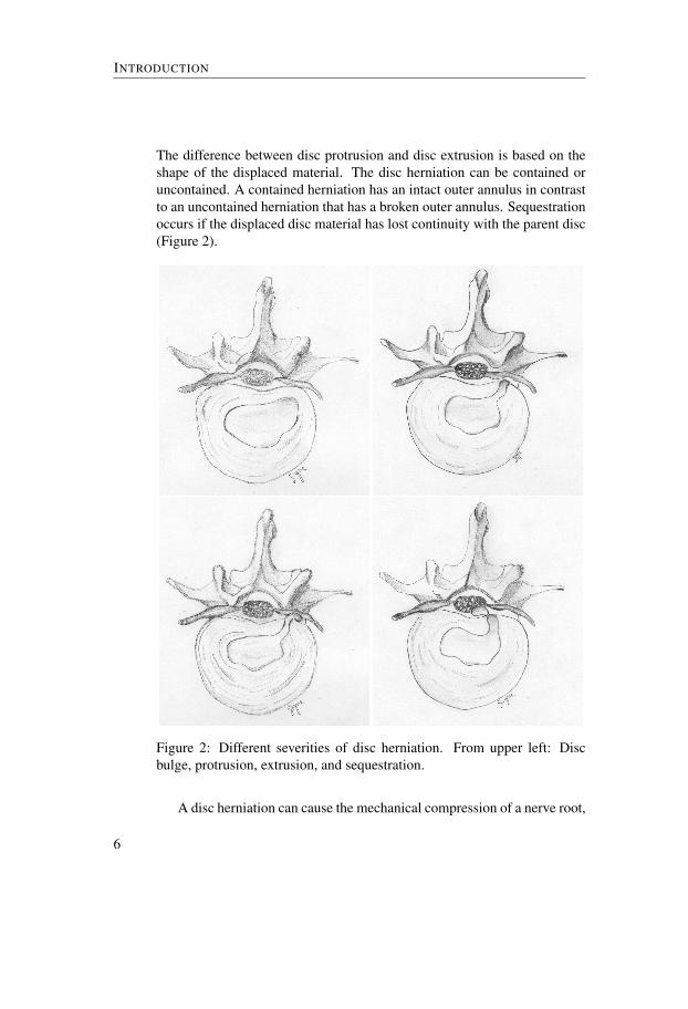
The difference between disc protrusion and disc extrusion is based on theshape of the displaced material. The disc herniation can be contained oruncontained. A contained herniation has an intact outer annulus in contrastto an uncontained herniation that has a broken outer annulus. Sequestrationoccurs if the displaced disc material has lost continuity with the parent disc(Figure 2).
Figure 2: Different severities of disc herniation. From upper left: Discbulge, protrusion, extrusion, and sequestration.
A disc herniation can cause the mechanical compression of a nerve root,
6

Lumbar disc herniation
which can lead to symptoms and leg pain in particular (Rydevik et al.,1984). Furthermore, several studies have shown that sciatica depends notonly on mechanical nerve root compression but also on biochemical factors(Brisby et al., 2000; Kayama et al., 1996; Olmarker et al., 1996, 1993). Forexample, experiments in pigs have shown that the epidural application ofautologous nucleus pulposus without mechanical nerve root compressioninduced a pronounced reduction in nerve conduction velocity in the caudaequina nerve roots, compared with the epidural application of retroperi-toneal fat in control experiments (Olmarker et al., 1993). In addition, it hasbeen shown that the nucleus pulposus can induce morphological and struc-tural changes in the nerve root (Kayama et al., 1996; Olmarker et al., 1996).In conclusion, there appear to be several pathophysiological explanationsfor the generation of symptoms due to disc herniation.
Symptoms and clinical findings from lumbar disc herniation
Persons with lumbar disc herniation do not necessarily have any symptoms.With Magnetic Resonance Imaging (MRI) bulging discs have been demon-strated in 81% of healthy volunteers without back problems and focal discprotrusions in 33% of the included persons (Stadnik et al., 1998). Anotherstudy investigated a group of patients with sciatica severe enough to requirea discectomy and compared them with an age-, gender- and risk factor-matched group of asymptomatic individuals. The results showed that, in thematched group of asymptomatic individuals, there was a very high preva-lence (76%) of disc herniation. It was concluded that individuals with minordisc herniations (i.e. protrusion, contained disc) were at high risk that theirMRI findings not were a causal explanation of pain because a high rate ofasymptomatic subjects had comparable morphological findings (Boos et al.,1995). It is therefore most important to evaluate MRI findings together withclinical findings to be able to clarify whether or not a disc herniation is giv-ing the patient the symptoms.
When a person with a suspected lumbar disc herniation is examined,the clinical tests include a neurological examination of motor function, sen-sation, reflexes and the straight leg raising test (Hoppenfeldt, 1976). Backrange of motion is also often examined. According to Vucetic and Svens-son (1996), lumbar range of motion and the crossed Lasègue sign were theonly physical signs that predicted 71% of the ruptured annuli and 80% ofthe intact annuli. Moreover, a thorough anamnesis, including both presentand past history of pain and other symptoms, is necessary when patients
7

INTRODUCTION
with suspected disc herniation are examined, in order to make the correctdiagnosis (Koes et al., 2007).
Recurrent back pain is a common occurrence prior to the first appear-ance of radiating leg pain, which could be a sign of lumbar disc hernia-tion. The most common symptom associated with lumbar disc herniationis leg pain in the affected nerve root dermatome (Weber, 1994). In addi-tion, weakness or complete loss of motor function could appear and su-perficial sensibility can be reduced or totally lost in the affected nerve rootdermatome. Moreover, the patient may describe different qualities of painsuch as aching, pins and needles, numbness and muscle cramp pain.
Pain due to lumbar disc herniation is often known to be more severethan pain in other orthopaedic diagnoses. Furthermore, patients with signsof nerve root involvement are more severely affected than those with lowback pain and pain referred to the legs (Kongsted et al., 2012). A studyshowed that an excess of 30 mm corresponded to moderate pain and anexcess of 54 mm on the VAS was proposed as severe pain (Collins et al.,1997). Since the pain is often severe, it might influence the patients’ abilityto function in daily life. Pain can also be long-standing and lead to longperiods of sick leave due to pain and disability (Dawson et al., 2011). Sickleave often results in negative economic consequences for the individual, aswell as for society (Hansson and Hansson, 2005). Several studies have alsoreported that depressive symptoms can accompany disc herniation (Arpinoet al., 2004; Zieger et al., 2011, 2010).
Natural healing
Evaluating treatment effects on patients with symptoms from a lumbar discherniation is a challenge, as spontaneous healing is common. The symp-toms often vary over time and many discs heal spontaneously and the symp-toms cease. In patients with sciatica but without confirmed disc herniationon MRI, about one-third recover two weeks after the onset of sciatica andapproximately three-quarters recover after three months (Vroomen et al.,2002). Von Korff (1994) has pointed out problems associated with studyingthe natural course of back pain and argues that studies of natural historymust investigate the development of the back pain in the absence of clinicalintervention. This is a major problem, as it is ethically questionable notto offer treatment to patients with lumbar disc herniation who experiencesevere pain and/or long-standing pain.
8

Treatments and outcomes for patients with lumbar disc herniation
The natural course of sciatica, but without confirmed disc herniationon MRI, was evaluated in a randomised controlled trial, which comparednon-steroidal anti-inflammatory drugs (NSAID) with placebo. The patientswere, however, examined within 14 days after the onset of radiating legpain, which meant that the opportunity to draw definite conclusions waslimited. Approximately 40% of the 183 patients had back pain and restric-tions in work and leisure after three months, while the corresponding figureafter one year was 30% of 173 patients (Weber et al., 1993).
In a study designed to investigate the natural history of morphologicalchanges on MRI, it was found that 37 of 42 patients (88%) showed an ef-fective reduction in herniated mass on MRI 3-12 months after the onsetof symptoms (Takada et al., 2001). To be more precise, after three months,eight patients’ (19%) disc herniations were classified as being in regression.However, the so-called natural history included treatment with bed rest,non-steroidal, anti-inflammatory drugs, pelvic traction and caudal epiduralblock. The opportunity to draw definite conclusions about natural healingis therefore limited. Moreover, the results showed that sequestered herniasand transligamentous extrusions appear to be more easily and rapidly ab-sorbed than other types of herniation.
Saal et al. (1996) reviewed the literature on natural history and non-operative treatment for patients with lumbar disc herniation and concludedthat lumbar disc herniation has a favourable prognosis in the majority ofpatients. He also recommended that, because of the positive natural historywithin the first three months, surgery is rarely indicated before 6-12 weeks.A general recommendation is to wait 6-8 weeks before surgery (Bono et al.,2006)
Taken together, the true natural healing and history of lumbar disc her-niation disease is not clear.
Treatments and outcomes for patients with lumbar discherniation
Physiotherapy treatment
The general recommendation, when patients report symptoms from lumbardisc herniation, is to start with non-surgical treatment (Bono et al., 2006;Saal et al., 1996; Weber, 1994). There are many different treatment meth-ods for patients with low back pain and sciatica. However, there is limited
9
INTRODUCTION
evidence relating to the effects of physiotherapy treatments for patients di-agnosed with lumbar disc herniation.
Recent clinical guidelines for low back pain include guidelines for pa-tients with the ICD diagnosis of lumbago with sciatica and the associatedICF diagnosis of acute, sub-acute and chronic low back pain with radiatingpain (Delitto et al., 2012). The guidelines are extensive and include guide-lines for diagnosis, examination and interventions. The diagnosis is basedon impairment/function and no MRI is used. For this reason, the inter-ventions are not clearly formulated for patients with lumbar disc herniationand sciatica. However, there are some cohort studies, RCTs and system-atic reviews, which are presented below, designed to evaluate the efficacyof treatments for patients with lumbar disc herniation and sciatica. A sys-tematic review with the aim of evaluating the efficacy and adverse effectsof treatments for patients with lumbar disc herniation and radiculopathyreported moderate evidence favouring stabilisation exercises over no treat-ment, manipulation over sham manipulation and the addition of mechanicaltraction over medication and electrotherapy. Adverse events were primarilyexperienced in association with traction treatment (Hahne et al., 2010).
Another systematic review (Luijsterburg et al., 2007) was unable to con-clude whether physiotherapy, bed rest, manipulation or medication shouldbe recommended as the most suitable treatment for patients with disc her-niation. Traction, corticosteroid injections and acupuncture could not berecommended according to the same review, as several trials indicate no ev-idence of any effect. On the other hand, a recent RCT (Moustafa and Diab,2013) evaluated lumbar extension traction versus a control group in patientswith L5-S1 radiculopathy. At inclusion, the 64 patients had a duration ofsymptoms of more than three months and mild to moderate disability up to40% on the ODI (ranges 0-100%). Patients who were unable to tolerate ex-tension positions were excluded. The study showed that lumbar extensiontraction restored lumbar lordosis, reduced pain and disability and increasedsegmental intervertebral movements compared with a control group who re-ceived hot packs and interferential therapy. Another RCT (Unlu et al., 2008)compared three different physiotherapy treatments; traction, ultrasound andlow-power laser. The 60 patients were diagnosed as having lumbar disc her-niation with symptoms lasting less than three months. The treatments wereapplied over a period of three weeks, five days a week, and with a follow-upperiod of three months. The results showed that all three treatments wereequally effective in terms of pain and disability.
In a retrospective cohort study (Saal and Saal, 1989), all 58 patients un-
10

Treatments and outcomes for patients with lumbar disc herniation
derwent an aggressive physical rehabilitation programme including severaltreatment methods for pain control as well as for exercise training. All thepatients were diagnosed using CT or MRI showing lumbar disc herniation.The results for the total group were 90% good or excellent outcome on aself-reported 4-grade scale (excellent, good, fair and poor). However, theevaluation was only performed on one occasion, approximately 31 monthsafter treatment, which makes it difficult to disregard natural healing. An-other retrospective cohort study (Hahne et al., 2011) also reported goodresults for patients with lumbar disc herniation using a physiotherapeuticfunctional restoration programme. Like Saal and Saal (1989), this studyevaluated the effect of treatment several months after treatment began andhad in addition a long treatment period of 8.7 months. This long treatmentperiod makes it difficult to disregard natural healing. One randomised con-trolled study (Albert and Manniche, 2012) compared two types of activetreatment for patients with sciatica; one group with symptom-guided ex-ercises and the other with sham exercises, where both groups were giveninformation and advice to stay active. The conclusion was that both groupswere equally effective. However, this study did not control adequately fornatural healing, since, at inclusion, some patients had only had sciatica fortwo weeks and the patients’ diagnoses were not confirmed with an MRI.
One common management method for patients with low back pain andsciatica is Mechanical Diagnosis and Therapy (MDT), also known as theMcKenzie method, which aims to eliminate or minimise pain (McKenzieand May, 2003). A systematic review showed that patients with low backpain treated with MDT reported a greater, more rapid reduction in pain anddisability compared with NSAIDs, educational booklets, back massage andback care advice, strength training, spinal mobilisation and general exer-cises (Clare et al., 2004). In an RCT with a one-year follow-up, Paatelmaet al. (2008) found that the McKenzie method was only marginally moreeffective compared with only giving advice to patients with low back pain.For patients with sciatica and a verified lumbar disc herniation, it has, how-ever, been shown that a selected group of patients who responded to MDTafter five days of treatment reported that they were satisfied after 55 weeks(Brötz et al., 2003). The patients started treatment just 12 days after the on-set of symptoms and the effects of spontaneous healing cannot therefore beexcluded. However, according to the MDT method, the hydrostatic mech-anism in the disc is a prerequisite for being able to influence the internaldisc displacement. The conceptual model according to the MDT methodimplies that it is possible to influence the internal disc displacement by re-
11

INTRODUCTION
peating movements and positions, thereby influencing the patient’s symp-toms (McKenzie and May, 2003). When a disc has herniated, the annularwall is breached by the herniated material and the hydrostatic mechanismin the disc is lost. In this case, according to McKenzie and May (2003),the repeated movements and positions used with the MDT method can nolonger be expected to influence the symptoms. Consequently, physiothera-pists trained in the MDT method rarely use this method if disc herniationis confirmed with an MRI. However, symptom-guided treatment has beenshown to have an effect on patients with lumbar disc herniation (Albert andManniche, 2012), but further evaluation is required.
Trunk stabilisation exercises, which aim to restore deep trunk musclecontrol, have been used for the prevention and rehabilitation of low backpain (Hodges et al., 2003). A randomised controlled trial revealed a reduc-tion in the recurrence of low back pain episodes after specific trunk stabili-sation exercises compared with a control group receiving advice and the useof medication (Hides et al., 2001). Dynamic lumbar stabilisation exerciseshave been found to relieve pain and improve function in patients who haveundergone microdiscectomy (Yilmaz et al., 2003). The effects of trunk sta-bilisation exercises combined with MDT have, however, not been studiedin patients with non-operated lumbar disc herniation.
SurgeryAs stated earlier, disc herniation commonly heals spontaneously and withdecreasing symptoms over time (Weber et al., 1993). For this reason, it iscommon to allow some time to pass for healing before surgery is consid-ered. If other treatment does not succeed within 6-8 weeks, surgery may beconsidered in patients with severe symptoms (Bono et al., 2006). A Swedishstudy shows a mean annual incidence of lumbar disc surgery of 24/100,000inhabitants a year. The ten-year rate of re-operations in the same study was10%. The 30-day mortality rate was 0.5 per 1,000 operations (Jansson et al.,2004). Lumbar disc surgery rates vary widely between different countriesfrom 16 to 125/100,000 inhabitants (Rasmussen et al., 2005). The numberof back operations has been shown to be 40% higher in the United Statesthan in any other country (Cherkin et al., 1994).
One exception is the cauda equina syndrome, which is an acute con-dition that influences the function of the bladder, sometimes the intestinalfunction and the superficial sensibility in the genital area can be reduced.An absolute indication for lumbar disc surgery is a progressive neurolog-
12
Aspects of evaluations for patients with lumbar disc herniation
ical deficit commonly associated with the cauda equina syndrome (Bonoet al., 2006). Likewise, Cakir et al. (2009) state that the only clear andobjective indication for early surgery is the cauda equina syndrome. How-ever, the same authors also point out that striking evidence with regard tothe necessity for immediate surgery does not cover even this severe com-plication. The relative indications for discectomy vary between surgeonsand patients. According to Bono et al. (2006), it is incumbent on cliniciansto discuss the advantages, disadvantages, risks, alternatives and estimatedexpected outcomes with patients.
Most of the time, the primary aim of lumbar disc surgery is to relievethe patient from pain in the leg. Other symptoms, such as back pain andpossible muscle weakness in the leg, appear to be more difficult to reducewith surgery.
Aspects of evaluations for patients with lumbar discherniation
In connection with spinal disorders, evaluations of the following domainsare recommended for inclusion when evaluating the effects of treatment forpatients with lumbar disc herniation: back-specific function, generic healthstatus, pain, work disability and patient satisfaction (Bombardier, 2000).The results of a systematic review indicated that socio-demographic, clin-ical, work-related and psychological factors predict the outcome of lum-bar surgery (den Boer et al., 2006b). A systematic review of non-surgicaltreated sciatica concluded that psychological factors were rarely investi-gated and saw a need of a consistent definition of sciatica (Ashworth et al.,2011).
In this thesis, these recommendations were followed as basic evaluationdomains. In addition, the following domains are included: different aspectsof health, centralisation of pain and kinesiophobia.
Health from quantitative and qualitative perspectives
The WHO defines health as a state of complete physical, mental and so-cial well-being and not merely the absence of disease or infirmity (WorldHealth Organisation, 1948). Health is a fundamental human right and peo-ple should therefore have access to basic resources for health. Obviously,
13
INTRODUCTION
health is not a state that is easily measured, although several attempts havebeen made to measure quality of life and health (Garratt et al., 2002).
Health is an experience full of nuances and it is therefore not easilycaptured with standardised questionnaires. People’s perception of quality oflife also varies between individuals and is dynamic within them (Carr et al.,2001). For this reason, it may be suitable to evaluate health with interviewsin which people are free to speak out about their own experiences, makingit possible to give other perspectives than questionnaires permit.
In qualitative research, the interviewer is trained to put open-endedquestions and open-ended follow-up questions, in order to explore the uniqueindividual’s perspective. Open-ended questions are said to yield in-depthresponses about people’s experiences, perceptions, opinions, feelings andknowledge. The data consist of verbatim quotations with sufficient contextto be interpretable (Patton, 2002).
Taken together, standardised questionnaires and interviews illustrate dif-ferent perspectives of health and answer different research questions. Forthis reason, both types of evaluation are important and together they cangive a more detailed, deeper understanding of health than if just one per-spective is investigated.
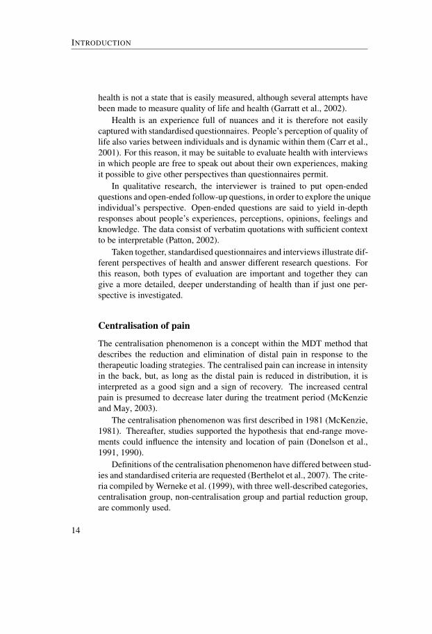
Centralisation of pain
The centralisation phenomenon is a concept within the MDT method thatdescribes the reduction and elimination of distal pain in response to thetherapeutic loading strategies. The centralised pain can increase in intensityin the back, but, as long as the distal pain is reduced in distribution, it isinterpreted as a good sign and a sign of recovery. The increased centralpain is presumed to decrease later during the treatment period (McKenzieand May, 2003).
The centralisation phenomenon was first described in 1981 (McKenzie,1981). Thereafter, studies supported the hypothesis that end-range move-ments could influence the intensity and location of pain (Donelson et al.,1991, 1990).
Definitions of the centralisation phenomenon have differed between stud-ies and standardised criteria are requested (Berthelot et al., 2007). The crite-ria compiled by Werneke et al. (1999), with three well-described categories,centralisation group, non-centralisation group and partial reduction group,are commonly used.
14
Aspects of evaluations for patients with lumbar disc herniation
Figure 3: Centralisation phenomenon, centralisation of distal pain to a morecentral location.
Centralisation group
1. A clinically induced change in the location of pain/symptoms referredfrom the spine moves from the most distal position toward the cervi-cal or lumbar midline. Note: For patients with only central or midlinepain, the midline pain must cease during initial visit.
2. The change in pain location or abolition of midline pain must remainbetter (i.e., the lateral or distal pain does not reappear), as a result ofmechanical movements/positions.
3. The change in pain location initially observed on the first visit mustcontinue its proximal movement on subsequent trials until all symp-toms are abolished. Note: Midline pain must remain abolished onsubsequent visits.
Non-centralisation group:
1. No changes in the location pain occurs, or
2. The location of pain changes from a central to a more distal locationthroughout all treatment visits.
15

INTRODUCTION
Partial reduction group
1. The location of pain changes from a more distal to a more centrallocation during each visit, without a progressive movement in initialpain location toward the midline at consecutive visits, or
2. No change in pain location occurs during any one visit, but the patienthas a gradual decrease in pain location over subsequent visits.
The centralisation phenomenon has been shown to be associated witha good prognosis; i.e. patients who centralise do better (Aina et al., 2004;Albert et al., 2012; Skytte et al., 2005; Werneke et al., 2008).
If the centralisation phenomenon and direction of preference (i.e. move-ments in one direction reduce the pain and movements in the opposite direc-tion increase it) are considered when exercise is prescribed and, when theexercise matches the direction of preferences, it leads to a better outcomein subgroups of patients with LBP than if exercise in the opposite directionis used (Long et al., 2004; Long, 1995).
Studies have shown that patients with sciatica and suspected disc herni-ation who have centralised will have better outcomes than non-centralisers(Albert et al., 2012; Broetz et al., 2010; Skytte et al., 2005). However, thesestudies have included patients with short duration of pain, which makes itdifficult to disregard natural healing. Moreover, MRI had not confirmed thedisc herniation. The centralisation phenomenon is not expected to occurin patients in whom uncontained disc herniation is confirmed with MRI,according to McKenzie and May (2003); for this reason, MDT is seldomrecommended when patients are diagnosed with disc herniation.
Kinesiophobia and fear of movementFear of movement and kinesiophobia are two concepts, which are frequentlyused synonymously in the literature. Another term used to describe fear inrelation to pain is pain-related fear. Pain-related fear is a broad, generalterm that incorporates all kinds of fear related to pain (Crombez et al.,1999). Fear of movement/(re-)injury is described as “a specific fear ofmovement and physical activity that is (wrongfully) assumed to cause rein-jury” (Vlaeyen et al., 1995b). In the most extreme situation of fear of move-ment, the term ‘kinesiophobia’ can be used, according to Kori et al. (1990).
The cognitive-behavioural fear-avoidance model (Figure 4) is often usedwhen describing the different paths a patient with chronic pain can follow
16

Aspects of evaluations for patients with lumbar disc herniation
!
Figure 4: A cognitive-behavioural model of fear of movement/(re)injury byVlaeyen et al. (1995b). This figure has been reproduced with permissionof the International Association for the Study of Pain® (IASP). The figuremay not be reproduced for any other purpose without permission.
(Vlaeyen et al., 1995b). The model suggests two responses to pain afteran injury; catastrophising, with fear of fear of movement/(re)injury andavoidance followed by disability and consequently a vicious circle, or non-catastropising and confrontation, which are assumed to lead to recovery 4.
Originally, the fear-avoidance model was used on patients with chroniclow back pain (Boersma and Linton, 2006; Picavet et al., 2002; Vlaeyenet al., 1995a). Kinesiophobia is thought to play a negative role in the out-come of rehabilitation for patients with low back pain and a high prevalenceof kinesiophobia has been observed among patients with persistent low backpain (Lundberg et al., 2004; Picavet et al., 2002). During the last decade thenumber of studies concerning the fear-avoidance model have increased sub-stantially (Vlaeyen and Linton, 2012). The model and conception of fear ofmovement have spread and have been used for patients with cervical radicu-lopathy (Dedering and Börjesson, 2012), upper extremity disability (Das Deet al., 2013; Feleus et al., 2007), patients with knee problems (Domenechet al., 2012) and acute low back pain (Ostelo et al., 2007).
17
INTRODUCTION
Summary of problem areas
Lumbar disc herniation is fairly common in the general population and canlead to severe, long-standing pain (Hoy et al., 2012). Many lumbar discherniations heal spontaneously, but many patients have to endure a longperiod of pain and symptoms. Non-surgical treatment including pain med-ication are recommended as the first choice for patients with severe painfrom a lumbar disc herniation (Bono et al., 2006). However, there is littleevidence to support the effect of physiotherapy treatment methods. In or-der to account for the complexity of pain, symptoms, impaired function anddisability these patients present, it seems necessary to design a structuredphysiotherapy treatment model. The treatment should aim for a reductionin the patients’ pain and disability and also to empower the patients and in-crease their self-efficacy, so that they will be able to cope more easily withtheir back problem in the future.
Health-related quality of life (HRQoL) is assessed at an earlier stage inpatients in relation to lumbar disc surgery (Hansson and Hansson, 2007). Amore detailed description of patients’ experience of health a couple of yearsafter structured physiotherapy treatment or surgery is, however, lacking.A qualitative interview study with open-ended questions to patients withlumbar herniation could yield in-depth responses about their experiences,perceptions, opinions, feelings and knowledge.
The centralisation of pain is not expected to occur in patients when discherniation is confirmed with MRI and, for this reason, MDT is seldom rec-ommended when patients are diagnosed with disc herniation (McKenzieand May, 2003). At our hospital, however, we have good clinical expe-rience of MDT treatment for this group of patients. An evaluation of thecentralisation phenomenon in relation to a structured physiotherapy treat-ment model for patients with lumbar disc herniation therefore appeared tobe justified.
Fear of movement is thought to play a negative role in the outcomeof rehabilitation for patients with low back pain and a high prevalence ofkinesiophobia has been observed among patients with persistent low backpain (Lundberg et al., 2004; Picavet et al., 2002). It could therefore beassumed that fear of movement might also influence patients with lumbardisc herniation.
18

Aims
The overall aim of this thesis was to evaluate a structured physiotherapytreatment model for patients, who qualified for lumbar disc surgery by hav-ing severe, long-standing pain and an MRI-verified lumbar disc herniation.
The specific aims were to:
• study kinesiophobia in patients who were treated surgically for lum-bar disc herniation and relate the results to established outcome mea-sures (Study I)
• evaluate a structured physiotherapy treatment model in patients whoqualified for lumbar disc surgery (Study II)
• describe the experience of health among patients three years aftertreatment with a structured physiotherapy model or surgery due tolumbar disc herniation (Study III)
• evaluate the occurrence of the centralisation phenomenon in relationto the patients’ disability, self-efficacy and kinesiophobia, after twoweeks of McKenzie therapy for patients who qualified for lumbar discsurgery (Study IV)
19


Patients and Methods
All the patients in this thesis qualified for lumbar disc surgery. However,there were two study populations (Table 1) — patients who were treatedsurgically at Södra Älvsborg Hospital (Study I) and patients who were iden-tified as surgical candidates at Sahlgrenska University Hospital (Studies II-IV). Orthopaedic surgeons determined whether the patients qualified forlumbar disc surgery after MRI and a physical examination according to therecommendations of the American Academy of Orthopaedic Surgeons forpatients with lumbar disc herniation (Nachemson, 1993) and also accordingto more recent recommendations by Bono et al. (2006).
Inclusion criteria
Patients were included if they were between 18-65 years of age, had an MRIconfirming disc herniation and explaining the clinical findings, had severeleg pain and symptoms for at least six weeks (minimising the effects ofspontaneous healing), and pain distribution with concomitant neurologicaldisturbances correlated to the affected nerve root.
Exclusion criteria
Patients were excluded from participation in Studies II, III and IV if theyhad the cauda equina syndrome, previous spinal surgery, other spinal dis-eases, such as spinal stenosis and spondylolisthesis, and inadequate com-mand of Swedish language.
Study I
All 97 patients between 18 and 65 years of age who had undergone stan-dardised open discectomy in 2004 and 2005 at Södra Älvsborg Hospital(Sweden) were invited to participate in the study. Questionnaires were sentto the patients in September 2006. If no response was received after twomailed reminders, the patients were reminded by telephone. Eighty-four(48 men) of 97 patients (87%) returned the questionnaires. The patientshad a mean age of 43 (SD 11) years.
21

PATIENTS AND METHODS
Table 1: Overview of the studies included in the thesis
Study Numberofpatients
Treatments Aim Studypopulation
I 84 Surgery Study kinesiophobia Södra ÄlvsborgHospital
II 41 Structuredphysiotherapytreatment
Evaluate structuredphysiotherapytreatment
SahlgrenskaUniversityHospital
III 20(10+10)
Structuredphysiotherapytreatment and surgery
Describe experienceof health
SahlgrenskaUniversityHospital
IV 41 Structuredphysiotherapytreatment
Evaluatecentralisationphenomenon
SahlgrenskaUniversityHospital
Studies II and IV
One hundred and fifty patients, who were referred to the orthopaedic clinicat Sahlgrenska University Hospital, Gothenburg, Sweden, from November2003 to January 2008, were identified as potential participants in StudiesII and IV. The patients were examined and disc herniation was confirmedby MRI. The spontaneous resolution of symptoms occurred in 70 patients(Figure 5). The remaining 80 patients had MRI-verified disc herniation, metthe inclusion criteria and qualified for surgery. Orthopaedic surgeons deter-mined whether the patients qualified for lumbar disc surgery after MRI anda physical examination according to the recommendations of the AmericanAcademy of Orthopaedic Surgeons for patients with lumbar disc herniation(Nachemson, 1993).
Initially, Study II was planned as a randomised controlled trial (RCT)comparing a structured physiotherapy treatment model and surgery, but thenumber of patients was not large enough to obtain acceptable power, de-spite a long period of inclusion. Eighteen of the 80 patients were initiallyrandomised to physiotherapy, 17 patients were randomised to surgery and45 patients did not accept randomisation. Twenty-seven of the 45 patients
22
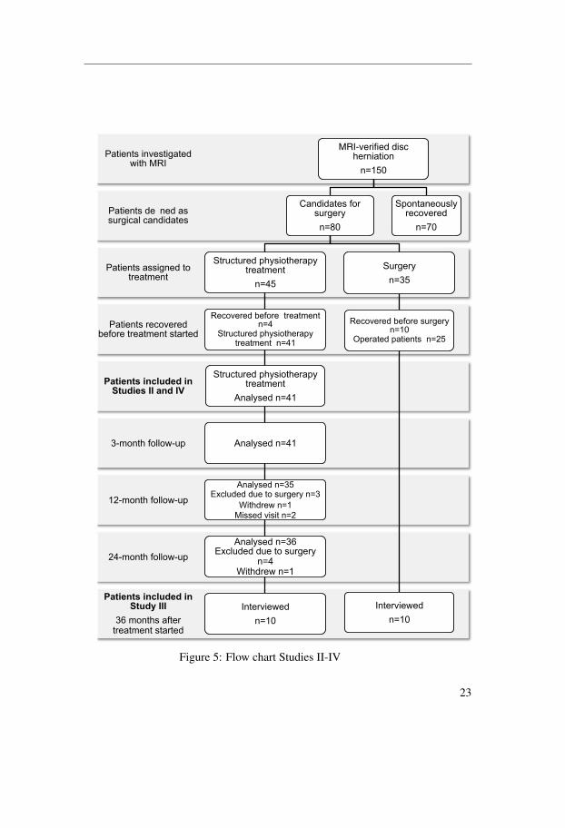
!"#$%&#'($&)*+,%,($&(-#+,.(///((((((((((((((((((((((
!"#$%&'()#*+',-#'-,*'$,&'#)'*-',.#
/01$%&'(#+%22%3145#
6/1$%&'(#+%22%3145#
!1$%&'(#+%22%3145#
!"#$%&#'($&)*+,%,($&(-#+,$%'(..("&,(./
!"#$%&#'()%*+,%)%-(.%/+)%(#)%"#0%&#('#")#%-
!"#$%&#'("''$1&%-(#+(#)%"#0%&#
'2)1$*"3(*"&-$-"#%'(
!"#$%&#'($&,%'#$1"#%-(4$#5(678
MRI-verified disc 5%)&$"#$+&(
&9:;<
7*&.8.*',)#+%-#)4-9,-:#
&;<=#
>'-4?'4-,.#5(:)8%'(,-*5:#'-,*'$,&'#
&;0@#
A,?%B,-,.#C,+%-,##'-,*'$,&'##&;0#
>'-4?'4-,.#5(:)8%'(,-*5:#'-,*'$,&'##&;06#
>'-4?'4-,.#5(:)8%'(,-*5:#'-,*'$,&'##
D&*2:),.#&;06#
D&*2:),.#&;06#
D&*2:),.#&;!@#EF?24.,.#.4,#'%#)4-9,-:#&;!#
G8'(.-,3#&;6#H8)),.#B8)8'#&;/#
D&*2:),.#&;!"#EF?24.,.#.4,#'%#)4-9,-:#
&;0#G8'(.-,3#&;6#
I&',-B8,3,.#
&;6=#
>4-9,-:#
&;!@#
A,?%B,-,.#C,+%-,#)4-9,-:##&;6=#
J5,-*',.#5*'8,&')##&;/@#
I&',-B8,3,.##
&;6=#
>5%&'*&,%4)2:#-,?%B,-,.#
&;K=#
Figure 5: Flow chart Studies II-IV
23
PATIENTS AND METHODS
who did not accept randomisation agreed to take part in the structured phys-iotherapy treatment and 18 patients decided to undergo surgery. A decisionwas therefore made solely to present a cohort of 45 patients treated accord-ing to the structured physiotherapy treatment protocol. Before the struc-tured physiotherapy treatment began, four patients recovered to the extentthat they could no longer be accepted as surgical candidates and they weretherefore excluded from the studies. The remaining 41 patients treated ac-cording to the structured physiotherapy model will be presented in StudiesII and IV (Figure 5).
Independent examiners, who were not involved in the treatment, dis-tributed the questionnaires before treatment and at the three-, 12- and 24-month follow-ups.
The patients had a mean age of 42 (SD 9.1) years. Of the 41 patients,19 (46%) were men.
Study III
Three years after completing either structured physiotherapy treatment(n=10) or surgery (n=10), the patients were selected for this interview study(Figure 5). The patients were selected from the cohort that was initiallyplanned as an RCT (Study II) of a structured physiotherapy treatment modeland surgery and a cohort of patients who had chosen treatment (structuredphysiotherapy treatment model or surgery). Earlier quantitative studiesshow no differences between surgery and non-surgical treatments after oneand two years (Jacobs et al., 2011). A decision was therefore made to in-clude both patients who were treated with surgery and patients who weretreated with structured physiotherapy, as these patients could be regardedas a homogeneous group. There was, however, no intention to comparethe two groups. A convenience sample of ten patients from each treatmentgroup was consecutively selected, meaning that ten patients had undergonesurgery, of whom five were randomised to treatment and five had chosensurgery in Study II. This group was named the Operative Group (OG). Cor-respondingly, ten patients had been treated with structured physiotherapy,of whom five patients were randomised to treatment and five patients chosephysiotherapy treatment in Study II. This group was named the StructuredPhysiotherapy Group (SPG). Patients that had surgery on more than oneoccasion and patients who first received physiotherapy treatment but thenrequired surgery were not selected for this study. In order to prevent unevendistribution in the two groups (SPG and OG), a check was made of the in-
24

Treatment methods
tensity of pain in the leg and back two years after treatment. In both groups,the selected patients had a wide spread of pain intensity in the leg and back,documented with a Visual Analogue Scale (VAS) two years after treatment.
The twenty patients were 25-66 years old (median age 43.5), ninewomen and eleven men. The patients in the SPG, six women and four men,were 31-66 years old (median age 49.5) and the patients in the OG, threewomen and seven men, were 25-59 years old (median age 40.5). Duringthe interviews, three patients reported other diagnoses that could influencetheir health. In the SPG, one patient had a whiplash disorder and anotherhad concentration problems following a virus in the CNS. In the OG, onepatient had varicose ulcers. Two years after treatment, the patients answeredquestionnaires, which revealed that three patients experienced kinesiopho-bia, 13 patients had no leg pain and likewise 13 patients reported no backpain. No disability was reported by 15 patients.
Treatment methods
Surgical treatment was performed on all the patients in Study I. The struc-tured physiotherapy treatment model was used for all the patients in StudiesII and IV. In Study III, ten patients were treated with structured physiother-apy and ten with surgery.
Surgical treatment, Studies I and III
The surgical treatment comprised a standardised open discectomy per-formed by spinal surgeons. The post-surgery rehabilitation included earlyactive rehabilitation according to Kjellby-Wendt and Styf (1998). The sur-gical treatment is expected to reduce leg pain and thereafter the post-surgeryrehabilitation aims to restore function, such as strength and flexibility, in or-der to return to work and physical activity.
Structured physiotherapy treatment model, Studies II-IV
Six physiotherapists, with MDT credentials, examined and treated the pa-tients during a nine-week period (Figure 6).
25

PATIENTS AND METHODS
Mechanical Diagnosis and Therapy from week 1
Home-based stabilisation exercises from week 3
Stabilisation training with equipment at the physiotherapy department from week 4
1 2 3 4 5 6 7 8 9
Weeks
Figure 6: A graphic illustration of the structured physiotherapy treatmentmodel. A total of nine weeks of treatment and thereafter a follow-up inweek 13. Patients were empowered to continue the training on their own.
Figure 7: The best-known movements associated with MDT are extensionexercises. Picture: Anders Agetorp
Phase 1 – MDT, weeks 1-2
For the first two weeks, an MDT protocol was followed based on individualclinical examinations of mechanical and symptomatic responses to posi-tions and movements (Figure 6). The aim of the protocol was to minimisepain and it was conducted with the emphasis on self-management and theempowerment of the patient. The key management decision is to determine
26

Treatment methods
Figure 8: Side-gliding as the patient’s own exercise and as aphysiotherapist-assisted exercise. Photo: Göte Norgren
Figure 9: Rotation in flexion as patient’s own exercise and as a physiother-apist assisted exercise. Photo: Göte Norgren
27

PATIENTS AND METHODS
the direction of loading that is necessary primarily to reduce the symptomsin the leg. The best-known movement associated with MDT is extensionexercises (Figure 7). However, in patients with lumbar disc herniation, itis often movements in other directions that reduce pain, so-called lateralprocedures such as side glide(Figure 8) and rotation in flexion (Figure 9).The patients were instructed to perform exercises several times a day withthe aim of reducing the leg pain. The fact that the patients were aware ofthe effect of different postures and mechanical loads and were able to adjustposture and loads from symptomatic responses was just as important as theexercises. The patients were educated in the principles of the MDT methodin order to evaluate the effect of the home-based exercises themselves. Thismeant that the patients could decide whether to continue with the exerciseor interrupt it until the next meeting with the physiotherapist. Sometimes, itmay be necessary to introduce manual techniques performed by the physio-therapist in order to produce a reduction in pain. Most patients will then beable to continue with their home exercises several times a day (McKenzieand May, 2003). The MDT method is characterised by the collaboration be-tween the patient and the physiotherapist. The aim with the collaboration isto encourage empowerment and give the patients tools to treat themselves.
Evaluation of the centralisation phenomenon (Study IV)
Two weeks after MDT treatment began, the physiotherapist who treated thepatient evaluated the centralisation phenomenon on the basis of the self-reported pain drawings and the assessments made by the physiotherapist,see also Assessment of the centralisation phenomenon, page 34.
Phase 2 – Home-based stabilisation exercises, week 3
During the third week (Figure 6), graded trunk stabilisation exercises in ly-ing, sitting, and standing were added to the MDT. The purpose of gradedtrunk stabilisation exercises was to improve muscle control. Initially, thestabilisation exercises were home based and performed without any equip-ment.
Phase 3 – Stabilisation training with equipment at the physiotherapydepartment, weeks 4-9
The training was then scheduled at the physiotherapy department threetimes a week (Figure 6). In the training, dumbbells, expanders and weight
28
Treatment methods
Figure 10: Training program. The pictures are from the MOBILUSw train-ing application.
29
PATIENTS AND METHODS
machines for strength training were used. The low-load muscular enduranceexercises were gradually increased in intensity on an individual basis withrespect to the patients’ reported leg pain and the observed movement con-trol and quality. A schedule was used to record the progress in the numberof exercises and weights throughout the training period. An example of thetraining program can be seen in Figure 10). During the last weeks, the pa-tients were encouraged to continue exercising on their own at a gym or toperform some other type of physical training of their own choice. Through-out the training period, the patients proceeded with the MDT exercises,which were continuously discussed and evaluated by the physiotherapist incollaboration with the patient.
Follow-up visit
Four weeks after the completion of the nine-week physiotherapy treatmentperiod, the patients attended a follow-up visit to the physiotherapist. Theaim of this visit was to encourage a high level of compliance with respectto continued trunk stabilisation exercises and MDT practice.
Evaluation methods
In this thesis, there are three types of evaluation methods; questionnaires,interviews and assessments of the centralisation phenomenon. The datawere then analysed statistically (Studies I-II, IV) or using content analysis(Study III).
Questionnaires
All the patients presented in this thesis answered questionnaires, which havebeen found to be reliable and valid. The questionnaires included descriptivedata including age, gender and duration of pain before treatment. Patientsin Study I also answered questions about their history of previous disc her-niation surgery.
Pain
Pain intensity was rated on two Visual Analogue Scales (VAS), one for legpain and one for back pain (Scott and Huskisson, 1976). The VAS ranges
30

Evaluation methods
from 0 to 100 mm, from “no pain” to “maximum pain”. A score of 0-10mm on the VAS was defined as no pain (Öberg et al., 2003).
Back-specific function
The Oswestry Disability Index (ODI) comprises ten items assessing back-specific function (Fairbank et al., 1980). Each item is scored from 0 to 5.The total score is expressed as a percentage, where 0% represents no dis-ability. An ODI disability score of 0-20% was defined as minimal or nodisability, 21-40% moderate disability, 41-60% severe disability, 61-80%crippled and a score above 80% was defined as either bedbound or exag-gerating their symptoms (Fairbank et al., 1980). According to Öberg et al.(2003), a score of 0-10% was defined as no disability. Good reliability andvalidity have been reported (Fairbank and Pynsent, 2000; Grönblad et al.,1993).
Kinesiophobia
The Tampa Scale for Kinesiophobia (TSK) questionnaire comprises 17items assessing the subjective rating of kinesiophobia. Each item has a4-point Likert scale with scoring alternatives ranging from “strongly dis-agree” to “strongly agree”. A total sum is calculated after inversion of theindividual scores for items 4, 8, 12 and 16. The total score varies between17 and 68. A high TSK value indicates a high degree of kinesiophobia.Vlaeyen et al. (1995a) defined a cut-off of >37 as a high degree of kine-siophobia. The TSK-SV has been found to be reliable and valid for use inSwedish patients with persistent low back pain (Lundberg et al., 2004).
Self-efficacy
The Self-Efficacy Scale (SES) consists of eight items assessing functionalself-efficacy beliefs specifically related to various basic physical activities(Estlander et al., 1994). Each category is scored on an 8-point Likert scalewhereby the patients estimate how long they believe they would be able toendure the activity, from less than 2 minutes to more than 45 minutes. Thetotal score range is 8-64, with higher scores indicating more positive beliefs.The reliability has been tested in a Swedish population of LBP (Johansson,1999).
31

PATIENTS AND METHODS
Catastrophising thoughts
The Pain Catastrophising Scale (PCS) comprises 13 items assessing catas-trophising thoughts (Sullivan et al., 1995). Each item is scored from 0 to4 and the scores are added up from 0 to 52, where 0 is no catastrophisingthoughts. Patients scoring above 24 on the PCS are classified as catas-trophisers and below 15 as non-catastrophisers (Sullivan et al., 1995). ThePCS is a reliable and valid measurement of catastrophising (Sullivan et al.,1995).
Health-related quality of life
The European Quality of Life in 5 Dimensions (EQ-5D) questionnaire wasused to measure health-related quality of life (HRQL) (Rabin and de Charro,2001). The EQ-5D consists of two parts; the first part involves five dimen-sions with three levels of answers. Possible values range from –0.59 to 1.0,where 1.0 is optimal health. The mean EQ-5D index is 0.86 for a Swedishpopulation aged 40-49 years (Burström et al., 2001b) and a value of ≤ 0.86could therefore be defined as normal for this age group. The second part isthe EuroQol Visual Analogue Scale (EQ-VAS) ranging from 0 (worst pos-sible health state) to 100 (best possible health state). The EQ-5D has beentested and validated (Burström et al., 2001a).
Depressive symptoms
The Zung Self-Rating Depression Scale (ZDS) consists of 20 items assess-ing depressive features (Zung, 1965). Each item has a 4-point Likert scalefrom “seldom” to “almost always”. The scores are added up from 20 to 80.The more depressed the patient is, the higher the score obtained. A scoreof 35 or higher would indicate depressive symptoms (Arpino et al., 2004;Zung, 1965). The ZDS is a reliable and valid measurement (Zung, 1965).
Patient satisfaction
Patient satisfaction with treatment was measured on a three-grade Likertscale – satisfied, less satisfied and dissatisfied (Strömqvist et al., 2001).
32

Evaluation methods
Work disability
Work status was measured using a three-grade Likert scale – working fulltime, full-time sick leave and part-time sick leave.
Pain drawing
The area of pain distribution was marked on a body outline drawing (frontand back, full view) to record the location of pain or symptoms. Four dif-ferent symbols were used for different qualities of pain; aching, pins andneedles, numbness and muscle cramps. These features were chosen withregard to the fact that all patients had disc herniation. The pain drawingswere evaluated with a clear overlay body template and the most distal painwas coded (Donelson et al., 1991; Long, 1995; Werneke et al., 1999). Fig-ure för att visa områdena.
Figure 11: Body outline template used for indicating pain distribution.
InterviewData were collected through interviews in Study III. An interview guidewith open-ended question areas was composed with regard to health andeveryday living. For the purpose of this study, the following question wasanalysed; Could you please describe how you are feeling?
33

PATIENTS AND METHODS
The patients were contacted by phone, informed about the study andasked if they would like to participate. The interviews were conducted ina separate room at the physiotherapy department by a researcher familiarwith the rehabilitation process for patients with disc herniation.
The interviews took place over a period of four months in 2009, ap-proximately three years after treatment started. In the SPG, the interviewslasted 25-46 minutes (median 31.5 minutes) and, in the OG, 18-97 min-utes (median 31 minutes). In all, 11 hours and 58 minutes of interviewswere tape-recorded and then transcribed verbatim. The researcher listenedto the interviews and corrected the transcripts as necessary before startingthe analysis. The text was then analysed according to content analysis, seepage 36.
Assessment of the centralisation phenomenonBefore and after each visit to the physiotherapist, the patients completeda pain drawing. Two weeks after the treatment started, centralisation ornon-centralisation was determined on the basis of the self-reported paindrawings and the assessments of the physiotherapist who treated the pa-tient. There were three definitions: the centralisation group, the non-centralisation group and the partial reduction group. The definitions byWerneke et al. (1999) were somewhat modified in this study to suit pa-tients with lumbar disc herniation, all of whom had radiating leg pain andqualified for lumbar disc surgery on the basis of their symptoms and MRIverification. Later on, the non-centralisation group and the partial reduc-tion group were merged into one group, named non-centralisers, in order toobtain an acceptable sample size.
Centralisation group
1. A clinically induced change in the location of pain referred from thespine goes from the most distal position toward the lumbar midline.At a minimum, the pain must move from one body part to the next(for example, from the foot to the calf or from the calf to the thigh).
2. The change in pain location must remain positive, i.e. centralised(the lateral or distal pain does not reappear), as a result of mechan-ical movements/positions. Pain that was centralised during repeatedmovements or positions must remain positive/centralised after resum-ing weight-bearing position.
34
Analysis
3. The changes in pain location initially observed on the first visit mustcontinue their proximal movement on subsequent trials (until allsymptoms have disappeared).
Non-centralisation group
1. No change in pain location occurs, or
2. The location of the pain changes from a central to a more distal loca-tion on all treatment visits.
Partial reduction group
1. The pain location changes from a more distal to a more central lo-cation during each visit, without any progressive movement of theinitial pain location toward the midline at consecutive visits, or
2. No change in pain location occurs during any one visit, but the patientexperiences a gradual decrease in pain and a shift in pain locationduring subsequent visits.
Analysis
In this thesis, a different statistical analysis was used for questionnairesand for the assessment of the centralisation phenomenon. In the qualitativestudy, content analysis was performed (Table 2).
Statistical analysis
Study I: The results are presented as median values and range, except forage, which is presented as the mean and standard deviation (SD). The sig-nificance level was set at 5%. Statistical comparisons between those withand without kinesiophobia were made from logistic regression with adjust-ment for age and gender. Comparison without adjustment was calculatedwith the chi-square test, with pooling of categories when necessary.
For five patients, one TSK item was missing and we used imputationwith linear regression to replace the lost information. The imputation tech-nique used here may lead to an underestimation of the variance, but thesmall number of imputed data made this a minor problem. The ODI score
35

PATIENTS AND METHODS
Table 2: Statistical analyses used in the studies
Analysis Study
Median values and IQR II, IVMedian values and range I, IIIMean SD I, II, IVLogistic regression with adjustment for age and gender IMann-Whitney U test II, IVWilcoxon’s signed rank test II, IVChi-square test ILinear regression to replace the lost information IContent analysis III
was calculated as the sum of the ODI items divided by the number of validitems. In four patients, one item was missing.
Study II and IV: The results are presented as median values and in-terquartile ranges (IQR), except for age, which is presented as the meanand SD. Changes over time within groups were analysed with Wilcoxon’ssigned rank test. Changes between groups were analysed with the Mann-Whitney U test. Statistical significance was set at an alpha level of 0.05.
For two patients, one item on the TSK was missing and missing dataimputation was used to recover the lost information. For nine patients, oneODI item was missing. The ODI score was calculated as the sum of theODI items divided by the number of valid items.
Content analysis
Study III: The interview texts were analysed by content analysis. Contentanalysis can be used both qualitatively and quantitatively. Content analysisis useful both as a method and a technique in analyses of texts. Krippendorff(2012) emphasises the importance of making replicable, valid inferencesfrom texts or other meaningful matter to the contexts of their use. Contentanalysis proceeds step by step, in order to recognise patterns, themes andsub-themes (Figure 12) (Patton, 2002).
36
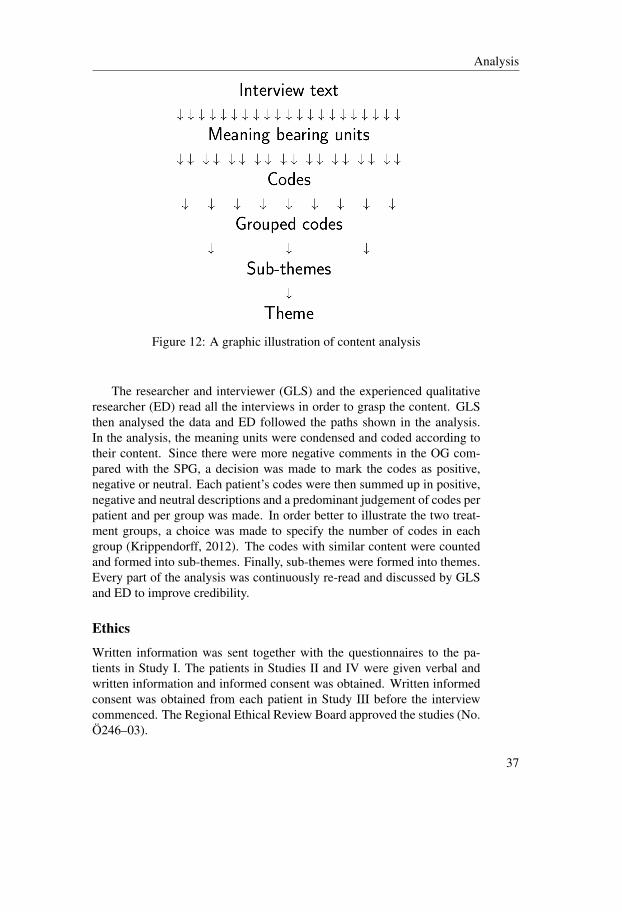
Analysis
Figure 12: A graphic illustration of content analysis
The researcher and interviewer (GLS) and the experienced qualitativeresearcher (ED) read all the interviews in order to grasp the content. GLSthen analysed the data and ED followed the paths shown in the analysis.In the analysis, the meaning units were condensed and coded according totheir content. Since there were more negative comments in the OG com-pared with the SPG, a decision was made to mark the codes as positive,negative or neutral. Each patient’s codes were then summed up in positive,negative and neutral descriptions and a predominant judgement of codes perpatient and per group was made. In order better to illustrate the two treat-ment groups, a choice was made to specify the number of codes in eachgroup (Krippendorff, 2012). The codes with similar content were countedand formed into sub-themes. Finally, sub-themes were formed into themes.Every part of the analysis was continuously re-read and discussed by GLSand ED to improve credibility.
EthicsWritten information was sent together with the questionnaires to the pa-tients in Study I. The patients in Studies II and IV were given verbal andwritten information and informed consent was obtained. Written informedconsent was obtained from each patient in Study III before the interviewcommenced. The Regional Ethical Review Board approved the studies (No.Ö246–03).
37


Summary of results
Study I – High degree of kinesiophobia after lumbar discherniation surgery
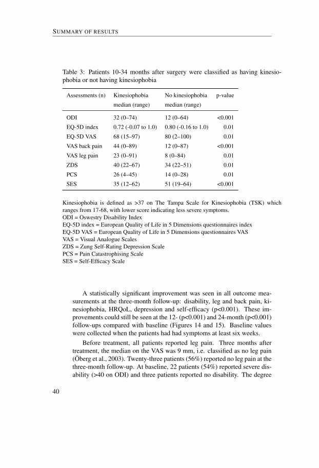
Study I was a cross-sectional study of 84 patients who were operated on dueto lumbar disc herniation, at Södra Älvsborg Hospital, 10-34 months priorto this study. Twenty of the 84 patients had previously undergone surgeryfor lumbar disc herniation. Of these 20, 18 were operated on twice, onepatient had been operated on three times and one patient had been operatedon five times.
Eighty patients answered the Tampa Scale for Kinesiophobia (TSK).Approximately half of them (36/80) were classified as having kinesiopho-bia, scores of more than 37 on the TSK. Descriptive data were compara-ble between the groups with and without kinesiophobia in terms of age,gender, place of birth, number of operations and disc herniation level. Be-fore surgery, patients with kinesiophobia had not experienced symptomsany longer than patients without kinesiophobia.
Patients classified as having kinesiophobia obtained statistically signif-icantly poorer results in eight of ten outcome measurements in comparisonto those without kinesiophobia (Table 3). In Figure 13, the number of pa-tients with each score on the TSK is shown.
Study II – A structured physiotherapy treatment modelcan give rapid relief to patients who qualify for lumbardisc surgery
Study II was a prospective cohort study with a 24-month follow-up afterstructured physiotherapy treatment. No patient had undergone surgery atthe three-month follow-up. At the 12-month follow-up, three patients hadundergone surgery and, at the 24-month follow-up, one additional patienthad been operated on. After surgery, these four patients were excluded fromfurther follow-ups.
39
SUMMARY OF RESULTS
Table 3: Patients 10-34 months after surgery were classified as having kinesio-phobia or not having kinesiophobia
Assessments (n) Kinesiophobia No kinesiophobia p-value
median (range) median (range)
ODI 32 (0–74) 12 (0–64) <0.001
EQ-5D index 0.72 (-0.07 to 1.0) 0.80 (-0.16 to 1.0) 0.01
EQ-5D VAS 68 (15–97) 80 (2–100) 0.01
VAS back pain 44 (0–89) 12 (0–87) <0.001
VAS leg pain 23 (0–91) 8 (0–84) 0.01
ZDS 40 (22–67) 34 (22–51) 0.01
PCS 26 (4–45) 14 (0–28) 0.01
SES 35 (12–62) 51 (19–64) <0.001
Kinesiophobia is defined as >37 on The Tampa Scale for Kinesiophobia (TSK) whichranges from 17-68, with lower score indicating less severe symptoms.ODI = Oswestry Disability IndexEQ-5D index = European Quality of Life in 5 Dimensions questionnaires indexEQ-5D VAS = European Quality of Life in 5 Dimensions questionnaires VASVAS = Visual Analogue ScalesZDS = Zung Self-Rating Depression ScalePCS = Pain Catastrophising ScaleSES = Self-Efficacy Scale
A statistically significant improvement was seen in all outcome mea-surements at the three-month follow-up: disability, leg and back pain, ki-nesiophobia, HRQoL, depression and self-efficacy (p<0.001). These im-provements could still be seen at the 12- (p<0.001) and 24-month (p<0.001)follow-ups compared with baseline (Figures 14 and 15). Baseline valueswere collected when the patients had had symptoms at least six weeks.
Before treatment, all patients reported leg pain. Three months aftertreatment, the median on the VAS was 9 mm, i.e. classified as no leg pain(Öberg et al., 2003). Twenty-three patients (56%) reported no leg pain at thethree-month follow-up. At baseline, 22 patients (54%) reported severe dis-ability (>40 on ODI) and three patients reported no disability. The degree
40

A structured physiotherapy treatment model can give rapid relief
0
1
2
3
4
5
20 21 22 23 24 25 26 27 28 29 30 31 32 33 34 35 36 37 38 39 40 41 42 43 44 45 46 47 48 49 50 51 53 54Tampa Scale for Kinesiophobia (TSK)
Num
ber o
f pat
ients
Figure 13: Number of patients for each score on the TSK. The TSK rangesfrom 17-68. Patients with a score of more than 37 were classified as havingkinesiophobia.
of disability decreased at the three-month follow-up, as only nine patients(22%) reported severe disability and 26 patients (64%) reported no disabil-ity.
At the three-month follow-up, 32 (78%) of 41 patients were satisfiedwith the structured physiotherapy treatment. At the two-year follow-up, thenumber of satisfied patients was 29 (78%) of 37 patients.
41

SUMMARY OF RESULTS
Figure 14: The figure shows the rapid relief of the patients’ disability (ODI= Oswestry Disability Index), pain intensity in the leg and back (VAS =Visual Analogue Scale) and fear of movement (TSK = Tampa Scale forKinesiophobia). A substantial and significant improvement was seen at thethree-month follow-up and it could still be seen at 12 and 24 months. Atbaseline, the patients had had symptoms for at least six weeks. The graphshows the median values from all outcome measurements at baseline and atthe 3-, 12- and 24-month follow-ups.
42
Months
value
10152025303540
5
10
15
20
25
30
35
40
ODI●
●
●
●
VAS_back_pain●
●
●
●
0 3 12 24
10
20
30
40
50
60
30
32
34
36
38
40
42
VAS_leg_pain●
●
● ●
TSK●
●
●
●
0 3 12 24
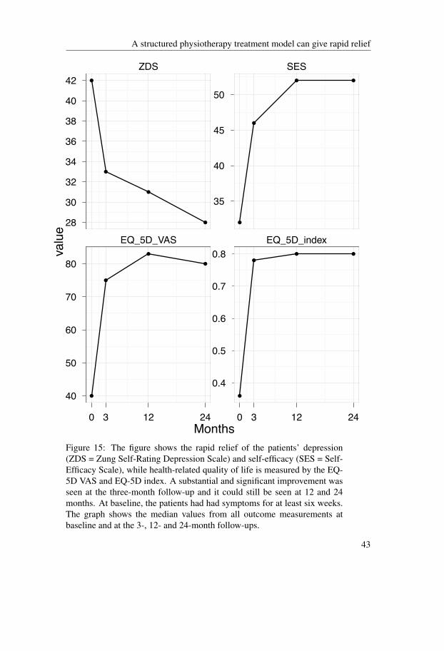
A structured physiotherapy treatment model can give rapid relief
Figure 15: The figure shows the rapid relief of the patients’ depression(ZDS = Zung Self-Rating Depression Scale) and self-efficacy (SES = Self-Efficacy Scale), while health-related quality of life is measured by the EQ-5D VAS and EQ-5D index. A substantial and significant improvement wasseen at the three-month follow-up and it could still be seen at 12 and 24months. At baseline, the patients had had symptoms for at least six weeks.The graph shows the median values from all outcome measurements atbaseline and at the 3-, 12- and 24-month follow-ups.
43
Months
value
28
30
32
34
36
38
40
42
40
50
60
70
80
ZDS●
●
●
●
EQ_5D_VAS
●
●
●
●
0 3 12 24
35
40
45
50
0.4
0.5
0.6
0.7
0.8
SES
●
●
● ●
EQ_5D_index
●
●● ●
0 3 12 24

SUMMARY OF RESULTS
Study III – Patients’ experience of health three years afterstructured physiotherapy or surgery for lumbar discherniation
Study III was a qualitative interview study. The interviews showed that thepatients experienced both a feeling of well-being and a feeling of ill-being.The patients who felt good said that they felt good most of the time but thatthey sometimes had symptoms of various kinds (Figure 13). Some patientssaid that they did not take that much notice of their symptoms and that theywere active despite symptoms. The patients also said that, when symptomsoccurred, they could deduce why they did so, which made it easier to handleand cope with the symptoms. Patients treated with structured physiotherapyexpressed a high level of well-being.
!""#$%&'()'*"##+,"$%&'
-('./012(0.'
3"$%&')$%"'
452$6"'7".1$2"'./012(0.'
Figure 16: An illustration of the theme ”Feeling of well-being” includingthe sub-themes. It describes the patients’ experience of feeling fine, havingno symptoms and being physically active despite symptoms.
The patients who said that they felt bad (ill-being) had both physical andpsychological symptoms. They also stated that they were anxious that theirsymptoms would return. This anxiety led to their avoiding physical activity.It was only patients in the Operative Group (OG) that expressed anxiety.
Both the patients that were operated on and the patients that were treated
44

Patients’ experience of health three years after treatment
!""#$%&'()'$##*+"$%&'
,-./$01#'/.234(2/'
,/.0-(#(&$01#'/.234(2/'
56($7$%&'3-./$01#'104$6$4.'
Figure 17: An illustration of the theme ”Feeling of ill-being” including thesub-themes. It describes the patients’ experience of feeling bad, with bothphysical and psychological symptoms, including anxiety. The anxiety ledthe patients who had undergone surgery to avoid physical activity.
with structured physiotherapy (SPG) expressed feelings of well-being andill-being. However, when a quantitative analysis of the number of codeswas performed, it showed that, in the SPG, a high number of codes werefound for the theme Feeling of well-being and, in the OG, a high number ofcodes were found for the theme Feeling of ill-being. The codes were thensummarised per patient, Table 4.
Table 4: Predominant judgement of codes per patient.
Group Positive patients Neutral patients Negative patients
SPG 3 4 3OG 0 2 8
45

SUMMARY OF RESULTS
Study IV – The occurrence of centralisation of pain afterMcKenzie therapy for patients with MRI-verified lumbardisc herniation and long-standing pain
This study was a prospective cohort study with prognostic value. At base-line, the majority of the patients had experienced severe leg pain for morethan three months. Twenty-one of the 41 patients centralised two weeksafter the McKenzie therapy had started, 14 patients achieved a partial re-duction in symptoms and six patients were non-centralisers. Patients in thepartial reduction group and the non-centralisers were pooled into one group,labelled non-centralisers, resulting in two groups; centralisers (n=21) andnon-centralisers (n=20). At baseline, the pain distribution and pain inten-sity were equal in both groups. Thirty-two patients reported symptoms inthe foot as the most distal symptom. After two weeks, ten patients (24%)had no pain; they were all centralisers. Fourteen patients still had symp-toms in the foot; twelve of them were non-centralisers (Figure 18). One ofthe two centralisers with symptoms in the foot described “pins and needles”and numbness, while the other patient had pain but only 7 mm on the VAS(VAS 0-10 mm was defined as no pain). The change in pain distribution cor-responded well with the physiotherapists’ assessment of the centralisationphenomenon. At the three-month follow-up, 23 patients (56 %) reported noleg pain (0-10 mm on the VAS). Of these, 15 were centralisers and eightwere non-centralisers.
Both groups had improved significantly with regard to their leg painat the three-month follow-up (p < 0.001) and centralisers also improvedwith regard to back pain (p = 0.001). Furthermore, centralisers reportedstatistically significantly lower pain intensity in both the leg (p = 0.014) andthe back (p = 0.006), compared with non-centralisers at the three-monthfollow-up. However, there were no statistically significant differences inthe degree of change between the two groups regarding pain intensity in theleg or back over time.
At baseline, there was a statistical difference between centralisers andnon-centralisers (p = 0.038) regarding disability, as evaluated with the ODI.The non-centralisers were more disabled than the centralisers. In spite of thebaseline difference the centralisers had improved statistically significantly(p = 0.024) more than the non-centralisers at the three-month follow-up.The centralisers’ results at the three-month follow-up were significantly bet-ter than those of the non-centralisers (p < 0.001). However, both centralisers
46
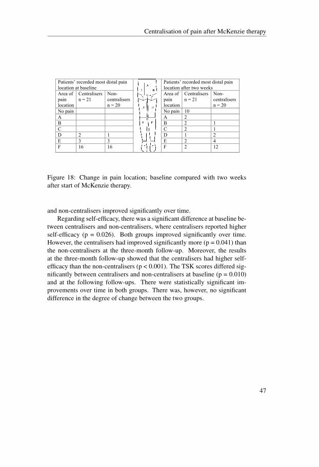
Centralisation of pain after McKenzie therapy
!!
!!
"#$%&'$()!*&+,*-&-!.,($!-%($#/!0#%'!!/,+#$%,'!#$!1#(&/%'&!
!
"#$%&'$()!*&+,*-&-!.,($!-%($#/!0#%'!/,+#$%,'!#2$&*!$3,!3&&4(!
5*&#!,2!0#%'!/,+#$%,'!
6&'$*#/%(&*(!'!7!89!
:,';+&'$*#/%(&*(!'!7!8<!
5*&#!,2!0#%'!/,+#$%,'!
6&'$*#/%(&*(!'!7!89!
:,';+&'$*#/%(&*(!'!7!8<!
:,!0#%'! ! ! :,!0#%'! 9<! !5! ! ! 5! 8! !=! ! ! =! 8! 9!6! ! ! 6! 8! 9!>! 8! 9! >! 9! 8!?! @! @! ?! 8! A!B! 9C! 9C! B! 8! 98!
Figure 18: Change in pain location; baseline compared with two weeksafter start of McKenzie therapy.
and non-centralisers improved significantly over time.Regarding self-efficacy, there was a significant difference at baseline be-
tween centralisers and non-centralisers, where centralisers reported higherself-efficacy (p = 0.026). Both groups improved significantly over time.However, the centralisers had improved significantly more (p = 0.041) thanthe non-centralisers at the three-month follow-up. Moreover, the resultsat the three-month follow-up showed that the centralisers had higher self-efficacy than the non-centralisers (p < 0.001). The TSK scores differed sig-nificantly between centralisers and non-centralisers at baseline (p = 0.010)and at the following follow-ups. There were statistically significant im-provements over time in both groups. There was, however, no significantdifference in the degree of change between the two groups.
47
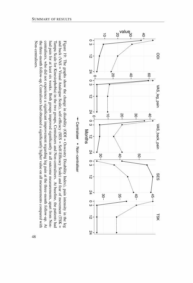
●
●
●
●
●
●●
●
●
●●
●
●
●
●●
●
●●
●
ODIVAS_leg_pain
VAS_back_painSES
TSK
10 20 30 40
0 20 40 60
0 10 20 30 40
30 40 50
30 35 40 45
03
1224
03
1224
03
1224
03
1224
03
1224
Months
value
●Centraliser
Non!centraliser
SUMMARY OF RESULTS
Figure19:
Thegraphs
showthe
changein
disability(O
DI
=O
swestry
Disability
Index),painintensity
inthe
legand
back(VA
S=
VisualA
nalogueScale),self-efficacy
(SES=
Self-EfficacyScale)
andfear
ofm
ovement(TSK
=Tam
paScale
forKinesiophobia)in
thetw
ogroups,C
entralisersand
Non-centralisers.
Atbaseline,the
patientshad
hadpain
foratleastsixw
eeks.B
othgroups
improved
significantlyin
alloutcome
measurem
ents,apartfromN
on-centralisers,w
hodid
notexperiencea
significantimprovem
entregardingleg
painatthe
three-month
follow-up.
At
thethree-m
onthfollow
-up,Centralisershad
obtaineda
significantlyhighervalue
onallm
easurementscom
paredw
ithN
on-centralisers.
48

Discussion
This thesis shows that the structured physiotherapy treatment model pro-duces early, significant and substantial improvements in all the measureddomains for patients with severe, long-standing pain due to lumbar discherniation. Furthermore, the improvements could still be seen at the two-year follow-up. Moreover, half the patients with lumbar disc herniationcentralised their pain after two weeks of McKenzie therapy, in spite of thefact that they were surgical candidates. Study III, which had a qualitativeresearch approach, showed that, three years after treatment, patients treatedwith structured physiotherapy experienced well-being and were physicallyactive despite their symptoms. Patients treated with surgery experiencedill-being and were avoiding physical activity due to anxiety.
Evaluation of patients with lumbar disc herniation
One aim of medical research is to produce evidence of the effects of treat-ment. When it comes to patients with disc herniation, there are problemsmeasuring the effects of treatment, as natural healing is regarded as fairlyextensive and is difficult to estimate. It has been shown that one third ofthe patients with sciatica recovered within two weeks and approximately75% within three months (Vroomen et al., 2002). According to anotherstudy, 60% of the patients with disc herniation had recovered after threemonths and 70% after twelve months (Weber et al., 1993). With these re-sults in mind and the guideline of waiting six to eight weeks before surgeryis considered (Bono et al., 2006), the inclusion criterion for the presentstudies was set at six weeks in order to minimise the effect of natural heal-ing. Moreover, the majority of patients in the present studies had had painfor more than three months at inclusion, which would further minimise theeffect of natural healing.
To date, the present studies are unique because of the patients’ long pe-riod with severe pain before the structured physiotherapy treatment began.The intention was to try to avoid patients whose disc herniation had healednaturally. There are some studies that have investigated physiotherapy treat-
49

DISCUSSION
ment for patients with disc herniation, but they have had a period of painshorter than six to eight weeks before treatment began (Albert et al., 2012;Albert and Manniche, 2012; Brötz et al., 2003). These studies were there-fore unable to control for natural healing. Likewise, a large, randomised,multicentre study, the spine patient outcomes research trial, had difficultycontrolling for natural healing. Its aim was to compare surgery with so-called usual care. The problem consisted of cross-over between the tworandomised groups. Due to natural healing, patients randomised to surgerycrossed over to usual care and, the other way around, patients who wererandomised to usual care had undergone surgery within six weeks (Wein-stein et al., 2008, 2006b). To summarise, RCTs and cohort studies have haddifficulty controlling for the natural healing of the disc herniation and theresults are therefore inconclusive and difficult to interpret.
Surgery for lumbar disc herniation has been investigated in numerousstudies. Surgery has been compared with a variety of treatments such aseducation, chiropractic, unspecified physiotherapy, acupuncture, injectionsand medication (Atlas et al., 2005; Peul et al., 2008; Weber, 1983; Weinsteinet al., 2006a,b). Surgery has been well described in these studies, but theother treatments have only been vaguely described and various treatmentshave been used. Previous studies have reported favourable short-term (afterthree months - one year) outcomes for surgery, but no major differencesbetween surgical and other treatments have been demonstrated in the longterm (more than two years) (Jacobs et al., 2011; Osterman et al., 2006;Weber, 1983; Weinstein et al., 2006a,b). The conclusions that are drawnfrom the comparison between surgery and non-systematic other treatmentsmay thus be misleading.
In order to compare the effects of surgery with physiotherapy, it is nec-essary that not only surgery but also physiotherapy has a well-structuredtreatment protocol. In this thesis, we have evaluated a structured physiother-apy treatment model, but we have not compared it with surgical treatment.However, Studies II and IV illustrated a rapid (two weeks and a three-monthfollow-up), statistically significant and substantial improvement from struc-tured physiotherapy treatment. The improvements could still be seen at thetwo-year follow-up in terms of all outcome measurements.
50

Different aspects of health and empowerment of patients
Different aspects of health and empowerment of patients
In Study II, the change over time in Health Related Quality of Life(HRQoL) was measured using the EQ-5D instrument; it had improved sig-nificantly after only three months. However, health is a subjective experi-ence of the utmost importance to the individual but without apparent diseasecorrelates. The WHO has defined health as “a state of complete physical,mental, and social well-being and not merely the absence of disease or in-firmity” (World Health Organisation, 1948).
Many of the most commonly used quality of life measurements are con-sidered incomplete in their ability to capture the quality of life of the indi-vidual patient (Carr and Higginson, 2001). Since quality of life is seen as anindividual construct, it was decided also to conduct interviews on patients’experiences of health in Study III. Surprisingly, despite the fact that the in-terviews were conducted as long as three years after treatment, a variationwas found between patients who had undergone surgery and those that hadhad physiotherapy treatment.
Patients who were treated with structured physiotherapy described ahigh degree of well-being and were active despite their symptoms. Patientswho had undergone surgery reported the opposite – namely, a high degreeof ill-being and the avoidance of physical activity because of fear of pain. Itcan be speculated that the feeling of well-being might be explained by theability of the structured physiotherapy treatment to empower the patientsto increase their self-efficacy. This is in line with Bandura (2004), whostates that health is greatly influenced by lifestyle habits and increased self-efficacy enables people to take some control of their health. The patientswho underwent surgery were not treated with the structured physiotherapymodel.
The philosopher Gadamer (1996) stated that the question ”Do you feelhealthy?” is an absurd question since health is not a feeling. Health in-stead means being engaged in what happens around us in private life, aswell as in the world around us, being together with people around us andbeing actively involved and rewarded in our everyday lives. From the op-posite perspective, he also claims that the medical field is oriented towardsmastery and gaining control of illness. This is perhaps what happened tothe patients who underwent surgery in Study III; the operation masteredthe illness on behalf of the patients. They were unable to master their ill-ness by themselves. It is possible to speculate about whether the patientsbecame worried after surgery when they felt symptoms and they therefore
51

DISCUSSION
avoided activity, in order not to end up in the same position again, need-ing surgery to master illness. On the other hand, the patients who weretreated with structured physiotherapy may have learnt to master illness bythemselves and their self-efficacy increased. If the symptoms recurred, theywould probably be able to master their illness again. Moreover, the patientstreated with physiotherapy described themselves as active. Being active andbeing involved were defined as a part of the condition of health by Gadamer(1996).
The high degree of well-being in the physiotherapy group in Study IIIand the positive results at the three- and 24-month follow-ups (Study II)might be explained by the strategies for patient empowerment in the MDTmethod. The use of empowerment strategies has been promoted, as it isthought to improve the effects of physiotherapy treatment (Perreault, 2008).Furthermore, the results of a study by Pellino et al. (1998), comparing thepreoperative education of patients randomised between an empowermentteaching method and a traditional teaching method, showed that patients inthe empowerment group obtained significantly higher self-efficacy than thecontrol group. One desired outcome in traditional teaching is compliancewith the prescribed treatment. In empowerment teaching, the desired out-come is an informed patient who is qualified to make his/her own decisionsabout his/her own health care (Funnell et al., 1991). In the empowermentmodel, the patients and the provider form a partnership in teaching/learningand shared decision-making. It is shown that only a minority of patients donot wish to have a role in, or responsibility for, treatment decision-making(van Til et al., 2010). This description of empowerment teaching and shareddecision-making corresponds well with the goal of the MDT method and ofthe treatment that the patients in the structured physiotherapy group usedin this thesis were offered. However, interestingly enough, the concept ofempowering the patient is not mentioned by McKenzie and May (2003). Itis possible to speculate about whether patient empowerment might be themost powerful part of the MDT and of the structured physiotherapy treat-ment model used here, but this has to be evaluated in more detail in futurestudies.
Centralisation of pain
A key part of the MDT concept is centralisation of pain, which is evaluatedwith different purposes in several studies (Aina et al., 2004; Albert et al.,
52

Centralisation of pain
2012; Laslett et al., 2005; Long, 1995), including Study IV in this thesis.Study IV showed that, in half the patients, pain centralised after only twoweeks of MDT treatment, in spite of having MRI-verified disc herniationwith a broken outer annulus. However, McKenzie and May (2003) havestated that MDT is not effective if the patients have a disc herniation witha broken outer annulus, since the hydrostatic mechanism in the disc is aprerequisite for influencing the internal disc displacement and thereby en-abling centralisation. This statement has led physiotherapists educated inthe method to avoid using MDT when a disc herniation is confirmed onMRI.
In this thesis, what actually happens in the disc is not investigated, but,in Studies II, III and IV, we investigated the patients’ experience of reducedsymptoms. However, it has previously been shown that there is no obvi-ous link between MRI-verified improvements in disc herniation and nerveroot compression in relation to definite recovery (Jensen et al., 2007). Ear-lier studies have also questioned McKenzie’s statement, but there are somelimitations to those studies. The first limitation is that, in two studies,the disc herniation was not confirmed by an MRI (Albert and Manniche,2012; Skytte et al., 2005). Secondly, one study group only included pa-tients who responded with centralisation of pain during the first five dailyphysiotherapy sessions, which means they included a highly selected groupof patients who were already improving (Broetz et al., 2010; Brötz et al.,2003). Thirdly, one study showed that 43 of 46 patients with extruded orsequestered discs centralised, but a vague definition of the centralisationphenomenon was used. So-called unstable centralisers were defined as paincentralised during movements, but, after resuming a weight-bearing posi-tion, the pain reappeared (Albert et al., 2012). In spite of this, Albert et al.(2012) support the results of Study IV. Moreover, Adams et al. (2010) havere-interpreted the scientific literature regarding the healing of intervertebraldiscs. They suggest that physiotherapists who traditionally employ mechan-ical loading as a healing stimulus should evaluate therapies to relieve disco-genic pain by promoting healing in the disc periphery by stimulating cells,boosting metabolic transport and preventing adhesion and re-injury. Takentogether, the results of Study IV are important for physiotherapists work-ing clinically, as we now have evidence that patients with MRI-verified discherniation can be successfully treated with MDT.
53

DISCUSSION
Fear of movement and empowerment of patients
As presented in Study I, half the patients who had undergone surgery tento 34 months earlier were classified as having kinesiophobia. These re-sults were surprising, as it was a long time after surgery and the patientshad been recommended to exercise, according to an early, active trainingmodel (Kjellby-Wendt and Styf, 1998). It was therefore expected that thepatients would have again returned to an active life. However, 62 of 80 pa-tients (78%) were actually back at work, but half of them were classifiedas having kinesiophobia. The number of sick-listed patients correspondsfairly well with the findings of den Boer et al. (2006a), who reported re-duced work capacity in 25% of the patients six months after lumber discsurgery. Furthermore, fear of movement/(re)injury before surgery has pre-dicted more disability and severe pain six month after lumbar disc surgery(den Boer et al., 2006b).
In contrast, in the patients treated with structured physiotherapy in theprospective Study II, only four of 35 patients (11%) were classified as hav-ing kinesiophobia one year after treatment started and 33 patients (94%)were back at work. The discrepancy between the results in Studies I andII is interesting. One explanation could be that, in Study I, the patientswho had had surgery more than once were included. Twenty of 84 patients(24%) had previously undergone back surgery, so there was a large numberof re-operations compared with other studies that report a re-operation rateof 5-10% (Jansson et al., 2004; Morgan-Hough et al., 2003). As a result,several of the operated patients had had pain longer before surgery in StudyI than the patients had had before physiotherapy in Study II. Another rea-son for the difference in kinesiophobia between Study I and Study II maybe the effect of the structured physiotherapy treatment in Study II, whichaims at empowering the patients, increasing self-efficacy and reducing fearof movement.
It has previously been shown that patients with chronic pain and ahigh degree of kinesiophobia, who participated in a multidisciplinary pro-gramme, including physiotherapy, increased their physical activity and re-duced their degree of kinesiophobia at the six-month follow-up (Koho et al.,2011). The above reasons could all be possible mechanisms for reduc-ing the number of patients who were classified as having kinesiophobiain Study II. Another thing to bear in mind as a doctor or physiotherapistis that it has been shown that health-care providers who hold beliefs re-flecting fear avoidance may influence the patients to be more fear avoidant
54
Fear of movement and empowerment of patients
(Coudeyre et al., 2006; Linton et al., 2002). The way physiotherapistsintroduce the training post-surgery and the way doctors and physiothera-pists express themselves regarding post-surgery activities is therefore im-portant. If it is the empowerment component that makes the difference be-tween the two groups in Study I and Study II, then it seems reasonable thatthe post-surgery treatment should be updated and include a more patient-empowering treatment strategy.
Kinesiophobia and fear of movement are often used synonymously inthe literature. During the last decade, an increasing number of studies sup-porting the basic assumptions of the fear-avoidance model have been pub-lished (Vlaeyen and Linton, 2012). However, the fear-avoidance model hasalso been questioned and it needs to be conceptually expanded and furthertested in order to provide adequate clinical utility (Pincus et al., 2010).
In a study of 64 patients with LBP and a mean of 41.6 on the TSK, therelationship between kinesiophobia and avoidance of lifting was studiedand no relationship was confirmed (Reneman et al., 2003). In part, thesame authors performed an additional study with an extended study groupand concluded that the relationship between pain and pain-related fear andfunctional capacity was weak or non-existent in patients with chronic LBP(Reneman et al., 2007).
In an event-related functional MRI study of neural correlates of thefear of movement, 60 women (30 chronic LBP, 15 healthy controls and 15women with arachnophobia (fear of spiders)) were included. The chronicLBP patients were divided into a high and low fear-avoidance group accord-ing to the TSK, with a cut-off value of 35.5 on the TSK, with 15 patients ineach group. The participants viewed photos with neutral or back-stressingmovements and pictures of spiders, while the functional MRI data wereacquired. The patients with chronic LBP and high fear avoidance did notdiffer from those with low fear avoidance or pain-free participants in theirreaction to the back-stressing photos. However the arachnophobic individu-als reacted with the expected fear reaction to the pictures of spiders and acti-vations in “fear areas” in the brain. The authors conclude that the concept offear of movement is not really a fearful emotional state but something dif-ferent (Barke et al., 2012). These results are interesting when you considerthe results in Study III where just one of ten operated patients reported kine-siophobia, in spite of the fact that the operated group experienced ill-beingand avoided activity. Moreover, two patients reported kinesiophobia in thephysiotherapy group, despite the fact that they experienced well-being andwere active in spite of their symptoms.
55
DISCUSSION
Patient empowerment may be the most important part of the MDT concept,but how does the MDT succeed in empowering patients? There are proba-bly several reasons. Number one could be the expected immediate effect ofthe positions and the repeated movements the patient performs. The patientoften experiences an instant change in pain, due to different movements andpositions, and the patient learns how to evaluate the change. This immedi-ate effect of pain response is empowering for the patient and the patientlearns how he/she by him/herself can influence and control the pain, whichresults in increased self-efficacy. Another important factor is that the nextappointment with the physiotherapist is within a short period of time, oneor two days, to be able to guide the patient further (McKenzie and May,2003). An important motivator in learning is pain; when it comes to bothremoving pain and empowering the patient to increase his/her self-efficacyand probably also in the opposite way – increased pain can cause people tobecome fear avoidant and kinesiophobic.
RCTs are regarded as the most reliable method for determining the efficacyof different treatments and minimising bias. However, despite the good rep-utation of RCTs, conflicting conclusions among RCTs are not uncommon.It has also been shown that agreement between pairs of randomised trialsand non-randomised studies was most common when the pairs were of highmethodological quality and when there was a clinical similarity between thepairs in terms of settings, populations, interventions and outcomes (Furlanet al., 2008). In the absence of well-executed RCTs, evidence must rely onobservational studies (Rosén et al., 2009). Study II was planned as an RCT,but the number of patients available for inclusion was not sufficient, in spiteof a long period of inclusion. It was therefore decided to present the resultsas a prospective cohort study, since we judged the results to be interestingfor physiotherapists and doctors working clinically and their patients.
It should be pointed out that well-educated people are generally moreknowledgeable about medical options and are in better position to promotetheir own interests (Rosén et al., 2009). This might be a limitation in Stud-ies II, III and IV, as we did not investigate the level of education or socio-economic conditions. Moreover, 45 of 80 patients (56%) did not want to
56
Methodological considerations
Limitations
participate in the randomisation procedure; they wanted to decide for them-selves whether to obtain surgery or physiotherapy treatment. This could bea limitation in our study, but shared decision-making is also an importantand promoted part of today’s medical treatment.
In Studies II and IV, 41 patients were analysed after completing treat-ment according to a structured physiotherapy model. The number of pa-tients was quite small, but the statistically significant differences from base-line to the three-month follow-up were strong in all the measurements in-cluded in Study II and in all the measurements apart from pain intensity inthe back in non-centralisers in Study IV. Moreover, the minimal importantchange (MIC) is proposed as a 15-mm decrease on the VAS and a 10-degreedecrease on the ODI (Ostelo and Vlaeyen, 2008). The MIC was achievedon the VAS for leg and back pain and on the ODI in Study II and, cor-respondingly, for both centralisers and non-centralisers at the three-monthfollow-up in Study IV.
After structured physiotherapy treatment, there are no known risks, atleast not if the physiotherapist has an examination in the MDT method andis thereby experienced in the treatment of patients with LBP. However, thepatient needs to invest time and energy in movements and training in orderto succeed and get better. On the other hand, after surgery, the patients maybe pain free immediately on the day after surgery, without any training andwithout any effort of their own. However, there are several risks, such asinfection, when it comes to surgery. Clinical practice today is that manypatients are recommended simply to wait for healing and stay as active aspossible during the healing period. If patients cannot bear to wait until thepain decreases, surgery is presented as a leg-pain-reducing treatment. Sincewe have shown positive results just two weeks after structured physiother-apy treatment and significant and substantial results at three months, thiswould not be a long time to wait for surgery, especially since the waitingtime for an appointment with an orthopaedic surgeon in Sweden can befairly long. It would probably save time for the patient to begin a structuredphysiotherapy treatment at an early stage, before an appointment is madewith a surgeon. There may also be a risk of persistent pain if patients onlywait at home and are worried because of the intense pain. Furthermore, it iscostly for both the individual and society just to wait and be on sick leave.We therefore argue that it is beneficial for the patient to reduce pain withproper treatment and economically beneficial for society to start structuredphysiotherapy treatment before surgery is considered.
In Study IV, the physiotherapist who treated the patient also made an
57
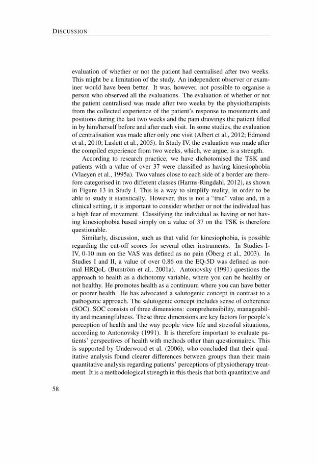
DISCUSSION
evaluation of whether or not the patient had centralised after two weeks.This might be a limitation of the study. An independent observer or exam-iner would have been better. It was, however, not possible to organise aperson who observed all the evaluations. The evaluation of whether or notthe patient centralised was made after two weeks by the physiotherapistsfrom the collected experience of the patient’s response to movements andpositions during the last two weeks and the pain drawings the patient filledin by him/herself before and after each visit. In some studies, the evaluationof centralisation was made after only one visit (Albert et al., 2012; Edmondet al., 2010; Laslett et al., 2005). In Study IV, the evaluation was made afterthe compiled experience from two weeks, which, we argue, is a strength.
According to research practice, we have dichotomised the TSK andpatients with a value of over 37 were classified as having kinesiophobia(Vlaeyen et al., 1995a). Two values close to each side of a border are there-fore categorised in two different classes (Harms-Ringdahl, 2012), as shownin Figure 13 in Study I. This is a way to simplify reality, in order to beable to study it statistically. However, this is not a “true” value and, in aclinical setting, it is important to consider whether or not the individual hasa high fear of movement. Classifying the individual as having or not hav-ing kinesiophobia based simply on a value of 37 on the TSK is thereforequestionable.
Similarly, discussion, such as that valid for kinesiophobia, is possibleregarding the cut-off scores for several other instruments. In Studies I-IV, 0-10 mm on the VAS was defined as no pain (Öberg et al., 2003). InStudies I and II, a value of over 0.86 on the EQ-5D was defined as nor-mal HRQoL (Burström et al., 2001a). Antonovsky (1991) questions theapproach to health as a dichotomy variable, where you can be healthy ornot healthy. He promotes health as a continuum where you can have betteror poorer health. He has advocated a salutogenic concept in contrast to apathogenic approach. The salutogenic concept includes sense of coherence(SOC). SOC consists of three dimensions: comprehensibility, manageabil-ity and meaningfulness. These three dimensions are key factors for people’sperception of health and the way people view life and stressful situations,according to Antonovsky (1991). It is therefore important to evaluate pa-tients’ perspectives of health with methods other than questionnaires. Thisis supported by Underwood et al. (2006), who concluded that their qual-itative analysis found clearer differences between groups than their mainquantitative analysis regarding patients’ perceptions of physiotherapy treat-ment. It is a methodological strength in this thesis that both quantitative and
58

Limitations
qualitative methods were used to evaluate experiences of health. This hasprovided a deeper understanding of the patients’ experiences, which wouldnot have been achieved using nothing but questionnaires.
59

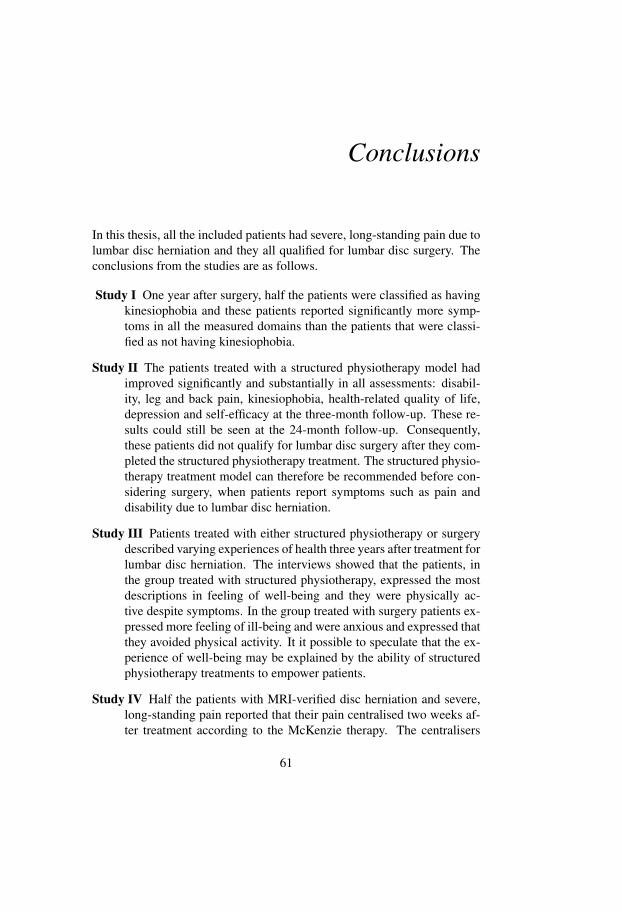
Conclusions
In this thesis, all the included patients had severe, long-standing pain due tolumbar disc herniation and they all qualified for lumbar disc surgery. Theconclusions from the studies are as follows.
Study I One year after surgery, half the patients were classified as havingkinesiophobia and these patients reported significantly more symp-toms in all the measured domains than the patients that were classi-fied as not having kinesiophobia.
Study II The patients treated with a structured physiotherapy model hadimproved significantly and substantially in all assessments: disabil-ity, leg and back pain, kinesiophobia, health-related quality of life,depression and self-efficacy at the three-month follow-up. These re-sults could still be seen at the 24-month follow-up. Consequently,these patients did not qualify for lumbar disc surgery after they com-pleted the structured physiotherapy treatment. The structured physio-therapy treatment model can therefore be recommended before con-sidering surgery, when patients report symptoms such as pain anddisability due to lumbar disc herniation.
Study III Patients treated with either structured physiotherapy or surgerydescribed varying experiences of health three years after treatment forlumbar disc herniation. The interviews showed that the patients, inthe group treated with structured physiotherapy, expressed the mostdescriptions in feeling of well-being and they were physically ac-tive despite symptoms. In the group treated with surgery patients ex-pressed more feeling of ill-being and were anxious and expressed thatthey avoided physical activity. It it possible to speculate that the ex-perience of well-being may be explained by the ability of structuredphysiotherapy treatments to empower patients.
Study IV Half the patients with MRI-verified disc herniation and severe,long-standing pain reported that their pain centralised two weeks af-ter treatment according to the McKenzie therapy. The centralisers
61
CONCLUSIONS
improved significantly more than the non-centralisers in terms of dis-ability and self-efficacy. The centralisers and the non-centralisers im-proved significantly and substantially over time in terms of leg pain,disability, self-efficacy and kinesiophobia. The findings in this studysupport the belief that patients with lumbar disc herniation can betreated successfully with the MDT method.
Thesis The overall conclusion from this thesis is that a structured physio-therapy treatment model for patients with pain and disability due toa lumbar disc herniation should be recommended before surgery isconsidered
62
Clinical implications
When planning the treatment protocol for patients with lumbar disc hernia-tion, it appears to be important to remember that many patients experiencevarious degrees of fear of movement. As many as about half the patientsin this thesis were classified as having kinesiophobia, i.e. a fairly high de-gree of fear of movement, one year after surgery. As treatment after surgeryinvolves physical training, the physiotherapist needs to have a knowledgeof how to deal with patients with a high degree of fear of movement whenintroducing the various movements, positions and training exercises in thetreatment protocol. The MDT method aims to reduce the patients’ pain us-ing various positions and movements and to encourage empowerment andgive the patients tools to treat themselves. This was probably the reasonfor the low degree of fear of movement in Study II (approximately 10%were classified as having kinesiophobia) one year after the structured phys-iotherapy treatment. Strategies of empowerment can be recommended forimplementation in the treatment protocol for postsurgical rehabilitation.
This thesis recommends the structured physiotherapy treatment modelfor nine weeks before considering surgery, when patients report severe painand disability due to lumbar disc herniation. It appears to be important togive the patients with severe pain the opportunity to obtain effective struc-tured physiotherapy treatment at an early stage, rather than passively wait-ing for healing. The treatment model can therefore be recommended forpatients with back and leg pain assessed by physiotherapists educated ac-cording to the MDT method, even though a lumbar disc herniation has notbeen confirmed with an MRI.
In Study III, the importance of the patients’ own experiences andthoughts about their pain and their relationship with surgery, physiotherapy,attitudes to physical activity and their own health was illustrated. It is possi-ble that, if a more structured physiotherapy model including empowermenthad been used for the patients that underwent surgery, these patients wouldnot have reported ill-being. Well-being reported by the patients treated withstructured physiotherapy after three years is a pleasure to see. The reasonsfor this need to be explored in detail in future research, as the results can beexpected to be of great importance for not only patients with lumbar discherniation but also patients with other musculoskeletal problems.
63

Future perspectives
To further confirm the results of these studies, it would be appropriate toconduct an RCT comparing the structured physiotherapy treatment modelwith natural healing or surgery for patients with lumbar disc herniation.
Future research is also needed to examine the impact of psychologi-cal components in relation to the centralisation phenomenon and how theempowerment of patients, according to the MDT method, influences thepatients’ symptoms.
The qualitative interview (Study III) provided a deeper understandingof health from the patients’ perspective than was possible using the ques-tionnaires. It seems reasonable to use qualitative interview studies morefrequently in order to include the patients’ perspectives and thereby addnew knowledge and hypotheses for future research.
65


Acknowledgements
I wish to express my gratitude to all the people who have contributed tothis work. In particular, I want to thank:
Gunilla Kjellby Wendt PhD, RPT and my tutor, for inviting me into thisproject a long time ago and supporting me to become a PhD student.We have had a long and crooked journey, but at last we have reachedthe goal! Thank you for standing by on the whole journey.
Roland Thomeé professor, RPT and my latest co-tutor, for positive sup-port, sharing your scientific knowledge and most of all for believingin me. It has made a huge difference.
Ella Danielson professor and my co-tutor, it has been such a joy to learnfrom you. We have always had positive meetings when you haveshared your vast knowledge and guided me most professionally inthe field of qualitative research. And for all the support you havegiven me when I have needed it most.
Hans Christian Östgaard associate professor, co-author of the firstarticle, for employing me in the project and thereafter assessing andincluding the patients in the studies and introducing me into science.
Mari Lundberg associate professor, RPT, co-author of the first articleand my kinesiophobia mentor, for sharing your knowledge ofkinesiophobia and your positive support.
Jón Karlsson professor and head of Section for Anesthesiology, Bio-materials, Orthopaedics and Otorhinolaryngology, the SahlgrenskaAcademy at University of Gothenburg and the person who accepted
67

ACKNOWLEDGEMENTS
me as a PhD student. For friendly support and interest during myjourney.
Helena Brisby professor and chairman at the Department of Orthopaedics,the Sahlgrenska Academy at University of Gothenburg for friendlysupport on my scientific journey.
Patrik Drevander, Sofia Fridén, Christina Grundén and Eva FahlgrenRPTs with credentials in MDT. Thank you for your professionaltreatment of the patients according to the structured physiotherapytreatment model and filling in all the sheets of paper.
Anders Björk, Linda Carlsson, Ole-Jörgen Ellefsen, Mia Gustafsson,Åse Hauta, Matilda Söderlund and Jenny Wingqvist Bergh,RPTs, for excellent and blinded distribution of questionnaires andtesting the patients before and after treatment all the way to thetwo-year follow-up.
The orthopaedic surgeons and the staff at the Department of Or-thopaedics at Sahlgrenska University Hospital, who helped meinclude patients.
Peter Nyberg for his work on collecting patients for the study at theDepartment for Orthopaedics, Södra Älvsborg Hospital in Borås.Svante Ahlstrand and Daniel Björneroth, RPTs, for assisting withthe questionnaires to the patients operated on at Södra ÄlvsborgHospital.
Björn Rydevik professor, for friendly help with the summary.
Åke Blixt and Olle Hägg PhDs for fruitful discussions during the plan-ning of the studies.
68
Linda Johansson for outstanding assistance in academic guidance,practical matters and for always being kind and helpful in spite of allthe possible and impossible questions.
Eva Beckung professor and head of the Department of Physiotherapyand Occupational Therapy at Sahlgrenska University Hospitaland Staffan Skarrie, head of the Physiotherapy Department atSahlgrenska University Hospital in Mölndal, for support, interestand providing me with the opportunity to carry on with my research.
My colleagues and dear friends at the Lundberg Laboratory KarinLarsson, Eva Runesson, Roy Tranberg, Roland Zügner, UllaThang, Elin Nilsson, Helena Barreto Henriksson, CeciliaElam Edwén, and Annelie Brorsson for all the laughs around thescientific kitchen table and for support in both better and worse times.
Monika Fagevik-Olsén PhD and my supportive mentor on scientificmatters.
Annelie Bilberg RPT, for inspiring lunches containing so much more thanfood, you are the next PhD in line, I wish you good luck!
Lars Degerfeldt RPT with a diploma exam and MDT teacher, for fruitfuldiscussions about the structured physiotherapy treatment modelbefore the studies started.
Gill Asplin, Jeanette Kliger and Yvonne Tizard for excellent revisionof the English text.
Lena Kjäll and Eva Rodelius for verbatim transcription of the interviews.
Valter Sundh for statistical support.
69

ACKNOWLEDGEMENTS
Göte Norgren, Titti Strandberg and Jan-Ola Vestin RPTs, photogra-pher and photographer’s models, for letting me use your instructivephotos in my thesis.
Anders Agetorp RPT, for letting me use your pictures in my thesis.
Maria Bäck PhD and RPT, for answering all my questions during the lastperiod before the defence of my thesis.
The former “Satellite group” , Kerstin Hagberg, Mari Lundberg,Annelie Gutke, Elisabeth Hansson, Pia Thomeé and AnnaDanielsson, you have all had your doctor’s hat and ring for manyyears. Finally, it is my turn too! Thanks for qualitative discussionsand good laughs when I started my life as a PhD student.
All my happy, positive and intelligent colleagues at the PhysiotherapyDepartment at Sahlgrenska University Hospital in Mölndal, to all ofyou for support and positive exclamations!
My brothers, Magnus and Martin Limbäck for the past few years wehave together taken care of our mother. I am so glad that we havemanaged so well. Thank you both. And on 12 April you must put onyour dancing shoes!
Min mamma, Kerstin, och min avlidne pappa, Ingvar för att ni gavmig en trygg uppväxt. Tack också för att ni gav mig eget ansvar ochdärmed möjligheten att växa.
Simon, Nils and Signe our wonderful children, who grew up with medicalscience around the dinner table. Two of you are now studying atChalmers, I wonder why? Thank you for reminding me at times whatis significant in life. I love all three of you enormously! An extrathank you to Signe, who has made the extraordinary illustrations onthe cover and the anatomic pictures in the book.
70

Erik my husband, first of all, for believing in me and supporting me in mo-ments of despair, due to the scientific life. And for love, data support,layout of this thesis, being my favourite cook, my husband and friend.
All patients for generously participating in the studies.
The thesis was financially supported by grants from: The Health & MedicalCare Committee of the Västra Götaland Region, the Local Research andDevelopment Board for Gothenburg and Södra Bohuslän, the Renée EanderFoundation, the Swedish McKenzie Institute, the Doctor Felix NeuberghFoundation, Wilhelm & Martina Lundgren’s Foundation of Science andAlice Swenzon’s Foundation for Scientific Research.
71


References
Adams, M. A. and Roughley, P. J. (2006). What is intervertebral disc degeneration,and what causes it? Spine, 31(18):2151–61.
Adams, M. A., Stefanakis, M., and Dolan, P. (2010). Healing of a painful interver-tebral disc should not be confused with reversing disc degeneration: implicationsfor physical therapies for discogenic back pain. Clinical biomechanics (Bristol,Avon), 25(10):961–71.
Aina, A., May, S., and Clare, H. (2004). The centralization phenomenon of spinalsymptoms–a systematic review. Manual Therapy, 9(3):134–43.
Albert, H. B., Hauge, E., and Manniche, C. (2012). Centralization in patients withsciatica: are pain responses to repeated movement and positioning associatedwith outcome or types of disc lesions? European Spine Journal, 21(4):630–6.
Albert, H. B. and Manniche, C. (2012). The efficacy of systematic active conser-vative treatment for patients with severe sciatica: a single-blind, randomized,clinical, controlled trial. Spine, 37(7):531–42.
Allan, D. B. and Waddell, G. (1989). An historical perspective on low back painand disability. Acta Orthopaedica Scandinavica. Supplementum, 234:1–23.
Antonovsky, A. (1991). Hälsans mysterium, volume 1. Bokförlaget Natur ochKultur, Finland, 1st edition.
Arpino, L., Iavarone, A., Parlato, C., and Moraci, A. (2004). Prognostic role ofdepression after lumbar disc surgery. Neurological sciences, 25(3):145–7.
Ashworth, J., Konstantinou, K., and Dunn, K. M. (2011). Prognostic factors in non-surgically treated sciatica: a systematic review. BMC Musculoskeletal Disorders,12:208.
Atlas, S. J., Keller, R. B., Wu, Y. A., Deyo, R. A., and Singer, D. E. (2005). Long-term outcomes of surgical and nonsurgical management of sciatica secondary to alumbar disc herniation: 10 year results from the maine lumbar spine study. Spine,30(8):927–35.
Bandura, A. (2004). Swimming against the mainstream: the early years fromchilly tributary to transformative mainstream. Behaviour Research and Therapy,42(6):613–30.
73
REFERENCES
Barke, A., Baudewig, J., Schmidt-Samoa, C., Dechent, P., and Kroner-Herwig, B.(2012). Neural correlates of fear of movement in high and low fear-avoidantchronic low back pain patients: an event-related fMRI study. Pain, 153(3):540–52.
Bergmark, A. (1989). Stability of the lumbar spine. A study in mechanical engi-neering. Acta Orthopaedica Scandinavica. Supplementum, 230:1–54.
Berthelot, J. M., Delecrin, J., Maugars, Y., and Passuti, N. (2007). Contribution ofcentralization phenomenon to the diagnosis, prognosis, and treatment of disko-genic low back pain. Joint, bone, spine, 74(4):319–23.
Björck-van Dijken, C., Fjellman-Wiklund, A., and Hildingsson, C. (2008). Lowback pain, lifestyle factors and physical activity: a population based-study. Jour-nal of Rehabilitation Medicine, 40(10):864–9.
Boersma, K. and Linton, S. J. (2006). Expectancy, fear and pain in the predictionof chronic pain and disability: a prospective analysis. European Journal of Pain,10(6):551–7.
Bombardier, C. (2000). Outcome assessments in the evaluation of treatment ofspinal disorders: summary and general recommendations. Spine, 25(24):3100–3.
Bono, C. M., Wisneski, R., and Garfin, S. R. (2006). Lumbar Disc Herniations.In Herkowitz, H. N., Garfin, S. R., Eismont, F. J., Bell, G. R., and Balderston,R. A., editors, Rothman-Simeone The Spine, volume II, chapter 59, pages 979–80.Saunders, Philadelphia, 5th edition.
Boos, N., Rieder, R., Schade, V., Spratt, K. F., Semmer, N., and Aebi, M. (1995).1995 Volvo Award in Clinical Sciences. The diagnostic accuracy of magnetic res-onance imaging, work perception, and psychosocial factors in identifying symp-tomatic disc herniations. Spine, 20(24):2613–25.
Brisby, H., Byröd, G., Olmarker, K., Miller, V. M., Aoki, Y., and Rydevik, B. (2000).Nitric oxide as a mediator of nucleus pulposus-induced effects on spinal nerveroots. Journal of Orthopaedic Research, 18(5):815–20.
Broetz, D., Burkard, S., and Weller, M. (2010). A prospective study of mechani-cal physiotherapy for lumbar disk prolapse: five year follow-up and final report.NeuroRehabilitation, 26(2):155–8.
Brötz, D., Kuker, W., Maschke, E., Wick, W., Dichgans, J., and Weller, M. (2003). Aprospective trial of mechanical physiotherapy for lumbar disk prolapse. Journalof Neurology, 250(6):746–9.
74
References
Burström, K., Johannesson, M., and Diderichsen, F. (2001a). Health-related qualityof life by disease and socio-economic group in the general population in Sweden.Health policy, 55(1):51–69.
Burström, K., Johannesson, M., and Diderichsen, F. (2001b). Swedish populationhealth-related quality of life results using the EQ-5D. Quality of Life Research,10(7):621–35.
Cakir, B., Schmidt, R., Reichel, H., and Käfer, W. (2009). Lumbar disk herniation:what are reliable criterions indicative for surgery? Orthopedics, 32(8):589.
Carr, A. J., Gibson, B., and Robinson, P. G. (2001). Measuring quality of life: Isquality of life determined by expectations or experience? BMJ (Clinical ResearchEd.), 322(7296):1240–3.
Carr, A. J. and Higginson, I. J. (2001). Are quality of life measures patient centred?BMJ (Clinical research ed.), 322(7298):1357–60.
Chan, S. C., Ferguson, S. J., and Gantenbein-Ritter, B. (2011). The effects of dy-namic loading on the intervertebral disc. European Spine Journal, 20(11):1796–812.
Cherkin, D. C., Deyo, R. A., Loeser, J. D., Bush, T., and Waddell, G. (1994). Aninternational comparison of back surgery rates. Spine, 19(11):1201–6.
Clare, H. A., Adams, R., and Maher, C. G. (2004). A systematic review of efficacyof McKenzie therapy for spinal pain. The Australian Journal of Physiotherapy,50(4):209–16.
Collins, S. L., Moore, R. A., and McQuay, H. J. (1997). The visual analogue painintensity scale: what is moderate pain in millimetres? Pain, 72(1-2):95–7.
Coudeyre, E., Rannou, F., Tubach, F., Baron, G., Coriat, F., Brin, S., Revel, M., andPoiraudeau, S. (2006). General practitioners’ fear-avoidance beliefs influencetheir management of patients with low back pain. Pain, 124(3):330–7.
Crombez, G., Vlaeyen, J. W., Heuts, P. H., and Lysens, R. (1999). Pain-related fearis more disabling than pain itself: evidence on the role of pain-related fear inchronic back pain disability. Pain, 80(1-2):329–39.
Das De, S., Vranceanu, A. M., and Ring, D. C. (2013). Contribution of kinesophobiaand catastrophic thinking to upper-extremity-specific disability. The Journal ofBone and Joint Surgery. American vol, 95(1):76–81.
Dawson, A. P., Schluter, P. J., Hodges, P. W., Stewart, S., and Turner, C. (2011).Fear of movement, passive coping, manual handling, and severe or radiating painincrease the likelihood of sick leave due to low back pain. Pain, 152(7):1517–24.
75

REFERENCES
Dedering, A. and Börjesson, T. (2012). Assessing fear-avoidance beliefs in patientswith cervical radiculopathy. Physiotherapy Research International.
Delitto, A., George, S. Z., Van Dillen, L. R., Whitman, J. M., Sowa, G., Shekelle,P., Denninger, T. R., and Godges, J. J. (2012). Low back pain. The Journal ofOrthopaedic and Sports Physical Therapy, 42(4):A1–57.
den Boer, J. J., Oostendorp, R. A., Beems, T., Munneke, M., and Evers, A. W.(2006a). Reduced work capacity after lumbar disc surgery: the role of cognitive-behavioral and work-related risk factors. Pain, 126(1-3):72–8.
den Boer, J. J., Oostendorp, R. A., Beems, T., Munneke, M., Oerlemans, M., andEvers, A. W. (2006b). A systematic review of bio-psychosocial risk factors foran unfavourable outcome after lumbar disc surgery. European Spine Journal,15(5):527–36.
Domenech, J., Sanchis-Alfonso, V., Lopez, L., and Espejo, B. (2012). Influenceof kinesiophobia and catastrophizing on pain and disability in anterior knee painpatients. Knee Surgery, Sports Traumatology, Arthroscopy, page [Epub ahead ofprint].
Donelson, R., Grant, W., Kamps, C., and Medcalf, R. (1991). Pain response tosagittal end-range spinal motion. A prospective, randomized, multicentered trial.Spine, 16(6 Suppl):S206–12.
Donelson, R., Silva, G., and Murphy, K. (1990). Centralization phenomenon. Itsusefulness in evaluating and treating referred pain. Spine, 15(3):211–3.
Edmond, S. L., Werneke, M. W., and Hart, D. L. (2010). Association betweencentralization, depression, somatization, and disability among patients with non-specific low back pain. The Journal of Orthopaedic and Sports Physical Therapy,40(12):801–10.
Estlander, A. M., Vanharanta, H., Moneta, G. B., and Kaivanto, K. (1994). Anthro-pometric variables, self-efficacy beliefs, and pain and disability ratings on theisokinetic performance of low back pain patients. Spine, 19(8):941–7.
Fairbank, J. C., Couper, J., Davies, J. B., and O’Brien, J. P. (1980). The oswestrylow back pain disability questionnaire. Physiotherapy, 66(8):271–3.
Fairbank, J. C. and Pynsent, P. B. (2000). The Oswestry Disability Index. Spine,25(22):2940–52; discussion 2952.
Fardon, D. F. and Milette, P. C. (2001). Nomenclature and classification of lumbardisc pathology. Recommendations of the Combined Task Forces of the NorthAmerican Spine Society, American Society of Spine Radiology, and AmericanSociety of Neuroradiology. Spine, 26(5):E93–E113.
76

References
Feleus, A., van Dalen, T., Bierma-Zeinstra, S. M., Bernsen, R. M., Verhaar, J. A.,Koes, B. W., and Miedema, H. S. (2007). Kinesiophobia in patients with non-traumatic arm, neck and shoulder complaints: a prospective cohort study in gen-eral practice. BMC Musculoskeletal Disorders, 8:117.
Funnell, M. M., Anderson, R. M., Arnold, M. S., Barr, P. A., Donnelly, M., Johnson,P. D., Taylor-Moon, D., and White, N. H. (1991). Empowerment: an idea whosetime has come in diabetes education. The Diabetes educator, 17(1):37–41.
Furlan, A. D., Tomlinson, G., Jadad, A. A., and Bombardier, C. (2008). Method-ological quality and homogeneity influenced agreement between randomized tri-als and nonrandomized studies of the same intervention for back pain. Journal ofclinical epidemiology, 61(3):209–31.
Gadamer, H.-G. (1996). The Enigma of Health, volume 1. Polity Press in associationwith Blackwell Publishers Ltd, Oxford OX4 1JF, UK.
Garratt, A., Schmidt, L., Mackintosh, A., and Fitzpatrick, R. (2002). Quality of lifemeasurement: bibliographic study of patient assessed health outcome measures.BMJ (Clinical research ed.), 324(7351):1417.
Grönblad, M., Hupli, M., Wennerstrand, P., Järvinen, E., Lukinmaa, A., Kouri, J. P.,and Karaharju, E. O. (1993). Intercorrelation and test-retest reliability of the PainDisability Index (pdi) and the Oswestry Disability Questionnaire (ODQ) and theircorrelation with pain intensity in low back pain patients. The Clinical Journal ofPain, 9(3):189–95.
Hagins, M. and Lamberg, E. M. (2011). Individuals with low back pain breathe dif-ferently than healthy individuals during a lifting task. The Journal of Orthopaedicand Sports Physical Therapy, 41(3):141–8.
Hahne, A. J., Ford, J. J., Hinman, R. S., Taylor, N. F., Surkitt, L. D., Walters, A. G.,and McMeeken, J. M. (2011). Outcomes and adverse events from physiotherapyfunctional restoration for lumbar disc herniation with associated radiculopathy.Disability and Rehabilitation, 33(17-18):1537–47.
Hahne, A. J., Ford, J. J., and McMeeken, J. M. (2010). Conservative management oflumbar disc herniation with associated radiculopathy: a systematic review. Spine,35(11):E488–504.
Handa, T., Ishihara, H., Ohshima, H., Osada, R., Tsuji, H., and Obata, K. (1997).Effects of hydrostatic pressure on matrix synthesis and matrix metalloproteinaseproduction in the human lumbar intervertebral disc. Spine, 22(10):1085–91.
Hansson, E. and Hansson, T. (2007). The cost-utility of lumbar disc herniationsurgery. European Spine Journal, 16(3):329–37.
77
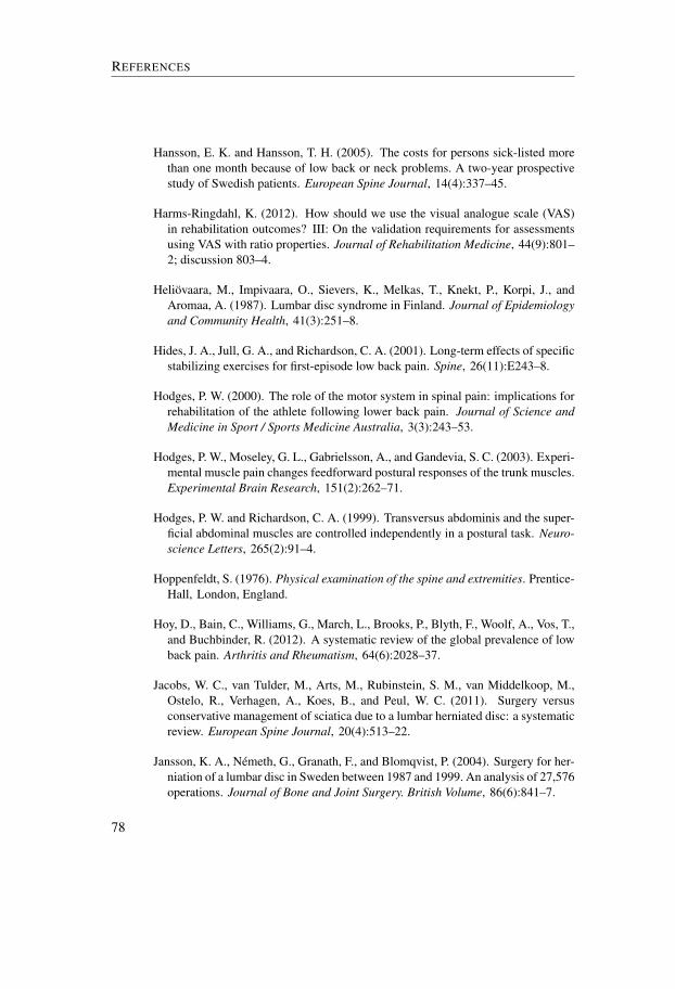
REFERENCES
Hansson, E. K. and Hansson, T. H. (2005). The costs for persons sick-listed morethan one month because of low back or neck problems. A two-year prospectivestudy of Swedish patients. European Spine Journal, 14(4):337–45.
Harms-Ringdahl, K. (2012). How should we use the visual analogue scale (VAS)in rehabilitation outcomes? III: On the validation requirements for assessmentsusing VAS with ratio properties. Journal of Rehabilitation Medicine, 44(9):801–2; discussion 803–4.
Heliövaara, M., Impivaara, O., Sievers, K., Melkas, T., Knekt, P., Korpi, J., andAromaa, A. (1987). Lumbar disc syndrome in Finland. Journal of Epidemiologyand Community Health, 41(3):251–8.
Hides, J. A., Jull, G. A., and Richardson, C. A. (2001). Long-term effects of specificstabilizing exercises for first-episode low back pain. Spine, 26(11):E243–8.
Hodges, P. W. (2000). The role of the motor system in spinal pain: implications forrehabilitation of the athlete following lower back pain. Journal of Science andMedicine in Sport / Sports Medicine Australia, 3(3):243–53.
Hodges, P. W., Moseley, G. L., Gabrielsson, A., and Gandevia, S. C. (2003). Experi-mental muscle pain changes feedforward postural responses of the trunk muscles.Experimental Brain Research, 151(2):262–71.
Hodges, P. W. and Richardson, C. A. (1999). Transversus abdominis and the super-ficial abdominal muscles are controlled independently in a postural task. Neuro-science Letters, 265(2):91–4.
Hoppenfeldt, S. (1976). Physical examination of the spine and extremities. Prentice-Hall, London, England.
Hoy, D., Bain, C., Williams, G., March, L., Brooks, P., Blyth, F., Woolf, A., Vos, T.,and Buchbinder, R. (2012). A systematic review of the global prevalence of lowback pain. Arthritis and Rheumatism, 64(6):2028–37.
Jacobs, W. C., van Tulder, M., Arts, M., Rubinstein, S. M., van Middelkoop, M.,Ostelo, R., Verhagen, A., Koes, B., and Peul, W. C. (2011). Surgery versusconservative management of sciatica due to a lumbar herniated disc: a systematicreview. European Spine Journal, 20(4):513–22.
Jansson, K. A., Németh, G., Granath, F., and Blomqvist, P. (2004). Surgery for her-niation of a lumbar disc in Sweden between 1987 and 1999. An analysis of 27,576operations. Journal of Bone and Joint Surgery. British Volume, 86(6):841–7.
78

References
Jensen, T. S., Albert, H. B., Sorensen, J. S., Manniche, C., and Leboeuf-Yde, C.(2007). Magnetic resonance imaging findings as predictors of clinical outcomein patients with sciatica receiving active conservative treatment. Journal of Ma-nipulative and Physiological Therapeutics, 30(2):98–108.
Johansson, E. (1999). Exercise-Based Physiotherapy Management of Patients withPersistent, Non-Specific Low Back Pain. PhD-thesis, Uppsala University, Upp-sala.
Kayama, S., Konno, S., Olmarker, K., Yabuki, S., and Kikuchi, S. (1996). Incisionof the anulus fibrosus induces nerve root morphologic, vascular, and functionalchanges. An experimental study. Spine, 21(22):2539–43.
Kjellby-Wendt, G. and Styf, J. (1998). Early active training after lumbar discectomy.A prospective, randomized, and controlled study. Spine, 23(21):2345–51.
Koes, B. W., van Tulder, M. W., and Peul, W. C. (2007). Diagnosis and treatment ofsciatica. BMJ (Clinical research ed.), 334(7607):1313–7.
Koho, P., Orenius, T., Kautiainen, H., Haanpaa, M., Pohjolainen, T., and Hurri,H. (2011). Association of fear of movement and leisure-time physical ac-tivity among patients with chronic pain. Journal of Rehabilitation Medicine,43(9):794–9.
Kongsted, A., Kent, P., Albert, H., Jensen, T. S., and Manniche, C. (2012). Patientswith low back pain differ from those who also have leg pain or signs of nerveroot involvement – a cross-sectional study. BMC Musculoskeletal Disorders,13(1):236.
Konstantinou, K. and Dunn, K. M. (2008). Sciatica: review of epidemiologicalstudies and prevalence estimates. Spine, 33(22):2464–72.
Kori, S. H., Miller, R. P., and Todd, D. D. (1990). Kinisophobia: A new view ofchronic pain behavior. Pain Management, 3:35–43.
Krippendorff, K. (2012). Content analysis: An introduction to its methodology.Number 1. Sage Publications, Inc., Thounds Oaks, Ca, 3rd edition.
Lamberg, E. M. and Hagins, M. (2012). The effects of low back pain on naturalbreath control during a lowering task. European Journal of Applied Physiology,112(10):3519–24.
Laslett, M., Oberg, B., Aprill, C. N., and McDonald, B. (2005). Centralizationas a predictor of provocation discography results in chronic low back pain, andthe influence of disability and distress on diagnostic power. The Spine Journal :official journal of the North American Spine Society, 5(4):370–80.
79
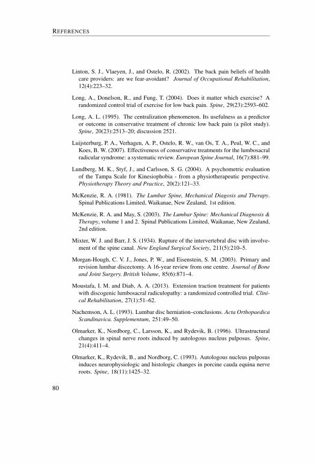
REFERENCES
Linton, S. J., Vlaeyen, J., and Ostelo, R. (2002). The back pain beliefs of healthcare providers: are we fear-avoidant? Journal of Occupational Rehabilitation,12(4):223–32.
Long, A., Donelson, R., and Fung, T. (2004). Does it matter which exercise? Arandomized control trial of exercise for low back pain. Spine, 29(23):2593–602.
Long, A. L. (1995). The centralization phenomenon. Its usefulness as a predictoror outcome in conservative treatment of chronic low back pain (a pilot study).Spine, 20(23):2513–20; discussion 2521.
Luijsterburg, P. A., Verhagen, A. P., Ostelo, R. W., van Os, T. A., Peul, W. C., andKoes, B. W. (2007). Effectiveness of conservative treatments for the lumbosacralradicular syndrome: a systematic review. European Spine Journal, 16(7):881–99.
Lundberg, M. K., Styf, J., and Carlsson, S. G. (2004). A psychometric evaluationof the Tampa Scale for Kinesiophobia - from a physiotherapeutic perspective.Physiotherapy Theory and Practice, 20(2):121–33.
McKenzie, R. A. (1981). The Lumbar Spine, Mechanical Diagosis and Therapy.Spinal Publications Limited, Waikanae, New Zealand, 1st edition.
McKenzie, R. A. and May, S. (2003). The Lumbar Spine: Mechanical Diagnosis &Therapy, volume 1 and 2. Spinal Publications Limited, Waikanae, New Zealand,2nd edition.
Mixter, W. J. and Barr, J. S. (1934). Rupture of the intervertebral disc with involve-ment of the spine canal. New England Surgical Society, 211(5):210–5.
Morgan-Hough, C. V. J., Jones, P. W., and Eisenstein, S. M. (2003). Primary andrevision lumbar discectomy. A 16-year review from one centre. Journal of Boneand Joint Surgery. British Volume, 85(6):871–4.
Moustafa, I. M. and Diab, A. A. (2013). Extension traction treatment for patientswith discogenic lumbosacral radiculopathy: a randomized controlled trial. Clini-cal Rehabilitation, 27(1):51–62.
Nachemson, A. L. (1993). Lumbar disc herniation–conclusions. Acta OrthopaedicaScandinavica. Supplementum, 251:49–50.
Olmarker, K., Nordborg, C., Larsson, K., and Rydevik, B. (1996). Ultrastructuralchanges in spinal nerve roots induced by autologous nucleus pulposus. Spine,21(4):411–4.
Olmarker, K., Rydevik, B., and Nordborg, C. (1993). Autologous nucleus pulposusinduces neurophysiologic and histologic changes in porcine cauda equina nerveroots. Spine, 18(11):1425–32.
80

References
Ostelo, R. W., Swinkels-Meewisse, I. J., Knol, D. L., Vlaeyen, J. W., and de Vet,H. C. (2007). Assessing pain and pain-related fear in acute low back pain: whatis the smallest detectable change? International Journal of Behavioral Medicine,14(4):242–8.
Ostelo, R. W. and Vlaeyen, J. W. (2008). Attitudes and beliefs of health careproviders: extending the fear-avoidance model. Pain, 135(1-2):3–4.
Osterman, H., Seitsalo, S., Karppinen, J., and Malmivaara, A. (2006). Effectivenessof microdiscectomy for lumbar disc herniation: a randomized controlled trialwith 2 years of follow-up. Spine, 31(21):2409–14.
Paatelma, M., Kilpikoski, S., Simonen, R., Heinonen, A., Alen, M., and Videman,T. (2008). Orthopaedic manual therapy, McKenzie method or advice only forlow back pain in working adults: a randomized controlled trial with one yearfollow-up. Journal of Rehabilitation Medicine, 40(10):858–63.
Panjabi, M. M. (1992). The stabilizing system of the spine. Part I. Function, dys-function, adaptation, and enhancement. Journal of Spinal Disorders, 5(4):383–9;discussion 397.
Patton, M. Q. (2002). Qualitative research & evaluation methods. Sage publica-tions, Inc., Thousand Oaks, Ca, 3rd edition.
Pellino, T., Tluczek, A., Collins, M., Trimborn, S., Norwick, H., Engelke, Z. K., andBroad, J. (1998). Increasing self-efficacy through empowerment: preoperativeeducation for orthopaedic patients. Orthopedic Nursing, 17(4):48–51, 54–9.
Perreault, K. (2008). Linking health promotion with physiotherapy for low backpain: a review. Journal of Rehabilitation Medicine, 40(6):401–9.
Peul, W. C., van den Hout, W. B., Brand, R., Thomeer, R. T., and Koes, B. W.(2008). Prolonged conservative care versus early surgery in patients with sciaticacaused by lumbar disc herniation: two year results of a randomised controlledtrial. BMJ (Clinical research ed.), 336(7657):1355–8.
Picavet, H. S., Vlaeyen, J. W., and Schouten, J. S. (2002). Pain catastrophizingand kinesiophobia: predictors of chronic low back pain. American Journal ofEpidemiology, 156(11):1028–34.
Pincus, T., Smeets, R. J. E. M., Simmonds, M. J., and Sullivan, M. J. L. (2010).The fear avoidance model disentangled: improving the clinical utility of the fearavoidance model. The Clinical Journal of Pain, 26(9):739–46.
Rabin, R. and de Charro, F. (2001). EQ-5D: a measure of health status from theEuroQol Group. Annals of Medicine, 33(5):337–43.
81

REFERENCES
Rasmussen, C., Nielsen, G. L., Hansen, V. K., Jensen, O. K., and Schioettz-Christensen, B. (2005). Rates of lumbar disc surgery before and after imple-mentation of multidisciplinary nonsurgical spine clinics. Spine, 30(21):2469–73.
Reneman, M. F., Jorritsma, W., Dijkstra, S. J., and Dijkstra, P. U. (2003). Relation-ship between kinesiophobia and performance in a functional capacity evaluation.Journal of Occupational Rehabilitation, 13(4):277–85.
Reneman, M. F., Schiphorts Preuper, H. R., Kleen, M., Geertzen, J. H. B., and Di-jkstra, P. U. (2007). Are pain intensity and pain related fear related to functionalcapacity evaluation performances of patients with chronic low back pain? Jour-nal of Occupational Rehabilitation, 17(2):247–58.
Rosén, M., Axelsson, S., and Lindblom, J. (2009). Observational studies versusrcts: what about socioeconomic factors? Lancet, 373(9680):2026.
Rydevik, B., Brown, M. D., and Lundborg, G. (1984). Pathoanatomy and patho-physiology of nerve root compression. Spine, 9(1):7–15.
Saal, J. A. and Saal, J. S. (1989). Nonoperative treatment of herniated lumbar inter-vertebral disc with radiculopathy. An outcome study. Spine, 14(4):431–7.
Saal, J. S., Saal, J. A., and Yurth, E. F. (1996). Nonoperative management of herni-ated cervical intervertebral disc with radiculopathy. Spine, 21(16):1877–83.
Scott, J. and Huskisson, E. C. (1976). Graphic representation of pain. Pain,2(2):175–84.
Skytte, L., May, S., and Petersen, P. (2005). Centralization: its prognostic value inpatients with referred symptoms and sciatica. Spine, 30(11):E293–9.
Stadnik, T. W., Lee, R. R., Coen, H. L., Neirynck, E. C., Buisseret, T. S., and Os-teaux, M. J. (1998). Annular tears and disk herniation: prevalence and contrastenhancement on MR images in the absence of low back pain or sciatica. Radiol-ogy, 206(1):49–55.
Strömqvist, B., Jönsson, B., Fritzell, P., Hägg, O., Larsson, B. E., and Lind, B.(2001). The Swedish National Register for lumbar spine surgery: Swedish Soci-ety for Spinal Surgery. Acta Orthopaedica Scandinavica, 72(2):99–106.
Sullivan, M., Bishop, S., and Pivik, J. (1995). The Pain Catastrophizing Scale:Development and validation. Psychological Assessment, 7(4):524–32.
Takada, E., Takahashi, M., and Shimada, K. (2001). Natural history of lumbar dischernia with radicular leg pain: Spontaneous MRI changes of the herniated massand correlation with clinical outcome. Journal of Orthopaedic Surgery (HongKong), 9(1):1–7.
82
References
Underwood, M. R., Harding, G., and Klaber Moffett, J. (2006). Patient percep-tions of physical therapy within a trial for back pain treatments (UK BEAM)[ISRCTN32683578]. Rheumatology (Oxford, England), 45(6):751–6.
Unlu, Z., Tasci, S., Tarhan, S., Pabuscu, Y., and Islak, S. (2008). Comparison of3 physical therapy modalities for acute pain in lumbar disc herniation measuredby clinical evaluation and magnetic resonance imaging. Journal of Manipulativeand Physiological Therapeutics, 31(3):191–8.
van Til, J. A., Drossaert, C. H., Punter, R. A., and Ijzerman, M. J. (2010). Thepotential for shared decision-making and decision aids in rehabilitation medicine.Journal of Rehabilitation Medicine, 42(6):598–604.
Vlaeyen, J., Kole-Snijders, A., Rotteveel, A., Ruesink, R., and Heuts, P. (1995a).The role of fear of movement/(re)injury in pain disability. Journal of Occupa-tional Rehabilitation, 5(4):235–51.
Vlaeyen, J. W., Kole-Snijders, A. M., Boeren, R. G., and van Eek, H. (1995b). Fearof movement/(re)injury in chronic low back pain and its relation to behavioralperformance. Pain, 62(3):363–72.
Vlaeyen, J. W. S. and Linton, S. J. (2012). Fear-avoidance model of chronic muscu-loskeletal pain: 12 years on. Pain, 153(6):1144–7.
Von Korff, M. (1994). Studying the natural history of back pain. Spine, 19(18Suppl):2041S–6S.
Vroomen, P. C., de Krom, M. C., Wilmink, J. T., Kester, A. D., and Knottnerus,J. A. (2002). Diagnostic value of history and physical examination in patientssuspected of lumbosacral nerve root compression. Journal of Neurology, Neuro-surgery and Psychiatry, 72(5):630–4.
Vucetic, N. and Svensson, O. (1996). Physical signs in lumbar disc hernia. ClinicalOrthopaedics and Related Research, (333):192–201.
Weber, H. (1983). Lumbar disc herniation. a controlled, prospective study with tenyears of observation. Spine, 8(2):131–40.
Weber, H. (1994). The natural history of disc herniation and the influence of inter-vention. Spine, 19(19):2234–8; discussion 2233.
Weber, H., Holme, I., and Amlie, E. (1993). The natural course of acute sciaticawith nerve root symptoms in a double-blind placebo-controlled trial evaluatingthe effect of piroxicam. Spine, 18(11):1433–8.
83
REFERENCES
Weinstein, J. N., Lurie, J. D., Tosteson, T. D., Skinner, J. S., Hanscom, B., Toste-son, A. N., Herkowitz, H., Fischgrund, J., Cammisa, F. P., Albert, T., and Deyo,R. A. (2006a). Surgical vs nonoperative treatment for lumbar disk herniation: theSpine Patient Outcomes Research Trial (SPORT) observational cohort. JAMA,296(20):2451–9.
Weinstein, J. N., Lurie, J. D., Tosteson, T. D., Tosteson, A. N., Blood, E. A., Abdu,W. A., Herkowitz, H., Hilibrand, A., Albert, T., and Fischgrund, J. (2008). Surgi-cal versus nonoperative treatment for lumbar disc herniation: four-year results forthe Spine Patient Outcomes Research Trial (SPORT). Spine, 33(25):2789–800.
Weinstein, J. N., Tosteson, T. D., Lurie, J. D., Tosteson, A. N., Hanscom, B., Skin-ner, J. S., Abdu, W. A., Hilibrand, A. S., Boden, S. D., and Deyo, R. A. (2006b).Surgical vs nonoperative treatment for lumbar disk herniation: the Spine PatientOutcomes Research Trial (SPORT): a randomized trial. JAMA, 296(20):2441–50.
Werneke, M., Hart, D. L., and Cook, D. (1999). A descriptive study of the central-ization phenomenon. A prospective analysis. Spine, 24(7):676–83.
Werneke, M. W., Hart, D. L., Resnik, L., Stratford, P. W., and Reyes, A. (2008).Centralization: prevalence and effect on treatment outcomes using a standardizedoperational definition and measurement method. The Journal of Orthopaedic andSports Physical Therapy, 38(3):116–25.
White, A. A. and Panjabi, M. M. (1978). Clinical Biomechanics of the Spine. J. B.Lippincott Company, Philadelphia, Pa.
World Health Organisation (1948). Preamble to the Constitution of the WorldHealth Organization as adopted by the International Health Conference, (OfficialRecords of the World Health Organization, no. 2, p. 100).
Yilmaz, F., Yilmaz, A., Merdol, F., Parlar, D., Sahin, F., and Kuran, B. (2003).Efficacy of dynamic lumbar stabilization exercise in lumbar microdiscectomy.Journal of Rehabilitation Medicine, 35(4):163–7.
Zieger, M., Luppa, M., Matschinger, H., Meisel, H. J., Gunther, L., Meixensberger,J., Toussaint, R., Angermeyer, M. C., Konig, H. H., and Riedel-Heller, S. G.(2011). Affective, anxiety, and substance-related disorders in patients under-going herniated disc surgery. Social Psychiatry and Psychiatric Epidemiology,46(11):1181–90.
Zieger, M., Schwarz, R., Konig, H. H., Harter, M., and Riedel-Heller, S. G. (2010).Depression and anxiety in patients undergoing herniated disc surgery: relevantbut underresearched - a systematic review. Central European Neurosurgery,71(1):26–34.
84

References
Zung, W. W. (1965). A self-rating depression scale. Archives of General Psychiatry,12:63–70.
Öberg, B., Enthoven, P., Kjellman, G., and Skargren, E. (2003). Back pain in pri-mary care: a prospective cohort study of clinical outcome and healthcare con-sumption. Advances in Physiotherapy, 5(3):98.
85