european association of veterinary … · of veterinary diagnostic imaging european college of...
TRANSCRIPT
�
E A V D I 2 0 0 7 A N N U A L M E E T I N G
EISAGOGIKOEUROPEAN ASSOCIATION
OF VETERINARY DIAGNOSTIC IMAGING
EUROPEAN COLLEGEOF VETERINARY DIAGNOSTIC IMAGING
ARISTOTLE UNIVERSITY OF THESSALONIKISCHOOL OF VETERINARY MEDICINE
SECTION OF RADIOLOGY
ANNUAL MEETING 2007CONFERENCE GUIDE & ABSTRACTS BOOK
PORTO CARRAS, CHALKIDIKI, GREECE
2 9 . 0 8 - 0 1 . 0 9 . 2 0 0 7
��
E A V D I 2 0 0 7 A N N U A L M E E T I N G
POSTEr SESSiONStudyofGasterointestinalTractFunctionofGoldenHamsterwithBariumSulfate. . . . . . . . . . . .97
DopplerImagingoftheExtrnalOphtalmicArteryandInternalOphtalmicArteryinNormalDshCat.. . . . . . . . . . . . . . . . . . . . . . . . . . . . . . . . . . . . . . . . . . .97
Multi-Detector-RowComputedTomographyoftheCarpalJointinDogs. . . . . . . . . . . . . . . . . . . . .97
HighResolution16DetectorRowComputedTomographyExaminationoftheCanineThorax. . . . . . . . . . . . . . . . . . . . . . . . . . . . . . . . . . . . . . . . . . . . . . . . . . . . . . . . . .98
UltrasonographyandHistologyoftheEquineMenisci:AComparativeStudyoftheMedialMeniscus. . . . . . . . . . . . . . . . . . . . . . . . . . . . . . . . . . . . . . . . . . . . . . .99
EvaluationoftheUseofThreeDimensionalUltrasonogeraphyoftheEyeandMeasurementofOpticalNerveSheetDiameterinPersianCat . . . . . . . . . . . . . . . .99
KneeJointUltrasonographyoftheCcltRabbitExperimentalModelofOsteoarthritis. . . . . . . 100
HelicalandThirdGenerationComputedTomographyoftheNormalCaninePelvicCavity. . .101
HelicalComputedTomographicAnatomyoftheEquineTemporomandibularJoint:NormalAppearance. . . . . . . . . . . . . . . . . . . . . . . . . . . . . . . . . . . . . . . . . . . .101
PrevalenceofPkdinPersianandExoticShorthairCatsinItalyandUsefulnessofUltrasonographyintheEarlyDiagnosis. . . . . . . . . . . . . . . . . . . . . . . . . . 102
IncidenceofElbowDysplasiainSouthAfrica . . . . . . . . . . . . . . . . . . . . . . . . . . . . . . . . . . . . . . . . . . . . . 103
RadiographyicStudyofDistalSesamoidBoneofNormalClawsinCattle. . . . . . . . . . . . . . . . . . . 104
WallStentPlacementtoPreventCoilsMigrationDuringIntrahepaticEmbolizationofPssinADog. . . . . . . . . . . . . . . . . . . . . . . . . . . . . . . . . . . . . . . . . 104
AbdominalUltrasonographicFindingsin38DogswithBabesiosis(BabesiaCanisCanis). . . . . . . . . . . . . . . . . . . . . . . . . . . . . . . . . . . . . . . . . . . . . . . . . . . . . 105
UltrasonographicAssessmentofNewBoneFormationDuringDistractioninARabbitModel. . . . . . . . . . . . . . . . . . . . . . . . . . . . . . . . . . . . . . . . . . . . . . . . . . . . . 106
ImagingofTwoTraumaCasesinLoggerheadSeaTurtle(CarettaCaretta). . . . . . . . . . . . . . . . . . .107
InvestigationonthePharmacodynamicsofTwoDifferentPsychopharmacaintheDog’sBrainwithSpect . . . . . . . . . . . . . . . . . . . . . . . . . . . . . . . . . . . . . . . . . . . .107
RadiographicAtlasofOsteoarthitisintheRabbitExperimentalModelKneeJoint. . . . . . . . . . . . . . . . . . . . . . . . . . . . . . . . . . . . . . . . . . . . . 108
MagneticResonanceImagingofMelanomasin3Horses. . . . . . . . . . . . . . . . . . . . . . . . . . . . . . . . . . 109
CervicalMyelopathySecondarytoCongenitalIncompleteOssificationoftheDorsalLaminaoftheAtlas:CaseReportandLiteratureReview. . . . . . . . . . . . . . . . . . . . . . 110
MdctAttenuationValuesoftheLiverinCaninePituitary-DependentHyperadrenocorticism. . . . . . . . . . . . . . . . . . . . . . . . . . . . . . . . . . . . . . . . . . . . . . 110
1.
2.
3.
4.
5.
6.
7.
8.
9.
10.
11.
12.
13.
14.
15.
16.
17.
18.
19.
20.
21.
Close this window to return to IVIS
Published with the permission of the European Association of Veterinary Diagnostic Imaging (EAVDI)
E A V D I 2 0 0 7 A N N U A L M E E T I N G
RadiographicFeaturesofPulmonaryEdemaAssociatedwithMitralRegurgitationinDogs. . . . . . . . . . . . . . . . . . . . . . . . . . . . . . . . . . . . . . . . . . . . . .111
MorphometricAnalysisoftheCaudalFossainCavalierKingCharlesSpaniel. . . . . . . . . . . . . . . . . . . . . . . . . . . . . . . . . . . . . . . . . . . . . . . . . . . . . . . . . . 112
FirstThreeYearsofCTExaminationinAPrivatePracticeinItaly. . . . . . . . . . . . . . . . . . . . . . . . . . . . 113
HemodynamicAlterationsCausedBy3TypesofIntravenousContrastMediainAnesthetizedCats . . . . . . . . . . . . . . . . . . . . . . . . . . . . . . . . . . . . . . . . . . . . . . . . . . . . . 113
ComputedTomographicAnatomyoftheEquineMetacarpophalangealJoint. . . . . . . . . . . . . . 114
ImagingDiagnosticofGastricUlcersinDogs-CaseReport. . . . . . . . . . . . . . . . . . . . . . . . . . . . . . . . 115
SclerotherapyWithoutDrainageof95%EthanolforTreatmentofAneurismalBoneCystinADog. . . . . . . . . . . . . . . . . . . . . . . . . . . . . . . . . . . . . . . . . . 115
Clinician’sGuidetotheComputerTomographicandGrossAnatomyoftheSandtigerShark(CarchariasTaurus). . . . . . . . . . . . . . . . . . . . . . . . . . . . 116
ComparisonoftheArterialBloodSupplyoftheLumbarSpineinDog,SwineandRabbitByCe-Mra. . . . . . . . . . . . . . . . . . . . . . . . . . . . . . . . . .117
ArteriographicStudyoftheBodyCavityintheCommonStork(CiconiaCiconia) . . . . . . . . . . . .117
ImagingofRetrobulbarMassesinDogsandCats:RetrospectiveStudyof16Cases. . . . . . . . . . . . . . . . . . . . . . . . . . . . . . . . . . . . . . . . . . . . . . . . . . . . . . . . . . 118
ASeverityScoringSystem(Sss)forRadiographicFindings(Rf )intheLimbsofYoungHorses . . . . . . . . . . . . . . . . . . . . . . . . . . . . . . . . . . . . . . . . . . . . . . . . . . . . . . . . 119
AccuracyofTrans-RectalUltrasonographytoAssesstheNumberofOvarianPre-OvulatoryFolliclesinSows. . . . . . . . . . . . . . . . . . . . . . . . . . 119
EvaluationofRadiographicalFindingsofthePelvisinCatswithDystocia. . . . . . . . . . . . . . . . . . 120
AbscessonLeftThoracicWallDuetoReticularForeignBody . . . . . . . . . . . . . . . . . . . . . . . . . . . . . . .121
EchocardiographicFeaturesofQuadricuspidAorticValveinThreeBoxerDogs. . . . . . . . . . . . . .121
DiagnosticOcularUltrasonographyinDonkeys. . . . . . . . . . . . . . . . . . . . . . . . . . . . . . . . . . . . . . . . . . . 122
RadiographicandMRIFindingsofNasalLymphoma.ACaseReportinASiameseCat.. . . . . . . . . . . . . . . . . . . . . . . . . . . . . . . . . . . . . . . . . . . . . . . . . . . . . . . . . . 122
AbdominalUltrasoundandThoracicRadiographinDogswithPrimaryImmune-MediatedHemolyticAnemia . . . . . . . . . . . . . . . . . . . . . . . . . . . . . . 123
ComparisonofIodixanolwithIohexolinExcretoryUrographyofCat . . . . . . . . . . . . . . . . . . . . . . 124
MagneticResonanceImaginginALabradorRetrieverwithLeukoencephalopathy. . . . . . . . . 124
theUseofIntra-ArticularAirinLowFieldMRArthrography(Mrar)fortheArtificiallyProducedCartilageLesionsDetectiononEquineMetacarpophalangealJoint.. . . . . . . . . . . . . . . . . . . . . . . . . . . . . . . . . . . . . . . . . 125
PreliminaryResultsofRadiographicandUltrasoundExaminationoftheThoracicSpinousProcessesandInterspinousSpacesinHalf-BreedSportHorses.. . . . . . . . . . . . . . . . . . . . . . . . . . . . . . . . . . . . . . 126
UltrasonographicChangesoftheThyroidGlandinHyperthyroidCats6MonthsAfter131iRadioactiveIodineTherapy . . . . . . . . . . . . . . . . . . . . . .127
DiscDegenerationandSpondylosisinoldDachhounds.Afollowupstudyover8years. . . . .127
22.
23.
24.
25.
26.
27.
28.
29.
30.
31.
32.
33.
34.
35.
36.
37.
38.
39.
40.
41.
42.
43.
44.
45.
46.
��
Close this window to return to IVIS
Published with the permission of the European Association of Veterinary Diagnostic Imaging (EAVDI)
��
E A V D I 2 0 0 7 A N N U A L M E E T I N G
STUdy OF gASTErOiNTESTiNAL TrACT FUNCTiONOF gOLdEN hAmSTEr WiTh BAriUm SULFATE
Kabir f.1, Vajhi A.2, Masoudifard M.2
1 Islamic Azad University, Science and Research branch, Faculty of Veterinary Medicine, Clinical Science Department, Tehran - IRAN
2 University of Tehran, Faculty of Veterinary Medicine, Clinical Science Department, Tehran - IRAN
Today, because some families like to keep Hamster as a pet and it involved with a variety of gastrointestinal tract problems, we decided to find a standard radiographic time pattern of gastrointestinal functions. So we utilized Bariumsulfate, a contrast media agent, as a pasty food. Six Golden Hamsters were included in this study. Ventrodorsal and Laterolateral plain radiographs with mammography specialized film and cassette were performed and their healths were confirmed. They were not had any food for 12 hours, then 2cc of 30% bariumsulfate were eaten. Radiographs were taken from 0 minuets till 24 hours laterolateral and ventrodorsaly. Stomach evacuation to deodenom was begun in 15th minute, and after 60 minute there was no contrast media agent in the stomach pouches. Bariumsulphate were remained in the main stomach till 5 hours. Entrance of contrast medium to cecum was begun at 60th minute and was left till the end of examination. the first feces observed in colon in 100th minute. At 24th hour of the study contrast medium was observed in the first part of the stomach because of hamster’s feces eating.
dOPPLEr imAgiNg OF ThE EXTrNAL OPhTALmiC ArTEryANd iNTErNAL OPhTALmiC ArTEry iN NOrmAL dSh CAT.
Vosough Dariush 1,Masodifard Majid2
1 Faculty of Veterinary Medicine, University of Bahonar, Kerman - Iran2 Faculty of Veterinary Medicine, University of Tehran, Iran.
Blood velocity parameters of the orbital and ocular vasculature can be no invasively assessed and measured by Doppler imaging. the purpose of this study was to blood velocity measurement in orbital vasculature. A total of 10 (Female) previously healthy domestic short hair cats were selected. General Electrics Voluson 730-Pro ultrasound equipment with linear trapezoid 5-12 MHz transducer was applied for all the examinations. Vessels identified a majority of the time, include: external ophthalmic artery (EOA), and internal ophthalmic artery (IOA) and the following Doppler parameters were measured, peak systolic velocity (PSV), end diastolic velocity (EDV), Mean PSV, EDV, at the EOA were 10.3, 5.1, and the mean PSV, EDV, at the IOA were 10.8, 3.5, and 0.307. Doppler imaging has the potential for determining no invasively and consecutively the blood velocity parameters found in orbital and ocular diseases, including orbital inflammations and neoplasia;intraocular inflammations and neoplasia;vascular diseases including systemic vascular disease (hypertension)vasculopathies,and anemia; the glaucoma; and document able follow-up after medical and surgical treatment of these diseases.
1-Kathleen J.Gelatt-Nicholson and et al: Doppler imaging of the ophthalmic vasculature of the normal dog, blood velocity measurement and reproducibility, Veterinary Ophthalmology (1999)2.87-96
mULTi-dETECTOr-rOW COmPUTEd TOmOgrAPhy OF ThE CArPAL jOiNT iN dOgS
Cavrenne R., Bolen G., De Busscher V. , Snaps F.Diagnostic Imaging Departement of the Veterinary Faculty of the University of Liège (Belgium)
INTRODUCTIONCarpal joint is a complex articulation. Radiographic evaluation of this region is difficult because of superimposition of bony structures. Multidector-row computed tomography ( MDCT ) is widely used in bone and joint imaging in humans. the aim of this study is to present the MDCT examination of the carpal joint in dogs.
Close this window to return to IVIS
Published with the permission of the European Association of Veterinary Diagnostic Imaging (EAVDI)
��
E A V D I 2 0 0 7 A N N U A L M E E T I N G
MATERIAL AND METhODSFour carpal joints were used. Radiography was performed to rule out any bony changes. Computed tomography (CT) was performed on a 16 detector-row system (Somatom 16, Siemens, Erlagen). Native 2mm thick contiguous slices were obtained. Multiplanar reformatted images of 0.6mm thickness were obtained in sagittal, transverse and dorsal planes.Three dimensional images were also created. After CT examination, carpal joint were frozen and sectioned into slab sections.
RESULTSNormal anatomy of the carpal joint was presented in the three sectional planes. the computed tomography images were matched with the structured identified in the corresponding anatomy section. Three dimensional reconstructed anatomy was also presented
CONCLUSIONSMultidetector-row computed tomography is a precise method for evaluation of the carpal joint. the entire joint could be evaluated in three sectional planes.
high rESOLUTiON 16 dETECTOr rOW COmPUTEd TOmOgrAPhyEXAmiNATiON OF ThE CANiNE ThOrAX
De Busscher V., Bolen G., Cavrenne R., Clercx C., Snaps F.ULg-Facultι de Mιdecine Vιtιrinaire-Sciences Cliniques-Service d’Imagerie Mιdicale-Liθge-Belgium
INTRODUCTIONMultidetector row computed tomography (MDCT) has brought about major advances in thorax imaging. MDCT of the lungs is the accepted diagnostic method for detection and characterisation of various pulmonary parenchymal abnormalities involving the airways, air space and interstitium in humans. the aim of this study is to present MDCT examination of the canine thorax.
MATERIALS AND METhODSthe thorax examinations were performed on a 16 detector row scanner (Somatom 16, Siemens, Erlagen, Germany). Native transverse acquisitions of 5 mm thickness were obtained in two different window settings: high resolution lungs and mediastinal windows. Mediastinal window acquisition was also performed after intravenous injection of iodine contrast media. Time of acquisition was less than 15 seconds for each scanning sequences with a total acquisition time of less than 60 seconds for the completed examination. High resolution multiplanar reformatted images of 1 mm thickness were obtained in sagittal, transverse and dorsal planes. Three dimensional (3D) volume rendering reconstruction images were also created.
RESULTSTransverse, sagittal and dorsal images of the normal lungs, bronchial, mediastinal and cardio-vascular structures were presented. Different pathologic conditions as alveolar, interstitial, bronchial patterns, mediastinal pathologies were illustrated in the three sectional planes. Three dimensional (3D) volume rendering reconstruction anatomy was presented, especially the central airways. Technical aspects (slices thickness, gantry rotation time, pitch, window levels) were also discussed.
CONCLUSIONMDCT is a method of choice for evaluation of the canine thorax. It allows high resolution thin images in three sectional planes.
Assheuer J. and Sager M., MRI and CT Atlas of the Dog, Blackwell Science 1997,315-346; Rivero MA. et al., Anat. Histol. Embryol. 2005,34(4):215-219; Smallwood JE. and George TF., Vet. Radiol. Ultrasound. 1993,34(2):65-83.
Close this window to return to IVIS
Published with the permission of the European Association of Veterinary Diagnostic Imaging (EAVDI)
��
E A V D I 2 0 0 7 A N N U A L M E E T I N G
ULTrASONOgrAPhy ANd hiSTOLOgy OF ThE EqUiNE mENiSCi:A COmPArATivE STUdy OF ThE mEdiAL mENiSCUS
De Busscher V.*, Gabriel A.**, Cassart D.**, Heimann M.***, Antoine N.**, Busoni V.** ULg-Facultι de Mιdecine Vιtιrinaire-Sciences Cliniques-Service d’Imagerie Mιdicale-Liθge-Belgium** ULg-Facultι de Mιdecine Vιtιrinaire-Morphologie et Pathologie-Liθge-Belgium*** Institut de Pathologie et de Gιnιtique-Dιpartement Anatomie Pathologique-Gosselies-Belgium
INTRODUCTIONHypoechoic areas are commonly seen in the equine medial menisci at ultrasonography and have been associated with fibres disruption and collapse, edema, or degenerative processes such as fibroplasias or necrosis. in horses, no comparative study of ultrasonographic and histological appearance of the menisci has been reported. This study aimed to compare ex-vivo ultrasonographic and histological features of the equine medial meniscus.
MATERIALS AND METhODSMenisci were examined post-mortem in situ and after excision in a water bath with a 7.5 MHz linear transducer. Vertical (abaxio/axial) sections were made and stained with toluidine blue
RESULTSThirteen medial menisci of 12 warmblood horses were scanned. Two menisci showed a normal homogenous echogenicity. Eleven menisci contained either a central hypoechoic area and/or a linear, horizontal hypoechoic zone. At histology, in the 2 ultrasonographically normal menisci, dense collagen fibres were found in the middle of the meniscus; more matrix was seen in the periphery. the hypoechoic defects seen in 11 menisci were mainly associated with internal architectural changes: modified orientation and/or increased quantity of collagen fibres. One central lesion was associated with thick trabeculae with increased cellularity and increased amount of matrix. One meniscus presented edema and one horizontal lesion corresponded to very dense fibrous tissue with neovascularization.
DISCUSSION - CONCLUSIONThis study demonstrates that hypoechoic areas seen at ultrasonography in the medial meniscus correspond to different types of degenerative or regenerative lesions with architectural changes.
1. De Busscher V. et al., J. Equine Vet. Sci. 2006,26:453-461 ; 2.Denoix JM., in: Joint Disease in the Horse, McIlwraith CW, Trotter GW (Eds.), Saunders WB 1996:165-202 ; 3.Denoix JM. et al., Pferdeheilkunde 1996;12:629-631 ; 4.Ferrer-Roca O. et al., Clin. Orthop. Relat. Res. 1980, 146:289-307 ; 5.Noble J. et al., J Bone Surg. 1975,57-B:180-186.
EvALUATiON OF ThE USE OF ThrEE dimENSiONAL ULTrASONOgErAPhy OF ThE EyE ANd mEASUrEmENT OF OPTiCAL NErvE ShEET diAmETEr iN PErSiAN CAT
Vosough Dariush*, Masoudifard Majid, Veshkini Abbas, Vajhi Alireza, Soroory*Department of Clinical Sciences, Faculty of Veterinary Medicine, University of Shahid Bahonar, Kerman - Iran. Department of Clinical Sciences, Faculty of Veterinary Medicine, University of Tehran - Iran
This study was for determine the possibility of three-dimensional ultrasonogeraphy (3DUS) and measurement of optical nerve sheet by this method. 10 Persian cat( 5males,5 females), age 1-1.5 year, and weighting 4-8 kg were selected. 3D ultrasounds of the eyes were evaluated and the normal optical nerves in 3DUS images were measured using Volouson 730 and Statistical analysis- by paired sample T-test. in the obtained 3D images vitreous body, anterior chamber, and lens cortex and nucleus showed a distinct anechogenic to hypoechogenic. Details of the eyes compartments were better observed by rotating the images in all possible angles and planes using 3D facilities. Anterior and posterior lens capsule and the optic disk were hyperechogenic. the mean optical nerve in males was: 1.35 mm and in females it was 1.40mm. There weren’t a significant difference between ocular nerve measurements of male and female dogs and left and right e yes. -the 3DUS gives useful images for teaching and diagnostic purpose and Lesions of the caudal portion of the orbit (e.g. optic nerve atrophy) are better
Close this window to return to IVIS
Published with the permission of the European Association of Veterinary Diagnostic Imaging (EAVDI)
�00
E A V D I 2 0 0 7 A N N U A L M E E T I N G
visualized by this technique. the results of the eye 3DU in cats showed marked advantages in image acquisition for interpretation of all aspects of the ocular structures. Measurement of the optic nerve by 3D ultrasonography and other methods such as CT scan and direct measurement did not have any significant difference.
1- Downey D.B, Nicoll D.A, (1996). Three dimensional ultrasound imaging of the eye. Congress of EADI department of diagnostic Radiology University of western Ontario. 10:75-81
kNEE jOiNT ULTrASONOgrAPhy OF ThE CCLT rABBiTEXPErimENTAL mOdEL OF OSTEOArThriTiS
Boulocher C, Arnault F, Duclos ME, Roualdes O, Hartmann D, Roger T, Vignon E, Viguier E.Boulocher C., Arnault F, Duclos ME, Roger R., Viguier E. UMR MA 3990. Ecole Nationale Vétérinaire de Lyon (ENVL), Marcy l’Etoile, FranceVignon E. UMR MA 3990. UCBL (Université Claude Bernard Lyon 1), Centre Hospitalier Lyon-Sud, Pierre Benite,France.Roualdes O, Hartmann D. UMR MA 3990. UCBL (Université Claude Bernard Lyon 1),Lyon,France
ObjECTIvESto develop a protocol for knee joint ultrasonography (US) of the Cranial Cruciate Ligament Transection (CCLT) rabbit model of osteoarthritis(OA); to evaluate the correlation between US and macroscopic medial and lateral meniscal injuries (MMI and LMI) with tibial cartilage damage1,2, depending on the age and weight3.
METhODSone group of skeletally mature White New Zealand Rabbits and one adolescent group were used for the study. Clinical examination, in vivo US and final macroscopy were compared 5 months after CCLT. MMI and LMI were graded semi-quantitatively. Tibial cartilage damage was scored quantitatively with the Visual Analogical Evaluation (EVA) 4.
RESULTSthe CCLT rabbit knee joint US protocol was standardized. Positive correlation was found between US and macroscopic MMI (p=10-5, r= 0.79) and LMI (p= 0.001, r= 0.63). US MI predictive positive value was 92.3% and predictive negative value 81.25%, compared with macroscopy. the total tibial EVA was well correlated with the total menisci score (p=0.008, r=0.70). Medial tibial EVA were significantly higher in the adult than in the adolescent operated group (p=0.04).
CONCLUSIONin the rabbit OA model, age and weight when the CCLT is performed influence the severity of meniscal and cartilage damages. A significant relationship for the MI between macroscopic and US grading as well as a significant correlation between tibial cartilage lesions and MI was observed. US is relevant and effective in detecting meniscal lesions and we propose US as a non invasive, non expensive, in vivo imaging technique for preclinical studies in the CCLT rabbit OA model.
RefeRences1. Hellio Le Graverand MP, Vignon E, Otterness IG, Hart DA. Early changes in lapine menisci during osteoarthritis
development: Part I: cellular and matrix alterations. Osteoarthritis Cartilage. 2001 Jan;9(1):56-64.2. Hellio Le Graverand MP, Vignon E, Otterness IG, Hart DA. Early changes in lapine menisci during osteoarthritis
development: Part II: molecular alterations. Osteoarthritis Cartilage. 2001;9(1):65-72.3. Ding C., Martel-Pelletier J., Pelletier J.-P., Abram F., Raynauld J.-P., Cicuttini F. and Jones G. Osteoarthritis risk factors: a
cross-sectional study of associations between meniscal tear and knee structure, radiographic changes and symptoms in an undiagnosed OA Cohort. Abstract. Osteoarthritis and Cartilage. 2006. 14(2):S147
4. Ayral X, Dougados M, Listrat V, et al. Arthroscopic Evaluation of chondropathy in osteoarthritis of the knee. J Rheumatology 1996;23:698–706
Close this window to return to IVIS
Published with the permission of the European Association of Veterinary Diagnostic Imaging (EAVDI)
�0�
E A V D I 2 0 0 7 A N N U A L M E E T I N G
hELiCAL ANd Third gENErATiON COmPUTEd TOmOgrAPhyOF ThE NOrmAL CANiNE PELviC CAviTy
Vazquez JM.1, Teixeira M.2, Arencibia A.3, Cardoso L.2, Gil F.1, Soler M.4, López O.1, Ramirez G.1, Agut A.4
1 University of Murcia. Anatomy Department, Murcia, Spain2 University Lutheran of Brazil, Rio Grande do Sul, Brazil3 University of Las Palmas. Anatomy Department, Las Palmas de Gran Canaria, Spain4 University of Murcia. Medicine and Surgery Department, Murcia, Spain
SUbjECTComputed Tomography (CT) is a valuable technique for diagnosis of intrapelvic disorders and in human medicine it has become the imaging modality of choice for the pelvic cavity. CT can provide information that is just not attainable by other means. the aim of this study is to describe the helical and conventional third generation CT appearance of the pelvic region in the normal dog.
MATERIALS AND METhODSEight cross-breed dogs were used, four males and four females. Helical and third-generation CT scans were performed in each dog under general anaesthesia. Injection of iodinated contrast medium was made in four dogs (two males and two females) through cephalic venous catheter and a dose of 10 ml/kg of oral contrast medium was given two hours before scanning. the windows chosen had soft tissues setting. the images were acquired from the 7th lumbar vertebra to the ischiatic tuberosity.
RESULTSTwelve representative images were selected, six from females with contrast medium obtained with the helical CT scan and six from males without contrast achieved with the third-generation CT scan. Osseous and articular structures, intrapelvic organs, iliac vessels and muscles were located and identified. Different atlas of cross-sectional anatomy (Feeney et al., 1991; Vazquez et al., 2000) were used to identify the structures of the pelvic cavity and correlate to analogous structures on the CT images.
CONCLUSIONSHelical CT provides a good detail of pelvic structures. Normal anatomy is identified when compared with anatomical sections.
Feeney, D.A., Fletcher, T.F., Hardy, R.M., 1991. Atlas of correlative imaging anatomy of the normal dog. Ultrasound and computed tomography. W.B. Saunders Co., Philadelphia. Smallwood, J.E., George II, T.F., 1993. Anatomic atlas for computed tomography in the mesaticephalic dog: caudal abdomen and pelvis. Veterinary Radiology & Ultrasound 34 (3), 143-167. Vázquez, J.M., Ramírez, G., Gil, F., Latorre, R., Moreno, F., López, O., Orenes, M., Arencibia, A., 2000. Atlas de Anatomía Clínica: perro y gato. Cavidades torácica, abdominal y pelviana. A.G. Novograf, S.A.. Murcia. España.
hELiCAL COmPUTEd TOmOgrAPhiC ANATOmyOF ThE EqUiNE TEmPOrOmANdiBULAr jOiNT: NOrmAL APPEArANCE
Rodríguez, M.J.1, Latorre, R.2, Soler, M. 1, López, O. 2, Agut, A.11 Veterinary School, University of Murcia, Department of Medicine and Surgery, 30100 Espinardo, Murcia, Spain. 2 Veterinary School, University of Murcia,Department of Anatomy, 30100 Espinardo, Murcia, Spain.
INTRODUCTIONDiagnoses of temporomandibular joint (TMJ) disorders are awkward due to its complex anatomy, the non-specific symptomatology and the difficulty to interpret the radiographic views. Computed tomography (CT) is a valuable imaging tool that provides a reliable evaluation of the osseous TMJ structures. However, a thorough knowledge of the TMJ cross-sectional anatomy is critical for accurate interpretation of CT study. the aim of this study was to describe the normal computed tomographic imaging of the equine TMJ.
Close this window to return to IVIS
Published with the permission of the European Association of Veterinary Diagnostic Imaging (EAVDI)
�0�
E A V D I 2 0 0 7 A N N U A L M E E T I N G
MATERIAL AND METhODSEight TMJs from Pure-Bred Spanish adult horses were used to perform the CT study. A helical CT scanner was employed to acquire contiguous 1 mm transverse slices of the TMJ region. Transverse images were reformatted into sagittal and dorsal planes and processed with a detailed algorithm to enhance bony and soft tissue structures. A three-dimensional reconstructed imaging of the joint was obtained. for the anatomic study, transverse, sagittal and dorsal cryosections of the TMJ area were obtained and plastinated using the P-40 method. CT images and anatomic sections were studied and compared to identify the structures.
RESULTSthe best definition of TMJ components was acquired with a bone window obtaining a good delineation between cortex and medulla. the articular cartilage was observed as a hyperdense stripe over the subchondral bone. the soft tissue-TMJ structures were not well visualised using a soft tissue window.
DISCUSSION-CONCLUSIONHelical CT provides an excellent evaluation of the TMJ bone components but not of soft tissues. Normal CT anatomy is identified comparing with plastinated sections.
1. Devine, D.V., Moll, H.D., Bahr, R.J. (2005). Fracture, luxation, and chronic septic arthritis of the temporomandibular joint in a juvenile horse. J Vet Dent, 22(2):96-99. 2. Morrow, K.L., Park, R.D., Spurgeon, T.L., Stashak, T.S., Arceneaux, B. (2000). Computed tomographic imaging of the equine head. Vet Radiol & Ultrasound, 41(6):491-497. 3. Soler, M., Murciano, J., Latorre, R., Belda, E., Rodríguez, M.J., Agut, A. (2006). Ultrasonography, computed tomographic and magnetic resonance imaging anatomy of the normal canine stifle joint. the Veterinary Journal (2006), doi:10.1016/j.tvjl.2006.08.019. 4. Smallwood, J.E., Wood, B.C., Taylor, W.E., Tate, L.P. (2002). Anatomic reference for computed tomography of the head of the foal. Vet Radiol & Ultrasound, 43(2):99-117.
PrEvALENCE OF Pkd iN PErSiAN ANd EXOTiC ShOrThAir CATS iN iTALy ANd USEFULNESS OF ULTrASONOgrAPhy iN ThE EArLy diAgNOSiS
Bonazzi M1, Volta A.1, Gnudi G1, Gazzola M2, Bertoni G1
1 Università di Parma, Dip. di Salute Animale, Sez. Radiologia e D.I.2 Sez. Patologia e Anat. Patologica, Parma, Italy
INTRODUCTIONthe aims of the study were to determine the prevalence of Polycystic Kidney Disease in Persians (PKD) and Exotic Shorthairs in Italy and to evaluate ultrasonography for the diagnosis prior to 9 months of age.
MATERIALS AND METhODSTwo-hundred-eighty-eight Persian and 44 Exotic Shorthair cats that underwent ultrasonographic (US) screening for PKD between July 2003 and December 2005 were reviewed. Cats were divided in two groups, one including cats aged <9 months (G1) and one cats aged ≥9 months (G2). Cats were classified as PKD-positive when at least one renal cyst was found. for all the examinations a 10 MHz linear transducer was used.Sixteen cats of 5 different litters with at least one parent affected by PKD were selected from G1 and examined four times from 3 to 18 months of age.
RESULTSOne-hundred-thirty-six cats (41%) showed more than one cyst in at least one kidney. the prevalence of PKD was similar in G1 and G2. Eight PKD-positive cats had cystic liver (5.9%).Among the 16 cats examined four times from 3 to 18 months of age, 4 resulted PKD-positive at 3 months of age, while the others never showed any renal cyst from the first to the last examination.
DISCUSSION-CONCLUSIONFeline PKD is common in Italy and the resulting prevalence is similar to those reported in the literature1-5.
Close this window to return to IVIS
Published with the permission of the European Association of Veterinary Diagnostic Imaging (EAVDI)
�0�
E A V D I 2 0 0 7 A N N U A L M E E T I N G
This study suggests also a better sensibility of US in the early diagnosis of PKD compared to literature1, although only a few number of cases has been examined.
References1-Barrs VR et al. (2001) Australian Veterinary Journal, 79, 4, 257-259. 2-Barthez PY, Rivier P, Begon D (2003) Jour Feline Med and Surg, 5, 6, 345-347. 3-Beck C, Lavelle RB (2001) Australian Vet Jour, 79, 3, 181-184. 4-Cannon MJ et al. (2001) Vet Rec,149, 409-411. 5-Ottesen N (2004) Vet Rad & Ultr, 45, 6, 600.
the full results of this study will be published in a paper that has been accepted for the Journal of Feline Medicine and Surgery.
iNCidENCE OF ELBOW dySPLASiA iN SOUTh AFriCAKirberger Robert M, Stander NerissaUniversity of Pretoria, Faculty of Veterinary Science, Department of Companion Animal Clinical Studies, Diagnostic Imaging Section, Onderstepoort, Republic of South Africa
INTRODUCTIONAn elbow dysplasia (ED) scheme was initiated according to the International Elbow Working Group guidelines in South Africa in 1998. in order to encourage client compliance only maximally flexed ML views were required which were often made at the same time as hip dysplasia radiographs.
MATERIAL AND METhODS1827 cases evaluated by the senior author were examined. Age, breed, sex and grading were recorded and statistically evaluated. Data of the top 6 breed incidence rankings were compared to those of the Orthopaedic Foundation of America.
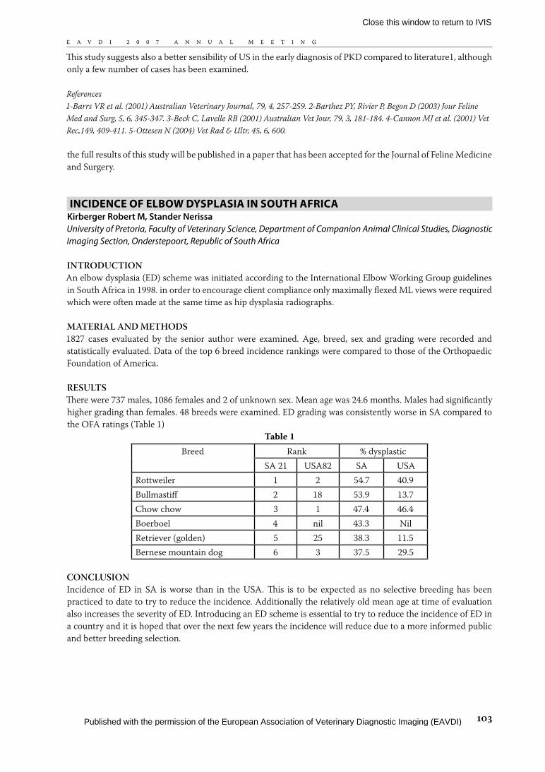
RESULTSThere were 737 males, 1086 females and 2 of unknown sex. Mean age was 24.6 months. Males had significantly higher grading than females. 48 breeds were examined. ED grading was consistently worse in SA compared to the OFA ratings (Table 1)
Table 1Breed Rank % dysplastic
SA 21 USA82 SA USARottweiler 1 2 54.7 40.9Bullmastiff 2 18 53.9 13.7Chow chow 3 1 47.4 46.4Boerboel 4 nil 43.3 NilRetriever (golden) 5 25 38.3 11.5Bernese mountain dog 6 3 37.5 29.5
CONCLUSIONIncidence of ED in SA is worse than in the USA. This is to be expected as no selective breeding has been practiced to date to try to reduce the incidence. Additionally the relatively old mean age at time of evaluation also increases the severity of ED. Introducing an ED scheme is essential to try to reduce the incidence of ED in a country and it is hoped that over the next few years the incidence will reduce due to a more informed public and better breeding selection.
Close this window to return to IVIS
Published with the permission of the European Association of Veterinary Diagnostic Imaging (EAVDI)
�0�
E A V D I 2 0 0 7 A N N U A L M E E T I N G
rAdiOgrAPhyiC STUdy OF diSTAL SESAmOid BONEOF NOrmAL CLAWS iN CATTLE
Meimandi Parizi, A.*; Tadjalli, M.** and Ghanberizadeh M.**** Department of Clinical Sciences, School of Veterinary Medicine, Shiraz University, Shiraz, Iran** Department of Basic Sciences, School of Veterinary Medicine, Shiraz University, Shiraz, Iran*** Graduated in School of Veterinary Medicine, Shiraz University, Shiraz, Iran
Sesamoid bones are typically found in locations where a tendon passes over a joint. Functionally, they act to protect the tendon and to increase its mechanical effect. There is no available any document about radiography of the distal sesamoid bone of cattle. This study was carried out on the radiography of the distal sesamoid bone of cattle with normal claws. 80 distal sesamoid bones of 40 specimens (fore and hind limbs) were collected from Shiraz slaughterhouse, Shiraz, Iran. Standard radiographs of lateromedial, dorsopalmar or dorsoplantar and oblique views were taken from each sample. Radiographic study showed distal sesamoid bone was elliptical shape and its palmar / plantar surface was slightly convex with a blunt sagittal ridge. It’s distal border lies to the last quarter of distal end of middle phalanx and the proximal border lies at the middle of 2nd phalanx. in the cranio- caudal view, the distal sesamoid bone was covered width of middle phalanx. This bone in lateral view was diagonal and its density was close to the middle phalanx. the trabecular patterns of osseous tissue is observeable in the lateral view. the normal radiography of the distal sesamoid helps a clinician to recognize any radiographic changes of the bone in diseases or any abnormal condition in the digital region.
RefeRences1. Baggot DG, Russel AM. Lameness in dairy cattle. Br. Vet. J. 1988; 144: 114-132. 2. Bargai, A. B., Pharr J. W. (1989): Bovine radiography. Iowa State, University Press. pp: 35-50 3. Berry C.R. Pool R.R., Stovers, O’Brien T.R. and koblick P.D. Radiographic/morphologic investigation of a radiolucent cresseent within the flexor central eminence of the navicular bone in thoroughloreds. Am. J. Vet. Res.1992; 53: 1604-1611. 4. Burt JK, Myers VS, Hillmann DJ, Getty R. the radiographic locations of epiphyseal lines in bovine limbs. Am. J. Vet. Med. Assoc. 1968; 152: 168-174.Cited by Getty R. Sisson and Grossman’s. the anatomy of the domestic animals. 5th ed. Philadelphia: WB Saunders Co, 1975; 1: 431, 753-755, 789, 844, 859. 5. Carlson WD. Veterinary radiography. 2nd ed. Philadelphia: Lea and Febiger, 1967; 54-56, 585-591. 6. Doige C.E. Hoffer M.A. Pathological changes in the navicular bone and associated structures of the horse. Can. J. Comp. Med. 1983; 47: 387-395. 7. Getty R. Sisson and Grossman’s the anatomy of the domestic animals. 5th ed. Philadelphia: WB Saunders Co, 1975; 753-755, 789, 844, 859, 1208-1209. 8. Greenough PR, MacCallum FJ, Weaver AD. Lameness in cattle. 2nd ed. Bristol: John Wright and Sons, 1981; 109, 38-39, 174-182, 228-262, 286-294, 328-337. 9. Meimandi-Parizi A, Raddanipour M. Radiological observation of bone disorders of lame cattle. Proceeding of 11th international symposium on disorders ruminants digit and 3rd international conference on bovine lameness. September 3-7, 2000; Parma, Italy 233-241. 10. Meimandi Parizi A, Shakeri M. the abattoir study of radiographic changes in bone and joint of digital region in cattle. Proceeding of 12th international symposium on lameness in ruminants. January 9-13 2002; Orlando Florida 239. 11. O,brien, R. T., Biller D. S. (1996): Clinical applications of radiography and ancillary imaging. Vet.Clin. North Am. (Food Anim. Pract. ). 12, 263-275. 12. Pharr, J. W. (1985): Ancillary diagnostic imaging.Angiography, Ultrasonography, Scintigraphy, and Xeroradiography. Vet.Clin. North Am. (Food Anim. Pract.) 1, 53-56. 13. Wright I.M., Kidd L. and thorp B.H. (1998). Gross histological and histomorphometric features of the navicular bone related structure in the horse. Equine Vet. J. 30: 220-234.
WALL STENT PLACEmENT TO PrEvENT COiLS migrATiONdUriNg iNTrAhEPATiC EmBOLizATiON OF PSS iN A dOg.
Bolen G.*, De Busscher V.*, Cavrenne R.*, Peeters D.**, Dondelinger R.***, Snaps F.** ULg – Faculty of Veterinary Medicine – Clinical Sciences Department – Medical Imaging Section – Liège – Belgium ** ULg – Faculty of Veterinary Medicine – Clinical Sciences Department – Internal Medicine Section – Liège –Belgium *** ULg – Faculty of Medicine – Medical Imaging Section – Liège – Belgium
INTRODUCTIONIntravascular embolization is a minimally invasive technique for treatment of single congenital intrahepatic
Close this window to return to IVIS
Published with the permission of the European Association of Veterinary Diagnostic Imaging (EAVDI)
�0�
E A V D I 2 0 0 7 A N N U A L M E E T I N G
portosystemic shunt (PSS) in dogs. Coils migration is a potentially life-threatening complication of this method. the aim of this poster is to present the use of a wall stent during coils embolization to prevent coils migration.
MATERIAL AND METhODSA five months female irish wolfhound was presented with clinical signs and biochemistry compatible with a PSS. Ultrasonographic examination confirmed a 12mm intrahepatic porto-caval shunt. Vena cava was catheterized via the jugular vein with a. 5F cobra catheter to find the shunt. A trans-shunt portography was done. Due to the large shunt ostium a 24 x 70mm Wall stent-uni endoprothesis® (Boston Scientific, Ireland) was used to prevent coils migration. the wall-stent was placed into the vena cava to cover the shunt ostium. A 3F radiofocus catheter (Terumo, Belgium) was placed into the shunt through the stent. Intravascular coils embolization using Tornado embolization microcoils® (Cook, Denmark) was done. Seventeen microcoils (7x3mm, 8 x 5mm and 10 x 5mm) were placed into the shunt to reduce the shunt outflow.
RESULTS No coil migration was observed with this procedure.
CONCLUSIONSTransvenous coil embolization is one of the techniques for occlusion of intrahepatic PSS. One disadvantage of this technique is coils migration. Vena cava wall-stent placement is an effective method to prevent coils migration during intravascular embolization.
ABdOmiNAL ULTrASONOgrAPhiC FiNdiNgS iN 38 dOgSWiTh BABESiOSiS (BABESiA CANiS CANiS).
Fraga, E; Goicoa, A; Fraga, G; Seoane, A; Barreiro, A.Department of Veterinary Clinical Sciences, Faculty of Veterinary Medicine, University of Santiago de Compostela, E-27002 Lugo, Spain.
INTRODUCTION/PURPOSEBabesia canis canis, is widespread in Galicia (Spain). Babesiosis can involve multiple organs and result in a wide variety of clinical manifestations. This study evaluates ultrasonographic changes of the spleen, liver and kidneys, in dogs naturally infected with Babesia canis.
MATERIALS AND METhODSThirty-eight dogs of different breeds and ages diagnosed with babesiosis were studied. A complete blood count and the main biochemical parameters were obtained to classify them into 2 groups, uncomplicated and complicated babesiosis. Twenty-seven dogs had uncomplicated babesiosis and 11 had complicated babesiosis. Ultrasonographic examination was performed using a MyLab 70 machine with a 3-9 MHz multifrequency transducer. Colour and pulsed Doppler was used to measure the renal resistive indices (RI).
RESULTSin the uncomplicated group 20/27 dogs had splenic lesions, 5/27 had diffuse liver diseases and 9/27 had abnormalities of the renal parenchyma. 10/11 dogs with complicated babesiosis had splenic lesions, 4/11 had liver lesions and 8/11 had lesions in the kidneys. No significant correlation was found in splenic or liver diseases between complicated and uncomplicated groups but significant differences were found in renal diseases. the mean kidney RI was 0,660±0,012 for the uncomplicated group and 0,681±0,023 for the complicated group. No significant differences were found.
DISCUSSION/CONCLUSIONSthe ultrasound investigation showed that in most animals the spleen was affected (91% of the complicated group and 74% of the uncomplicated group) and few dogs had liver lesions (36% and 18% respectively). Abnormalities of the renal parenchyma were present in 73% of the dogs with complicated babesiosis, and in 33% of the animals
Close this window to return to IVIS
Published with the permission of the European Association of Veterinary Diagnostic Imaging (EAVDI)
�0�
E A V D I 2 0 0 7 A N N U A L M E E T I N G
with uncomplicated babesiosis, although this fact remained unnoticed in their clinical exam and in their serum parameters, proving that ultrasound may be useful in the early detection of renal involvement in babesiosis.
1.Burk, RL and Feeney, DA. 2003. Small Animal Radiology and Ultrasonography. A diagnostic Atlas and Text. Third Edition, Saunders, St. Louis, Missouri (USA). 2.Jacobson, LS and Clark, IA. 1994. the pathophysiology of canine babesiosis: new approaches to an old puzzle. J S Afr Vet Assoc. 65 (3): 134-145. 3.Koma, LM; Kirberger, RM; Leisewitz, AL; Jacobson, LS; Becker, PJ and Bland Van den Berg, P. 2005. Comparison of effects of uncomplicated canine babesiosis and canine normovolaemic anaemia on abdominal splanchnic Doppler characteristics--a preliminary investigation. J S Afr Vet Assoc. 76 (3): 138-145. 4.Novellas, R; Espada, Y and Ruiz de Gopegui, R. 2007. Doppler ultrasonographic estimation of renal and ocular resistive and pulsatility indices in normal dogs and cats. Vet Radiol Ultrasound. 48 (1): 69-73. 5.Nyland, TG and Matoon, JS. Small Animal Diagnostic Ultrasound. Second Edition, Saunders, Philadelphia (USA).
ULTrASONOgrAPhiC ASSESSmENT OF NEW BONE FOrmATiONdUriNg diSTrACTiON iN A rABBiT mOdEL
Savet.A1. , DVM, Huguet.T1., DVM, Sailhan.F2, MD, Chousta.A2, MD, Viguier.E,1 DVM, PhD, Dipl ECVS1 UMR 3090 Biomatériaux et biocompatibilité des matériaux médicaux. Ecole Nationale Vétérinaire de Lyon, 1,
Avenue BOURGELAT, 69280 MARCY L’ETOILE. France2 Hôpital Debrousse. 29 rue des Sœurs Bouvier 69005 LYON. FRANCE
INTRODUCTIONthe goal of this study is to evaluate normal sonographic aspect of new bone production during lengthening in a rabbit model of tibiae lengthening.
MATERIALS AND METhODS12 sub-mature New-Zealand male white rabbits, body weight 2.0-3.0 kg were used for this study. An unilateral external fixator (ORTHOFIX® M-103) 7 cm length is placed on lateral side of tibiae, after a mid-diaphyseal tibial osteotomy. After a 7 days latency period, a 2.1cm distraction was carried out at a rate of 0.5 mm twice a day for 21 days. Rabbits were sacrified at 28, 35, 42 and 49 days after osteotomy. Radiographic and ultrasonographic examinations were made weekly from the first week of distraction to the sacrifice. All examinations were interpreted by two independents blinded observers according to a grading previously used in similar studies. Evaluations included distance between native bone ends, misalignment of bony segments, aspect and maturity of new callus, and signs of complication.
RESULTSDistraction gap first appears as a sonolucent defect between the two ends of cortical bone. the distance between native bone ends progressively decreases during consolidation while echogenicity increased at the distraction site. After 1-2 weeks, few disorganized foci are seen in the distraction gap. After 3-4 weeks, these areas become more aligned along the long axis of the bone, and increase in size and number until they coalesce as echodense bone at 7-8 weeks following distraction. on a transversal view, new bone formation appears as an “electric cable”. A complication appears on a rabbit as a sonolucent area round a pin and a well-defined fluid collection in the subcutaneous tissues. Bone cysts can also been detected by sonography. Those complications were not detected on standard radiography.
CONCLUSIONUsing ultrasonography can considerably decrease the X-ray exposure during limb lengthening monitoring and provides different information in earliest stages of new bone formation. It can also monitor the quality of new bone formation showing complications, and the distraction rate can thus be optimized.
1. Donnan, L.T., et al., Radiographic assessment of bone formation in tibia during distraction osteogenesis. J Pediatr Orthop. 200; 22(5): 645-51 2. Eyres, K.S., M.J. Bell, and J.A. Kanis, Methods of assessing new bone formation during limb lengthening. Ultrasonography, dual energy X-ray absorptiometry and radiography compared. J Bone Joint Surg Br, 1993;
Close this window to return to IVIS
Published with the permission of the European Association of Veterinary Diagnostic Imaging (EAVDI)
�0�
E A V D I 2 0 0 7 A N N U A L M E E T I N G
75(3):358-64 3. Eyres, K.S., M.J. Bell, and J.A. Kanis, New bone formation during leg lengthening. Evaluated by dual energy X-Ray absorptiometry J Bone Joint Surg Br, 199; 75(1):96-106 4. Hugues, T.H and al. Imaginig in bone lengthening. A review. Clin Orthop Relat Res, 1994(308):50-3 5. Li, G., et al., Bone consolidation is enhanced by rhBMP-2 in a rabbit model of distraction osteogenesis. J Orthop Res, 200; 20(4):779-88 6. Yasko AW, Lane JM, Fellinger EJ, Rosen V, Wozney JM, Wang EA. the healing of segmental bone defects, induced by recombinant human bone morphogenetic protein (rhBMP-2). A radiographic, histological, and biomechanical study in rats. J Bone Joint Surg Am. 1992 Jun;74(5):659-70. Erratum in: J Bone Joint Surg Am 1992; 74(7):1111.
imAgiNg OF TWO TrAUmA CASES iN LOggErhEAd SEA TUrTLE(CArETTA CArETTA)
Vignoli M.1,2, Nardini G.2, Bielli M.2, Rossi F.1, Terragni R.1, Leone V.1,2
1 Clinica Veterinaria dell’Orologio, Sasso Marconi (BO)- Italy2 Exotic Veterinary Team - Italy
in the Mediterranean Sea Caretta caretta is the most common sea turtle and trauma is the prevalent disease diagnosed in rescued turtles1,2. Two C. caretta (Titania 71 kg and Sole 13 kg) were admitted at the Fondazione Cetacea Turtle Hospital; both turtles suffered head trauma with bone fractures and skin wounds resulting from boat collision. Survey radiographs excluded bony lesions of the distal left forelimb of Titania, but showed comminuted fracture of the left maxilla and jugal bones3. the CT scans were taken under anesthesia (propofol 8 mg/kg i.v.) and revealed multiple cranial fractures in both individuals. Ultrasonography of the brain of Titania was possible trough the fractures of the skull. Within the celomatic cavity several eggs were visible with mineralized shell and distal shadowing. Both turtles underwent low level laser therapy (LLLT) for two months. CT scans were repeated two months later. in case of Titania the CT scan showed fibrous and osseous callus formation at fracture sites and the turtle has been successfully released one month later. At the moment there are no reports on LLLT in reptiles. the clinical improvements in tissue regeneration and wound healing in other species is documented4,5. Since LLLT may help in cases of neurological deficit6, Sole is still undergoing this therapy. the results we have had on these sea turtles are encouraging and we think that LLLT had a role in shortening the healing process. CT was very useful to assess the severity and extent of the skull fractures and healing process.
1) Schofield G. and Kopsida H 1999. Head Injury Rehabilitation of Sea Turtles; the positive Side of a Negative Conundrum. pp.41-43 Proc. 19th Ann. Symp. Sea Turt. Cons. Biol. 2-6March South Padre Isl. Texas, USA. 2) Nardini G., Bielli M., Scaravelli D., Vignoli M. New Technologies Helping Chelonian Conservation: Computed Tomography (CT) and Laser (Lllt) Therapy. International Conference, Parco Le Navi, 2006. 3) Wyneken J. the anatomy of the sea turtles. Miami 2001, pp.8-25. 4) Ihsan F.R. 2005. Low-level laser therapy accelerates collateral circulation and enhances microcirculation. Photomed Laser Surg. 2005; 23(3) pp.289-294. 5) Gal P., Vidinski B., Toporcer T., Mokry M., Mozes S., Langauer F., Sabo J. 2006. Histological assessment of effect of laser irradiation on skin wound healing in rats. Photomed laser Surg. 2006; 24(4) pp.480-488. 6) Detaboada L, Ilic S, Leichliter-Martha S, Oron U, Oron A, Streeter J. 2006. Transcranial application of low-energy laser irradiation improves neurological deficits in rats following acute stroke. Lasers Surg Med. 2006;38(1) pp.70-3.
iNvESTigATiON ON ThE PhArmACOdyNAmiCS OF TWOdiFFErENT PSyChOPhArmACA iN ThE dOg’S BrAiN WiTh SPECT
Vermeire Simon1, Audenaert Kurt2, Vandermeulen Eva1, Peremans Kathelijne1
1 Ghent University, Faculty of Veterinary Medicine, Department of Medical Imaging, Ghent, Belgium2 University Hospital, Faculty of Medicine, Department of Psychiatry and Medical Psychology, Ghent, Belgium
INTRODUCTIONCurrently, certain behavioural disorders in dogs are treated with psychopharmaca derived from human psychiatry. Many act on the serotonin system. Selective serotonin reuptake inhibitors (SSRIs), such as S-citalopram, are used to elevate the synaptic serotonin by blocking the serotonin transporter (SERT). Antipsychotic drugs, such
Close this window to return to IVIS
Published with the permission of the European Association of Veterinary Diagnostic Imaging (EAVDI)
�0�
E A V D I 2 0 0 7 A N N U A L M E E T I N G
as the serotonin-2A receptor antagonist pipamperon, are used to act on the serotonin-2A receptor.the aim of this study was to evaluate the pharmacodynamics of both serotonergic psychopharmaca in the dog’s brain.
MATERIALS AND METhODSS-Citalopram (SSRI):Two dogs (female, age 2y and 6y) were included.123I-beta-CIT SPECT was used to image the SERT.Scans were obtained in blank conditions and after IV administration of S-citalopram.Acquisition was performed 3 hours after injection of the tracer.PipamperonThree female dogs (female, age 8y) were included.[123I]-R91150 SPECT was used to evaluate the 5-HT2A receptor binding index.Scans were obtained in blank conditions and after oral administration of 0,25ml or 0,5ml pipamperon.Acquisition was performed 90 minutes after injection of the tracer.All acquisitions were performed with a triple headed gamma camera (Trionix LEHR parallel hole collimators).
RESULTSAdministration of citalopram prior to the tracer showed a decreased binding of 123I-beta-CIT to the SERT, thereby proving the effective blocking of SERT.Administration of pipamperon prior to the tracer showed a decreased binding of [123I]-R91150 to the5-HT2A receptor, thereby proving the effective blocking of the serotonin-2A receptors.
DISCUSSION-CONCLUSIONThis study demonstrates the feasibility to investigate the mode of action of psychopharmaca with in vivo imaging in dogs.
(Peremans et al.) (Peremans et al.) (Stengler-Wenzke et al.)
RefeRence listPeremans, K., et al. “Regional binding index of the radiolabeled selective 5-HT2A antagonist 123I-5-I-R91150 in the normal canine brain imaged with single photon emission computed tomography.” Vet.Radiol.Ultrasound 44.3 (2003): 344-51.Peremans, K., et al. “the effect of citalopram hydrobromide on 5-HT2A receptors in the impulsive-aggressive dog, as measured with 123I-5-I-R91150 SPECT.” Eur.J.Nucl.Med.Mol.Imaging 32.6 (2005): 708-16.Stengler-Wenzke, K., et al. “Serotonin transporter imaging with [123I]beta-CIT SPECT before and after one year of citalopram treatment of obsessive-compulsive disorder.” Neuropsychobiology 53.1 (2006): 40-45.
rAdiOgrAPhiC ATLAS OF OSTEOArThiTiS iN ThE rABBiT EXPErimENTAL mOdEL kNEE jOiNT
C. Boulocher, E. Viguier, M.E. Duclos, T. Roger, E. VignonEcole Nationale Vétérinaire de Lyon, Département des Animaux de Compagnie UMR MA 3990
ObjECTIvESTo create a radiographic atlas for grading knee joint osteoarthritis (OA) in the rabbit experimental model of OA and compare the radiographic and macroscopic grades.
METhODSin vivo digital radiographs of the left knee of 10 control and 40 operated rabbits were performed at 5 months.Two blinded observers graded the osteophytes with a 4 grades scale for the medial femoro-tibial compartment and a 3 grades scale for the femoral trochlea. Joint space narrowing and subchondral cysts were graded with a 3 grades scale. Radiography and final macroscopy were compared.
Close this window to return to IVIS
Published with the permission of the European Association of Veterinary Diagnostic Imaging (EAVDI)
�0�
E A V D I 2 0 0 7 A N N U A L M E E T I N G
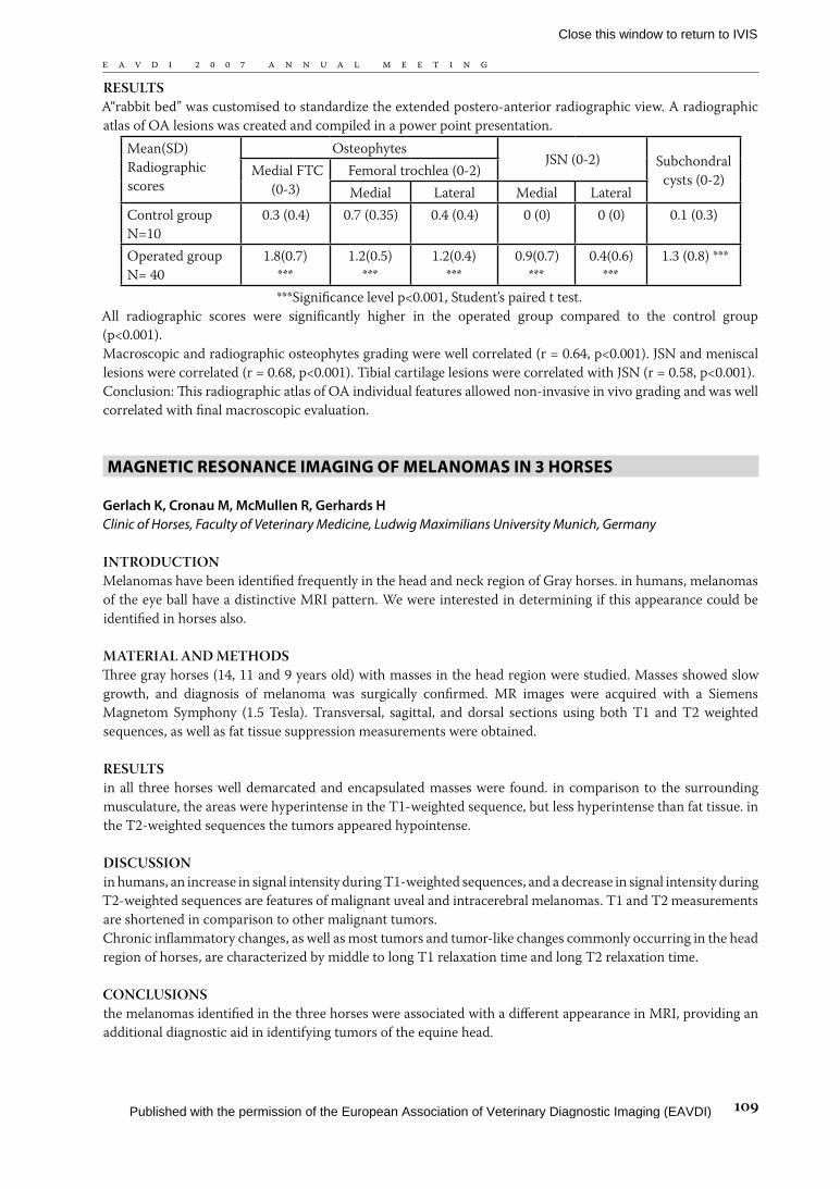
RESULTSA“rabbit bed” was customised to standardize the extended postero-anterior radiographic view. A radiographic atlas of OA lesions was created and compiled in a power point presentation.
Mean(SD) Radiographic scores
OsteophytesJSN (0-2) Subchondral
cysts (0-2) Medial FTC (0-3)
Femoral trochlea (0-2)Medial Lateral Medial Lateral
Control group N=10
0.3 (0.4) 0.7 (0.35) 0.4 (0.4) 0 (0) 0 (0) 0.1 (0.3)
Operated group N= 40
1.8(0.7)***
1.2(0.5)***
1.2(0.4)***
0.9(0.7)***
0.4(0.6)***
1.3 (0.8) ***
***Significance level p<0.001, Student’s paired t test.All radiographic scores were significantly higher in the operated group compared to the control group (p<0.001).Macroscopic and radiographic osteophytes grading were well correlated (r = 0.64, p<0.001). JSN and meniscal lesions were correlated (r = 0.68, p<0.001). Tibial cartilage lesions were correlated with JSN (r = 0.58, p<0.001).Conclusion: This radiographic atlas of OA individual features allowed non-invasive in vivo grading and was well correlated with final macroscopic evaluation.
mAgNETiC rESONANCE imAgiNg OF mELANOmAS iN 3 hOrSES
Gerlach K, Cronau M, McMullen R, Gerhards HClinic of Horses, Faculty of Veterinary Medicine, Ludwig Maximilians University Munich, Germany
INTRODUCTIONMelanomas have been identified frequently in the head and neck region of Gray horses. in humans, melanomas of the eye ball have a distinctive MRI pattern. We were interested in determining if this appearance could be identified in horses also.
MATERIAL AND METhODSThree gray horses (14, 11 and 9 years old) with masses in the head region were studied. Masses showed slow growth, and diagnosis of melanoma was surgically confirmed. MR images were acquired with a Siemens Magnetom Symphony (1.5 Tesla). Transversal, sagittal, and dorsal sections using both T1 and T2 weighted sequences, as well as fat tissue suppression measurements were obtained.
RESULTSin all three horses well demarcated and encapsulated masses were found. in comparison to the surrounding musculature, the areas were hyperintense in the T1-weighted sequence, but less hyperintense than fat tissue. in the T2-weighted sequences the tumors appeared hypointense.
DISCUSSIONin humans, an increase in signal intensity during T1-weighted sequences, and a decrease in signal intensity during T2-weighted sequences are features of malignant uveal and intracerebral melanomas. T1 and T2 measurements are shortened in comparison to other malignant tumors.Chronic inflammatory changes, as well as most tumors and tumor-like changes commonly occurring in the head region of horses, are characterized by middle to long T1 relaxation time and long T2 relaxation time.
CONCLUSIONSthe melanomas identified in the three horses were associated with a different appearance in MRI, providing an additional diagnostic aid in identifying tumors of the equine head.
Close this window to return to IVIS
Published with the permission of the European Association of Veterinary Diagnostic Imaging (EAVDI)
��0
E A V D I 2 0 0 7 A N N U A L M E E T I N G
CErviCAL myELOPAThy SECONdAry TO CONgENiTAL iNCOmPLETE OSSiFiCATiON OF ThE dOrSAL LAmiNA OF ThE ATLAS: CASE rEPOrT ANd LiTErATUrE rEviEW
Owen MC, Davis SH & Worth AJVeterinary Teaching Hospital Institute of Veterinary, Animal & Biomedical Sciences Massey University, New Zealand
INTRODUCTIONCongenital abnormalities of the cranial cervical vertebrae in dogs are only occasionally described in veterinary medicine, and have involved malformation of the dens, combined occipitoatlantoaxial malformations or occipital dysplasia. Isolated C1 congenital abnormalities are rare and may not produce neurological disease unless the abnormality predisposes the spinal cord to injury.
MATERIALS & METhODSA thirteen-week old Wirehaired Fox Terrier was presented to the hospital with acute neurological deficits following a fall. Radiographs showed a widened atlantoaxial distance, which did not alter during flexion views, and a suggestion of a deficient dorsal lamina to C1, but the presence of normal transverse processes. the dens of C2 appeared normal. A CT was performed which showed the absence of the dorsal laminae of the atlas.
RESULTSSurgical stabilisation was considered, but conservative therapy of a neck brace and cage rest has returned the dog to normal neurological function.
DISCUSSIONthe atlas has three separate ossification centres – the body (ventral arch) and one on each side combining the lateral arches and the transverse processes and meets in the midline dorsally to form the dorsal lamina. the abnormality described here was unusual in that it involved only a portion of the dorsal lamina and did not include the transverse processes. C1 congenital vertebral abnormalities are rare in both human and veterinary medicine. They appear to predispose patients to myelopathies following often minor trauma. This case is unusual in that its appearance does not follow the normal ossification pattern of the developing atlas.
Bailey CS, Morgan JP. Congenital Spinal Malformations. Vet clinics of NA: small animal practice 1992;22(4):985–1015. Watson AG, Evans HE & de Lahunta A. Ossification of the Atlas-Axis Complex in the Dog. Anat.Histol. Embryol. 1986;15:122-138
mdCT ATTENUATiON vALUES OF ThE LivEr iN CANiNEPiTUiTAry-dEPENdENT hyPErAdrENOCOrTiCiSm
Bertolini G1, DVM, Borsetto A1, DVM, Furlanello T2, DVM, Dip. ECVCP and Caldin M2, DVM, Dip. ECVCP1 ’San Marco’ Private Veterinary Clinic, Padova, Italy2 ’San Marco’ Private Veterinary Laboratory, Padova, Italy
INTRODUCTIONHepatic steatosis may occur in association with canine pituitary dependent hyperadrenocorticism (PDH). in humans, a liver-to-spleen (L/S) computed tomography (CT) attenuation values (CT number) ratio <1 is indicative of hepatic steatosis in Cushing syndrome.
MATERIALS AND METhODSLiver and spleen CT numbers of dogs having clinical, clinicopathological, imaging evidence of PDH, and histopathologic diagnosis of liver steatosis were recorded and compared with data of dogs without any clinical, clinicopathological, and imaging evidence of liver/spleen disease. All dogs underwent multidetector CT (MDCT) examination. Three 100 mm2 regions of interest (ROIs) were drawn on the liver and one on the spleen, in non-
Close this window to return to IVIS
Published with the permission of the European Association of Veterinary Diagnostic Imaging (EAVDI)
���
E A V D I 2 0 0 7 A N N U A L M E E T I N G
enhanced 2D multiplanar reformatted images. Care was taken not to include any vessels in the ROIs. the mean values of the three liver ROIs and the splenic ROI were related (L/S).the standard deviation of each ROI was also recorded, as well as the liver CT features.
RESULTSTwenty dogs (10 normal dogs and 10 with PDH) were recruited for the study (13 males, 7 females, 10 years median age, 10 Kg median body weight). the mean values of the liver CT number were 62,04 ± 5,33 and 60,76 ± 5,56 respectively in normal dogs and in dogs with PDH. the L/S was 0,99 in normal dogs and 0,98 in dogs with PDH. Four dogs presented diffuse liver hypodensity and six dogs had focal hypodense lesions.
DISCUSSION-CONCLUSIONSin this preliminary study, the CT characteristics of the liver in canine PDH were determined.
RefeRences• Bydder G M, Chapman R W, Harry D, Bassan L, Sherlock S, Kreel L, Computed tomography attenuation values in fatty
liver, J. Comput. Tomogr., 1981 Mar; 5 (1):33-35.• Limanond P, Raman S S, Lassman C, Sayre J, Ghobrial R M, Busuttil R W, Saab S, Lu D S K, Macrovescicular Hepatic
Steatosis in Living Related Liver Donors: correlation between CT and histologic findings, Radiology 2004; 230:276-280.• Piekarski J, Goldberg H I, Royal S A, Axel L, Moss A A, Difference between liver and spleen CT numbers in the normal
adult: its usefulness in predicting the presence of diffuse liver disease. Radiology,1980; 137 (3): 727-729.• Ricci C, Longo R, Gioulis E, Bosco M, Pollesello P, Masutti F, Croce L S, Paoletti S, De Bernard B, Tiribelli C, Dalla
Palma L, Noninvasive in vivo quantitative assessment of fat content in human liver, J.Hepatol.,1997 Jul;27 (1):108-113.• Rockall A G, Sohaib S A, Evans D, Kaltsas G, Isidori A M, Monson J P, Besser G M Grossman A B and Reznek R H,
Hepatic steatosis in Cushing’s syndrome: a radiological assessment using computed tomography. European Journal of Endocrinology 2003;149:543-548.
rAdiOgrAPhiC FEATUrES OF PULmONAry EdEmAASSOCiATEd WiTh miTrAL rEgUrgiTATiON iN dOgS
Diana A., Sanacore A., Guglielmini C1., Pivetta M., Cepparulo A., Cipone M.University of Bologna, Faculty of Veterinary Medicine, Veterinary Clinical Department, Ozzano dell’Emilia (BO), Italy 1 University of Teramo, Department of Veterinary Clinical Sciences, Teramo, Italy
INTRODUCTIONAn asymmetric distribution of pulmonary edema (PE) has been documented in humans with mitral regurgitation (MR). On the contrary, few studies focused on the distribution of PE in dogs with MR. the purpose of this study was to evaluate the distribution of PE in dogs with MR.
MATERIALS AND METhODSThoracic radiographs of dogs diagnosed with MR from January 1998 to March 2007 were reviewed. Inclusion criteria were availability of 2 orthogonal projections and good quality thoracic radiographs. Evaluation of shape and dimension of the cardiac silhouette, including calculation of the vertebral heart size (VHS), pulmonary vasculature, and aspect and distribution of any interstitial and/or alveolar pattern was performed.
RESULTSPulmonary edema was diagnosed in 44 (34%) out of 130 dogs with MR. the VHS (mean ± SD) of dogs with PE was 12.25 ± 2.05. Vascular congestion was observed in 34/44 dogs (77%). An interstitial pattern was evident in all dogs with PE and a combined interstitial-alveolar was found in 9 dogs (20%). A symmetric distribution of PE was found in 28/44 dogs (64%). An asymmetric distribution of PE was evidenced in 16 dogs (36%). Unique involvement of the right caudal lung lobe was observed in 13 dogs (30%), while unique involvement of the middle and right caudal lung lobe, the left caudal lobe, and the right apical and caudal lobes was found in 1 dog (2%), respectively.
Close this window to return to IVIS
Published with the permission of the European Association of Veterinary Diagnostic Imaging (EAVDI)

���
E A V D I 2 0 0 7 A N N U A L M E E T I N G
DISCUSSION-CONCLUSIONUnique involvement of the right caudal lung lobe may be frequently observed in dogs with PE due to MR.
RefeRencesSuter PF and Lord A, Text atlas. Thoracic Radiography. Thoracic disease of the dog and the cat. Wettswill (CH), Suter PF Eds, 1984: 558-567; Schynder P et al., AJR 1993;161:33-36; Grenon H and Bilodeau S, Can Assoc Radiol J 1994;45:97-100.
mOrPhOmETriC ANALySiS OF ThE CAUdAL FOSSA iN CAvALiEr kiNg ChArLES SPANiEL
Carrera, I., Dennis, R*., Sullivan, M.Companion Animal Clinical Science, University of Glasgow, Glasgow, UK
* Animal Health Trust, Diagnostic Imaging, Newmarket, UK
INTRODUCTIONin humans, Chiari type I malformation is a disorder of the para-axial mesoderm characterized by an underdevelopment of the posterior cranial fossa and an overcrowding of the normally developed hindbrain. This anomaly is a leading cause of syringohydromyelia (SHM) and can occur in association with osseous abnormalities at the cranio-vertebral junction. A similar disorder to human Chiari type I malformation occurs in Cavalier King Charles Spaniel (CKCS) dogs. the aim of this study was to determine the dimensions of the caudal fossa and signs of occipital dysplasia in CKCS by morphometric studies.
MATERIALS AND METhODSSeventy CKCS patients were selected in this study. Multiple morphometric measurements (including linear, angular and area measures) were made using midline sagittal T2-weighted MRI scans. the results were compared to a control group which consisted of forty Labradors, and forty mesatephalic Spaniel breeds (Springer and Cocker Spaniel). Several specific MRI findings were also recorded in CKCS patients, which included amongst others the presence of SHM, displacement of the cerebellum, and signs of occipital dysplasia.
RESULTSStatistically significant differences were detected between CKCS patients and the control group (p=0.01). All measurements indicated that both the depth and the surface area of the caudal fossa were smaller in CKCS. When dividing CKCS into patients with SHM or without SHM, the former showed even more dramatic differences to the control group.
CONCLUSIONThis study suggests that the bony components of the caudal fossa in CKCS are not fully developed, being more severe in CKCS with SHM.
Aydin, S., H. Hanimoglu, et al. (2005). “Chiari type I malformations in adults: a morphometric analysis of the posterior cranial fossa.” Surg Neurol 64(3): 237-41; discussion 241. Churcher, R. K. and G. Child (2000). “Chiari 1/syringomyelia complex in a King Charles Spaniel.” Aust Vet J 78(2): 92-5. Karagoz, F., N. Izgi, et al. (2002). “Morphometric measurements of the cranium in patients with Chiari type I malformation and comparison with the normal population.” Acta Neurochir (Wien) 144(2): 165-71; discussion 171. Lu, D., C. R. Lamb, et al. (2003). “Neurological signs and results of magnetic resonance imaging in 40 cavalier King Charles spaniels with Chiari type 1-like malformations.” Vet Rec 153(9): 260-3. Marin-Padilla, M. (1991). “Cephalic axial skeletal-neural dysraphic disorders: embryology and pathology.” Can J Neurol Sci 18(2): 153-69. Rusbridge, C. (2005). “Neurological diseases of the Cavalier King Charles spaniel.” J Small Anim Pract 46(6): 265-72. Rusbridge, C., D. Greitz, et al. (2006). “Syringomyelia: current concepts in pathogenesis, diagnosis, and treatment.” J Vet Intern Med 20(3): 469-79. Rusbridge, C., P. Knowler, et al. (2005). “Inherited occipital hypoplasia/syringomyelia in the cavalier King Charles spaniel: experiences in setting up a worldwide DNA collection.” J Hered 96(7): 745-9. Rusbridge, C. and S. P. Knowler (2003). “Hereditary aspects of occipital bone hypoplasia and syringomyelia (Chiari type I malformation) in cavalier King Charles spaniels.” Vet Rec 153(4): 107-12. Rusbridge, C. and S. P. Knowler (2006). “Coexistence of occipital
Close this window to return to IVIS
Published with the permission of the European Association of Veterinary Diagnostic Imaging (EAVDI)
���
E A V D I 2 0 0 7 A N N U A L M E E T I N G
dysplasia and occipital hypoplasia/syringomyelia in the cavalier King Charles spaniel.” J Small Anim Pract 47(10): 603-6. Rusbridge, C., J. E. MacSweeny, et al. (2000). “Syringohydromyelia in Cavalier King Charles spaniels.” J Am Anim Hosp Assoc 36(1): 34-41. Stovner, L. J., U. Bergan, et al. (1993). “Posterior cranial fossa dimensions in the Chiari I malformation: relation to pathogenesis and clinical presentation.” Neuroradiology 35(2): 113-8.
FirST ThrEE yEArS OF CT EXAmiNATiON iN A PrivATE PrACTiCE iN iTALy
Vignoli M., Laganga P., Rossi F., Terragni R..Clinica Veterinaria dell’Orologio – Sasso Marconi (BO) – Italy
AIMTo present the CT caseload handled at the Clinica Veterinaria dell’Orologio during the first three years of activity.
MATERIAL AND METhODSthe CT studies were performed and read by two radiologists (VM, RF) using a spiral GE Pro Speed Power unit.
RESULTS1051 animals were scanned, in some cases two different body areas were examined so that a total of 1210 CT studies were performed. 772 (73.45 %) cases were referred from other Clinics, while 279 (26.54%) came to our Clinic as first opinion. of a total of 1051 animals, 921 (87.63%) were dogs, 117 (11.13%) cats, 4 (0.38%) turtles, 4 (0.38%) rabbits, 2 (0.19%) aspis aspis, 1 (0.09%) prairie dog, 1 (0.09%) ferret and 1 (0.09%) wolf. of the 1210 CTs examinations 348 (28.76%) were head examinations, 295 neurocranium and 53 splanchnocranium (46 noses/retrobulbar spaces, 4 temporomandibular joints and 3 CT-guided radiofrequency treatments), 320 (26.44%) spines, 295 (24.37%) chests, 133 (10.99%) abdomens (in 76 cases combined thorax and abdomen scan was taken for tumor stadiation), 72 (5.95%) limbs, 24 (1.98%) pelvis, 18 (1.48%) necks (soft tissues). of 320 spine examinations 52 were myelo-CT, performed if the neurological localisation was uncertain or more then one lesion was suspected. in 980 cases (285 spines, 283 heads, 183 chests, 75 limbs, 74 thorax-abdomens, 45 abdomens, 23 pelvis and 12 necks) abnormalities were found. A total of 156 studies were considered normal.
CONCLUSIONSin our clinic, neurological cases represented more than one half of the CT examinations performed in the first three years of activity.
hEmOdyNAmiC ALTErATiONS CAUSEd By 3 TyPESOF iNTrAvENOUS CONTrAST mEdiA iN ANESThETizEd CATS
Rachel E. Pollard DVM, PhD, Sarah M. Puchalski DVM, Peter J. Pascoe BVSc.Department of Surgical and Radiological Sciences University of California, Davis School of Veterinary Medicine, Davis, CA 95616
INTRODUCTIONthe purpose of this retrospective study was to determine the incidence of alterations in physiological parameters such as heart rate (HR) and peak systolic blood pressure (BP) associated with intravenous (IV) administration of ionic iodinated, non-ionic iodinated and gadolinium contrast agents in anesthetized cats.
MATERIALS AND METhODSUC Davis SVM records were searched for cats receiving IV ionic iodinated (Conray), non-ionic iodinated (Isovue) and gadolinium (Magnevist) contrast agents during anesthesia between 4/2005-4/2006. for both control and study groups, HR and BP were recorded at 5-minute intervals for 20 minutes. Baseline data for each cat was defined as the measurement immediately prior to contrast administration in the study group and15 minutes
Close this window to return to IVIS
Published with the permission of the European Association of Veterinary Diagnostic Imaging (EAVDI)
���
E A V D I 2 0 0 7 A N N U A L M E E T I N G
after the beginning of the MRI in the control group. A ≥15% change in HR or BP was considered a response. the percent change from baseline was calculated for each time point.
RESULTS15/59 (25%) of cats receiving Conray had a response in HR at 1 or more time points. 33/60 (55%) of cats receiving Conray had a response in BP. 4/11 (36%) of cats receiving Isovue had a response in HR while 6/12 (50%) had a response in BP. 22/81 (27%) of cats receiving Magnevist had a response in HR while 33/83 (40%) had a response in BP. 3/34 (9%) of control cats had a response in HR while 7/35 (20%) had a response in BP.
DISCUSSION/CONCLUSIONSAdministration of IV iodine and gadolinium contrast agents is more commonly associated with a ≥15% change in HR and/or BP than control cats.
COmPUTEd TOmOgrAPhiC ANATOmy OF ThE EqUiNEmETACArPOPhALANgEAL jOiNT
Vanderperren K1, Ghaye B2, Hoegaerts M3, Snaps F4, Saunders JH1
1 Ghent University, Veterinary Faculty, Department of Medical Imaging, Merelbeke, Belgium2 University of Liège, University Hospital, Department of Medical Imaging, Liège, Belgium3 Veterinary Clinic “Visdonk”, Roosendaal, the Netherlands4 University of Liège, Veterinary faculty, Department of Clinical Sciences, Liège, Belgium
INTRODUCTIONComputed tomography (CT) has become an important diagnostic imaging modality in the diagnosis of equine musculoskeletal disorders1. the aim of this study was to describe a detailed CT reference of the normal equine metacarpophalangeal (MCP) joint.
MATERIALS AND METhODSCT of the MCP joint was performed on 4 normal equine cadaver forelimbs. in one of these limbs, CT was also achieved after intra-articular injection of 30 mL contrast (40 mg iodine/mL). One-mm thick transverse slices were made (120 kV, 160 mA) and sagittal and dorsal planes were reformatted with a slice thickness of 2 mm. the CT images were matched with corresponding anatomical slices.
RESULTSthe third metacarpal bone (condyles, sagittal ridge), proximal sesamoid bones and proximal phalanx could be clearly visualised. the common digital extensor tendon, accessory digital extensor tendon, lateral digital extensor tendon, superficial digital flexor tendon (including manica flexoria), deep digital flexor tendon, suspensory ligament (including its attachment), extensor branches of the suspensory ligament, collateral ligaments, straight, oblique and cruciate distal sesamoidean ligaments, palmar ligament, annular ligament and joint capsule could be obviously seen. the collateral sesamoidean ligaments and short distal sesamoidean ligaments could be localized but not always clearly identified, while the metacarpointersesamoidean ligament could not be identified. the cartilage of the MCP joint could be evidently assessed on the post-contrast sequence.
DISCUSSION-CONCLUSIONCT of the equine MCP joint can be of great value when radiography and ultrasonography are inconclusive. the images of this study will serve as a CT reference of the equine MCP joint.
1Bienert A, Stadler P. Computed tomographic examination of the locomotor apparatus of horses-a review. Pferdeheilk 2006;22:218-226.
Close this window to return to IVIS
Published with the permission of the European Association of Veterinary Diagnostic Imaging (EAVDI)
���
E A V D I 2 0 0 7 A N N U A L M E E T I N G
imAgiNg diAgNOSTiC OF gASTriC ULCErS iN dOgS - CASE rEPOrT
Prof. Dr Vanja KrsticFaculty of Veterinary Medicine, Department of Equine and Small Animal disease, Belgrade, SerbiaProf. Dr Nikola KrsticFaculty of Veterinary Medicine, Department of Radiology and PhysiotherapyAssist. Dr Darko MarinkovicFaculty of Veterinary Medicine, Department of Pathology
A 10-year old, mixed breed bitch was found upon clinical examination to have suffered of lack of appetite and frequent vomiting of a whitish foamy content with traces of blood. the mucosae were pale, body temperature 38oC, heart and breathing rate 130/min and 20/min, respectively. Erythrocyte count was 5.5 x 10¹²/L and leukocyte count was 20 x 109.Biochemical parameters were within the physiological limits.
METhODSEndoscopic examination was carried out with the Xion® device. Ultrasonography was carried out by Aloka 2000® device with 3.5MHz and 7.5 MHz probes. Radiography was performed with a Selenos 4® device with a focal length of 60-70cm and appropriate exposure. Histological samples were examined under the light microscope.
RESULTSEndoscopic examination revealed hyperemia and oedema of the gastric mucosa with an evident 4 cm ulcerous defect filled with hemorrhagic content.Ultrasound examination revealed an anechogenic shadow of the fundus wall 9.1 mm thick. in the lumen, a hypoechogenic defect of the wall, filled with a coagulum was prominent.the barium contrast roentgenogram showed dysplastic mucosa on the cranioventral gastric margin. the mucosal defect, filled with barium contrast had an irregular bud-shape silhouette. the gastric caudal margin displayed a prominent proliferation in the lumen mucosa.Upon histological examination, it was established that the defect reached the tunica muscularis with proliferating connective tissue in the ulcus floor. Tissue samples taken from the margins of the ulcus contained an infiltrate which consisted of lymphocytes, plasmocytes, and macrophages.
SCLErOThErAPy WiThOUT drAiNAgE OF 95% EThANOLFOr TrEATmENT OF ANEUriSmAL BONE CyST iN A dOg
Agut A, Soler M, Murciano J, Belda E, Guillem R.University of Murcia. Departamento de Medicina y Cirugía Animal. Murcia. Spain
INTRODUCTIONAn aneurysmal bone cyst (ABC) is an expansible osteolytic lesion with a thin wall and blood-filled. ABC is rare in dogs. Amputation, bloc resection, and curettage with cancellous bone graft have been reported as effective treatments. To the best of the authors´ knowledge the treatment of an ABC with percutaneous injection of a fibrosing agent has not been reported in the dog. We describe an ABC in the ulna of a dog treated by sclerotherapy.
MATERIAL AND METhODSA three-month-old female, St Bernard was referred with left forelimb lameness and painful swelling of the antebrachium region. On radiographs, a geographic lysis of the dyaphises of the ulna, with thinning of the cortices and several septa dividing the lytic structure into chambers were noted. Brachial arteriography was performed. A cyst-like structure with hypoechoic fluid content was seen by ultrasonography. A sanguineous fluid was obtained from it and then analysed. On the basis of theses findings, ABC diagnosis was made. Under ultrasonographic guidance, cystic fluid was aspirated and then a volume of 95 % ethanol equivalent to 25 % of the aspirated volume was injected into the cavity and left there. Radiographic examinations were made at 15 days, 1, 3, 6, 9 and 12 months after the treatment.
Close this window to return to IVIS
Published with the permission of the European Association of Veterinary Diagnostic Imaging (EAVDI)
���
E A V D I 2 0 0 7 A N N U A L M E E T I N G
RESULTSPainful inflammatory reaction occurred following the injection .the radiological healing was progressive, and classified as good at 12 months after treatment.
CONCLUSIONPercutaneous sclerotherapy with ethanol is a safe alternative to conventional surgery for the treatment of an ABC.
RefeRencesBarnhart MD. Malignant transformation of an aneurismal bone cyst in a dog. Vet Surgery, 31:519-524. 2002.Dowdle SM, Spotswood TC, Lambrechts NE; Duncan NM. Aneurysmal bone cyst in the distal radius of a dog:diagnostic
imaging and surgical treatment. Vet Comp Orthop Traumatol, 16: 116-121. 2003.Topouchian V, Mazda K, Hamze, B, Laredo JD, Pennecot GF. Aneurysmal bone cysts in children: complications of fibrosing
agent injection. Radiology, 232: 522-526. 2004.
CLiNiCiAN’S gUidE TO ThE COmPUTEr TOmOgrAPhiCANd grOSS ANATOmy OF ThE SANdTigEr ShArk (CArChAriAS TAUrUS)
E. Mairi Frame1, Peter W. Scott2, Alastair A. Macdonald3, Kirsty A. MacLean4
1 Diagnostic Imaging Service, Department of Veterinary Clinical Studies, Royal (Dick) School of Veterinary Studies, the University of Edinburgh, Easter Bush, Roslin, Midlothian, EH25 9RG
2 Zoo & Aquatic Veterinary Group, Biotope Ltd, Keanter, Stoke Charity Rd, Kings Worthy, Winchester, SO23 7LS3 Department of Veterinary Biomedical Sciences, Royal (Dick) School of Veterinary Studies, the University of Edinburgh,
Summerhall, Edinburgh EH9 1QH4 Scottish Agricultural College, West Mains Road Edinburgh EH9 3JG
INTRODUCTIONOur aim was to obtain anatomical data from a sandtiger shark cadaver using CT imaging and frozen sections, relating these to external landmarks to provide a reference of applied anatomy for the shark clinician.
MATERIALS AND METhODSA two metre, 100 kg male, captive sandtiger shark (Carcharias taurus) was presented immediately post mortem for imaging. Spiral computed tomography (CT)* was performed to evaluate a suspected spinal injury and to provide anatomical data.the shark was positioned in ventral recumbency to simulate normal body position and organ distribution. Contiguous 5mm slices of the entire length of the cadaver were made using exposure values of 130kV and 67mA. the cadaver was frozen and later sectioned using a band saw. Ten 10cm thick sections were made at levels predetermined to coincide with CT slices to demonstrate key areas of anatomical interest. Positioning in lateral recumbency was necessary during this procedure to avoid exceeding the height of the band saw.
RESULTSComparison of the CT sections with the corresponding anatomical sections greatly enhanced understanding of the sectional anatomy and provided an overview of the location and extent of individual organs relative to palpable external landmarks.
DISCUSSIONthe size of the patient was challenging. Since its length exceeded that of the CT table, it was necessary to turn the shark part way through the examination. the height of the animal precluded making the frozen sections in ventral recumbency. the resultant variation in slice shape between CT and frozen section did not adversely affect interpretation.
Close this window to return to IVIS
Published with the permission of the European Association of Veterinary Diagnostic Imaging (EAVDI)
���
E A V D I 2 0 0 7 A N N U A L M E E T I N G
COmPAriSON OF ThE ArTEriAL BLOOd SUPPLy OF ThE LUmBAr SPiNEiN dOg, SWiNE ANd rABBiT By CE-mrA
Sager M, Assheuer JHeinrich-Heine-University, Düsseldorf, GermanyInstitute for Magnetic Resonance Imaging, Köln, Germany
INTRODUCTIONDog, swine and rabbit are frequently used species in animal spine models. the validity of experimental animal models is very close related to the comparability of the anatomical and physiological conditions. the purpose of this study was to delineate differences in the arterial blood supply of the lumbar spine in these species by use of contrast enhanced magnetic resonance angiography (CE-MRA).
MATERIAL AND METhODSthe MR- angiographic studies were performed in 10 dogs of different breeds, 6 NZW-rabbits and 6 Troll® minipigs in general anaesthesia. the MR-images were acquired on a 1.5 Teslar scanner (Magnetom Symphony, Siemens). Coils were chosen dependent on the animal’s size. MR-angiography was performed using contrast enhanced angiography (CE-MRA). After IV administration of 0,16 mmol/ kg of bodyweight (0,33 ml/kg) Gd-contrast medium in the V. antebrachii respective the ear vein a set of 8 dynamic successive 3D time-of-flight acquisitions orientated dorsal to the spine was started the data were post processed by the maximum-intensity projection manner (MIP). the vessels were identified in the post processed images using slice-by-slice technique and by comparison with images illustrated in anatomical textbooks.
RESULTSthe following vessels and branches can be described in all species: ramification of the abdominal aorta, ramification and different origin of lumbar arteries with ventral and dorsal branches, later in time from the venous system the internal vertebral venous plexus and in dog only the basivertebral veins of the vertebral bodies. the lumbar arteries of the pig showed the lowest signal intensities representing less arterial blood supply. Very distinct intraforaminal vessels and also very marked lumbar arteries were constantly depicted in the rabbit’s spine. These arteries arise mostly in the different segments as a singular common branch from the aorta which divides shortly ventral the vertebral body. in pig there is usually only a short common trunk. in rabbit and pig a very short circulation time makes the differentiation between arteries and veins more difficult than in dogs.
CONCLUSIONthe exact knowledge of the anatomy of the vessels is necessary to delineate potential changes in circulatory patterns by drugs, aging or pathological conditions.
ArTEriOgrAPhiC STUdy OF ThE BOdy CAviTyiN ThE COmmON STOrk (CiCONiA CiCONiA)
Barreiro, A.; Fdz. de Trocóniz, P.*; Vila, M.; López, A.University of Santiago de Compostela. Faculty of Veterinary Medicine.Lugo, Spain. Department of Clinical Veterinary Sciences
* Department of Anatomy and Animal Production
INTRODUCTIONin order to establish the arterial pattern of the coelomic cavity of the Common Stork (Ciconia ciconia), an arteriographic study was carried out by means of a postmorten technique.
MATERIAL AND METhODSFive mature specimens of Common Stork (Ciconia ciconia) of both sexes brought lifeless from the wild animal recovery center of O VERAL-LUGO, or euthanized because of irreversible wounds, were used. Three of them
Close this window to return to IVIS
Published with the permission of the European Association of Veterinary Diagnostic Imaging (EAVDI)
���
E A V D I 2 0 0 7 A N N U A L M E E T I N G
were studied fresh and the other two were fixed by infusion of 10% formalin through the external jugular veins. Arteriographies were performed in fresh animals after injection of a 150% Pb3O4 solution as contrast medium via internal carotid arteries. Contrast radiographs and routine dissection were been performed.
RESULTSPictures of the anatomical dissection and radiological projections with their corresponding schemes were shown.
DISCUSSION/CONCLUSIONthe distribution of arteries in Ciconia ciconia closely follows the model of the N.A.A. (1), similar to what was described in Buteo buteo (2). the differences found when comparing with seabirds (3) encourage our aim in deepening our knowledge of blood flow in wild birds.
1. Handbook of avian anatomy: Nomina Anatomica Avium. 2nd. Edition. Edited by J.J. Baumel. Editorial Cambridge. Massachusetts. 1993. 2. Vila, M.; Barreiro, A.; Fdz.de Trocóniz, P.; López, A.; Barreiro, J.D. “Arteriographic study of the body cavity in birds of prey: Common Buzzard”. Vet. Radiol. Ultrasound, 44/2:232. 2003. 3. Barreiro, A.;.Fernández-Trocóniz, P.; López, A.M.; Vila, M.; Cerdeira, T. “Arteriographic study of the body cavity in seabirds: Northern Gannet (Sula bassana). Vet. Radiol. Ultrasound, 47/4:430. 2006.
imAgiNg OF rETrOBULBAr mASSES iN dOgS ANd CATS:rETrOSPECTivE STUdy OF 16 CASES.
Hoffmann A.C.*, Busoni V.**, Monclin S.***, Snaps F.*** Faculty of Veterinary of Liege - Department of Clinical Sciences - Small Animal Section - Belgium** Faculty of Veterinary Medecine - Department of Clinical Sciences - Diagnostic Imaging Section - University of
Liege - Belgium*** Faculty of Veterinary of Liege - Department of Clinical Sciences - Ophthalmology Section - Belgium
INTRODUCTIONAdvances in imaging technology have changed the evaluation methods of retrobulbar tissues. the objectives of this poster was to describe imaging findings in patients with retrobulbar masses and to attempt to correlate imaging and tissue diagnosis.
METhODSHospital records were reviewed to select cats and dogs with a retrobulbar mass that underwent an imaging assessment and for whom a cytological or histopathological diagnosis was available. Clinical signs, imaging findings and cyto/histopathology were reviewed.
RESULTSSeven dogs had undergone ultrasonography and radiography. A cavitary lesion was recognized ultrasonographically in 2 dogs with a retrobulbar abscess. Three masses were heterogeneous at US and were neoplasms. One out of the 6 neoplasms showed an orbital bone lesion on radiographs. One case showed retrobulbar hyperechogenic fat at US and was diagnosed as a retrobulbar cellulitis.Nine cases had undergone cross-sectional imaging (5 CT and 4 MRI). Two had an imaging diagnosis of salivary mucocele, 1 was diagnosed as a dental abcess, 5 as neoplasms, 1 as myositis. Cyto/histopathology confirmed the imaging diagnosis. Five retrobulbar tumors were secondary to an aggressive sinonasal neoplasia. An invasion of masticatory muscles, zygomatic gland, nasal cavity or frontal sinus was seen in six neoplasms.
CONCLUSIONAs in literature, the most common diagnosis in this study was malignant neoplasia and four tumors were primary. Ultrasonography correlated well with the final cyto/histological diagnosis. Sectional imaging (CT and MRI) provided the best detail of retrobulbar tissue and the best evaluation of the extent of the disorder.
Close this window to return to IVIS
Published with the permission of the European Association of Veterinary Diagnostic Imaging (EAVDI)
���
E A V D I 2 0 0 7 A N N U A L M E E T I N G
A SEvEriTy SCOriNg SySTEm (SSS) FOr rAdiOgrAPhiC FiNdiNgS(rF) iN ThE LimBS OF yOUNg hOrSES
Lepeule J.1, Robert C.2, Bareille N.1, Valette J.P., Jacquet S., Seegers H.1, Denoix J.M.2
1 Animal Health Management Group, Nantes, France2 Unité de Biomécanique et Pathologie Locomotrice du Cheval, Maisons-Alfort, France
bACkGROUNDRadiography is considered an important additional diagnostic procedure to determine the osteo-articular status of horses, and frequently, the price of horses depends on it (van Hoogmoed et al. 2003). the purpose was to provide a SSS for RF applies to all limb joints that could be x-rayed with a mobile unit, and to evaluate its internal consistency.
METhODSA standardised scoring system of RF based on 4 criteria (the size, associated complications, position in the joint, and interference with joint motion) applied to multiple limb joints was developed to assess the severity of RF. To assess the consistency of the SSS, the severity of RF was graded twice by the same three experienced equine veterinarians who gave a common assessment. Then, 551 anatomical sites located on the front- and hind-limb digits, the carpi, the hock, and the stifle were evaluated.
RESULTSOverall agreement on the presence/absence of RF was good with a kappa of 0.63 [Confidence Interval (CI) 95%= 0.56-0.69], and was excellent for the severity of RF with an intraclass correlation coefficient (ICC) of 0.82 [CI 95% = 0.75-0.87]. the carpi displayed the worse agreement results, which could be due to several osseous structures implying difficult interpretation of X-rays.
CONCLUSIONOur results have shown that the SSS of RF for young horses was a stable and reliable intra-observer procedure for most limb joints. It may be used in routine practice to assess the RF severity in young horses.
van Hoogmoed, L. M., Snyder, J. R., Thomas, H. L. and Harmon, F. A. (2003) Retrospective evaluation of equine prepurchase examinations performed 1991-2000. Equine Vet. J. 35 (4), 375-381.
ACCUrACy OF TrANS-rECTAL ULTrASONOgrAPhy TO ASSESSThE NUmBEr OF OvAriAN PrE-OvULATOry FOLLiCLES iN SOWS
Bolarin A, Vazquez JM, Parrilla I, Vazquez JL, Martinez EA, Roca JUniversity of Murcia, Veterinary Science, Department of Medicine and Animal Surgery. 30100 Murcia. Spain.
INTRODUCTIONB-mode ultrasonography is used for monitoring ovarian activity and in predicting ovulation time in pigs. However, their accuracy to determine the number of pre-ovulatory follicles (pre-OF) has not been evaluated. the aim of the present study was to investigate the accuracy of trans-rectal ultrasound (trU) for assessing the number of pre-OF in sows.
MATERIAL AND METhODSOvaries of 63 hormonally treated (eCG/hCG) weaned sows were checked by trU (7.5 MHz) at three times: 12 h (as a control to check the feasibility of the trU examination and assess the ovarian healthy), and 24-25 h and 37-38 h (to check the number of pre-OF) post hCG treatment. the ovaries were classified into three groups according to the number of pre-OF: (A) <7, (B) 7 to 14, and (C) >14. After the second trU-checking, the real number of pre-OF was counted using laparoscopy exploration (L).
Close this window to return to IVIS
Published with the permission of the European Association of Veterinary Diagnostic Imaging (EAVDI)
��0
E A V D I 2 0 0 7 A N N U A L M E E T I N G
RESULTSA significant correlation (r=0.724; P<0.01) was found between pre-OF counted by trU and L. However, within each group, the correlation was significant only in A (r=0.525; P<0.05) and C (r=0.752; P<0.01) groups. the trU undervalue the number of pre-OF (2.59 less than L, P<0.001). However, the undervaluation varied among groups. It was not significant in A (0.1 more than L, P>0.05), and significant (P<0.001) in B (2.16 less than L) and C (5.67 less than L).
CONCLUSIONTrU is a good procedure to estimate the number of pre-OF in sows, but undervalue it when the number is more high than 6.Supported by MEC-DGI (AGF2005-00760) and SENECA (03002/PI/05), Spain.
EvALUATiON OF rAdiOgrAPhiCAL FiNdiNgS OF ThE PELviSiN CATS WiTh dySTOCiA
Yilmazbas G1, Seyrek Intas K1, Celimli N2, Keskin A1, Kumru IH1, Seyrek Intas D2
1 Obstetrics and Gynecology Department, Uludag University, Faculty of Veterinary Medicine Bursa / Turkey2 Surgery Department, Uludag University, Faculty of Veterinary Medicine Bursa / Turkey
INTRODUCTIONOne of the main reasons of maternal dystocia is the congenital or acquired narrowing of the birth canal (1,2,4). Pelvic narrowing/deformation may occur due to malunion of a previously fractured pelvis or metabolic disorders such as rickets or secondary nutritive hyperparathyroidism (2,3,4). the aim of this study was to evaluate the radiological findings of the pelvis causing dystocia in native Turkish cats.
MATERIAL AND METhODSForty-three queens between 0.5-6 years were presented with dystocia to the Clinic for Obstetrics and Gynecology, Uludag University, Faculty of Veterinary Medicine between 1995-2007. After clinical, ultrasonographical and radiographical examinations en-bloc ovariohysterectomy was performed.
RESULTSTwelve (28%) out of 43 queens with dystocia showed a deformation/narrowing of the pelvis radiographically. Seven cats had old fractures at different regions of the pelvis and six of them had a narrowed pelvic canal. Regions of the fractures (n=13) were acetabulum (n=5), body of ilium (n=3), ischiatic table (n=3) and cranial ramus of pubis (n=2). Five cats had deformations of both pelvic bones and lumbar vertebrae following rickets/secondary nutritive hyperparathyroidism resulting in a narrow birth canal.
DISCUSSION AND CONCLUSIONAs these cats are free-living animals they are more prone to nutritional deficiencies/imbalances, and pathological or traumatic fractures and its consequences may stay unobserved. This is why we encountered quite a high rate of traumatic/developmental pelvic narrowing compared to other studies (2,4). To prevent life-threatening dystocia for queen and kittens, radiographic examination of the pelvis before mating (2), or, if breeding is not intended, castration of free-living cats is recommended.
1. Feldman EC, Nelson RW: Feline Reproduction. in: Canine and Feline Endocrinology and Reproduction, 3rd Ed, Saunders comp., 2004 2. Jackson PGG: Handbook of Veterinary Obstetrics, 2nd Ed, Dystocia in the dog and cat, Saunders comp., 2004 3. Kealy KJ, McAllister H, Diagnostic Radiology Ultrasonography of the dog and cat, Elsevier Saunders, 4th Ed, 2005 4. Sohst S, Munich A, Grussel T, Sohst E, Busch W: Evaluations of clinical dates about dystocia in cats, Kleintierpraxis 48 (12), 745-754, 2003
Close this window to return to IVIS
Published with the permission of the European Association of Veterinary Diagnostic Imaging (EAVDI)
���
E A V D I 2 0 0 7 A N N U A L M E E T I N G
ABSCESS ON LEFT ThOrACiC WALL dUE TO rETiCULAr FOrEigN BOdy
Omidi, A. Ghadiri, A. R1) Department of Animal Sciences, Faculty of Agriculture, University of Birjand. Birjand, Iran2) Department of Clinical Sciences, Faculty of Veterinary, University of Shahid Chamran, Ahvaz, Iran
Penetrating reticular foreign body is a cause of traumatic reticulitis, parareticular abscessation, or peritonitis. An extrareticular wire or nail is the most likely cause of reticulitis or peritonitis in an animal with compatible clinical signs. An immobile reticular foreign body may be trapped in the reticular mucosa, penetrating a mucosal fold, or piercing the wall of the reticulum. Some wires have an unusually pattern of movement. in some cases five cows with abscess on posterior of left humerus were examined clinically, haematologically, radiographically and ultrasonographically. They all had clinical signs including chronic indigestion, pyrexia, an absence or reduced ruminal motility and weight loss. A hematological examination revealed anemia, increased concentrations of plasma protein and fibrinogen. Radiography revealed foreign bodies penetrating the reticulum of some cows. All of these foreign bodies were visualised by ultrasonography revealed a large reticular abscess with a well developed capsule appeared as echogenic deposits that sometimes accompanied by hypoechogenic fluid. Abscesses had an echogenic capsule with a hypoechogenic center. the abscess was elongated toward left thoracic wall and appeared in posterior of left humerus. in one case the tip of wire was seen on abscess surface. the abscess was incised and drained from body surface and reticulum during a rumenotomy. All the cows after abscess drainage and treatment with antibiotics recovered their health.
Ref:Braun U, Gotz M, Marmier O. Ultrasonographic findings in cows with traumatic reticuloperitonitis. Vet Rec. 1993 Oct
23;133(17):416-22.Braun U, Iselin U, Lischer C, Fluri E. Ultrasonographic findings in five cows before and after treatment of reticular abscesses.
Vet Rec. 1998 Feb 21;142(8):184-9.Farrow CS .Reticular foreign bodies. Causative or coincidence? Vet Clin North Am Food Anim Pract. 1999 Jul;15(2):397-408
EChOCArdiOgrAPhiC FEATUrES OF qUAdriCUSPid AOrTiC vALvEiN ThrEE BOXEr dOgS
Della Santa Daniele, Vannini Simone, Domenech Oriol, Bussadori ClaudioVetsuisse Faculty – University of Bern - Division of Clinical Radiology - Department of Clinical Veterinary Medicine
– Bern, Switzerland Clinica Veterinaria Gran Sasso – Milano, Italy
INTRODUCTIONQuadricuspid aortic valve (QAV) is a rare congenital heart defect in which four, rather than three, aortic valvular cusps are present. Only three cases have been reported in the dog. in the present paper the echocardiographic findings detected in three affected boxers are described.
MATERIALS AND METhODSthe archives of the Clinica Veterinaria Gran Sasso (Milano, Italy) and Department of Veterinary Clinical Sciences of the University of Pisa (Italy) were searched for cases of quadricuspid aortic valve in boxer dogs.
RESULTSThree cases of QAV were retrieved. QAV, severe aortic insufficiency, left ventricular volume overload and hypokinesis were identified in a ten-year-old male boxer evaluated because of exercise intolerance. in the second case, quadricuspid aortic morphology was associated with moderate subaortic stenosis in an asymptomatic six-month-old female boxer evaluated because of a systolic heart murmur. in the third case, QAV was observed in an asymptomatic four-year-old female boxer evaluated because of a systolic heart murmur; the abnormality was responsible for moderate aortic insufficiency and was associated with mild thickening of the aortic leaflets and moderate valvular aortic stenosis. in the latter two cases, echocardiography did not show enlargement of any cardiac chamber.
Close this window to return to IVIS
Published with the permission of the European Association of Veterinary Diagnostic Imaging (EAVDI)
���
E A V D I 2 0 0 7 A N N U A L M E E T I N G
DISCUSSIONQAV is a rare condition, often diagnosed incidentally by echocardiography and not responsible for relevant hemodynamic consequences. Nevertheless associated aortic insufficiency may possibly lead to significant left ventricular volume overload. A breed predisposition might exist in boxers, where QAV can occur isolated or associated with other abnormalities of the aortic valve apparatus.
Sisson DD, Riepe R. Congenital quadricuspid aortic valve anomaly in two dogs. J Vet Cardiol 2000;2:25-28 Kettner F, Côté E, Kirberger RM. Quadricuspid aortic valve and associated abnormalities in a dog. J Am Anim Hosp Assoc 2005;41:406-412
diAgNOSTiC OCULAr ULTrASONOgrAPhy iN dONkEyS
Hussein M. El-Maghraby, BVSc, PhD Adel M. Al-Akraa, BVSc, PhDDiagnostic Imaging Unit, Department of Surgery, Faculty of Veterinary Medicine, Benha University, Egypt
the ultrasonographic evaluation of the eyes of 20 donkeys was studied by using all purpose scanners. Many ocular pathological lesions in donkeys were illustrated by using B-mode ultrasonograms as capsular and cortical cataract, luxated lens, vitreal hemorrhage, retinal separation and panophthalmitis. This study proved that using all purpose scanners and 7.5 MHz transducers gives excellent degree of reliability, good penetration for ophthalmic use with excellent resolution for examination of the ocular lesions in donkeys. Evaluation of different techniques for examination of the eye (corneal contact, eyelid contact and using stand off) showed that corneal contact was more suitable for examination of the eye and orbit in donkeys in spite of the poor definition of the anterior chamber and cornea. the most common encountered artifacts were studied and evaluated. This study proved that ophthalmic diagnostic ultrasound in donkeys is a valuable, safe, painless and non invasive technique
Bently, E, Miller, PE and Diehl, KA: Use of high-resolution ultrasound as a diagnostic tool in veterinary ophthalmology. J Am Vet Med Assoc. 2003 Dec 1;223(11):1617-22, 1599. Gelatt KN. Veterinary Ophthalmology.” 3rd ed, Lea & Febiger, 1999, Philadelphia. Scotty, NC; Cutler, TJ; Brooks, DE and Ferrel, E: Diagnostic ultrasonography of equine lens and posterior segment abnormalities. Vet Ophthalmol. 2004 Mar-Apr;7(2):127-39.
rAdiOgrAPhiC ANd mri FiNdiNgS OF NASAL LymPhOmA.A CASE rEPOrT iN A SiAmESE CAT.
Arvanitis GeorgiosSMALL ANIMAL PRACTICE, ATHENS, GREECE.
Lymphoma is a malignancy of the lymphoid tissue. There are four types of lymphoma : 1 multicentric, 2 mediastinal, 3 alimentary, and 4 extranodal. in cats, the extranodal lymphoma includes the nasopharyngeal, ocular, renal, and neural lymphomas. A 14 years old, 3,450 Kg, male castrated, Siamese cat was presented with a history of depression, anorexia, sneezing, stertor, weight loss, nasal discharge, and dacryorrhea of the right eye. the symptoms begun 2 months ago and they were deteriorating. the cat has been treated for about 1.5 months with antibiotics without clinical improvement. in physical examination the cat was sneezing, has a mucosanguineous nasal discharge from the right nostril and serous discharge from the right eye, and stertor mainly during inspiration. in palpation mandibular lymph nodes were enlarged. the cat was slightly dehydrated (2-3%) and its haircoat was dull. Otherwise the physical examination was normal. Laboratory findings were within normal range apart from a slight elevation in serum globulins. the test for FeLV and FIV were negative. the cat was sedated and underwent radiography of the skull and of the thorax. in the DV view of the skull there was a diffuse soft tissue opacity in the right nasal cavity, ethmoturbinates and frontal sinuses without bone destruction or deviation of the nasal septum. No abnormalities were detected in the right lateral thoracic radiograph. the radiological differential diagnosis was : 1 nasal neoplasia, 2 chronic rhinitis, 3 nasal foreign body, and 4 nasal polyps. Abdominal ultrasonography was unremarkable. An MRI of nasal cavities and cranium was performed in a human private practice with a 1T scanner. the cat was under general anaesthesia, in dorsal recumbency, and
Close this window to return to IVIS
Published with the permission of the European Association of Veterinary Diagnostic Imaging (EAVDI)
���
E A V D I 2 0 0 7 A N N U A L M E E T I N G
a head coil was used. T1- and T2-weighted sequences were performed in the sagittal, transverse, and coronal planes. the right nasal cavity was occupied by a soft tissue mass which was extended to the ethmoturbianates and frontal sinuses. There was no intracranial extension. A blind biopsy was taken from the right nostril of the cat. for that purpose was used a small dogs’ urinary catheter with a deliberately oblique cut at its end. Five fragments of tissue measuring 0.5-1.5 cm was sent to a veterinary laboratory. the histopathologic diagnosis was high grade lymphoma of nasal mucous membranes. After 2 months of chemotherapy the cat has improved both clinically and radiographically. in the DV view there were no abnormalities detected.
1 Allen, H.S., Broussard, J., Noone, K. (1999) Nasopharyngeal Diseases in Cats: A retrospective Study of 53 Cases (1991-1998). Journal of the American Animal Hospital Association, 35, 457-461. 2 Chang, Y., Thompson, H., Reed, N., and Penderis, J. (2006) Clinical and magnetic resonance imaging features of nasopharyngeal lymphoma in two cats with concurrent intracranial mass. Journal of Small Animal Practice 47, pp 678-681. 3 Coulson, A. with Lewis, N. (2002) An Atlas of Interpretative Radiographic Anatomy of the Dog & Cat. 1st edn. Blackwell Publishing Ltd., Oxford, U.K. pp. 440-441. 4 Lamb, Christopher R. (2006) Skull-nasal chambers and frontal sinuses. in: BSAVA Manual of Canine and Feline Muskuloskeletal imaging. 1st edn. BSAVA, Quedgeley, U.K. pp 192-205. 5 Myer W. (1998) Nasal Cavity and Paranasal Sinuses. in: Thrall D.E. Textbook of Veterinary Diagnostic Rdiology, 3rd edn. Saunders Co., Philadelphia, PA, U.S.A. pp 59-65. 6 Petite, A.F.B., Dennis, R. (2006) Comparison of radiography and magnetic resonance imaging for evaluating the extent of nasal neoplasia in dogs. Journal of Small Animal Practice 47, pp 529-536. 7 Russo, M., Lamb, C.R., Jakovljevic, S. (2000) Distinguishing rhinitis and nasal neoplasia by radiography. Veterinary Radiology & Ultrasound, 41, pp 118-124.
ABdOmiNAL ULTrASOUNd ANd ThOrACiC rAdiOgrAPh iN dOgSWiTh PrimAry immUNE-mEdiATEd hEmOLyTiC ANEmiA
Cruz - Arambulo Robert, Mitchell Kelly, Kruth StephenUniversity of Guelph, Clinical Studies, Guelph, Canada
INTRODUCTIONPrimary immune-mediated hemolytic anemia (IMHA) is a common cause of anemia in dogs. To the author’s knowledge, the ultrasonographic and radiological findings associated with this syndrome have not been described.
MATERIALS AND METhODSAbdominal ultrasound and thoracic radiographs were performed on 27 dogs with primary IMHA.
RESULTSUltrasonographic abnormalities were detected in 59.3% (16/27) of the dogs. 11/27 had hepatic, 7/27 had splenic and 1/27 had renal abnormalities, respectively. the dog with renal abnormalities also had splenic changes (patchy hypoechoic areas in both kidneys and markedly splenomegaly with patchy hypoechoic nodules and a heteroechoic nodule with anechoic lesions). 29% (8/27) of the dogs had only a small amount of abdominal fluid. One liver with ill defined hypoechoic small nodules had cholestasis and extramedullary hematopoesis (EMH). One spleen with a single heteroechoic nodule had nodular lymphoid hyperplasia. Ill defined irregular shape hypoechoic nodules in the spleen and both kidneys were multiple acute infarcts. One dog with hepatic multiple ill defined hypoechoic nodules and mild splenomegaly with normal echogenicity had EMH in liver and spleen and steroid hepatopathy. Thoracic radiographs were abnormal in 18.6 % (5/27). Abnormalities included a small amount of pleural effusion (2/5), mild atrial enlargement (MAE) (1/5), MAE with mild pulmonary edema (1/5) and mixed alveolar interstitial pattern (1/5).
DISCUSSION-CONCLUSIONHepatic and splenic abnormalities are commonly identified by ultrasonography in dogs with primary IMHA. These lesions appear to be associated with EMH or lymphoid hyperplasia. We hypothesize that small effusions are associated with vasculitis and/or fluid therapy.
Close this window to return to IVIS
Published with the permission of the European Association of Veterinary Diagnostic Imaging (EAVDI)
���
E A V D I 2 0 0 7 A N N U A L M E E T I N G
COmPAriSON OF iOdiXANOL WiTh iOhEXOL iN EXCrETOry UrOgrAPhy OF CAT
Ghadiri, Alireza, Avizeh Reza and Ghaderi, ZahraShahid Chamran University, Faculty of Veterinary Medsicine, Department of Clinial Sciences, Ahvaz, Iran
INTRODUCTIONthe aim of the study was to investigate differences in image quality between a non-ionic dimeric and isotonic iodinated X-ray contrast medium Iodixanol, (Visipaque) and a non-ionic monomer contrast medium Iohexol, (Omnipaque) in cat excretory urography.
MATERIALS AND METhODSTen adult healthy cats were studied after injection of iodixanol 320mgI/ml and iohexol 300mgI/ml. the dose for every cat was 400 mgI/kg of body weight of both agents. Ventrodorsal and left to right lateral radiographs of the abdomen were obtained immediately (0), 1, 5, 10, 20, 40 and 60 minutes after injection. After completion of the study, the radiographs obtained were evaluated and compared independently by a radiologist who was unaware of the contrast agent used. the urogram was scored and analyzed for statistical significance.
RESULTSDiagnostically adequate urographic examinations were obtained with both agents. Nephrographic and pyelographic phases were visualized with both contrast media. Immediately after, 1, 5, 10 and 20 minutes after administration of iodixanol the quality of nephrograms were significantly better than iohexal. Pyelocalyceal system and ureters were visible with each agent 5 minutes after injection in all cats. On the 10 and 20 minute radiographs, the density of the pyelocalyceal system and ureters were significantly better with the iodixanol than iohexol groups. Evaluation of each phase of the radiographic study showed almost uniform good-to-excellent opacification. No adverse event occurred.
DISCUSSIONIodixanol proved effective contrast material for intravenous urography in cats and at the dose of 400 mgI/kg of body weight produces urograms of better quality than iohexol.
Agut A, Murciano J, Sanchez-Valverde MA, Laredo FG, Tovar MC (1999): Comparison of different doses of iohexol with amidotrizoate for excretory urography in cats. Res Vet Sci. Aug;67(1):73-82 Feeny, D.A. and Johnston, G.R. (2002): the kidney and ureters in Thrall, D.E. (Ed). Textbook of Veterinary Diagnostic Radiology. 4th ed. W.B. Saunders Company, Philadelphia, pp: 556-570.
mAgNETiC rESONANCE imAgiNg iN A LABrAdOr rETriEvErWiTh LEUkOENCEPhALOPAThy
Flatz Katharina M, Bruehschwein Andreas, Holz Ines, Foltin Isa, Matiasek Kaspar, Matis Ulrike1,2,3,6 Department of Veterinary Surgery, University of Munich, Germany 4 Praxis Dr. Rieden, Heidelberg, Germany5 Chair of General Pathology & Neuropathology, Institute of Veterinary Pathology, University of Munich,
Germany
INTRODUCTIONMRI is a well-established method of diagnosing leukoencephalopathy in human medicine. Leukoencephalopathy has also been described in the veterinary literature.
MATERIAL AND METhODA 12-year-old Labrador retriever was presented with episodes of progressive head tremor and front limb weakness. the patient had difficulty rising and stumbled while walking. Postural reactions were normal in the front limbs and delayed in both hind limbs. the cervical spine was slightly painful on extension. MRI of the brain and cervical spinal cord was carried out at 1.5 Tesla. the T2-TSE and FLAIR sequences showed bilateral
Close this window to return to IVIS
Published with the permission of the European Association of Veterinary Diagnostic Imaging (EAVDI)
���
E A V D I 2 0 0 7 A N N U A L M E E T I N G
symmetric hyperintensities within the periventricular white matter of the parietal and occipital lobes. the spinal cord showed no abnormal signal intensities. Based on the MRI findings, the differential diagnosis included leukoencephalopathy, edema and multilobar CNS tumours. the neurological signs worsened during the following year, all postural reactions were delayed. A second MRI examination was carried out. No significant changes in size and signal intensities were detected in the hyperintense areas that were seen in the T2-weighted images the year before. the lateral ventricles were larger than in the previous images, probably due to loss of periventricular parenchyma.
RESULTSBecause of the severity of the clinical signs, the dog was euthanased and underwent a postmortem examination. the diagnosis of a bilateral leukoencephalopathy was confirmed by histological examination.
CONCLUSIONLeukoencephalopathies are rare in dogs, but should be considered in the differential diagnosis when there is bilaterally increased signal intensity of cerebral white matter in T2-weighted MR images.Bagley, Rodney S. „Fundamentals of veterinary clinical neurology“-1st ed., 2005; Blackwell Publishing Company; Oxford (UK),Carlton (AUS) Dewey, Curtis W. „A practical guide to canine and feline Neurology“, 2003, Blackwell Publishing Company, Iowa (USA) Jaggy, A. „Atlas und Lehrbuch der Kleintierneurologie“, 2004, Schlütersche Verlagsgesellschaft mbH & Co. KG, Hannover (GER) Assheuer, J.; Sager, M. „MRI and CT Atlas of the Dog“, 1999, Blackwell Publishing Company; Oxford (UK), Gamble, D.; Chrisman, C. „A leukoenchephalomyelopathy of rottweiler dogs“, 1984 May; 21(3):274-80, Vet Pathol. Dähnert, W. „Radiology Review Manual“-5th ed., 2003; Lippincott Williams&Wilkins; Philadelphia (USA)
ThE USE OF iNTrA-ArTiCULAr Air iN LOW FiELd mr ArThrOgrAPhy (mrAr)FOr ThE ArTiFiCiALLy PrOdUCEd CArTiLAgE LESiONS dETECTiON ON EqUiNE mETACArPOPhALANgEAL jOiNT.
Zani D.1; Scandella M.2; Biggi M.1; Romanò L.1; Travetti O.1; Di Giancamillo M.1
1 Fac. di Med. Vet., Dip. di Sc. Clin. Vet., Sez. di Rad. Vet. Clin.e Sper., Ospedale dei Grandi Animali, Lodi, Italia.2 Fac. di Med. Vet., Dip. di Sc. Clin. Vet., Sez. di Clin. Chir. Vet., Ospedale dei Grandi Animali, Lodi, Italia.
the present research was conducted to assess the capability of air in magnetic resonance arthrography (MRAr) in order to investigate articular cartilage lesions of equine metacarpophalangeal joint4,5,7. To achieve this purpose, we utilized sixteen isolated forelimbs belonging to two years old eight sound horses just slaughtered. All limbs were immediately stored at 4°C prior to magnetic resonance investigation (MRI), or frozen at -20°C if scanning was not possible within 48 hours1. X-ray examination of all limbs was made to role out gross alteration. Then, by arthroscopy eight standard lesions for each fetlock were created2. All joints were put into a low field (0,2-T) dedicated MR unit in a knee dual phase array receiving coil. in order to obtain the best visualization of cartilage surface, the fetlock was also investigated in a flexed position. MRI was made using conventional T1W sequence, Turbo Multi Echo, 3DCE, T1W Gradient Echo (GE) and GE STIR6. Afterwards, joints were distended with air and MRAr were performed. Air MRAr allows to detect small different type of cartilage lesions, thanks to signal void that provides high contrast between intra-articular structures, synovium and air-filled joint. Furthermore, air in comparison to gadolinium is less irritant, it does not require US Food and Drug Administration (FDA) approval, it is not expensive and there is no known risk of allergic reaction3. Therefore, the use of air as contrast medium in MRAr might be useful as an investigational method for the evaluation of articular cartilage damage as an alternative contrast agent for clinical use.
1) Brommer H., Rijkenhuizen A. B. M., Brama P. A. J., Barneveld A. and Van Weeren P. R. (2004): Accuracy of diagnostic arthroscopy for the assessment of cartilage damage in the equine metacarpophalangeal joint. Equine Vet Journal 36 (4), 331-335
2) Link T.M. LindnerN. Haeussler M., Reimer P. Allkemper T. Jerosch J. Peterfy C. Majumdar S. and Peters P.E. (1997): Artificially produced cartilage lesions in small joints: detection with optimized MRI-sequences. Magnetic Resonance Imaging 15 (8) 949-956.
Close this window to return to IVIS
Published with the permission of the European Association of Veterinary Diagnostic Imaging (EAVDI)
���
E A V D I 2 0 0 7 A N N U A L M E E T I N G
3) Maes R.M., Morrison W.B., Lewin J. S., Duerk J. L., Kiewiet C.J.M. and Wacker F.K. (2006): Use of intra-articular carbon dioxide and air for MR arthrography: a feasibility study. Contrast Media Mol Imaging 1: 147-152
4) Martinelli M.J., Baker G.J., Clarkson R.B. et al.(1996) : Correlation between anatomic features and low-field magnetic resonance imaging of the equine metacarpophalangeal joint. American Journal of Veterinary Research. 57 (10), 1421-1426.
5) Mosher T. J., and. Pruett S. W (1999): Magnetic Resonance Imaging of Superficial Cartilage Lesions: Role of Contrast in Lesion Detection. Journal of Magnetic Resonance Imaging 10:178–182.
6) Murray R. C., Branch M. V., Tranquille C. and Woods S. (2005): Validation of magnetic resonance imaging for measurement of equine articular cartilage and subchondral bone thickness. Am. J. Vet. Rad. 66 (11), 1999-2005.
7) Zanetti M. and Holder J. (1997): Contrast media in MR arthrograohy of the glenohumeral joint: intra-articular gadopentate vs saline: preliminary results. European Radiology 7, 498-502.
PrELimiNAry rESULTS OF rAdiOgrAPhiC ANd ULTrASOUNd EXAmiNATiON OF ThE ThOrACiC SPiNOUS PrOCESSES ANd iNTErSPiNOUS SPACES iN hALF-BrEEd SPOrT hOrSES.
Henklewski Radomir RH, Atamaniuk Wojciech WAWroclaw University of Environmentaland Life Sciences the Faculty of Veterinary Medicine Department and Clinic of Veterinary Surgery
INTRODUCTIONSpinal diseases in sport horses take place in about 60% of clinically examined animals. the purpose of this study was to evaluate incidence of ultrasonographically and radiologically detectable thoracic spinous processes and interspinous spaces lesions in half-breed sport horses.
MATERIALS AND METhODSPreliminary study included 20 horses; 12 of them were used for racing, 8 of them were used for jumping. These horses were of different age ranging from 4 to 12 years as well as of both sexes. Clinically, three stages of pain were determined: painless, light to moderate and severe pain. Each horse was then diagnosed by means of RX and US examination of the thoracic spine area. Radiological examination played monitoring function. Sonographic examination. was applied in two planes: longitudinal and transverse.
RESULTSSex hadn`t a statistical significance for the examination’s results as opposed to horses usage. in jumping horses more serious clinical symptoms were observed radiologically and sonographically than in the racing ones. Radiographic and ultrasound changes were more obvious in older horses. Ultrasound results were more correlated with clinical symptoms. in more advanced cases, RX images confirmed diagnosis. in 15 % of cases typical clinical symptoms of back pain as well as US changes were observed, however there were no changes in radiograms.
DISCUSSION AND CONCLUSIONThis preliminary study showed correlation between the way the horses are used and radiographic and ultrasound changes. in jumping horses lesions were more evident both in ultrasound and radiographic examinations. for more reliable diagnosis of thoracic spinous processes and interspinous spaces the clinical examination, US and RX is required, although ultrasound results seem to be more reliable and accurate than radiography.
P.Rene van Weeren.: Structure and biomechanical concept of the eqiune back. Pferdeheilkunde 20 (2004) 341-348 A. von Rotz, R. Frey, A. E. Friees, U. Schatzmann, H. P. Meier.: Die sonographische Untersuchung des Pferderuckens. Pferdeheilkunde 18 (2002)157-163 N.W. Rantanen, A.O. McKinnon.: Equine diagnostic ultrasonography. Willaims & Wilkins1998, 515-521
Close this window to return to IVIS
Published with the permission of the European Association of Veterinary Diagnostic Imaging (EAVDI)
���
E A V D I 2 0 0 7 A N N U A L M E E T I N G
ULTrASONOgrAPhiC ChANgES OF ThE ThyrOid gLANdiN hyPErThyrOid CATS 6 mONThS AFTEr 131i rAdiOACTivE iOdiNE ThErAPy
Elke Van der Vekens1, Olivier Taeymans1, Kathelijne Peremans1, Ingrid van Hoek2, Sylvie Daminet2, Jimmy H. Saunders1
Department of Medical Imaging1 and Small Animal Clinic2, Veterinary Medicine Faculty, Ghent University, Belgium
INTRODUCTIONUltrasonography (US) has been proved useful for the characterization of the thyroid gland in hyperthyroid cats.1 the aim of this study was to describe the US changes of the thyroid gland in hyperthyroid cats observed after 131I radioactive iodine therapy.
MATERIALS AND METhODSFifteen hyperthyroid cats were used. the diagnosis of hyperthyroidism was made by clinical examination, serum total T4 concentration and scintigraphy. US was performed in all cats at initial presentation and 6 months after 131I radioactive iodine therapy (1,4-5,0 mCi intravenously) using a multi-frequency linear transducer set at 12MHz (Logiq 7). the following US criteria were evaluated: size (length, width, height), shape, homogeneity, and vascularization (using power-Doppler US) of the thyroid gland.
RESULTSAll 30 thyroid lobes were visualized on both pre- and post-treatment US.On pre-treatment US, 8 cats showed bilateral abnormal thyroid lobes and 7 cats showed only one abnormal lobe with the contralateral lobe being normal or reduced in size.On post-treatment US, changes compared to the initial examination were observed in 16 of the 23 abnormal thyroid lobes, corresponding to 12 of the 15 cats. the changes were a reduction in size (10-60% of the volume), modification of the shape (reduced rounding), reduction of the heterogeneity and decrease of vascularization (from moderately/severely vascularized to absence of power Doppler signal).
CONCLUSIONUS changes are observed in diseased thyroid lobes in the majority of hyperthyroid cats 6 months after 131I radioactive iodine therapy. A severe reduction in size, with consequent change in shape, and vascularization are observed.
1 Wisner ER, Théon AP, Nyland TG, Hornof WJ. Ultrasonographic examination of the thyroid gland of hyperthyroid cats: comparison to 99mTcO4- scintigraphy
diSC dEgENErATiON ANd SPONdyLOSiS iN OLd dAChShOUNdS.A FOLLOW UP STUdy OvEr 8 yEArS
Jens Arnbjerg
INTRODUCTIONIt has been shown that degenerated disks can disappear again in Dachshund (Jensen 2001). in this study it is shown that older dachshunds have fewer disk degenerations than they had at the age of 2 years, whereas spondylosis might develop in the older dachshunds.
MATERIAL & METhODAt the age of 2 years 62 dachshunds were radiographed to examine the vertebral columna for disk degeneration and spondylosis.the same dogs were radiographed again at the age of 8 – 11 years, however only 37 were able to show up for the examination due to different reasons.
Close this window to return to IVIS
Published with the permission of the European Association of Veterinary Diagnostic Imaging (EAVDI)
���
E A V D I 2 0 0 7 A N N U A L M E E T I N G
RESULTSAt the age of 2 years 62 dachshunds had disk degeneration in 275 disks. At the age of 8 – 11 years only 27 out of 36 dogs had disk degenerations. There was a 47 % reduction of degenerated disks in the 36 dogs. There were no differences between male and female nor difference between dogs originally having a few and a greater number of degenerated discs. Resolution of the signs of degenerative disk disease was seen most commonly in the thoracic spine.19 out of 36 (53%) had developed spondylosis in moderate degree at the age of 8 – 11 years. None of them had spondylosis at the age of 2 years. the spondylosis was observed most often at T9-T10 and the lumbal area. There were no statistical correlation between disk degeneration and spondylosis. the dog showing the most severe and greatest extension of spondylosis had no disk degeneration. There was more spondylosis in the female than in males.
DISCUSSION AND CONCLUSIONthe study supports a breeding program evaluating Dachshunds at the age of 2 years, as this age has the highest number of degeneration disks. in the Dachshund there is no correlation between the disk degeneration and spondylosis, and the spondylosis appears later in dachshunds, than often seen in other breeds.
Jensen, V.F.: Asymptomatic radiographic disappearance of calcified intervertebral disk material in the Dachshund. Vet Radiol ultrasound 2001, 42: 141-148.Jensen, V.F. & Arnbjerg, J.: Development of intervertebral disk calcification in the Dachshund: A prospective longitudinal radiological study. J Am Anim Hosp Assoc 2001; 37:274-282.Jensen, V.F. & Christensen, K.A.: Inheritance of disc calcification in the Dachshund. J Vet Med A 2000, 47: 331-346.
Close this window to return to IVIS
Published with the permission of the European Association of Veterinary Diagnostic Imaging (EAVDI)
���
E A V D I 2 0 0 7 A N N U A L M E E T I N G
SCiENTiFiC SESSiON 12CHAIRMAN: Francisco LIabres
Keynote Lecturer: Erik WisnerDiagnosticImagingofBrainTumours . . . . . . . . . . . . . . . . . . . . . . . . . . . . . . . . . . . . . . . . 130
C. NoellerCT-AnatomyoftheNormalandBrachycephalicFelineNasolacrimalDrainageSystem. . . . . . . . . . . . . . . . . . . . . . . . . . . . . . . . . . . . . . . . . . . . . . . 132
P.A. RachedCT-andMR-DacryocystographyoftheNormalCanineNasolacrimalDrainageSystem:PreliminaryResultsin15Dogs. . . . . . . . . . . . . . . . 133
M. PekarkovaNormalMagneticResonanceImaging(MRI)AnatomyoftheEquineLarynxandPharynx. . . . . . . . . . . . . . . . . . . . . . . . . . . . . . . . . . . . . . . . . . . . 134
T. LiutiScintigraphyforEvaluationofCanineandFelineNeckMasses. . . . . . . . . . . . . . . . . 135
Close this window to return to IVIS
Published with the permission of the European Association of Veterinary Diagnostic Imaging (EAVDI)
��0
E A V D I 2 0 0 7 A N N U A L M E E T I N G
diAgNOSTiC imAgiNg OF BrAiN TUmOUrS
Erik R. Wisner, DVM, DACVR
University of California, Davis
INTRODUCTIONthe diagnosis of brain tumors has vastly improved with the advent of clinical MRI and this modality continues to be the mainstay of evaluation of patients with intracranial disease. CT may also play a role in imaging diagnosis and is particularly valuable for guided brain biopsy.
GENERAL PRINCIPLES Of MR IMAGE fORMATIONfor routine clinical MR imaging, water hydrogen protons within a patient are aligned with the axis of a magnetic field produced by the magnetic resonance scanner. Pulsed radiofrequency (RF) waves directed at the patient cause the hydrogen protons to wobble after which they return to alignment. This phenomenon is referred to as proton relaxation. By manipulating the applied RF pulse, characteristic signals emitted by the tissues can be detected by a receiver within the scanner. Because the relaxation behavior of hydrogen protons depends on their microenvironment, variations in tissue-emitted signals can be used to discriminate between different tissue types. By systematically altering the applied RF pulses, anatomy can be mapped in all three dimensions. in addition, by varying the RF pulse sequence, the returning signal from different tissues can be either enhanced (tissues are hyperintense and appear white on MR images) or suppressed (tissues are hypointense and appear darker on MR images). These types of manipulations can be used to enhance the signal produced by contrast media to more clearly delineate a neoplastic mass or to suppress the signal of normal CSF to provide a clearer image of periventricular edema in a patient with inflammatory brain disease. Now that the terms hyperintense and hypointense have been mentioned, the obvious question is “in relation to what?” Typically we use the intensity of normal cortical gray matter as the basis for comparison. Tissues that appear white or lighter gray are hyperintense; those that appear darker gray are termed hypointense.MR images are typically displayed as thin slices of cross-sectional anatomy using a square or rectangular matrix with slice thickness ranging between 1 to 10 mm. Unlike CT, in which image formation depends solely on tissue density differences, MR relies on differences in chemical properties and resulting magnetic resonance responses of tissues for image formation and tissue discrimination. Contrast resolution of MR is excellent making it particularly useful for imaging soft tissues that are not adequately evaluated using other imaging modalities. Spatial resolution is approximately 0.2-0.3 mm for current clinical magnets.
COMMONLy USED PULSE SEqUENCESAs mentioned above, different pulse sequences can be applied to enhance or suppress signals arising from different tissues. By generating brain images using a variety of pulse sequences, we can fully characterize brain anatomy and pathology. Dozens of pulse sequences have been developed, each for a very special purpose. It is not necessary for us, the end users, to fully understand how these pulse sequences are created or the basis for how they produce the tissue responses that the do. Following are brief descriptions of the most common imaging pulse sequences used for intracranial MR imaging. the terms used for each sequence reflect either the way in which the water protons respond to an applied RF pulse or the manner in which the RF pulse is applied.T1-weighting: in general, this sequence produces images with excellent anatomical detail. Brain parenchyma appears medium gray but cortical white and gray matter cannot be clearly differentiated. Regions with high water content such as the ventricular system and highly edematous brain parenchyma appear dark or hypointense. As is true of most of the commonly used neuroimaging pulse sequences, fat appears hyperintense and dense cortical bone appears black (signal void). Surrounding temporal musculature appears medium gray. T1-weighted sequences are also used for contrast-enhanced imaging.T2-weighting: T2 images tend to appear “noisier” or grainier than comparable T1 images due to a lower signal response from the tissues that form the images. Cortical gray matter appears medium gray and, ironically, white matter appears hypointense, or darker, than gray matter. CSF and tissues with high water content appear
Close this window to return to IVIS
Published with the permission of the European Association of Veterinary Diagnostic Imaging (EAVDI)
���
E A V D I 2 0 0 7 A N N U A L M E E T I N G
hyperintense. T2-weighted sequences are particularly good for identifying tissue edema. Lesion margins, particularly those of neoplastic mass lesions, can be more clearly delineated than on non-contrast T1-weighted images. Fat appears hyperintense, dense bone produces a signal void and muscle appears very hypointense on T2-weighted images.Proton Density-weighting: Proton density-weighted (PD-weighted) sequences produce images with excellent anatomical detail and are particularly good for discriminating between gray and white matter. Because of the characteristics of the pulse sequence it is typically acquired simultaneously with the T2 imaging sequence and therefore there is no cost in terms of additional imaging time. in reality this sequence provides minimal information beyond that gained from the other routinely used imaging sequences. However, it may be particularly well-suited for evaluation of patients with developmental or metabolic disorders of the cortex in which the anatomy and signal characteristics of the grey and white matter are diffusely altered.FLAIR: This acronym is short for the rather intimidating term “fluid attenuated inversion recovery.” This sequence is very sensitive for detecting edema and was first used to assist in identifying periventricular lesions in people with multiple sclerosis. This sequence causes normal CSF to appear black resulting in a relatively dark image overall. Because edema and inflammatory fluids contain relatively high concentrations of macromolecules that affect the behavior of the water protons in the magnetic field, they will respond differently to the applied RF pulse and they will appear hyperintense on FLAIR images.Gradient Echo: Gradient Echo or GE sequences (aka: gradient recalled echo, GRE) are sometimes used in conjunction with T1 and T2 sequences to verify a suspicion of intracranial hemorrhage or to stage the maturity of a known hemorrhagic lesion. Some GE sequences are specifically designed to accentuate the magnetic effects of blood degradation products resulting in a signal void (appears black) at the site of the hemorrhage.
CONTRAST MEDIAthe commercially available MRI contrast media used today are small, relatively low molecular weight molecules that distribute into the extracellular fluid space and are excreted by the kidneys. Contrast agents for MRI are formulated from gadolinium atoms attached to small chelation molecules such as diethylenetriaminepentacetate (DTPA). Chelation of gadolinium reduces its toxicity and alters the pharmacokinetics of these contrast agents. Contrast enhancement of lesions immediately after contrast media injection relies on a relatively high concentration of the agent remaining in the vascular space before equilibration into the extracellular space. Delayed contrast enhancement relies on high contrast concentration in the extracellular space within or around a lesion due to increased vascular permeability.
GENERAL MR fEATURES Of INTRACRANIAL NEOPLASMSTo aid in imaging diagnosis, neoplasms may be classified anatomically as being extraaxial or intraaxial. in some classification schemes, extraaxial tumors include not only those neoplasms arise external to the brain (e.g. meningioma, pituitary adenoma/adenocarcinoma) but those that originate from within the ventricular system as well (e.g. choroid plexus tumor, ependymoma). Other schemes include choroids plexus tumors and ependymomas in the intraaxial category due to their embryonic origin. Intraaxial tumors include primary brain tumors (e.g. astrocytoma, oligodendroglioma), metastatic tumors or round cell tumors that arise within the brain parenchyma. Virtually all neoplasms except for some very invasive gliomas produce a mass effect and most are accompanied by a variable amount of perilesional edema. Contrast enhancement is also variable but is most pronounced and often most uniform with extraaxial neoplasms since these tumors are not shielded by the blood-brain barrier. Metastatic neoplasms may sometimes have a nonuniform peripheral enhancement pattern and gliomas are, in general, less contrast enhancing than other intracranial tumors. Because they are spread hematogenously, metastatic tumors may be multiple and may preferentially distribute in the cortex in terminal vessels.
Close this window to return to IVIS
Published with the permission of the European Association of Veterinary Diagnostic Imaging (EAVDI)
���
E A V D I 2 0 0 7 A N N U A L M E E T I N G
CT-ANATOmy OF ThE BrAChyzEPhALANd NOrmAL FELiNE NASOLACrimAL drAiNAgE SySTEm
Nöller, C.
University of Leipzig, Department of Small Animals, Leipzig, Germany
Introduction: As a consequence of their head conformation Persian cats regularly suffer from epiphora. the obvious differences between brachy- and normocephalic cats are the „missing“or shortened nose and discoloring of skin. Therefore the objective of this study was to evaluate the course of the nasolacrimal drainage system (NDS) in brachycephalic cats in comparison to normal shaped breeds using computed tomography.
Materials and Methods: This anatomic and computed tomographic study is based on 47 heads (29 brachycepahlic, 18 mesatocephalic) of euthanized or anaesthetized cats. in six cases CT-dacryocytorhinography of the NDS were carried out using a casting media (mixture of barium sulphate and silicone rubber) or an iodinated contrast medium (mixture of contrast medium and methyl cellulose), respectively. Additionally 3D-reconstructions, dissections, and macerations were produced.
Results: A final stage of brachycephaly results in a high-graded dislocation of nasal structures (especially the ventral nasal concha) caused by dorso-rotation of teeth. the canine tooth is the main barrier for the NDS which originates laterally and drains medially into the nose. in case of a missing nose, the duct passed below this tooth. Furthermore the NDS is characterized by an increased angle and a steeper course.
Discussion-Conclusion: Normally tears pass into the NDS by a process of gravity feed (among others). the dorso-rotation of the canine tooth and the ventral nasal concha is the reason for the steeper course that forces the lacrimal fluid to drain uphill. This hinders the drainage and might result in epiphora, the obvious resenting sign in Persian cats.
RefeRences:BREIT, S., KÜNZEL, W., OPPEL, M. (2003):the Course of the Nasolacrimal Duct in Brachycephalic Cats.Anat Histol Embryol, 32, S. 224-227.
NÖLLER, C., HENNINGER, W., GRÖNEMEYER, D. H. W., HIRSCHBERG, R. M., BUDRAS, K.-D. (2006):Computed tomography-Anatomy of the normal feline nasolacrimal drainage system.Vet Radiol Ultrasound, 47 (1), S. 53-60
Close this window to return to IVIS
Published with the permission of the European Association of Veterinary Diagnostic Imaging (EAVDI)
���
E A V D I 2 0 0 7 A N N U A L M E E T I N G
CT-ANd mr- dACryOCySTOgrAPhy OF ThE NOrmAL CANiNENASOLACrimAL drAiNAgE SySTEm: PrELimiNAry rESULTS iN 15 dOgS
Rached, P.A.; Ludewig, E.; Oechtering, G.; Nöller, C.
University of Leipzig, Department of Small Animals, Leipzig, Germany
CT-AND MR- DACRYOCYSTOGRAPHY OF THE NORMAL CANINE NASOLACRIMAL DRAINAGE SYSTEM: PRELIMINARY RESULTS IN 15 DOGS Rached, Paula Abi; Ludewig, Eberhard; Oechtering, Gerhard; Nöller, Claudia University of Leipzig, Department of Small Animals, Leipzig, Germany Introduction: Conventional radiographic cannulation dacryocystography is a commonly used technique for visualizing disorders of nasolacrimal drainage system (NDS) in dogs. However, superimposition of structures can compromise the diagnosis. Although there are no studies with CT-and MR-dacryocystography (CTD and MRD) in dogs, it is expected that they could be useful in the diagnosis of nasolacrimal diseases in dogs. the aims of this study are to develop CTD and MRD protocols and to describe possible variations in the NDS anatomy of dogs with different skull conformations. Materials and Methods: Up to now, cadavers from 15 dogs free of ocular and nasal disease were included. the upper lacrimal canaliculi were cannulated and contrast media were injected (Imeron® and Omniscan® 1:200, for CTD and MRD respectivelly). CT transverse and 3D images were obtained using 0.8 to 2mm-thick slices. MRD protocol included transverse images obtained by T1W/3D/FFE and PDW/TSE sequences. Results: in CT scans, the upper and the lower lacrimal canaliculi, lacrimal sac and nasolacrimal duct could be well described in all 15 dogs. the differentiation between the bony and membranous part of the nasolacrimal duct was easily performed. MRD images required longer scan time and allowed the visualization of NDS structures in a lower percentage of dogs. Discussion/Conclusion: CTD and MRD are useful techniques for evaluation of the NDS in dogs. the protocols can be recommended. CTD was shown to be a more reliable technique for anatomical description of the NDS with high spatial resolution. MRD provided more information about the surrounding soft tissue structures.
RefeRences:NÖLLER, C., HENNINGER, W., GRÖNEMEYER, D. H. W., HIRSCHBERG, R. M., BUDRAS, K.-D. (2006): Computed tomography-Anatomy of the normal feline nasolacrimal drainage system. Vet Radiol Ultrasound, 47 (1), S. 53-60 NYKAMP, S.G.; SCRIVANI, P.V.; PEASE, A.P (2004): Computed tomography dacryocystography evaluation of the nasolacrimal apparatus. Vet. Radiol. Ultrasound 45(1), S. 23 – 28
Close this window to return to IVIS
Published with the permission of the European Association of Veterinary Diagnostic Imaging (EAVDI)
���
E A V D I 2 0 0 7 A N N U A L M E E T I N G
NOrmAL mAgNETiC rESONANCE imAgiNg (mri) ANATOmyOF ThE EqUiNE LAryNX ANd PhAryNX.
M. Pekarkova1, M. Konar1, J. Lang1, C. Tessier2
1 Division of Clinical Radiology, Department of Clinical Veterinary Medicine, Vetsuisse Faculty Berne, Switzerland2 Equine Hospital, Department of Clinical Veterinary Medicine, Vetsuisse Faculty Berne, Switzerland
the purpose of the present study was to define normal MRI anatomy of the larynx and pharynx and to define the optimal protocol, sequences and possible limitations of this examination technique in the horse. Two horses without clinical signs of upper airway disease were used. Low-field MRI (Hitachi Airis II, 0.3 Tesla) of the laryngeal and pharyngeal regions was performed under general anesthesia. the protocol consisted of sagittal and transverse T2w Fast Spin Echo, transverse T1w Spin Echo and dorsal high resolution T1w gradient echo (both plain and contrast enhanced) sequences. Euthanasia was performed at the end of the imaging procedure. After death the heads were removed and frozen immediately. the first head was sectioned in sagittal, the second in transverse direction according to the MR imaging planes, then photographed, and compared with the MR images. the laryngeal cartilages, hyoid apparatus, and upper airway muscle groups with their attachments could be identified. However, it was not always possible to isolate individual muscle bellies in every plane (e.g. lateral pharyngeal muscles in the sagittal plane). Most useful were both T2 weighted and T1 weighted transverse sequences. Contrast was helpful to identify blood vessels. the MR images corresponded well with the macroscopic anatomy of cadaver sections. MRI is useful for assessing the equine larynx and pharynx. There was excellent differentiation of soft tissue structures. the main limitation of MRI is the need for general anesthesia. This study provides an atlas of normal anatomy of the equine larynx and pharynx, which can help evaluating laryngeal and pharyngeal diseases in horses.
1. Arencibia A, Vazquez JM, Jaber R, Gil F, Ramirez JA, Rivero M, Gonzalez N, Wisner ER. Magnetic resonance imaging and cross sectional anatomy of the normal equine sinuses and nasal passages. Vet. Radiol. and Ultrasound, Vol 41, No. 4, 2000, pp 313-319. 2. Arencibia A, Vazquez JM, Ramirez JA; Ramirez G, Vilar JM, Rivero MA, Alayon S, Gil F. Magnetic resonance imaging of the normal equine brain. Vet. Radiol. and Ultrasound, Vol. 42, No. 5, 2001, pp 405-408. 3. Ashdown RR and Done SH. Colour atlas of veterinary anatomy. Vol. 2: the horse. Baillière tindall, Gower Medical Publishing 1987. 4. Chaffin MK, Walker MA, McArthur NH, Perris EE, Matthews NS. Magnetic resonance imaging of the brain of normal neonatal foal. Vet. Radiol. and Ultrasound, Vol 38, No. 2, 1997, pp 102-111. 5. Chalmers HJ, Cheetham J, Yeager AE, Ducharme NG. Ultrasonography of the equine larynx. Vet. Radiol. and Ultrasound, Vol 47, No. 5, 2006, pp 476-481. 6. Davenport-Goodall CLM, Parente EJ. Disorders of the Larnyx. Vet. Clin. Equine 19 (2003) 169-187. 7. Fava D, Bitelli G, Cappi M, Diana A, Pietra M, Cinotti S. Endoscopic examination of equine upper respiratory tract by computed image analysis. Vet. Research Communication, 27 Suppl 14 (2003) 749-753 8. Holcombe SJ. Neuromuscular regulation of the larynx and nasopharynx in the horse. Am. Ass. Eq. Pract. Proceedings, Vol. 44, 1998, pp 26-29 9. Latorre R, Arencibia A, Gil F, Rivero M, Henry RW, Ramirez G, Vazquez JM. Correlation of magnetic resonance images with anatomic features of the equine tarsus. AJVR, Vol. 67, No. 5, May 2006, pp 756-761 10. Nickel RSA and Seiferle E. Lehrbuch der Anatomie der Haustiere/4. neubearb. Aufl. ed., Verlag Paul Parey, Berlin (1984
Close this window to return to IVIS
Published with the permission of the European Association of Veterinary Diagnostic Imaging (EAVDI)
���
E A V D I 2 0 0 7 A N N U A L M E E T I N G
SCiNTigrAPhy FOr EvALUATiON OF CANiNE ANd FELiNE NECk mASSES
Τ. Liuti, E. Vandermeulen, S. Vermeire, K. Peremans
Faculty of Veterinary Medicine, Ghent University, Belgium
INTRODUCTIONIdentification of tumours and staging are important for management of the oncological patient.in this study scintigraphy was evaluated to this purpose.
MATERIALS & METhODSFourteen patients, thirteen dogs, (5-14 years; eight female,six male),and one cat, (female;13 years), were presented with a neck mass.99m Tc- pertechnetate was used in twelve cases (eleven dogs, one cat) to evaluate the primary lesion and presence of metastases.in six cases with a negative 99m Tc-pertechnetate scan, 99m Tc-MIBI was used.in three cases 99m Tc-MIBI was performed without a prior 99m Tc-pertechnetate scan.Further examinations included radiographic, ultrasonographic and fine-needle aspiration (FNA)/biopsy.
RESULTSIdentification of the mass was possible in seven cases with 99m Tc-pertechnetate (six high uptake in affected thyroid gland, one abnormal uptake in affected salivary gland).in five cases no uptake was seen in the affected thyroid gland and in two cases both lobes were normal.Eigth cases showed abnormal uptake of 99m Tc-MIBI (one lymphoma, seven thyroid carcinoma).in two cases scintigraphy correctly identified the origin of the mass as opposed to ultrasound.Metastatic disease was correctly identified in four cases (three with 99m Tc-MIBI, one with 99m Tc-pertechnetate).in three cases metastases was suspected but not confirmed with radiographs/ultrasound.FNA/biopsy: tumour involvement was confirmed in all cases (three low malignant thyroid tumours, ten thyroid carcinoma, one carcinoma of the salivary gland, one lymphoma).
DISCUSSION-CONCLUSIONScintigraphy can be used to identify neck masses, evaluate candidate thyroid masses for I131 treatment and check for metastatic disease.
Close this window to return to IVIS
Published with the permission of the European Association of Veterinary Diagnostic Imaging (EAVDI)