epilepsy emergencies: diagnosis and management...epilepsy emergencies: diagnosis and management...
TRANSCRIPT

EpilepsyEmergencies:Diagnosis andManagement
Brandon Foreman, MDa, Lawrence J. Hirsch, MDb,*
KEYWORDS
� Epilepsy � Seizures � Status epilepticus � Coma� Intensive care unit � Nonconvulsive seizures � Critical care
Emergencies in epilepsy may be encountered in the home, en route to a nearbyhospital, in the emergency department (ED), in the hospital, or in an intensive careunit (ICU). Adequate and effective treatment delivered in a timely fashion is crucialto the successful management of these patients. This review addresses the diagnosisand management of acute seizures, status epilepticus, refractory status epilepticus,and seizures in the critically ill.
DEFINITIONS AND EPIDEMIOLOGY
Acute seizures (Szs) are the most common epilepsy emergency and comprise 1million, or 1%, of all ED visits.1 Szs are formally defined as transient occurrences ofsigns or symptoms related to abnormal synchronous neuronal activity.2 The manifes-tations of Szs are protean. While in the acute setting, most Szs are identified by motorsymptoms (especially clonic jerking, termed convulsive seizures [CSz]), it should berecognized that the majority of Szs in adults do not have prominent motor activity. Ifacute alteration of awareness occurs, these are known as complex partial seizures.For patients with impaired consciousness to begin with, Szs without prominent motor
Statement of disclosure: Dr Hirsch has received lecture fees from UCB-Pharma, Lundbeck, andGlaxoSmithKline; consulting fees from Lundbeck and Upsher-Smith; research funding fromLundbeck, Pfizer, Upsher-Smith, Eisai, and UCB-Pharma; and receives royalties from authoringchapters in UpToDate: Neurology as well as from coauthoring Atlas of EEG in Critical Care(Wiley, 2010). There are no disclosures related to stock ownership or options, or expert testi-mony. Dr Foreman has nothing to disclose.a Comprehensive Epilepsy Center, Neurological Institute of New York, Columbia UniversityMedical Center, 710 West 168th Street, New York, NY 10032, USAb Division of Epilepsy and EEG, Yale University School of Medicine, PO Box 208018, New Haven,CT 06520, USA* Corresponding author.E-mail address: [email protected]
Neurol Clin 30 (2012) 11–41doi:10.1016/j.ncl.2011.09.005 neurologic.theclinics.com0733-8619/12/$ – see front matter � 2012 Elsevier Inc. All rights reserved.
Foreman & Hirsch12
activity are typically referred to as nonconvulsive seizures (NCSzs). Typical seizures inambulatory patients are self-limited and last less than 3 minutes.3
The annual incidence of Szs ranges from 70 to 100 in 100,000.4 The annual cost ofprehospital and ED care alone has been estimated at $1 billion.5 More than half ofpatients with a first Sz will go on to have another. The occurrence of 2 or more unpro-voked Szs constitutes a diagnosis of epilepsy.When Szs are prolonged or recurrent without a return to baseline, they are referred
to as status epilepticus (SE); the best studied form is convulsive SE (CSE). Moststudies of SE use 30 minutes to define “prolonged,” a classic distinction based onanimal models of neuronal injury that originated with Gastaut.6 Clinically, Szs dobecome self-sustaining with increased mortality when they last longer than 30minutes,7 but Szs lasting just 10 minutes are significantly more likely to extend intoSE.8 Further, despite clear guidelines that early treatment leads to better outcomes,9
almost 60% of patients experience a delay of more than 30 minutes to treatment inhospitals.10 Therefore, most neurologists use an operational definition of any Szlasting more than 5 minutes, or 2 or more Szs between which the patient does notreturn to baseline, to facilitate more timely and therefore more effective treatment.11
For practical purposes, SE should be diagnosed in “any patient that is still seizing.”12
The definition of nonconvulsive status epilepticus (NCSE) has been more elusive.Electroencephalography (EEG) is required. The most common definitions include 30to 60 minutes of impaired consciousness in conjunction with some form of Sz activityon EEG.13 The eletrophysiologic definition of NCSE is beyond the scope of this article,but one definition has been suggested by Chong and Hirsch.14
SE complicates about 6% of Szs in the ED.5 In the United States, the overalladjusted incidence of SE is 10.3 to 61 in 100,000.15 Around a quarter are NCSEwith a cumulative incidence of up to 14.1 in 100,000, although this is likely underascer-tained13; this means up to 152,000 individuals develop SE yearly16 at a cost of around$4 billion.17 Direct costs are between 60% and 90% higher for those with SE than forsimilar patients with intracerebral hemorrhage (ICH), myocardial infarction, or conges-tive heart failure without SE.17
Of those who develop SE, about a quarter develop refractory status epilepticus(RSE), a condition variably defined as SE wherein there is failure to respond to first-line and second-line antiepileptic drugs (AEDs).18 Before EEG became a prominenttool in epilepsy emergencies, there were an estimated 2000 to 6000 cases per year;based on prospective data, this number is likely closer to 45,000 cases peryear.16,18 Up to a third of CSE patients continue to have continuous electrographicSz activity after convulsive activity stops. When this is accompanied by periodicdischarges on the EEG, it has been referred to as “subtle status epilepticus,”18 ormore recently as “status epilepticus terminans,” a final stage of SE.19 About a thirdof patients with RSE will have recurrence of Szs within 5 days of tapering an anestheticmedication, a condition referred to as “malignant” SE.20
Szs and SE are common among patients who are already hospitalized, although largeprospectiveepidemiologicstudiesare lacking.Themajority (about75%overall in the liter-ature, andup to92%inone largeseries21) of critically ill patientswithSzshavepurelynon-convulsive Szs that cannot be recognized without continuous EEG monitoring (cEEG)(Table 1). Even after excluding those with prior clinical Sz activity or subtle motor signs,8% of unexplained coma patients will have NCSE.31 NCSzs have been documented in19% of 570 inpatients with altered mental status in whom cEEG was requested.21 Inthe neurologic ICU, 27% to 35% of patients undergoing cEEG will have NCSzs.29,34
These statistics reflect a potentially huge number of patients for whom NCSzs or NCSEconstitute a frequently delayed or misdiagnosed “brain-threatening emergency.”35

Table 1Prevalence of electrographic seizures and percentages that are clinically unrecognized
Study Population EEG Type Design N% of Patients with AnySeizures While on cEEG
% of Patients with PurelyNonconvulsive Seizures* References
Altered consciousness or suspectedsubclinical seizures anywhere inmedical center undergoing urgentroutine EEG
Routine Prospective 198 37 100 (32% had no subtleclinical signs)
Privitera et al30
Neuro-ICU cEEG Retrospective 124 35 74 Jordan26
Prior convulsive status epilepticus,altered consciousness withoutclinical seizure activity
cEEG Prospective 164 48 100 (by definition) DeLorenzo et al24
Moderate-to-severe traumatic braininjury, Neuro-ICU
cEEG Retrospective 94 22 52 Vespa et al32
ICU, coma, without evidence of priorcurrent clinical seizures
Routine Retrospective 236 8 100 (by definition) Towne et al31
Neuro-ICU with infarct or ICH cEEG Prospective 109 19% overall (Lobar ICH:34% Deep ICH 21%)
79 Vespa et al33
All inpatients undergoingnonelective cEEG
cEEG Retrospective 570 19 92 Claassen et al21
Neuro-ICU cEEG Retrospective 105 27 68 Pandian et al29
Under 18 years old, in an ICU cEEG Retrospective 117 44 75 Jette et al25
ICH cEEG Retrospective 102 31 58 Claassen et al23
Medical ICU patients without knownacute brain injury
cEEG Retrospective 201 10 (Sepsis: 16%) 67 Oddo et al28
All inpatients undergoingnonelective cEEG
cEEG Retrospective 300 28 “Most” Kilbride et al27
Pediatric ICU cEEG Prospective 100 46 70 Abend et al22
* This column refers to the following: Of all patients with seizures on cEEG, what percent of them had purely nonconvulsive seizures (ie, no clinically recognizedones) that could only be recognized with EEG.
Abbreviations: cEEG, continuous electroencephalographic monitoring; ICH, intracerebral hemorrhage; ICU, intensive care unit.Data from Hirsch LJ. Urgent continuous EEG (cEEG) monitoring leads to changes in treatment in half of cases. Epilepsy Curr 2010;10(4):82–5; and Friedman D,
Hirsch LJ. Diagnosing and monitoring seizures in the ICU: the role of continuous EEG for detection and management of seizures in critically ill patients. In: VarelasP, editor. Seizures in critical care: a guide to diagnosis and therapeutics. 2nd edition. New York: Humana Press; 2010. p. 23.
Epile
psy
Emergencie
s:Diagnosis
andManagement
13
Foreman & Hirsch14
PRESENTATION AND CAUSE
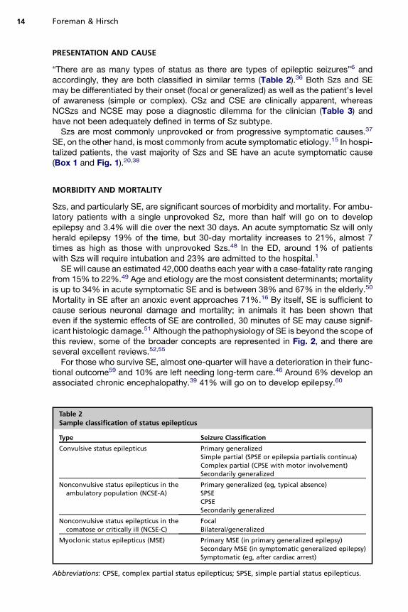
“There are as many types of status as there are types of epileptic seizures”6 andaccordingly, they are both classified in similar terms (Table 2).36 Both Szs and SEmay be differentiated by their onset (focal or generalized) as well as the patient’s levelof awareness (simple or complex). CSz and CSE are clinically apparent, whereasNCSzs and NCSE may pose a diagnostic dilemma for the clinician (Table 3) andhave not been adequately defined in terms of Sz subtype.Szs are most commonly unprovoked or from progressive symptomatic causes.37
SE, on the other hand, is most commonly from acute symptomatic etiology.15 In hospi-talized patients, the vast majority of Szs and SE have an acute symptomatic cause(Box 1 and Fig. 1).20,38
MORBIDITY AND MORTALITY
Szs, and particularly SE, are significant sources of morbidity and mortality. For ambu-latory patients with a single unprovoked Sz, more than half will go on to developepilepsy and 3.4% will die over the next 30 days. An acute symptomatic Sz will onlyherald epilepsy 19% of the time, but 30-day mortality increases to 21%, almost 7times as high as those with unprovoked Szs.48 In the ED, around 1% of patientswith Szs will require intubation and 23% are admitted to the hospital.1
SE will cause an estimated 42,000 deaths each year with a case-fatality rate rangingfrom 15% to 22%.49 Age and etiology are the most consistent determinants; mortalityis up to 34% in acute symptomatic SE and is between 38% and 67% in the elderly.50
Mortality in SE after an anoxic event approaches 71%.16 By itself, SE is sufficient tocause serious neuronal damage and mortality; in animals it has been shown thateven if the systemic effects of SE are controlled, 30 minutes of SE may cause signif-icant histologic damage.51 Although the pathophysiology of SE is beyond the scope ofthis review, some of the broader concepts are represented in Fig. 2, and there areseveral excellent reviews.52,55
For those who survive SE, almost one-quarter will have a deterioration in their func-tional outcome59 and 10% are left needing long-term care.46 Around 6% develop anassociated chronic encephalopathy.39 41% will go on to develop epilepsy.60
Table 2Sample classification of status epilepticus
Type Seizure Classification
Convulsive status epilepticus Primary generalizedSimple partial (SPSE or epilepsia partialis continua)Complex partial (CPSE with motor involvement)Secondarily generalized
Nonconvulsive status epilepticus in theambulatory population (NCSE-A)
Primary generalized (eg, typical absence)SPSECPSESecondarily generalized
Nonconvulsive status epilepticus in thecomatose or critically ill (NCSE-C)
FocalBilateral/generalized
Myoclonic status epilepticus (MSE) Primary MSE (in primary generalized epilepsy)Secondary MSE (in symptomatic generalized epilepsy)Symptomatic (eg, after cardiac arrest)
Abbreviations: CPSE, complex partial status epilepticus; SPSE, simple partial status epilepticus.

Table 3Semiological spectrum of nonconvulsive seizures and nonconvulsive status epilepticus
Negative Symptoms Positive Symptoms
AnorexiaAphasia/mutismAmnesiaCatatoniaComaConfusionLethargyStaring
Agitation/aggressionAutomatismsBlinkinga
CryingDeliriumDelusionsEcholaliaFacial twitching
LaughterNausea/vomitingNystagmusa
Eye deviationa
PerseverationPsychosisTremulousnessHippusa
a Applicable only to acute symptomatic comatose patients.Data from Jirsch J, Hirsch LJ. Nonconvulsive seizures: developing a rational approach to the diag-
nosis and management in the critically ill population. Clin Neurophysiol 2007;118:1660–70; and Ka-plan PW. Nonconvulsive status epilepticus in the emergency room. Epilepsia 1996;37(7):643–50.
Epilepsy Emergencies: Diagnosis and Management 15
There is significant debate over the morbidity and mortality of NCSzs and NCSE(Fig. 3). Studies have yielded inconsistent associations. For instance, generalizedNCSE overall is associated with fairly high mortality47; yet there is stark contrastbetween the absence status of primary epilepsy, which leads to no measureablemorbidity or mortality, and NCSE in comatose patients, which is associated withmortality rates of 51% to 65%.18,24 Kaplan uses mental status as the major determi-nant of mortality in NCSE; in one study, death occurred in 39%who had severe mentalstatus impairment compared with only 7% with mild impairment.61 However, the vastmajority of severely impaired patients die of their underlying comorbidities, leaving
Box 1
Some medications that lower the seizure threshold
� Analgesics: meperidine, fentanyl, tramadol
� Antiarrhythmics: mexiletine, lidocaine, digoxin
� Antibiotics: b-lactams (benzylpenicillin > semisynthetic penicillin; cefazolin; imipenem),quinolones, isoniazid, antimalarials (primaquine), metronidazole
� Antidepressants, especially bupropion and maprotiline
� AEDs: Phenytoin at supratherapeutic levels, tiagabine
� Baclofen
� Calcineurin inhibitors: cyclosporine, tacrolimus
� Chemotherapeutic agents: alkylating agents (chlorambucin, busulfan), a-interferons
� Neuroleptics, especially clozapine but also phenothiazines
� Lithium
� Multiple sclerosis medications: dalfampridine, 4-aminopyridine, b-interferons
� Radiographic contrast agents (intrathecal and intravenous)
� Theophylline
� Withdrawal from: opiates, alcohol, AEDs (especially benzodiazepines, barbiturates)
Data from Abou Khaled KJ, Hirsch LJ. Updates in the management of seizures and status epi-lepticus in critically ill patients. Neurol Clin 2008;26(2):385–408.

Fig. 1. Causes of seizures and status epilepticus in the United States. ADEM, acute dissemi-nated encephalomyelitis; AED, antiepileptic drug; CJD, Creutzfeldt-Jakob disease; CNS,central nervous system; GAD, glutamic acid decarboxylase; HIV, human immunodeficiencyvirus; ICH, intracerebral hemorrhage; INH, isoniazid; MS, multiple sclerosis; NMDA, N-methyl-D-aspartate (receptor); PRES, posterior reversible leukoencephalopathy syndrome;SAH, subarachnoid hemorrhage; SDH, subdural hematoma; SSPE, subacute sclerosing pan-encephalitis; TBI, traumatic brain injury; TTP, thrombotic thrombocytopenic purpura;VGKC, voltage-gated potassium channel. *See Box 1. (Data from Refs.16,20,37–47)
Foreman & Hirsch16
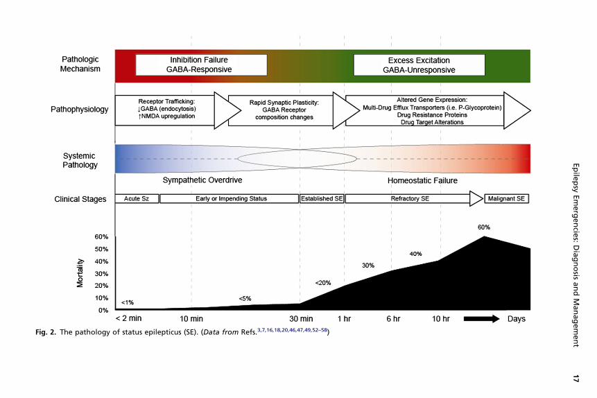
Fig. 2. The pathology of status epilepticus (SE). (Data from Refs.3,7,16,18,20,46,47,49,52–58)
Epile
psy
Emergencie
s:Diagnosis
andManagement
17

Fig. 3. Mortality and neuronal injury in subtypes of status epilepticus. Absence, absencestatus epilepticus; CPSE, complex partial status epilepticus; EPC, epilepsia partialis continua;GCSE, generalized convulsive status epilepticus; MSE, myoclonic status epilepticus; NCSE-A,nonconvulsive status epilepticus in the ambulatory patient; NCSE-C, nonconvulsive statusepilepticus in the comatose or critically ill; SET, status epilepticus terminans; SPSE, simplepartial status epilepticus; SSE, subtle status epilepticus.
Foreman & Hirsch18
a question as to the role of NCSE as an independent source of mortality. There aresome compelling clinical data suggesting harmful effects of the Sz activity itself,including increased mortality when age and etiology are controlled24; associationsbetween both the delay to diagnosis of NCSE and the duration of NCSE andmortality62; increased glutamate, glycerol, and lactate-pyruvate ratio on cerebralmicrodialysis63; increased neuron-specific enolase, a biomarker of neuronaldamage64; increased glutamate, lactate-pyruvate ratio, and intracranial pressureduring NCSzs compared with interictal periods within the same patient65; increasedmass effect and shift on serial brain imaging after ICH33; and eventual hippocampalatrophy ipsilateral to NCSzs experienced after traumatic brain injury (TBI).66
Szs or SE in hospitalized patients is an independent risk factor for morbidity andmortality.28,67 In one study of 41 hospitalized patients with SE, death occurred in61% and only 1 in 5 returned to baseline on discharge.42 In 201 medical ICU patients,Szs on cEEG were associated with death or poor outcome in 89% (vs 39% in thosewithout Szs); Szs remained associated with worse outcome even when controlledfor age, examination, and organ dysfunction.28 In both medical and surgical ICUs,mortality associated with SE approaches 67%.43 CSE and NCSE also act synergisti-cally with acute brain pathology to worsen outcomes in situations such as stroke,68
ICH,33 subarachnoid hemorrhage (SAH),69 and TBI.32 Animal studies suggest this:for example, NCSzs in rat models of acute focal ischemia were associated withincreased infarct size and higher mortality,70 and a low-dose pilocarpine model ofa single episode of NCSE in rats demonstrated permanent histologic, motor, andsocial behavior changes.71 It stands to reason that treating Szs and SE quickly andeffectively may potentially create better outcomes. Vespa’s motto for cEEG moni-toring in the ICU setting is: “to detect and protect.”72
In the only prospective study of RSE, mortality was 39.3% despite a low percentagerequiring intubation,75 itself an independent risk factor for mortality in SE.73 Rates of

Epilepsy Emergencies: Diagnosis and Management 19
about 50% are more widely cited. Hospitalization is longer and there is a significantassociation with deterioration on functional measures74; only 20% return to baselineon discharge.75
The overall duration of SE may play a role in mortality. Studies stratifying dura-tion7,47,76 found a mortality of 2.7% at less than 30 minutes, 19% at less than 1hour, 32% at over an hour, and logarithmically up to 6 hours thereafter. However,once RSE has become quite prolonged, duration may no longer be an independentpredictor of outcome54 even in RSE lasting longer than 7 days.41
PREHOSPITAL MANAGEMENT AND PRIMARY EVALUATION
An initial evaluation should take place either in the field or immediately on arrival to theED in conjunctionwithmedication administration (Box 2). Aswith any emergency, strictattention should be paid to the patient’s airway, breathing, and circulation (the ABCs).Treatment that is initiated early is much more likely to be effective18 and improve
outcomes.77 Studies have reported that emergency medical services (EMS) maytake 15 minutes to get to the ED77; only 41% of patients receive treatment before 30minutes10; and delays to treatment of up to 50 minutes may occur despite establishedprotocols.78 When first-responders (ie, family members, EMS) are able to give medica-tion, Sz time and recurrence decrease and patient outcomes are likely to improve.Based on 11 randomized controlled studies, diazepam and lorazepam are clearly
superior to placebo for stopping Szs and reducing the incidence of SE.79 Their promptprehospital administration actually leads to significantly decreased rates of intuba-tion77 compared with placebo. Intravenous (IV) lorazepam in particular has beenshown to be superior to both diazepam alone and phenytoin (PHT) alone as first-line therapy in adults.18,77 At times, IV access is not available. A variety of formulationsof midazolam (intranasal, buccal, and intramuscular [IM]) have been used in childrenwith prolonged Szs. Although buccal midazolam has been shown in a prospectiverandomized controlled trial to be more effective than rectal diazepam in abortingSzs in children,80 the only approved non-IV benzodiazepine for adult patients remainsrectal diazepam. A prospective study of adult prehospital IM versus IV benzodiazepinetreatment is under analysis as of this review,81 and the authors offer nasal midazolam,
Box 2
Initial steps in the prehospital management of seizures and status epilepticus
ABCs
Place in left-lateral decubitus position and remove any foreign objects from mouth(ie, dentures); no need for spinal precautions
Pulse oximetry and supplementary oxygen
Suction
Bag-valve mask or secure airway, as indicated
Cardiac monitoring
Insert peripheral intravenous line
Glucometer; if <80 mg/dL, administer 100 mg thiamine followed by 25–50 g dextrose 50%
Benzodiazepine administration based on availability
Data fromMichael GE, O’Connor RE. The diagnosis and management of seizures and status ep-ilepticus in the prehospital setting. Emerg Med Clin North Am 2011;29(1):29–39.
Foreman & Hirsch20
an effective and preferred route of administration,82 to many of their patients at risk forSz clusters or prolonged Szs to use at home.Once the patient has arrived at the treating facility, the focus should remain basic
and advanced life support measures, adequate monitoring of cardiopulmonary func-tion, and rapid treatment. SE frequently occurs with systemic manifestations (Table 4).In addition, recognition of Szs or SE mandates an evaluation for its cause. An initialevaluation strategy is outlined in the first row of Fig. 4.The value of imaging in the acute evaluation should be balanced against the risks of
delaying treatment. Acute symptomatic causes should be urgently evaluated with non-contrast computed tomography in any patient without a known history of epilepsy orwith focal findings on examination. EEG is an important tool to evaluate patients inSE. The resolution of clinical symptomsmay belie 48%who are still having intermittentNCSzs in thesubsequent24hours, and14%still inNCSEaftermovements stop.24Simi-larly, Szs may occur in up to 34% of patients with acute brain injury,34 and often mani-festsasadecrease inmental statusout of proportion to thedegreeof injury. If the level ofconsciousness after a convulsive Sz of any duration does not improve by 20minutes ornormalize by 60 minutes, NCSE should be suspected89 and an urgent EEG obtained.Another consideration may include lumbar puncture if there is clinical suspicion for
meningoencephalitis or SAH, but only once it is safe to do so and provided there is nodelay to treatment of the Szs and possible infection. Empiric antibiotics should include1 to2gceftriaxone, 1g vancomycin, and10mg/kgacyclovir. For theelderly, theadditionof 30mg/kg ampicillinmay bewarranted to cover for Listeria spp. In addition to cerebro-spinal fluid, blood and urine should be concurrently evaluated for organisms.
PRIMARY MANAGEMENT OF SE
Just as in prehospital treatment, the authors’ algorithm begins with use of g-aminobu-tyric acid A (GABAA) agonists: benzodiazepines (see Fig. 4). While multiple benzodiaz-epines will abort Szs with similar efficacy, Sz recurrence occurs less frequently withlorazepam than with diazepam owing to its decreased volume of distribution andlong-lasting central nervous system (CNS) levels, up to 12 hours.90 If diazepam or mid-azolam are used, a longer-acting maintenance AED, such as PHT or valproate (VPA),should be started concurrently. The response to lorazepam as a first-line agent fallsbetween 59% and 89%.18,77,90
The choice of second-line agent has traditionally been PHT,91 which works toprolong the recovery of voltage-gated sodium channels (see Fig. 4; Table 5). PHT isfrequently underdosed78 and should be based on weight. Complications from PHTinclude hypotension (27%–58%), respiratory depression (8%–10%), and cardiacarrhythmia (7%).18 PHT may also impede motor recovery after stroke.103 In addition,extravasation of IV PHT and its solvents (including propylene glycol) may cause softtissue necrosis and/or distal ischemia as part of the “purple-glove syndrome.” Theincidence of this complication in one prospective study was 1.7%.104 A safer butmore expensive alternative is a water-soluble diphosphate sodium ester of PHT, fos-phenytoin (FosPHT). Although FosPHT is a prodrug and must be converted to PHT,the conversion half-life is roughly 15 minutes, which is offset by faster infusion rates(about triple). Its primary benefit derives from avoiding hypotension and not havingto slow down the infusion as often. Cardiac arrhythmia can still occur as FosPHT isconverted to PHT,105 and both cardiac and hemodynamic monitoring should continuefor at least 15 minutes after the infusion of the prodrug is complete.VPA acts on sodium channels, but has effects on calcium channels and GABAA as
well. Two prospective, randomized trials have compared VPA with PHT for patients

Table 4Systemic manifestations of status epilepticus
System Effect Result
Temperature Increased systemic temperature due to sustainedmuscle activity; occursin up to 83%
Neuronal injury, particularly in the cerebellum
Vascular Blood pressure increases by up to 85mmHg systolic in the initial 30 mindue to sympathetic overdrive; as cardiac output decreases withincreasing mean arterial pressure, sudden loss of homeostaticmechanisms leads to hypotension as SE progresses >30 min
Hypertensive injury due to sympathetic tone followed by loss ofperfusion to metabolically sensitive cortex
Cardiac Potentially fatal cardiac arrhythmias occur in up to 58% Increased mortalityTakotsubo cardiomyopathy and contraction band necrosis may occur
due to endogenous catecholamines
Cerebrospinal fluid Pleocytosis; typically <10 � 106/L, up to 80 � 106/L � mild transientelevation in protein
Misdiagnosis of meningitis or encephalitis
Pulmonary Elevated minute ventilation and pulmonary hypertension Pulmonary edema
Other Acidosis; pH <7.3 in up to 81% Refractory hypotension, decreased respiratory driveRhabdomyolysis � hyperkalemiaa Renal failure, cardiac arrhythmiaHyperglycemia Exacerbation of acidosis
a Depolarizing paralytics (ie, succinylcholine) are relatively contraindicated, as they may result in worsened muscle damage and hyperkalemia, thus facilitatingcardiac arrhythmia.
Data from Refs.39,55,83–88
Epile
psy
Emergencie
s:Diagnosis
andManagement
21

Fig. 4. Convulsive status epilepticus treatment algorithm in adults at Columbia University’sComprehensive Epilepsy Center. ABG, arterial blood gas; AED, antiepileptic drug; BMP, basicmetabolic panel; BP, blood pressure; Ca, calcium; CBC, complete blood count; D50W, 50%dextrose in water; EKG, electrocardiogram; FSBG, finger stick blood glucose; HCG, humanchorionic gonadotropin; HR, heart rate; IM, intramuscular; IV, intravenous; LFTs, liver func-tion tests; Mg, magnesium; PO4, phosphorus; PR, per rectum.
Foreman & Hirsch22
withSE.Onestudy randomized100patientswithSE refractory todiazepam to20mg/kgof either VPA or PHT, and found no difference in either efficacy or tolerability.92 ThesecondstudyusedVPA (30mg/kg) orPHT (18mg/kg) as first-line treatment for SEusinga crossover design. VPA was more effective in controlling SE both as the initial drug(66% vs 42%, P 5 .046) and as the second drug after the first had failed (79% vs25%, P 5 .004).100 Adverse effects associated with VPA based on studies in otherscenarios include hyperammonemia, hepatic dysfunction, pancreatitis, parkinsonism,dose-dependent thrombocytopenia,83 and other potential mechanisms that may leadto impaired coagulation such as lower platelet activation and prolonged thrombintime.106Of importance, there are fewcardiovascular ormental statuseffects.One studyreported loading doses of up to 32.7 mg/kg in elderly inpatients with no reported hypo-tension or arrhythmias.107 This finding makes VPA a particularly attractive AED in theelderly, the critically ill, or those with advanced directives precluding intubation.Two additional AEDs have recently been studied in SE. The first is levetiracetam
(LEV), which is a synaptic vesicle (SV2A) ligand and inhibits high-voltage–gatedcalcium-channel currents. Adverse effects in two case series were limited to mildsedation (often when given close to benzodiazepines), mild nausea, transient asymp-tomatic thrombocytopenia, and a transient elevation in liver function tests.94,99
Response varies: SPSE and CPSE were effectively treated by 1 to 2 g LEV assecond-line therapy 60% to 79% of the time.93,97 Generalized NCSE or subtle statusresponded 30% of the time, whereas none of the patients with GCSE in one seriesresponded.97 A small study of SE in elderly patients with medical comorbidities re-ported an 88% EEG response to 1.5 g IV bolus as first-line therapy.108
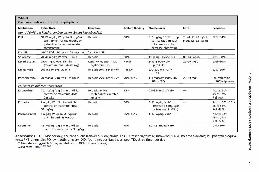
Table 5Common medications in status epilepticus
Medication Initial Dose Clearance Protein Binding Maintenance Level Response
Non-cIV (Without Respiratory Depression, Except Phenobarbital)
PHT 18–20 mg/kg IV up to 50 mg/min(25 mg/min for the elderly orpatients with cardiovascularcompromise)
Hepatic 90% 5–7 mg/kg PO/IV div upto TID; caution withtube feedings thatdecrease absorption
Total: 15–20 mg/mLFree: 1.5–2.5 mg/mL
37%–84%
FosPHT 18–20 PE/kg IV up to 150 mg/min Same as PHT
Valproate 20–40 mg/kg IV over 10 min Hepatic 90% 1000 mg PO/IV q 6 h 80–140 mg/mL 79%–88%
Levetiracetam 2500 mg IV over 15 min(maximum bolus dose: 4 g)
Renal 67%, enzymatichydrolysis 33%
<10% 2–12 g PO/IV divup to QID
25–60 mg/L 60%–90%
Lacosamide 300 mg IV over 30 min Hepatic 60%, renal 40% <15%a 200–300 mg PO/IVq 12 h
— 57%–60%
Phenobarbital 20 mg/kg IV up to 60 mg/min Hepatic 75%, renal 25% 20%–45% 1–3 mg/kg/d PO/IV divBID or TID
20–50 mg/L Equivalent toPHT/valproate
cIV (With Respiratory Depression)
Midazolam 0.2 mg/kg IV q 5 min until Szcontrol or maximum dose2 mg/kg
Hepatic; activemetabolites excretedrenally
95% 0.1–2.9 mg/kg/h cIV — Acute: 82%48-h: 37%7-d: N/A
Propofol 2 mg/kg IV q 5 min until Szcontrol or maximum dose
10 mg/kg
Hepatic 90% 2–15 mg/kg/h cIV(limited to 5 mg/kg/hfor treatment >48 h)
— Acute: 67%–73%48-h: 54%7-d: 43%
Pentobarbital 5 mg/kg IV up to 50 mg/minq 5 min until Sz control
Hepatic 35%–55% 1–10 mg/kg/h cIV — Acute: 92%48-h: 57%7-d: 22%
Ketamine 1.5 mg/kg IV q 5 min until Szcontrol or maximum 4.5 mg/kg
Hepatic 45% 1.2–7.5 mg/kg/h cIV — Unknown
Abbreviations: BID, Twice per day; cIV, continuous intravenous; div, divide; FosPHT, fosphenytoin; IV, intravenous; N/A, no data available; PE, phenytoin equiva-lents; PHT, phenytoin; PO, by mouth; q, every; QID, four times per day; Sz, seizure; TID, three times per day.
a New data suggest LCS may exhibit up to 90% protein binding.Data from Refs.74,92–102
Epile
psy
Emergencie
s:Diagnosis
andManagement
23

Foreman & Hirsch24
The second agent is the recently available AED lacosamide (LCS), which enhancesthe slow inactivation of voltage-dependent sodium channels. Similar to LEV and VPA,there are minimal if any effects on respiratory function, and cardiovascular effects arelimited to potential PR interval prolongation. Although data on nonrefractory SE arelimited, one case series of 39 patients has been published.98 Dose ranges between200 and 400 mg were given to patients, 85% of whom were in NCSE-A. Almost halfof patients needed no further medication when LCS was used, and there were noadverse effects related directly to LCS.
PRIMARY MANAGEMENT OF RSE
Even prolonged RSE lasting months can resolve with good outcome.41 Despite theincredible challenges of treating RSE and the lack of consensus on how to best todo so, it is crucial to establish a rapid, effective agent in patients who continue tohave Szs. As only a minority of patients will respond to a third bolus AED,18 a contin-uous IV (cIV) medication should be considered, either midazolam (MDZ) or propofol(PRO) initially (see Fig. 4). It should be emphasized that RSE is very often electro-graphic only, or has subtle clinical correlate, even after “successful” treatment ofconvulsions18,24,74; transfer to a unit or hospital with cEEG should be considered forthe most effective use of these continuous drips.MDZ is a rapid-acting benzodiazepine. Its major adverse effect is hypotension, often
requiring pressors, and respiratory suppression. In addition, tachyphylaxis may occurand there is a theoretical concern of secondary downregulation of GABA receptorsfrom either prolonged benzodiazepine use or SE. Because breakthrough Szs predictSz recurrence after anesthetic weaning, in the authors’ center MDZ infusion is nowused at much higher doses than in the past, up to 2.9 mg/kg/h, 10 times higher thanaverage rates in older studies. Sz suppression is typically maintained for 24 hours fol-lowed by slow weaning over 6 to 24 hours while on cEEG to evaluate for Sz recurrence.PRO is a GABAA agonist with rapid onset (about 3 minutes) and easy reversibility. In
addition, it inhibits N-methyl-D-aspartate (NMDA) and modulates calcium influx.Adverse effects include hypotension, respiratory suppression, transient movementdisorders that may be misconstrued as Szs,102 and the propofol infusion syndrome(PIS; Box 3). Originally described in children, PIS has been recognized in adultswho receive propofol at more than 5 mg/kg/h for longer than 48 h, particularly afterhead injury.109 A maximum of 5 mg/kg/h is recommended if PRO is to be maintainedfor more than 48 hours, and creatine kinase, lactic acid, pH, and triglycerides shouldbe checked daily. To maintain efficacy while reducing the dose, some use an adjunc-tive “dose-sparing” cIV benzodiazepine.102 As with MDZ, PRO is typically weanedover 6 to 24 hours.
Box 3
Propofol infusion syndrome: diagnostic criteria greater than 6 hours after initiation of
propofol
Creatine kinase >2000 U/L
Triglycerides >500 mg/dL
Progressive lactic acidosis >2.5 mmol/L
1 Bicarbonate <20 mmol/L >6 hours after propofol infusion (not due to sepsis)
Data from Rossetti AO, Milligan TA, Vulliemoz S, et al. A randomized trial for the treatment ofrefractory status epilepticus. Neurocrit Care 2011;14:4–10.
Epilepsy Emergencies: Diagnosis and Management 25
For decades, the preferred anesthetic for ICU treatment of RSE has been barbitu-rates. In the United States this is typically pentobarbital, which acts on different GABAreceptor isoforms than do benzodiazepines. While benzodiazepine-sensitive receptorsare internalized in experimental models of SE, barbiturate-sensitive receptors maintaintheir responsiveness.110 The adverse effects of pentobarbital are principally cardiovas-cular: cardiac depression, vasodilatation and hypotension, and poikilothermia.MDZ, PRO, and barbiturates have been compared in a systematic review of 193
patients,96 andmore recently PROandbarbiturates have been compared in a prospec-tive, randomized controlled trial of 23 patients.101 In the systematic review, pressor-requiring hypotension occurred in about 30% of patients on MDZ, significantly fewerthanwith pentobarbital (77%). This result was likely related to dosing and goals of treat-ment: MDZ is typically titrated to Sz control whereas pentobarbital is titrated to back-ground suppression. Related to this, MDZ was associated with significantly morebreakthrough Szs (more than half) compared with PRO (15%) and pentobarbital(12%). However, cEEG was only performed in one-quarter of patients receiving pento-barbital (compared with 80% of those on MDZ), so it remains unclear if the rates ofbreakthrough Szs (usually NCSzs) were truly lower. Finally, doses of PRO averaging3.2 mg/kg/h only succeeded in achieving burst suppression 38% of the time, muchless often than with pentobarbital (96%). By contrast, the prospective trial randomizedpatients to either PRO or barbiturates with the goal of achieving background suppres-sion. Seven-day Sz freedom rates after 36 to 48 hours of suppression-burst weresimilar between PROand barbiturates, but patients receiving barbiturates experiencedlonger mechanical ventilation time than patients receiving PRO.There does not appear to be any significant difference in mortality between MDZ,
PRO, and barbiturates when used for RSE.96,101 Mortality also remains the samewhen only one versus more than one anesthetic is administered.111 Therefore, thereare no clear guidelines as to which agent should be used first. Also, goals are not clearregarding titration of anesthetics to suppression burst versus Sz freedom, and for howlong to do this. On one hand, barbiturate treatment seems to demonstrate significantlymore suppression burst and possibly less Sz recurrence,96 a predictor of relapse andsubsequently mortality.112 On the other hand, there is no clear difference in mortalitybetween patients who achieve suppression burst and those who do not.111 At leastoneorganizationhas formally recommended24hoursof suppressionbeforemedicationweaning,113 but data from a randomized prospective study found an overall Sz-freeresponse rate of only 35%after suppression burst for 36 to 48 hours on cEEG.101 Couldlonger durations therefore be required to establish efficacy of treatment? Older studiesdid not incorporate cEEG and therefore data are limited. Szs can still occur duringsuppression burst, and even during complete background EEG suppression.
MAINTENANCE THERAPY IN RSE
In RSE, maintenance AEDs facilitate continued Sz control after weaning from cIVmedications. Agent(s) used in the initial treatment of SE should be maximized andcontinued, but additional AEDs are commonly required (see Table 5). PHT can beused IV or transitioned to oral administration. For patients receiving continuous tubefeedings, dosing should remain IV to avoid a dramatic (up to 71%) decrease in absorp-tion of PHT.114 Daily total and free levels should be monitored. PHT is highly proteinbound and hepatically cleared, thus free levels may be quite elevated in the settingof low protein binding or when using other highly protein-bound medications suchas VPA or benzodiazepines. In critically ill patients on PHT plus full doses of VPA,the free PHT level is usually close to 3 times the expected level, that is, it is close to

Foreman & Hirsch26
30% of the total PHT rather than the usual 10%. In these patients, a total PHT level of 8to 10 mg/mL will provide a high-therapeutic free level of 2.4 to 3.0 mg/mL. In somecenters, free levels may take longer to report than total levels; if there is a predictablerelationship between the two over several consecutive days of steady dosing, the totallevel may be used reliably.VPA, like PHT, is highly protein bound and hepatically cleared. However, it is a cyto-
chrome P450 inhibitor rather than inducer. Another interaction of particular concernhas been the interaction between VPA and meropenem, and probably carbapenemsas a class, which are widely used for resistant gram-negative infections. These antibi-otics may dramatically reduce VPA levels, due to a variety of mechanisms involvingdecreased glucuronidation and increased renal excretion.115
LEV is only minimally protein bound and is cleared mostly by the kidneys. The signif-icance of levels is not well established. Similarly, LCS is purported to have minimalprotein binding, although a recent study demonstrated that perhaps 90% is proteinbound.116 LCS is both hepatically and renally cleared. Specifically for RSE, an uncon-trolled, unblinded study reported that LCS has a response rate of 20%98; all 4 cases ofrefractory partial status responded to doses of 100 mg or less despite NCSE lasting upto 50 hours.117 The authors’ algorithm includes LEV and LCS as later add-on medica-tion unless there are contraindications to PHT and VPA.Phenobarbital, a barbiturate, may be used as a bolus for second-line or third-line
treatment. Adverse reactions include respiratory depression, impaired mental status,hypotension, and rash. Like PHT, phenobarbital may affect motor recovery afterstroke.103 Phenobarbital can be useful in patients requiring cIV barbiturates, as pheno-barbital reduces the risk of relapse even if levels are subtherapeutic.112 It remainsa reasonable alternative for the early treatment of SE, as it was not statistically inferiorto lorazepam in the VA Cooperative study and did not have greater acute adverseevents.18 Because of hypotension, prolonged sedation, the need to load it fairly slowly,and its strong induction of P450 enzymes, the authors prefer other agents, but includephenobarbital as an option for select patients only.Any oral agent may be used, such as topiramate (TPM), gabapentin (GPN), prega-
balin (PGB), oxcarbazepine, or carbamazepine (CBZ). Two of these agents, PGB andTPM, have been studied in this setting. PGB was used in mostly partial RSE in 11patients.118 After a median of 5 days of RSE, 150 to 600mg of PGBwas given enterallyin divided doses with Sz cessation within 24 hours in 5 patients for a response rate of45%. TPM exhibits a variety of actions that may be helpful in SE including sodium-channel inhibition, GABA potentiation, high-voltage–gated calcium-channel inhibition,and antagonism of excitatory transmission. Rapid titration of up to 1600 mg/d (a veryhigh dose) may be useful in treating RSE based on one series of 6 patients, althoughthis has yet to be confirmed in further studies.119 Animal studies suggest TPM mayhave neuroprotective properties, and may act synergistically with NMDA antago-nists.120 When using TPM, it should be kept in mind that it is a carbonic anhydraseinhibitor and may exacerbate acidosis. The authors avoid the use of TPM while usingpropofol, as it theoretically may exacerbate the acidosis associated with PIS. In addi-tion, cases have been reported of acute hepatic failure associated with the use of TPMin combination with VPA.121
ALTERNATIVE MANAGEMENT OF RSE
As described, failure of an anesthetic agent or Sz recurrence after its withdrawal hasbeen termed “malignant” status.20 The authors might revise this definition as Sz recur-rence at any time after a period of at least 24 hours of suppression burst on cEEG plus

Epilepsy Emergencies: Diagnosis and Management 27
trials of at least 3 AEDs titrated to appropriate serum levels. Alternative strategies mayneed to be considered in these patients.Ketamine is an NMDA antagonist that has been used by anesthesiologists for
decades. In experimental models of prolonged SE, ketamine has been found to beless effective early, but is increasingly effective as Szs become self-sustained.122
This process is the reverse of that of GABA agonists (including benzodiazepinesand barbiturates), likely a result of internalization of GABA receptors during SE.Thus, glutamate blockade becomes more effective than GABA agonism in later stagesof SE (see Fig. 2). In addition, because excitatory amino acid mediated toxicity isthought to underlie neuronal injury in SE, NMDA antagonism may be neuroprotec-tive.49 Animal models have suggested a synergistic effect when ketamine is usedtogether with benzodiazepines in SE.123 These models have also demonstratedpotential ketamine toxicity, and a single anecdotal human case of cerebellar atrophyallegedly due to prolonged (72 hours) ketamine use in SE has been reported.124
Despite the theoretical benefits of ketamine and its short-term efficacy in abortingSzs, very few studies support its use. At the authors’ center ketamine is used inconjunction with MDZ, often in the instance that maximal MDZ cIV has not resultedin Sz control on cEEG. It is also used as a bridge when tapering off of cIV barbituratesto limit withdrawal-associated Sz recurrence. Increased cardiovascular output,including tachycardia and hypertension, is both an adverse effect and a strength ofketamine, in contrast to the hypotension induced by other anesthetic agents usedfor treating RSE. Dissociative effects such as hallucinations or psychosis occur at sub-anesthetic dosing in awake patients, but are not a problem in this setting. Cautionshould be exercised prior to infusion in patients with elevated intracranial pressure;cardiovascular disease including hypertension; myocardial ischemia, or congestiveheart failure; autonomic dysregulation; and TBI.Inhaled anesthetics have been used with some success, but are impractical and
have not demonstrated sustained benefit after cessation. Mirsattari and colleagues125
reported a case series of isoflurane and desflurane in patients with RSE at end-tidalconcentrations of 1.2% to 5%. Suppression burst was initiated within minutes andwas maintained for a median period of 11 days while maintenance AEDs were opti-mized. Four of the 7 patients included in the study survived with good outcomes.Adverse effects included hypotension, along with ICU-associated problems such asatelectasis and ileus. With prolonged use of more than 30 days, thalamic and cere-bellar hyperintensities on T2-weighted magnetic resonance imaging (MRI) may occur,indicating potentially reversible neurotoxicity.126
Lidocaine is a nonanesthetic class IB antiarrhythmic agent that has been used incardiology for many years. It works by blocking sodium-channel conduction, attenu-ating depolarization, and decreasing automaticity. In addition, lidocaine has beenused as a nonsedating, rapidly acting agent in SE. The largest prospective studycomprised 36 chronic obstructive pulmonary disease patients with SE127 in whomlidocaine given at cardiac doses led to immediate Sz cessation in almost three-quarters. Szs recurred quickly in more than half. Initial bolus is 1 to 3 mg/kg, and aninfusion of up to 4 mg/kg/h is recommended to prevent recurrence.128 Caution shouldbe exercised in using lidocaine in patients with hepatic disease and the elderly,although it is noted that doses typically associated with hypotension or myocardialdepression are about twice as high, and those associated with induction of Szs areabout 3 times as high.128
Magnesium (Mg) is an important component of the NMDA receptor, effectivelyblocking transmission at membrane resting potential. Mg repletion may saturateNMDA receptors to restore tonic blockade. Its use is largely anecdotal in adults

Foreman & Hirsch28
with the exception of eclampsia, in which it is the treatment of choice. The mechanismof action in eclampsia, however, is likely related to endothelial stabilization and there-fore the treatment of the underlying condition, not the Szs directly. One recent studyfound Mg to be effective in 2 young adults with mitochondrial disease (POLG1 muta-tion).129 Given the ever-growing spectrum of juvenile-onset and adult-onset presenta-tions of mitochondrial disease, Mg should be considered in patients with cryptogenicRSE. Typical loading doses are 2 to 4 g IV, but optimal serum levels are notestablished.Adjunctive medications may be useful; coadministration of benzodiazepines with
PRO or ketamine has already been discussed. Pathologic studies have implicateddrug efflux transporters such as P-glycoprotein (Pgp) in the refractory nature of pro-longed RSE.130 Verapamil, a calcium-channel antagonist and Pgp inhibitor, wasused in one patient with recurrent Szs after prolonged RSE.131 A dosage of 120 mg/d,roughly half the starting dose for cardiovascular indications, significantly reduced theburden of the patient’s nocturnal Szs over the course of 2 weeks while levels of PHTand phenobarbital remained essentially constant. However, a recent animal studyfound no benefit of calcium-channel blockers for phenobarbital-resistant recurrentSzs, and 3 of the 11 animals actually developed Sz clusters.132 Animal studies havealso recently hinted at the use of HMG-CoA reductase inhibitors, or statins, in long-lasting SE.133 Lovastatin decreased the expression of proinflammatory cytokinemRNA and body temperature in rats, which may provide neuroprotection. In addition,erythropoietin (EPO), a cytokine hormone with receptors located widely within theCNS, has been studied in low-dose pilocarpine rat models of SE. When administeredafter SE, EPO reduced blood-brain barrier disruption, decreased neuronal cell death,and attenuated microglial activation. Clinically, spontaneous recurrent Szs were lessfrequent during prolonged monitoring (>1 month).134 Further studies will be requiredto test these agents for use in humans with SE.There is a variety of nonpharmacologic interventions for RSE including hypothermia,
electroconvulsive therapy (ECT), the ketogenic diet, and music therapy. Hypothermia,typically used in cardiac arrest patients, has been used for RSE in a small caseseries,135 based on animal data136 that suggests hypothermia has anticonvulsantand neuroprotective properties. Four patients refractory to MDZ and barbituratesreceived endovascular cooling to 31� to 33�C. cIV anesthetics were weaned, and aftera 24-hour Sz-free period, 0.5�C rewarming every 4 hours to 36.5�C took place. RSEwas stopped in all patients; 2 of the 4 remained Sz free. Adverse effects were prom-inent and included shivering, acidosis, coagulopathy, thrombosis (ie, deep vein throm-bosis, pulmonary embolism), cardiac arrhythmia, and immunosuppression. Althoughthese risks might be mitigated by a short duration of cooling, controlled studies areneeded. ECT presumably increases endogenous anticonvulsant signaling pathwaysand induces a refractory period that aborts SE. One case series and review137
suggests weaning all maintenance AEDs while on an anesthetic titrated to suppres-sion burst followed by sessions of ECT for 3 to 8 days. If Szs recur, suppressioncan be resumed along with maintenance medications; however, 70% of the cases re-ported in the literature remained Sz free. Lastly, music has been reported to abort SE.Miranda and colleagues138 have now reported two cases of RSE that spontaneouslyaborted within hours of continuously played classical music from Mozart and Bach.The ketogenic diet (KD) is a carbohydrate-restricted diet with high fat content that
induces ketosis and suppresses Szs. KD is typically used in children. In the ambula-tory pediatric population, KD has been shown in a randomized controlled trial to effec-tively treat daily Szs (>90% reduction in Sz frequency in 7% vs 0% of controlsand >50% reduction in Sz frequency in 38% vs 6% of controls).139 Recently, KD

Epilepsy Emergencies: Diagnosis and Management 29
has been used in two adult cases of RSE.140 In both, KetoCal 4:1 tube feedings weregradually introduced to patients with malignant RSE. Ketosis was achieved in 8 to 10days coincident with Sz control. There are a variety of adverse effects including hypo-glycemia, gastrointestinal upset, and acidosis; long-term effects are not well reported,but include renal stones and growth delay. With the use of KD a nutritional specialistshould be involved, a urinalysis should be checked for ketones daily, and serum b-hydroxybutyrate should be checked weekly.Data on the effectiveness of epilepsy surgery for RSE are limited and are mostly
from pediatric populations. Nonetheless, surgery may be considered in selectpatients with malignant RSE with evidence of one main Sz focus. One case seriesdocumented resection and multiple subpial transections in one patient with focalstatus and callosotomy in two patients with multifocal status, all with resolution ofSE.141 The patient with focal status continued to have “occasional brief partialseizures,” one of the patients with multifocal status remained Sz free for 2 years,and the other had monthly Szs with one recurrence of treatable SE. A more recentcase reported resection in one patient with focal status who remained Sz free at16 months postsurgery.142 Some investigators advocate for 2 weeks of failed treat-ment as a justification for surgery.143 However, concordance is crucial for accuratelocalization, and MRI demonstrating focal restricted diffusion, ictal single-photonemission computed tomography (SPECT),141 ictal 18F-fluorodeoxyglucose positronemission tomography (PET),142 or electrocorticography141,142 may all be helpful. AsRSE becomes malignant, physicians should consider acquiring data early so thatsurgical options become available. Other strategies that need further evaluationinclude vagus nerve stimulation and deep brain stimulation. The latter has beensuccessfully used in intractable epilepsy to good effect,144 but has been evaluatedonly in animal models of SE.145
RSE caused by CNS “infection” infrequently yields a proven pathogen.40 A variety ofautoimmune or paraneoplastic disorders may be implicated, although the incidence isunknown. It is reasonable, then, to consider steroids, adrenocorticotropic hormone,intravenous immunoglobulin, and/or plasma exchange to treat cryptogenic RSE inconjunction with other AEDs. It is even postulated that systemic exposure of braintissue through damaged blood-brain barrier associated with prolonged Szs mayinduce a “secondary immune-mediated encephalitis,”146 further justification forempiric immunomodulatory therapy in RSE.
TREATMENT CONSIDERATIONS IN THE COMATOSE OR CRITICALLY ILL
Szs and SE in the critically ill are largely nonconvulsive. As opposed to NCSE in theambulatory population, the typical presentation of NCSE in the comatose or criticallyill is nonlocalizing coma.147Despitemorewidely available cEEG, diagnosis is frequentlydelayed: by 24 hours in 16% of patients in the neuro-ICU,62 by 48 hours in the medicalICU, and by 72 hours in the surgical ICU.43 Routine EEG (20–60 minutes) will miss atleast half of patients who are having NCSzs. In noncomatose patients, 24 hours ofcEEG will identify up to 95% of patients with NCSzs. In comatose patients, only 80%will be diagnosed by 24 hours. Therefore, a full 48 hours of cEEG should be used incomatose patients to increase the sensitivity of NCSz detection to almost 90%.21
The critically ill will often do poorly regardless of the presence or absence of Szs.However, in multivariate analyses, NCSE,18,24 delays to treatment,62 duration ofSzs,24,62 and coma54 portend worse prognosis. In the elderly, there is some retrospec-tive evidence for increased mortality and longer hospitalization with IV benzodiazepinetreatment,148 but others suggest there were not adequate controls of dosage and

Foreman & Hirsch30
timing in the study, and that “lack of clinical prudence [rather] than an inherent dangerfrom the benzodiazepines” played a role.35
Complicating the diagnosis and management of Szs and SE in the critically ill are so-calledboundary conditions,13whereby theEEGmayappear potentially ictal yet doesnotstrictly fulfill criteria for definite NCSzs or NCSE. In ambulatory patients, much of thedifferentiationbetween ictal andnonictal canbemadebasedonmental status. In thecrit-ically ill, on the other hand, these conditions occur in the context of coma. Chong andHirsch14 provide a thorough review of patterns such as lateralized periodic discharges(LPDs), generalized periodic discharges (GPDs), and their potential clinical significance.It is clear that LPDs are highly associated with Szs149 and they may also be associ-
ated with worse outcomes after SE.150,151 The authors’ recent analysis of 200 patientswith GPDs and a matched control group showed that GPDs are associated withNCSzs and NCSE but do not appear to be associated with worse outcome after thor-ough adjustment for age, neurologic examination, and etiology.152 One subset ofGPDs, triphasic waves, are seen in metabolic encephalopathy and degenerativediseases, but cannot be reliably distinguished from generalized epileptiformdischarges or NCSE in a given individual based on EEG alone.153,154 It should be notedthat LPDs are occasionally ictal without question, for instance when they are associ-ated with time-locked jerking on the contralateral side14 or with aphasia that resolvesalong with the LPDs in response to AED.155 At times, LPDs are associated withincreased glucose metabolism on PET or increased cerebral blood flow on SPECT.14
The authors view these periodic patterns as part of an ictal-interictal continuum, in anattempt to avoid the false dichotomy of interictal versus ictal EEG patterns in enceph-alopathic patients. Ongoing efforts are being made to define and therefore facilitatestudy of these and other confusing EEG phenomena.156
When there is reasonable suspicion for NCSE, an urgent EEG is indicated. A lack ofrapid clinical response to an AED does not help rule out NCSE. In the VA CooperativeStudy, 100% of the patients with subtle SE remained comatose 12 hours after treat-ment.18 In another study, more than half of nonanoxic patients with NCSE in the ICUimproved in alertness after treatment with AEDs, but the response was almost neverimmediate, sometimes only minimal, and not always sustained.43 Yet if EEG is notavailable or the EEG findings lie along the ictal-interictal continuum, it may be usefulto perform a trial of a rapid-acting AED at the bedside to evaluate for clinical improve-ment (Box 4). A major restriction of empiric AED trials in possible NCSE in the criticallyill or comatose is that many EEG patterns resolve, leaving a comatose patient withdiffuse slowing. Resolution of an abnormal pattern does not represent proof of itsepileptic nature; for example, in patients believed to have pure metabolic encephalop-athy without Szs, triphasic waves resolve with benzodiazepines as well.153
In the ICU most patients do not have a history of epilepsy, and new Szs may be thepresenting symptom of a new cerebral insult.45 For patients in whom the EEG is equiv-ocal or clearly ictal, an assessment for acute brain injury (stroke, hemorrhage) or newsystemic syndrome (hepatic failure, renal failure, sepsis), medication review, andconsideration of lumbar puncture may be appropriate as treatment is initiated.35 Inpatients with periodic discharges, equivocal patterns or only brief intermittent NCSzs,the authors generally try to avoid coma-inducing doses of medications, and typicallystart with IV PHT, VPA, or LEV, and possibly LCS.In patients with frequent or periodic epileptiform discharges, cEEG monitoring is
recommended for 48 hours, as these patients are more likely to have a delay beforerecording their first definite Sz.27 The authors advocate a nonsedating AED for Szprophylaxis for patients with frequent or periodic epileptiform discharges during theacute illness only (typically a couple of weeks) if there have been no definite Szs,

Box 4
Antiepileptic drug trial for diagnosis of suspected nonconvulsive status epilepticus
Indication:
Rhythmic or periodic focal or generalized epileptiform discharges on EEG with neurologicimpairment
Contraindication:
Patients who are heavily sedated/paralyzed
Patients who have a clear reason for their level of consciousness
Monitoring:
EEG, pulse oximetry, blood pressure, electrocardiography, respiratory rate with dedicatednurse
Antiepileptic Drug Trial:
� Sequential small doses of rapidly acting short-duration benzodiazepine such as midazolam1mg per dose or nonsedating AED such as levetiracetam, valproate, phenytoin, or lacosamide
� Between doses, repeated clinical and EEG assessment
� Trial is stopped after any of the following
� Persistent resolution of the EEG pattern (and examination repeated)
� Definite clinical improvement
� Respiratory depression, hypotension, or other adverse effect
� A maximum dose is reached (such as 0.2 mg/kg midazolam, though higher may be neededif on chronic benzodiazepines)
Test is considered positive if there is resolution of the potentially ictal EEG pattern AND eitheran improvement in the clinical state or the appearance of previously absent normal EEGpatterns (eg, posterior-dominant ‘‘alpha’’ rhythm). If EEG improves but patient does not, theresult is equivocal.
Adapted from Jirsch J, Hirsch LJ. Nonconvulsive seizures: developing a rational approach to thediagnosis andmanagement in the critically ill population. Clin Neurophysiol 2007;118:1660–70.
Epilepsy Emergencies: Diagnosis and Management 31
clinically or electrographically. If Szs develop, the authors typically treat with an AEDfor about 3months, obtain another EEG, and re-assess at that point. There are minimaldata to guide these decisions, but extensive experience with TBI, brain tumors, andother scenarios suggests that prolonged prophylactic AEDs in those without clearepilepsy (ie, recurrent unprovoked Szs, which excludes Szs during the acute illness)will not be effective and may cause unnecessary adverse effects.
SPECIAL SITUATIONS: ORGAN FAILURE
In patients with liver failure and Szs or SE, serum levels of hepatically metabolizedmedications will increase. In addition, decreased protein synthesis may lead to hypo-albuminemia, further increasing serum free (unbound) drug levels. Medications thatare not hepatically metabolized and demonstrate minimal protein binding, such asLEV, GPN, or PGB, are easier to use in these settings.Renal failure causes metabolic disturbances including hyponatremia, acidosis, and
hypoalbuminemia. Medications that are renally cleared such as LEV, GPN, PGB, andto a lesser degree phenobarbital and TPM, should be dosed significantly lower anda dose should be given immediately following dialysis. Medications such as TPMand zonisamide have carbonic anhydrase activity, which can precipitate acidosis or

Foreman & Hirsch32
renal calculi in patients with poor renal function. Highly protein-bound medicationssuch as PHT, VPA, benzodiazepines, and possibly LCS116 will experience higher-than-usual unbound levels for a given total serum level, but are only minimally dialyzedbecause protein-bound drug is not removed during dialysis.
SPECIAL SITUATIONS: ORGAN TRANSPLANTATION
Szs in transplant patients occur frequently in the context of the following: significantmetabolic abnormalities, depending on the organ involved; infection or neurotoxicityfrom medications; and complications from surgery, such as hypoxia (Table 6). Livertransplant patients in particular appear to develop Szs 4 to 6 days postoperatively.157
Abou Khaled and Hirsch83 speculate that the cause may be withdrawal from highendogenous benzodiazepines as the new organ begins functioning. Patients on calci-neurin inhibitors such as tacrolimus are at risk for the posterior reversible encephalop-athy syndrome (PRES), characterized by Szs and cerebral edema associated withcortical blindness, aphasia, or altered mental status. Other factors implicated in thedevelopment of this syndrome include hypertension, hypomagnesemia, and supra-therapeutic immunosuppressant levels.Medications should not interfere with the new organ; for instance, certain AEDs
(PHT, CBZ, and several others) can affect cardiac conduction and should be used judi-ciously after heart transplant. Similarly, barbiturates can cause myocardial depres-sion. The choice of medication must also navigate the difficult interactions thatoften occur with transplant medications. Cyclosporine and methylprednisolone aremetabolized through the cytochrome P450 pathway, thus inducers such as PHT,CBZ, and phenobarbital may increase clearance and reduce levels, sometimes quitedramatically.
SPECIAL SITUATIONS: PREGNANCY
As in the critically ill, Szs in a pregnant woman should warrant an evaluation for newacute brain injury, as only 15% to 30% of women with epilepsy develop increasesin Sz frequency during pregnancy.158 The reversible vasoconstriction syndrome(sometimes referred to as the Call-Fleming syndrome, or peripartum vasculopathy)may occur at any time during or even several weeks after pregnancy, and relativehypercoagulability increases the incidence of venous thrombosis. In addition,pregnancy-specific systemic illness such as hyperemesis gravidarum, the HELLP
Table 6Incidence of clinical seizures (%) in organ failure or transplantation
Liver failure 2–33
Renal failure (on hemodialysis) 2–10
Transplant
Liver 25–30
Kidney 1–31
Heart 2–15
Lung 22–27
Bone marrow 3–12.5
Pancreas 13
Data from Abou Khaled KJ, Hirsch LJ. Updates in the management of seizures and status epilepti-cus in critically ill patients. Neurol Clin 2008;26(2):385–408.

Epilepsy Emergencies: Diagnosis and Management 33
syndrome (hemolysis, elevated liver enzymes, and low platelets), and eclampsia mayprecipitate Szs. Eclampsia is the most common cause of Szs during pregnancy andmay occur during or up to 3 weeks after pregnancy.158 In patients with hypertension,Mg may be used to treat the underlying endothelial dysfunction in preeclampsia andeclampsia, and thereby help prevent Szs or SE.A recent study group found that among pregnant women with epilepsy, SE occurred
in 1.8% with no maternal mortality and one stillbirth related to CSE.159 For a pregnantwoman in SE, emphasis should be placed on positioning the woman into a left-lateraldecubitus position if she is obviously gravid. Thiamine and glucose should be usedempirically. Lorazepam should be used as a first-line agent to abort prolonged Szs,followed by Mg. A load of 2 to 4 g Mg may be given IV, but if Sz cessation is notprompt, alternative AEDs should be added according to the standard algorithm. It isimportant to recognize that unless a woman has previously been on VPA or phenobar-bital with good control, it is not recommended to begin and maintain treatment withthese medications.160 In addition, hormone binding and increased renal and hepaticclearance of medications may lead to decreased drug concentrations, which willrequire close monitoring as Szs are controlled. Theoretical concerns for the fetusare derived from systemic effects of CSE such as hypoxia and lactic acidosis. IfRSE develops, medications should be given aggressively as in nonpregnant patients,although boluses of anesthetic medications should perhaps be given somewhat moreslowly and with fetal monitoring if possible. An obstetrics team should be closelyinvolved to determine if and when the baby can be delivered to either treat (ifeclamptic) or facilitate further treatment.
SUMMARY
Szs and SE are epilepsy emergencies with high morbidity and mortality. Early treat-ment is crucial, and the identification of an underlying etiology informs both continuedtreatment and prognosis. Many patients have underdiagnosed NCSzs or NCSE,particularly the comatose or critically ill, as well as those with acute or remote braininjury, prior convulsions, or sepsis. How aggressively to treat is controversial, buttimely EEG can be useful for diagnosis, management, optimizing treatment response,and determining prognosis in these patients. Refractory conditions can be quitecomplicated, with limited evidence-based guidance, but treatment should not berestricted by nihilism even in the most prolonged cases, especially if there is not wide-spread irreversible brain injury. Further studies are needed to identify faster deliverymechanisms, appropriate monitoring, and more effective treatment in addition to clar-ifying our basic understanding of how these emergencies occur.
REFERENCES
1. Pallin DJ, Goldstein JN, Moussally JS, et al. Seizure visits in US emergencydepartments: epidemiology and potential disparities in care. Int J Emerg Med2008;1(2):97–105.
2. Fisher RS, van Emde Boas W, Blume W, et al. Epileptic seizures and epilepsy:definitions proposed by the International League Against Epilepsy (ILAE) andthe International Bureau for Epilepsy (IBE). Epilepsia 2005;46(4):470–2.
3. Theodore WH, Porter RJ, Albert P, et al. The secondarily generalized tonic-clonic seizure: a videotape analysis. Neurology 1994;44(8):1403–7.
4. Hauser WA, Beghi E. First seizure definitions and worldwide incidence andmortality. Epilepsia 2008;49(Suppl 1):8–12.

Foreman & Hirsch34
5. Martindale JL, Goldstein JN, Pallin DJ. Emergency department seizure epidemi-ology. Emerg Med Clin North Am 2011;29(1):15–27.
6. Gastaut H. Classification of status epilepticus. Adv Neurol 1983;34:15–35.7. DeLorenzo RJ, Garnett LK, Towne AR, et al. Comparison of status epilepticus
with prolonged seizure episodes lasting from 10 to 29 minutes. Epilepsia1999;40(2):164–9.
8. Jenssen S, Gracely EJ, Sperling MR. How long do most seizures last? A system-atic comparison of seizures recorded in the epilepsy monitoring unit. Epilepsia2006;47(9):1499–503.
9. Treatment of convulsive status epilepticus. Recommendations of the EpilepsyFoundation of America’s Working Group on Status Epilepticus. JAMA 1993;270(7):854–9.
10. Pellock JM, Marmarou A, DeLorenzo R. Time to treatment in prolonged seizureepisodes. Epilepsy Behav 2004;5(2):192–6.
11. Lowenstein DH, Bleck T, Macdonald RL. It’s time to revise the definition of statusepilepticus. Epilepsia 1999;40(1):120–2.
12. Hirsch LJ, Claassen J. The current state of treatment of status epilepticus. CurrNeurol Neurosci Rep 2002;2(4):345–56.
13. Shorvon S. The definition, classification and frequency of NCSE. In: Walker M,Cross H, Smith S, et al, editors. Nonconvulsive status epilepticus: EpilepsyResearch Foundation Workshop Reports. Epileptic Disord 2005;7(3):253–96.
14. Chong DJ, Hirsch LJ. Which EEG patterns warrant treatment in the critically ill?Reviewing the evidence for treatment of periodic epileptiform discharges andrelated patterns. J Clin Neurophysiol 2005;22(2):79–91.
15. Chin RF, Neville BG, Scott RC. A systematic review of the epidemiology of statusepilepticus. Eur J Neurol 2004;11(12):800–10.
16. DeLorenzo RJ, Hauser WA, Towne AR, et al. A prospective, population-basedepidemiologic study of status epilepticus in Richmond, Virginia. Neurology1996;46(4):1029–35.
17. Penberthy LT, Towne A, Garnett LK, et al. Estimating the economic burden ofstatus epilepticus to the health care system. Seizure 2005;14(1):46–51.
18. Treiman DM, Meyers PD, Walton NY, et al. A comparison of four treatments forgeneralized convulsive status epilepticus. Veterans Affairs Status EpilepticusCooperative Study Group. N Engl J Med 1998;339(12):792–8.
19. Claassen J, Hirsch LJ. Refractory status epilepticus. In: Sirven JI, Stern JM,editors. Atlas of video-EEG monitoring. 1st edition. New York: McGraw-Hill;2011. p. 473–505.
20. Holtkamp M, Othman J, Buchheim K, et al. A “malignant” variant of status epi-lepticus. Arch Neurol 2005;62(9):1428–31.
21. ClaassenJ,MayerSA,KowalskiRG,et al.Detectionof electrographicseizureswithcontinuous EEGmonitoring in critically ill patients. Neurology 2004;62(10):1743–8.
22. Abend NS, Gutierrez-Colina AM, Topjian AA, et al. Nonconvulsive seizures arecommon in critically ill children. Neurology 2011;76(12):1071–7.
23. Claassen J, Jette N, Chum F, et al. Electrographic seizures and periodicdischarges after intracerebral hemorrhage. Neurology 2007;69(13):1356–65.
24. DeLorenzo RJ, Waterhouse EJ, Towne AR, et al. Persistent nonconvulsive statusepilepticus after the control of convulsive status epilepticus. Epilepsia 1998;39(8):833–40.
25. Jette N, Claassen J, Emerson RG, et al. Frequency and predictors of nonconvul-sive seizures during continuous electroencephalographic monitoring in criticallyill children. Arch Neurol 2006;63(12):1750–5.

Epilepsy Emergencies: Diagnosis and Management 35
26. Jordan KG. Neurophysiologic monitoring in the neuroscience intensive careunit. Neurol Clin 1995;13(3):579–626.
27. Kilbride RD, Costello DJ, Chiappa KH. How seizure detection by continuouselectroencephalographic monitoring affects the prescribing of antiepilepticmedications. Arch Neurol 2009;66(6):723–8.
28. Oddo M, Carrera E, Claassen J, et al. Continuous electroencephalography inthe medical intensive care unit. Crit Care Med 2009;37(6):2051–6.
29. Pandian JD, Cascino GD, So EL, et al. Digital video-electroencephalographicmonitoring in the neurological-neurosurgical intensive care unit: clinical featuresand outcome. Arch Neurol 2004;61(7):1090–4.
30. Privitera M, Hoffman M, Moore JL, et al. EEG detection of nontonic-clonic statusepilepticus in patients with altered consciousness. Epilepsy Res 1994;18(2):155–66.
31. Towne AR, Waterhouse EJ, Boggs JG, et al. Prevalence of nonconvulsive statusepilepticus in comatose patients. Neurology 2000;54(2):340–5.
32. Vespa PM, NuwerMR, Nenov V, et al. Increased incidence and impact of noncon-vulsive and convulsive seizures after traumatic brain injury as detected by contin-uous electroencephalographic monitoring. J Neurosurg 1999;91(5):750–60.
33. Vespa PM, O’Phelan K, Shah M, et al. Acute seizures after intracerebral hemor-rhage: a factor in progressive midline shift and outcome. Neurology 2003;60(9):1441–6.
34. Jordan KG. Continuous EEG and evoked potential monitoring in the neurosci-ence intensive care unit. J Clin Neurophysiol 1993;10(4):445–75.
35. Jordan KG, Hirsch LJ. In nonconvulsive status epilepticus (NCSE), treat toburst-suppression: pro and con. Epilepsia 2006;47(Suppl 1):41–5.
36. Engel J Jr. A proposed diagnostic scheme for people with epileptic seizuresand with epilepsy: report of the ILAE Task Force on Classification and Termi-nology. Epilepsia 2001;42(6):796–803.
37. Hauser WA, Annegers JF, Kurland LT. Incidence of epilepsy and unprovokedseizures in Rochester, Minnesota: 1935-1984. Epilepsia 1993;34(3):453–68.
38. Annegers JF, Hauser WA, Lee JR, et al. Incidence of acute symptomaticseizures in Rochester, Minnesota, 1935-1984. Epilepsia 1995;36(4):327–33.
39. Aminoff MJ, Simon RP. Status epilepticus. Causes, clinical features and conse-quences in 98 patients. Am J Med 1980;69(5):657–66.
40. Bleck TP. Less common etiologies of status epilepticus. Epilepsy Curr 2010;10(2):31–3.
41. Cooper AD, Britton JW, Rabinstein AA. Functional and cognitive outcome in pro-longed refractory status epilepticus. Arch Neurol 2009;66(12):1505–9.
42. Delanty N, French JA, Labar DR, et al. Status epilepticus arising de novo inhospitalized patients: an analysis of 41 patients. Seizure 2001;10(2):116–9.
43. Drislane FW, Lopez MR, Blum AS, et al. Detection and treatment of refractorystatus epilepticus in the intensive care unit. J Clin Neurophysiol 2008;25(4):181–6.
44. Hesdorffer DC, Logroscino G, Cascino G, et al. Incidence of status epilepticusin Rochester, Minnesota, 1965-1984. Neurology 1998;50(3):735–41.
45. Holtkamp M, Othman J, Buchheim K, et al. Predictors and prognosis of refrac-tory status epilepticus treated in a neurological intensive care unit. J Neurol Neu-rosurg Psychiatry 2005;76(4):534–9.
46. Lowenstein DH, Alldredge BK. Status epilepticus at an urban public hospital inthe 1980s. Neurology 1993;43(3 Pt 1):483–8.
47. Towne AR, Pellock JM, Ko D, et al. Determinants of mortality in status epilepti-cus. Epilepsia 1994;35(1):27–34.
Foreman & Hirsch36
48. Hesdorffer DC, Benn EK, Cascino GD, et al. Is a first acute symptomatic seizureepilepsy? Mortality and risk for recurrent seizure. Epilepsia 2009;50(5):1102–8.
49. Fountain NB. Status epilepticus: risk factors and complications. Epilepsia 2000;41(Suppl 2):S23–30.
50. Logroscino G, Hesdorffer DC, Cascino G, et al. Short-term mortality after a firstepisode of status epilepticus. Epilepsia 1997;38(12):1344–9.
51. Meldrum BS, Vigouroux RA, Brierley JB. Systemic factors and epileptic braindamage. Prolonged seizures in paralyzed, artificially ventilated baboons. ArchNeurol 1973;29(2):82–7.
52. Chen JW, Wasterlain CG. Status epilepticus: pathophysiology and managementin adults. Lancet Neurol 2006;5(3):246–56.
53. Costello DJ, Cole AJ. Treatment of acute seizures and status epilepticus.J Intensive Care Med 2007;22(6):319–47.
54. Drislane FW, Blum AS, Lopez MR, et al. Duration of refractory status epilepticusand outcome: loss of prognostic utility after several hours. Epilepsia 2009;50(6):1566–71.
55. Fountain NB, Lothman EW. Pathophysiology of status epilepticus. J Clin Neuro-physiol 1995;12(4):326–42.
56. Mazarati AM, Baldwin RA, Sankar R, et al. Time-dependent decrease in theeffectiveness of antiepileptic drugs during the course of self-sustaining statusepilepticus. Brain Res 1998;814(1–2):179–85.
57. Sisodiya SM, Thom M. Widespread upregulation of drug-resistance proteins infatal human status epilepticus. Epilepsia 2003;44(2):261–4.
58. Wasterlain CG, Mazarati AM, Naylor D, et al. Short-term plasticity of hippo-campal neuropeptides and neuronal circuitry in experimental status epilepticus.Epilepsia 2002;43(Suppl 5):20–9.
59. Claassen J, Lokin JK, Fitzsimmons BF, et al. Predictors of functional disabilityand mortality after status epilepticus. Neurology 2002;58(1):139–42.
60. Hesdorffer DC, Logroscino G, Cascino G, et al. Risk of unprovoked seizure afteracute symptomatic seizure: effect of status epilepticus. Ann Neurol 1998;44(6):908–12.
61. Shneker BF, Fountain NB. Assessment of acute morbidity and mortality in non-convulsive status epilepticus. Neurology 2003;61(8):1066–73.
62. Young GB, Jordan KG, Doig GS. An assessment of nonconvulsive seizures inthe intensive care unit using continuous EEG monitoring: an investigation of vari-ables associated with mortality. Neurology 1996;47(1):83–9.
63. Vespa P, Prins M, Ronne-Engstrom E, et al. Increase in extracellular glutamatecaused by reduced cerebral perfusion pressure and seizures after human trau-matic brain injury: a microdialysis study. J Neurosurg 1998;89(6):971–82.
64. DeGiorgio CM, Heck CN, Rabinowicz AL, et al. Serum neuron-specific enolasein the major subtypes of status epilepticus. Neurology 1999;52(4):746–9.
65. Vespa PM, Miller C, McArthur D, et al. Nonconvulsive electrographic seizuresafter traumatic brain injury result in a delayed, prolonged increase in intracranialpressure and metabolic crisis. Crit Care Med 2007;35(12):2830–6.
66. Vespa PM, McArthur DL, Xu Y, et al. Nonconvulsive seizures after traumatic braininjury are associated with hippocampal atrophy. Neurology 2010;75(9):792–8.
67. Vignatelli L, Rinaldi R, Baldin E, et al. Impact of treatment on the short-termprognosis of status epilepticus in two population-based cohorts. J Neurol2008;255(2):197–204.
68. Waterhouse EJ, Vaughan JK, Barnes TY, et al. Synergistic effect of status epilep-ticus and ischemic brain injury on mortality. Epilepsy Res 1998;29(3):175–83.

Epilepsy Emergencies: Diagnosis and Management 37
69. Dennis LJ, Claassen J, Hirsch LJ, et al. Nonconvulsive status epilepticus aftersubarachnoid hemorrhage. Neurosurgery 2002;51(5):1136–43 [discussion:1144].
70. Williams AJ, Tortella FC, Lu XM, et al. Antiepileptic drug treatment of nonconvul-sive seizures induced by experimental focal brain ischemia. J Pharmacol ExpTher 2004;311(1):220–7.
71. Krsek P, Mikulecka A, Druga R, et al. Long-term behavioral and morphologicalconsequences of nonconvulsive status epilepticus in rats. Epilepsy Behav 2004;5(2):180–91.
72. Vespa P. Continuous EEG monitoring for the detection of seizures in traumaticbrain injury, infarction, and intracerebral hemorrhage: “to detect and protect”.J Clin Neurophysiol 2005;22(2):99–106.
73. Koubeissi M, Alshekhlee A. In-hospital mortality of generalized convulsive statusepilepticus: a large US sample. Neurology 2007;69(9):886–93.
74. Mayer SA, Claassen J, Lokin J, et al. Refractory status epilepticus: frequency,risk factors, and impact on outcome. Arch Neurol 2002;59(2):205–10.
75. Novy J, Logroscino G, Rossetti AO. Refractory status epilepticus: a prospectiveobservational study. Epilepsia 2010;51(2):251–6.
76. Waterhouse EJ, Garnett LK, Towne AR, et al. Prospective population-basedstudy of intermittent and continuous convulsive status epilepticus in Richmond,Virginia. Epilepsia 1999;40(6):752–8.
77. Alldredge BK, Gelb AM, Isaacs SM, et al. A comparison of lorazepam, diaz-epam, and placebo for the treatment of out-of-hospital status epilepticus.N Engl J Med 2001;345(9):631–7.
78. Muayqil T, Rowe BH, Ahmed SN. Treatment adherence and outcomes in themanagement of convulsive status epilepticus in the emergency room. EpilepticDisord 2007;9(1):43–50.
79. Prasad K, Al-Roomi K, Krishnan PR, et al. Anticonvulsant therapy for status epi-lepticus. Cochrane Database Syst Rev 2005;4:CD003723.
80. McIntyre J, Robertson S, Norris E, et al. Safety and efficacy of buccal midazo-lam versus rectal diazepam for emergency treatment of seizures in children:a randomised controlled trial. Lancet 2005;366(9481):205–10.
81. Silbergleit R, LowensteinD, Durkalski V. Adouble-blind randomized clinical trial ofthe efficacy of IM midazolam versus IV lorazepam in the pre-hospital treatment ofstatus epilepticus by paramedics. In: Clinicaltrials.gov. Bethesda (MD): NationalLibrary of Medicine (US); 2000 [cited 6 Jun 2011]. Available at: https://clinitaltrials.gov/ct2/show/NCT00809146. Accessed June6, 2011.NLM Identifier:00809146.
82. Nocero M, Svoronos A, Arif H, et al. Nasal midazolam as a seizure-abortingtherapy and comparison with oral and rectal benzodiazepines in adults[abstract S855.006]. Neurology Suppl 2011;76(9):A1–816.
83. Abou Khaled KJ, Hirsch LJ. Updates in the management of seizures and statusepilepticus in critically ill patients. Neurol Clin 2008;26(2):385–408, viii.
84. Boggs JG, Painter JA, DeLorenzo RJ. Analysis of electrocardiographic changesin status epilepticus. Epilepsy Res 1993;14(1):87–94.
85. Legriel S, Bruneel F, Dalle L, et al. Recurrent takotsubo cardiomyopathy trig-gered by convulsive status epilepticus. Neurocrit Care 2008;9(1):118–21.
86. Manno EM, Pfeifer EA, Cascino GD, et al. Cardiac pathology in status epilepti-cus. Ann Neurol 2005;58(6):954–7.
87. Seow SC, Lee YP, Teo SG, et al. Takotsubo cardiomyopathy associated withstatus epilepticus. Eur J Neurol 2008;15(6):e46.
Foreman & Hirsch38
88. Willmore LJ. Epilepsy emergencies: the first seizure and status epilepticus.Neurology 1998;51(5 Suppl 4):S34–8.
89. Arif H, Hirsch LJ. Treatment of status epilepticus. Semin Neurol 2008;28(3):342–54.
90. Leppik IE, Derivan AT, Homan RW, et al. Double-blind study of lorazepam anddiazepam in status epilepticus. JAMA 1983;249(11):1452–4.
91. Claassen J, Hirsch LJ, Mayer SA. Treatment of status epilepticus: a survey ofneurologists. J Neurol Sci 2003;211(1–2):37–41.
92. Agarwal P, Kumar N, Chandra R, et al. Randomized study of intravenous val-proate and phenytoin in status epilepticus. Seizure 2007;16(6):527–32.
93. Aiguabella M, Falip M, Villanueva V, et al. Efficacy of intravenous levetiracetamas an add-on treatment in status epilepticus: a multicentric observational study.Seizure 2011;20(1):60–4.
94. Berning S, Boesebeck F, van Baalen A, et al. Intravenous levetiracetam as treat-ment for status epilepticus. J Neurol 2009;256(10):1634–42.
95. Claassen J, Hirsch LJ, Emerson RG, et al. Continuous EEG monitoring and mid-azolam infusion for refractory nonconvulsive status epilepticus. Neurology 2001;57(6):1036–42.
96. Claassen J, Hirsch LJ, Emerson RG, et al. Treatment of refractory status epilep-ticus with pentobarbital, propofol, or midazolam: a systematic review. Epilepsia2002;43(2):146–53.
97. EueS,GrumbtM,MullerM, et al. Two years of experience in the treatment of statusepilepticus with intravenous levetiracetam. Epilepsy Behav 2009;15(4):467–9.
98. Kellinghaus C, Berning S, Immisch I, et al. Intravenous lacosamide for treatmentof status epilepticus. Acta Neurol Scand 2011;123(2):137–41.
99. Knake S, Gruener J, Hattemer K, et al. Intravenous levetiracetam in the treat-ment of benzodiazepine refractory status epilepticus. J Neurol NeurosurgPsychiatry 2008;79(5):588–9.
100. Misra UK, Kalita J, Patel R. Sodium valproate vs phenytoin in status epilepticus:a pilot study. Neurology 2006;67(2):340–2.
101. Rossetti AO, Milligan TA, Vulliemoz S, et al. A randomized trial for the treatmentof refractory status epilepticus. Neurocrit Care 2011;14(1):4–10.
102. Rossetti AO, Reichhart MD, Schaller MD, et al. Propofol treatment of refractorystatus epilepticus: a study of 31 episodes. Epilepsia 2004;45(7):757–63.
103. Camilo O, Goldstein LB. Seizures and epilepsy after ischemic stroke. Stroke2004;35(7):1769–75.
104. Burneo JG, Anandan JV, Barkley GL. A prospective study of the incidence of thepurple glove syndrome. Epilepsia 2001;42(9):1156–9.
105. DeToledo JC, Ramsay RE. Fosphenytoin and phenytoin in patients with statusepilepticus: improved tolerability versus increased costs. Drug Saf 2000;22(6):459–66.
106. Zeller JA, Schlesinger S, Runge U, et al. Influence of valproate monotherapy onplatelet activation and hematologic values. Epilepsia 1999;40(2):186–9.
107. Sinha S, Naritoku DK. Intravenous valproate is well tolerated in unstable patientswith status epilepticus. Neurology 2000;55(5):722–4.
108. Fattouch J, Di Bonaventura C, Casciato S, et al. Intravenous levetiracetam asfirst-line treatment of status epilepticus in the elderly. Acta Neurol Scand2010;121(6):418–21.
109. Vasile B, Rasulo F, Candiani A, et al. The pathophysiology of propofol infusionsyndrome: a simple name for a complex syndrome. Intensive Care Med 2003;29(9):1417–25.

Epilepsy Emergencies: Diagnosis and Management 39
110. Kapur J. Hippocampal neurons express GABA A receptor insensitive to diaz-epam in hyperexcitable conditions. Epilepsia 2000;41(Suppl 6):S86–9.
111. Rossetti AO, Logroscino G, Bromfield EB. Refractory status epilepticus: effect oftreatment aggressiveness on prognosis. Arch Neurol 2005;62(11):1698–702.
112. Krishnamurthy KB, Drislane FW. Relapse and survival after barbiturate anes-thetic treatment of refractory status epilepticus. Epilepsia 1996;37(9):863–7.
113. Meierkord H, Boon P, Engelsen B, et al. EFNS guideline on the management ofstatus epilepticus in adults. Eur J Neurol 2010;17(3):348–55.
114. Bauer LA. Interference of oral phenytoin absorption by continuous nasogastricfeedings. Neurology 1982;32(5):570–2.
115. Sander JW, Perucca E. Epilepsy and comorbidity: infections and antimicrobialsusage in relation to epilepsy management. Acta Neurol Scand Suppl 2003;180:16–22.
116. Greenaway C, Ratnaraj N, Sander JW, et al. Saliva and serum lacosamideconcentrations in patients with epilepsy. Epilepsia 2011;52(2):258–63.
117. Koubeissi MZ, Mayor CL, Estephan B, et al. Efficacy and safety of intravenouslacosamide in refractory nonconvulsive status epilepticus. Acta Neurol Scand2011;123(2):142–6.
118. Novy J, Rossetti AO. Oral pregabalin as an add-on treatment for status epilep-ticus. Epilepsia 2010;51(10):2207–10.
119. Towne AR, Garnett LK, Waterhouse EJ, et al. The use of topiramate in refractorystatus epilepticus. Neurology 2003;60(2):332–4.
120. Fisher A, Wang X, Cock HR, et al. Synergism between topiramate and budipinein refractory status epilepticus in the rat. Epilepsia 2004;45(11):1300–7.
121. Hamer HM, Knake S, Schomburg U, et al. Valproate-induced hyperammonemicencephalopathy in the presence of topiramate. Neurology 2000;54(1):230–2.
122. Borris DJ, Bertram EH, Kapur J. Ketamine controls prolonged status epilepticus.Epilepsy Res 2000;42(2–3):117–22.
123. Martin BS, Kapur J. A combination of ketamine and diazepam synergisticallycontrols refractory status epilepticus induced by cholinergic stimulation. Epilep-sia 2008;49(2):248–55.
124. Ubogu EE, Sagar SM, Lerner AJ, et al. Ketamine for refractory status epilepticus:acaseofpossibleketamine-inducedneurotoxicity. EpilepsyBehav2003;4(1):70–5.
125. Mirsattari SM, Sharpe MD, Young GB. Treatment of refractory status epilepticuswith inhalational anesthetic agents isoflurane and desflurane. Arch Neurol 2004;61(8):1254–9.
126. Fugate JE, Burns JD, Wijdicks EF, et al. Prolonged high-dose isoflurane forrefractory status epilepticus: is it safe? Anesth Analg 2010;111(6):1520–4.
127. Pascual J, Ciudad J, Berciano J. Role of lidocaine (lignocaine) in managingstatus epilepticus. J Neurol Neurosurg Psychiatry 1992;55(1):49–51.
128. Walker IA, Slovis CM. Lidocaine in the treatment of status epilepticus. AcadEmerg Med 1997;4(9):918–22.
129. VisserNA,BraunKP, LeijtenFS,et al.Magnesium treatment forpatientswith refrac-tory status epilepticus due to POLG1-mutations. J Neurol 2011;258(2):218–22.
130. Loscher W. Mechanisms of drug resistance in status epilepticus. Epilepsia2007;48(Suppl 8):74–7.
131. Schmitt FC, Dehnicke C, Merschhemke M, et al. Verapamil attenuates the malig-nant treatment course in recurrent status epilepticus. Epilepsy Behav 2010;17(4):565–8.
132. Jambroszyk M, Tipold A, Potschka H. Add-on treatment with verapamil in phar-macoresistant canine epilepsy. Epilepsia 2011;52(2):284–91.

Foreman & Hirsch40
133. Gouveia TL, Scorza FA, Silva MJ, et al. Lovastatin decreases the synthesis ofinflammatory mediators in the hippocampus and blocks the hyperthermia ofrats submitted to long-lasting status epilepticus. Epilepsy Behav 2011;20(1):1–5.
134. Chu K, Jung KH, Lee ST, et al. Erythropoietin reduces epileptogenic processesfollowing status epilepticus. Epilepsia 2008;49(10):1723–32.
135. Corry JJ, Dhar R, Murphy T, et al. Hypothermia for refractory status epilepticus.Neurocrit Care 2008;9(2):189–97.
136. Schmitt FC, Buchheim K, Meierkord H, et al. Anticonvulsant properties of hypo-thermia in experimental status epilepticus. Neurobiol Dis 2006;23(3):689–96.
137. Kamel H, Cornes SB, Hegde M, et al. Electroconvulsive therapy for refractorystatus epilepticus: a case series. Neurocrit Care 2010;12(2):204–10.
138. Miranda M, Kuester G, Rios L, et al. Refractory nonconvulsive status epilepticusresponsive to music as an add-on therapy: a second case. Epilepsy Behav2010;19(3):539–40.
139. Neal EG, Chaffe H, Schwartz RH, et al. The ketogenic diet for the treatment ofchildhood epilepsy: a randomised controlled trial. Lancet Neurol 2008;7(6):500–6.
140. Wusthoff CJ, Kranick SM, Morley JF, et al. The ketogenic diet in treatment of twoadults with prolonged nonconvulsive status epilepticus. Epilepsia 2010;51(6):1083–5.
141. Ma X, Liporace J, O’Connor MJ, et al. Neurosurgical treatment of medicallyintractable status epilepticus. Epilepsy Res 2001;46(1):33–8.
142. Costello DJ, Simon MV, Eskandar EN, et al. Efficacy of surgical treatment of denovo, adult-onset, cryptogenic, refractory focal status epilepticus. Arch Neurol2006;63(6):895–901.
143. Lhatoo SD, Alexopoulos AV. The surgical treatment of status epilepticus. Epilep-sia 2007;48(Suppl 8):61–5.
144. Fisher R, Salanova V, Witt T, et al. Electrical stimulation of the anterior nucleus ofthalamus for treatment of refractory epilepsy. Epilepsia 2010;51(5):899–908.
145. Hamani C, Hodaie M, Chiang J, et al. Deep brain stimulation of the anteriornucleus of the thalamus: effects of electrical stimulation on pilocarpine-induced seizures and status epilepticus. Epilepsy Res 2008;78(2–3):117–23.
146. Robakis TK, Hirsch LJ. Literature review, case report, and expert discussion ofprolonged refractory status epilepticus. Neurocrit Care 2006;4(1):35–46.
147. Jordan KG. Nonconvulsive status epilepticus in acute brain injury. J Clin Neuro-physiol 1999;16(4):332–40 [discussion: 353].
148. Litt B, Wityk RJ, Hertz SH, et al. Nonconvulsive status epilepticus in the criticallyill elderly. Epilepsia 1998;39(11):1194–202.
149. Orta DS, Chiappa KH, Quiroz AZ, et al. Prognostic implications of periodicepileptiform discharges. Arch Neurol 2009;66:985–91.
150. Jaitly R, Sgro JA, Towne AR, et al. Prognostic value of EEG monitoring afterstatus epilepticus: a prospective adult study. J Clin Neurophysiol 1997;14(4):326–34.
151. Nei M, Lee JM, Shanker VL, et al. The EEG and prognosis in status epilepticus.Epilepsia 1999;40(2):157–63.
152. Foreman B, Claassen J, Hirsch LJ. A controlled study of 202 patients with gener-alized periodic discharges (GPDs): relationship to seizures and outcome. NewOrleans (LA): American Clinical Neurophysiology Society; 2011.
153. Fountain NB, Waldman WA. Effects of benzodiazepines on triphasic waves:implications for nonconvulsive status epilepticus. J Clin Neurophysiol 2001;18(4):345–52.

Epilepsy Emergencies: Diagnosis and Management 41
154. Brenner RP. Is it status? Epilepsia 2002;43(Suppl 3):103–13.155. Ono S, Chida K, Fukaya N, et al. Dysphasia accompanied by periodic lateral-
ized epileptiform discharges. Intern Med 1997;36(1):59–61.156. Hirsch LJ, Brenner RP, Drislane FW, et al. The ACNS subcommittee on research
terminology for continuous EEG monitoring: proposed standardized terminologyfor rhythmic and periodic EEG patterns encountered in critically ill patients.J Clin Neurophysiol 2005;22(2):128–35.
157. Wijdicks EF, Plevak DJ, Wiesner RH, et al. Causes and outcome of seizures inliver transplant recipients. Neurology 1996;47(6):1523–5.
158. Beach RL, Kaplan PW. Seizures in pregnancy: diagnosis and management. IntRev Neurobiol 2008;83:259–71.
159. EURAP Study Group. Seizure control and treatment in pregnancy: observationsfrom the EURAP epilepsy pregnancy registry. Neurology 2006;66(3):354–60.
160. Meador K, Reynolds MW, Crean S, et al. Pregnancy outcomes in women withepilepsy: a systematic review and meta-analysis of published pregnancy regis-tries and cohorts. Epilepsy Res 2008;81(1):1–13.