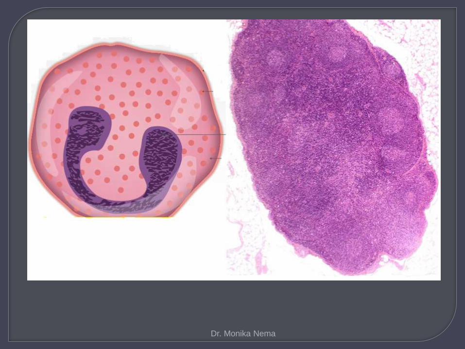
eosinophils in lymph node
TRANSCRIPT

Dr. Monika Nema
Dr. Monika Nema

Non neoplastic conditions
Filaria lymphadenitis.
Drug induced lymphadenopathy.
Kimura disease.
Angiolymphoid hyperplasia with
eosinophilia.
Dermatopathic lymphadenopathy.
Eosinophilic granuloma of lymph node.
Dr. Monika Nema

Neoplastic conditions
Mixed cellularity Hodgkin disease
Eosinophilic myeloid disorder
Angioimmunoblastic T cell Lymphoma
Dr. Monika Nema

Dr. Monika Nema
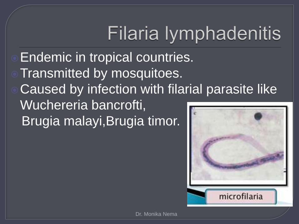
Endemic in tropical countries.
Transmitted by mosquitoes.
Caused by infection with filarial parasite like
Wuchereria bancrofti,
Brugia malayi,Brugia timor.
Dr. Monika Nema

In humans, adult filariae worms colonize lymphatic vessels and lymph nodes.
In men, the worms are most commonly found in the lymphatics of the epididymis and testis, and in women in the lymphatics of the breast.
They also invade the lymphatics of the legs and the inguinal and pelvic lymph nodes.
The lymphatics become occluded and inflammed.
Dr. Monika Nema

Blockage of the lymphatics in the lower
limbs may cause elephantiasis of the legs,
more often in the elderly persons.
Dr. Monika Nema

It is very rare to see microfilaria in the lymph node tissue which is an accidentally trapped site.
The larve can migrates through the blood vessels and lymphatics to be lodged in the lymphatics and mature to adult worm.
Viable microfilariae in lymphatics usually do not cause lesions.
When the adult worm or larva lodge in the lymph node and die, they produce an intense inflammatory reaction with the larva in the center accompanied by dense eosinophil infiltrate with microabscess and multinucleated giant cells.
Dr. Monika Nema
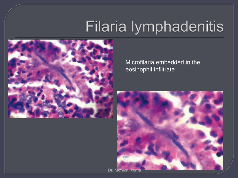
Microfilaria embedded in the
eosinophil infiltrate
Dr. Monika Nema

The longitudinal, loosely
arranged nuclear column
typical of W. bancrofti
species and the
adherence of
inflammatory cells to the
border of the sheath are
visible
Dr. Monika Nema

Dr. Monika Nema

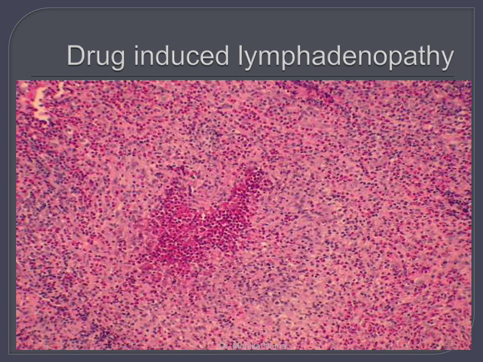
Associated with a systemic hypersensitivity
reaction to arene oxide- producing
anticonvulsant drugs.
Triad of fever,rash and lymphadenopathy.
Lymph node abormalities usually appear
early, within weeks or months, after the
initiation of anticonvulsant drug therapy.
Dr. Monika Nema

Clinical feature- hepatitis, gingival
hyperplasia,fever,
skin rash,eosinophilia,gum hyperplasia
and lymphadenopathy,
hepatospleenomegaly ,facial edema.
Dr. Monika Nema

Lymphodenopathy,regress after
anticonvulsants are discontinued, but they
may reappear if the same drug is resumed.
Dr. Monika Nema
Dr. Monika Nema

Dr. Monika Nema

Over the years, there has been considerable confusion between Kimura disease and angiolymphoid hyperplasia with eosinophilia(ALHE).
Clinically, both conditions present as soft tissue swellings that usually arise in the head and neck region with an indolent, prolonged clinical course. Microscopically, both processes show eosinophilic infiltrates and vascular proliferations.
Dr. Monika Nema

Features Kimura
lymphadenopathy
Angiolymphoid
hyperplasia with
eosinophilia
Age group Young Elderly
Race Asians Caucasian
Sex Males Females
Most affected site Deep subcutaneous
cervical masses with
regional lymph node and
salivary gland
involvement.
The lesions usually
involve skin in the form
of multiple small
superficial papules,
frequently
erythrematous.
Peripheral eosinophilia
and elevated serum IgE
levels
Often Rare
Dr. Monika Nema
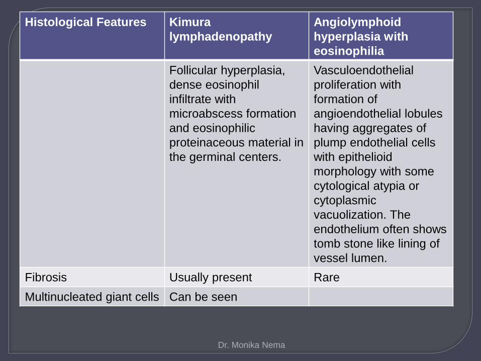
Histological Features Kimura
lymphadenopathy
Angiolymphoid
hyperplasia with
eosinophilia
Follicular hyperplasia,
dense eosinophil
infiltrate with
microabscess formation
and eosinophilic
proteinaceous material in
the germinal centers.
Vasculoendothelial
proliferation with
formation of
angioendothelial lobules
having aggregates of
plump endothelial cells
with epithelioid
morphology with some
cytological atypia or
cytoplasmic
vacuolization. The
endothelium often shows
tomb stone like lining of
vessel lumen.
Fibrosis Usually present Rare
Multinucleated giant cells Can be seen
Dr. Monika Nema

KIMURA
LYMPHADENOPATHY
ANGIOLYMPHOID
HYPERPLASIA WITH
EOSINOPHILIA
Dr. Monika Nema

Dr. Monika Nema

Lymphadenopathy associated with chronic dermatologic lesions representing the lymph node reaction to the drainage of melanin and various skin antigens.
Axillary and inguinal lymph nodes are most commonly involved.
Lymph nodes are enlarged,firm,movable and nontender.
Peripheral eosinophilia is frequently present.
Dr. Monika Nema

Maintained lymph node architecture with paracortical T zone expansion.
Lymphoid follicles and germinal centres are present.
Histiocytes are located in the cortex towards the periphery of node.
Intermingled with the histiocytesare variable number of plasma cells,eosinophils and occasionally neutrophils.
The lymph node medulla contains pronounced infiltrates of plasma cells,and medullary sinuses are distended and filled with histiocytes,plasma cells and eosinophils.
Dr. Monika Nema

Dr. Monika Nema

It is a form of Langerhans Cell
Histiocytosis that inolves only lymph nodes
and does not infiltrate any other organ.
Considered as a benign disease and
resolves spontaneously.
Occurs mainly in children and young adults
and show a slight preponderance of males.
Dr. Monika Nema

Lymph nodes are predominately infiltrated by Langerhans cells.
Langerhans cells are - Mononuclear histiocyte like cells with oval nuclei with
well defined round or oval cytoplasm. A prominent nuclear groove (coffee bean nuclei) can
be seen in most of the nuclei. Eosinophilic cytoplasm. Contain Birbeck granules on electron microscopy and
are lysozyme negative.
Mixture of inflammatory cells. Giant cells can be present.
Dr. Monika Nema
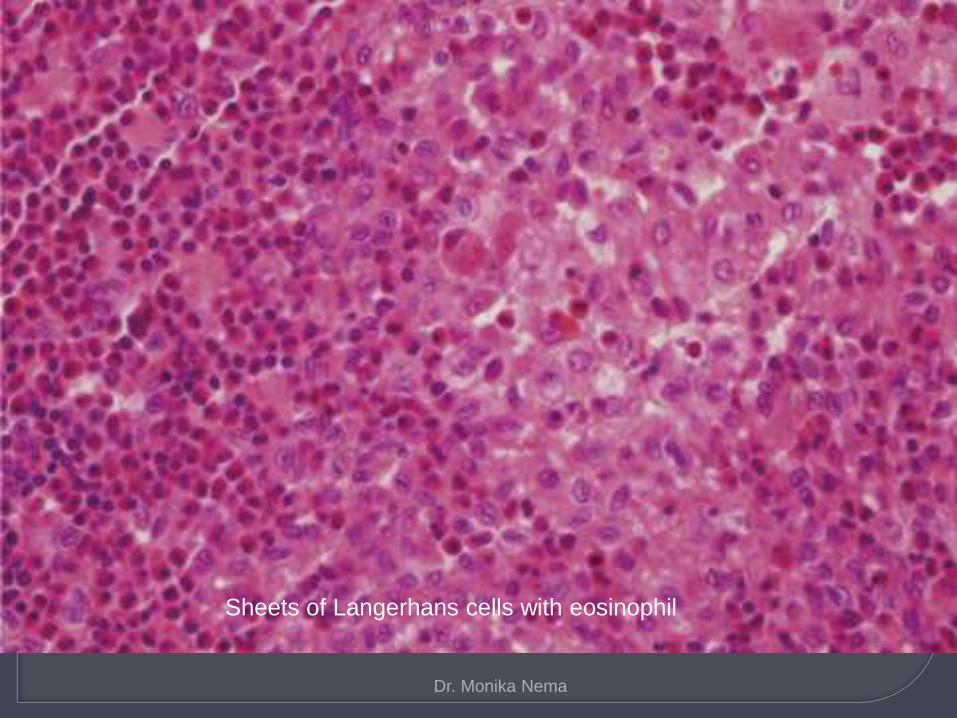
Sheets of Langerhans cells with eosinophil
Sheets of Langerhans cells with eosinophil
Dr. Monika Nema

Dr. Monika Nema

• 2nd most common type of Hodgkin
lymphoma in general population.
• The most common variety in HIV+
patients.
• Most patients present with peripheral
and/or abdominal adenopathy and B-
symptoms (fever, night sweats, and
weight-loss).
Dr. Monika Nema

The lymph node architecture is diffusely
effaced by a polymorphous population of
small lymphocytes, histiocytes, plasma
cells, and eosinophils in varying
proportions along with Reed-Sternberg
cells
Dr. Monika Nema

Dr. Monika Nema

Dr. Monika Nema

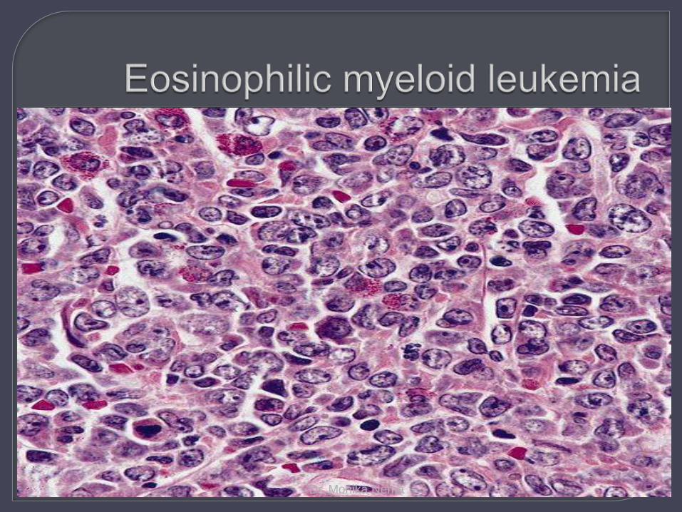
Represent a heterogenous group of disorders.
WHO classification- (1) myeloid and lymphoid neoplasms with
PDGFRA rearrangement. (2) myeloid neoplasms with PDGFRB
rearrangement. (3) myeloid and lymphoid neoplasms with FGFR1
abnormalities. (4) chronic eosinophilic leukemia not otherwise
specified. (5) idiopathic hypereosinophilic syndrome. (6) idiopathic hypereosinophilia.
Dr. Monika Nema
Dr. Monika Nema

Dr. Monika Nema
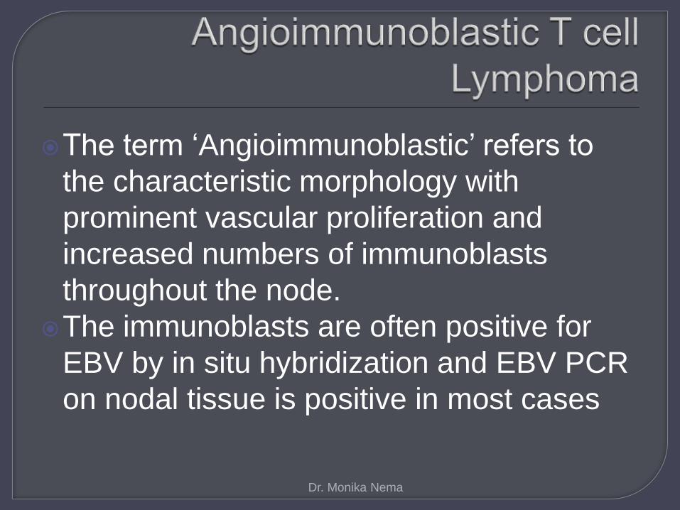
The term ‘Angioimmunoblastic’ refers to
the characteristic morphology with
prominent vascular proliferation and
increased numbers of immunoblasts
throughout the node.
The immunoblasts are often positive for
EBV by in situ hybridization and EBV PCR
on nodal tissue is positive in most cases
Dr. Monika Nema

Dr. Monika Nema

Dr. Monika Nema
Effaced lymph node architecture.Diffuse cellular proliferation.Characteristic triad of (a)
arborization,hyperplasia of small vessels; (b) immunoblasts,predominately T-cell type;(c) PAS positive material,clear cell immunoblasts,Reed Sternberg like cells,plasma cells,eosinophils,epithelioidcells.
Bone marrow,spleen,liver,lung may be involved.
Dr. Monika Nema

Whenever there is tissue or peripheral blood eosinophilia, especially in a patient from tropics or subtropics, the possibility of a parasitic infection should be thought.
If the organism is not seen in the initial sections, extensive sampling, adequate serial sectioning and vigilant search should be made to arrive at a correct diagnosis and to avoid misdiagnosis and mismanagement of the patient.
Dr. Monika Nema

Presentation by – Dr. Monika Nema
Dr. Monika Nema