eosinophilic esophagitis: extraesophageal manifestations · eosinophilic esophagitis:...
TRANSCRIPT
Eosinophilic Esophagitis:Extraesophageal Manifestations
Karen B. Zur, MD
Director, Pediatric Voice Program
Associate Director, Center for Pediatric Airway Disorders
The Children’s Hospital of Philadelphia
Associate Professor, Otolaryngology: Head & Neck Surgery
Perelman School of Medicine, The University of Pennsylvania
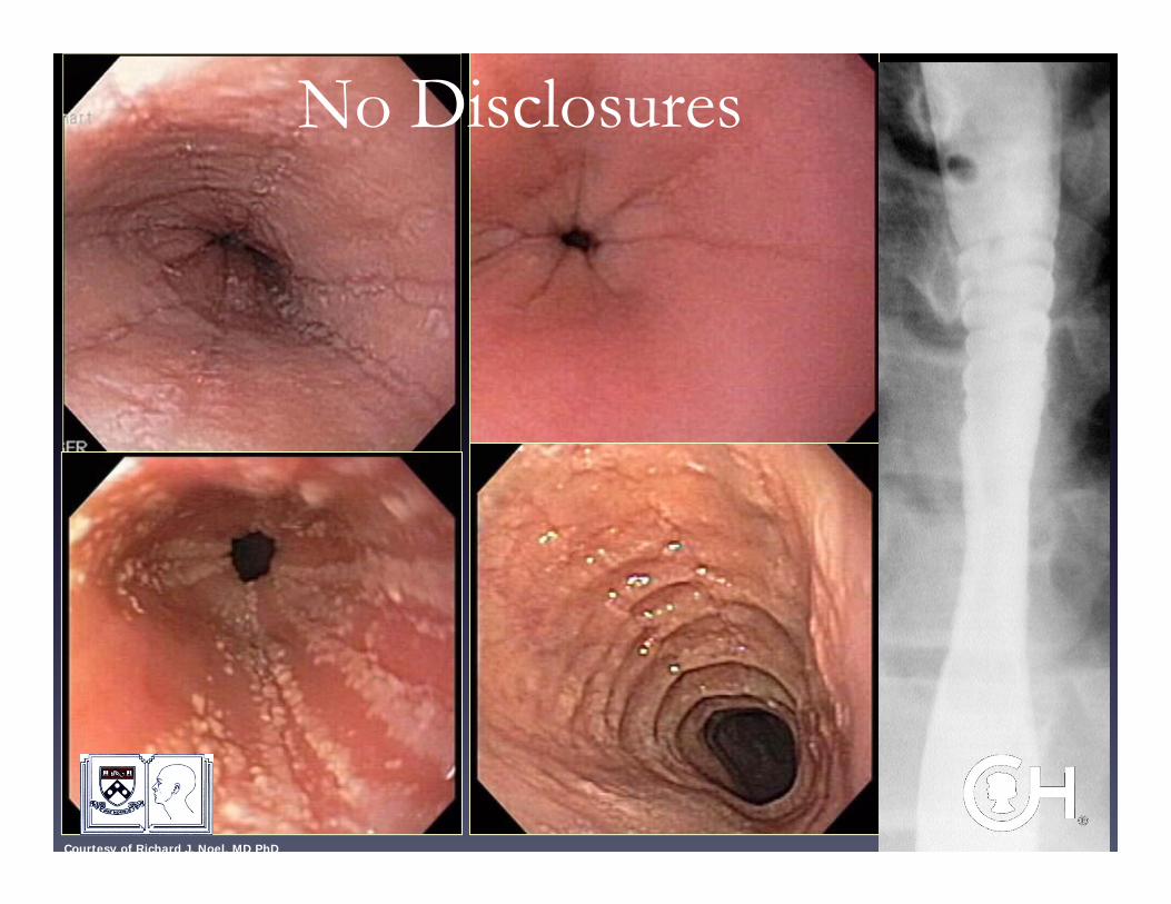
Courtesy of Richard J. Noel, MD PhD
No Disclosures

Introduction• EoE is a relatively new and unique
clinicopathologic entity
• Important cause of complaints relating to the
upper aerodigestive tract
• Asthma is the most common lower airway
finding
Introduction
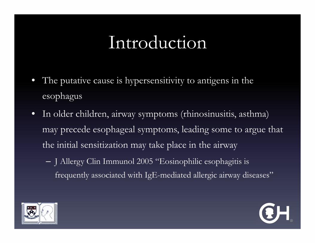
• The putative cause is hypersensitivity to antigens in the esophagus
• In older children, airway symptoms (rhinosinusitis, asthma) may precede esophageal symptoms, leading some to argue that the initial sensitization may take place in the airway
– J Allergy Clin Immunol 2005 “Eosinophilic esophagitis is frequently associated with IgE-mediated allergic airway diseases”
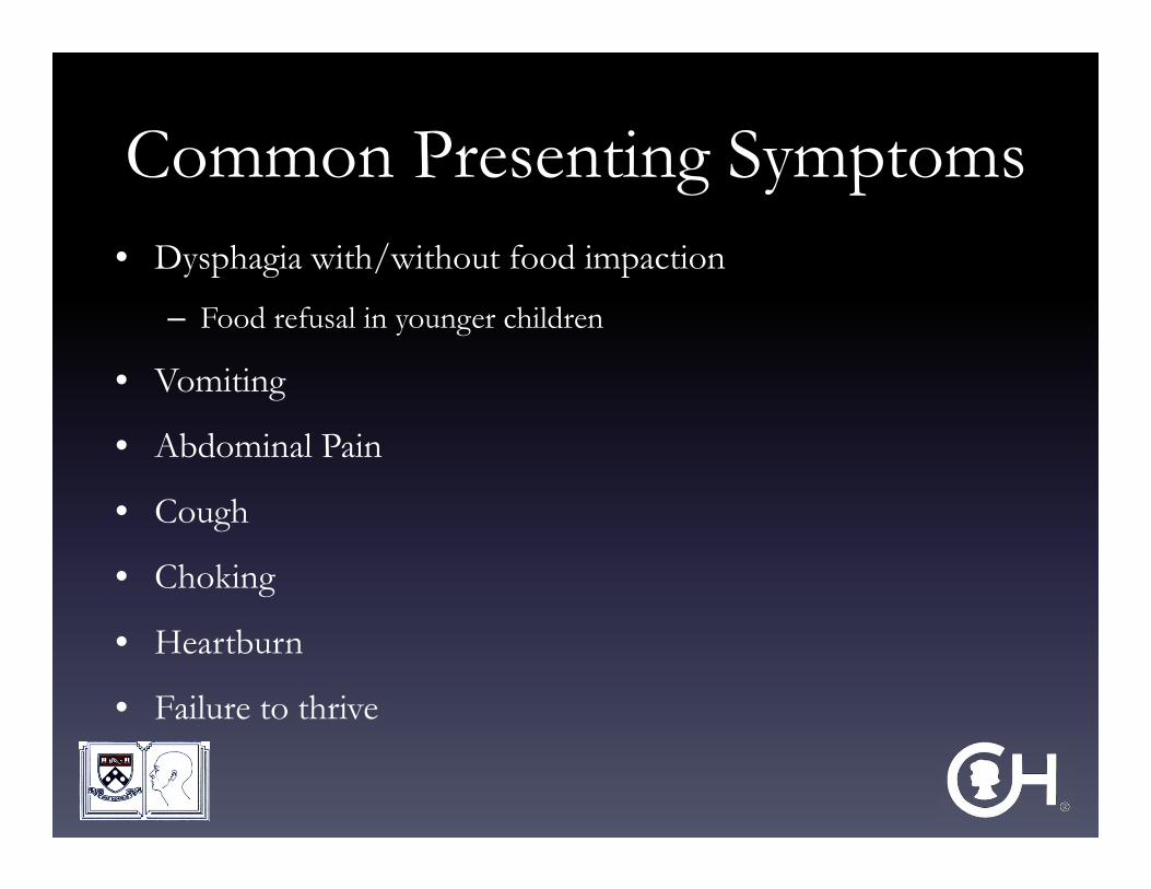
Common Presenting Symptoms• Dysphagia with/without food impaction
– Food refusal in younger children
• Vomiting
• Abdominal Pain
• Cough
• Choking
• Heartburn
• Failure to thrive
Presentation
• 15% of patients may present first to an otolaryngologist!
• A large proportion will get GERD treatment
• If no response to maximal medical therapy, need to think
about EoE
• Worsening symptoms during high pollen season noted
E.H. Dauer, et al, Clinical characteristics of eosinophilicesophagitis in children, Ann. Otol. Rhinol. Laryngol. 114 (11) (2005) 827–833.
ENT Symptoms• Rhinosinusitis
– Some believe it is unlikely that EE actually CAUSES rhinosinusitis, but that Eosinophils activation by allergic triggers affect the nose directly
• Laryngeal disorders
– Hoarseness
– Cough
• Recurrent croup
• Airway inflammation
– Subglottic stenosis
Mechanism of Action
• Postulated mechanism of action
– Direct deposition of pollen in the nose and/or
pharynx
– Subsequent swallowing of secretions

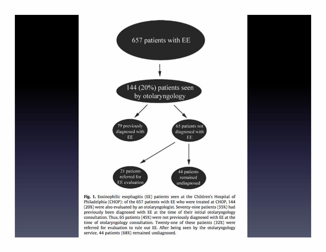
CHOP Study Findings
• 657 patient in the EoE IRB database 1994-2007– Of note, the Center for Airway Disorders formalized in 2005
• 144 were also seen by the general ENT division– 79% male
– Average age at diagnosis of EoE 5.5 yr (7mo-18yr)
– 60% were atopic (n=86)• Allergic rhinitis, asthma or atopic dermatitis
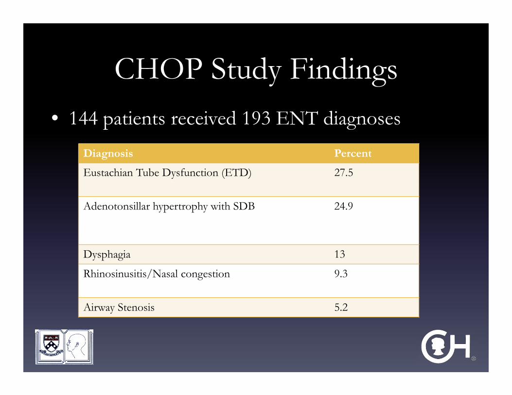
CHOP Study Findings• 144 patients received 193 ENT diagnoses
Diagnosis Percent
Eustachian Tube Dysfunction (ETD) 27.5
Adenotonsillar hypertrophy with SDB 24.9
Dysphagia 13
Rhinosinusitis/Nasal congestion 9.3
Airway Stenosis 5.2
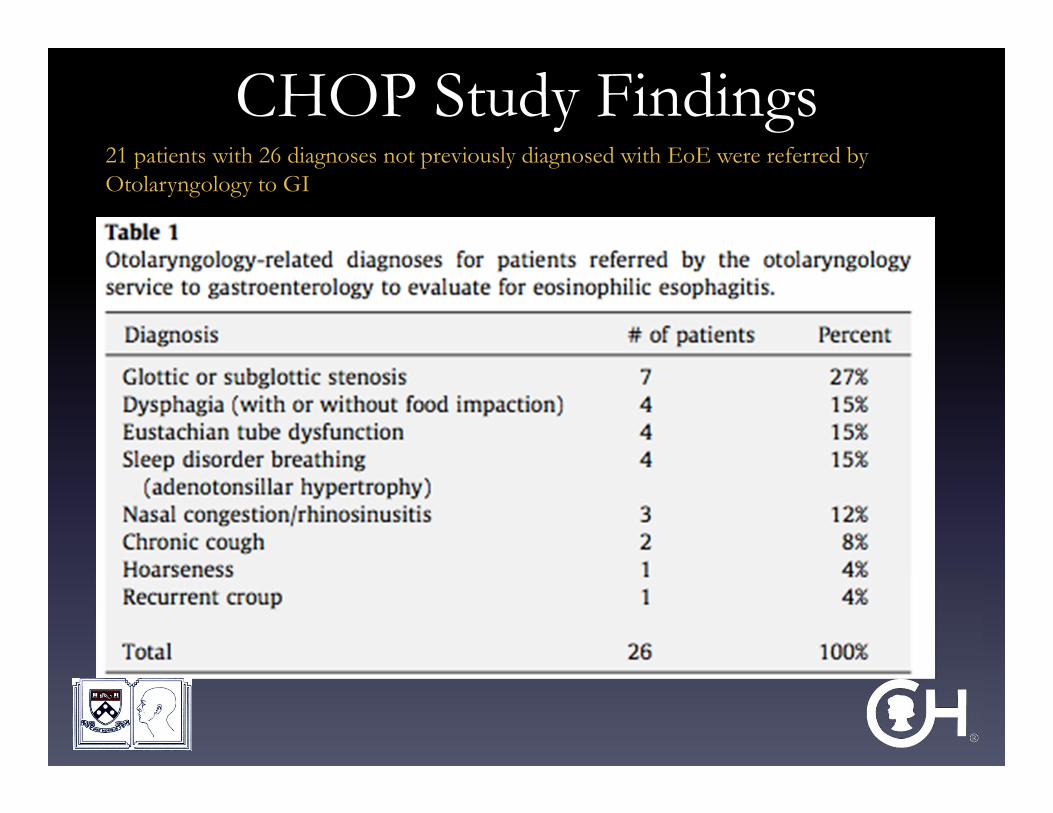
CHOP Study Findings21 patients with 26 diagnoses not previously diagnosed with EoE were referred by Otolaryngology to GI

Center for Pediatric EosinophilicDisorders at CHOP: Criteria for Dx• At least 1 clinical symptom
• No response to 2 month therapeutic trial of PPI
• EGD with biopsies from proximal, mid and distal
esophagus stomach and duodenum
• +/- colonoscopies as indicated
• EoE diagnosed if >20 Eos/HPF in esophagus with
normal stomach and duodenal biopsies
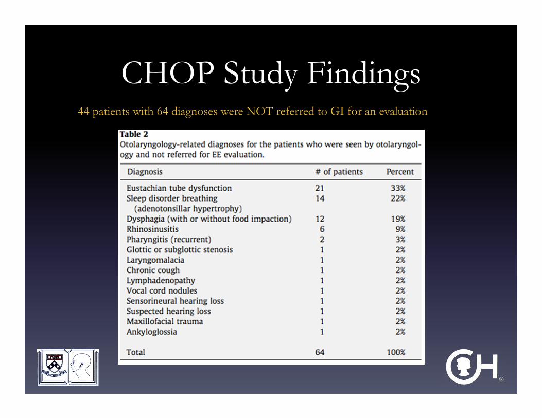
CHOP Study Findings44 patients with 64 diagnoses were NOT referred to GI for an evaluation
CHOP Study Findings:You Need a High Level of Suspicion
• Most children with EoE present to
otolaryngologist with routine complaints
• The 3rd most common complaint was dysphagia
+/- bolus impaction
Take Home Message
• A thorough review of systems with regards to
GI complains (dysphagia, vomiting, FTT)
• Ask about multiple food allergies
• Ask about dermatitis/eczema
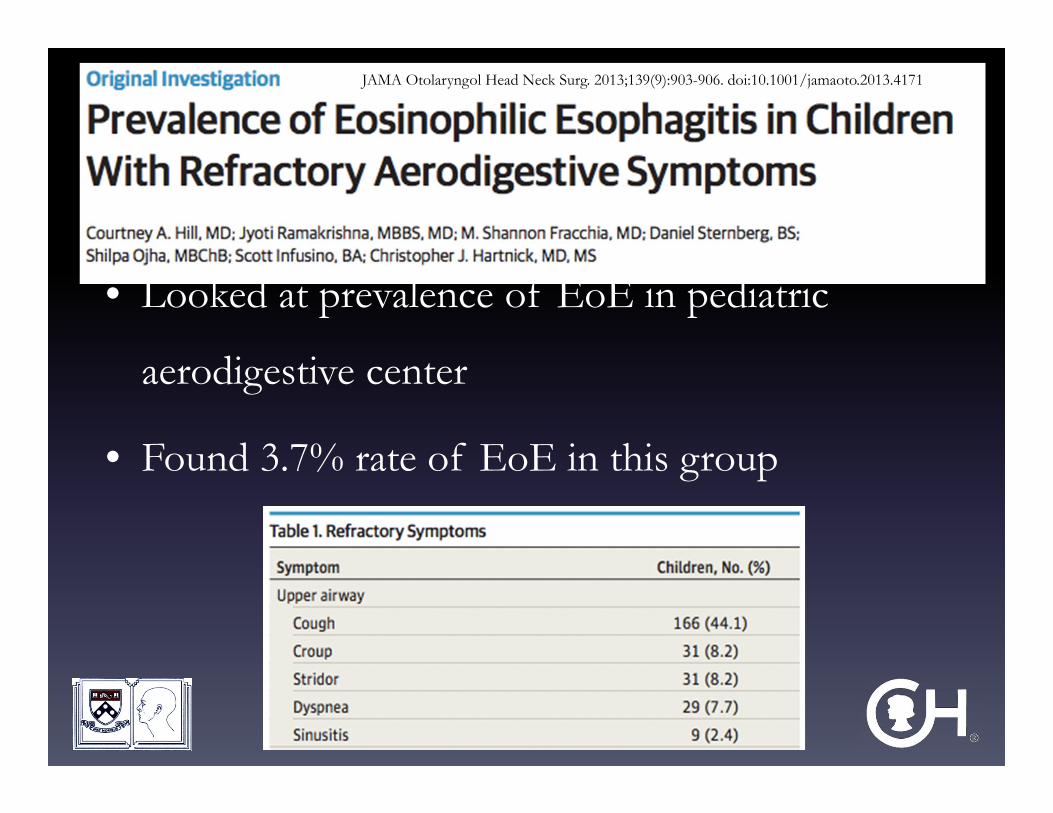
JAMA Otolaryngol Head Neck Surg. 2013;139(9):903-906. doi:10.1001/jamaoto.2013.4171
• Looked at prevalence of EoE in pediatric
aerodigestive center
• Found 3.7% rate of EoE in this group
Eosinophilic Esophagitis and the Airway
Why Does it Matter?
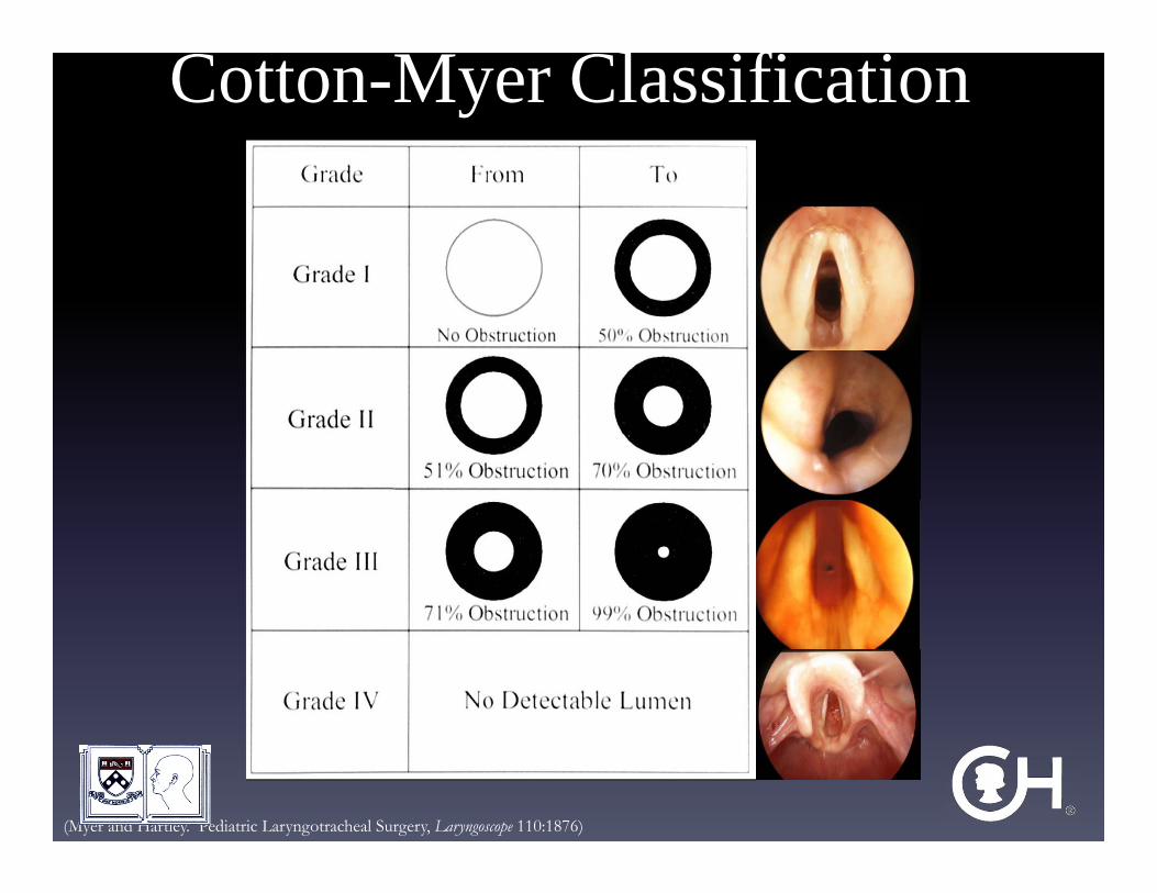
(Myer and Hartley. Pediatric Laryngotracheal Surgery, Laryngoscope 110:1876)
Cotton-Myer Classification
Gastroenterological Evaluation
• Why should we bother?
• Because 80% of airway reconstruction failures
have been linked to active laryngopharyngeal
reflux and/or Eosinophilic Esophagitis (EE)
– And 80% success when EoE was discovered and
treated Johnson & Rutter, ABEA 2003
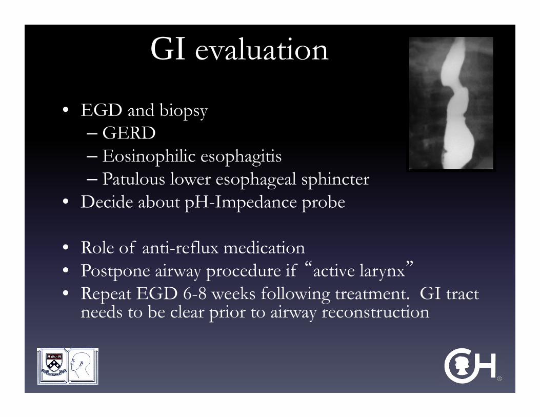
GI evaluation
• EGD and biopsy– GERD– Eosinophilic esophagitis– Patulous lower esophageal sphincter
• Decide about pH-Impedance probe
• Role of anti-reflux medication• Postpone airway procedure if “active larynx”• Repeat EGD 6-8 weeks following treatment. GI tract
needs to be clear prior to airway reconstruction
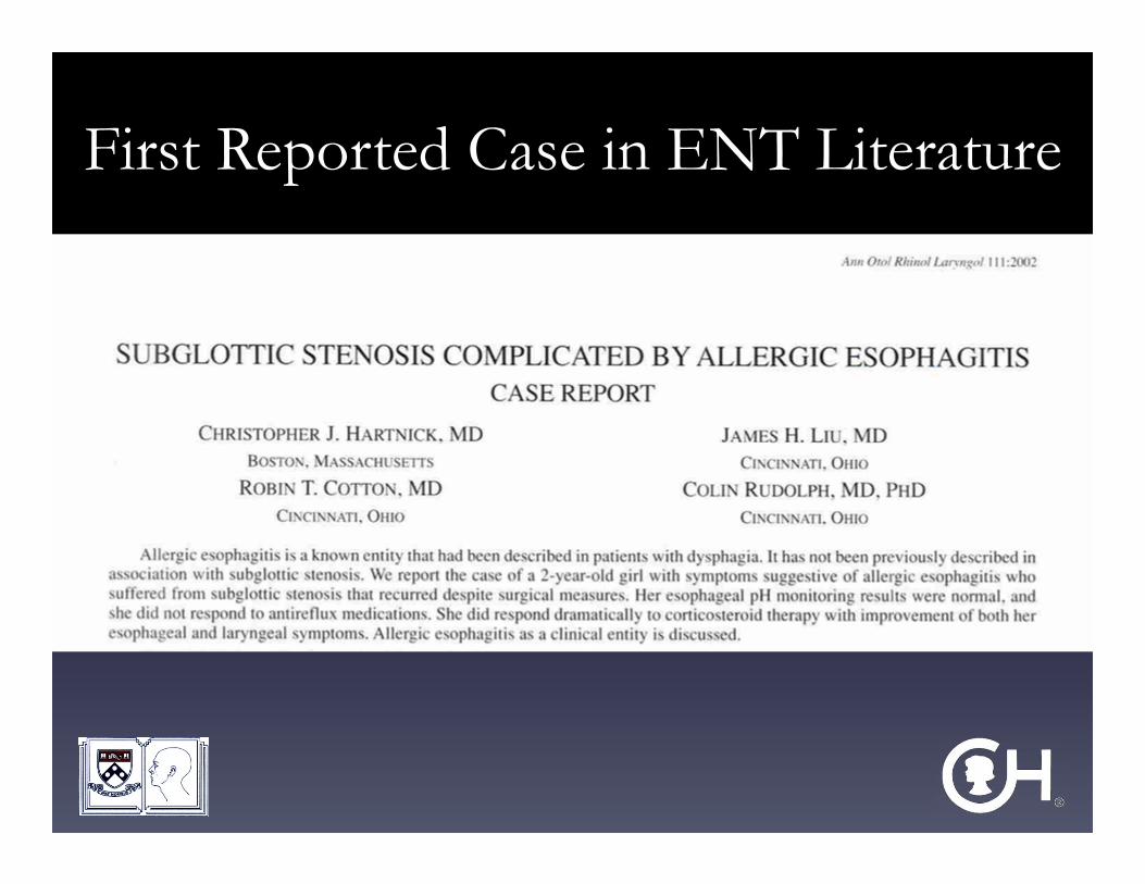
First Reported Case in ENT Literature
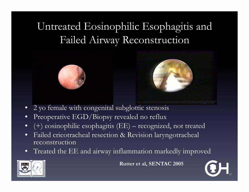
Untreated Eosinophilic Esophagitis and Failed Airway Reconstruction
• 2 yo female with congenital subglottic stenosis• Preoperative EGD/Biopsy revealed no reflux• (+) eosinophilic esophagitis (EE) – recognized, not treated• Failed cricotracheal resection & Revision laryngotracheal
reconstruction• Treated the EE and airway inflammation markedly improved
Rutter et al, SENTAC 2005
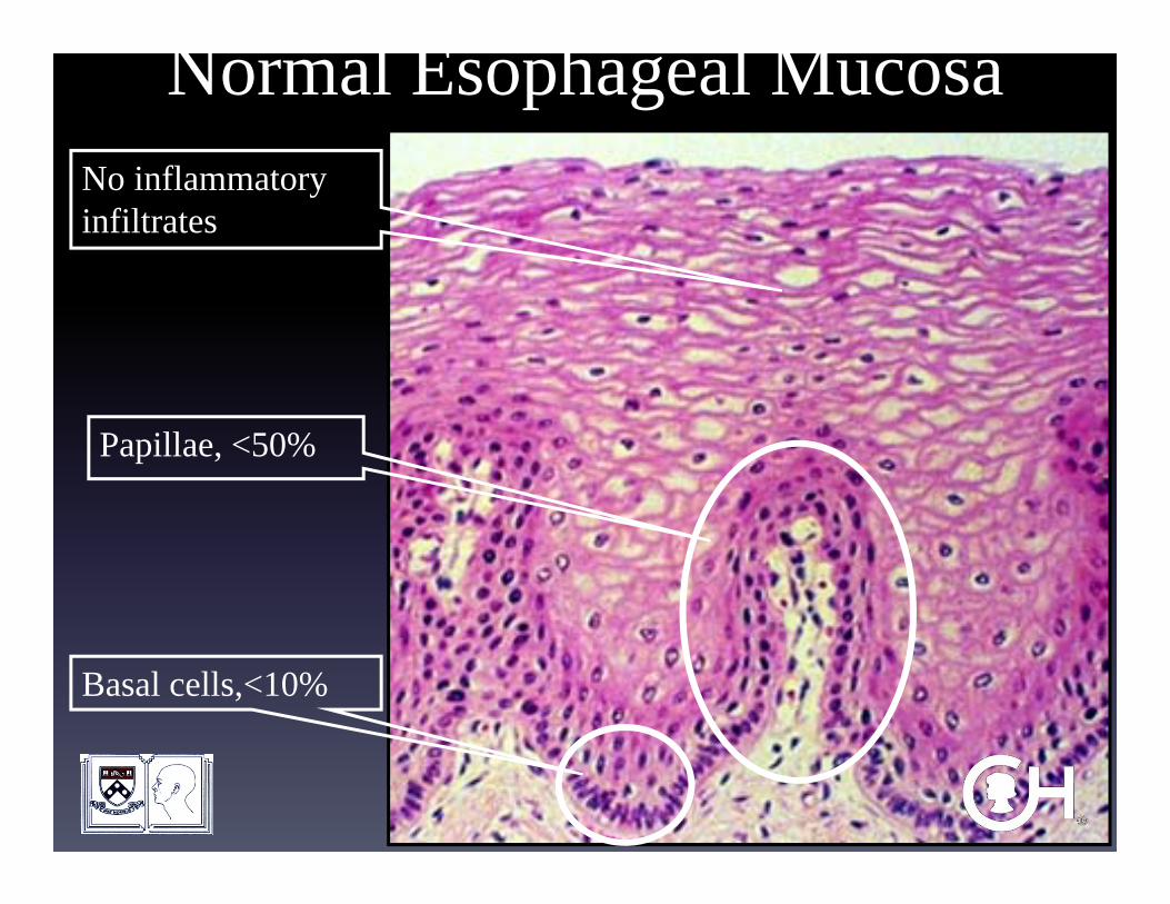
Papillae, <50%
Basal cells,<10%
No inflammatory infiltrates
Normal Esophageal Mucosa
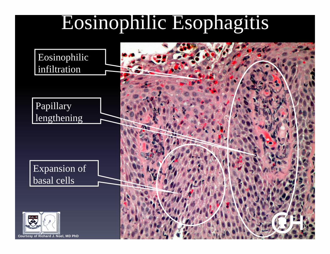
Eosinophilic infiltration
Papillary lengthening
Expansion of basal cells
Eosinophilic Esophagitis
Courtesy of Richard J. Noel, MD PhD
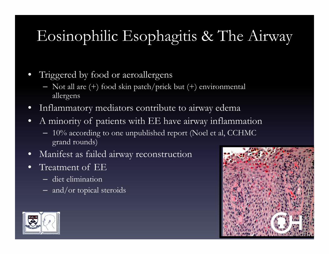
Eosinophilic Esophagitis & The Airway
• Triggered by food or aeroallergens– Not all are (+) food skin patch/prick but (+) environmental
allergens• Inflammatory mediators contribute to airway edema• A minority of patients with EE have airway inflammation
– 10% according to one unpublished report (Noel et al, CCHMC grand rounds)
• Manifest as failed airway reconstruction• Treatment of EE
– diet elimination – and/or topical steroids
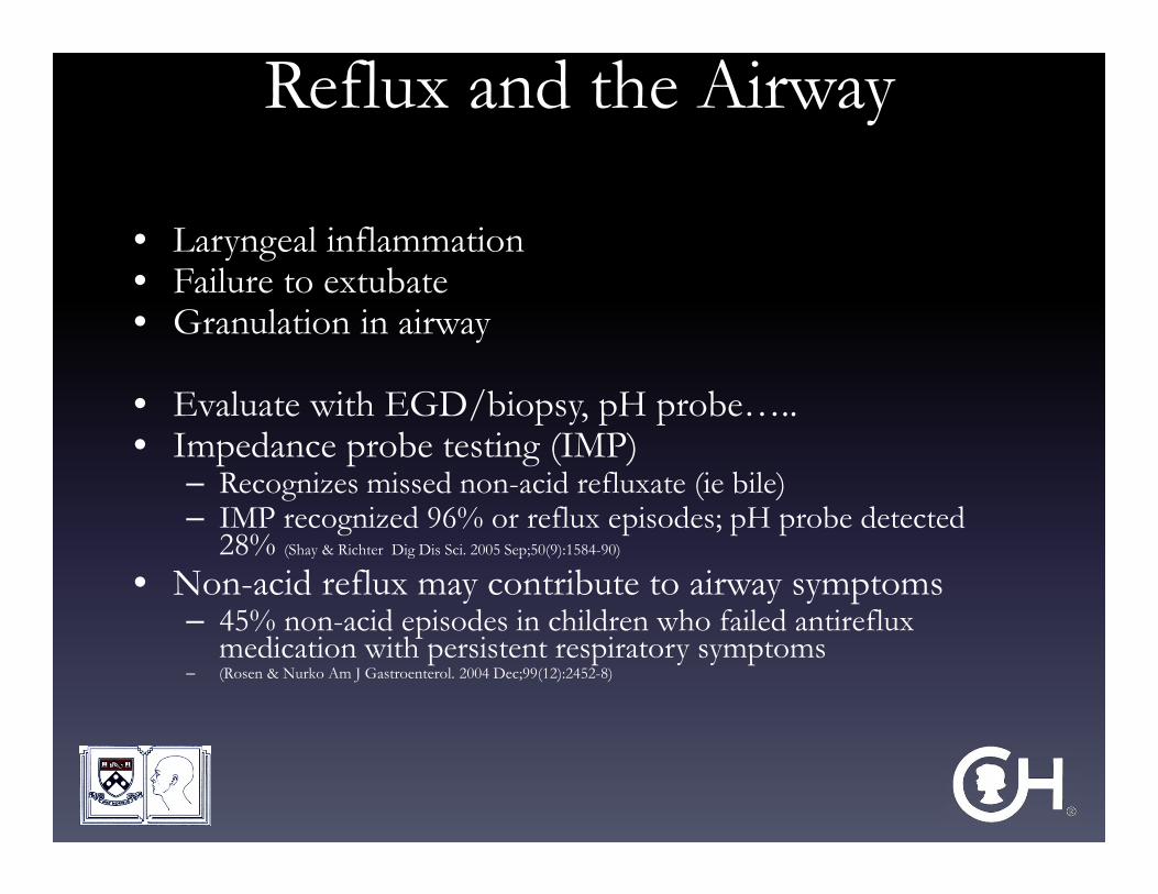
Reflux and the Airway
• Laryngeal inflammation• Failure to extubate• Granulation in airway
• Evaluate with EGD/biopsy, pH probe…..• Impedance probe testing (IMP)
– Recognizes missed non-acid refluxate (ie bile)– IMP recognized 96% or reflux episodes; pH probe detected
28% (Shay & Richter Dig Dis Sci. 2005 Sep;50(9):1584-90)
• Non-acid reflux may contribute to airway symptoms– 45% non-acid episodes in children who failed antireflux
medication with persistent respiratory symptoms – (Rosen & Nurko Am J Gastroenterol. 2004 Dec;99(12):2452-8)
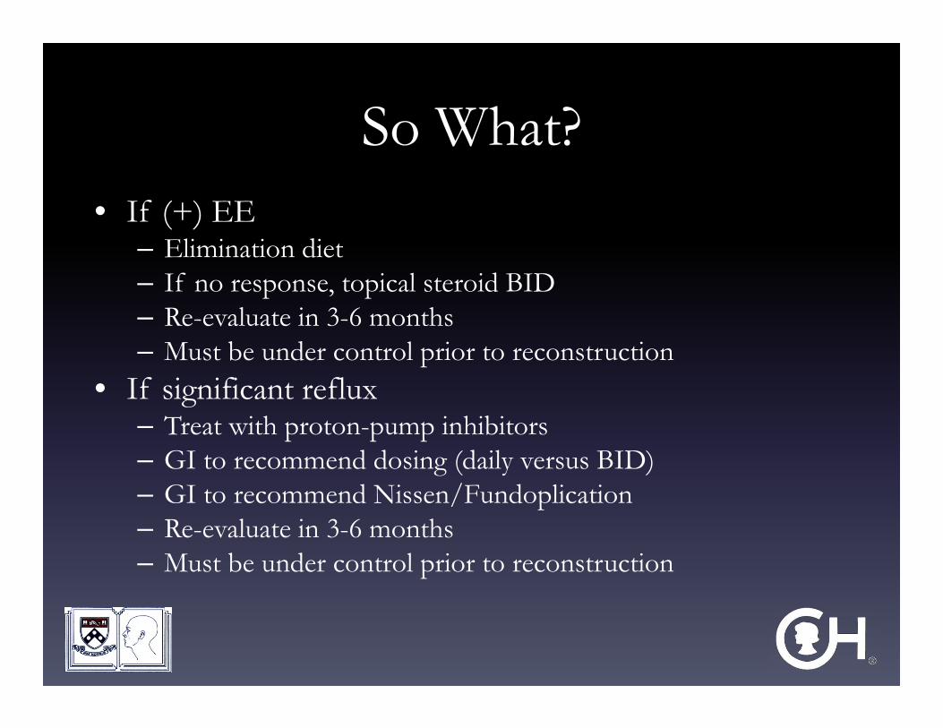
So What?• If (+) EE
– Elimination diet– If no response, topical steroid BID– Re-evaluate in 3-6 months– Must be under control prior to reconstruction
• If significant reflux– Treat with proton-pump inhibitors– GI to recommend dosing (daily versus BID)– GI to recommend Nissen/Fundoplication– Re-evaluate in 3-6 months– Must be under control prior to reconstruction
Collaboration is Essential• Airway patients often medically complicated
• Pre- and postoperative follow-up of chronic pulmonary conditions– Manage nebulizers
– Alternative non-invasive ventilation
• Pre- and postoperative follow-up of GI issues
• Do not miss treatable conditions!
• As a group, decide on appropriate timing of reconstruction