environmental sensing and signal transduction pathways ... · glcnac catabolic pathway of c....
TRANSCRIPT
MICROBIOLOGY AND MOLECULAR BIOLOGY REVIEWS, June 2007, p. 348–376 Vol. 71, No. 21092-2172/07/$08.00�0 doi:10.1128/MMBR.00009-06Copyright © 2007, American Society for Microbiology. All Rights Reserved.
Environmental Sensing and Signal Transduction Pathways RegulatingMorphopathogenic Determinants of Candida albicans
Subhrajit Biswas,1†‡ Patrick Van Dijck,2,3‡ and Asis Datta1*National Centre for Plant Genome Research, New Delhi 110 067, India,1 and Department of Molecular Microbiology,
VIB,2 and Laboratory of Molecular Cell Biology,3 Katholieke Universiteit Leuven, Kasteelpark Arenberg 31,B-3001 Leuven-Heverlee, Flanders, Belgium
INTRODUCTION .......................................................................................................................................................349DIMORPHISM, AN IMPORTANT VIRULENCE FACTOR................................................................................349GENETIC MANIPULATION WITH C. ALBICANS ..............................................................................................349ENVIRONMENTAL SENSING PATHWAYS REQUIRED FOR MORPHOGENESIS AND
PATHOGENESIS................................................................................................................................................350Amino Acid Availability Regulates Morphogenesis of C. albicans ...................................................................350
Amino acid sensing by Csy1..............................................................................................................................350Amino acid sensing (and transport) by the general amino acid permease Gap1 .....................................350Amino acid sensing by Gpr1 .............................................................................................................................351Ammonium sensing by Mep2 ............................................................................................................................351Role of Gcn4 in nitrogen-regulated morphogenesis in C. albicans ..............................................................352
Signal Transduction, Quorum Sensing, and the MAPK Cascade Module.....................................................353cAMP-PKA Pathway...............................................................................................................................................354
Upstream components of the cAMP-PKA pathway .......................................................................................354Ras1, the master hyphal regulator...................................................................................................................355Cyr1, Srv2, and Pde2..........................................................................................................................................355Adenylate cyclase and CO2 sensing..................................................................................................................355PKA.......................................................................................................................................................................356
Efg1 and Efh1, Major Transcriptional Regulators............................................................................................356Efg1 is a downstream component in the cAMP-PKA pathway ....................................................................356Roles of Efg1 and Efh1 in morphogenesis in C. albicans..............................................................................356Efg1 and embedded growth ...............................................................................................................................357Efg1 and phenotype switching...........................................................................................................................358Efg1 and cell wall dynamics ..............................................................................................................................358
Convergent Regulation of Cph1 and Efg1: Involvement of Tec1 and Cph2...................................................358Other Positive Regulators: Cdc5, G1 Cyclins, Int1, Mcm1, and Fkh2............................................................359Transcriptional Repressor Tup1...........................................................................................................................360
Tup1 repression with Nrg1, Mig1, and Rfg1 ..................................................................................................361Other Negative Regulators: Rap1, Rbf1, and Rad6...........................................................................................361Other MAPK Pathways Involved in Morphogenesis .........................................................................................362
The HOG MAPK pathway: response to oxidative stress ..............................................................................362Role of the PKC pathway in C. albicans morphology ....................................................................................362
GlcNAc Catabolic Pathway of C. albicans............................................................................................................363�-N-Acetylglucosaminidase and virulence.......................................................................................................364
pH Regulation in C. albicans .................................................................................................................................365Genes involved in pH regulation ......................................................................................................................365Rim101-dependent and -independent pathways .............................................................................................365Participation of endocytic machinery in pH signaling ..................................................................................366
DOWNSTREAM TARGETS OF THE DIFFERENT ENVIRONMENTAL SENSING PATHWAYSINVOLVED IN MORPHOGENESIS...............................................................................................................366
Adhesins ...................................................................................................................................................................366Role of Hwp1 in morphogenesis and pathogenicity.......................................................................................367ALS family and adhesion...................................................................................................................................367
Extracellular Hydrolytic Enzymes ........................................................................................................................368SAP gene expression in the yeast-to-hypha transition ..................................................................................368Phospholipase B..................................................................................................................................................368
* Corresponding author. Mailing address: National Centre for PlantGenome Research, New Delhi 110 067, India. Phone: 91-11-39440511.Fax: 91-11-26109186. E-mail: [email protected].
† Present address: Department of Biochemistry and Molecular Bio-logy, Medical University of South Carolina, Charleston, SC 29425.
‡ These authors contributed equally to the work.
348
on April 27, 2019 by guest
http://mm
br.asm.org/
Dow
nloaded from
FUTURE CHALLENGES ..........................................................................................................................................368FINAL OUTLOOK .....................................................................................................................................................369ACKNOWLEDGMENTS ...........................................................................................................................................369REFERENCES ............................................................................................................................................................369
INTRODUCTION
Opportunistic fungal pathogens, such as Candida albicans,are found in the normal gastrointestinal flora and the oralmucosa of most healthy humans. However, in immunocompro-mised patients, bloodstream infections often cause death, de-spite the use of antifungal therapies (152). The underlyingmolecular mechanisms for survival inside the human body andadaptation to various environments are probably distinct butoverlapping. Dietary factors, such as an excess of or deficiencyin certain nutrients, may alter the endogenous microbial flora.Mechanical factors, such as trauma or occlusive injury, can alsoalter the microenvironment, deplete the system of “friendlybacteria,” and enable the pathogenic fungus to take over. Im-munocompromised or immunosuppressed persons, includingAIDS patients, neonates, and transplant recipients, are alsoparticularly susceptible to fungal infections.
The most common systemic fungal infection is candidiasis,which accounts for well over half of these invasive mycoses. Asingle species, C. albicans, causes the majority of these infec-tions. Its success stems in part from its capacity to live as abenign commensal in a variety of body locations, most notablythe oral cavity, genitalia, and gastrointestinal tract (272). C.albicans expresses various traits critical for existence on muco-sal surfaces, where a constant but dynamic interplay occursbetween innate and acquired host defense mechanisms. Thepathogenic Candida species also establish well-developed bio-films, which occur easily on various implants and are resistantto antifungal agents (76). The nature of disease resulting fromtissue invasion by this organism is complex and depends on avariety of physical and physiological conditions in the host andon specific C. albicans traits. The capacity of C. albicans torapidly acquire resistance to antifungal drugs, such as ampho-tericin B, flucytosine, and a series of azoles, means that con-tinued development of new antifungals remains an importantfocus for clinicians and pharmaceutical companies.
An important feature of C. albicans, relevant to its patho-genesis, is its ability to switch between different morphologicalforms. C. albicans can grow in a single-celled, budding yeastform (blastospore) or in a filamentous form (including bothpseudohyphae and true hyphae) (31). A crucial component ofthis versatility is the ability to survive as a commensal in severalanatomically distinct sites, each with its own specific set ofenvironmental pressures. Thus, C. albicans must be able toadapt its growth to a range of physiological extremes. Toachieve adaptability, the fungus has evolved sophisticatedmechanisms of sensing and responding to environmental cuesby activating developmental switches that result in coordinatedchanges in cell physiology, morphology, and adherence.Progress in understanding many aspects of the biology of C.albicans has been hindered by the inability to carry out simple,large-scale genetic screens because of the diploid nature of thisorganism. A major breakthrough in assessing the contributionof specific genes to morphogenesis and virulence occurred withthe development of transformation protocols and methods of
deleting both alleles of a gene sequentially. Also, whole-ge-nome microarray analysis has now become an important toolfor probing signal transduction pathways during morphogene-sis in C. albicans (102).
Additional interest in the molecular mechanisms of C. albi-cans morphopathogenic determinants originated from the ne-cessity of identifying new drug targets due to increased drugresistance in clinical isolates. There is hope that recently de-veloped techniques of manipulating C. albicans and the se-quencing of its whole genome will lead to a thorough under-standing of its virulence and biology, thus offering thepossibility of a knowledge-based approach to the developmentof novel antifungal agents. A major strategy for determiningvirulence genes as molecular targets for antifungal drugs andvaccines is to identify a specific biochemical or structural targetunique to C. albicans (or to fungi in general) in an attempt tospecifically and selectively disrupt them and determine theireffects on virulence.
In this review, we focus on recent advances in the environ-mental sensing and signal transduction pathways that mediatethe morphogenesis and pathogenesis of C. albicans. Wherepossible, we compare the pathways of C. albicans with theanalogous pathways/genes in Saccharomyces cerevisiae.
DIMORPHISM, AN IMPORTANT VIRULENCE FACTOR
The terms “dimorphism” and “dimorphic fungus” (i.e., ex-isting in two morphological forms) are commonly accepted inreference to C. albicans. Strictly speaking, however, this fungushas the ability to adopt a spectrum of morphologies; thus, C.albicans can be considered a “polymorphic” or “pleomorphic”organism (71, 289). The production of germ tubes results inconversion to a filamentous growth phase or hypha, also calledthe mycelial form. The formation of pseudohyphae occurs bypolarized cell division when yeast cells growing by buddinghave elongated without detaching from adjacent cells. Undercertain nonoptimal growth conditions, C. albicans can undergothe formation of chlamydospores, which are round, retractilespores with a thick cell wall. These morphological transitionsoften represent a response of the fungus to changing environ-mental conditions and may permit adaptation to a differentbiological niche. The transition from a commensal to patho-genic lifestyle may also involve changes in environmental con-ditions and dispersion within the human host. Althoughprogress has been achieved in recent years, the molecularmechanisms governing these morphogenetic conversions arestill not fully understood, partly due to the difficulty of geneticmanipulations with C. albicans (164), an issue we addressbriefly below.
GENETIC MANIPULATION WITH C. ALBICANS
Sequencing of the genome of C. albicans has recently beencompleted. C. albicans has a diploid genome consisting of eightpairs of chromosomes that can be separated by pulsed-field gel
VOL. 71, 2007 SIGNAL TRANSDUCTION NETWORK IN C. ALBICANS 349
on April 27, 2019 by guest
http://mm
br.asm.org/
Dow
nloaded from
electrophoresis. With a size of �16 Mb, the haploid genome isslightly larger than that of the model yeast, S. cerevisiae. Moreinformation on the C. albicans genome can be found at www.candidagenome.org (68). Many genes are conserved betweenS. cerevisiae and C. albicans, and it is based on this similaritythat the mechanisms of many biological processes in C. albi-cans have been discovered. As highlighted throughout thisreview, in many cases, although the specific components ofrelevant signaling pathways are conserved, molecular mecha-nisms and environmental signals have often diverged, mostlikely because of the coevolution of C. albicans and its humanhost.
C. albicans poses special problems for scientists interested instudying gene function because it is diploid and because CUGis translated into a serine instead of a leucine (164). To analyzethe function of a gene, one must disrupt each of the two allelesby transformation. Although methods of transformation(spheroplast-polyethylene glycol, lithium acetate, and electro-poration) are patterned after those used with S. cerevisiae, thetransformation efficiency is poor. For the study of gene func-tion in C. albicans, a number of disruption protocols and se-lectable markers are commonly utilized (300). The most widelyused marker is the URA3 gene, which encodes orotidine-5�-phosphate decarboxylase and confers uracil prototrophy. How-ever, this marker must be used with caution; URA3 expressionlevels can affect virulence and are susceptible to chromosomeposition effects, thereby complicating the analysis of strainsconstructed with URA3 as a selectable marker (62, 261, 281,292). To avoid the use of the URA3 marker, various otherauxotrophic markers, as well as dominant markers, have beendeveloped and are currently used (206, 224, 241, 263). To studythe functions of essential genes, the C. albicans MET3 pro-moter, which is regulated by the level of methionine and/orcysteine in the medium, or the tetracycline on/off system can beused. These systems allow conditional expression so that theconsequences of depletion of a gene product may be investi-gated (57, 231, 254). In order to study the expression of certaingenes at either the RNA level or the protein level, a number ofC. albicans-optimized reporter constructs, or tags, have beengenerated (32). The development of these molecular tools hasgreatly accelerated the elucidation of morphogenesis andpathogenesis in C. albicans.
ENVIRONMENTAL SENSING PATHWAYS REQUIREDFOR MORPHOGENESIS AND PATHOGENESIS
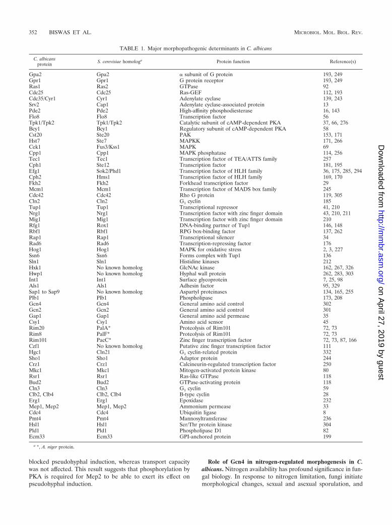
All organisms, from bacteria and yeast to higher eukaryotes,respond to changes that occur in the environment. In C. albi-cans, the yeast-to-hypha transition is triggered by variousenvironmental cues, such as serum, N-acetylglucosamine(GlcNAc), neutral pH, high temperature, starvation, CO2, andadherence. In recent years, receptors/sensors that may mediateenvironmental responses have been identified and partiallycharacterized. Many in vivo and in vitro experiments point toa prominent role for amino acids as nitrogen sources and asligands for membrane receptors involved in the regulation ofcellular morphology and virulence (summarized in Fig. 1 andTable 1). In the following sections, we highlight work aimed atidentifying receptors, ligands, and signaling pathways involvedin the sensing of environmental signals in C. albicans.
Amino Acid Availability Regulates Morphogenesisof C. albicans
Amino acid sensing by Csy1. Serum- and amino acid-basedmedia are known to induce filamentous growth in C. albicans.Although the mechanism by which amino acids induce fila-mentation is not well established, several genes have beenimplicated in the amino acid response. In S. cerevisiae, anamino acid-sensing complex (SPS sensor for Ssy1, Ptr3, andSsy5) that is required for amino acid transporter expressionthrough the induction of proteolytic cleavage of two latenttranscription factors has been described (4, 94). Recently asimilar sensing complex in C. albicans was described (45). Fur-thermore, it was shown that the amino acid sensor Csy1 (thehomolog of the yeast protein Ssy1) plays an important role infilamentation. Loss of Csy1 results in a lack of amino acid-mediated activation of amino acid transport and a lack ofinduction of transcription of specific amino acid permeasegenes. Csy1 mutants also show altered colony morphology andhyphal formation in serum- and amino acid-based solid mediabut not in media that do not contain amino acids. As in S.cerevisiae, Csy1 appears to discharge its critical role in aminoacid transport and filamentation by promoting proteolyticcleavage of two latent transcription factors, Stp1 and Stp2.Truncated Stp2 migrates to the nucleus and induces the ex-pression of genes required for amino acid uptake, and trun-cated Stp1 induces genes required for the degradation of ex-tracellular proteins (SAP2) and for uptake of peptides (OPT1)(198). The Ljungdahl group also discovered that Csh3, thefunctional homolog of S. cerevisiae Shr3, which is involved inendoplasmic reticulum exit after packaging, is essential for theproper uptake and sensing of extracellular amino acids andthat a null mutant is unable to switch morphology in responseto inducing amino acids (197).
Amino acid sensing (and transport) by the general aminoacid permease Gap1. In S. cerevisiae, the Gap1 protein func-tions not only as an amino acid transporter but also as anamino acid sensor for rapid activation of a fermentable growthmedium-induced signaling pathway that controls protein ki-nase A (PKA) targets (83, 140). Receptors with dual roles assensors have been dubbed “transceptors” (124). Our group hasdiscovered that the C. albicans Gap1 transporter, which is afunctional homolog of the budding yeast gene, may also be atransceptor important for initiating signal transduction path-ways resulting in morphogenesis and virulence. GAP1 was iso-lated in a screen to identify and characterize genes that couldbe involved in the regulation of morphogenesis and virulenceinduced by GlcNAc, an important hyphal induction regulatorof C. albicans (35) (see below). A null mutant of GAP1 has adefect in filamentation under nitrogen starvation conditions, aswell as upon the addition of GlcNAc (35). Addition of serum,however, still resulted in the induction of filamentation in thegap1 mutant, suggesting that Gap1 is not required for serum-induced hyphal formation. As in S. cerevisiae, the expressionof GAP1 is strongly repressed in medium containing ammo-nium as a good nitrogen source. How exactly amino acidtransport (and/or sensing) results in hyphal formation re-mains to be determined. One interesting link is the fact thatGlcNAc induces GAP1 expression at the yeast-to-germ-tubetransition. This induction depends on Cph1, the transcrip-
350 BISWAS ET AL. MICROBIOL. MOL. BIOL. REV.
on April 27, 2019 by guest
http://mm
br.asm.org/
Dow
nloaded from
tion factor that is the downstream target of the mitogen-activated protein kinase (MAPK) pathway (see below). Thismay indicate that Gap1-mediated filamentation is regulatedby this pathway.
Amino acid sensing by Gpr1. In S. cerevisiae, ScGpr1 is a Gprotein-coupled receptor that functions upstream of the cyclicAMP (cAMP)-PKA pathway and promotes a rapid cAMP in-crease after the addition of glucose or sucrose (156). In con-trast, C. albicans Gpr1 is not required for the glucose-inducedincrease in cAMP (193) but seems to directly or indirectlysense methionine (193). Addition of methionine to wild-typecells results in a rapid internalization of Gpr1, reminiscent ofligand-induced internalization, a typical feature of GPCR sys-tems in higher eukaryotic cells. Gpr1 is also required for themethionine-induced yeast-to-hypha transition (193). Furtherresearch is required to determine the mechanism by whichGpr1 activates the PKA pathway and what the role of methi-onine is in this mechanism. Interestingly, a role for methioninein morphogenesis is not limited to C. albicans. Recently, Heit-man and colleagues discovered that the addition of methionineresults in the internalization of a G protein-coupled receptor(Gpr4) in Cryptococcus neoformans and that a gpr4 null mutant
is still responsive to glucose (323). We discuss the Gpr1 recep-tor in more detail in “cAMP-PKA Pathway,” below.
Ammonium sensing by Mep2. Ammonium transport in C.albicans is mediated by Mep1 and Mep2 (33). Recently it wasshown that Mep2, but not Mep1, also functions as a receptorfor the induction of filamentous growth under nitrogen star-vation conditions, placing Mep2 in the transceptor family ofproteins (33, 124). More detailed analysis has shown that, aswith the budding yeast protein ScGap1, the C terminus ofMep2 is required for proper sensing but not for transport (33).Signaling by Mep2 functions through both the MAPK and thecAMP-PKA pathways and is Ras1 dependent (see below formore information on the Ras pathway). Under high ammo-nium conditions, when expression of Mep2 is repressed, fila-mentation is blocked. A role for Mep2 in polarized morpho-genesis is supported by work with both budding yeast andUstilago maydis. In S. cerevisiae, ScMep2 is required forpseudohyphal induction (186). The Mep2 homolog of U. maydis(Ump1) is able to complement an S. cerevisiae mep1 mep2mep3 strain both for ammonium transport and for pseudo-hyphal induction (269). Interestingly, mutation of a putativePKA phosphorylation site in either Mep2 or Ump1 specifically
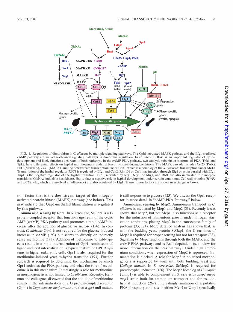
FIG. 1. Regulation of dimorphism in C. albicans by multiple signaling pathways. The Cph1-mediated MAPK pathway and the Efg1-mediatedcAMP pathway are well-characterized signaling pathways in dimorphic regulation. In C. albicans, Ras1 is an important regulator of hyphaldevelopment and likely functions upstream of both pathways. In the cAMP-PKA pathway, two catalytic subunits or isoforms of PKA, Tpk1 andTpk2, have differential effects on hyphal morphogenesis under different hypha-inducing conditions. The MAPK cascade includes Cst20 (PAK),Hst7 (MAPKK), Cek1 (MAPK), and the downstream transcription factor Cph1, which is a homolog of the S. cerevisiae transcription factor Ste12.Transcription of the hyphal regulator TEC1 is regulated by Efg1 and Cph2. Rim101 or Czf1 may function through Efg1 or act in parallel with Efg1.Tup1 is the negative regulator of the hyphal transition. Tup1, recruited by Rfg1, Nrg1, or Mig1, and Rbf1 are also implicated in dimorphictransitions. GlcNAc-inducible hexokinase, Hxk1, plays a negative role in hyphal development under certain conditions. Cell wall proteins (HWP1and ECE1, etc., which are involved in adherence) are also regulated by Efg1. Transcription factors are shown in rectangular boxes.
VOL. 71, 2007 SIGNAL TRANSDUCTION NETWORK IN C. ALBICANS 351
on April 27, 2019 by guest
http://mm
br.asm.org/
Dow
nloaded from
blocked pseudohyphal induction, whereas transport capacitywas not affected. This result suggests that phosphorylation byPKA is required for Mep2 to be able to exert its effect onpseudohyphal induction.
Role of Gcn4 in nitrogen-regulated morphogenesis in C.albicans. Nitrogen availability has profound significance in fun-gal biology. In response to nitrogen limitation, fungi initiatemorphological changes, sexual and asexual sporulation, and
TABLE 1. Major morphopathogenic determinants in C. albicans
C. albicansprotein S. cerevisiae homologa Protein function Reference(s)
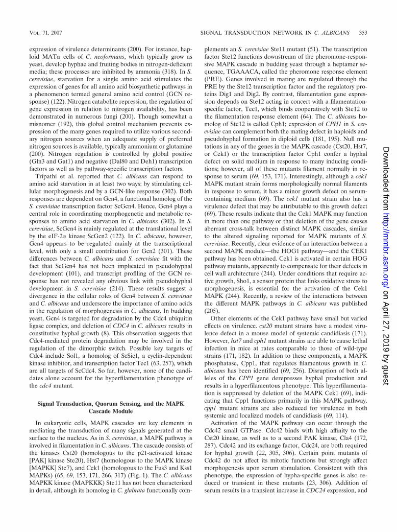
Gpa2 Gpa2 � subunit of G protein 193, 249Gpr1 Gpr1 G protein receptor 193, 249Ras1 Ras2 GTPase 92Cdc25 Cdc25 Ras-GEF 112, 193Cdc35/Cyr1 Cyr1 Adenylate cyclase 139, 243Srv2 Cap1 Adenylate cyclase-associated protein 13Pde2 Pde2 High-affinity phosphodiesterase 16, 143Flo8 Flo8 Transcription factor 56Tpk1/Tpk2 Tpk1/Tpk2 Catalytic subunit of cAMP-dependent PKA 37, 66, 276Bcy1 Bcy1 Regulatory subunit of cAMP-dependent PKA 58Cst20 Ste20 PAK 153, 171Hst7 Ste7 MAPKK 171, 266Cek1 Fus3/Kss1 MAPK 69Cpp1 Cpp1 MAPK phosphatase 114, 256Tec1 Tec1 Transcription factor of TEA/ATTS family 257Cph1 Ste12 Transcription factor 181, 195Efg1 Sok2/Phd1 Transcription factor of HLH family 36, 175, 285, 294Cph2 Hms1 Transcription factor of HLH family 169, 170Fkh2 Fkh2 Forkhead transcription factor 29Mcm1 Mcm1 Transcription factor of MADS box family 245Cdc42 Cdc42 Rho G protein 119, 305Cln2 Cln2 G1 cyclin 185Tup1 Tup1 Transcriptional repressor 41, 210Nrg1 Nrg1 Transcription factor with zinc finger domain 43, 210, 211Mig1 Mig1 Transcription factor with zinc finger domain 210Rfg1 Rox1 DNA-binding partner of Tup1 146, 148Rbf1 Rbf1 RPG box-binding factor 137, 262Rap1 Rap1 Transcriptional silencer 34Rad6 Rad6 Transcription-repressing factor 176Hog1 Hog1 MAPK for oxidative stress 2, 3, 227Ssn6 Ssn6 Forms complex with Tup1 136Sln1 Sln1 Histidine kinases 212Hxk1 No known homolog GlcNAc kinase 162, 267, 326Hwp1 No known homolog Hyphal wall protein 262, 283, 303Int1 Int1 Surface glycoprotein 7, 25, 98Als1 Als1 Adhesin factor 95, 329Sap1 to Sap9 No known homolog Aspartyl proteinases 134, 165, 255Plb1 Plb1 Phospholipase 173, 208Gcn4 Gcn4 General amino acid control 302Gcn2 Gcn2 General amino acid control 301Gap1 Gap1 General amino acid permease 35Csy1 Csy1 Amino acid sensor 45Rim20 PalA* Proteolysis of Rim101 72, 73Rim8 PalF* Proteolysis of Rim101 72, 73Rim101 PacC* Zinc finger transcription factor 72, 73, 87, 166Czf1 No known homolog Putative zinc finger transcription factor 111Hgc1 Cln21 G1 cyclin-related protein 332Sho1 Sho1 Adaptor protein 244Crz1 Crz1 Calcineurin-regulated transcription factor 250Mkc1 Mkc1 Mitogen-activated protein kinase 80Rsr1 Rsr1 Ras-like GTPase 118Bud2 Bud2 GTPase-activating protein 118Cln3 Cln3 G1 cyclin 59Clb2, Clb4 Clb2, Clb4 B-type cyclin 28Erg1 Erg1 Epoxidase 232Mep1, Mep2 Mep1, Mep2 Ammonium permease 33Cdc4 Cdc4 Ubiquitin ligase 8Pmt4 Pmt4 Mannosyltransferase 236Hsl1 Hsl1 Ser/Thr protein kinase 304Pld1 Pld1 Phospholipase D1 82Ecm33 Ecm33 GPI-anchored protein 199
a *, A. niger protein.
352 BISWAS ET AL. MICROBIOL. MOL. BIOL. REV.
on April 27, 2019 by guest
http://mm
br.asm.org/
Dow
nloaded from
expression of virulence determinants (200). For instance, hap-loid MAT� cells of C. neoformans, which typically grow asyeast, develop hyphae and fruiting bodies in nitrogen-deficientmedia; these processes are inhibited by ammonia (318). In S.cerevisiae, starvation for a single amino acid stimulates theexpression of genes for all amino acid biosynthetic pathways ina phenomenon termed general amino acid control (GCN re-sponse) (122). Nitrogen catabolite repression, the regulation ofgene expression in relation to nitrogen availability, has beendemonstrated in numerous fungi (200). Though somewhat amisnomer (192), this global control mechanism prevents ex-pression of the many genes required to utilize various second-ary nitrogen sources when an adequate supply of preferrednitrogen sources is available, typically ammonium or glutamine(200). Nitrogen regulation is controlled by global positive(Gln3 and Gat1) and negative (Dal80 and Deh1) transcriptionfactors as well as by pathway-specific transcription factors.
Tripathi et al. reported that C. albicans can respond toamino acid starvation in at least two ways: by stimulating cel-lular morphogenesis and by a GCN-like response (302). Bothresponses are dependent on Gcn4, a functional homolog of theS. cerevisiae transcription factor ScGcn4. Hence, Gcn4 plays acentral role in coordinating morphogenetic and metabolic re-sponses to amino acid starvation in C. albicans (302). In S.cerevisiae, ScGcn4 is mainly regulated at the translational levelby the eIF-2� kinase ScGcn2 (122). In C. albicans, however,Gcn4 appears to be regulated mainly at the transcriptionallevel, with only a small contribution for Gcn2 (301). Thesedifferences between C. albicans and S. cerevisiae fit with thefact that ScGcn4 has not been implicated in pseudohyphaldevelopment (101), and transcript profiling of the GCN re-sponse has not revealed any obvious link with pseudohyphaldevelopment in S. cerevisiae (214). These results suggest adivergence in the cellular roles of Gcn4 between S. cerevisiaeand C. albicans and underscore the importance of amino acidsin the regulation of morphogenesis in C. albicans. In buddingyeast, Gcn4 is targeted for degradation by the Cdc4 ubiquitinligase complex, and deletion of CDC4 in C. albicans results inconstitutive hyphal growth (8). This observation suggests thatCdc4-mediated protein degradation may be involved in theregulation of the dimorphic switch. Possible key targets ofCdc4 include Sol1, a homolog of ScSic1, a cyclin-dependentkinase inhibitor, and transcription factor Tec1 (63, 257), whichare all targets of ScCdc4. So far, however, none of the candi-dates alone account for the hyperfilamentation phenotype ofthe cdc4 mutant.
Signal Transduction, Quorum Sensing, and the MAPKCascade Module
In eukaryotic cells, MAPK cascades are key elements inmediating the transduction of many signals generated at thesurface to the nucleus. As in S. cerevisiae, a MAPK pathway isinvolved in filamentation in C. albicans. The cascade consists ofthe kinases Cst20 (homologous to the p21-activated kinase[PAK] kinase Ste20), Hst7 (homologous to the MAPK kinase[MAPKK] Ste7), and Cek1 (homologous to the Fus3 and Kss1MAPKs) (65, 69, 153, 171, 266, 317) (Fig. 1). The C. albicansMAPKK kinase (MAPKKK) Ste11 has not been characterizedin detail, although its homolog in C. glabrata functionally com-
plements an S. cerevisiae Ste11 mutant (51). The transcriptionfactor Ste12 functions downstream of the pheromone-respon-sive MAPK cascade in budding yeast through a heptamer se-quence, TGAAACA, called the pheromone response element(PRE). Genes involved in mating are regulated through thePRE by the Ste12 transcription factor and the regulatory pro-teins Dig1 and Dig2. By contrast, filamentation gene expres-sion depends on Ste12 acting in concert with a filamentation-specific factor, Tec1, which binds cooperatively with Ste12 tothe filamentation response element (64). The C. albicans ho-molog of Ste12 is called Cph1; expression of CPH1 in S. cer-evisiae can complement both the mating defect in haploids andpseudohyphal formation in diploid cells (181, 195). Null mu-tations in any of the genes in the MAPK cascade (Cst20, Hst7,or Cek1) or the transcription factor Cph1 confer a hyphaldefect on solid medium in response to many inducing condi-tions; however, all of these mutants filament normally in re-sponse to serum (69, 153, 171). Interestingly, although a cek1MAPK mutant strain forms morphologically normal filamentsin response to serum, it has a minor growth defect on serum-containing medium (69). The cek1 mutant strain also has avirulence defect that may be attributable to this growth defect(69). These results indicate that the Cek1 MAPK may functionin more than one pathway or that deletion of the gene causesaberrant cross-talk between distinct MAPK cascades, similarto the altered signaling reported for MAPK mutants of S.cerevisiae. Recently, clear evidence of an interaction between asecond MAPK module—the HOG1 pathway—and the CEK1pathway has been obtained. Cek1 is activated in certain HOGpathway mutants, apparently to compensate for their defects incell wall architecture (244). Under conditions that require ac-tive growth, Sho1, a sensor protein that links oxidative stress tomorphogenesis, is essential for the activation of the Cek1MAPK (244). Recently, a review of the interactions betweenthe different MAPK pathways in C. albicans was published(205).
Other elements of the Cek1 pathway have small but variedeffects on virulence. cst20 mutant strains have a modest viru-lence defect in a mouse model of systemic candidiasis (171).However, hst7 and cph1 mutant strains are able to cause lethalinfection in mice at rates comparable to those of wild-typestrains (171, 182). In addition to these components, a MAPKphosphatase, Cpp1, that regulates filamentous growth in C.albicans has been identified (69, 256). Disruption of both al-leles of the CPP1 gene derepresses hyphal production andresults in a hyperfilamentous phenotype. This hyperfilamenta-tion is suppressed by deletion of the MAPK Cek1 (69), indi-cating that Cpp1 functions primarily in this MAPK pathway.cpp1 mutant strains are also reduced for virulence in bothsystemic and localized models of candidiasis (69, 114).
Activation of the MAPK pathway can occur through theCdc42 small GTPase. Cdc42 binds with high affinity to theCst20 kinase, as well as to a second PAK kinase, Cla4 (172,287). Cdc42 and its exchange factor, Cdc24, are both requiredfor hyphal growth (22, 305, 306). Certain point mutants ofCdc42 do not affect its mitotic functions but strongly affectmorphogenesis upon serum stimulation. Consistent with thisphenotype, the expression of hypha-specific genes is also re-duced or transient in these mutants (23, 306). Addition ofserum results in a transient increase in CDC24 expression, and
VOL. 71, 2007 SIGNAL TRANSDUCTION NETWORK IN C. ALBICANS 353
on April 27, 2019 by guest
http://mm
br.asm.org/
Dow
nloaded from
this increased expression depends on Tec1, the second tran-scription factor (the homolog of ScTec1) downstream of theCek1 MAPK module (23). Cdc24 is then recruited to the tip ofthe hypha. The regulatory relationship between Tec1 andCdc24 suggests a positive feedback loop that contributes to thelevel of active Cdc42 at the tip of the germ tube.
Inputs into the Cek1 MAPK pathway may also occur byquorum sensing. Quorum-sensing molecules allow bacteria tomonitor their growth and to control cell density-dependentphenomena. Recently, similar regulatory molecules, tyrosoland farnesol, were identified in C. albicans, and they are in-volved in morphogenesis. Studies of the morphological transi-tion from a filamentous to a budding yeast form in C. albicansrevealed excretion of an autoregulatory substance, 3,7,11-tri-methyl-2,6,10-dodecatrienoate, or farnesol, into the medium.Farnesol inhibits filamentous growth and may be involved indevelopmental signaling (226). Whereas farnesol is a com-pound that prevents hyphal formation (it is produced duringhigh-density growth), tyrosol stimulates the growth of Candidacells and, under the proper conditions, hyphal formation (61,125). Recent work shows that farnesol may function throughthe Cek1 MAPK pathway to inhibit morphogenesis, as theaddition of farnesol represses the expression of CPH1 andHST7 (253). How farnesol is sensed and how the signal istransmitted to this MAPK pathway remains to be determined.Interestingly, C. albicans mutants lacking the histidine kinaseChk1 (see below) are refractory to the inhibitory effect offarnesol both in cell suspension and during the formation of abiofilm (158). This indicates a role of two-component signaltransduction proteins in quorum sensing and as upstream com-ponents of the Cek1 MAPK pathway.
cAMP-PKA Pathway
The cAMP-PKA pathway plays a very important role infilamentation in S. cerevisiae, C. albicans, and other fungi (177).Nitrogen starvation in S. cerevisiae results in the formation ofelongated buds termed pseudohyphae, which is dependent onactivation of the cAMP pathway (110, 157, 177). In C. albicans,an increase in cAMP levels accompanies the yeast-to-hyphatransition, and inhibition of the cAMP phosphodiesterase in-duces this transition (247). Previous reports of cAMP levelsduring the yeast-to-hypha transition (13, 85, 193, 247) are dif-ficult to compare because of differences in strains and experi-mental conditions. Nonetheless, it is clear is that the cAMPsignal is less pronounced in C. albicans than in S. cerevisiae.Our understanding of the C. albicans cAMP-PKA pathway isbased on considerable work with budding yeast, which webriefly review below. We then discuss the different componentsof the cAMP pathway in C. albicans and their roles in mor-phogenesis in more detail.
Upstream components of the cAMP-PKA pathway. In S.cerevisiae, the cAMP-PKA pathway is activated by a G protein-coupled receptor system consisting of the G protein-coupledreceptor ScGpr1 and the G� protein ScGpa2. ScGpr1 wasidentified in two-hybrid screens with the G� protein ScGpa2 asbait (156, 324) and in a screen for mutants deficient in glucose-induced loss of heat resistance, a property controlled by thecAMP-PKA pathway (156). Apart from the receptor, ScGpa2also interacts with ScGbp1/ScKrh2 and ScGbp2/ScKrh1, two
proteins that appear to act as G�-mimicking subunits, based onstructural resemblance with classical G� proteins (24, 117).Recently, two different molecular mechanisms by which thesekelch repeat proteins function were described (116, 233). Itseems that activated ScGpa2 relieves the inhibition imposed bythe kelch repeat proteins on PKA, thereby bypassing adenylatecyclase for direct regulation of PKA. It is also clear that bothScKrh1 and ScKrh2 may bind different components of thepathway, thereby functioning as scaffolding proteins. Anotherinteraction partner of ScGpa2 is ScGpg1, which has predictedstructural properties typical of a G�-like subunit, althoughScGpg1 lacks apparent target sequences for posttranslationalmodifications, which are typical of G� subunits (117). Theactivity of ScGpa2 is also controlled by the RGS proteinScRgs2 (309). Hence, a G protein-coupled receptor (GPCR)system composed of ScGpr1, ScGpa2, and ScRgs2 has beenproposed to act as a glucose-sensing system for control of thecAMP pathway (296, 310). Recently we obtained evidence thatScGpr1 is a sensor for sucrose and glucose and that mannoseacts as an antagonist (174). This GPCR system is required forpseudohyphal and invasive growth induction, and ScGPR1 orScGPA2 mutants, deficient in this morphogenesis, can be sup-pressed by the addition of cAMP (187, 188, 293).
Genes similar to ScGPA2 and ScGPR1 have been identifiedin C. albicans, but their precise functions remain unclear. Ge-netic evidence suggests that GPA2 may function upstream ofboth the Cek1 MAPK pathway (249) and the cAMP-PKApathway (193, 204). Deletion of GPA2 or GPR1 producesdefects in hyphal formation and morphogenesis in C. albicans,which are reversed by exogenous addition of (db)cAMP or byoverexpression of downstream components in the pathway. Asexpected, epistasis analysis revealed that Gpa2 (the G� pro-tein) acts downstream of Gpr1 (the receptor) in the samesignaling pathway, and a two-hybrid assay indicated that thecarboxy terminus of Gpr1 interacts with Gpa2. Moreover, ex-pression levels of HWP1 and ECE1, which are cAMP-depen-dent hypha-specific genes, are reduced in both mutants (204).Interestingly, the morphogenesis defect is present only whencells are cultured on solid media. A possible link betweenGpa2 and the CEK1 MAPK pathway was recently solidified bythe discovery that Gpa2 is involved in mating. Thus, Gpa2seems to integrate the nutrient-sensing pathway with the pher-omone response MAPK pathway, providing an explanation forwhy the function of the latter pathway strongly depends onnutritional conditions (27).
Conflicting data regarding the possible ligand for Gpr1 exist.Miwa et al. (204) showed that, as in S. cerevisiae, Gpr1 isrequired for the glucose-induced cAMP signal, but we foundthat the gpr1 strain showed the same glucose-induced increasein cAMP as did a wild-type strain (193). However, deletion ofeither CDC25 or RAS1 affected the glucose- and serum-in-duced cAMP signal, consistent with mediation of the glucose-induced cAMP increase in C. albicans by the Cdc25-Ras1branch of the glucose response pathway and probably not viathe Gpr1-Gpa2 branch (193). Addition of serum results in arapid response, similar to that seen with glucose, supporting arecent finding that glucose is the main factor in serum affectingCandida morphogenesis (135). Two important gaps remain inour understanding of the upstream components of the cAMP-PKA pathway in C. albicans: (i) the identity of the receptor
354 BISWAS ET AL. MICROBIOL. MOL. BIOL. REV.
on April 27, 2019 by guest
http://mm
br.asm.org/
Dow
nloaded from
activating the Cdc25 guanine nucleotide exchange protein and(ii) the nature of the ligand for Gpr1. No data from buddingyeast or C. albicans regarding the Cdc25 receptor are available,but, as mentioned above, preliminary investigations aimed atidentifying the possible ligand for Gpr1 seem to point to aminoacids, specifically methionine, although glucose or other sugarscannot be excluded (193, 194).
The concentration of glucose in the medium is a very im-portant parameter for the hyphal response. Most researchgroups use normal yeast extract-peptone-dextrose or syntheticcomplete medium containing 2% glucose. Under these condi-tions, a wild-type strain displays smooth colonies. However, inthe presence of more physiologically relevant concentrations(around 0.1% glucose in the blood), and when there is methi-onine present in the medium, a wild-type strain rapidly starts toform hyphae. This methionine-dependent, low glucose concen-tration-induced hyphal production is completely absent in thegpr1 mutant (194), as well as in an hgt12 mutant (189). Hgt12is homologous to S. cerevisiae Snf3, which is the high-affinityglucose sensor required for the expression of glucose trans-porter genes. The pathway that is activated by Hgt12 in C.albicans remains to be investigated, but the Hgt12 receptor isrequired for hyphal development during macrophage infec-tion. Hgt12 does not have the long C-terminal tail that istypical of the yeast Snf3 and Rgt2 glucose sensors. Recently,conflicting data regarding the role of Hgt12 as a sensor havebeen presented. Brown and colleagues constructed indepen-dent Hgt12 mutants and were unable to reproduce the growthor filamentation abnormalities. They showed that Hgt4, an-other homolog of the yeast Snf3 glucose sensor that doescontain a long C-terminal tail, might be the sensor regulatingthe expression of Hgt12, which, according to these authors, isa normal glucose transporter (48).
Ras1, the master hyphal regulator. Mutants of the singleRas homolog, Ras1, in C. albicans are viable but have a severedefect in hyphal growth in response to serum and other induc-ing conditions (92). In addition, while a dominant-negativeRas1 mutation [Ras1(A16)] causes a defect in filamentation, adominant-active Ras1 mutation [Ras1(V13)] enhances the for-mation of hyphae (92). As mentioned above, the morphogen-esis defect of a ras1 mutant can be suppressed by exogenouscAMP, and ras1 mutants are defective in cAMP induction inresponse to glucose and serum stimulation. Both phenotypesare shared with mutants lacking the guanine nucleotide ex-change factor Cdc25 (193), suggesting that the Cdc25-Ras1pathway functions upstream of the cAMP-PKA pathway. Thisis supported by a recent study in which a clear interactionbetween Ras1 and adenylate cyclase, Cyr1, is demonstrated(90). However, as with the situation in S. cerevisiae, Ras1 alsoappears to function upstream of the Cek1 MAPK pathway(Fig. 1), as the morphogenesis defect of a ras1 mutant can alsobe suppressed by overexpressing components of the filament-inducing MAPK cascade. Other evidence for a role in bothpathways is that ras1 mutant strains exhibit a filamentationdefect that is similar to that of the efg1 cph1 mutant strain (seebelow).
Cyr1, Srv2, and Pde2. C. albicans has a single gene homol-ogous to the S. cerevisiae adenylate cyclase gene (CYR1/CDC35). Like Ras1, the cyclase is not essential for growth in C.albicans but is required for hyphal development (243). Also as
in budding yeast, genes encoding an adenylate cyclase-associ-ated protein (SRV2) and two phosphodiesterases (PDE1 andPDE2) are present in the C. albicans genome. The Srv2 proteinregulates adenylate cyclase activity and is required for wild-type germ tube formation and for virulence in a mouse modelof systemic infection; it is the homolog of the S. cerevisiae Cap1protein. Like other mutants in the pathway, srv2 mutants havedefects in morphogenesis, but they can be rescued by theaddition of cAMP (13). Interestingly, the basal level of cAMPis higher in the srv2 mutant than in the wild type (61.8 � 6.5versus 45.3 � 4.6 pmol/mg protein), indicating a possible neg-ative-feedback inhibition.
To characterize more fully the effects of hyperactivation ofthe cAMP pathway, two groups disrupted the PDE2 gene,which encodes the high-affinity phosphodiesterase (PDEase)(16, 143). An advantage of studying cAMP hyperactivation bydisrupting PDE2 rather than overexpressing TPK1 and TPK2,which encode the catalytic subunits of PKA, is that the pde2mutant displays phenotypes that result solely from an increasein the cAMP level, thereby preempting any unpredictable ef-fects of inequitable overexpression of one cAMP signalingcomponent relative to the others. ScPDE1 and ScPDE2 encodethe low- and high-affinity cAMP PDEases, respectively, in S.cerevisiae (190, 221, 252). Budding yeast Scpde1 and Scpde2mutants exhibit sensitivity to heat shock and nutrient starva-tion (190), and Scpde2 mutants also have cell wall-related phe-notypes, such as lysis upon hypo-osmotic shock, suggesting arole for ScPde2 (and/or cAMP) in the maintenance of cell wallintegrity in S. cerevisiae (299). Although C. albicans also has alow-affinity PDEase encoded by PDE1 (127), so far only PDE2has been studied in detail. As with the situation in buddingyeast, pde2 mutants show cell wall- and membrane-relatedphenotypes, such as increased sensitivity to membrane-per-turbing agents such as sodium dodecyl sulfate or antifungalssuch as amphotericin B and a strong reduction in the thicknessof the cell wall, caused mainly by alterations in the ergosteroland glucan composition (144). PDE2 mutants are also sensitiveto nutrient deprivation and defective in entry into stationaryphase and are avirulent in a mouse model of systemic candi-diasis (16). Under conditions in which wild-type strains formsmooth colonies composed of budding yeasts on agar media,the homozygous pde2 mutant displayed a wrinkled colony phe-notype. The wrinkled colonies exhibited mixtures of elongatedyeasts and filamentous forms (pseudohyphae and true hyphae).Germ tube formation by the homozygous pde2 mutant wasaccelerated in liquid media compared to wild-type strains.Thus, it is clear that Pde2 is required to regulate the level ofcAMP in the cells and that disruption of this enzyme results ina constitutive activation of the PKA pathway.
Adenylate cyclase and CO2 sensing. C. albicans is present invarious parts of the body where the CO2 concentration is morethan 150-fold higher (5%) than in atmospheric air (0.033%). Inother words, cells growing on the skin face much lower CO2
concentrations than do cells present in the intestine or blood.The presence of 5% CO2 strongly induces pseudohyphal de-velopment and invasion of the underlying agar (151), a re-sponse that requires the catalytic domain of adenylate cyclase(Cyr1/Cdc35) but not Ras1. In many cases, cellular effectsobserved with CO2 can be mediated via its hydrated form,bicarbonate. C. albicans NCE103 encodes a carbonic anhy-
VOL. 71, 2007 SIGNAL TRANSDUCTION NETWORK IN C. ALBICANS 355
on April 27, 2019 by guest
http://mm
br.asm.org/
Dow
nloaded from

drase that greatly accelerates bicarbonate formation. Nce103 isrequired for C. albicans cells to grow in air but not under highCO2 concentrations (�0.5%). Nce103 is also required for tis-sue damage under atmospheric conditions but not under highCO2 conditions. Whether there is indeed a specific CO2 sensorprotein on the cell membrane (e.g., through aquaporin waterchannels as in plant and mammalian cells) remains to be es-tablished. Klengel et al. have already excluded C. albicansAqy1 as a possible candidate (151). A similar CO2-sensingpathway in Cryptococcus neoformans has also been described(14), and a review of this topic was recently published (15).
PKA. Growth and cellular differentiation of eukaryotic cellsdepends to a large extent on the activity of cAMP-dependentprotein kinases (PKA). PKAs are structurally conserved, con-sisting of two catalytic subunits that are inactivated by thebinding of a homodimer of regulatory subunits. External cueselevate intracellular levels of cAMP, whose binding to theregulatory subunits liberates and thereby activates the catalyticsubunits. A comparison of PKA subunit isoforms in C. albicansand S. cerevisiae reveals several significant differences. (i) In S.cerevisiae, PKA is encoded by three paralogues (ScTPK1 toScTPK3) (298), whereas only two paralogues (TPK1 and TPK2)are present in the genome of C. albicans (66). (ii) In C. albi-cans, both isoforms have a positive, stimulatory function on theformation of true hyphae, whereas in S. cerevisiae, ScTpk2 hasa positive function and ScTpk1 and ScTpk3 have negativefunctions in pseudohyphal development (229).
Although both C. albicans Tpk isoforms act positively toregulate hyphal formation, they have various phenotypic ef-fects. For example, tpk1 mutants are defective in hyphal for-mation on solid media but are less affected in liquid media(276) (Fig. 1). In contrast, hyphal formation in tpk2 mutants ispartially affected on solid media but is blocked in liquid me-dium. Because Tpk1 and Tpk2 mainly differ in their N-terminaldomains (80 to 90 amino acids), hybrid Tpk proteins, withexchanged N-terminal domains, were tested by Bockmuhl et al.(37). Tests of hybrid proteins suggested that the catalytic do-main mediates Tpk protein specificities in filamentation,whereas agar invasion is mediated by the N-terminal domain ofTpk2.
The regulatory subunits of PKA in S. cerevisiae and C. albi-cans are encoded by ScBCY1 and BCY1, respectively (58, 298).Cantore’s group examined the morphogenetic behavior of C.albicans yeast cells lacking the PKA regulatory subunit andgenerated a bcy1 tpk2 double mutant strain, because a homozy-gous bcy1 mutant in a wild-type genetic background could notbe obtained. In the bcy1 tpk2 mutant, PKA activity (due to thepresence of the TPK1 gene) was cAMP independent, indicat-ing that the cells harbored an unregulated phosphotransferaseactivity. This mutant has constitutive protein kinase activityand displays a defective germinative phenotype in GlcNAc andin serum-containing media. In addition, a Tpk1-green fluores-cent protein (GFP) fusion is dispersed throughout the cell inthe bcy1 tpk2 double mutant, while it is normally predomi-nantly nuclear in wild-type cells. These genetic studies, to-gether with biochemical evidence, suggest that C. albicans Bcy1may tether the PKA catalytic subunit to the nucleus andthereby perform a pivotal role in regulating the enzymaticactivity and availability of PKA in response to growth phase-related nutritional requirements (58).
Efg1 and Efh1, Major Transcriptional Regulators
The C. albicans EFG1 and EFH1 genes encode basic helix-loop-helix (bHLH) transcription factors that are members ofthe APSES family of fungus-specific regulators and involved inmorphogenetic processes (81). This class includes StuAp ofAspergillus nidulans and Asm1 in Neurospora crassa, as well asthe Phd1 and Sok2 proteins of S. cerevisiae. StuA is involved inthe formation of conidiophores, which are important for asex-ual reproduction in A. nidulans (202, 264), while Asm1 of N.crassa has a role in ascospore maturation (6). Phd1 enhancesand Sok2 suppresses pseudohyphal formation in S. cerevisiae(109, 314). In C. albicans, Efg1 plays a central role in morpho-genesis. Below, we first discuss Efg1 as a downstream compo-nent of the PKA pathway. Next, we discuss target genes in-duced or repressed by Efg1, followed by a more generaldiscussion of the genetic network regulating the expression ofyeast- or hypha-specific genes. Finally, we discuss the role ofEfg1 in morphogenesis under embedded conditions.
Efg1 is a downstream component in the cAMP-PKA path-way. Several lines of evidence suggest that Efg1 functionsdownstream of PKA. Overexpression of TPK2 is unable tosuppress the mutant phenotype of efg1, whereas overexpres-sion of EFG1 can bypass the filamentation defect in a tpk2mutant (276). The suppression activity of Efg1 depends on athreonine residue at position 206, a potential phosphorylationsite for PKA (36). Induction of morphogenesis after the addi-tion of serum, a process known to be regulated by the PKApathway, requires Efg1. When EFG1 transcript levels wereexamined in transformants overexpressing TPK1 or TPK2, areduction (about fourfold) of EFG1 mRNA was observed(294). This finding suggests that Efg1 is down-regulated by itsimmediate upstream regulator, PKA, but other observationspoint to autoregulation of EFG1 gene expression. Overexpres-sion of EFG1 increases the levels of a smaller 2.2-kb transcriptproduced by the locus, while the levels of the endogenous3.2-kb major EFG1 transcript decline dramatically (294) (Fig.2). Serial deletion analysis of the promoter region revealedthat the TATA box region is required for EFG1 autoregula-tion. Binding of Efg1 to the EFG1 transcriptional initiationregion by chromatin immunoprecipitation was also shown. Insummary, it appears that the levels and/or activities of Efg1,Tpk1, and Tpk2 are able to down-regulate the major EFG1promoter. In addition, Efh1, the homolog of Efg1, also affectsEfg1 down-regulation, indicating that at least four proteins cancontribute to EFG1 expression. Negative autoregulation andPKA-mediated down-regulation are probably mediated thoughthe Sin3-Rpd3 (279)-containing histone deacetylase complex(Fig. 2).
Roles of Efg1 and Efh1 in morphogenesis in C. albicans.Overexpression of Efg1 or Efh1 induces pseudohyphae andopaque-white switching, a specific morphogenetic transition(81, 182, 285). An efg1 mutant strain has a moderate but notcomplete defect in hyphal growth in response to many envi-ronmental conditions (182). Addition of serum or GlcNAc asan inducer in liquid or on solid medium completely blockshyphal formation in efg1 null mutants. In contrast, under mi-croaerophilic or embedded conditions, hyphal formation isunaffected—and may be stimulated—in homozygous efg1 mu-tants (111). These results indicate that, depending on environ-
356 BISWAS ET AL. MICROBIOL. MOL. BIOL. REV.
on April 27, 2019 by guest
http://mm
br.asm.org/
Dow
nloaded from

mental cues (and depending on the genetic background), Efg1may act as a transcriptional activator and repressor for differ-ent subsets of genes, whose balanced activities are essential foryeast, pseudohyphal, and hyphal morphogenesis of C. albicans.An efg1 cph1 double mutant strain has an extreme filamentousgrowth defect, with no detectable filamentation under almostany conditions tested, including the presence of serum (182).In addition, while the efg1 mutant strain has a minor reductionin virulence and the cph1 mutant has little or no defect, an efg1cph1 double mutant strain is essentially avirulent in a mousemodel of systemic infection (182). Recently, it was shown thatthis double mutant is still able to cause infections in the kid-neys of infected mice. So, although this mutant is avirulent,local proliferation of C. albicans cells can occur in certaintissues (60). Thus, Cph1 and Efg1 define elements of twoseparate pathways that together are essential for both filamen-tation and virulence in C. albicans.
Efg1 and embedded growth. As we have just described, efg1mutants have a drastic block in true hyphal formation undermost standard induction conditions, but considerable filamen-tation occurs in certain environments (46, 276). A limited sup-ply of oxygen, as occurs under a coverslip during induction ofchlamydospores, allows wild-type cells to form filaments, whichis enhanced in efg1 mutants (275). Similarly, growth of wild-type colonies embedded in agar stimulates filamentation,which still occurs in homozygous efg1 cph1 mutants (111, 242).
Thus, an Efg1-independent pathway of filamentation that isoperative under microaerophilic/embedded conditions appearsto exist in C. albicans. In wild-type strains, the filaments pro-duced under microaerophilic conditions are mostly pseudo-hyphae; however, they are mostly true hyphae in efg1 mutants(276). Interestingly, the alternative filamentation pathway isnot only independent of Efg1 but may be repressed by it to acertain degree. The enhanced filamentation in efg1 mutantsdoes not depend on the Cph1 MAPK, because a homozygousefg1 cph1 strain is as hyperfilamentous as the homozygous efg1mutant (276). It is possible that agar embedding generatesmicroaerophilic conditions, which activate the same Efg1-in-dependent pathway of morphogenesis under both conditions.The putative transcription factor Czf1 is probably an importantelement of the alternative pathway of filamentation in C. albi-cans (47). The central portion of Czf1 contains four clusters ofglutamine residues, and the C terminus contains a cysteine-richregion similar to zinc finger elements. Overexpression of CZF1stimulates filamentous growth but only under embedded con-ditions and in certain media lacking glucose. The expression ofCzf1 is strongly up-regulated in a vps34 mutant; as a result, avps34 mutant is derepressed in hyphal formation under mi-croaerophilic conditions (150). Vps34 is phosphatidylinositol-3-kinase, which influences vesicular intracellular transport, fil-amentous growth, and virulence. The exact role of Vps34 inCzf1-mediated morphogenesis is not yet clear. Homozygous
FIG. 2. (Top) White-opaque switching in C. albicans. Shown are scanning electron micrographs at a magnification of 1,000 and bright-fieldimages of white and opaque cells. (Reprinted, with permission, from the Annual Review of Microbiology [26], volume 59, © 2005 by AnnualReviews.) (Bottom) Model of morphogenetic regulation by Efg1. Under hypha-inducing conditions (e.g., serum, GlcNAc), Efg1 is induced andactivated; under microaerophilic conditions, however, it is repressed. The activated Efg1 (by PKA isoforms Tpk1 and Tpk2) initiates hyphalformation by inducing genes involved in hyphal formation and/or repressing genes directing the yeast form. Efg1 also induces the cell wall proteins(HWP1, HWP2, and RBE1) that are involved in adherence. The phase-specific genes (at the white-to-opaque-phase transition period) are alsoinduced by Efg1. In parallel, Efg1, in conjunction with the Sin3-Rpd3 deacetylase complex, silences chromatin and thereby down-regulates EFG1promoter activity (294). The 3.2-kb major transcript of EFG1 is expressed in the white phase, and the less-abundant 2.2-kb transcript is expressedin opaque cells. The Hda1-Rpd3 deacetylase complex regulates the white-to-opaque-phase transition as well as EFG1 down-regulation (279).
VOL. 71, 2007 SIGNAL TRANSDUCTION NETWORK IN C. ALBICANS 357
on April 27, 2019 by guest
http://mm
br.asm.org/
Dow
nloaded from
czf1 null mutants filament normally under standard inductionconditions, but they are defective in hyphal development whenembedded in agar. This defective phenotype occurs only dur-ing embedding in certain media, such as complex mediumcontaining sucrose or galactose as carbon sources at 25°C, butnot at 37°C, or in media containing strong inducers, includingserum and GlcNAc. These characteristics suggest that factorsother than Czf1 contribute to filamentation under embeddedconditions. The defective phenotype of a czf1 mutant is exac-erbated by the presence of a cph1 mutation, which by itselfshows defects in the types of media used for monitoring theczf1 phenotype. Thus, although the cph1 mutant phenotypedoes not appear to be specific for embedded conditions, itworsens the filamentation defects caused by the czf1 mutation.Hyperfilamentation of efg1 single and efg1 cph double mutantssuggests that Efg1 is a negative modulator of the Czf1 pathwayunder microaerophilic/embedded conditions. However, recentdata show that this hyperfilamentation phenotype of efg1 mu-tants may be caused by a Czf1-independent pathway, as efg1mutants do not express CZF1. Chromatin immunoprecipita-tion analysis has further indicated that Efg1 and Czf1 interactwith the promoter of CZF1. This indicates that, like EFG1,CZF1 is autoregulated (312).
Other aspects of C. albicans morphogenesis are also depen-dent on Efg1. Chlamydospore formation requires Efg1 proteinin PKA-dependent regulation, as T206 is required (275). Also,recent studies revealed the involvement of EFG1 in normalbiofilm formation (237), as well as in normal filamentationunder oxygen limitation conditions (260), which are probablysimilar to the microaerophilic conditions. Under these condi-tions, Efg1 seems to function as a repressor of filamentation. Incontrast to efg1, an efh1 mutant does not give any clear phe-notype except in an efg1 mutant background. In this case, anefh1 null mutant is hyperfilamentous under embedded or hyp-oxic conditions, indicating the cooperation of Efg1 and Efh1 insuppression of an alternative morphogenetic signaling pathway(81).
Efg1 and phenotype switching. As noted briefly above, oneform of phenotypic switching is the white-opaque transition,first described by Soll and colleagues in 1987 (268). C. albicanscolonies of some strains (e.g., WO-1) can switch from thenormal size, shape, and white color with high frequency (274)to larger, flatter, and gray colonies. White cells are similar inshape, size, and budding pattern to cells of common laboratorystrains. Opaque cells, however, are bean shaped and exhibitthree times the volume and twice the mass of white cells (Fig.2) (268). Transcription of EFG1 is regulated in a unique fash-ion during the white-opaque transition (280); the more-abun-dant 3.2-kb transcription is expressed in the white phase, whilethe less-abundant 2.2-kb transcription is detected in theopaque phase (Fig. 2). Experiments using deletion or overex-pression of EFG1 show that Efg1 functions downstream of theswitch event in the regulation of a subset of white-phase-spe-cific genes involved in the generation of the round white cellshape (278). Furthermore, a detailed promoter analysis sug-gests that the upstream region of EFG1 contains overlappingpromoters for the expression of white-phase-specific andopaque-phase-specific transcripts (280). Overlapping promot-ers are also seen for the � and � mRNAs of the EFG1 homologStuA in A. nidulans (320). It should be noted that EFG1 is not
the only phase-regulated gene expressed as an alternative-molecular-weight transcript during the white-opaque transitionin C. albicans. The deacetylase HOS3 is also transcribed as a2.5-kb transcript in the white phase and as a less-abundant,lower-molecular-weight 2.3-kb transcript in the opaque phase(279). Recently, a key transcription factor required to establishand maintain the opaque growth phase was described by dif-ferent groups. Like Efg1, this transcription factor, Wor1(Tos9), binds to its own regulatory region, thereby activating itsown expression. It is believed that high levels of Wor1 regulatethe epigenetic inheritance of the opaque phase of growth (60,131, 277).
Efg1 and cell wall dynamics. Recently, several genes encod-ing cell wall components were shown to be regulated by Efg1,implying cell wall regulation in C. albicans morphogenesis.These genes include the cell wall mannoprotein Hwp1 (42,262), the glycosylphosphatidylinositol (GPI)-anchored cell wallprotein HYR1 (17), and ALS1, which encodes a cell surfaceglycoprotein (95). In order to analyze cell wall dynamics andthe regulatory function of Cph1 and Efg1 in the transcriptionalcontrol of cell wall genes in a systematic manner, Sohn et al.used a DNA microarray to assay transcriptional profiles fromwild-type cells and cph1, efg1, and cph1 efg1 double-mutantstrains cultured under various yeast- or hypha-inducing condi-tions (271). Overall, their data demonstrate that Efg1 plays amajor role in the regulation of cell wall genes analyzed underboth yeast- and hypha-inducing conditions, while Cph1 plays aminor role. During induction of filamentation, many hypha-specific genes that are important for adhesion and virulenceare expressed in wild-type cells (in an Efg1-dependent man-ner). Among those genes are the hyphal wall proteins HWP1and HWP2. Concomitant with the up-regulation of hyphal wallproteins, the expression of yeast-specific genes such as YWP1 isdown-regulated during the yeast-to-hypha transition. Thus, thereduced virulence of the efg1 mutant likely reflects both a lackof expression of hypha-specific genes and a change in theoverall organization of the cell wall (155, 182). Microarrayanalysis has further demonstrated that apart from yeast- orhypha-specific genes, Efg1 is also important for the expressionof metabolism genes, inducing glycolytic genes and repressinggenes essential for oxidative metabolism (81). Finally, Erg3,which is involved in drug resistance, is also down-regulated byEfg1, and efg1 mutants are more resistant to antifungal agents(183).
Convergent Regulation of Cph1 and Efg1: Involvement ofTec1 and Cph2
Tec1, a member of the TEA/ATTS family of transcriptionfactors, has been shown to regulate hyphal development andvirulence in C. albicans (257). TEA/ATTS family membersAbaA in A. nidulans and ScTec1 in S. cerevisiae are involved inthe regulation of conidiophore formation and pseudohyphalgrowth, respectively (5, 106). As mentioned before, in S. cer-evisiae ScTec1 and ScSte12 form a transcription factor complexto specifically activate genes involved in pseudohyphal growth(64, 191). Such complexes have not yet been identified in C.albicans. In C. albicans, TEC1 transcription is not regulated bythe ScSte12 homolog Cph1 (171) but by Cph2 and Efg1. Cph2,an myc family bHLH protein, regulates hyphal development in
358 BISWAS ET AL. MICROBIOL. MOL. BIOL. REV.
on April 27, 2019 by guest
http://mm
br.asm.org/
Dow
nloaded from
C. albicans (169). cph2 mutant strains are impaired in hyphaldevelopment and in the induction of hypha-specific genes inliquid Lee’s medium, and Cph2 is necessary for the transcrip-tional induction of TEC1 (Fig. 1). Cph2 binds directly to twosterol regulatory element 1-like elements upstream of TEC1.Furthermore, the ectopic expression of TEC1 suppresses thedefect of cph2 in hyphal development. The function of Cph2 inhyphal transcription is therefore mediated, in part, throughTec1. tec1 mutants exhibit suppressed filamentation in liquidserum-containing media. EFG1 overexpression does not sup-press the morphological defect of the tec1 mutant, whereasTEC1 overexpression has a partial phenotype in the efg1 mu-tant (257). These results, coupled with the fact that efg1 strainshave a more severe defect in hyphal development than do tec1strains, suggest that Tec1 is downstream of Efg1. In summary,then, it is clear that Tec1 is downstream of both Cph2 andEfg1.
The study of the C. albicans yeast-to-hypha transition hasbeen informed by studies performed with budding yeast. Veryrecently, the group of Snyder performed a detailed analysis ofthe transcription factor network involved in the yeast-to-pseudohypha transition in S. cerevisiae. Predicted binding sitesfor two transcription factor-encoding genes, ScPHD1 andScMGA, appear upstream of most genes involved in pseudo-hyphal growth, including Flo8 (38). Overexpression of eitherScPHD1 or ScMGA1 induces pseudohyphal growth, even un-der noninducing conditions. This indicates that ScPhd1 andScMga1 are the master regulators in this system. ScPhd1 is thehomolog of C. albicans Efg1, and ScMga1 is homologous toorf19.3969, which has not yet been characterized in C. albicans(44). A common theme for regulation of gene expression dur-ing pseudohyphal development is the cooperative binding oftranscription factors (probably in preformed complexes, as de-scribed above) to target promoters. Cooperative binding be-tween ScTec1 and ScSte12 is well established (64, 191), andScFlo8 and ScMss1 cooperate to activate ScFLO11 or ScSTA1expression (149, 307). Other cooperating pairs include ScMga1and ScFlo8 (38) and the C. albicans APSES proteins Efg1 andEfh1 (81). Recently the C. albicans FLO8 gene was character-ized (56). As in S. cerevisiae, Flo8 is required for hyphal de-velopment and for hypha-specific gene expression. Coopera-tive binding of two different transcription factors may alsooccur in C. albicans, as Flo8 and Efg1 interact with each otherand Flo8 controls a subset of the target genes of Efg1 (56).
Other Positive Regulators: Cdc5, G1 Cyclins, Int1,Mcm1, and Fkh2
Several other genes in C. albicans may contribute to mor-phogenesis, but information is limited or not well focused onpathogenicity relationships. These include genes encoding thepolo-like kinase Cdc5, three major G1 cyclins, and MADS boxand forkhead transcription factor families, among others (Ta-ble 1). C. albicans cells lacking the polo-like kinase CDC5 areblocked early in nuclear division. Cell cycle arrest is character-ized by the formation of hypha-like filaments under yeastgrowth conditions. The filaments resemble serum-induced hy-phae, and filament formation is independent of Cph1 or Efg1but requires Cyr1/Cdc35 (11). Microarray experiments con-firmed the resemblance of cell cycle-arrested filaments to se-
rum-induced hyphae, as in both cases several targets of hypha-signaling pathways were expressed (10).
Cyclin-dependent protein kinases (Cdks) regulate major cellcycle transitions in eukaryotic cells. In S. cerevisiae, three G1
cyclins, ScCln1, ScCln2, and ScCln3, activate the Cdc28 Cdkto promote G1-phase progression (30). C. albicans containshomologs of the S. cerevisiae G1 cyclins, including Ccn1 (ScCln1), Hgc1, and Cln3 (www.candidagenome.org). Deletion ofCCN1 results in the inability to maintain hyphal growth undercertain conditions (185), while deletion of HGC1 prevents hy-phal growth under all hypha-inducing conditions (332). Ccn1and Hgc1 are not essential for progression through the cellcycle (they have a slow-growth phenotype), suggesting thatthese G1 cyclin homologs in C. albicans have evolved importantroles in hyphal morphogenesis as opposed to cell cycle pro-gression. The induction of three known hypha-specific genes,HYR1, ECE1, and HWP1, in ccn1 mutant cells in response toserum is slightly reduced (about 50%) in comparison to that inwild-type cells. On the other hand, in liquid amino acid-basedLee’s medium, the ccn1 strain shows a profound defect in thetranscriptional activation of all three hypha-specific genes.Thus, Ccn1 may coordinately regulate hyphal developmentwith signal transduction pathways in response to various envi-ronmental cues (185). Cln3, however, is essential for the C.albicans yeast cell cycle. When unbudded cells are depleted forCln3, they increase in size but do not bud (12, 59). Eventually,however, these enlarged cells spontaneously form true hyphae.Cln3 seems to be less important for the hyphal cell cycle,although morphological abnormalities are observed in Cln3-depleted cells when they are treated with hypha-inducing sig-nals such as serum (59). Apart from the G1 cyclins, C. albicansalso contains two B-type cyclins, Clb2 and Clb4, that are im-portant for morphogenesis but affect polarized growth nega-tively (28).
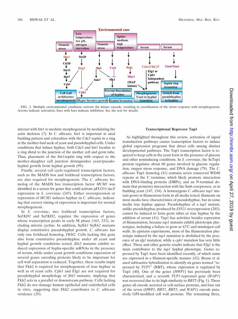
Other kinases have also been investigated for their role inpseudohyphal development. The Nim1 kinases, Gin4 and Hsl1,are important for the formation of septin structures, and bothgin4 and hsl1 mutants form pseudohyphae constitutively (319).In S. cerevisiae, ScHsl1 regulates the tyrosine phosphorylationof the cyclin-dependent protein kinase Cdc28 by deactivatingthe protein kinase ScSwe1, which regulates the G2/M transition(295). Interestingly, in C. albicans, the Hsl1-Swe1-Cdc28 path-way appears to be important for cell elongation of both theyeast and hyphal forms and for virulence (304). Gin4-depletedpseudohyphae are unable to form hyphae when challengedwith serum, but this can be overcome by ectopic expression ofGin4 from the MET3 promoter. Thus, Gin4 plays an importantrole in regulating the developmental switch from pseudo-hyphae to hyphae (Fig. 3) (319).
Another morphogenesis factor, Int1, was originally clonedby virtue of its limited homology to vertebrate leukocyte inte-grins (98). Like cph1 mutants, C. albicans int1 strains have areduced ability to form hyphae in response to most hypha-inducing conditions but form apparently normal hyphae in thepresence of serum (100). The C terminus of Int1 has homologyto S. cerevisiae Bud4, which is localized to the septin rings atthe mother-bud neck (7). C. albicans INT1 induces filamentousgrowth in S. cerevisiae, and this property has been used toexplore the cytoskeleton components required for INT1-in-duced filamentous growth. Sla2, a cytoskeleton protein, may
VOL. 71, 2007 SIGNAL TRANSDUCTION NETWORK IN C. ALBICANS 359
on April 27, 2019 by guest
http://mm
br.asm.org/
Dow
nloaded from
interact with Int1 to mediate morphogenesis by modulating theactin skeleton (7). In C. albicans, Int1 is important in axialbudding pattern and colocalizes with the Cdc3 septin in a ringat the mother-bud neck of yeast and pseudohyphal cells. Underconditions that induce hyphae, both Cdc3 and Int1 localize toa ring distal to the junction of the mother cell and germ tube.Thus, placement of the Int1/septin ring with respect to themother-daughter cell junction distinguishes yeast/pseudo-hyphal growth from hyphal growth (99).
Finally, several cell cycle-regulated transcription factors,such as the MADS box and forkhead transcription factors,are also required for morphogenesis. The C. albicans ho-molog of the MADS box transcription factor MCM1 wasidentified in a screen for genes that could activate pFLO11::lacZexpression in S. cerevisiae (245). Either overexpression orrepression of MCM1 induces hyphae in C. albicans, indicat-ing that correct timing of expression is important for normalmorphogenesis.
In S. cerevisiae, two forkhead transcription factors,ScFKH1 and ScFKH2, regulate the expression of geneswhose transcription peaks in early M phase (163, 333), in-cluding mitotic cyclins. In addition, Scfkh1 Scfkh2 mutantsdisplay constitutive pseudohyphal growth. C. albicans hasonly one forkhead homolog, FKH2. Cells lacking this genealso form constitutive pseudohyphae under all yeast andhyphal growth conditions tested. fkh2 mutants exhibit re-duced expression of hypha-specific mRNAs in the presenceof serum, while under yeast growth conditions expression ofseveral genes encoding proteins likely to be important forcell wall separation is reduced. Together, these results implythat Fkh2 is required for morphogenesis of true hyphae aswell as of yeast cells. Cph1 and Efg1 are not required forpseudohyphal morphology of fkh2 mutants, implying thatFkh2 acts in a parallel or downstream pathway. Cells lackingFkh2 do not damage human epithelial and endothelial cellsin vitro, suggesting that Fkh2 contributes to C. albicansvirulence (29).
Transcriptional Repressor Tup1
As highlighted throughout this review, activation of signaltransduction pathways causes transcription factors to induceglobal expression programs that direct cells among distinctdevelopmental pathways. The Tup1 transcription factor is re-quired to keep cells in the yeast form in the presence of glucoseand other noninducing conditions. In S. cerevisiae, the ScTup1protein regulates about 60 genes involved in glucose regula-tion, oxygen stress response, and DNA damage (79). The C.albicans Tup1 homolog (41) contains seven conserved WD40repeats at the C terminus, which likely promote interactionwith DNA-binding proteins (DBPs), and an N-terminal do-main that promotes interaction with the Ssn6 corepressor, as inbudding yeast (147, 154). A homozygous C. albicans tup1 mu-tant grows in filamentous form in all media tested; filaments onmost media have characteristics of pseudohyphae, but in somemedia true hyphae appear. Pseudohyphae of a tup1 mutant,unlike pseudohyphae produced by EFG1 overexpression (285),cannot be induced to form germ tubes or true hyphae by theaddition of serum (41). Tup1 has activities besides repressionof filamentation, because tup1 mutants exhibit pleiotropic phe-notypes, including a failure to grow at 42°C and misshapen cellwalls. In epistasis experiments, most of the filamentation phe-notype induced by the tup1 mutation is abolished by the pres-ence of an efg1 mutation, while a cph1 mutation has very littleeffect. These and other genetic results indicate that Efg1 is themain contributor to the tup1 hyphal phenotype. Genes re-pressed by Tup1 have been identified recently, of which someare expressed in a filament-specific manner (42). Braun et al.used subtractive hybridization to identify six genes termed “re-pressed by TUP1” (RBT), whose expression is regulated byTup1 (40). One of the genes (HWP1) has previously beencharacterized, and a seventh TUP1-repressed gene (WAP1)was recovered due to its high similarity to RBT5 (Fig. 1). Thesegenes all encode secreted or cell surface proteins, and four outof the seven (HWP1, RBT1, RBT5, and WAP1) encode puta-tively GPI-modified cell wall proteins. The remaining three,
FIG. 3. Multiple environmental conditions activate the kinase cascade, resulting in coordination of the stress response with morphogenesis.Arrows indicate activation; lines with bars indicate inhibition. See the text for details.
360 BISWAS ET AL. MICROBIOL. MOL. BIOL. REV.
on April 27, 2019 by guest
http://mm
br.asm.org/
Dow
nloaded from
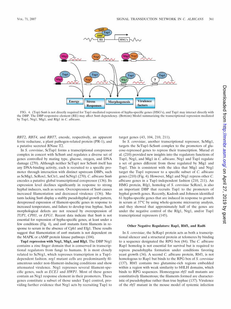
RBT2, RBT4, and RBT7, encode, respectively, an apparentferric reductase, a plant pathogen-related protein (PR-1), anda putative secreted RNase T2.
In S. cerevisiae, ScTup1 forms a transcriptional corepressorcomplex in concert with ScSsn6 and regulates a diverse set ofgenes controlled by mating type, glucose, oxygen, and DNAdamage (270). Although neither ScTup1 nor ScSsn6 itself hasany DNA-binding activity, each is recruited to a specific pro-moter through interaction with distinct upstream DBPs, suchas ScMig1, ScRox1, ScCtr1, and ScNrg1 (270). C. albicans Ssn6encodes a putative global transcriptional corepressor (136). Itsexpression level declines significantly in response to stronghyphal inducers, such as serum. Overexpression of Ssn6 causesincreased filamentation and decreased virulence (136). Mu-tants lacking Ssn6 display a stubby pseudohyphal growth pattern,derepressed expression of filament-specific genes in response toincreased temperature, and failure to develop true hyphae. Suchmorphological defects are not rescued by overexpression ofTUP1, CPH1, or EFG1. Recent data indicate that Ssn6 is notessential for repression of hypha-specific genes, at least under afew conditions (Fig. 4), and ssn6 mutants form filaments in re-sponse to serum in the absence of Cph1 and Efg1. These resultssuggest that filamentation of ssn6 mutants is not dependent onthe MAPK or cAMP protein kinase pathways (104).
Tup1 repression with Nrg1, Mig1, and Rfg1. The DBP Nrg1contains a zinc finger domain that is conserved in transcrip-tional regulators from fungi to humans. It is most closelyrelated to ScNrg1, which represses transcription in a Tup1-dependent fashion. nrg1 mutant cells are predominantly fil-amentous under non-filament-inducing conditions and showattenuated virulence. Nrg1 represses several filament-spe-cific genes, such as ECE1 and HWP1. Most of these genescontain an Nrg1 response element in their promoters. Thesegenes constitute a subset of those under Tup1 control, pro-viding further evidence that Nrg1 acts by recruiting Tup1 to
target genes (43, 104, 210, 211).In S. cerevisiae, another transcriptional repressor, ScMig1,
targets the ScTup1-ScSsn6 complex to the promoters of glu-cose-repressed genes to repress their transcription. Murad etal. (210) provided new insights into the regulatory functions ofTup1, Nrg1, and Mig1 in C. albicans. Nrg1 and Tup1 regulatea set of genes different from those regulated by Mig1 andTup1. This is consistent with the idea that Mig1 and Nrg1target the Tup1 repressor to a specific subset of C. albicansgenes (210) (Fig. 4). However, Mig1 and Nrg1 repress other C.albicans genes in a Tup1-independent fashion (210, 211). AnHMG protein, Rfg1, homolog of S. cerevisiae ScRox1, is alsoan important DBP that recruits Tup1 to the promoters ofhyphal growth genes. Recently, Kadosh and Johnson identified61 hypha-specific genes that are induced in response to growthin serum at 37°C by using whole-genome microarray analysis,and they showed that approximately half of the genes areunder the negative control of the Rfg1, Nrg1, and/or Tup1transcriptional repressors (145).
Other Negative Regulators: Rap1, Rbf1, and Rad6
In S. cerevisiae, the ScRap1 protein acts as both a transcrip-tional silencer and a structural protein at telomeres by bindingto a sequence designated the RPG box (84). The C. albicansRap1 homolog is not essential for survival but is required torepress pseudohypha formation under conditions favoringyeast growth (34). A second C. albicans protein, Rbf1, is nothomologous to Rap1 but binds to the RPG box of S. cerevisiae(137). Rbf1 contains two glutamine-rich regions embeddedwithin a region with weak similarity to bHLH domains, whichbinds to RPG sequences. Homozygous rbf1 null mutants areconstitutively filamentous; the filaments formed are character-istic of pseudohyphae rather than true hyphae (137). Virulenceof the rbf1 mutant in the mouse model of systemic infection
FIG. 4. (Top) Ssn6 is not directly required for Tup1-mediated repression of hypha-specific genes (HSG’s), and Tup1 may interact directly withthe DBP. The DBP-responsive element (RE) may affect Ssn6 dependency. (Bottom) Model summarizing the transcriptional repression mediatedby Tup1, Nrg1, Mig1, and Rfg1 in C. albicans.
VOL. 71, 2007 SIGNAL TRANSDUCTION NETWORK IN C. ALBICANS 361
on April 27, 2019 by guest
http://mm
br.asm.org/
Dow
nloaded from
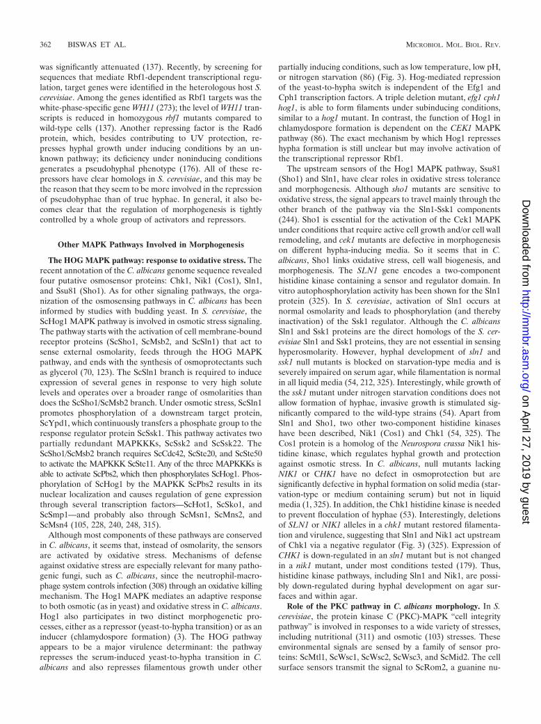
was significantly attenuated (137). Recently, by screening forsequences that mediate Rbf1-dependent transcriptional regu-lation, target genes were identified in the heterologous host S.cerevisiae. Among the genes identified as Rbf1 targets was thewhite-phase-specific gene WH11 (273); the level of WH11 tran-scripts is reduced in homozygous rbf1 mutants compared towild-type cells (137). Another repressing factor is the Rad6protein, which, besides contributing to UV protection, re-presses hyphal growth under inducing conditions by an un-known pathway; its deficiency under noninducing conditionsgenerates a pseudohyphal phenotype (176). All of these re-pressors have clear homologs in S. cerevisiae, and this may bethe reason that they seem to be more involved in the repressionof pseudohyphae than of true hyphae. In general, it also be-comes clear that the regulation of morphogenesis is tightlycontrolled by a whole group of activators and repressors.
Other MAPK Pathways Involved in Morphogenesis
The HOG MAPK pathway: response to oxidative stress. Therecent annotation of the C. albicans genome sequence revealedfour putative osmosensor proteins: Chk1, Nik1 (Cos1), Sln1,and Ssu81 (Sho1). As for other signaling pathways, the orga-nization of the osmosensing pathways in C. albicans has beeninformed by studies with budding yeast. In S. cerevisiae, theScHog1 MAPK pathway is involved in osmotic stress signaling.The pathway starts with the activation of cell membrane-boundreceptor proteins (ScSho1, ScMsb2, and ScSln1) that act tosense external osmolarity, feeds through the HOG MAPKpathway, and ends with the synthesis of osmoprotectants suchas glycerol (70, 123). The ScSln1 branch is required to induceexpression of several genes in response to very high solutelevels and operates over a broader range of osmolarities thandoes the ScSho1/ScMsb2 branch. Under osmotic stress, ScSln1promotes phosphorylation of a downstream target protein,ScYpd1, which continuously transfers a phosphate group to theresponse regulator protein ScSsk1. This pathway activates twopartially redundant MAPKKKs, ScSsk2 and ScSsk22. TheScSho1/ScMsb2 branch requires ScCdc42, ScSte20, and ScSte50to activate the MAPKKK ScSte11. Any of the three MAPKKKs isable to activate ScPbs2, which then phosphorylates ScHog1. Phos-phorylation of ScHog1 by the MAPKK ScPbs2 results in itsnuclear localization and causes regulation of gene expressionthrough several transcription factors—ScHot1, ScSko1, andScSmp1—and probably also through ScMsn1, ScMns2, andScMsn4 (105, 228, 240, 248, 315).
Although most components of these pathways are conservedin C. albicans, it seems that, instead of osmolarity, the sensorsare activated by oxidative stress. Mechanisms of defenseagainst oxidative stress are especially relevant for many patho-genic fungi, such as C. albicans, since the neutrophil-macro-phage system controls infection (308) through an oxidative killingmechanism. The Hog1 MAPK mediates an adaptive responseto both osmotic (as in yeast) and oxidative stress in C. albicans.Hog1 also participates in two distinct morphogenetic pro-cesses, either as a repressor (yeast-to-hypha transition) or as aninducer (chlamydospore formation) (3). The HOG pathwayappears to be a major virulence determinant: the pathwayrepresses the serum-induced yeast-to-hypha transition in C.albicans and also represses filamentous growth under other
partially inducing conditions, such as low temperature, low pH,or nitrogen starvation (86) (Fig. 3). Hog-mediated repressionof the yeast-to-hypha switch is independent of the Efg1 andCph1 transcription factors. A triple deletion mutant, efg1 cph1hog1, is able to form filaments under subinducing conditions,similar to a hog1 mutant. In contrast, the function of Hog1 inchlamydospore formation is dependent on the CEK1 MAPKpathway (86). The exact mechanism by which Hog1 represseshypha formation is still unclear but may involve activation ofthe transcriptional repressor Rbf1.
The upstream sensors of the Hog1 MAPK pathway, Ssu81(Sho1) and Sln1, have clear roles in oxidative stress toleranceand morphogenesis. Although sho1 mutants are sensitive tooxidative stress, the signal appears to travel mainly through theother branch of the pathway via the Sln1-Ssk1 components(244). Sho1 is essential for the activation of the Cek1 MAPKunder conditions that require active cell growth and/or cell wallremodeling, and cek1 mutants are defective in morphogenesison different hypha-inducing media. So it seems that in C.albicans, Sho1 links oxidative stress, cell wall biogenesis, andmorphogenesis. The SLN1 gene encodes a two-componenthistidine kinase containing a sensor and regulator domain. Invitro autophosphorylation activity has been shown for the Sln1protein (325). In S. cerevisiae, activation of Sln1 occurs atnormal osmolarity and leads to phosphorylation (and therebyinactivation) of the Ssk1 regulator. Although the C. albicansSln1 and Ssk1 proteins are the direct homologs of the S. cer-evisiae Sln1 and Ssk1 proteins, they are not essential in sensinghyperosmolarity. However, hyphal development of sln1 andssk1 null mutants is blocked on starvation-type media and isseverely impaired on serum agar, while filamentation is normalin all liquid media (54, 212, 325). Interestingly, while growth ofthe ssk1 mutant under nitrogen starvation conditions does notallow formation of hyphae, invasive growth is stimulated sig-nificantly compared to the wild-type strains (54). Apart fromSln1 and Sho1, two other two-component histidine kinaseshave been described, Nik1 (Cos1) and Chk1 (54, 325). TheCos1 protein is a homolog of the Neurospora crassa Nik1 his-tidine kinase, which regulates hyphal growth and protectionagainst osmotic stress. In C. albicans, null mutants lackingNIK1 or CHK1 have no defect in osmoprotection but aresignificantly defective in hyphal formation on solid media (star-vation-type or medium containing serum) but not in liquidmedia (1, 325). In addition, the Chk1 histidine kinase is neededto prevent flocculation of hyphae (53). Interestingly, deletionsof SLN1 or NIK1 alleles in a chk1 mutant restored filamenta-tion and virulence, suggesting that Sln1 and Nik1 act upstreamof Chk1 via a negative regulator (Fig. 3) (325). Expression ofCHK1 is down-regulated in an sln1 mutant but is not changedin a nik1 mutant, under most conditions tested (179). Thus,histidine kinase pathways, including Sln1 and Nik1, are possi-bly down-regulated during hyphal development on agar sur-faces and within agar.
Role of the PKC pathway in C. albicans morphology. In S.cerevisiae, the protein kinase C (PKC)-MAPK “cell integritypathway” is involved in responses to a wide variety of stresses,including nutritional (311) and osmotic (103) stresses. Theseenvironmental signals are sensed by a family of sensor pro-teins: ScMtl1, ScWsc1, ScWsc2, ScWsc3, and ScMid2. The cellsurface sensors transmit the signal to ScRom2, a guanine nu-
362 BISWAS ET AL. MICROBIOL. MOL. BIOL. REV.
on April 27, 2019 by guest
http://mm
br.asm.org/
Dow
nloaded from
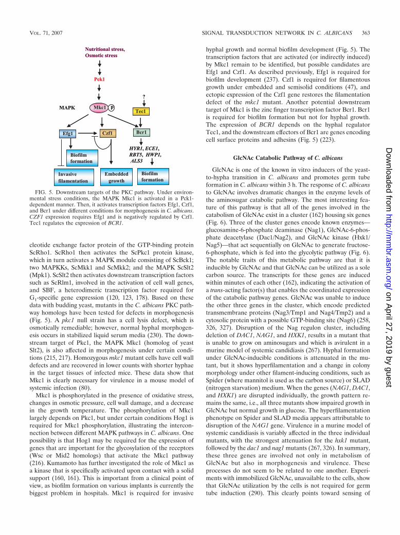
cleotide exchange factor protein of the GTP-binding proteinScRho1. ScRho1 then activates the ScPkc1 protein kinase,which in turn activates a MAPK module consisting of ScBck1;two MAPKKs, ScMkk1 and ScMkk2; and the MAPK ScSlt2(Mpk1). ScSlt2 then activates downstream transcription factorssuch as ScRlm1, involved in the activation of cell wall genes,and SBF, a heterodimeric transcription factor required forG1-specific gene expression (120, 123, 178). Based on thesedata with budding yeast, mutants in the C. albicans PKC path-way homologs have been tested for defects in morphogenesis(Fig. 5). A pkc1 null strain has a cell lysis defect, which isosmotically remediable; however, normal hyphal morphogen-esis occurs in stabilized liquid serum media (230). The down-stream target of Pkc1, the MAPK Mkc1 (homolog of yeastSlt2), is also affected in morphogenesis under certain condi-tions (215, 217). Homozygous mkc1 mutant cells have cell walldefects and are recovered in lower counts with shorter hyphaein the target tissues of infected mice. These data show thatMkc1 is clearly necessary for virulence in a mouse model ofsystemic infection (80).
Mkc1 is phosphorylated in the presence of oxidative stress,changes in osmotic pressure, cell wall damage, and a decreasein the growth temperature. The phosphorylation of Mkc1largely depends on Pkc1, but under certain conditions Hog1 isrequired for Mkc1 phosphorylation, illustrating the intercon-nection between different MAPK pathways in C. albicans. Onepossibility is that Hog1 may be required for the expression ofgenes that are important for the glycosylation of the receptors(Wsc or Mid2 homologs) that activate the Mkc1 pathway(216). Kumamoto has further investigated the role of Mkc1 asa kinase that is specifically activated upon contact with a solidsupport (160, 161). This is important from a clinical point ofview, as biofilm formation on various implants is currently thebiggest problem in hospitals. Mkc1 is required for invasive
hyphal growth and normal biofilm development (Fig. 5). Thetranscription factors that are activated (or indirectly induced)by Mkc1 remain to be identified, but possible candidates areEfg1 and Czf1. As described previously, Efg1 is required forbiofilm development (237). Czf1 is required for filamentousgrowth under embedded and semisolid conditions (47), andectopic expression of the Czf1 gene restores the filamentationdefect of the mkc1 mutant. Another potential downstreamtarget of Mkc1 is the zinc finger transcription factor Bcr1. Bcr1is required for biofilm formation but not for hyphal growth.The expression of BCR1 depends on the hyphal regulatorTec1, and the downstream effectors of Bcr1 are genes encodingcell surface proteins and adhesins (Fig. 5) (223).
GlcNAc Catabolic Pathway of C. albicans
GlcNAc is one of the known in vitro inducers of the yeast-to-hypha transition in C. albicans and promotes germ tubeformation in C. albicans within 3 h. The response of C. albicansto GlcNAc involves dramatic changes in the enzyme levels ofthe aminosugar catabolic pathway. The most interesting fea-ture of this pathway is that all of the genes involved in thecatabolism of GlcNAc exist in a cluster (162) housing six genes(Fig. 6). Three of the cluster genes encode known enzymes—glucosamine-6-phosphate deaminase (Nag1), GlcNAc-6-phos-phate deacetylase (Dac1/Nag2), and GlcNAc kinase (Hxk1/Nag5)—that act sequentially on GlcNAc to generate fructose-6-phosphate, which is fed into the glycolytic pathway (Fig. 6).The notable traits of this metabolic pathway are that it isinducible by GlcNAc and that GlcNAc can be utilized as a solecarbon source. The transcripts for these genes are inducedwithin minutes of each other (162), indicating the activation ofa trans-acting factor(s) that enables the coordinated expressionof the catabolic pathway genes. GlcNAc was unable to inducethe other three genes in the cluster, which encode predictedtransmembrane proteins (Nag3/Tmp1 and Nag4/Tmp2) and acytosolic protein with a possible GTP-binding site (Nag6) (258,326, 327). Disruption of the Nag regulon cluster, includingdeletion of DAC1, NAG1, and HXK1, results in a mutant thatis unable to grow on aminosugars and which is avirulent in amurine model of systemic candidiasis (267). Hyphal formationunder GlcNAc-inducible conditions is attenuated in the mu-tant, but it shows hyperfilamentation and a change in colonymorphology under other filament-inducing conditions, such asSpider (where mannitol is used as the carbon source) or SLAD(nitrogen starvation) medium. When the genes (NAG1, DAC1,and HXK1) are disrupted individually, the growth pattern re-mains the same, i.e., all three mutants show impaired growth inGlcNAc but normal growth in glucose. The hyperfilamentationphenotype on Spider and SLAD media appears attributable todisruption of the NAG1 gene. Virulence in a murine model ofsystemic candidiasis is variably affected in the three individualmutants, with the strongest attenuation for the hxk1 mutant,followed by the dac1 and nag1 mutants (267, 326). In summary,these three genes are involved not only in metabolism ofGlcNAc but also in morphogenesis and virulence. Theseprocesses do not seem to be related to one another. Experi-ments with immobilized GlcNAc, unavailable to the cells, showthat GlcNAc utilization by the cells is not required for germtube induction (290). This clearly points toward sensing of
FIG. 5. Downstream targets of the PKC pathway. Under environ-mental stress conditions, the MAPK Mkc1 is activated in a Pck1-dependent manner. Then, it activates transcription factors Efg1, Czf1,and Bcr1 under different conditions for morphogenesis in C. albicans.CZF1 expression requires Efg1 and is negatively regulated by Czf1.Tec1 regulates the expression of BCR1.
VOL. 71, 2007 SIGNAL TRANSDUCTION NETWORK IN C. ALBICANS 363
on April 27, 2019 by guest
http://mm
br.asm.org/
Dow
nloaded from
GlcNAc at the cell surface, but which receptor is involved isnot yet understood.
The capacity to metabolize GlcNAc is present in variousbacteria, such as Escherichia coli (313), Klebsiella pneumoniae,and Vibrio species (328), but, surprisingly, is absent in S. cer-evisiae. The main reason for this seems to be the absence ofgenes homologous to NAG1, DAC1, and HXK1. A notableexception is S. cerevisiae strain 3059, a clinical isolate, whichgrew well on both glucose- and GlcNAc-containing media(265). How this GlcNAc is metabolized in this particular strainis not yet known. The functions of the other genes in the Nagregulon are not yet clear. NAG3 and NAG4 have more than80% homology to each other and have significant sequencehomology to S. cerevisiae Ypr156C and Ygr138C (both trans-porters of spermine) (162, 327). NAG3 and NAG4 are notrequired for GlcNAc uptake, as there was no difference be-tween the growth of a nag3 nag4 mutant in GlcNAc-containingmedium and the growth of the wild-type strain (258). The nag3nag4 mutant became sensitive to cycloheximide, 4-nitroquino-line 1-oxide, and 1,10-phenanthroline. The above observations
indicate that though NAG3 and NAG4 are present in the samegene cluster as the catabolic pathway genes, they are not in-dispensable for GlcNAc metabolism or GlcNAc-mediated sig-naling (258). NAG6 encodes a protein involved in drug sensi-tivity that has no human or murine homolog (327). This,together with the fact that Nag6 is required for virulence,makes it an interesting target for antifungals.
�-N-Acetylglucosaminidase and virulence. �-N-Acetylglu-cosaminidase is a chitobiase that likely acts together with chi-tinase to release GlcNAc from chitin by hydrolysis (20). Thisprocess could have two biological effects: first, it may providean appropriate carbon source to the fungus; second, it mayfacilitate adhesion of Candida cells to host tissues by bringingabout conformational changes in the cell wall (55). The en-zyme is reported to be induced by GlcNAc and repressed byother sugars, such as glucose (94-fold lower), fructose, galac-tose, and mannose (220). Thus, GlcNAc not only regulates itsown catabolic pathway but also regulates the availability ofGlcNAc to the cell. �-N-Acetylglucosaminidase is secreted anddeposited into various parts of the cell wall of both the yeast
FIG. 6. (Top) Chromosomal organization of the NAG gene cluster in C. albicans. The organization of the cluster of genes on chromosome 6and their direction of transcription are illustrated. NAG1 and DAC1 are transcribed in opposite directions from a bidirectional promoter. (Bottom)Metabolism of GlcNAc in C. albicans. GlcNAc is synthesized by C. albicans from fructose-6-phosphate by sequential action of glutamine: Frc 6-Pamidotransferase (Gfp1), GlcN6-P acetyltransferase (Gna1), GlcNAc6P mutase (Agm1), UDP-GlcNAc pyrophosphorylase (Uap1). On the otherhand, GlcNAc permease, GlcNAc kinase, GlcNAc-6-phosphate deacetylase, and GlcN-6-phosphate deaminase act sequentially on GlcNAc togenerate the fructose-6-phosphate that is fed into the glycolytic pathway. N-Acetylmannosamine epimerase converts N-acetylmannosamine, whichis imported in the cell by an unknown permease (?), into GlcNAc. Glucosamine can directly enter the cell via a general sugar permease and isconverted to glucosamine-6-phosphate by the action of a GlcN-kinase.
364 BISWAS ET AL. MICROBIOL. MOL. BIOL. REV.
on April 27, 2019 by guest
http://mm
br.asm.org/
Dow
nloaded from
and hyphal forms. A mutant deficient in �-N-acetylglu-cosaminidase exhibits attenuated virulence in a mouse modelof candidiasis (141), although it shows normal growth. TheHEX1 gene encodes �-N-acetylglucosaminidase and producesa transcript that is 200 nucleotides longer when isolated fromGlcNAc-grown cells than from glucose-grown cells, implyingthat alternate transcription termination sites are used depend-ing on the growth conditions (55). Temperature and pHchanges also affect the level of HEX1 transcripts and �-N-acetylglucosaminidase activity.
pH Regulation in C. albicans
C. albicans causes infections in a broad range of host niches,which show significant differences in ambient pH. For example,the mouse systemic pH is 7.3, whereas the pH in the rat vaginais 4.5. The ability of Candida to react appropriately to (amongother environmental variables) rather different pH environ-ments is crucial for its pathogenicity. The environmental pHacts as a manipulator for many physiological functions, includ-ing morphogenesis (93). Under optimal (37°C) temperatureconditions, filamentation is favored by ambient pH values closeto neutral and is considerably reduced at pH values lower than6. In contrast, the yeast form predominates almost exclusivelyat pH 4 (50).
Genes involved in pH regulation. The pathway controllingpH-responsive gene expression has been most extensively dis-sected for the ascomycete Aspergillus nidulans (234). Central tothe pH response is the pH-dependent activation of the zincfinger transcription factor encoded by pacC (297). PacC issynthesized in an inactive form, which is activated at alkalinepH by proteolytic removal of the carboxy terminus (203). Pro-teolysis is dependent on six genes: palA, palB, palC, palF, palH,and palI (297). The activated form of PacC induces the expres-sion of alkaline pH-expressed genes and represses acidic pH-expressed genes (297). This regulatory pathway is apparentlyconserved, as various elements have been identified in Yar-rowia lipolytica and S. cerevisiae. The Y. lipolytica RIM101 geneproduct is activated by carboxy-terminal truncation and is re-quired for alkaline pH-dependent expression of XPR2-en-coded protease (168). In S. cerevisiae, the PacC homolog,ScRIM101, was initially identified as controlling meiosis andhaploid invasiveness (180, 286). ScRIM101 and the yeast ho-molog of PalB, ScCpl1, have been implicated in a pH-depen-dent growth response of yeast (97). Genes homologous to palAand palI are also present in S. cerevisiae (77, 218). Saporito-Irwin et al. used differential screening techniques to isolategenes with a pH-dependent expression profile and identifiedPHR1, a prototypical alkaline pH-expressed gene encoding aprotein of 548 residues, carrying a glycosylphosphatidylinositollipid anchor, which is required for apical cell growth and mor-phogenesis (251). PHR1 is homologous to ScGAS1, which en-codes a GPI-anchored protein of the S. cerevisiae cell surface(225). PHR1 is strongly expressed under conditions of alkalinepH but not at any pH below 5.5 (251). A second pH-regulatedgene, designated PHR2 (207), is expressed at an ambient pHbelow 5.5 and also plays a role in morphogenesis (251). Dele-tion of either one or both genes has a strong effect on viru-lence. Deletion of PHR1 results in pH-conditional defects ingrowth, morphogenesis, and virulence, evident at neutral to
alkaline pHs. Conversely, a phr2 null mutant exhibits similarpH-conditional defects in growth and morphogenesis thatmanifest at acidic rather than alkaline pH values. Engineeredexpression of PHR1 at acidic pH in a phr2 mutant strain andPHR2 at alkaline pH in a phr1 mutant strain complements thedefects in the opposing mutant. A strain lacking both PHR1and PHR2 shows pH-independent constitutive growth andmorphological defects (108, 207). When such strains weretested for pathogenicity in various niches of the host, the vir-ulence phenotype paralleled the pH dependence of the in vitrophenotypes. The phr1 mutant was avirulent in a mouse modelof systemic infection but uncompromised in its ability to causevaginal infection in rats. The virulence phenotype of a phr2mutant was the inverse. Heterozygous mutants exhibited par-tial reductions in their pathogenic potential, suggesting a genedosage effect (75). Another pH-regulated gene, PRA1, a ho-molog of surface antigens of Aspergillus species, with maximalexpression at neutral pH, has an important role in host-patho-gen interactions during candidal infection (259).
To investigate the pH-dependent regulation of PHR1 andPHR2 in C. albicans, Fonzi et al. isolated and characterizedPRR1 (pH response regulator), which is the C. albicans ho-molog of palF (now renamed RIM8). Expression of RIM8 is pHdependent, and a mutant lacking RIM8 is defective in pH-dependent regulation of gene expression. PHR1 is no longerinduced at alkaline pH, and PHR2 is no longer repressed inboth alkaline and acidic pH. Thus, Rim8 is a component of thepH response pathway in C. albicans. A rim8 mutant exhibitedno morphological abnormalities at either pH; however, it can-not form hyphae on serum-containing medium (235). The abil-ity to form hyphae on serum is not restored by forced expres-sion of PHR1, indicating that additional RIM8-dependentgenes are required for hyphal development. As discussedabove, HWP1 is an Efg1 target gene required for normal fila-mentation, but it also appears to be downstream of Rim8.Expression of HWP1 is induced in the absence of RIM8, indi-cating that the pH response pathway is not required for theexpression of EFG1-dependent genes. This suggests that thepH response pathway is distinct from the EFG1-dependentsignaling pathway (235). In this regard it might be noted thatEFG1 lies downstream of TPK2, which encodes a cAMP-de-pendent protein kinase (276). Rim101 of S. cerevisiae containsa functionally significant recognition site for phosphorylationby cAMP-dependent protein kinases (286). Although this siteis not conserved in the C. albicans homolog, two potentialphosphorylation sites are present, and this could provide aregulatory connection between Tpk2, Rim101, and Efg1.
Rim101-dependent and -independent pathways. Fonzi’sgroup characterized the full-length homolog of pacC, whichthey designated PRR2 (for “pH response regulator,” now re-named RIM101). C. albicans RIM101 encodes a protein of 604amino acids that shares two structural features with othermembers of the Rim101 family. First, the zinc finger domain ofthe Rim101 family is highly conserved; C. albicans Rim101 has57% identity and 87% similarity to the S. cerevisiae homolog inthis region. Second, C. albicans Rim101 has an 84-amino-acidC-terminal region with 32% D/E residues, which is conservedin other members of this family (in S. cerevisiae, Y. lipolytica,and A. nidulans).
As with its homologs in other species (see above), C. albi-
VOL. 71, 2007 SIGNAL TRANSDUCTION NETWORK IN C. ALBICANS 365
on April 27, 2019 by guest
http://mm
br.asm.org/
Dow
nloaded from
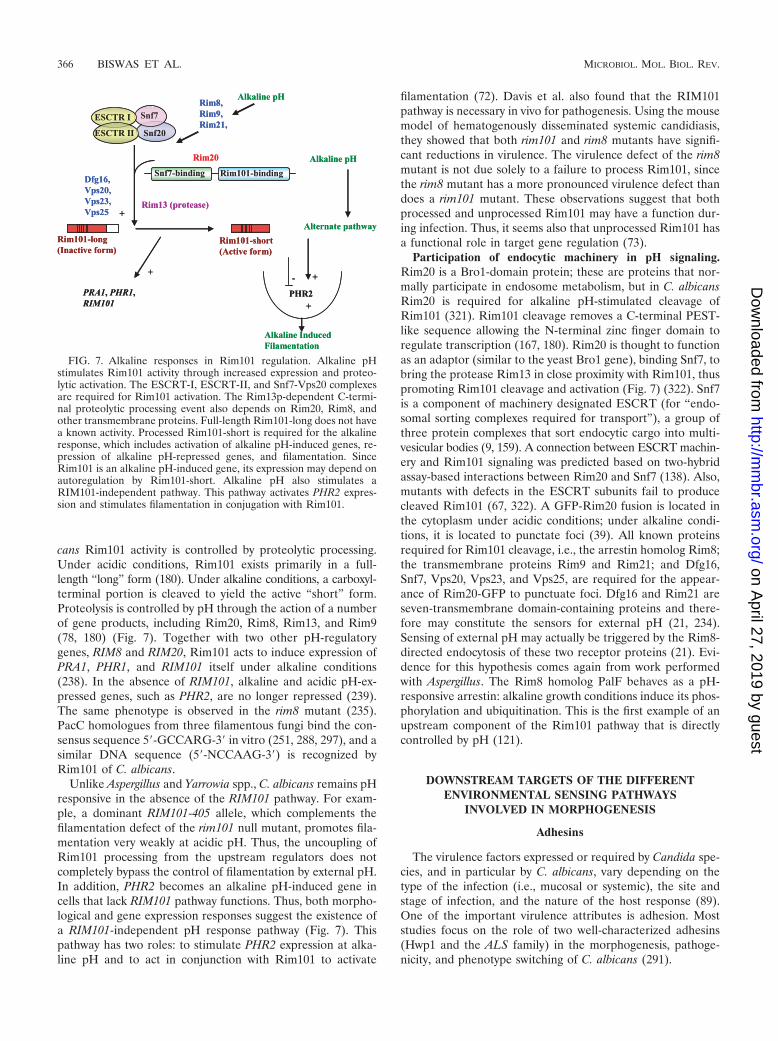
cans Rim101 activity is controlled by proteolytic processing.Under acidic conditions, Rim101 exists primarily in a full-length “long” form (180). Under alkaline conditions, a carboxyl-terminal portion is cleaved to yield the active “short” form.Proteolysis is controlled by pH through the action of a numberof gene products, including Rim20, Rim8, Rim13, and Rim9(78, 180) (Fig. 7). Together with two other pH-regulatorygenes, RIM8 and RIM20, Rim101 acts to induce expression ofPRA1, PHR1, and RIM101 itself under alkaline conditions(238). In the absence of RIM101, alkaline and acidic pH-ex-pressed genes, such as PHR2, are no longer repressed (239).The same phenotype is observed in the rim8 mutant (235).PacC homologues from three filamentous fungi bind the con-sensus sequence 5�-GCCARG-3� in vitro (251, 288, 297), and asimilar DNA sequence (5�-NCCAAG-3�) is recognized byRim101 of C. albicans.
Unlike Aspergillus and Yarrowia spp., C. albicans remains pHresponsive in the absence of the RIM101 pathway. For exam-ple, a dominant RIM101-405 allele, which complements thefilamentation defect of the rim101 null mutant, promotes fila-mentation very weakly at acidic pH. Thus, the uncoupling ofRim101 processing from the upstream regulators does notcompletely bypass the control of filamentation by external pH.In addition, PHR2 becomes an alkaline pH-induced gene incells that lack RIM101 pathway functions. Thus, both morpho-logical and gene expression responses suggest the existence ofa RIM101-independent pH response pathway (Fig. 7). Thispathway has two roles: to stimulate PHR2 expression at alka-line pH and to act in conjunction with Rim101 to activate
filamentation (72). Davis et al. also found that the RIM101pathway is necessary in vivo for pathogenesis. Using the mousemodel of hematogenously disseminated systemic candidiasis,they showed that both rim101 and rim8 mutants have signifi-cant reductions in virulence. The virulence defect of the rim8mutant is not due solely to a failure to process Rim101, sincethe rim8 mutant has a more pronounced virulence defect thandoes a rim101 mutant. These observations suggest that bothprocessed and unprocessed Rim101 may have a function dur-ing infection. Thus, it seems also that unprocessed Rim101 hasa functional role in target gene regulation (73).
Participation of endocytic machinery in pH signaling.Rim20 is a Bro1-domain protein; these are proteins that nor-mally participate in endosome metabolism, but in C. albicansRim20 is required for alkaline pH-stimulated cleavage ofRim101 (321). Rim101 cleavage removes a C-terminal PEST-like sequence allowing the N-terminal zinc finger domain toregulate transcription (167, 180). Rim20 is thought to functionas an adaptor (similar to the yeast Bro1 gene), binding Snf7, tobring the protease Rim13 in close proximity with Rim101, thuspromoting Rim101 cleavage and activation (Fig. 7) (322). Snf7is a component of machinery designated ESCRT (for “endo-somal sorting complexes required for transport”), a group ofthree protein complexes that sort endocytic cargo into multi-vesicular bodies (9, 159). A connection between ESCRT machin-ery and Rim101 signaling was predicted based on two-hybridassay-based interactions between Rim20 and Snf7 (138). Also,mutants with defects in the ESCRT subunits fail to producecleaved Rim101 (67, 322). A GFP-Rim20 fusion is located inthe cytoplasm under acidic conditions; under alkaline condi-tions, it is located to punctate foci (39). All known proteinsrequired for Rim101 cleavage, i.e., the arrestin homolog Rim8;the transmembrane proteins Rim9 and Rim21; and Dfg16,Snf7, Vps20, Vps23, and Vps25, are required for the appear-ance of Rim20-GFP to punctuate foci. Dfg16 and Rim21 areseven-transmembrane domain-containing proteins and there-fore may constitute the sensors for external pH (21, 234).Sensing of external pH may actually be triggered by the Rim8-directed endocytosis of these two receptor proteins (21). Evi-dence for this hypothesis comes again from work performedwith Aspergillus. The Rim8 homolog PalF behaves as a pH-responsive arrestin: alkaline growth conditions induce its phos-phorylation and ubiquitination. This is the first example of anupstream component of the Rim101 pathway that is directlycontrolled by pH (121).
DOWNSTREAM TARGETS OF THE DIFFERENTENVIRONMENTAL SENSING PATHWAYS
INVOLVED IN MORPHOGENESIS
Adhesins
The virulence factors expressed or required by Candida spe-cies, and in particular by C. albicans, vary depending on thetype of the infection (i.e., mucosal or systemic), the site andstage of infection, and the nature of the host response (89).One of the important virulence attributes is adhesion. Moststudies focus on the role of two well-characterized adhesins(Hwp1 and the ALS family) in the morphogenesis, pathoge-nicity, and phenotype switching of C. albicans (291).
FIG. 7. Alkaline responses in Rim101 regulation. Alkaline pHstimulates Rim101 activity through increased expression and proteo-lytic activation. The ESCRT-I, ESCRT-II, and Snf7-Vps20 complexesare required for Rim101 activation. The Rim13p-dependent C-termi-nal proteolytic processing event also depends on Rim20, Rim8, andother transmembrane proteins. Full-length Rim101-long does not havea known activity. Processed Rim101-short is required for the alkalineresponse, which includes activation of alkaline pH-induced genes, re-pression of alkaline pH-repressed genes, and filamentation. SinceRim101 is an alkaline pH-induced gene, its expression may depend onautoregulation by Rim101-short. Alkaline pH also stimulates aRIM101-independent pathway. This pathway activates PHR2 expres-sion and stimulates filamentation in conjugation with Rim101.
366 BISWAS ET AL. MICROBIOL. MOL. BIOL. REV.
on April 27, 2019 by guest
http://mm
br.asm.org/
Dow
nloaded from
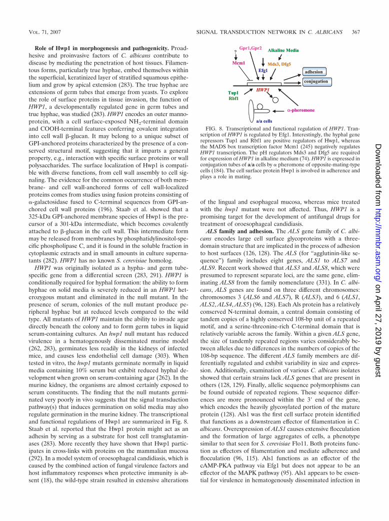
Role of Hwp1 in morphogenesis and pathogenicity. Proad-hesive and proinvasive factors of C. albicans contribute todisease by mediating the penetration of host tissues. Filamen-tous forms, particularly true hyphae, embed themselves withinthe superficial, keratinized layer of stratified squamous epithe-lium and grow by apical extension (283). The true hyphae areextensions of germ tubes that emerge from yeasts. To explorethe role of surface proteins in tissue invasion, the function ofHWP1, a developmentally regulated gene in germ tubes andtrue hyphae, was studied (283). HWP1 encodes an outer manno-protein, with a cell surface-exposed NH2-terminal domainand COOH-terminal features conferring covalent integrationinto cell wall �-glucan. It may belong to a unique subset ofGPI-anchored proteins characterized by the presence of a con-served structural motif, suggesting that it imparts a generalproperty, e.g., interaction with specific surface proteins or wallpolysaccharides. The surface localization of Hwp1 is compati-ble with diverse functions, from cell wall assembly to cell sig-naling. The evidence for the common occurrence of both mem-brane- and cell wall-anchored forms of cell wall-localizedproteins comes from studies using fusion proteins consisting of�-galactosidase fused to C-terminal sequences from GPI-an-chored cell wall proteins (196). Staab et al. showed that a325-kDa GPI-anchored membrane species of Hwp1 is the pre-cursor of a 301-kDa intermediate, which becomes covalentlyattached to �-glucan in the cell wall. This intermediate formmay be released from membranes by phosphatidylinositol-spe-cific phospholipase C, and it is found in the soluble fraction incytoplasmic extracts and in small amounts in culture superna-tants (282). HWP1 has no known S. cerevisiae homolog.
HWP1 was originally isolated as a hypha- and germ tube-specific gene from a differential screen (283, 291). HWP1 isconditionally required for hyphal formation: the ability to formhyphae on solid media is severely reduced in an HWP1 het-erozygous mutant and eliminated in the null mutant. In thepresence of serum, colonies of the null mutant produce pe-ripheral hyphae but at reduced levels compared to the wildtype. All mutants of HWP1 maintain the ability to invade agardirectly beneath the colony and to form germ tubes in liquidserum-containing cultures. An hwp1 null mutant has reducedvirulence in a hematogenously disseminated murine model(262, 283), germinates less readily in the kidneys of infectedmice, and causes less endothelial cell damage (303). Whentested in vitro, the hwp1 mutants germinate normally in liquidmedia containing 10% serum but exhibit reduced hyphal de-velopment when grown on serum-containing agar (262). In themurine kidney, the organisms are almost certainly exposed toserum constituents. The finding that the null mutants germi-nated very poorly in vivo suggests that the signal transductionpathway(s) that induces germination on solid media may alsoregulate germination in the murine kidney. The transcriptionaland functional regulations of Hwp1 are summarized in Fig. 8.Staab et al. reported that the Hwp1 protein might act as anadhesin by serving as a substrate for host cell transglutamin-ases (283). More recently they have shown that Hwp1 partic-ipates in cross-links with proteins on the mammalian mucosa(292). In a model system of oroesophageal candidiasis, which iscaused by the combined action of fungal virulence factors andhost inflammatory responses when protective immunity is ab-sent (18), the wild-type strain resulted in extensive alterations
of the lingual and esophageal mucosa, whereas mice treatedwith the hwp1 mutant were not affected. Thus, HWP1 is apromising target for the development of antifungal drugs fortreatment of oroesophageal candidiasis.
ALS family and adhesion. The ALS gene family of C. albi-cans encodes large cell surface glycoproteins with a three-domain structure that are implicated in the process of adhesionto host surfaces (126, 128). The ALS (for “agglutinin-like se-quence”) family includes eight genes, ALS1 to ALS7 andALS9. Recent work showed that ALS3 and ALS8, which werepresumed to represent separate loci, are the same gene, elim-inating ALS8 from the family nomenclature (331). In C. albi-cans, ALS genes are found on three different chromosomes:chromosomes 3 (ALS6 and ALS7), R (ALS3), and 6 (ALS1,ALS2, ALS4, ALS5) (96, 128). Each Als protein has a relativelyconserved N-terminal domain, a central domain consisting oftandem copies of a highly conserved 108-bp unit of a repeatedmotif, and a serine-threonine-rich C-terminal domain that isrelatively variable across the family. Within a given ALS gene,the size of tandemly repeated regions varies considerably be-tween alleles due to differences in the numbers of copies of the108-bp sequence. The different ALS family members are dif-ferentially regulated and exhibit variability in size and expres-sion. Additionally, examination of various C. albicans isolatesshowed that certain strains lack ALS genes that are present inothers (128, 129). Finally, allelic sequence polymorphisms canbe found outside of repeated regions. These sequence differ-ences are more pronounced within the 3� end of the gene,which encodes the heavily glycosylated portion of the matureprotein (128). Als1 was the first cell surface protein identifiedthat functions as a downstream effector of filamentation in C.albicans. Overexpression of ALS1 causes extensive flocculationand the formation of large aggregates of cells, a phenotypesimilar to that seen for S. cerevisiae Flo11. Both proteins func-tion as effectors of filamentation and mediate adherence andflocculation (96, 115). Als1 functions as an effector of thecAMP-PKA pathway via Efg1 but does not appear to be aneffector of the MAPK pathway (95). Als1 appears to be essen-tial for virulence in hematogenously disseminated infection in
FIG. 8. Transcriptional and functional regulation of HWP1. Tran-scription of HWP1 is regulated by Efg1. Interestingly, the hyphal generepressors Tup1 and Rbf1 are positive regulators of Hwp1, whereasthe MADS box transcription factor Mcm1 (245) negatively regulatesHWP1 transcription. The pH regulators Mds3 and Dfg5 are requiredfor expression of HWP1 in alkaline medium (74). HWP1 is expressed inconjugation tubes of a/a cells by � pheromone of opposite-mating-typecells (184). The cell surface protein Hwp1 is involved in adherence andplays a role in mating.
VOL. 71, 2007 SIGNAL TRANSDUCTION NETWORK IN C. ALBICANS 367
on April 27, 2019 by guest
http://mm
br.asm.org/
Dow
nloaded from
a murine model (52). Recently, a function for Als3 in biofilmformation was found by performing genome-wide expressionanalysis of the bcr1 mutant, which is affected in biofilm forma-tion. Als3 expression is strongly down-regulated in a bcr1 mu-tant (223). ALS3 overexpression rescues the bcr1 mutant bio-film defect in vivo, arguing for an important role for Als3 underthese in vivo conditions (222, 330).
Extracellular Hydrolytic Enzymes
In susceptible hosts, C. albicans enters the bloodstream andcauses deep-seated infection in target organs. Because theorganisms must cross the endothelial cells of the blood vesselsto enter these organs, the interaction between C. albicans andvascular endothelial cells is likely to be a critical step in theinitiation of a disseminated infection. One factor that contrib-utes to this process of virulence is hydrolytic enzyme produc-tion. The three most significant extracellular hydrolytic en-zymes produced by C. albicans are the secreted aspartylproteinases (Sap), phospholipase B enzymes, and lipases. All10 SAP genes of C. albicans encode preproenzymes approxi-mately 60 amino acids longer than the mature enzymes, whichare processed when transported via the secretory pathway(213).
SAP gene expression in the yeast-to-hypha transition. Thehypha-deficient efg1 mutant is, in contrast to the cph1 mutant,not able to invade or damage tissue (see above). However,whether this could be explained solely by a block in dimor-phism was not clear. Hube’s group investigated the expressionof the 10 SAP genes under hypha-inducing conditions (5%serum, different pH values, and different temperatures) in dif-ferent genetic backgrounds (wild type, efg1 mutant, and cph1mutant) (133). Expression of SAP4 to SAP6 was strongly re-duced in efg1 mutants in serum-containing medium, which is astrong environmental signal for hyphal growth of wild-typecells (316). However, expression of all three genes was en-hanced in both efg1 and cph1 mutants when hyphal formationwas induced via a pH and temperature shift protocol, suggest-ing that the transcription factors Efg1 and Cph1 can be eitherpositive or negative regulators of SAP4 to SAP6 expression.Since efg1 mutants do not produce hyphal cells under theseconditions, the expression of SAP4 to SAP6 is not strictlylinked to hyphal morphology but is regulated by another factorthat regulates hyphal formation. Expression of Sap antigenduring systemic infections has long been recognized in antigen-antibody studies (246). Staib et al. demonstrated high-levelexpression of SAP5 in all phases of infection with wild-typecells and a late induction of SAP2 and SAP6 using in vivoexpression technology. With Sap-specific antibodies, Sap1 toSap3 proteinases were detected on both yeast and hyphal wild-type cells, while the Sap4- to Sap6-specific antigen was identi-fied mostly on penetrating hyphal cells, conferring hypha-spe-cific expression of Sap4 and Sap6. The transcription factorsCph1 and Efg1 are involved in the expression of SAP5, as theavirulent cph1 efg1 mutant does not express this gene (284).Although expression of SAP2 was high in all in vivo samplesinvestigated and large amounts of Sap1 to Sap3 antigen werefound on all types of wild-type Candida cells, the level ofexpression did not correlate with the importance of the corre-sponding gene for invasion. Only mutants lacking SAP6 had
strongly reduced abilities to invade and damage parenchymalorgans, despite the fact that hyphal production was normal andall other proteinase genes were still expressed (91). It can beconcluded that the reduced virulence of hypha-deficient mu-tants is not only due to the inability to form hyphae but alsodue to modified expression of SAP genes normally associatedwith hyphal morphology.
Phospholipase B. Phospholipases are important pathogenic-ity determinants in C. albicans. They play a significant role indamaging cell membranes and invading host cells. High phos-pholipase production is correlated with increased adherenceand a higher mortality rate in animal models (201). Variousphospholipases reported from C. albicans include phospho-lipases A, B, C, and D (219). Phospholipase A and lysophos-pholipase activities are found in the cell wall of yeast cells andhyphae. Enzyme activity is higher in the walls of older yeastcells than of younger cells and is more prominent at the tip ofgrowing hyphae (113). A C. albicans strain deleted for thephospholipase B gene PLB1 has significantly attenuated viru-lence (107, 173). Although deletion of PLB1 does not produceany detectable effects on Candida adherence to human endo-thelial or epithelial cells, the ability of the plb1 null mutants topenetrate host cells is dramatically reduced (209). Thus, phos-pholipase B may well contribute to the pathogenicity of C.albicans by abetting the fungus in damaging and traversing hostcell membranes, processes which likely increase the rapidity ofdisseminated infection (173). Expression of PLB1 is regulatedby nutritional supplementation, environmental factors, and thegrowth phase of the C. albicans cells, as well as by physiologicalconditions. The differential expression of PLB1 in response toenvironmental factors may be correlated to host-specific com-ponents available to C. albicans (208). These data prove thatphospholipase B is essential for Candida virulence and pavethe way for studies directed at determining the mechanism(s)through which phospholipases modulate virulence in this or-ganism. The phospholipase D1 has been reported to be impor-tant for morphological transition under certain conditions(132).
FUTURE CHALLENGES
The mechanism of the yeast-to-hypha transition and identi-fication of potential virulence attributes of C. albicans is acritical problem that requires further investigation for the de-velopment and improvement of targeted antifungal therapy. Inthe past, progress in C. albicans research was slow due to thelack of effective molecular tools, such as stable plasmid expres-sion systems, limited mating ability, and the absence of knownsporulation. The recent annotation of the C. albicans genome(68), together with the development of a whole range of mo-lecular tools (e.g., codon-optimized reporter constructs), hasgreatly accelerated the pace of genetic research with this or-ganism. In this review, we have highlighted studies that revealthe dynamics of cellular signaling during the yeast-to-hyphatransition in C. albicans. Morphogenesis in C. albicans is reg-ulated by a limited number of transcription factors, such asEfg1, Cph1, Rim101, Tup1, Bcr1, Czf1, Tec1, Nrg1, and Rbf1.These transcription factors are induced and/or activated by awhole array of environmental signals through a number ofsignaling pathways. Many of these transcription factors have
368 BISWAS ET AL. MICROBIOL. MOL. BIOL. REV.
on April 27, 2019 by guest
http://mm
br.asm.org/
Dow
nloaded from
common downstream targets, which are the real triggers ofmorphogenesis. In the classical view, we see environmentalsensing as a system in which a membrane protein is the up-stream component of a signal transduction pathway. Recentstudies have shown that this is not always the case. The sensingof environmental CO2 concentrations seems to occur directlyat the level of carbonic anhydrase and adenylate cyclase anddoes not seem to involve a membrane receptor. Similar sys-tems may exist for the sensing of ammonia or other gases orsome of the small quorum-sensing molecules.
Several important questions remain unanswered. (i) How dothe different signals leading to morphogenesis and virulenceaffect the transcription of specific subsets of genes? What arethe transcription factors that mediate the responses? The ma-jor transcription factor Efg1 is a key regulator of the yeast andhyphal program, but it is not required, or may be even inhib-itory, for the later phase of filament formation. These obser-vations suggest that additional regulators are involved in thecontrol of morphogenesis and that there is a strong interplaybetween different signal transduction pathways. Alternatively,more feedback loops of gene regulation, such as for EFG1 andWOR1, will probably be discovered. A future challenge will beto identify all transcription factors involved in morphogenesisand how they interact with each other or how they are inter-dependent. (ii) How are morphogenesis and virulence regu-lated in vivo? Mutants that are either nonfilamentous (e.g.,cph1 efg1 mutants) or hyperfilamentous (e.g., pde2 or hxk1mutants) in vitro have strongly reduced virulence. Thus, itseems that a balance in filamentation is critical at the time ofinvasion and establishment of virulence. (iii) A third importantissue that needs to be clarified is how the aminosugar GlcNAcinduces cellular morphogenesis. Aminosugar utilization is anattribute of pathogenic yeast, and knockout of this catabolicpathway abolishes virulence. (iv) Finally, although severalmembrane receptors have been described, our knowledge ofligands remains limited. In this area of research, comparisonwith S. cerevisiae may be of limited use, as C. albicans hascoevolved with its host. For instance, whereas in S. cerevisiaewe have clearly shown that glucose and sucrose are the ligandsof the G protein-coupled receptor Gpr1, the ligand of thehomologous receptor in C. albicans remains to be identified. Inaddition, we will have to look into specific host components aspotential ligands for certain receptors.
FINAL OUTLOOK
C. albicans is a suitable organism for studying dimorphismand pathogenicity. Major challenges still remain in determin-ing how cells sense different sets of specific environmentalsignals and how the various signaling cascades process andintegrate these signals. The phrase “many components but fewconnections” can characterize the current knowledge of themechanisms of yeast-to-hypha transitions in C. albicans.Therefore, efforts should be directed toward analyses of func-tional linkages in addition to an expansion of the list of com-ponents affecting morphogenesis and virulence. A human-cu-rated assembly of the C. albicans genome sequence has beenreleased (www.candidagenome.org/) (44, 142). Bruno andMitchell describe different strategies for overcoming the limi-tation of C. albicans gene functional analysis (49), and Noble
and Johnson describe the use of new “lab” strains (avoiding theuse of the URA3 marker) and “high-throughput” gene deletionstrategies (224). Comparative genome analysis provides impor-tant clues about the evolution of the species and its mecha-nisms of pathogenesis (142). Comparative analysis of geneticand biochemical pathways in S. cerevisiae and C. albicans hasalso been extremely fruitful in revealing pathways regulatingvirulence in C. albicans. With the current speed of identifica-tion of genomes of other pathogens (e.g., C. glabrata and C.dubliniensis), comparative genomics will be an important toolin the future to identify broad-spectrum antifungal targets andto understand fungal pathogenesis. The ability of C. albicans topass through the diverse body tract requires niche-specific geneexpression (130). Recently, tools for monitoring the expressionof specific genes in different locations of the host have becomeavailable (19). The availability of new functional genomic tech-nologies, including DNA microarrays for high-throughputgene expression profiling and knock-out mutant collections,should enable functional characterization of novel genes. Ashas been achieved for S. cerevisiae, the C. albicans communityshould put together a consortium to generate a deletion straincollection, as well as conditional mutant collections for essen-tial genes. While technologies for rapid gene deletion construc-tion exist (88, 224), such collections should be constructed withthe use of dominant and reusable markers in order to avoidmarker problems and for the generation of multiple deletions.At the signaling level, new strategies for identifying nonlinearcomplexities in signal transduction, such as cross-talks andfeedback loops, must be developed. As a result, our view andappreciation of Candida biology and pathogenesis should un-dergo something just short of a revolution in the very nearfuture.
ACKNOWLEDGMENTS
We are grateful to Brenda Andrews, K. Natarajan, and C. W.Schweinfest for valuable suggestions during preparation of the manu-script. S. Ghosh and K. H. Rao are acknowledged for critical readingof the manuscript.
Recent studies of the GlcNAc signaling pathway in the laboratory ofA.D. have been supported by the Department of Biotechnology, India.Original work performed in the laboratory of P.V.D. was supported bygrants from the Research Fund of Flanders (G.0242.04) and from theEuropean Commission, Marie-Curie RTN Project CanTrain (MRTN-CT-2004-512841).
REFERENCES
1. Alex, L. A., C. Korch, C. P. Selitrennikoff, and M. I. Simon. 1998. COS1, atwo-component histidine kinase that is involved in hyphal development inthe opportunistic pathogen Candida albicans. Proc. Natl. Acad. Sci. USA95:7069–7073.
2. Alonso-Monge, R., F. Navarro-Garcia, G. Molero, R. Diez-Orejas, M. Gus-tin, J. Pla, M. Sanches, and C. Nombela. 1999. Role of the mitogen-activated protein kinase Hog1p in morphogenesis and virulence of Candidaalbicans. J. Bacteriol. 181:3058–3068.
3. Alonso-Monge, R., F. Navarro-Garcia, E. Roman, A. I. Negredo, B. Eisman,C. Nombela, and J. Pla. 2003. The Hog1 mitogen-activated protein kinaseis essential in the oxidative stress response and chlamydospore formation inCandida albicans. Eukaryot. Cell 2:351–361.
4. Andreasson, C., S. Heessen, and P. O. Ljungdahl. 2006. Regulation oftranscription factor latency by receptor-activated proteolysis. Genes Dev.20:1563–1568.
5. Andrianopoulos, A., and W. E. Timberlake. 1994. The Aspergillus nidulansabaA gene encodes a transcriptional activator that acts as a genetic switchto control development. Mol. Cell. Biol. 14:2503–2515.
6. Aramayo, R., Y. Peleg, R. Addison, and R. Metzenberg. 1996. Asm-1�, aNeurospora crassa gene related to transcriptional regulators of fungal de-velopment. Genetics 144:991–1003.
VOL. 71, 2007 SIGNAL TRANSDUCTION NETWORK IN C. ALBICANS 369
on April 27, 2019 by guest
http://mm
br.asm.org/
Dow
nloaded from
7. Asleson, C. M., E. S. Bensen, C. A. Gale, A. S. Melms, C. Kurischko, andJ. Berman. 2001. Candida albicans INT1-induced filamentation in Saccha-romyces cerevisiae depends on Sla2p. Mol. Cell. Biol. 21:1272–1284.
8. Atir-Lande, A., T. Gildor, and D. Kornitzer. 2005. Role of the SCFCDC4
ubiquitin ligase in Candida albicans morphogenesis. Mol. Biol. Cell 16:2772–2785.
9. Babst, M. 2005. A protein’s final ESCRT. Traffic 6:2–9.10. Bachewich, C., A. Nantel, and M. Whiteway. 2005. Cell cycle arrest during
S or M phase generates polarized growth via distinct signals in Candidaalbicans. Mol. Microbiol. 57:942–959.
11. Bachewich, C., D. Y. Thomas, and M. Whiteway. 2003. Depletion of apolo-like kinase in Candida albicans activates cyclase-dependent hyphal-like growth. Mol. Biol. Cell 14:2163–2180.
12. Bachewich, C., and M. Whiteway. 2005. Cyclin Cln3p links G1 progressionto hyphal and pseudohyphal development in Candida albicans. Eukaryot.Cell. 4:95–102.
13. Bahn, Y.-S., and P. Sundstrom. 2001. CAP1, an adenylate cyclase-associ-ated protein gene, regulates bud-hypha transitions, filamentous growth, andcyclic AMP levels and is required for virulence of Candida albicans. J.Bacteriol. 183:3211–3223.
14. Bahn, Y. S., G. M. Cox, J. R. Perfect, and J. Heitman. 2005. Carbonicanhydrase and CO2 sensing during Cryptococcus neoformans growth, dif-ferentiation, and virulence. Curr. Biol. 15:2013–2020.
15. Bahn, Y. S., and F. A. Muhlschlegel. 2006. CO2 sensing in fungi and beyond.Curr. Opin. Microbiol. 9:572–578.
16. Bahn, Y. S., J. Staab, and P. Sundstrom. 2003. Increased high-affinityphosphodiesterase PDE2 gene expression in germ tubes counteracts CAP1-dependent synthesis of cyclic AMP, limits hypha production and promotesvirulence of Candida albicans. Mol. Microbiol. 50:391–409.
17. Bailey, D. A., P. J. Feldmann, M. Bovey, N. A. Gow, and A. J. Brown. 1996.The Candida albicans HYR1 gene, which is activated in response to hyphaldevelopment, belongs to a gene family encoding yeast cell wall proteins. J.Bacteriol. 178:5353–5360.
18. Balish, E., T. F. Warner, C. J. Pierson, D. M. Bock, and R. D. Wagner. 2001.Oroesophageal candidiasis is lethal for transgenic mice with combinednatural killer and T-cell defects. Med. Mycol. 39:261–268.
19. Barelle, C. J., C. L. Priest, D. M. Maccallum, N. A. Gow, F. C. Odds, andA. J. Brown. 2006. Niche-specific regulation of central metabolic pathwaysin a fungal pathogen. Cell. Microbiol. 8:961–971.
20. Barrett-Bee, K., and M. Hamilton. 1984. The detection and analysis ofchitinase activity from the yeast form of Candida albicans. J. Gen. Micro-biol. 130:1857–1861.
21. Barwell, K. J., J. H. Boysen, W. Xu, and A. P. Mitchell. 2005. Relationshipof DFG16 to the Rim101p pH response pathway in Saccharomyces cerevi-siae and Candida albicans. Eukaryot. Cell 4:890–899.
22. Bassilana, M., J. Blyth, and R. A. Arkowitz. 2003. Cdc24, the GDP-GTPexchange factor for Cdc42, is required for invasive hyphal growth of Can-dida albicans. Eukaryot. Cell 2:9–18.
23. Bassilana, M., J. Hopkins, and R. A. Arkowitz. 2005. Regulation of theCdc42/Cdc24 GTPase module during Candida albicans hyphal growth.Eukaryot. Cell 4:588–603.
24. Battle, M., A. Lu, D. Green, Y. Xue, and J. P. Hirsch. 2003. Krh1p andKrh2p act downstream of the Gpa2p G alpha subunit to negatively regulatehaploid invasive growth. J. Cell Sci. 116:701–711.
25. Bendel, C. M., K. M. Kinneberg, R. P. Jechorek, C. A. Gale, S. L. Erlandsen,M. K. Hostetter, and C. L. Wells. 1999. Systemic infection followingintravenous inoculation of mice with Candida albicans int1 mutant strains.Mol. Genet. Metab. 67:343–351.
26. Bennett, R. J., and A. D. Johnson. 2005. Mating in Candida albicans and thesearch. Annu. Rev. Microbiol. 59:233–255.
27. Bennett, R. J., and A. D. Johnson. 2006. The role of nutrient regulation andthe Gpa2 protein in the mating pheromone response of C. albicans. Mol.Microbiol. 62:100–119.
28. Bensen, E. S., A. Clemente-Blanco, K. R. Finley, J. Correa-Bordes, and J.Berman. 2005. The mitotic cyclins Clb2p and Clb4p affect morphogenesis inCandida albicans. Mol. Biol. Cell 16:3387–3400.
29. Bensen, E. S., S. G. Filler, and J. Berman. 2002. A forkhead transcriptionfactor is important for true hyphal as well as yeast morphogenesis in Can-dida albicans. Eukaryot. Cell 1:787–798.
30. Benton, B. K., A. H. Tinkelenberg, D. Jean, S. D. Plump, and F. R. Cross.1993. Genetic analysis of Cln/Cdc28 regulation of cell morphogenesis inbudding yeast. EMBO J. 12:5267–5275.
31. Berman, J. 2006. Morphogenesis and cell cycle progression in Candidaalbicans. Curr. Opin. Microbiol. 9:595–601.
32. Berman, J., and P. Sudbery. 2002. Candida albicans: a molecular revolutionbuilt on lessons from budding yeast. Nat. Rev. Genet. 3:918–930.
33. Biswas, K., and J. Morchhauser. 2005. The Mep2 ammonium permeasecontrols nitrogen starvation-induced filamentous growth in Candida albi-cans. Mol. Microbiol. 56:649–669.
34. Biswas, K., K. J. Rieger, and J. Morschhauser. 2003. Functional analysis ofCaRAP1, encoding the repressor/activator protein 1 of Candida albicans.Gene 307:151–158.
35. Biswas, S., M. Roy, and A. Datta. 2003. N-Acetylglucosamine-inducibleCaGAP1 encodes a general amino acid permease which co-ordinates ex-ternal nitrogen source response and morphogenesis in Candida albicans.Microbiology 149:2597–2608.
36. Bockmuhl, D. P., and J. F. Ernst. 2001. A potential phosphorylation site foran A-type kinase in the Efg1 regulator protein contributes to hyphal mor-phogenesis of Candida albicans. Genetics 157:1523–1530.
37. Bockmuhl, D. P., S. Krishnamurthy, M. Gerads, A. Sonneborn, and J. F.Ernst. 2001. Distinct and redundant roles of the two protein kinase Aisoforms Tpk1 and Tpk2 in morphogenesis and growth of Candida albicans.Mol. Microbiol. 42:1243–1257.
38. Borneman, A. R., J. A. Leigh-Bell, H. Yu, P. Bertone, M. Gerstein, and M.Snyder. 2006. Target hub proteins serve as master regulators of develop-ment in yeast. Genes Dev. 20:435–448.
39. Boysen, J. H., and A. P. Mitchell. 2006. Control of Bro1-domain proteinRim20 localization by external pH, ESCRT machinery, and the S. cerevisiaeRIM101 pathway. Mol. Biol. Cell 17:1344–1353.
40. Braun, B. R., W. S. Head, M. X. Wang, and A. D. Johnson. 2000. Identifi-cation and characterization of TUP1-regulated genes in Candida albicans.Genetics 156:31–44.
41. Braun, B. R., and A. D. Johnson. 1997. Control of filament formation inCandida albicans by the transcriptional repressor Tup1. Science 277:105–109.
42. Braun, B. R., and A. D. Johnson. 2000. TUP1, CPH1 and EFG1 makeindependent contributions to filamentation in candida albicans. Genetics155:57–67.
43. Braun, B. R., D. Kadosh, and A. D. Johnson. 2001. NRG1, a repressor offilamentous growth in C. albicans, is down-regulated during filament induc-tion. EMBO J. 20:4753–4761.
44. Braun, B. R., M. Van het Hoog, C. d’Enfert, M. Martchenko, J. Dungan, A.Kuo, D. O. Inglis, M. A. Uhl, H. Hogues, M. Berriman, M. Lorenz, A.Levitin, U. Oberholzer, C. Bachewich, D. Harcus, A. Marcil, D. Dignard, T.Iouk, R. Zito, L. Frangeul, F. Tekaia, K. Rutherford, E. Wang, C. A. Munro,S. Bates, N. A. Gow, L. L. Hoyer, G. Kohler, J. Morchhauser, G. Newport,S. Znaidi, M. Raymond, B. Turcotte, G. Sherlock, M. Costanzo, J. Ihmels,J. Berman, D. Sanglard, N. Agabian, A. P. Mitchell, A. D. Johnson, M.Whiteway, and A. Nantel. 2005. A human-curated annotation of the Can-dida albicans genome. PLoS Genet. 1:36–57.
45. Brega, E., R. Zufferey, and B. C. Mamoun. 2004. Candida albicans Csy1 isa nutrient sensor important for activation of amino acid uptake and hyphalmorphogenesis. Eukaryot. Cell 3:135–143.
46. Brown, A. J. P., and N. A. R. Gow. 1999. Regulatory networks controllingCandida albicans morphogenesis. Trends Microbiol. 7:333–338.
47. Brown, D. H., Jr., A. D. Giusani, X. Chen, and C. A. Kumamoto. 1999.Filamentous growth of Candida albicans in response to physical environ-mental cues, and its regulation by the unique CZF1 gene. Mol. Microbiol.34:651–662.
48. Brown, V., J. A. Sexton, and M. Johnston. 2006. A glucose sensor inCandida albicans. Eukaryot. Cell 5:1726–1737.
49. Bruno, V. M., and A. P. Mitchell. 2004. Large-scale gene function analysisin Candida albicans. Trends Microbiol. 12:157–161.
50. Buffo, J., M. A. Herman, and D. R. Soll. 1984. A characterization ofpH-regulated dimorphism in Candida albicans. Mycopathologia 85:21–30.
51. Calcagno, A. M., E. Bignell, T. R. Rogers, M. Canedo, F. A. Muhlschlegel,and K. Haynes. 2004. Candida glabrata Ste20 is involved in maintaining cellwall integrity and adaptation to hypertonic stress, and is required for wild-type levels of virulence. Yeast 21:557–568.
52. Calderone, R. A., and W. A. Fonzi. 2001. Virulence factors of Candidaalbicans. Trends Microbiol. 9:327–335.
53. Calera, J. A., and R. Calderone. 1999. Flocculation of hyphae is associatedwith a deletion in the putative CaHK1 two-component histidine kinase genefrom Candida albicans. Microbiology 145:1431–1442.
54. Calera, J. A., X. J. Zhao, F. De Bernardis, M. Sheridan, and R. Calderone.1999. Avirulence of Candida albicans CaHK1 mutants in a murine model ofhematogenously dissemeinated candidiasis. Infect. Immun. 67:4280–4284.
55. Cannon, R. D., K. Niimi, H. F. Jenkinson, and M. G. Shepherd. 1994.Molecular cloning and expression of the Candida albicans �-N-acetylglu-cosaminidase (HEX1) gene. J. Bacteriol. 176:2640–2647.
56. Cao, F., S. Lane, P. P. Raniga, Y. Lu, Z. Zhou, K. Ramon, J. Chen, and H.Liu. 2006. The Flo8 transcription factor is essential for hyphal developmentand virulence in Candida albicans. Mol. Biol. Cell 17:295–307.
57. Care, R. S., J. Trevethick, K. M. Binley, and P. E. Sudbery. 1999. The MET3promoter: a new tool for Candida albicans molecular genetics. Mol. Micro-biol. 34:792–798.
58. Cassola, A., M. Parrot, S. Silberstein, B. B. Magee, S. Passeron, L. Giasson,and M. L. Cantore. 2004. Candida albicans lacking the gene encoding theregulatory subunit of protein kinase A displays a defect in hyphal formationand an altered localization of the catalytic subunit. Eukaryot. Cell 3:190–199.
59. Chapa y Lazo, B., S. Bates, and P. Sudbery. 2005. The G1 cyclin Cln3regulates morphogenesis in Candida albicans. Eukaryot. Cell 4:90–94.
60. Chen, C. G., Y. L. Yang, H. H. Chen, C. L. Su, S. F. Huang, C. T. Chen, Y. T.
370 BISWAS ET AL. MICROBIOL. MOL. BIOL. REV.
on April 27, 2019 by guest
http://mm
br.asm.org/
Dow
nloaded from
Liu, I. J. Su, and H. J. Lo. 2006. Non-lethal Candida albicans cph1/cph1efg1/efg1 transcription factor mutant establishing restricted zone of infec-tion in a mouse model of systemic infection. Int. J. Immunopathol. Phar-macol. 19:561–565.
61. Chen, H., M. Fujita, Q. Feng, J. Clardy, and G. R. Fink. 2004. Tyrosol is aquorum-sensing molecule in Candida albicans. Proc. Natl. Acad. Sci. USA101:5048–5052.
62. Cheng, S., M. H. Nguyen, Z. Zhang, H. Jia, M. Handfield, and C. J. Clancy.2003. Evaluation of the roles of four Candida albicans genes in virulence byusing gene disruption strains that express URA3 from the native locus.Infect. Immun. 71:6101–6103.
63. Chou, S., L. Huang, and H. Liu. 2004. Fus3-regulated Tec1 degradationthrough SCFCdc4 determines MAPK signaling specificity during mating inyeast. Cell 119:981–990.
64. Chou, S., S. Lane, and H. Liu. 2006. Regulation of mating and filamenta-tion genes by two distinct Ste12 complexes in Saccharomyces cerevisiae. Mol.Cell. Biol. 26:4794–4805.
65. Clark, K. L., P. J. Feldmann, D. Dignard, R. Larocque, A. J. Brown, M. G.Lee, D. Y. Thomas, and M. Whiteway. 1995. Constitutive activation of theSaccharomyces cerevisiae mating response pathway by a MAP kinase kinasefrom Candida albicans. Mol. Gen. Genet. 249:609–621.
66. Cloutier, M., R. Castilla, N. Bolduc, A. Zelada, P. Martineau, M. Bouillon,B. B. Magee, S. Passeron, L. Giasson, and M. L. Cantore. 2003. The twoisoforms of the cAMP-dependent protein kinase catalytic subunit are in-volved in the control of dimorphism in the human fungal pathogen Candidaalbicans. Fungal Gen. Biol. 38:133–141.
67. Cornet, M., F. Bidard, P. Schwarz, G. Da Costa, S. Blanchin-Roland, F.Dromer, and C. Gaillardin. 2005. Deletions of edocytic components VPS28and VPS32 affect growth at alkaline pH and virulence through bothRIM101-dependent and RIM101-independent pathways in Candida albi-cans. Infect. Immun. 73:7977–7987.
68. Costanzo, M. C., M. B. Arnaud, M. S. Skrzypek, G. Binkley, C. Lane, S. R.Miyasato, and G. Sherlock. 2006. The Candida Genome Database: facili-ating research on Candida albicans molecular biology. FEMS Yeast Res.6:671–684.
69. Csank, C., K. Schroppel, E. Leberer, D. Harcus, O. Mohamed, S. Meloche,D. Y. Thomas, and M. Whiteway. 1998. Roles of the Candida albicansmitogen-activated protein kinase homolog, Cek1p, in hyphal developmentand systemic candidiasis. Infect. Immun. 66:2713–2721.
70. Cullen, P. J., W. J. Sabbagh, E. Graham, M. M. Irick, E. K. van Olden, C.Neal, J. Delrow, L. Bardwell, and G. F. J. Sprague. 2004. A signaling mucinat the head of the Cdc42- and MAPK-dependent filamentous growth path-way in yeast. Genes Dev. 18:1695–1708.
71. Cutler, J. E. 1991. Putative virulence factors of Candida albicans. Annu.Rev. Microbiol. 45:187–218.
72. Davis, D., J. E. Edwards, Jr., A. P. Mitchell, and A. S. Ibrahim. 2000.Candida albicans RIM101 pH response pathway is required for host-patho-gen interactions. Infect. Immun. 68:5953–5959.
73. Davis, D., R. B. Wilson, and A. P. Mitchell. 2000. RIM101-dependent and-independent pathways govern pH responses in Candida albicans. Mol.Cell. Biol. 20:971–978.
74. Davis, D. A., V. M. Bruno, L. Loza, S. G. Filler, and A. P. Mitchell. 2002.Candida albicans Mds3p, a conserved regulator of pH responses and viru-lence identified through insertional mutagenesis. Genetics 162:1573–1581.
75. De Bernardis, F., F. A. Muhlchlegel, A. Cassone, and W. A. Fonzi. 1998.The pH of the host niche controls gene expression in and virulence ofCandida albicans. Infect. Immun. 66:3317–3325.
76. d’Enfert, C. 2006. Biofilms and their role in the resistance of pathogenicCandida to antifungal agents. Curr. Drug Targets 7:465–470.
77. Denison, S. H., S. Negrete-Urtasun, J. M. Mingot, J. Tilburn, W. A. Mayer,A. Goel, E. A. Espeso, M. A. Penalva, and H. N. Arst, Jr. 1998. Putativemembrane components of signal transduction pathways for ambient pHregulation in Aspergillus and meiosis in Saccharomyces are homologous.Mol. Microbiol. 30:259–264.
78. Denison, S. H., M. Orejas, and H. N. Arst, Jr. 1995. Signaling of ambientpH in Aspergillus involves a cysteine protease. J. Biol. Chem. 270:28519–28522.
79. DeRisi, J. L., V. R. Iyer, and P. O. Brown. 1997. Exploring the metabolicand genetic control of gene expression on a genomic scale. Science 278:680–686.
80. Diez-Orejas, R., G. Molero, F. Navarro-Garcia, J. Pla, C. Nombela, and M.Sanchez-Perez. 1997. Reduced virulence of Candida albicans MKC1 mu-tants: a role for mitogen-activated protein kinase in pathogenesis. Infect.Immun. 65:833–837.
81. Doedt, T., S. Krishnamurthy, D. P. Bockmuhl, B. Tebarth, C. Stempel, C. L.Russell, A. J. P. Brown, and J. F. Ernst. 2004. APSES proteins regulatemorphogenesis and metabolism in Candida albicans. Mol. Biol. Cell 15:3167–3180.
82. Dolan, J. W., A. C. Bell, B. Hube, M. Schaller, T. F. Warner, and E. Balish.2004. Candida albicans PLD1 activity is required for full virulence. Med.Mycol. 42:439–447.
83. Donaton, M. C., I. Holsbeeks, O. Lagatie, G. Van Zeebroeck, M. Crauwels,
J. Winderickx, and J. M. Thevelein. 2003. The Gap1 general amino acidpermease acts as an amino acid sensor for activation of protein kinase Atargets in the yeast Saccharomyces cerevisiae. Mol. Microbiol. 50:911–929.
84. Drazinic, C. M., J. B. Smerage, M. C. Lopez, and H. V. Baker. 1996.Activation mechanism of the multifunctional transcription factor repressor-activator protein 1 (Rap1p). Mol. Cell. Biol. 16:3187–3196.
85. Egidy, G., C. Paveto, S. Passeron, and M. A. Galvagno. 1990. cAMP levelsand in situ measurement of cAMP related enzymes during yeast-to-hyphaetransition in Candida albicans. Cell. Biol. Int. Rep. 14:59–68.
86. Eisman, B., R. Alonso-Monge, E. Roman, D. Arana, C. Nombela, and J. Pla.2006. The Cek1 and Hog1 mitogen-activated protein kinases play comple-mentary roles in cell wall biogenesis and chlamydospore formation in thefungal pathogen Candida albicans. Eukaryot. Cell 5:347–358.
87. El Barkani, A., O. Kurzai, W. A. Fonzi, A. Ramon, A. Porta, M. Frosch, andF. A. Muhlschlegel. 2000. Dominant active alleles of RIM101 (PRR2) bypassthe pH restriction on filamentation of Candida albicans. Mol. Cell. Biol.20:4635–4647.
88. Enloe, B., A. Diamond, and A. P. Mitchell. 2000. A single-transformationgene function test in diploid Candida albicans. J. Bacteriol. 182:5730–5736.
89. Fallon, K., K. Bausch, J. Noonan, E. Huguenel, and P. Tamburini. 1997.Role of aspartic proteases in disseminated Candida albicans infection inmice. Infect. Immun. 65:551–556.
90. Fang, H. M., and Y. Wang. 2006. RA domain-mediated interaction ofCdc35 with Ras1 is essential for increasing cellular cAMP level for Candidaalbicans hyphal development. Mol. Microbiol. 61:484–496.
91. Felk, A., M. Kretschmar, A. Albrecht, M. Schaller, S. Beinhauer, T. Nich-terlein, D. Sanglard, H. C. Korting, W. Schafer, and B. Hube. 2002. Can-dida albicans hyphal formation and the expression of the Efg1-regulatedproteinases Sap4 to Sap6 are required for the invasion of parenchymalorgans. Infect. Immun. 70:3689–3700.
92. Feng, Q., E. Summers, B. Guo, and G. R. Fink. 1999. Ras signaling isrequired for serum-induced hyphal differentiation in Candida albicans. J.Bacteriol. 181:6339–6346.
93. Fonzi, W. A. 2002. Role of pH response in Candida albicans virulence.Mycoses 45(Suppl. 1):16–21.
94. Forsberg, H., F. Gilstring, A. Zargari, P. Martinez, and P. O. Ljungdahl.2001. The role of the yeast plasma membrane SPS nutrient sensor in themetabolic response to extracellular amino acids. Mol. Microbiol. 42:215–228.
95. Fu, Y., A. S. Ibrahim, D. C. Sheppard, Y. C. Chen, S. W. French, J. E.Cutler, S. G. Filler, and J. E. Edwards, Jr. 2002. Candida albicans Als1p: anadhesin that is a downstream effector of the EFG1 filamentation pathway.Mol. Microbiol. 44:61–72.
96. Fu, Y., G. Rieg, W. A. Fonzi, P. H. Belanger, J. E. Edwards, Jr., and S. G.Filler. 1998. Expression of the Candida albicans gene ALS1 in Saccharo-myces cerevisiae induces adherence to endothelial and epithelial cells. In-fect. Immun. 66:1783–1786.
97. Futai, E., T. Maeda, H. Sorimachi, K. Kitamoto, S. Ishiura, and K. Suzuki.1999. The protease activity of a calpain-like cysteine protease in Saccharo-myces cerevisiae is required for alkaline adaptation and sporulation. Mol.Gen. Genet. 260:559–568.
98. Gale, C., D. Finkel, N. Tao, M. Meinke, M. McClellan, J. Olson, K. Kendrick,and M. Hostetter. 1996. Cloning and expression of a gene encoding an integrin-like protein in Candida albicans. Proc. Natl. Acad. Sci. USA 93:357–361.
99. Gale, C., M. Gerami-Nejad, M. McClellan, S. Vandoninck, M. S. Longtine,and J. Berman. 2001. Candida albicans Int1p interacts with the septin ringin yeast and hyphal cells. Mol. Biol. Cell 12:3538–3549.
100. Gale, C. A., C. M. Bendel, M. McClellan, M. Hauser, J. M. Becker, J.Berman, and M. K. Hostetter. 1998. Linkage of adhesion, filamentousgrowth, and virulence in Candida albicans to a single gene, INT1. Science279:1355–1358.
101. Gancedo, J. M. 2001. Control of pseudohyphae formation in Saccharomycescerevisiae. FEMS Microbiol. Rev. 25:107–123.
102. Garaizar, J., S. Brena, J. Bikandi, A. Rementeria, and J. Ponton. 2006. Useof DNA microarray technology and gene expression profiles to investigatethe pathogenesis, cell biology, antifungal susceptibility and diagnosis ofCandida albicans. FEMS Yeast Res. 6:987–998.
103. Garcia-Rodriguez, L. J., R. Valle, A. Duran, and C. Roncero. 2005. Cellintegrity signaling activation in response to hyperosmotic shock in yeast.FEBS Lett. 579:6186–6190.
104. Garcia-Sanchez, S., A. L. Mavor, C. L. Russell, S. Argimon, P. Dennison, B.Enjalbert, and A. J. Brown. 2005. Global roles of Ssn6 in Tup1- andNrg1-dependent gene regulation in the fungal pathogen, Candida albicans.Mol. Biol. Cell 16:2913–2925.
105. Gasch, A. P., P. T. Spellman, C. M. Kao, O. Carmel-Harel, M. B. Eisen, G.Storz, D. Botstein, and P. O. Brown. 2000. Genomic expression programs inthe response of yeast cells to environmental changes. Mol. Biol. Cell 11:4241–4257.
106. Gavrias, V., A. Andrianopoulos, C. J. Gimeno, and W. E. Timberlake. 1996.Saccharomyces cerevisiae TEC1 is required for pseudohyphal growth. Mol.Microbiol. 19:1255–1263.
VOL. 71, 2007 SIGNAL TRANSDUCTION NETWORK IN C. ALBICANS 371
on April 27, 2019 by guest
http://mm
br.asm.org/
Dow
nloaded from

107. Ghannoum, M. A. 1998. Extracellular phospholipases as universal virulencefactor in pathogenic fungi. Nippon Ishinkin Gakkai Zasshi 39:55–59.
108. Ghannoum, M. A., B. Spellberg, S. M. Saporito-Irwin, and W. A. Fonzi.1995. Reduced virulence of Candida albicans PHR1 mutants. Infect. Im-mun. 63:4528–4530.
109. Gimeno, C. J., and G. R. Fink. 1994. Induction of pseudohyphal growth byoverexpression of PHD1, a Saccharomyces cerevisiae gene related to tran-scriptional regulators of fungal development. Mol. Cell. Biol. 14:2100–2112.
110. Gimeno, C. J., P. O. Ljungdahl, C. A. Styles, and G. R. Fink. 1992. Unipolarcell divisions in the yeast Saccharomyces cerevisiae lead to filamentousgrowth: regulation by starvation and RAS. Cell 68:1077–1090.
111. Giusani, A. D., M. Vinces, and C. A. Kumamoto. 2002. Invasive filamenta-tous growth of Candida albicans is promoted by Czf1p-dependent relief ofEfg1p-mediated repression. Genetics 160:1749–1753.
112. Goldberg, D., M. Segal, and A. Levitzki. 1994. Cdc25 is not the signalreceiver for glucose induced cAMP response in S. cerevisiae. FEBS Lett.356:249–254.
113. Goyal, S., and G. K. Khuller. 1992. Phospholipid composition and subcel-lular distribution in yeast and mycelial forms of Candida albicans. J. Med.Vet. Mycol. 30:355–362.
114. Guhad, F. A., C. Csank, H. E. Jensen, D. Y. Thomas, M. Whiteway, and J.Hau. 1998. Reduced pathogenicity of a Candida albicans MAP kinasephosphatase (CPP1) mutant in the murine mastitis model. APMIS 106:1049–1055.
115. Guo, B., C. A. Styles, Q. Feng, and G. R. Fink. 2000. A Saccharomyces genefamily involved in invasive growth, cell-cell adhesion, and mating. Proc.Natl. Acad. Sci. USA 97:12158–12163.
116. Harashima, T., S. Anderson, J. R. R. Yates, and J. Heitman. 2006. TheKelch proteins Gpb1 and Gpb2 inhibit Ras activity via association with theyeast RasGAP neurofibromin homologs Ira1 and Ira2. Mol. Cell 22:819–830.
117. Harashima, T., and J. Heitman. 2002. The G alpha protein Gpa2 controlsyeast differentiation by interacting with Kelch repeat proteins that mimic Gbeta subunits. Mol. Cell 10:163–173.
118. Hausauer, D., M. Gerami-Nejad, C. Kistler-Anderson, and C. Gale. 2005.Hyphal guidance and invasive growth in Candida albicans require the Ras-like GTPase Rsr1p and its GTPase-activating protein Bud2p. Eukaryot.Cell 4:1273–1286.
119. Hazan, I., and H. Liu. 2002. Hyphal tip-associated localization of Cdc42 isF-actin dependent in Candida albicans. Eukaryot. Cell 1:856–864.
120. Heinisch, J. J., A. Lorberg, H. P. Schmitz, and J. J. Jacoby. 1999. Theprotein kinase C-mediated MAP kinase pathway involved in the mainte-nance of cellular integrity in Saccharomyces cerevisiae. Mol. Microbiol.32:671–680.
121. Herranz, S., J. M. Rodriguez, H. J. Bussink, J. C. Sanchez-Ferrero, H. N.Arst, Jr., M. A. Penalva, and O. Vincent. 2005. Arrestin-related proteinsmediate pH signaling in fungi. Proc. Natl. Acad. Sci. USA 102:12141–12146.
122. Hinnebusch, A. G. 1988. Mechanisms of gene regulation in the generalcontrol of amino acid biosynthesis in Saccharomyces cerevisiae. Microbiol.Rev. 52:248–273.
123. Hohmann, S. 2002. Osmotic stress signaling and osmoadaptation in yeasts.Microbiol. Mol. Biol. Rev. 66:300–372.
124. Holsbeeks, I., O. Lagatie, A. Van Nuland, S. Van De Velde, and J. M.Thevelein. 2004. The eukaryotic plasma membrane as a nutrient-sensingdevice. Trends Biochem. Sci. 29:556–564.
125. Hornby, J. M., E. C. Jensen, A. D. Lisec, J. J. Tasto, B. Jahnke, R. Shoe-maker, P. Dussault, and K. W. Nickerson. 2001. Quorum sensing in thedimorphic fungus Candida albicans is mediated by farnesol. Appl. Environ.Microbiol. 67:2982–2992.
126. Hoyer, L. L. 2001. The ALS gene family of Candida albicans. TrendsMicrobiol. 9:176–180.
127. Hoyer, L. L., L. B. Cieslinski, M. M. McLaughlin, T. J. Torphy, A. R.Shatzman, and G. P. Livi. 1994. A Candida albicans cyclic nucleotidephosphodiesterase: cloning and expression in Saccharomyces cerevisiae andbiochemical characterization of the recombinant enzyme. Microbiology140:1533–1542.
128. Hoyer, L. L., and J. E. Hecht. 2001. The ALS5 gene of Candida albicans andanalysis of the Als5p N-terminal domain. Yeast 18:49–60.
129. Hoyer, L. L., and J. E. Hecht. 2000. The ALS6 and ALS7 genes of Candidaalbicans. Yeast 16:847–855.
130. Hromatka, B. S., S. M. Noble, and A. D. Johnson. 2005. Transcriptionalresponse of Candida albicans to nitric oxide and the role of the YHB1 genein nitrosative stress. Mol. Biol. Cell 16:4814–4826.
131. Huang, G., H. Wang, S. Chou, X. Nie, J. Chen, and H. Liu. 2006. Bistableexpression of WOR1, a master regulator of white-opaque switching in Can-dida albicans. Proc. Natl. Acad. Sci. USA 103:12813–12818.
132. Hube, B., D. Hess, C. A. Baker, M. Schaller, W. Schafer, and J. W. Dolan.2001. The role and relevance of phospholipase D1 during growth anddimorphism of Candida albicans. Microbiology 147:879–889.
133. Hube, B., M. Monod, D. A. Schofield, A. J. Brown, and N. A. Gow. 1994.Expression of seven members of the gene family encoding secretory aspartylproteinases in Candida albicans. Mol. Microbiol. 14:87–99.
134. Hube, B., D. Sanglard, F. C. Odds, D. Hess, M. Monod, W. Schafer, A. J.Brown, and N. A. R. Gow. 1997. Disruption of each of the secreted aspartylproteinase genes SAP1, SAP2, and SAP3 of Candida albicans attenuatesvirulence. Infect. Immun. 65:3529–3538.
135. Hudson, D. A., Q. L. Sciascia, R. J. Sanders, G. E. Norris, P. J. Edwards,P. A. Sullivan, and P. C. Farley. 2004. Identification of the dialysable seruminducer of germ-tube formation in Candida albicans. Microbiology 150:3041–3049.
136. Hwang, C. S., J. H. Oh, W. K. Huh, H. S. Yim, and S. O. Kang. 2003. Ssn6,an important factor of morphological conversion and virulence in Candidaalbicans. Mol. Microbiol. 47:1029–1043.
137. Ishii, N., M. Yamamoto, F. Yoshihara, M. Arisawa, and Y. Aoki. 1997.Biochemical and genetic characterization of Rbf1p, a putative transcriptionfactor of Candida albicans. Microbiology 143:429–435.
138. Ito, T., T. Chiba, and M. Yoshida. 2001. Exploring the protein interactomeusing comprehensive two-hybrid projects. Trends Biotechnol. 19:S23–S27.
139. Jain, P., I. Akula, and T. Edlind. 2003. Cyclic AMP signaling pathwaymodulates susceptibility of Candida species and Saccharomyces cerevisiae toantifungal azoles and other sterol biosynthesis inhibitors. Antimicrob.Agents Chemother. 47:3195–3201.
140. Jauniaux, J. C., and M. Grenson. 1990. GAP1, the general amino acidpermease gene of Saccharomyces cerevisiae. Nucleotide sequence, proteinsimilarity with the other bakers yeast amino acid permeases, and nitrogencatabolite repression. Eur. J. Biochem. 190:39–44.
141. Jenkinson, H. F., and M. G. Shepherd. 1987. A mutant of Candida albicansdeficient in beta-N-acetylglucosaminidase (chitobiase). J. Gen. Microbiol.133:2097–2106.
142. Jones, T., N. A. Federspiel, H. Chibana, J. Dungan, S. Kalman, B. B.Magee, G. Newport, Y. R. Thorstenson, N. Agabian, P. T. Magee, R. W.Davis, and S. Scherer. 2004. The diploid genome sequence of Candidaalbicans. Proc. Natl. Acad. Sci. USA 101:7329–7334.
143. Jung, W. H., and L. I. Stateva. 2003. The cAMP phosphodiesterase en-coded by CaPDE2 is required for hyphal development in Candida albicans.Microbiology 149:2961–2976.
144. Jung, W. H., P. Warn, E. Ragni, L. Popolo, C. D. Nunn, M. P. Turner, andL. I. Stateva. 2005. Deletion of PDE2, the gene encoding the high-affinitycAMP phosphodiesterase, results in changes of the cell wall and membranein C. albicans. Yeast 22:285–294.
145. Kadosh, D., and A. D. Johnson. 2005. Induction of the Candida albicansfilamentous growth program by relief of transcriptional repression: a ge-nome-wide analysis. Mol. Biol. Cell 16:2903–2912.
146. Kadosh, D., and A. D. Johnson. 2001. Rfg1, a protein related to the Sac-charomyces cerevisiae hypoxic regulator Rox1, controls filamentous growthand virulence in Candida albicans. Mol. Cell. Biol. 21:2496–2505.
147. Keleher, C. A., M. J. Redd, J. Schultz, M. Carlson, and A. D. Johnson. 1992.Ssn6-Tup1 is a general repressor of transcription in yeast. Cell 68:709–719.
148. Khalaf, R. A., and R. S. Zitomer. 2001. The DNA binding protein Rfg1 isa repressor of filamentation in Candida albicans. Genetics 157:1503–1512.
149. Kim, T. S., H. Y. Kim, J. H. Yoon, and H. S. Kang. 2004. Recruitment of theSwi/Snf complex by Ste12-Tec1 promotes Flo8-Mss11-mediated activationof STA1 expression. Mol. Cell. Biol. 24:9542–9556.
150. Kitanovic, A., M. Nguyen, G. Vogl, A. Hartmann, J. Gunther, R. Wurzner,W. Kunkel, S. Wolfl, and R. Eck. 2005. Phospatidylinositol 3 kinase VPS34of Candida albicans is involved in filamentous growth, secretion of asparticproteases, and intracellular detoxification. FEMS Yeast Res. 5:431–439.
151. Klengel, T., W. J. Liang, J. Chaloupka, C. Ruoff, K. Schroppel, J. R. Naglik,S. E. Eckert, E. G. Mogensen, K. Haynes, M. F. Tuite, L. R. Levin, J. Buck,and F. A. Muhlschlegel. 2005. Fungal adenylyl cyclase integrates CO2 sens-ing with cAMP signaling and virulence. Curr. Biol. 15:2021–2026.
152. Klepser, M. 2006. Candida resistance and its clinical relevance. Pharmaco-therapy 26:68S–75S.
153. Kohler, J. R., and G. R. Fink. 1996. Candida albicans strains heterozygousand homozygous for mutations in mitogen-activated protein kinase signal-ing components have defects in hyphal development. Proc. Natl. Acad. Sci.USA 93:13223–13228.
154. Komachi, K., and A. D. Johnson. 1997. Residues in the WD repeats of Tup1required for interaction with �2. Mol. Cell. Biol. 17:6023–6028.
155. Korting, H. C., B. Hube, S. Oberhauser, E. Januschke, G. Hamm, A.Albrecht, C. Borelli, and M. Schaller. 2003. Reduced expression of thehyphal-independent Candida albicans proteinase genes SAP1 and SAP3 inthe efg1 mutant is associated with attenuated virulence during infection oforal epithelium. J. Med. Microbiol. 52:623–632.
156. Kraakman, L., K. Lemaire, P. Ma, A. W. R. H. Teunissen, M. C. V.Donaton, P. Van Dijck, J. Winderickx, J. H. de Winde, and J. M. Thevelein.1999. A Saccharomyces cerevisiae G-protein coupled receptor, Gpr1, isspecifically required for glucose activation of the cAMP pathway during thetransition to growth on glucose. Mol. Microbiol. 32:1002–1012.
157. Kronstad, J., A. D. De Maria, D. Funnell, R. D. Laidlaw, N. Lee, M. M. deSa, and M. Ramesh. 1998. Signaling via cAMP in fungi: interconnectionswith mitogen-activated protein kinase pathways. Arch. Microbiol. 170:395–404.
158. Kruppa, M., B. P. Krom, N. Chauhan, A. V. Bambach, R. L. Cihlar, and
372 BISWAS ET AL. MICROBIOL. MOL. BIOL. REV.
on April 27, 2019 by guest
http://mm
br.asm.org/
Dow
nloaded from

R. A. Calderone. 2004. The two-component signal transduction proteinChk1p regulates quorum sensing in Candida albicans. Eukaryot. Cell3:1062–1065.
159. Kullas, A. L., M. Li, and D. A. Davis. 2004. Snf7p, a component of theESCRT-III protein complex, is an upstream member of the RIM101 path-way in Candida albicans. Eukaryot. Cell 3:1609–1618.
160. Kumamoto, C. A. 2005. A contact-activated kinase signals Candida albicansinvasive growth and biofilm development. Proc. Natl. Acad. Sci. USA 102:5576–5581.
161. Kumamoto, C. A., and M. D. Vinces. 2005. Alternative Candida albicanslifestyles: growth on surfaces. Annu. Rev. Microbiol. 59:113–133.
162. Kumar, M. J., M. S. Jamaluddin, K. Natarajan, D. Kaur, and A. Datta.2000. The inducible N-acetylglucosamine catabolic pathway gene cluster inCandida albicans: discrete N-acetylglucosamine-inducible factors interact atthe promoter of NAG1. Proc. Natl. Acad. Sci. USA 97:14218–14223.
163. Kumar, R., D. M. Reynolds, A. Shevchenko, A. Shevchenko, S. D. Gold-stone, and S. Dalton. 2000. Forkhead transcription factors, Fkh1p andFkh2p, collaborate with Mcm1p to control transcription required for M-phase. Curr. Biol. 10:896–906.
164. Kurtz, M. B., D. R. Kirsch, and R. Kelly. 1988. The molecular genetics ofCandida albicans. Microbiol. Sci. 5:58–63.
165. Kvaal, C., S. A. Lachke, T. Srikantha, K. Daniels, J. McCoy, and D. R. Soll.1999. Misexpression of the opaque-phase-specific gene PEP1 (SAP1) in thewhite phase of Candida albicans confers increased virulence in a mousemodel of cutaneous infection. Infect. Immun. 67:6652–6662.
166. Lamb, T. M., and A. P. Mitchell. 2003. The transcription factor Rim101pgoverns ion tolerance and cell differentiation by direct repression of theregulatory genes NRG1 and SMP1 in Saccharomyces cerevisiae. Mol. Cell.Biol. 23:677–686.
167. Lamb, T. M., W. Xu, A. Diamond, and A. P. Mitchell. 2001. Alkalineresponse genes of Saccharomyces cerevisiae and their relationship to theRIM101 pathway. J. Biol. Chem. 276:1850–1856.
168. Lambert, M., S. Blanchin-Roland, F. Le Louedec, A. Lepingle, and C.Gaillardin. 1997. Genetic analysis of regulatory mutants affecting synthesisof extracellular proteinases in the yeast Yarrowia lipolytica: identification ofa RIM101/pacC homolog. Mol. Cell. Biol. 17:3966–3976.
169. Lane, S., C. Birse, S. Zhou, R. Matson, and H. Liu. 2001. DNA array studiesdemonstrate convergent regulation of virulence factors by Cph1, Cph2, andEfg1 in Candida albicans. J. Biol. Chem. 276:48988–48996.
170. Lane, S., S. Zhou, T. Pan, Q. Dai, and H. Liu. 2001. The basic helix-loop-helix transcription factor Cph2 regulates hyphal development in Candidaalbicans partly via TEC1. Mol. Cell. Biol. 21:6418–6428.
171. Leberer, E., D. Harcus, I. D. Broadbent, K. L. Clark, D. Dignard, K.Ziegelbauer, A. Schmidt, N. A. Gow, A. J. Brown, and D. Y. Thomas. 1996.Signal transduction through homologs of the Ste20p and Ste7p proteinkinases can trigger hyphal formation in the pathogenic fungus Candidaalbicans. Proc. Natl. Acad. Sci. USA 93:13217–13222.
172. Leberer, E., K. Ziegelbauer, A. Schmidt, D. Harcus, D. Dignard, J. Ash, L.Johnson, and D. Y. Thomas. 1997. Virulence and hyphal formation ofCandida albicans require the Ste20p-like protein kinase CaCla4p. Curr.Biol. 7:539–546.
173. Leidich, S. D., A. S. Ibrahim, Y. Fu, A. Koul, C. Jessup, J. Vitullo, W. Fonzi,F. Mirbod, S. Nakashima, Y. Nozawa, and M. A. Ghannoum. 1998. Cloningand disruption of CaPLB1, a phospholipase B gene involved in the patho-genicity of Candida albicans. J. Biol. Chem. 273:26078–26086.
174. Lemaire, K., S. Van De Velde, P. Van Dijck, and J. M. Thevelein. 2004.Nutrients as ligand for the G protein coupled receptor Gpr1 in the yeastSaccharomyces cerevisiae. Mol. Cell 16:293–299.
175. Leng, P., P. R. Lee, H. Wu, and A. J. Brown. 2001. Efg1, a morphogeneticregulator in Candida albicans, is a sequence-specific DNA binding protein.J. Bacteriol. 183:4090–4093.
176. Leng, P., P. E. Sudbery, and A. J. Brown. 2000. Rad6p represses yeast-hypha morphogenesis in the human fungal pathogen Candida albicans.Mol. Microbiol. 35:1264–1275.
177. Lengeler, K. B., R. C. Davidson, C. D’Souza, T. Harashima, W.-C. Shen, P.Wang, X. Pan, M. Waugh, and J. Heitman. 2000. Signal transductioncascades regulating fungal development and virulence. Microbiol. Mol.Biol. Rev. 64:746–785.
178. Levin, D. E. 2005. Cell wall integrity signaling in Saccharomyces cerevisiae.Microbiol. Mol. Biol. Rev. 69:262–291.
179. Li, D., V. Gurkovska, M. Sheridan, R. Calderone, and N. Chauhan. 2004.Studies on the regulation of the two-component histidine kinase geneCHK1 in Candida albicans using the heterologous LacZ reporter gene.Microbiology 150:3305–3313.
180. Li, W., and A. P. Mitchell. 1997. Proteolytic activation of Rim1p, a positiveregulator of yeast sporulation and invasive growth. Genetics 145:63–73.
181. Liu, H., J. Kohler, and G. R. Fink. 1994. Suppression of hyphal formationin Candida albicans by mutation of a STE12 homolog. Science 266:1723–1736.
182. Lo, H. J., J. R. Kohler, B. DiDomenico, D. Loebenberg, A. Cacciapuoti, andG. R. Fink. 1997. Nonfilamentous C. albicans mutants are avirulent. Cell90:939–949.
183. Lo, H. J., J. S. Wang, C. Y. Lin, C. G. Chen, T. Y. Hsiao, C. T. Hsu, C. L.Su, M. J. Fann, Y. T. Ching, and Y. L. Yang. 2005. Efg1 involved in drugresistance by regulating the expression of ERG3 in Candida albicans. An-timicrob. Agents Chemother. 49:1213–1215.
184. Lockhart, S. R., R. Zhao, K. J. Daniels, and D. R. Soll. 2003. �-Pheromone-induced “shmooing” and gene regulation require white-opaque switchingduring Candida albicans mating. Eukaryot. Cell 2:847–855.
185. Loeb, J. D., M. Sepulveda-Becerra, I. Hazan, and H. Liu. 1999. A G1 cyclinis necessary for maintenance of filamentous growth in Candida albicans.Mol. Cell. Biol. 19:4019–4027.
186. Lorenz, M. C., and J. Heitman. 1998. The MEP2 ammonium permeaseregulates pseudohyphal differentiation in Saccharomyces cerevisiae. EMBOJ. 17:1236–1247.
187. Lorenz, M. C., and J. Heitman. 1997. Yeast pseudohyphal growth is regu-lated by GPA2, a G protein � homolog. EMBO J. 16:7008–7018.
188. Lorenz, M. C., X. Pan, T. Harashima, M. E. Cardenas, Y. Xue, J. P. Hirsch,and J. Heitman. 2000. The G protein-coupled receptor Gpr1 is a nutrientsensor that regulates pseudohyphal differentiation in Saccharomyces cerevi-siae. Genetics 154:609–622.
189. Luongo, M., A. Porta, and B. Maresca. 2005. Homology, disruption andphenotypic analysis of CaGS Candida albicans gene induced during mac-rophage infection. FEMS Immunol. Med. Micriobiol. 45:471–478.
190. Ma, P., S. Wera, P. Van Dijck, and J. M. Thevelein. 1999. The PDE1-encoded low-affinity phosphodiesterase in the yeast Saccharomyces cerevi-siae has a specific function in controlling agonist-induced cAMP signaling.Mol. Biol. Cell 10:91–104.
191. Madhani, H. D., and G. R. Fink. 1997. Combinatorial control required forthe specificity of yeast MAPK signaling. Science 275:1314–1317.
192. Magasanik, B. 2003. Ammonia assimilation by Saccharomyces cerevisiae.Eukaryot. Cell 2:827–829.
193. Maidan, M. M., L. De Rop, J. Serneels, S. Exler, S. Rupp, H. Tournu, J. M.Thevelein, and P. Van Dijck. 2005. The G protein-coupled receptor Gpr1and the G� protein Gpa2 act through the cAMP-PKA pathway to inducemorphogenesis in Candida albicans. Mol. Biol. Cell 16:1971–1986.
194. Maidan, M. M., J. M. Thevelein, and P. Van Dijck. 2005. Carbon sourceinduced yeast-to-hypha transition in Candida albicans is dependent on thepresence of amino acids and on the G protein coupled receptor Gpr1.Biochem. Soc. Trans. 33:291–293.
195. Malathi, K., K. Ganesan, and A. Datta. 1994. Identification of a putativetranscription factor in Candida albicans that can complement the matingdefect of Saccharomyces cerevisiae ste12 mutants. J. Biol. Chem. 269:22945–22951.
196. Mao, Y., Z. Zhang, and B. Wong. 2003. Use of green fluorescent proteinfusions to analyse the N- and C-terminal signal peptides of GPI-anchoredcell wall proteins in Candida albicans. Mol. Microbiol. 50:1617–1628.
197. Martinez, P., and P. O. Ljungdahl. 2004. An ER packaging chaperonedetermines the amino acid uptake capacity and virulence of Candida albi-cans. Mol. Microbiol. 51:371–384.
198. Martinez, P., and P. O. Ljungdahl. 2005. Divergence of Stp1 and Stp2transcription factors in Candida albicans places virulence factors requiredfor proper nutrient acquisition under amino acid control. Mol. Cell. Biol.25:9435–9446.
199. Martinez-Lopez, R., L. Monteoliva, R. Diez-Orejas, C. Nombela, and C.Gil. 2004. The GPI-anchored protein CaEcm33p is required for cell wallintegrity, morphogenesis and virulence. Microbiology 150:3341–3354.
200. Marzluf, G. A. 1997. Genetic regulation of nitrogen metabolism in thefungi. Microbiol. Mol. Biol. Rev. 61:17–32.
201. Mayser, P., S. Laabs, K. U. Heuer, and K. Grunder. 1996. Detection ofextracellular phospholipase activity in Candida albicans and Rhodotorularubra. Mycopathologia 135:149–155.
202. Miller, K. Y., J. Wu, and B. L. Miller. 1992. StuA is required for cell patternformation in Aspergillus. Genes Dev. 6:1770–1782.
203. Mingot, J. M., J. Tilburn, E. Diez, E. Bignell, M. Orejas, D. A. Widdick, S.Sarkar, C. V. Brown, M. X. Caddick, E. A. Espeso, H. N. Arst, Jr., and M. A.Penalva. 1999. Specificity determinants of proteolytic processing of As-pergillus PacC transcription factor are remote from the processing site, andprocessing occurs in yeast if pH signalling is bypassed. Mol. Cell. Biol.19:1390–1400.
204. Miwa, T., Y. Takagi, M. Shinozaki, C.-W. Yun, W. A. Schell, J. R. Perfect,H. Kumagai, and H. Tamaki. 2004. Gpr1, a putative G-protein-coupledreceptor, regulates morphogenesis and hypha formation in the pathogenicfungus Candida albicans. Eukaryot. Cell 3:919–931.
205. Monge, R. A., E. Roman, C. Nombela, and J. Pla. 2006. The MAP kinasesignal transduction network in Candida albicans. Microbiology 152:905–912.
206. Morschhauser, J., P. Staib, and G. Kohler. 2005. Targeted gene deletion inCandida albicans wild-type strains by MPAR flipping. Methods Mol. Med.118:35–44.
207. Muhlschlegel, F. A., and W. A. Fonzi. 1997. PHR2 of Candida albicansencodes a functional homolog of the pH-regulated gene PHR1 with aninverted pattern of pH-dependent expression. Mol. Cell. Biol. 17:5960–5967.
VOL. 71, 2007 SIGNAL TRANSDUCTION NETWORK IN C. ALBICANS 373
on April 27, 2019 by guest
http://mm
br.asm.org/
Dow
nloaded from
208. Mukherjee, P. K., J. Chandra, D. M. Kuhn, and M. A. Ghannoum. 2003.Differential expression of Candida albicans phospholipase B (PLB1) undervarious environmental and physiological conditions. Microbiology 149:261–267.
209. Mukherjee, P. K., K. R. Seshan, S. D. Leidich, J. Chandra, G. T. Cole, andM. A. Ghannoum. 2001. Reintroduction of the PLB1 gene into Candidaalbicans restores virulence in vivo. Microbiology 147:2585–2597.
210. Murad, A. M., C. d’Enfert, C. Gaillardin, H. Tournu, F. Tekaia, D. Talibi,D. Marechal, V. Marchais, J. Cottin, and A. J. Brown. 2001. Transcriptprofiling in Candida albicans reveals new cellular functions for the tran-scriptional repressors CaTup1, CaMig1 and CaNrg1. Mol. Microbiol. 42:981–993.
211. Murad, A. M., P. Leng, M. Straffon, J. Wishart, S. Macaskill, D. MacCallum,N. Schnell, D. Talibi, D. Marechal, F. Tekaia, C. d’Enfert, C. Gaillardin, F. C.Odds, and A. J. P. Brown. 2001. NRG1 represses yeast-hypha morphogenesisand hypha-specific gene expression in Candida albicans. EMBO J. 20:4742–4752.
212. Nagahashi, S., T. Mio, N. Ono, T. Yamada-Okabe, M. Arisawa, H. Bussey,and H. Yamada-Okabe. 1998. Isolation of CaSLN1 and CaNIK1, the genesfor osmosensing histidine kinase homologues, from the pathogenic fungusCandida albicans. Microbiology 144:425–432.
213. Naglik, J. R., S. J. Challacombe, and B. Hube. 2003. Candida albicanssecreted aspartyl proteinases in virulence and pathogenesis. Microbiol.Mol. Biol. Rev. 67:400–428.
214. Natarajan, K., M. R. Meyer, B. M. Jackson, D. Slade, C. Roberts, A. G.Hinnebusch, and M. J. Marton. 2001. Transcriptional profiling shows thatGcn4p is a master regulator of gene expression during amino acid starva-tion in yeast. Mol. Cell. Biol. 21:4347–4368.
215. Navarro-Garcia, F., R. Alonso-Monge, H. Rico, J. Pla, R. Sentandreu, andC. Nombela. 1998. A role for the MAP kinase gene MKC1 in cell wallconstruction and morphological transitions in Candida albicans. Microbi-ology 144:411–424.
216. Navarro-Garcia, F., B. Eisman, S. M. Fiuza, C. Nombela, and J. Pla. 2005.The MAP kinase Mkc1 is activated under different stress conditions inCandida albicans. Microbiology 151:2737–2749.
217. Navarro-Garcia, F., M. Sanchez, J. Pla, and C. Nombela. 1995. Functionalcharacterization of the MKC1 gene of Candida albicans, which encodes amitogen-activated protein kinase homolog related to cell integrity. Mol.Cell. Biol. 15:2197–2206.
218. Negrete-Urtasun, S., S. H. Denison, and H. N. Arst, Jr. 1997. Character-ization of the pH signal transduction pathway gene palA of Aspergillusnidulans and identification of possible homologs. J. Bacteriol. 179:1832–1835.
219. Niewerth, M., and H. C. Korting. 2001. Phospholipases of Candida albicans.Mycoses 44:361–367.
220. Niimi, K., M. Niimi, M. G. Shepherd, and R. D. Cannon. 1997. Regulationof N-acetylglucosaminidase production in Candida albicans. Arch. Micro-biol. 168:464–472.
221. Nikawa, J., P. Sass, and M. Wigler. 1987. Cloning and characterization ofthe low-affinity cyclic AMP phosphodiesterase gene of Saccharomyces cer-evisiae. Mol. Cell. Biol. 7:3629–3636.
222. Nobile, C. J., D. R. Andes, J. E. Nett, F. J. Smith, F. Yue, Q. T. Phan, J. E.Edwards, Jr., S. G. Filler, and A. P. Mitchell. 2006. Critical role of Bcr1-dependent adhesins in C. albicans biofilm formation in vitro and in vivo.PLoS Pathog. 2:e63.
223. Nobile, C. J., and A. P. Mitchell. 2005. Regulation of cell-surface genes andbiofilm formation by the C. albicans transcription factor Bcr1p. Curr. Biol.15:1150–1155.
224. Noble, S. M., and A. D. Johnson. 2005. Strains and strategies for large-scalegene deletion studies of the diploid human fungal pathogen Candida albi-cans. Eukaryot. Cell 4:298–309.
225. Nuoffer, C., P. Jeno, A. Conzelmann, and H. Riezman. 1991. Determinantsfor glycophospholipid anchoring of the Saccharomyces cerevisiae GAS1protein to the plasma membrane. Mol. Cell. Biol. 11:27–37.
226. Oh, K. B., H. Miyazawa, T. Naito, and H. Matsuoka. 2001. Purification andcharacterization of an autoregulatory substance capable of regulating themorphological transition in Candida albicans. Proc. Natl. Acad. Sci. USA98:4664–4668.
227. O’Rourke, S. M., and I. Herskowitz. 1998. The Hog1 MAPK prevents crosstalk between the HOG and pheromone response MAPK pathways in Sac-charomyces cerevisiae. Genes Dev. 12:2874–2886.
228. O’Rourke, S. M., and I. Herskowitz. 2004. Unique and redundant roles forHOG MAPK pathway components as revealed by whole-genome expres-sion analysis. Mol. Biol. Cell 15:532–542.
229. Pan, X., and J. Heitman. 1999. Cyclic AMP-dependent protein kinaseregulates pseudohyphal differentiation in Saccharomyces cerevisiae. Mol.Cell. Biol. 19:4874–4887.
230. Paravicini, G., A. Mendoza, B. Antonsson, M. Cooper, C. Losberger, andM. A. Payton. 1996. The Candida albicans PKC1 gene encodes a proteinkinase C homolog necessary for cellular integrity but not dimorphism.Yeast 12:741–756.
231. Park, Y. N., and J. Morschhauser. 2005. Tetracycline-inducible gene ex-
pression and gene deletion in Candida albicans. Eukaryot. Cell 4:1328–1342.
232. Pasrija, R., S. Krishnamurthy, T. Prasad, J. F. Ernst, and R. Prasad. 2005.Squalene epoxidase encoded by ERG1 affects morphogenesis and drugsusceptibilities of Candida albicans. J. Antimicrob. Chemother. 55:905–913.
233. Peeters, T., W. Louwet, R. Gelade, D. Nauwelaers, J. M. Thevelein, and M.Versele. 2006. Kelch-repeat proteins interacting with the G alpha proteinGpa2 bypass adenylate cyclase for direct regulation of protein kinase A inyeast. Proc. Natl. Acad. Sci. USA 103:13034–13039.
234. Penalva, M. A., and H. N. Arst, Jr. 2002. Regulation of gene expression byambient pH in filamentous fungi and yeasts. Microbiol. Mol. Biol. Rev.66:426–446.
235. Porta, A., A. M. Ramon, and W. A. Fonzi. 1999. PRR1, a homolog ofAspergillus nidulans palF, controls pH-dependent gene expression and fila-mentation in Candida albicans. J. Bacteriol. 181:7516–7523.
236. Prill, S. K., B. Klinkert, C. Timpel, C. A. Gale, K. Schroppel, and J. F.Ernst. 2005. PMT family of Candida albicans: five protein mannosyltrans-ferase isoforms affect growth, morphogenesis and antifungal resistance.Mol. Microbiol. 55:546–560.
237. Ramage, G., K. VandeWalle, J. L. Lopez-Ribot, and B. L. Wickes. 2002. Thefilamentation pathway controlled by the Efg1 regulator protein is requiredfor normal biofilm formation and development in Candida albicans. FEMSMicrobiol. Lett. 214:95–100.
238. Ramon, A. M., and W. A. Fonzi. 2003. Diverged binding specificity ofRim101p, the Candida albicans ortholog of PacC. Eukaryot. Cell 2:718–728.
239. Ramon, A. M., A. Porta, and W. A. Fonzi. 1999. Effect of environmental pHon morphological development of Candida albicans is mediated via thePacC-related transcription factor encoded by PRR2. J. Bacteriol. 181:7524–7530.
240. Rep, M., M. Krantz, J. M. Thevelein, and S. Hohmann. 2000. The tran-scriptional response of Saccharomyces cerevisiae to osmotic shock. Hot1pand Msn2/Msn4p are required for the induction of subsets of high osmo-larity glycerol pathway-dependent genes. J. Biol. Chem. 275:8290–8300.
241. Reuß, O., A. Vik, R. Kolter, and J. Morschhauser. 2004. The SAT1 flipper,an optimized tool for gene disruption in Candida albicans. Gene 341:119–127.
242. Riggle, P. J., K. A. Andrutis, X. Chen, S. R. Tzipori, and C. A. Kumamoto.1999. Invasive lesions containing filamentous forms produced by a Candidaalbicans mutant that is defective in filamentous growth in culture. Infect.Immun. 67:3649–3652.
243. Rocha, C. R. C., K. Schroppel, D. Harcus, A. Marcil, D. Dignard, B. N.Taylor, D. Y. Thomas, M. Whiteway, and E. Leberer. 2001. Signallingthrough adenylyl cyclase is essential for hyphal growth and virulence in thepathogenic fungus Candida albicans. Mol. Biol. Cell 12:3631–3643.
244. Roman, E., C. Nombela, and J. Pla. 2005. The Sho1 adaptor protein linksoxidative stress to morphogenesis and cell wall biosynthesis in the fungalpathogen Candida albicans. Mol. Cell. Biol. 25:10611–10627.
245. Rottmann, M., S. Dieter, H. Brunner, and S. Rupp. 2003. A screen inSaccharomyces cerevisiae identified CaMCM1, an essential gene in Candidaalbicans crucial for morphogenesis. Mol. Microbiol. 47:943–959.
246. Ruchel, R. 1986. Cleavage of immunoglobulins by pathogenic yeasts of thegenus Candida. Microbiol. Sci. 3:316–319.
247. Sabie, F. T., and G. M. Gadd. 1992. Effect of nucleosides and nucleotidesand the relationship between cellular adenosine 3�:5�-cyclic monophos-phate (cyclic AMP) and germ tube formation in Candida albicans. Myco-pathologia 119:147–156.
248. Saito, H., and K. Tatebayashi. 2004. Regulation of the osmoregulatoryHOG MAPK cascade in yeast. J. Biochem. (Tokyo) 136:267–272.
249. Sanchez-Martinez, C., and J. Perez-Martin. 2002. Gpa2, a G-protein �subunit required for hyphal development in Candida albicans. Eukaryot.Cell 1:865–874.
250. Santos, M., and I. F. de Larrinoa. 2005. Functional characterization of theCandida albicans CRZ1 gene encoding a calcineurin-regulated transcriptionfactor. Curr. Genet. 48:88–100.
251. Saporito-Irwin, S. M., C. E. Birse, P. S. Sypherd, and W. A. Fonzi. 1995.PHR1, a pH-regulated gene of Candida albicans, is required for morpho-genesis. Mol. Cell. Biol. 15:601–613.
252. Sass, P., J. Field, J. Nikawa, T. Toda, and M. Wigler. 1986. Cloning andcharacterization of the high-affinity cAMP phosphodiesterase of Saccharo-myces cerevisiae. Proc. Natl. Acad. Sci. USA 83:9303–9307.
253. Sato, T., T. Watanabe, T. Mikami, and T. Matsumoto. 2004. Farnesol, amorphogenetic autoregularoty substance in the dimorphic fungus Candidaalbicans, inhibits growth through suppression of a mitogen-activated pro-tein kinase cascade. Biol. Pharm. Bull. 27:751–752.
254. Saville, S. P., A. L. Lazzell, C. Monteagudo, and J. L. Lopez-Ribot. 2003.Engineered control of cell morphology in vivo reveals distinct roles for yeastand filamentatous forms of Candida albicans during infection. Eukaryot.Cell 2:1053–1060.
255. Schaller, M., M. Bein, H. C. Korting, S. Baur, G. Hamm, M. Monod, S.Beinhauer, and B. Hube. 2003. The secreted aspartyl proteinases Sap1 andSap2 cause tissue damage in an in vitro model of vaginal candidiasis basedon reconstituted human vaginal epithelium. Infect. Immun. 71:3227–3234.
374 BISWAS ET AL. MICROBIOL. MOL. BIOL. REV.
on April 27, 2019 by guest
http://mm
br.asm.org/
Dow
nloaded from
256. Schroppel, K., K. Sprosser, M. Whiteway, D. Y. Thomas, M. Rollinghoff,and C. Csank. 2000. Repression of hyphal proteinase expression by themitogen-activated protein (MAP) kinase phosphatase Cpp1p of Candidaalbicans is independent of the MAP kinase Cek1p. Infect. Immun. 68:7159–7161.
257. Schweizer, A., S. Rupp, B. N. Taylor, M. Rollinghoff, and K. Schroppel.2000. The TEA/ATTS transcription factor CaTec1p regulates hyphal de-velopment and virulence in Candida albicans. Mol. Microbiol. 38:435–445.
258. Sengupta, M., and A. Datta. 2003. Two membrane proteins located in theNag regulon of Candida albicans confer multidrug resistance. Biochem.Biophys. Res. Commun. 301:1099–1108.
259. Sentandreu, M., M. V. Elorza, R. Sentandreu, and W. A. Fonzi. 1998.Cloning and characterization of PRA1, a gene encoding a novel pH-regu-lated antigen of Candida albicans. J. Bacteriol. 180:282–289.
260. Setiadi, E. R., T. Doedt, F. Cottier, C. Noffz, and J. F. Ernst. 2006. Tran-scriptional response of Candida albicans to hypoxia: linkage of oxygensensing and Efg1p-regulatory networks. J. Mol. Biol. 361:399–411.
261. Sharkey, L. L., W. L. Liao, A. K. Ghosh, and W. A. Fonzi. 2005. Flankingdirect repeats of hisG alter URA3 marker expression at the HWP1 locus ofCandida albicans. Microbiology 151:1061–1071.
262. Sharkey, L. L., M. D. McNemar, S. M. Saporito-Irwin, P. S. Sypherd, andW. A. Fonzi. 1999. HWP1 functions in the morphological development ofCandida albicans downstream of EFG1, TUP1, and RBF1. J. Bacteriol.181:5273–5279.
263. Shen, J., W. Guo, and J. R. Kohler. 2005. CaNAT1, a heterologous domi-nant selectable marker for transformation of Candida albicans and otherpathogenic Candida species. Infect. Immun. 73:1239–1242.
264. Sheppard, D. C., T. Doedt, L. Y. Chiang, H. S. Kim, D. Chen, W. C.Nierman, and S. G. Filler. 2005. The Aspergillus fumigatus StuA proteingoverns the up-regulation of a discrete transcriptional program during theacquisition of developmental competence. Mol. Biol. Cell 16:5866–5879.
265. Singh, B., and A. Datta. 1979. Regulation of N-acetylglucosamine uptake inyeast. Biochim. Biophys. Acta 557:248–258.
266. Singh, P., S. Ghosh, and A. Datta. 1997. A novel MAP-kinase kinase fromCandida albicans. Gene 190:99–104.
267. Singh, P., S. Ghosh, and A. Datta. 2001. Attenuation of virulence andchanges in morphology in Candida albicans by disruption of the N-acetyl-glucosamine catabolic pathway. Infect. Immun. 69:7898–7903.
268. Slutsky, B., M. Staebell, J. Anderson, L. Risen, M. Pfaller, and D. R. Soll.1987. “White-opaque transition”: a second high-frequency switching systemin Candida albicans. J. Bacteriol. 169:189–197.
269. Smith, D. G., M. D. Garcia-Pedrajas, S. E. Gold, and M. H. Perlin. 2003.Isolation and characterization from pathogenic fungi of genes encodingammonium permeases and their roles in dimorphism. Mol. Microbiol. 50:259–275.
270. Smith, R. L., and A. D. Johnson. 2000. Turning genes off by Ssn6-Tup1: aconserved system of transcriptional repression in eukaryotes. Trends Bio-chem. Sci. 25:325–330.
271. Sohn, K., C. Urban, H. Brunner, and S. Rupp. 2003. EFG1 is a majorregulator of cell wall dynamics in Candida albicans as revealed by DNAmicroarrays. Mol. Microbiol. 47:89–102.
272. Soll, D. R. 2002. Candida commensalism and virulence: the evolution ofphenotypic plasticity. Acta Trop. 81:101–110.
273. Soll, D. R. 1997. Gene regulation during high-frequency switching in Can-dida albicans. Microbiology 143:279–288.
274. Soll, D. R. 1992. High-frequency switching in Candida albicans. Clin. Mi-crobiol. Rev. 5:183–203.
275. Sonneborn, A., D. P. Bockmuhl, and J. F. Ernst. 1999. Chlamydosporeformation in Candida albicans requires the Efg1p morphogenetic regulator.Infect. Immun. 67:5514–5517.
276. Sonneborn, A., D. P. Bockmuhl, M. Gerads, K. Kurpanek, D. Sanglard, andJ. F. Ernst. 2000. Protein kinase A encoded by TPK2 regulates dimorphismof Candida albicans. Mol. Microbiol. 35:386–396.
277. Srikantha, T., A. R. Borneman, K. J. Daniels, C. Pujol, W. Wu, M. R.Seringhaus, M. Gerstein, S. Yi, M. Snyder, and D. R. Soll. 2006. TOS9regulates white-opaque switching in Candida albicans. Eukaryot. Cell5:1674–1687.
278. Srikantha, T., A. Klapach, W. W. Lorenz, L. K. Tsai, L. A. Laughlin, J. A.Gorman, and D. R. Soll. 1996. The sea pansy Renilla reniformis luciferaseserves as a sensitive bioluminescent reporter for differential gene expressionin Candida albicans. J. Bacteriol. 178:121–129.
279. Srikantha, T., L. Tsai, K. Daniels, A. J. Klar, and D. R. Soll. 2001. Thehistone deacetylase genes HDA1 and RPD3 play distinct roles in regulationof high-frequency phenotypic switching in Candida albicans. J. Bacteriol.183:4614–4625.
280. Srikantha, T., L. K. Tsai, K. Daniels, and D. R. Soll. 2000. EFG1 nullmutants of Candida albicans switch but cannot express the complete phe-notype of white-phase budding cells. J. Bacteriol. 182:1580–1591.
281. Staab, J., and P. Sundstrom. 2003. URA3 as a selectable marker for dis-ruption and virulence assessment of Candida albicans genes. Trends Mi-crobiol. 11:69–73.
282. Staab, J. F., Y. S. Bahn, C. H. Tai, P. F. Cook, and P. Sundstrom. 2004.
Expression of transglutaminase substrate activity on Candida albicans germtubes through a coiled, disulfide-bonded N-terminal domain of Hwp1 re-quires C-terminal glycosylphosphatidylinositol modification. J. Biol. Chem.279:40737–40747.
283. Staab, J. F., S. D. Bradway, P. L. Fidel, and P. Sundstrom. 1999. Adhesiveand mammalian transglutaminase substrate properties of Candida albicansHwp1. Science 283:1535–1538.
284. Staib, P., M. Kretschmar, T. Nichterlein, H. Hof, and J. Morschhauser.2002. Host versus in vitro signals and intrastrain allelic differences in theexpression of a Candida albicans virulence gene. Mol. Microbiol. 44:1351–1366.
285. Stoldt, V. R., A. Sonneborn, C. E. Leuker, and J. F. Ernst. 1997. Efg1p, anessential regulator of morphogenesis of the human pathogen Candidaalbicans, is a member of a conserved class of bHLH proteins regulatingmorphogenetic processes in fungi. EMBO J. 16:1982–1991.
286. Su, S. S., and A. P. Mitchell. 1993. Molecular characterization of the yeastmeiotic regulatory gene RIM1. Nucleic Acids Res. 21:3789–3797.
287. Su, Z., M. J. Osborne, P. Xu, X. Xu, Y. Li, and F. Ni. 2005. A bivalentdissectional analysis of the high-affinity interactions between Cdc42 and theCdc42/Rac interactive binding domains of signaling kinases in Candidaalbicans. Biochemistry 44:16461–16474.
288. Suarez, T., and M. A. Penalva. 1996. Characterization of a Penicilliumchrysogenum gene encoding a PacC transcription factor and its binding sitesin the divergent pcbAB-pcbC promoter of the penicillin biosyntheticcluster. Mol. Microbiol. 20:529–540.
289. Sudbery, P. E., N. A. Gow, and J. Berman. 2004. The distinct morphoge-netic states of Candida albicans. Trends Microbiol. 12:317–324.
290. Sullivan, P. A., and M. G. Shepherd. 1982. Gratuitous induction by N-acetylmannosamine of germ tube formation and enzymes for N-acetylglu-cosamine utilization in Candida albicans. J. Bacteriol. 151:1118–1122.
291. Sundstrom, P. 1999. Adhesins in Candida albicans. Curr. Opin. Microbiol.2:353–357.
292. Sundstrom, P., E. Balish, and C. M. Allen. 2002. Essential role of theCandida albicans transglutaminase substrate, hyphal wall protein 1, in lethaloroesophageal candidiasis in immunodeficient mice. J. Infect. Dis. 185:521–530.
293. Tamaki, H., T. Miwa, M. Shinozaki, M. Saito, C.-W. Yun, K. Yamamoto,and H. Kumagai. 2000. GPR1 regulates filamentous growth through FLO11in yeast Saccharomyces cerevisiae. Biochem. Biophys. Res. Commun. 267:164–168.
294. Tebarth, B., T. Doedt, S. Krishnamurthy, M. Weide, F. Monterola, A.Dominguez, and J. F. Ernst. 2003. Adaptation of the Efg1p morphogeneticpathway in Candida albicans by negative autoregulation and PKA-depen-dent repression of the EFG1 gene. J. Mol. Biol. 329:949–962.
295. Theesfeld, C. L., T. R. Zyla, E. G. Bardes, and D. J. Lew. 2003. A monitorfor bud emergence in the yeast morphogenesis checkpoint. Mol. Biol. Cell14:3280–3291.
296. Thevelein, J. M., and J. H. de Winde. 1999. Novel sensing mechanisms andtargets for the cAMP-protein kinase A pathway in the yeast Saccharomycescerevisiae. Mol. Microbiol. 33:904–918.
297. Tilburn, J., S. Sarkar, D. A. Widdick, E. A. Espeso, M. Orejas, J. Mungroo,M. A. Penalva, and H. N. Arst, Jr. 1995. The Aspergillus PacC zinc fingertranscription factor mediates regulation of both acid- and alkaline-ex-pressed genes by ambient pH. EMBO J. 14:779–790.
298. Toda, T., S. Cameron, P. Sass, M. Zoller, J. D. Scott, B. McMullen, M.Hurwitz, E. G. Krebs, and M. Wigler. 1987. Cloning and characterization ofBCY1, a locus encoding a regulatory subunit of the cyclic AMP-dependentprotein kinase in Saccharomyces cerevisiae. Mol. Cell. Biol. 7:1371–1377.
299. Tomlin, G. C., G. E. Hamilton, D. C. Gardner, R. M. Walmsley, L. I.Stateva, and S. G. Oliver. 2000. Suppression of sorbitol dependence in astrain bearing a mutation in the SRB1/PSA1/VIG9 gene encoding GDP-mannose pyrophosphorylase by PDE2 overexpression suggests a role for theRas/cAMP signal-transduction pathway in the control of yeast cell-wallbiogenesis. Microbiology 146:2133–2146.
300. Tournu, H., J. Serneels, and P. Van Dijck. 2005. Fungal pathogens re-search: novel and improved molecular approaches for the discovery ofantifungal drug targets. Curr. Drug Targets 6:909–922.
301. Tournu, H., G. Tripathi, G. Bertram, S. Macaskill, A. Mavor, L. Walker,F. C. Odds, N. A. Gow, and A. J. Brown. 2005. Global role of the proteinkinase Gcn2 in the human pathogen Candida albicans. Eukaryot. Cell4:1687–1696.
302. Tripathi, G., C. Wiltshire, S. Macaskill, H. Tournu, S. Budge, and A. J. P.Brown. 2002. Gcn4 co-ordinates morphogenetic and metabolic responses toamino acid starvation in Candida albicans. EMBO J. 21:5448–5456.
303. Tsuchimori, N., L. L. Sharkey, W. A. Fonzi, S. W. French, J. E. Edwards,Jr., and S. G. Filler. 2000. Reduced virulence of HWP1-deficient mutantsof Candida albicans and their interactions with host cells. Infect. Immun.68:1997–2002.
304. Umeyama, T., A. Kaneko, Y. Nagai, N. Hanaoka, K. Tanabe, Y. Takano, M.Niimi, and Y. Uehara. 2005. Candida albicans protein kinase CaHsl1pregulates cell elongation and virulence. Mol. Microbiol. 55:381–395.
305. Ushinsky, S. C., D. Harcus, J. Ash, D. Dignard, A. Marcil, J. Morchhauser,
VOL. 71, 2007 SIGNAL TRANSDUCTION NETWORK IN C. ALBICANS 375
on April 27, 2019 by guest
http://mm
br.asm.org/
Dow
nloaded from
D. Y. Thomas, M. Whiteway, and E. Leberer. 2002. CDC42 is required forpolarized growth in the human pathogen Candida albicans. Eukaryot. Cell1:95–104.
306. Vandenberg, A. L., A. S. Ibrahim, J. E. Edwards, Jr., K. A. Toenjes, andD. L. Johnson. 2004. Cdc42p GTPase regulates the budded-to-hyphal-formtransition and expression of hypha-specific transcripts in Candida albicans.Eukaryot. Cell 3:724–734.
307. Van Dyk, D., I. S. Pretorius, and F. F. Bauer. 2005. Mss11p is a centralelement of the regulatory network that controls FLO11 expression andinvasive growth in Saccharomyces cerevisiae. Genetics 169:91–106.
308. Vazquez-Torres, A., and E. Balish. 1997. Macrophages in resistance tocandidiasis. Microbiol. Mol. Biol. Rev. 61:170–192.
309. Versele, M., J. H. de Winde, and J. M. Thevelein. 1999. A novel regulatorof G-protein signalling in yeast, Rgs2, downregulates glucose-activation ofthe cAMP pathway through direct inhibition of Gpa2. EMBO J. 18:5577–5591.
310. Versele, M., K. Lemaire, and J. M. Thevelein. 2001. Sex and sugar in yeast:two distinct GPCR systems. EMBO Rep. 2:574–579.
311. Villela, F., E. Herrero, J. Torres, and M. A. de la Torre-Ruiz. 2005. Pkc1and the upstream elements of the cell integrity pathway in Saccharomycescerevisiae, Rom2 and Mtl1, are required for cellular responses to oxidativestress. J. Biol. Chem. 280:9149–9159.
312. Vinces, M. D., C. Haas, and C. A. Kumamoto. 2006. Expression of theCandida albicans morphogenesis regulator gene CZF1 and its regulation byEfg1p and Czf1p. Eukaryot. Cell 5:825–835.
313. Vogler, A. P., and J. W. Lengeler. 1989. Analysis of the nag regulon fromEscherichia coli K12 and Klebsiella pneumoniae and of its regulation. Mol.Gen. Genet. 219:97–105.
314. Ward, M. P., C. J. Gimeno, G. R. Fink, and S. Garrett. 1995. SOK2 mayregulate cyclic AMP-dependent protein kinase-stimulated growth andpseudohyphal development by repressing transcription. Mol. Cell. Biol.15:6854–6863.
315. Westfall, P. J., D. R. Ballon, and J. Thorner. 2004. When the stress of yourenvironment makes you go HOG wild. Science 306:1511–1512.
316. White, T. C., and N. Agabian. 1995. Candida albicans secreted aspartylproteinases: isoenzyme pattern is determined by cell type, and levels aredetermined by environmental factors. J. Bacteriol. 177:5215–5221.
317. Whiteway, M., D. Dignard, and D. Y. Thomas. 1992. Dominant negativeselection of heterologous genes: isolation of Candida albicans genes thatinterfere with Saccharomyces cerevisiae mating factor-induced cell cyclearrest. Proc. Natl. Acad. Sci. USA 89:9410–9414.
318. Wickes, B. L., M. E. Mayorga, U. Edman, and J. C. Edman. 1996. Dimor-phism and haploid fruiting in Cryptococcus neoformans: association with thealpha-mating type. Proc. Natl. Acad. Sci. USA 93:7327–7331.
319. Wightman, R., S. Bates, P. Amornrrattanapan, and P. Sudbery. 2004. InCandida albicans, the Nim1 kinases Gin4 and Hsl1 negatively regulatepseudohypha formation and Gin4 also controls septin organization. J. CellBiol. 164:581–591.
320. Wu, J., and B. L. Miller. 1997. Aspergillus asexual reproduction and sexualreproduction are differentially affected by transcriptional and translationalmechanisms regulating stunted gene expression. Mol. Cell. Biol. 17:6191–6201.
321. Xu, W., and A. P. Mitchell. 2001. Yeast PalA/AIP1/Alix homolog Rim20passociates with a PEST-like region and is required for its proteolytic cleav-age. J. Bacteriol. 183:6917–6923.
322. Xu, W., F. J. J. Smith, R. Subaran, and A. P. Mitchell. 2004. Multivesicularbody-ESCRT components function in pH response regulation in Saccha-romyces cerevisiae and Candida albicans. Mol. Biol. Cell 15:5528–5537.
323. Xue, C., Y. S. Bahn, G. M. Cox, and J. Heitman. 2006. G protein-coupledreceptor Gpr4 senses amino acids and activates the cAMP-PKA pathway inCryptococcus neoformans. Mol. Biol. Cell 17:667–679.
324. Xue, Y., M. Batlle, and J. P. Hirsch. 1998. GPR1 encodes a putative Gprotein-coupled receptor that associates with the Gpa2p G subunit andfunctions in a Ras-independent pathway. EMBO J. 17:1996–2007.
325. Yamada-Okabe, T., T. Mio, N. Ono, Y. Kashima, M. Matsui, M. Arisawa,and H. Yamada-Okabe. 1999. Roles of three histidine kinase genes inhyphal development and virulence of the pathogenic fungus Candida albi-cans. J. Bacteriol. 181:7243–7247.
326. Yamada-Okabe, T., Y. Sakamori, T. Mio, and H. Yamada-Okabe. 2001.Identification and characterization of the genes for N-acetylglucosaminekinase and N-acetylglucosamine-phosphate deacetylase in the pathogenicfungus Candida albicans. Eur. J. Biochem. 268:2498–2505.
327. Yamada-Okabe, T., and H. Yamada-Okabe. 2002. Characterization of theCaNAG3, CaNAG4, and CaNAG6 genes of the pathogenic fungus Candidaalbicans: possible involvement of these genes in the susceptibilities ofcytotoxic agents. FEMS Microbiol. Lett. 212:15–21.
328. Yamano, N., N. Oura, J. Wang, and S. Fujishima. 1997. Cloning andsequencing of the genes for N-acetylglucosamine use that construct diver-gent operons (nagE-nagAC) from Vibrio cholerae non-O1. Biosci. Biotech-nol. Biochem. 61:1349–1353.
329. Zhao, G. Q., Y. Zhang, M. A. Hoon, J. Chandrashekar, I. Erlenbach,N. J. P. Ryba, and C. S. Zuker. 2003. The receptors for mammalian sweetand umami taste. Cell 115:255–266.
330. Zhao, X., K. J. Daniels, S. H. Oh, C. B. Green, K. M. Yeater, D. R. Soll, andL. L. Hoyer. 2006. Candida albicans Als3p is required for wild-type biofilmformation on silicone elastomer surfaces. Microbiology 152:2287–2299.
331. Zhao, X., C. Pujol, D. R. Soll, and L. L. Hoyer. 2003. Allelic variation in thecontiguous loci encoding Candida albicans ALS5, ALS1 and ALS9. Micro-biology 149:2947–2960.
332. Zheng, X., Y. Wang, and Y. Wang. 2004. Hgc1, a novel hypha-specific G1cyclin-related protein regulates Candida albicans hyphal morphogenesis.EMBO J. 23:1845–1856.
333. Zhu, G., P. T. Spellman, T. Volpe, P. O. Brown, D. Botstein, T. N. Davis,and B. Futcher. 2000. Two yeast forkhead genes regulate the cell cycle andpseudohyphal growth. Nature 406:90–94.
376 BISWAS ET AL. MICROBIOL. MOL. BIOL. REV.
on April 27, 2019 by guest
http://mm
br.asm.org/
Dow
nloaded from