embryonic and fetal development in the … · embryonic and fetal development in the donkey ......
TRANSCRIPT
1
EMBRYONIC AND FETAL DEVELOPMENT IN THE DONKEY
A Capstone Experience Manuscript
Presented by
Weston H. Brown
With Research Assistance From:
Megan Gennings, UMass 2013
Danielle Youngman, UMass 2013
Alex Shailor, UMass 2013
Nicole Rapa, UMass 2013
Amanda Nee, UMass 2014
Brandie Amos, UMass 2014
Amber Henry, UMass 2012
Elizabeth Yanchak, UMass 2013
Completion Date:
May 2012
Approved By:
2
Abstract
The donkey, which is the common name for the species Equinis asinus and a member of
the Equidae family along with horses and mules, is a species that has relatively little information
available regarding its reproductive physiology. In the United States, donkeys are used primarily
as pets, with some using them for farming purposes, guard animals, and as therapy animals. In
this study four females were observed from the time of conception to parturition. Two out of the
four have carried their foals to term. Ultrasonography was used to observe embryonic and fetal
growth. For most of the examinations a 5 MHz linear transducer was used to perform transrectal
examinations. The jennets were examined three times a week until approximately 240 days of
gestation when the frequency was reduced to twice a week. Previous studies have indicated that
there are no major differences between the horse and the donkey pregnancies. The goal of this
study is to provide a resource for donkey breeders/owners and veterinarians that is more
comprehensive than what is currently available. It also provides preliminary information that
can be eventually compared to the horse pregnancy. For further study, more donkeys should be
included in the project, including mammoth sized donkeys, so that all sizes can be represented.
3
Introduction
The donkey, which is the common name for the species Equinis asinus and a member of
the Equidae family along with horses and mules, is a species that has relatively little information
available regarding its reproductive physiology. The donkey is found all over the world and is
considered a highly valuable and important animal in many countries. The donkey is the work
animal of most third world nations. They rely on the donkey for transportation, packing, and
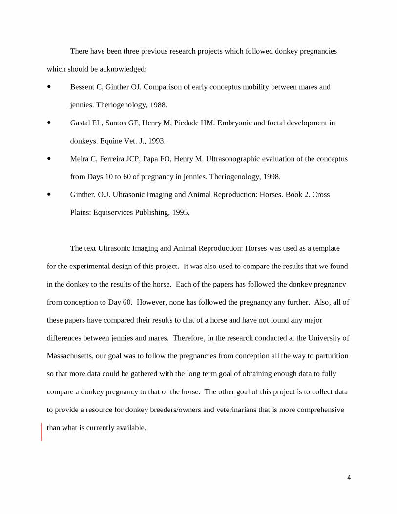
plowing, and they can also act as guard animals for livestock. Figure 1 shows donkeys being
used as pack animals in the Nuῆoa District, Peru. In the United States, donkeys are used
primarily as pets, with some using them for farming purposes, guard animals, and therapy
animals. Female donkeys are called jennets, while male donkeys are called jacks.
Figure 1: Donkeys being used as pack animals in the Nuñoa District, Peru
Reproduction is an important part of donkey stewardship whether in the United States or
around the world. With the world population on the rise and the need for more food production
worldwide, the importance of the donkey as a work animal in previously mentioned third world
countries should not be overlooked.
4
There have been three previous research projects which followed donkey pregnancies
which should be acknowledged:
Bessent C, Ginther OJ. Comparison of early conceptus mobility between mares and
jennies. Theriogenology, 1988.
Gastal EL, Santos GF, Henry M, Piedade HM. Embryonic and foetal development in
donkeys. Equine Vet. J., 1993.
Meira C, Ferreira JCP, Papa FO, Henry M. Ultrasonographic evaluation of the conceptus
from Days 10 to 60 of pregnancy in jennies. Theriogenology, 1998.
Ginther, O.J. Ultrasonic Imaging and Animal Reproduction: Horses. Book 2. Cross
Plains: Equiservices Publishing, 1995.
The text Ultrasonic Imaging and Animal Reproduction: Horses was used as a template
for the experimental design of this project. It was also used to compare the results that we found
in the donkey to the results of the horse. Each of the papers has followed the donkey pregnancy
from conception to Day 60. However, none has followed the pregnancy any further. Also, all of
these papers have compared their results to that of a horse and have not found any major
differences between jennies and mares. Therefore, in the research conducted at the University of
Massachusetts, our goal was to follow the pregnancies from conception all the way to parturition
so that more data could be gathered with the long term goal of obtaining enough data to fully
compare a donkey pregnancy to that of the horse. The other goal of this project is to collect data
to provide a resource for donkey breeders/owners and veterinarians that is more comprehensive
than what is currently available.
5
Methodology
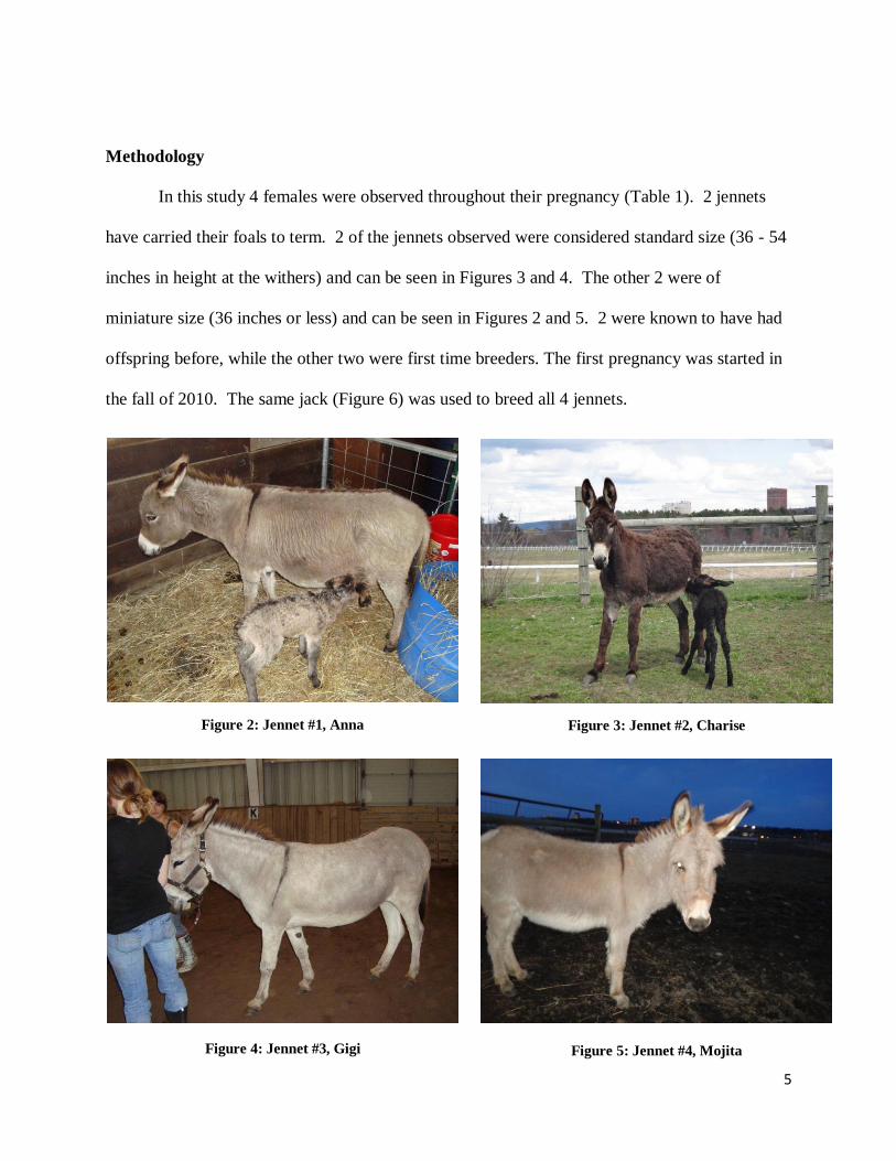
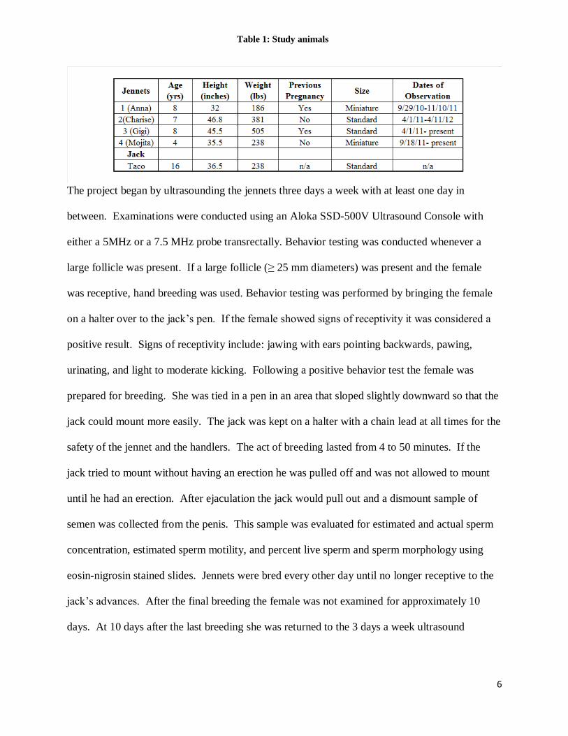
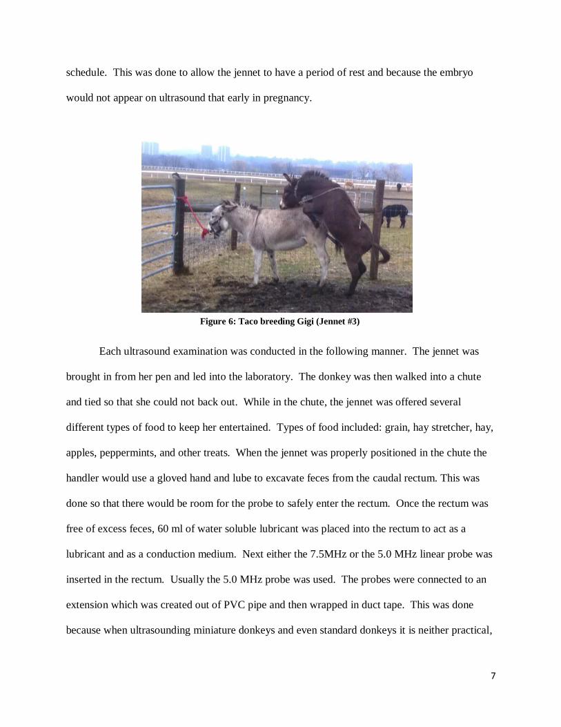
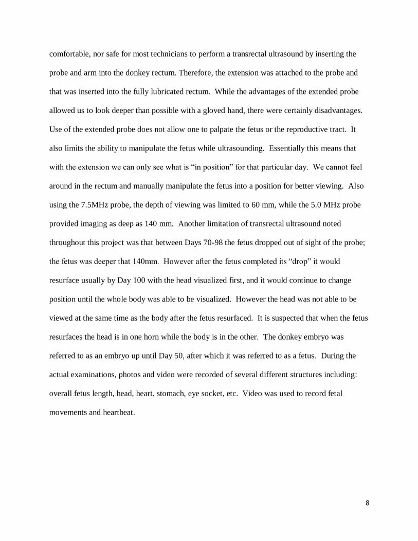
In this study 4 females were observed throughout their pregnancy (Table 1). 2 jennets
have carried their foals to term. 2 of the jennets observed were considered standard size (36 - 54
inches in height at the withers) and can be seen in Figures 3 and 4. The other 2 were of
miniature size (36 inches or less) and can be seen in Figures 2 and 5. 2 were known to have had
offspring before, while the other two were first time breeders. The first pregnancy was started in
the fall of 2010. The same jack (Figure 6) was used to breed all 4 jennets.
Figure 2: Jennet #1, Anna Figure 3: Jennet #2, Charise
Figure 4: Jennet #3, Gigi Figure 5: Jennet #4, Mojita
6
The project began by ultrasounding the jennets three days a week with at least one day in
between. Examinations were conducted using an Aloka SSD-500V Ultrasound Console with
either a 5MHz or a 7.5 MHz probe transrectally. Behavior testing was conducted whenever a
large follicle was present. If a large follicle (≥ 25 mm diameters) was present and the female
was receptive, hand breeding was used. Behavior testing was performed by bringing the female
on a halter over to the jack’s pen. If the female showed signs of receptivity it was considered a
positive result. Signs of receptivity include: jawing with ears pointing backwards, pawing,
urinating, and light to moderate kicking. Following a positive behavior test the female was
prepared for breeding. She was tied in a pen in an area that sloped slightly downward so that the
jack could mount more easily. The jack was kept on a halter with a chain lead at all times for the
safety of the jennet and the handlers. The act of breeding lasted from 4 to 50 minutes. If the
jack tried to mount without having an erection he was pulled off and was not allowed to mount
until he had an erection. After ejaculation the jack would pull out and a dismount sample of
semen was collected from the penis. This sample was evaluated for estimated and actual sperm
concentration, estimated sperm motility, and percent live sperm and sperm morphology using
eosin-nigrosin stained slides. Jennets were bred every other day until no longer receptive to the
jack’s advances. After the final breeding the female was not examined for approximately 10
days. At 10 days after the last breeding she was returned to the 3 days a week ultrasound
Table 1: Study animals
7
schedule. This was done to allow the jennet to have a period of rest and because the embryo
would not appear on ultrasound that early in pregnancy.
Each ultrasound examination was conducted in the following manner. The jennet was
brought in from her pen and led into the laboratory. The donkey was then walked into a chute
and tied so that she could not back out. While in the chute, the jennet was offered several
different types of food to keep her entertained. Types of food included: grain, hay stretcher, hay,
apples, peppermints, and other treats. When the jennet was properly positioned in the chute the
handler would use a gloved hand and lube to excavate feces from the caudal rectum. This was
done so that there would be room for the probe to safely enter the rectum. Once the rectum was
free of excess feces, 60 ml of water soluble lubricant was placed into the rectum to act as a
lubricant and as a conduction medium. Next either the 7.5MHz or the 5.0 MHz linear probe was
inserted in the rectum. Usually the 5.0 MHz probe was used. The probes were connected to an
extension which was created out of PVC pipe and then wrapped in duct tape. This was done
because when ultrasounding miniature donkeys and even standard donkeys it is neither practical,
Figure 6: Taco breeding Gigi (Jennet #3)
8
comfortable, nor safe for most technicians to perform a transrectal ultrasound by inserting the
probe and arm into the donkey rectum. Therefore, the extension was attached to the probe and
that was inserted into the fully lubricated rectum. While the advantages of the extended probe
allowed us to look deeper than possible with a gloved hand, there were certainly disadvantages.
Use of the extended probe does not allow one to palpate the fetus or the reproductive tract. It
also limits the ability to manipulate the fetus while ultrasounding. Essentially this means that
with the extension we can only see what is “in position” for that particular day. We cannot feel
around in the rectum and manually manipulate the fetus into a position for better viewing. Also
using the 7.5MHz probe, the depth of viewing was limited to 60 mm, while the 5.0 MHz probe
provided imaging as deep as 140 mm. Another limitation of transrectal ultrasound noted
throughout this project was that between Days 70-98 the fetus dropped out of sight of the probe;
the fetus was deeper that 140mm. However after the fetus completed its “drop” it would
resurface usually by Day 100 with the head visualized first, and it would continue to change
position until the whole body was able to be visualized. However the head was not able to be
viewed at the same time as the body after the fetus resurfaced. It is suspected that when the fetus
resurfaces the head is in one horn while the body is in the other. The donkey embryo was
referred to as an embryo up until Day 50, after which it was referred to as a fetus. During the
actual examinations, photos and video were recorded of several different structures including:
overall fetus length, head, heart, stomach, eye socket, etc. Video was used to record fetal
movements and heartbeat.
9
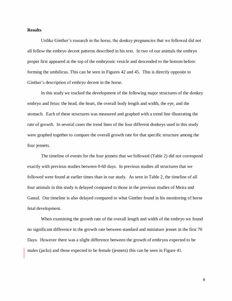
Results
Unlike Ginther’s research in the horse, the donkey pregnancies that we followed did not
all follow the embryo decent patterns described in his text. In two of our animals the embryo
proper first appeared at the top of the embryonic vesicle and descended to the bottom before
forming the umbilicus. This can be seen in Figures 42 and 45. This is directly opposite to
Ginther’s description of embryo decent in the horse.
In this study we tracked the development of the following major structures of the donkey
embryo and fetus: the head, the heart, the overall body length and width, the eye, and the
stomach. Each of these structures was measured and graphed with a trend line illustrating the
rate of growth. In several cases the trend lines of the four different donkeys used in this study
were graphed together to compare the overall growth rate for that specific structure among the
four jennets.
The timeline of events for the four jennets that we followed (Table 2) did not correspond
exactly with previous studies between 0-60 days. In previous studies all structures that we
followed were found at earlier times than in our study. As seen in Table 2, the timeline of all
four animals in this study is delayed compared to those in the previous studies of Meira and
Gastal. Our timeline is also delayed compared to what Ginther found in his monitoring of horse
fetal development.
When examining the growth rate of the overall length and width of the embryo we found
no significant difference in the growth rate between standard and miniature jennet in the first 70
Days. However there was a slight difference between the growth of embryos expected to be
males (jacks) and those expected to be female (jennets) this can be seen in Figure 41.
10
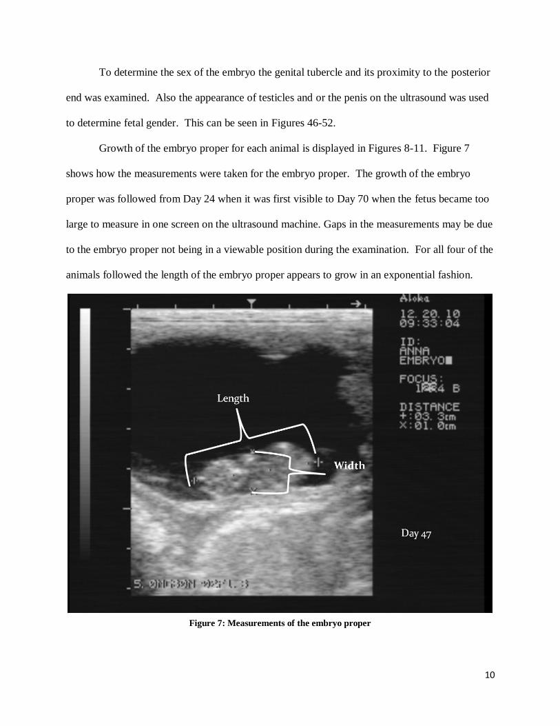
To determine the sex of the embryo the genital tubercle and its proximity to the posterior
end was examined. Also the appearance of testicles and or the penis on the ultrasound was used
to determine fetal gender. This can be seen in Figures 46-52.
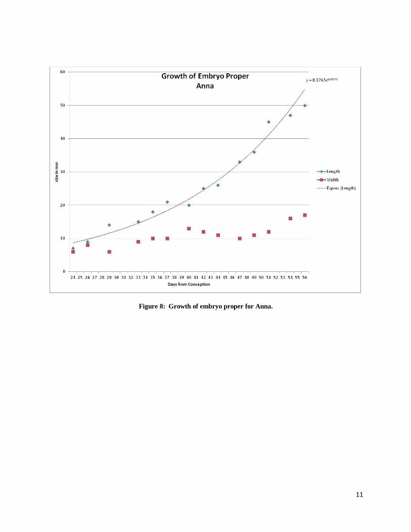
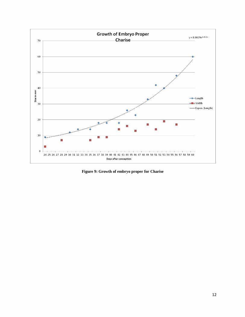
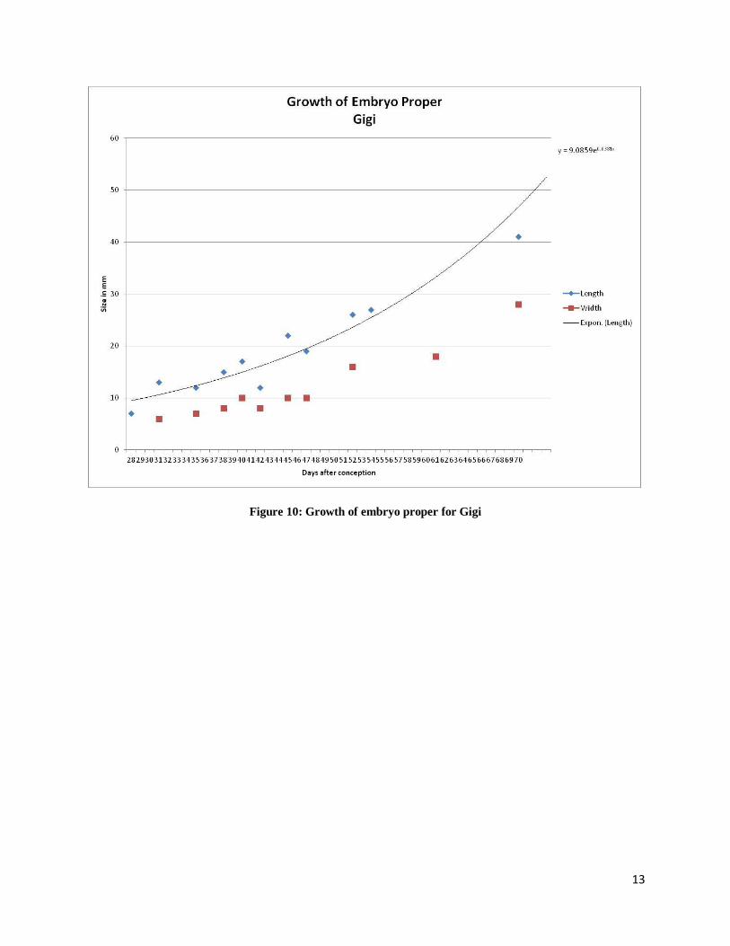
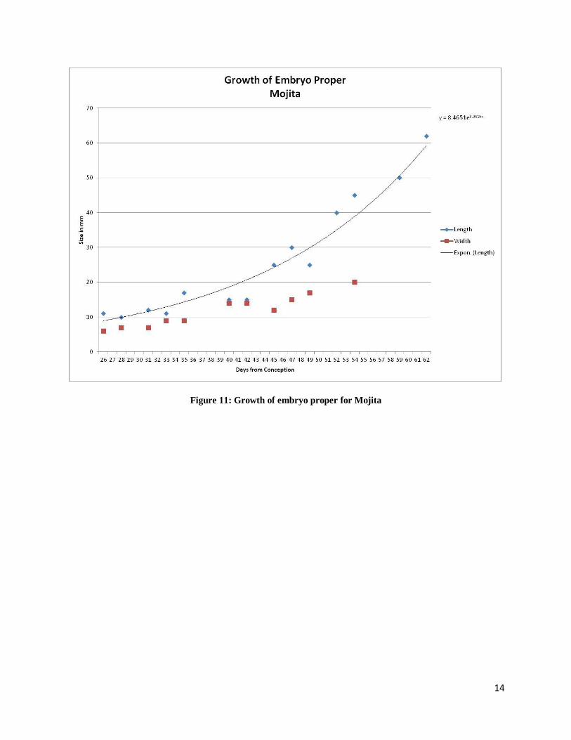
Growth of the embryo proper for each animal is displayed in Figures 8-11. Figure 7
shows how the measurements were taken for the embryo proper. The growth of the embryo
proper was followed from Day 24 when it was first visible to Day 70 when the fetus became too
large to measure in one screen on the ultrasound machine. Gaps in the measurements may be due
to the embryo proper not being in a viewable position during the examination. For all four of the
animals followed the length of the embryo proper appears to grow in an exponential fashion.
Figure 7: Measurements of the embryo proper
11
Figure 8: Growth of embryo proper for Anna.
12
Figure 9: Growth of embryo proper for Charise
13
Figure 10: Growth of embryo proper for Gigi
14
Figure 11: Growth of embryo proper for Mojita
15
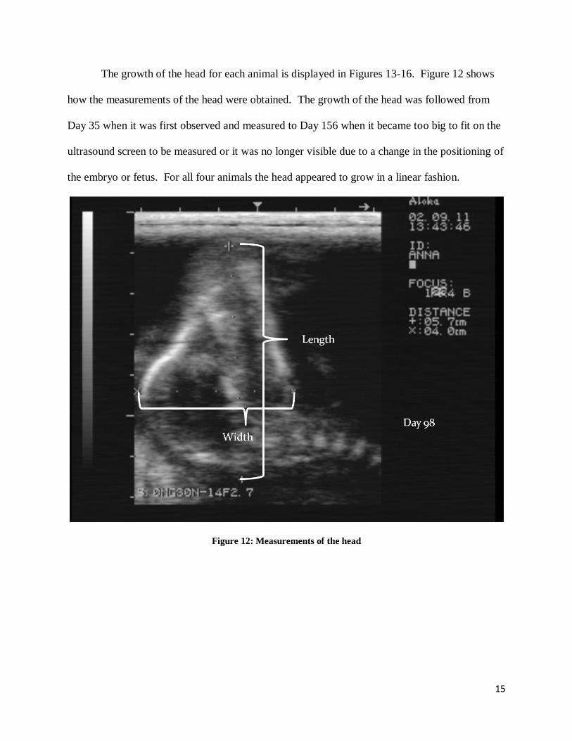
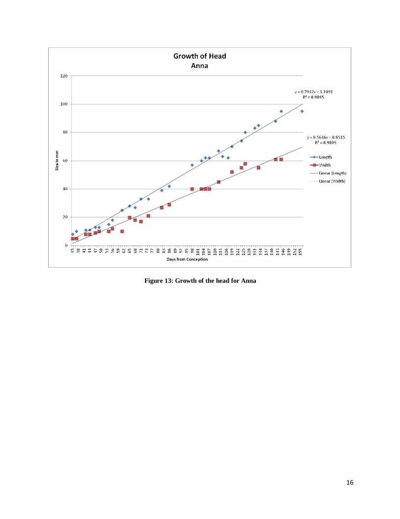
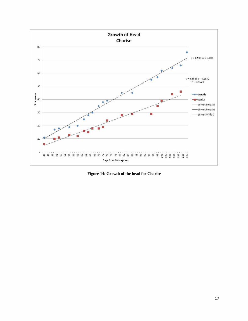
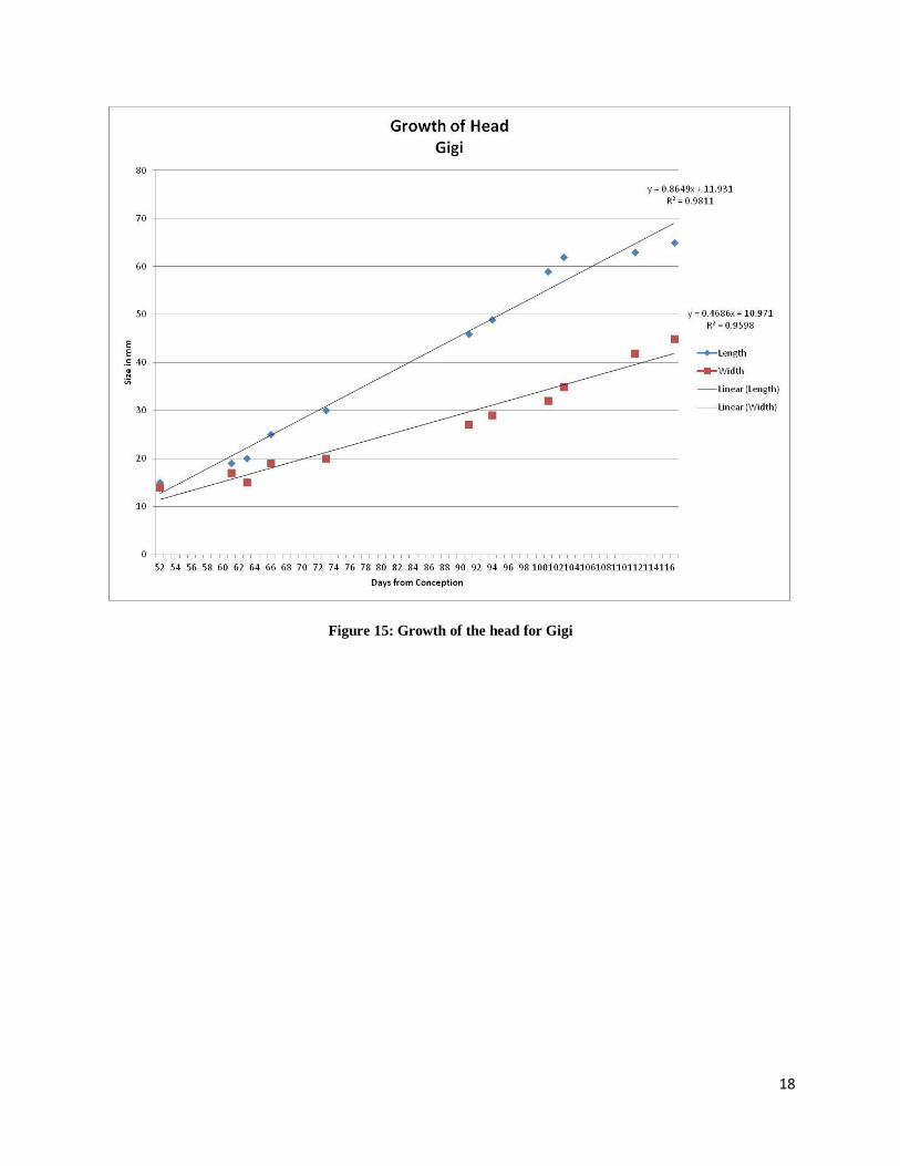
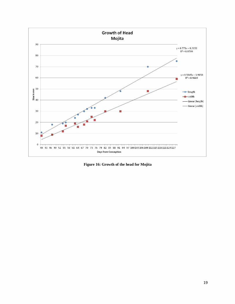
The growth of the head for each animal is displayed in Figures 13-16. Figure 12 shows
how the measurements of the head were obtained. The growth of the head was followed from
Day 35 when it was first observed and measured to Day 156 when it became too big to fit on the
ultrasound screen to be measured or it was no longer visible due to a change in the positioning of
the embryo or fetus. For all four animals the head appeared to grow in a linear fashion.
Figure 12: Measurements of the head
16
Figure 13: Growth of the head for Anna
17
Figure 14: Growth of the head for Charise
18
Figure 15: Growth of the head for Gigi
19
Figure 16: Growth of the head for Mojita
20
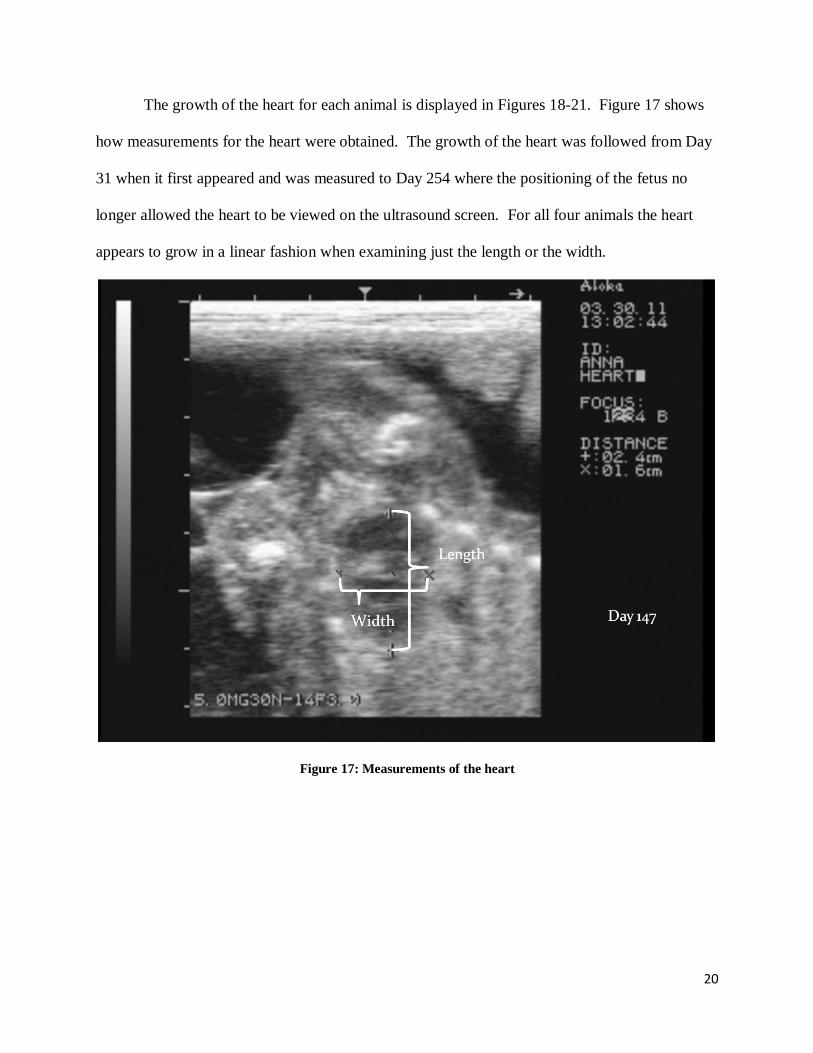
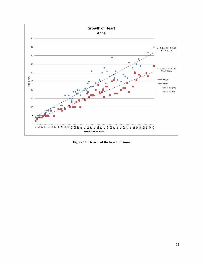
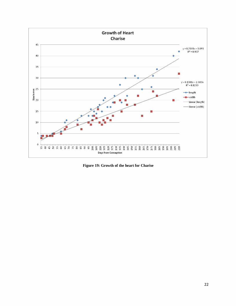
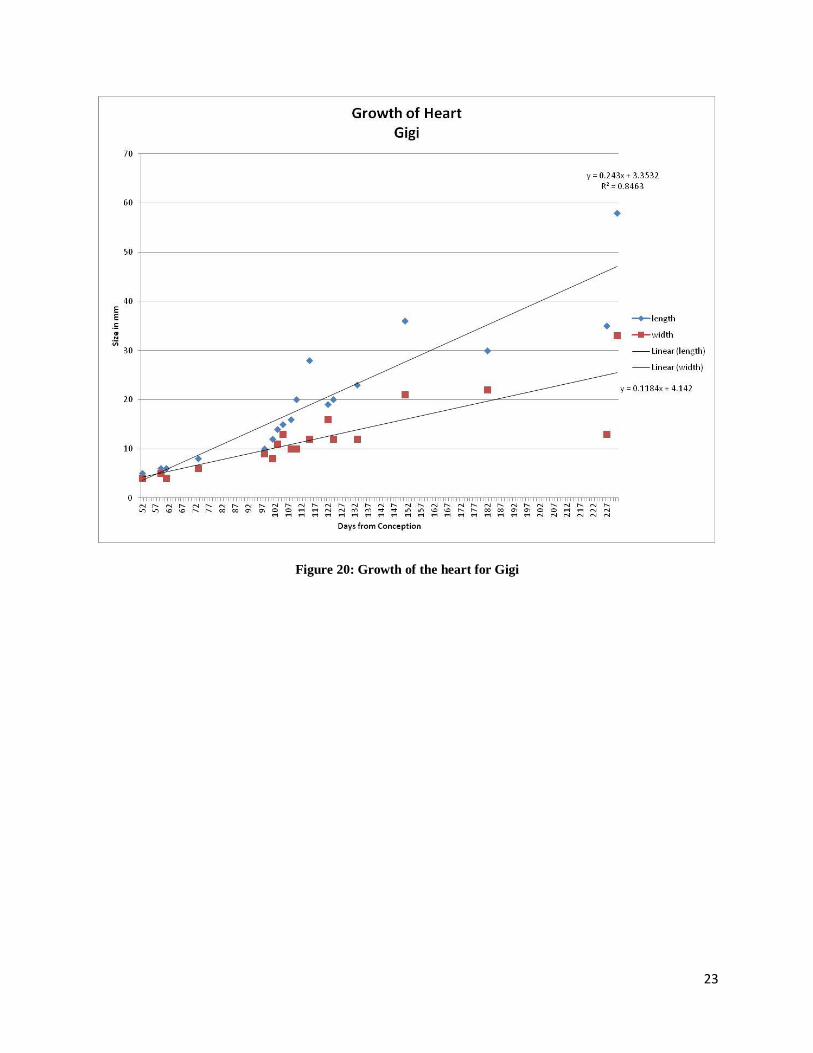
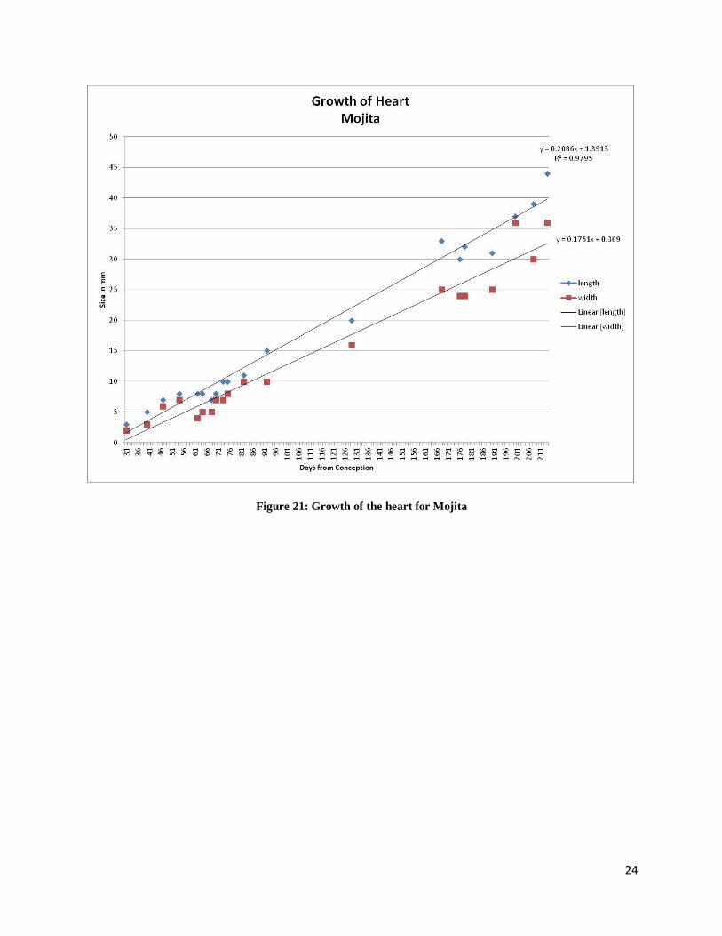
The growth of the heart for each animal is displayed in Figures 18-21. Figure 17 shows
how measurements for the heart were obtained. The growth of the heart was followed from Day
31 when it first appeared and was measured to Day 254 where the positioning of the fetus no
longer allowed the heart to be viewed on the ultrasound screen. For all four animals the heart
appears to grow in a linear fashion when examining just the length or the width.
Figure 17: Measurements of the heart
21
Figure 18: Growth of the heart for Anna
22
Figure 19: Growth of the heart for Charise
23
Figure 20: Growth of the heart for Gigi
24
Figure 21: Growth of the heart for Mojita
25
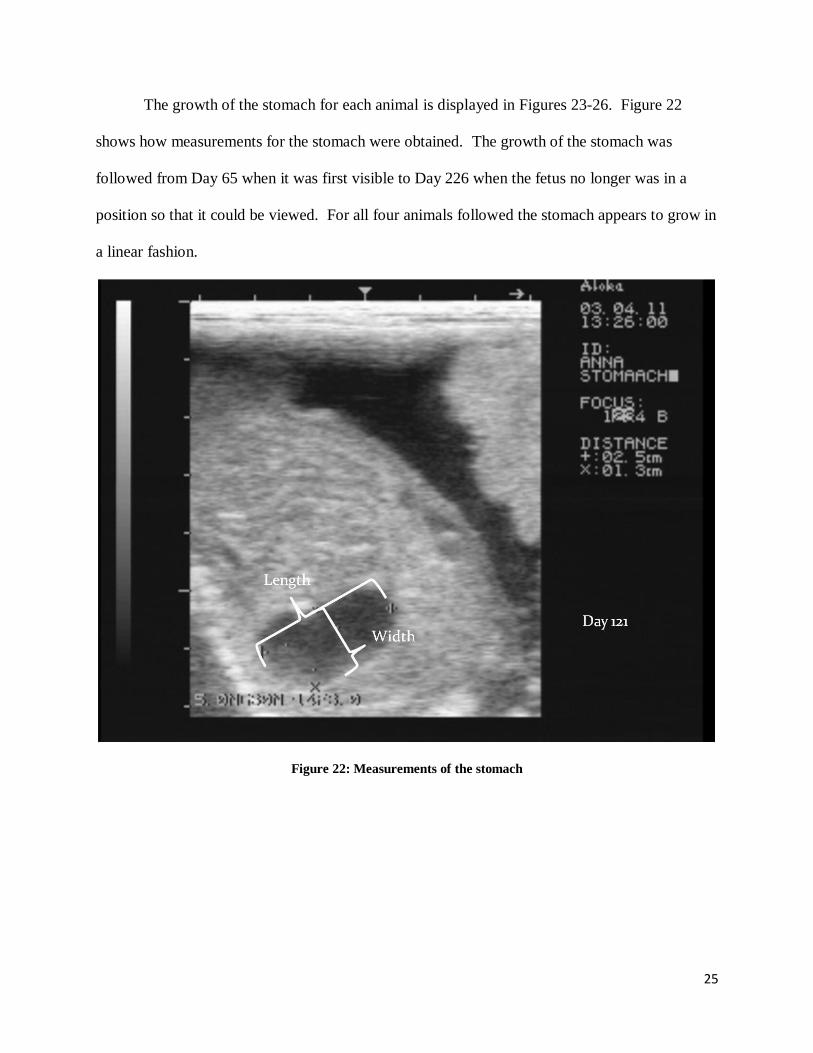
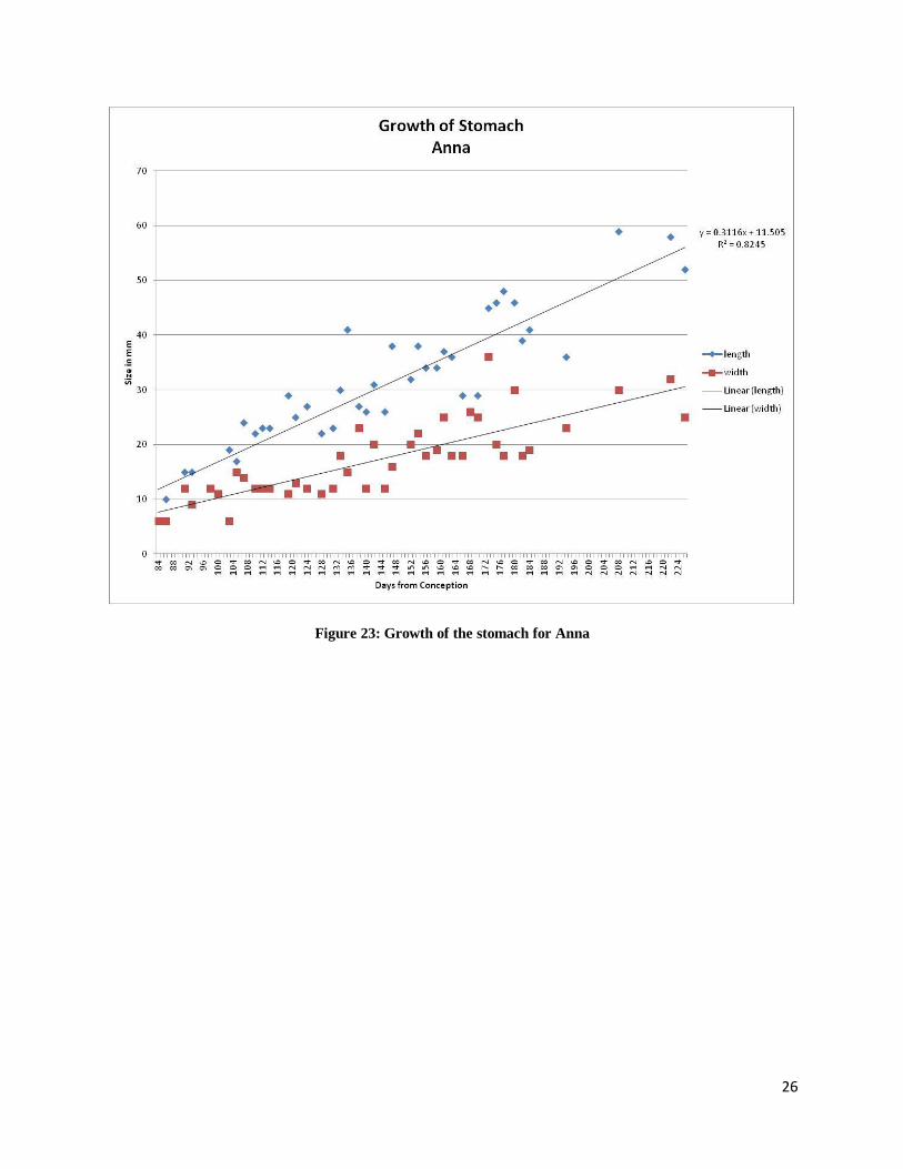
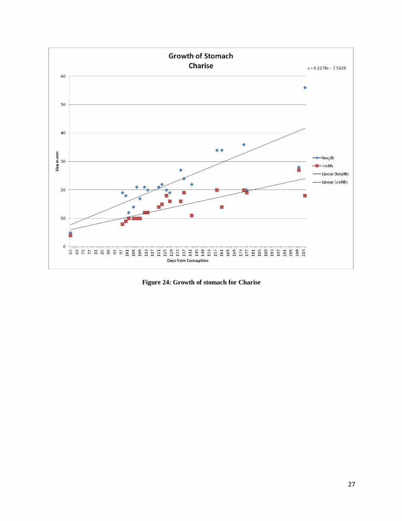
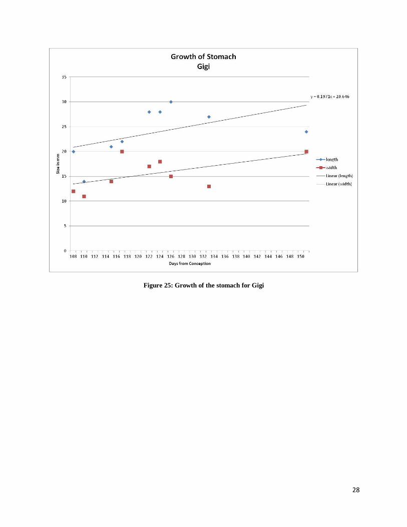
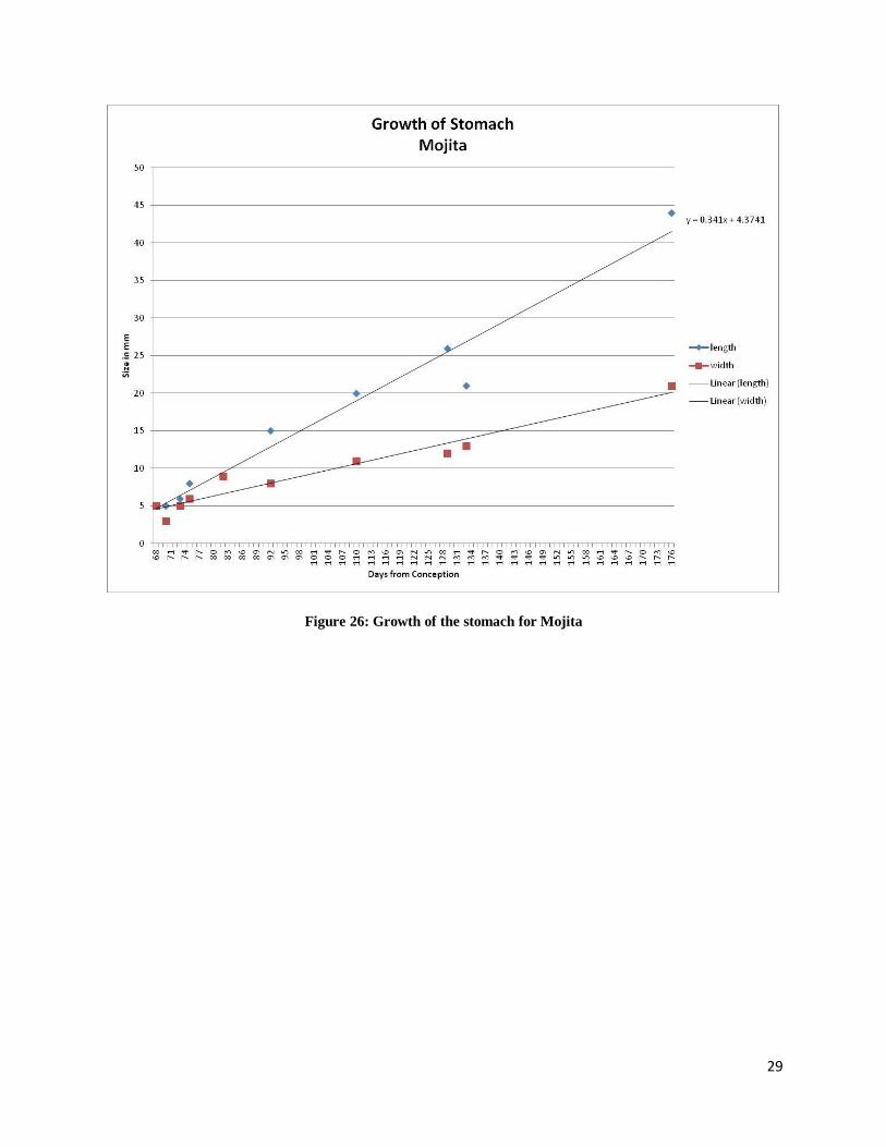
The growth of the stomach for each animal is displayed in Figures 23-26. Figure 22
shows how measurements for the stomach were obtained. The growth of the stomach was
followed from Day 65 when it was first visible to Day 226 when the fetus no longer was in a
position so that it could be viewed. For all four animals followed the stomach appears to grow in
a linear fashion.
Figure 22: Measurements of the stomach
26
Figure 23: Growth of the stomach for Anna
27
Figure 24: Growth of stomach for Charise
28
Figure 25: Growth of the stomach for Gigi
29
Figure 26: Growth of the stomach for Mojita
30
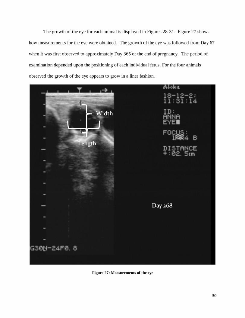
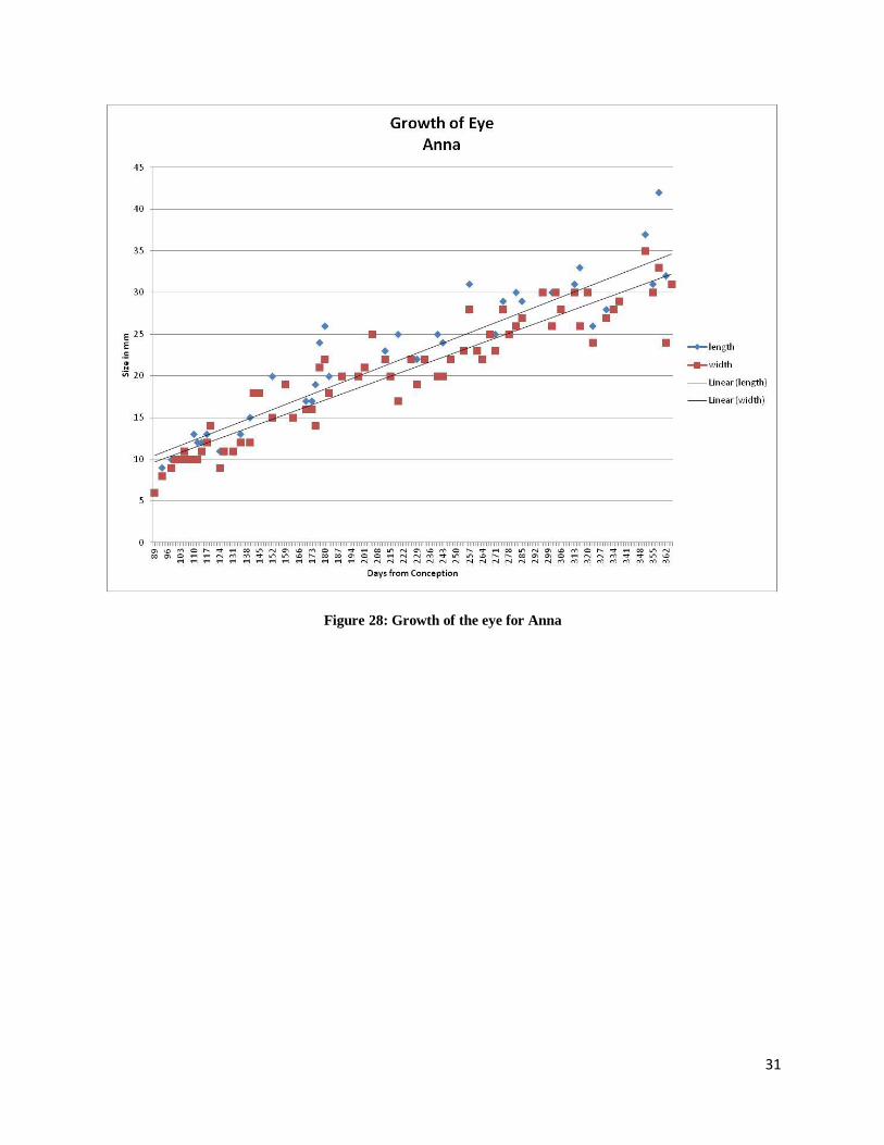
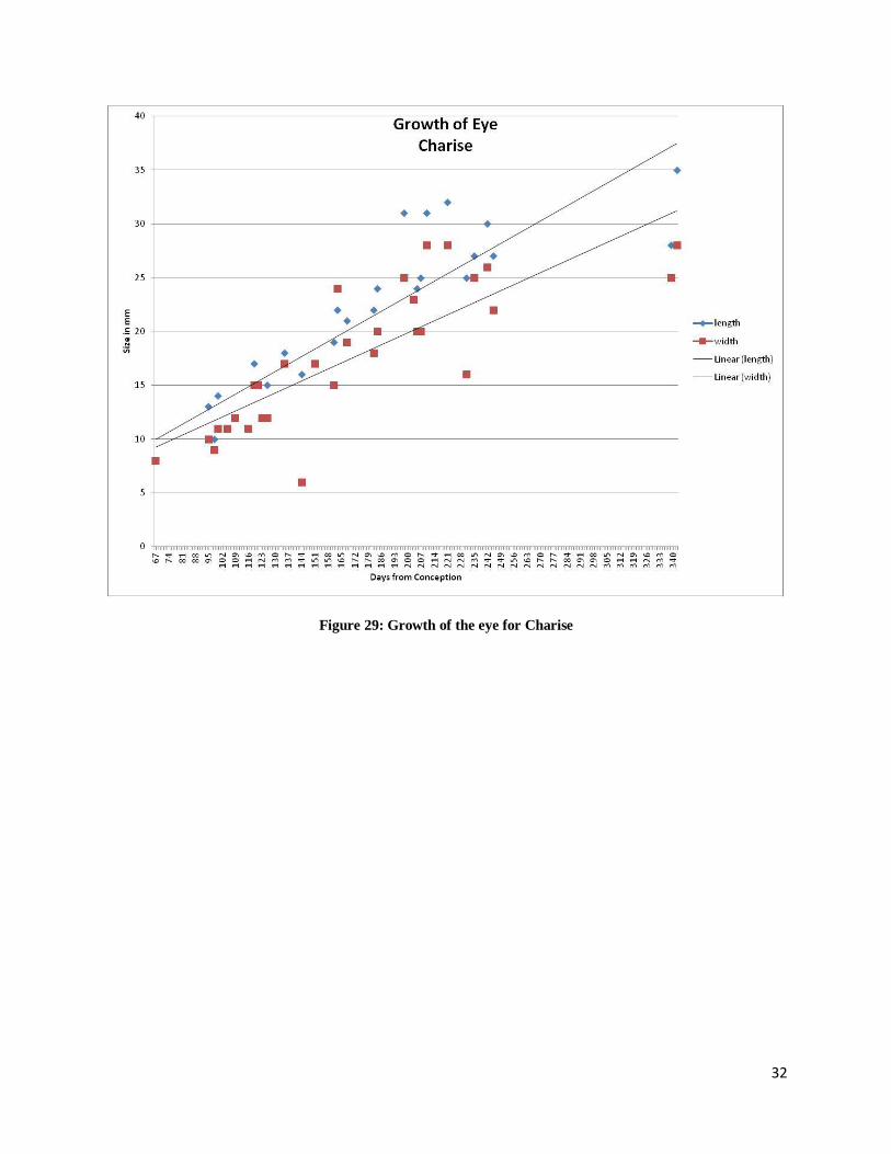
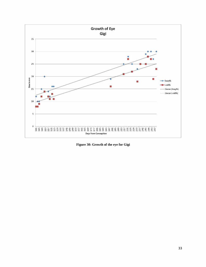
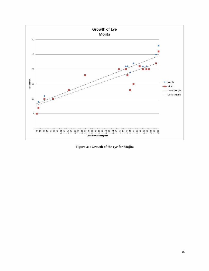
The growth of the eye for each animal is displayed in Figures 28-31. Figure 27 shows
how measurements for the eye were obtained. The growth of the eye was followed from Day 67
when it was first observed to approximately Day 365 or the end of pregnancy. The period of
examination depended upon the positioning of each individual fetus. For the four animals
observed the growth of the eye appears to grow in a liner fashion.
Figure 27: Measurements of the eye
31
Figure 28: Growth of the eye for Anna
32
Figure 29: Growth of the eye for Charise
33
Figure 30: Growth of the eye for Gigi
34
Figure 31: Growth of the eye for Mojita
35
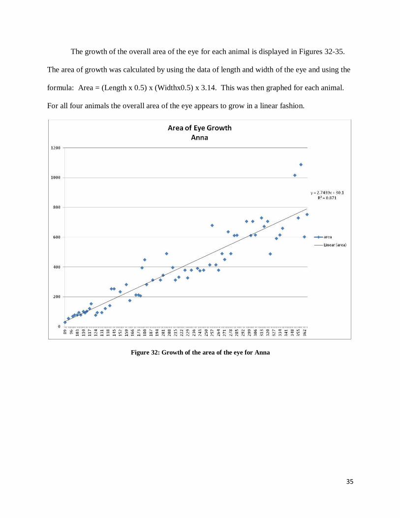
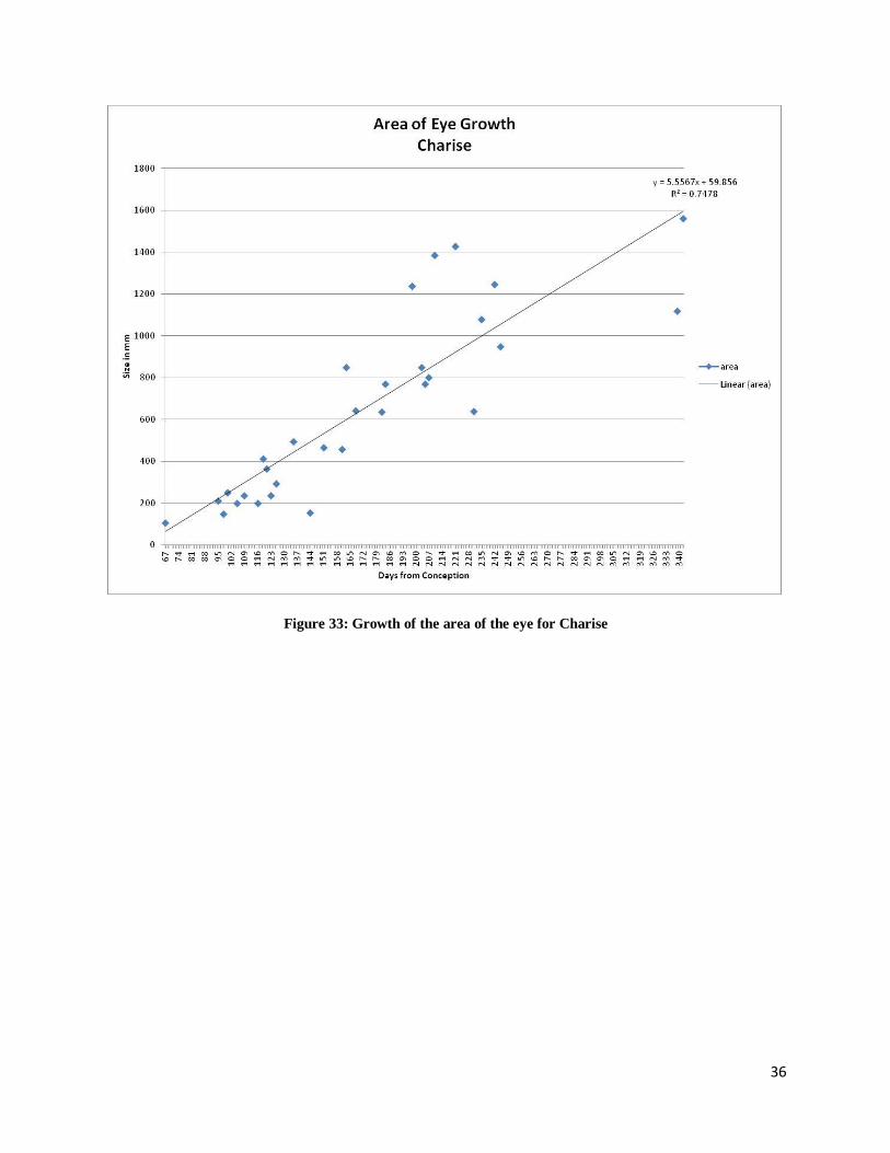
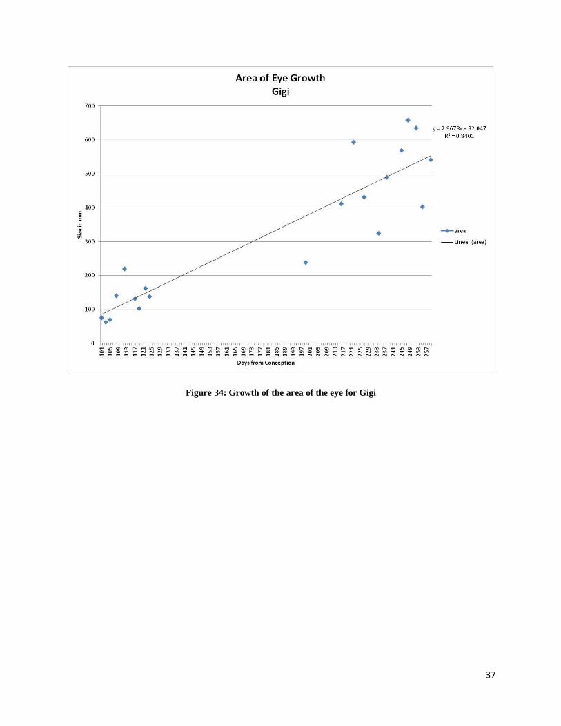
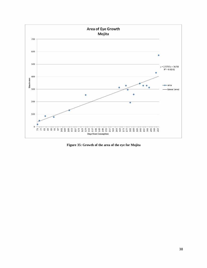
The growth of the overall area of the eye for each animal is displayed in Figures 32-35.
The area of growth was calculated by using the data of length and width of the eye and using the
formula: Area = (Length x 0.5) x (Widthx0.5) x 3.14. This was then graphed for each animal.
For all four animals the overall area of the eye appears to grow in a linear fashion.
Figure 32: Growth of the area of the eye for Anna
36
Figure 33: Growth of the area of the eye for Charise
37
Figure 34: Growth of the area of the eye for Gigi
38
Figure 35: Growth of the area of the eye for Mojita
39
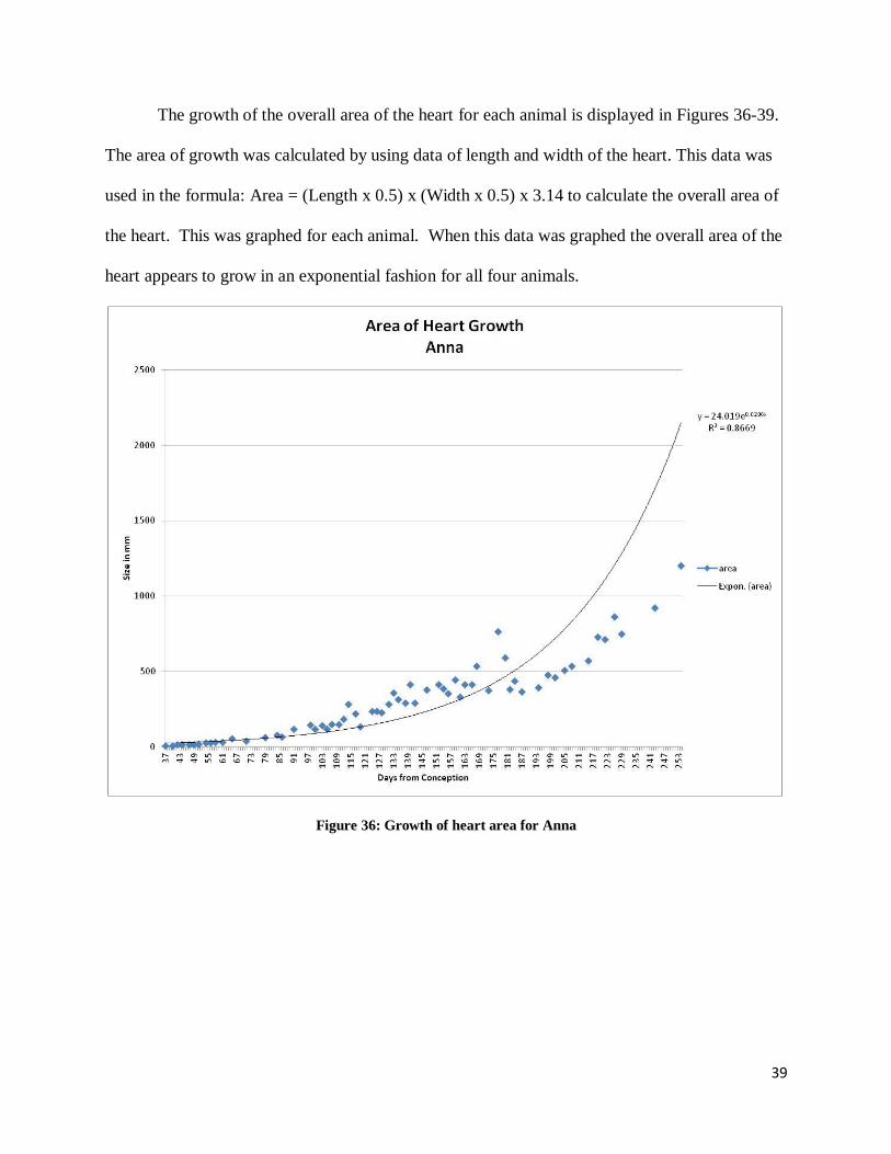
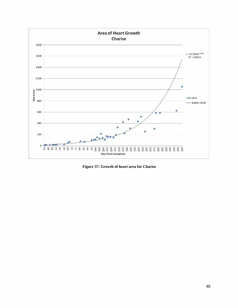
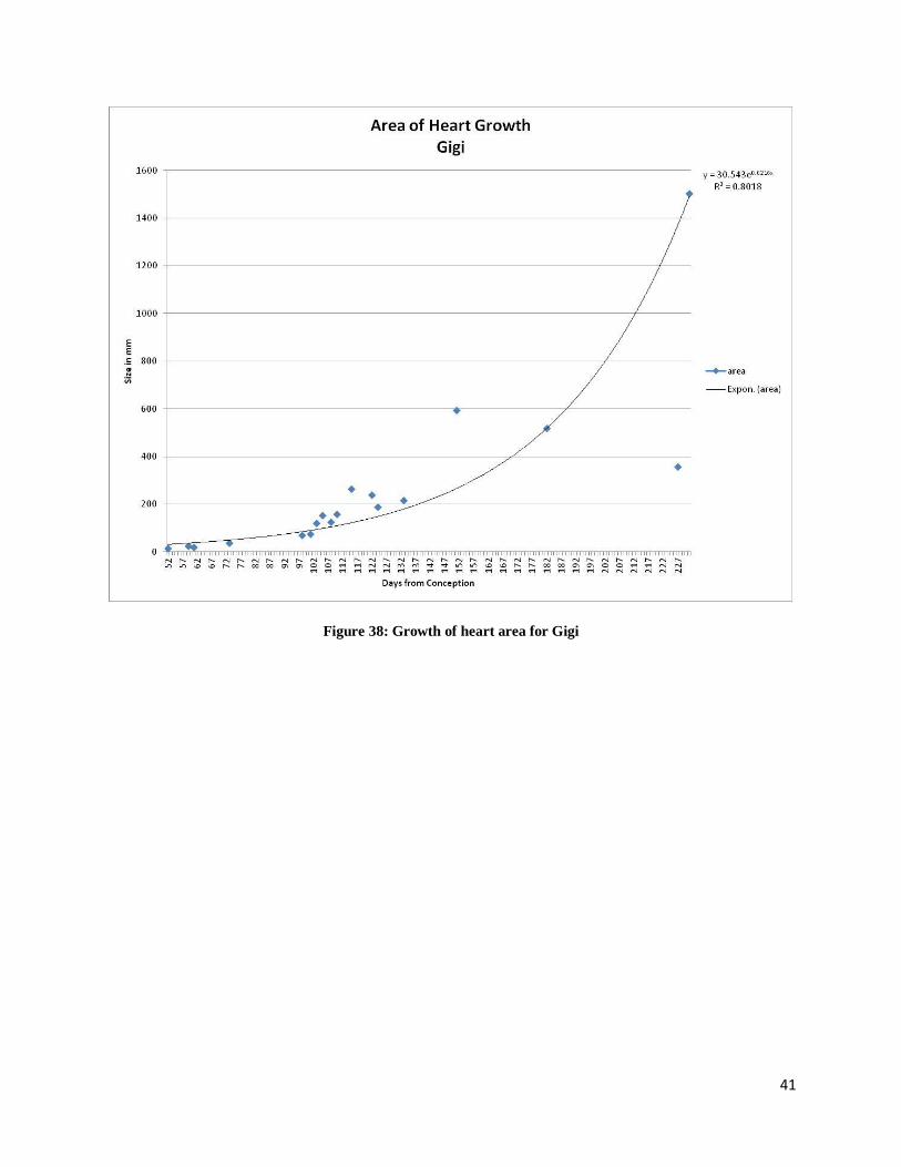
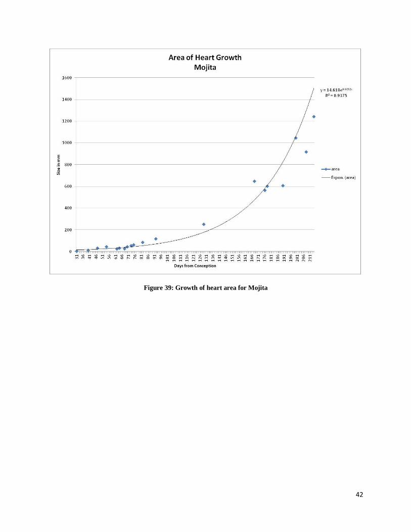
The growth of the overall area of the heart for each animal is displayed in Figures 36-39.
The area of growth was calculated by using data of length and width of the heart. This data was
used in the formula: Area = (Length x 0.5) x (Width x 0.5) x 3.14 to calculate the overall area of
the heart. This was graphed for each animal. When this data was graphed the overall area of the
heart appears to grow in an exponential fashion for all four animals.
Figure 36: Growth of heart area for Anna
40
Figure 37: Growth of heart area for Charise
41
Figure 38: Growth of heart area for Gigi
42
Figure 39: Growth of heart area for Mojita
43
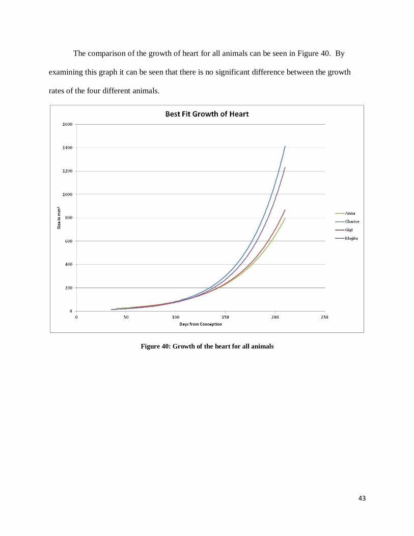
The comparison of the growth of heart for all animals can be seen in Figure 40. By
examining this graph it can be seen that there is no significant difference between the growth
rates of the four different animals.
Figure 40: Growth of the heart for all animals
44
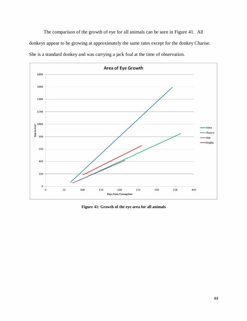
The comparison of the growth of eye for all animals can be seen in Figure 41. All
donkeys appear to be growing at approximately the same rates except for the donkey Charise.
She is a standard donkey and was carrying a jack foal at the time of observation.
Figure 41: Growth of the eye area for all animals
45
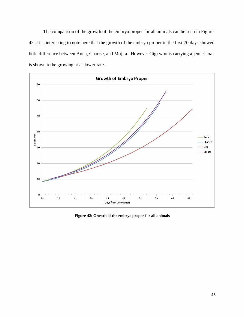
The comparison of the growth of the embryo proper for all animals can be seen in Figure
42. It is interesting to note here that the growth of the embryo proper in the first 70 days showed
little difference between Anna, Charise, and Mojita. However Gigi who is carrying a jennet foal
is shown to be growing at a slower rate.
Figure 42: Growth of the embryo proper for all animals
46
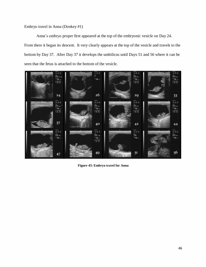
Embryo travel in Anna (Donkey #1)
Anna’s embryo proper first appeared at the top of the embryonic vesicle on Day 24.
From there it began its descent. It very clearly appears at the top of the vesicle and travels to the
bottom by Day 37. After Day 37 it develops the umbilicus until Days 51 and 56 where it can be
seen that the fetus is attached to the bottom of the vesicle.
Figure 43: Embryo travel for Anna
47
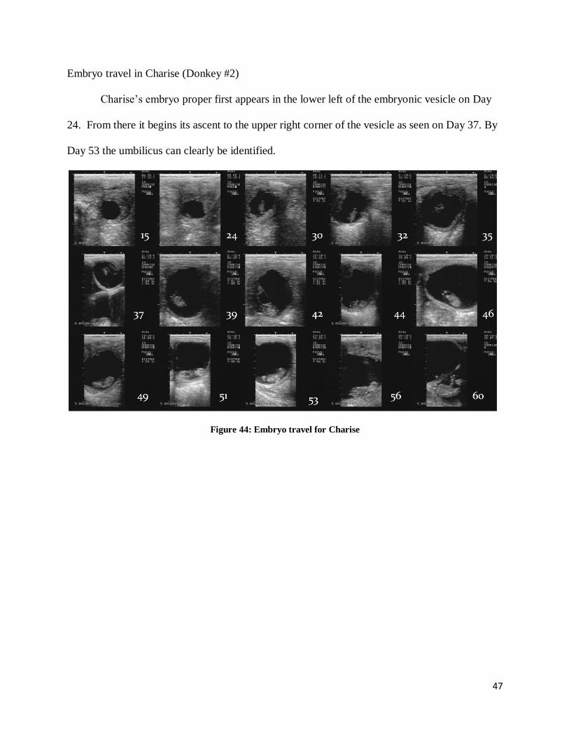
Embryo travel in Charise (Donkey #2)
Charise’s embryo proper first appears in the lower left of the embryonic vesicle on Day
24. From there it begins its ascent to the upper right corner of the vesicle as seen on Day 37. By
Day 53 the umbilicus can clearly be identified.
Figure 44: Embryo travel for Charise
48
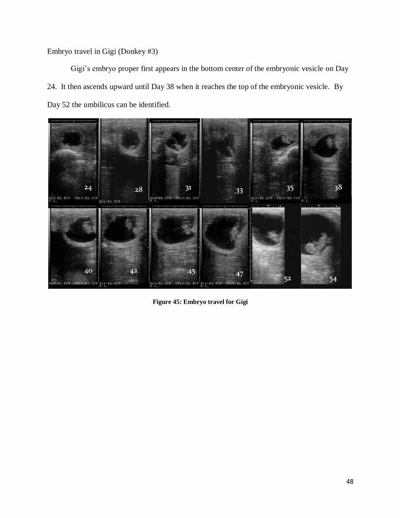
Embryo travel in Gigi (Donkey #3)
Gigi’s embryo proper first appears in the bottom center of the embryonic vesicle on Day
24. It then ascends upward until Day 38 when it reaches the top of the embryonic vesicle. By
Day 52 the umbilicus can be identified.
Figure 45: Embryo travel for Gigi
49
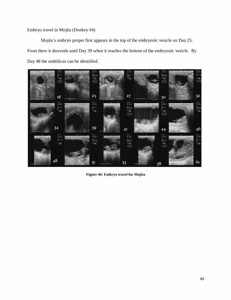
Embryo travel in Mojita (Donkey #4)
Mojita’s embryo proper first appears in the top of the embryonic vesicle on Day 25.
From there it descends until Day 39 when it reaches the bottom of the embryonic vesicle. By
Day 48 the umbilicus can be identified.
Figure 46: Embryo travel for Mojita
50
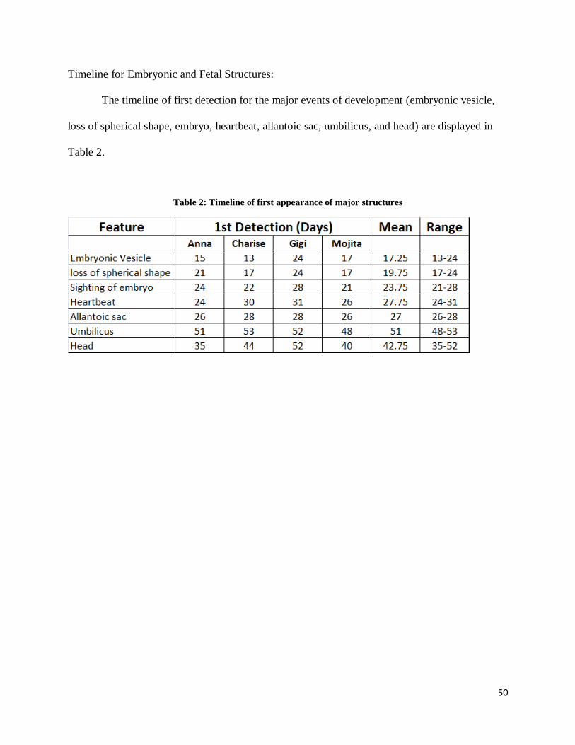
Timeline for Embryonic and Fetal Structures:
The timeline of first detection for the major events of development (embryonic vesicle,
loss of spherical shape, embryo, heartbeat, allantoic sac, umbilicus, and head) are displayed in
Table 2.
Table 2: Timeline of first appearance of major structures
51
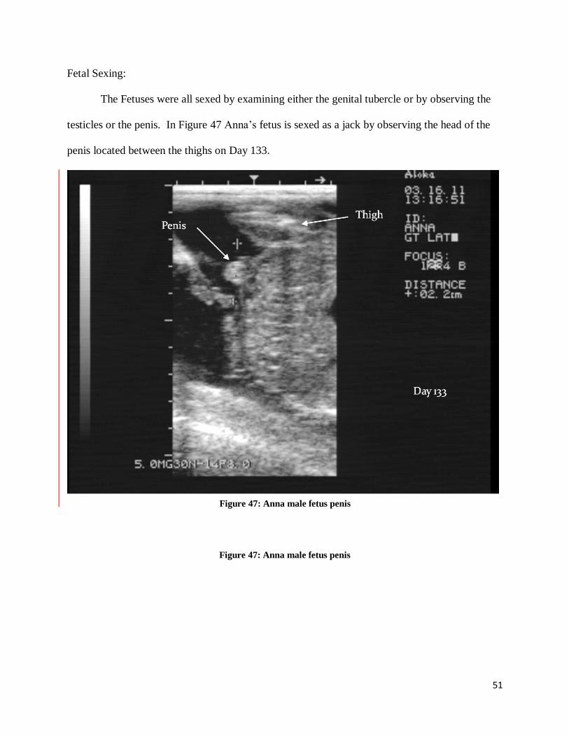
Fetal Sexing:
The Fetuses were all sexed by examining either the genital tubercle or by observing the
testicles or the penis. In Figure 47 Anna’s fetus is sexed as a jack by observing the head of the
penis located between the thighs on Day 133.
Figure 47: Anna male fetus penis
Figure 47: Anna male fetus penis
52
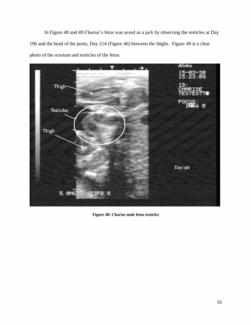
In Figure 48 and 49 Charise’s fetus was sexed as a jack by observing the testicles at Day
196 and the head of the penis, Day 214 (Figure 46) between the thighs. Figure 49 is a clear
photo of the scrotum and testicles of the fetus.
Figure 48: Charise male fetus testicles
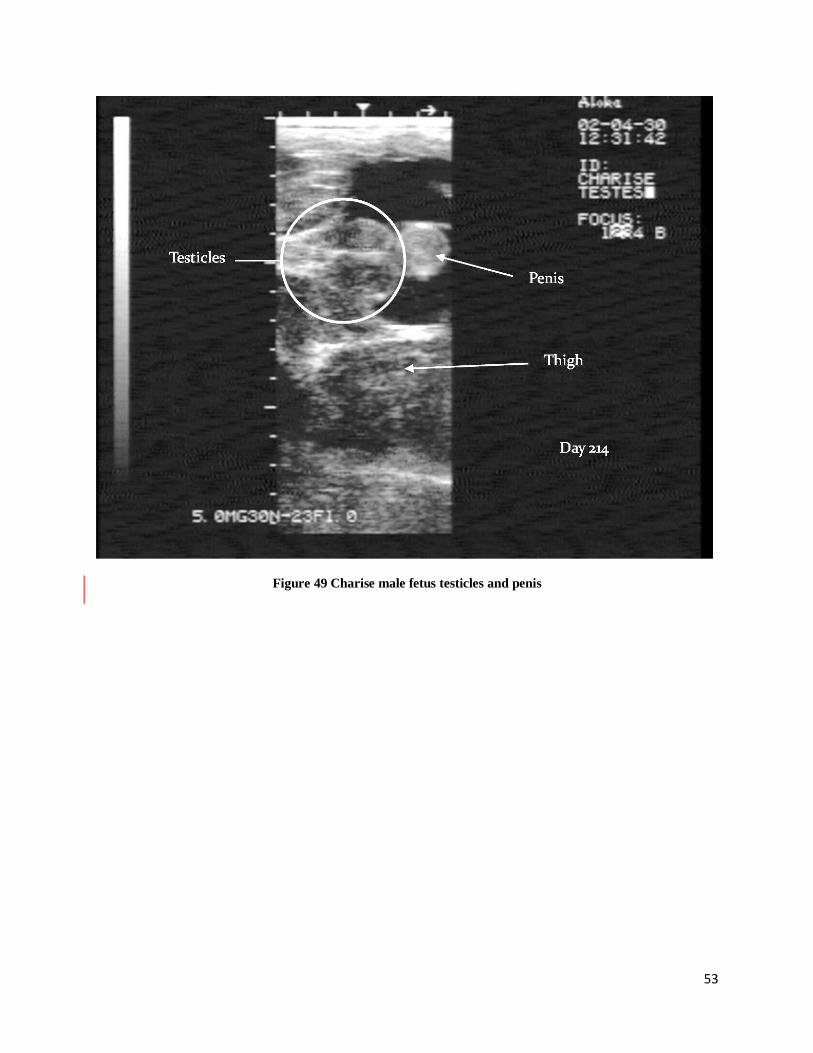
53
Figure 49 Charise male fetus testicles and penis
54
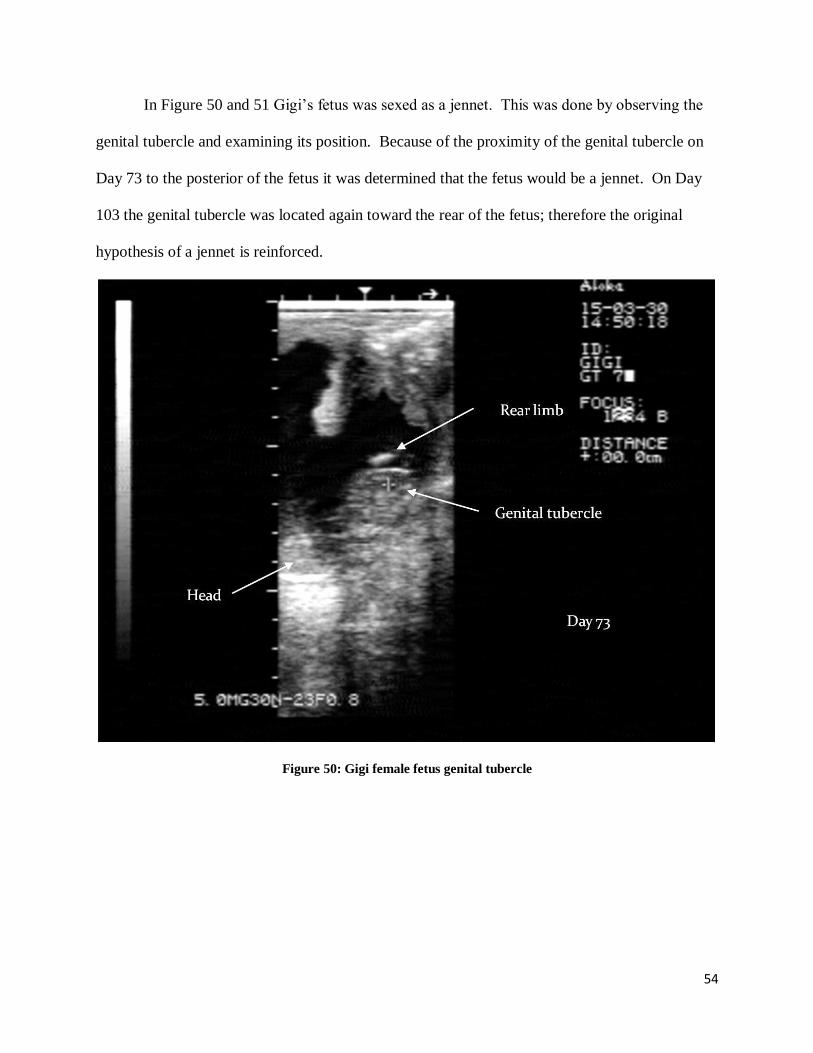
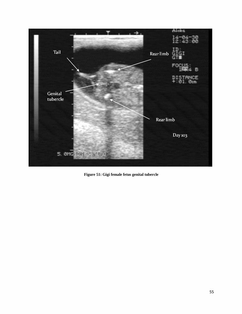
In Figure 50 and 51 Gigi’s fetus was sexed as a jennet. This was done by observing the
genital tubercle and examining its position. Because of the proximity of the genital tubercle on
Day 73 to the posterior of the fetus it was determined that the fetus would be a jennet. On Day
103 the genital tubercle was located again toward the rear of the fetus; therefore the original
hypothesis of a jennet is reinforced.
Figure 50: Gigi female fetus genital tubercle
55
Figure 51: Gigi female fetus genital tubercle
56
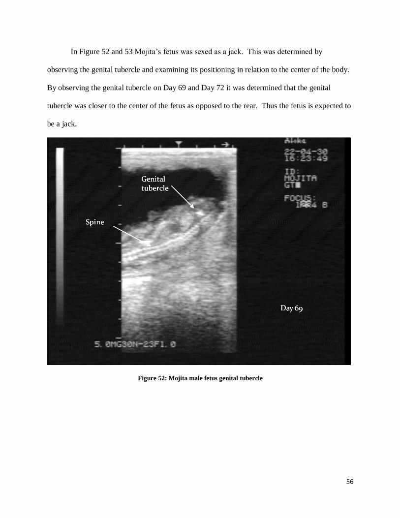
In Figure 52 and 53 Mojita’s fetus was sexed as a jack. This was determined by
observing the genital tubercle and examining its positioning in relation to the center of the body.
By observing the genital tubercle on Day 69 and Day 72 it was determined that the genital
tubercle was closer to the center of the fetus as opposed to the rear. Thus the fetus is expected to
be a jack.
Figure 52: Mojita male fetus genital tubercle
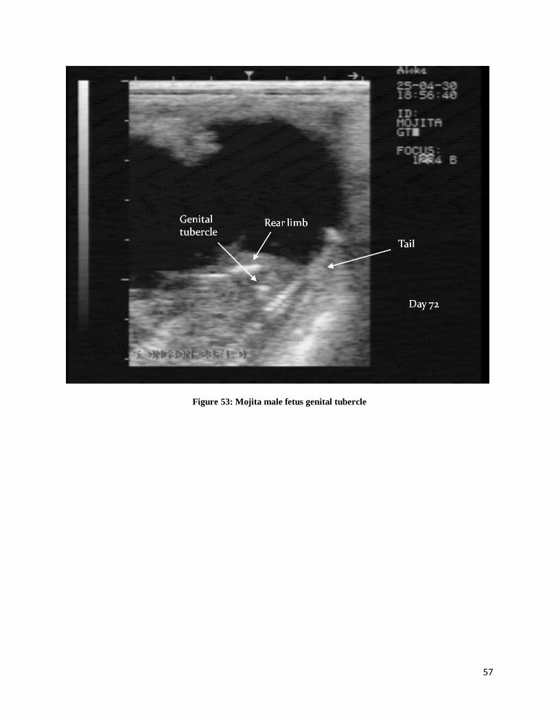
57
Figure 53: Mojita male fetus genital tubercle
58
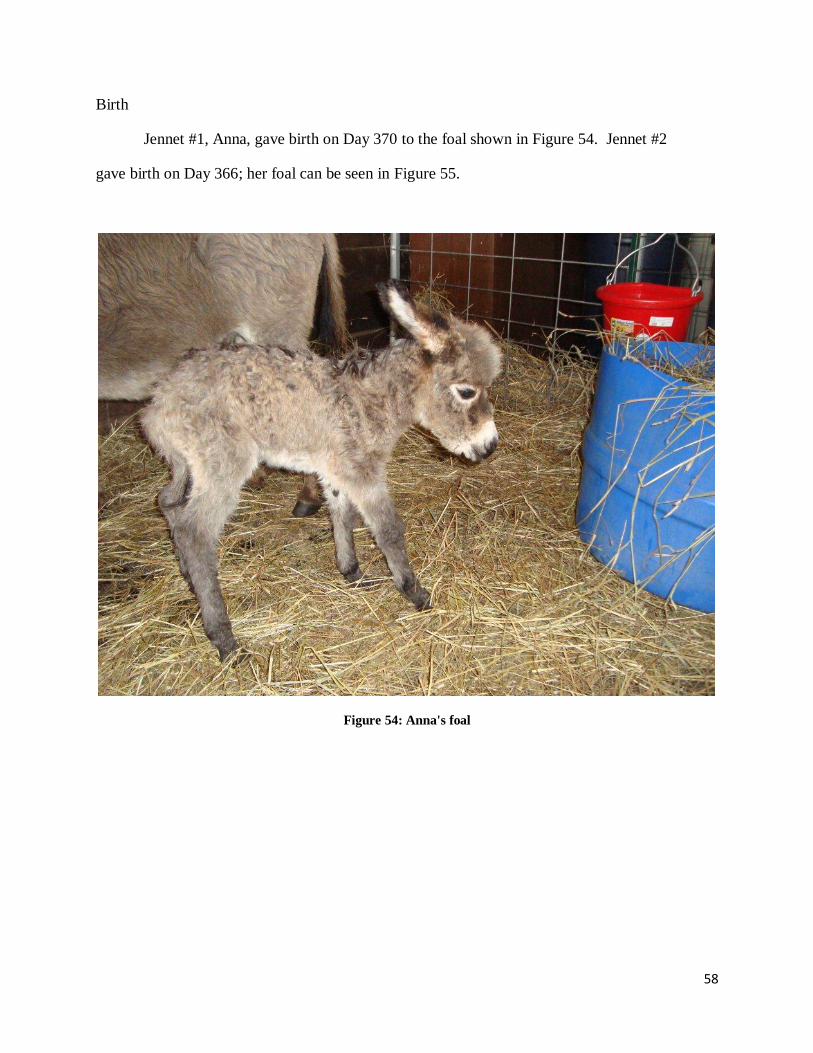
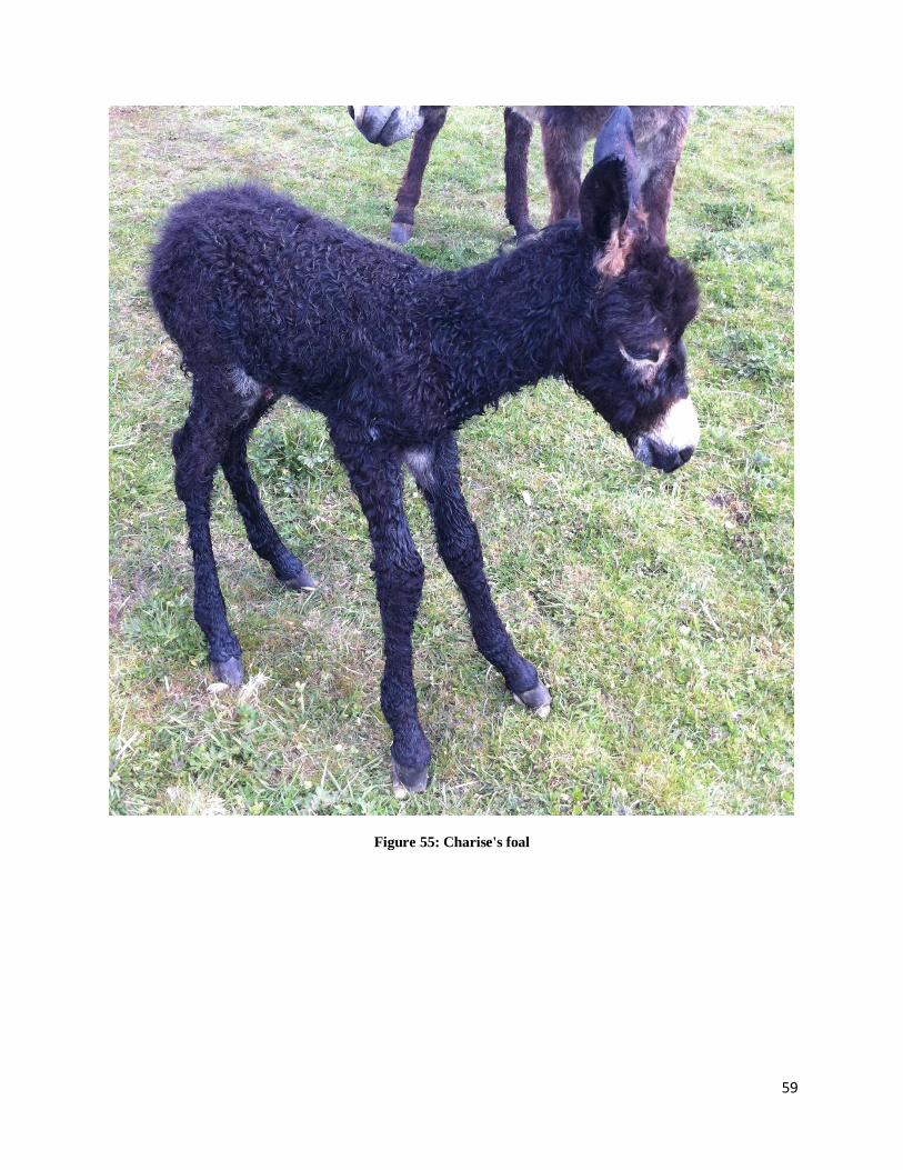
Birth
Jennet #1, Anna, gave birth on Day 370 to the foal shown in Figure 54. Jennet #2
gave birth on Day 366; her foal can be seen in Figure 55.
Figure 54: Anna's foal
59
Figure 55: Charise's foal
60
Conclusions
There were several different variables that were discovered during the course of this
project. For example, the largest variable encountered was the fact that we were not able to alter
the position of the embryo or fetus and were reliant upon whatever position that the embryo or
fetus was in during that particular exam period. Another variable was the skill of the operator.
This could have affected what structures were seen during the exam. Other variables worth
noting are that we had two different sizes of donkeys in this study, both miniatures and
standards. Finally the animals used in this study all had different reproductive backgrounds,
some had foals previously, others had been attempted to be bred, and others were never bred
before.
As previously mentioned, this study found that unlike Ginther’s research regarding the
developing horse embryo, 50% of the time the donkey embryo appeared at the top of the
embryonic vesicle as opposed to the bottom. Also this study’s timeline was consistently 3-4
Days behind the timelines developed in Meria’s and Gastal’s research. Another conclusion
reached from this project can be seen in Figure 38, the comparison of the growth of the embryo
proper. This study has found that the jennet carrying the female fetus grows at a slower rate
during the first 70 Days than the other three jennets carrying male fetuses. However this study
only examined four different animals; before strong correlations can be made more study is
needed. The reappearance of the fetus in ultrasound examinations is something that was found
across all four jennets and occurred at approximately the same time. This data suggests that at
around Day 70 the embryo will drop out deeper than 140mm into the uterus and will reappear
within 2 weeks in a different position, with the head in one horn and the body in another.
61
Finally the sexing of the fetuses was conducted using two different methods, one the
visual observation of reproductive organs and two the positioning of the genital tubercle. Both
visual observations were proven to be correct upon birth of the foal.
Future Study:
The overall goal of this project was to gather data so that more information could be
available for eventual comparison to the horse and to provide a source of reproductive
information for veterinarians, breeders, and owners. Several of our conclusions including the
differences in embryo travel, and the differences in growth of the embryo proper and fetus need
more data before they can be considered statistically significant. For future study I would
recommend several things. First, ultrasounds should be conducted sooner after conception, and
provided that there is no health hazard to the animal the ultrasounds should continue daily so that
more data can be gathered with less gaps between data points. Also a 3.0 MHz probe should be
used after approximately Day 175. This is when the fetus is deep into the abdomen and it is
quite large. Having a probe that could view a wider and deeper range would be extremely
helpful. In addition to the different probe, more measurements should be taken during the
ultrasound exams. This would make the overall exam time longer. As long as the animal
tolerates it well it would provide more data. Along with taking more measurements, video of
each exam should be recorded and saved for future reference in case of missing measurements or
discrepancies in the data. If video of the ultrasound could be recorded while recording a
voiceover of the examiner and what he or she was viewing or trying to view this would prove to
be extremely helpful. Furthermore, the organization of data should be established and written
down in precise protocols. This would prevent data from being missed, and data being confusing
62
or jumbled. Finally the most significant improvement to this project would be the addition of
more donkeys, both jacks and jennets. The addition of new donkeys would provide more data
for comparison. Mammoth donkeys should also be added to the project so that all sizes could be
represented in the study. With the addition of mammoth donkeys a rectal ultrasound conducted
by hand becomes a possibility, therefore increasing the ability to position the probe. This could
be useful in gathering more pertinent data.
Finally, this project is simply a start, more donkeys must be observed all the way from
conception to parturition so that a more complete library of records can be developed and
eventually compared to the horse development.
63
References
Bessent C, Ginther OJ. Comparison of early conceptus mobility between mares and jennies.
Theriogenology, 1988.
Gastal EL, Santos GF, Henry M, Piedade HM. Embryonic and foetal development in donkeys.
Equine Vet. J., 1993.
Ginther, O.J. Ultrasonic Imaging and Animal Reproduction: Horses. Book2. Cross Plains:
Equiservices Publishing, 1995.
Meira C, Ferreira JCP, Papa FO, Henry M. Ultrasonographic evaluation of the conceptus from
Days 10 to 60 of pregnancy in jennies. Theriogenology 1998
Purdy, SR. Donkeys A Veterinary Guide for Owners and Breeders. North Pomfret: Trafalgar
Square, 2010.