elementary steps in enzyme catalysis and...
TRANSCRIPT

ELEMENTARY STEPS IN ENZYME CATALYSIS ANDREGULATION
GORDON G. HAMMES
Department of Chemistry, Cornell University, Ithaca, New York 14850,USA
ABSTRACTA general approach to elucidating the molecular basis of enzyme catalysis andregulation is the time resolution of the complex reaction mechanisms involvedinto their elementary steps. Both enzyme catalysis and enzyme regulation areinitiated by the binding of small molecules to the protein. This associationreaction is generally quite rapid and i often followed by a conformational changeof the macromolecule. These conformational changes are discussed in terms ofthe dynamics of the underlying elementary processes, hydrogen bonding,solvation and hydrophobic interactions, all of which have been studied in modelsystems. Enzyme catalysis often involves acid—base catalysis. Consideration ofthe rates of both concerted and sequential proton transfer mechanisms in termsof model systems suggests that the observed turnover numbers of enzymesrequire that proton transfer steps in the catalytic process occur at close to theirmaximum possible rates. A general mechanism for enzyme catalysis is pro-posed and is illustrated by the mechanism of action of ribonuclease A. Adistinguishing feature of most regulatory enzymes is their multi-subunit nature.The regulation of enzyme activity usually is achieved by alteration of subunitinteractions through conformational changes triggered by ligand binding.These conformational transitions are important processes in the cooperativebinding of substrates and effectors to regulatory enzymes. As an example, themechanism of the regulation of aspartate transcarbamylase from Escherichiacoil is considered: in this case coupled conformational changes appear to be
utilized as interlocking on—off switches for the enzymic activity.
INTRODUCTIONEnzymes have two important physiological functions: efficient catalysis of
metabolic reactions and regulation of metabolic processes. In this presenta-tion the elementary steps and molecular bases of these two functions areconsidered.
The most remarkable features of enzyme catalysis are the very highefficiency and great specificity relative to model systems. For example, theenzyme fumarase, which catalyses the hydration of fumarate to L-malate,has a specific catalytic rate constant of about 2 x 10 s' at 25° when satura-ted with substrate', while the same reaction in 1 M hydrogen and hydroxideion has specific rate constants of approximately 2 x 10⁸ s_i and 2 x 10s 1, respectively2' 3, Moreover, no other substrates for this enzyme havebeen found other than fumarate and L-malate, except for cases where fluorine
525
PAC—40—-4---C

GORDON G. HAMMES
has been substituted for some of the substrate hydrogens. Although themechanism of enzyme action has been actively studied for many years, manyof the molecular details remain to be delineated.
The regulation of enzymes is a physiological process which does not yethave a true parallel in model systems. The turning on and off of enzymaticactivity by specific molecules is crucial for the control of metabolic fluxes.For example, aspartate transcarbamylase, which catalyses the carbamylationof aspartic acid by carbamyl phosphate, is inhibited by the ultimate end-product of the biosynthetic pathway, cytidine-5'-triphosphate (CTP), whicheffectively shuts down pyrimidine biosynthesis; this same enzyme is activated(turned on) by the purine, adenosine-5'-triphosphate (ATP)4. A second ex-ample of an enzyme with important regulatory properties is phosphofructo-kinase, which plays a central role in glycolysis and catalyses the transfer of aphosphoryl group from adenosine-5'-triphosphate to fructose-6-phosphateto give fructose-i,6-diphosphate and the nucleotide diphosphate. It isactivated by a variety of substances, including phosphate, fructose- 1,6-diphosphate, adenosine-5'-monophosphate and adenosine-5'-diphosphate,and is inhibited by several metabolites, including citrate and magnesiumadenosine-5'-triphosphate5'6 The regulation of enzymes is also controlledat the genetic level where the synthesis of enzymes can be turned on and off,but this important aspect of regulation will not be discussed.
The approaches to understanding enzyme catalysis stressed here arethermodynamics and kinetics. Basically this means that the chemical pro-cesses are studied as a function of concentrations and time. It is importantthat the accessible time range be as broad as possible in order that all of theindividual elementary steps can be isolated and studied. At the present timemethods are available, such as magnetic resonance, ultrasonic attenuationmeasurements, the temperature jump method and stopped flow techniques,which permit reaction time constants as short as 10 10_b- 's to be mea-sured (cf. reference 7). A summary of currently available experimental tech-niques for kinetic studies of enzyme reactions and their approximate timeresolution is given in Table 1. Since molecular vibrations occur in 10- 12_10 s, virtually the entire time range of chemical events is accessible. Thedelineation of the elementary steps in enzymatic processes permits thedevelopment of formal kinetic models, which ultimately must be inter-preted in structural terms.
In the discussion to be presented here the common elementary stepsinvolved in catalysis and regulation are considered. both in terms of reac-tions occurring in the actual enzymatic process and in terms of modelreactions. Relaxation methods are particularly useful in analysing complex
Table 1. Fast reaction techniques for the study of enzymes
TechniqueApplicable time
range (s) TechniqueApplicable time
range (s)
Nuclear magnetic resonance 10 6_> 1 Temperature jump 10 8.>IElectron magnetic resonance o —1 o 10 Pressure jump 10 —> 1
Rapid mixing iO>i Acoustic methods l0-10'526

ELEMENTARY STEPS IN ENZYME CATALYSIS AND REGULATION
mechanisms, since the number of relaxation processes or time constants(relaxation times) observed is a direct measure of the minimum number ofsteps in the mechanism. Two particular enzyme mechanisms will be dis-cussed in detail: the breakdown of ribonucleic acids by ribonuclease and theregulation of enzyme catalysis by aspartate transcarbamylase.
INITIATION OF CATALYSIS AND REGULATION
The initiation of catalysis and of regulation is similar, namely the bindingof a substrate or effector (i.e. regulator) molecule to the enzyme. The rate ofbinding of many small molecules to enzymes, including substrates, inhibitorsand regulatory ligands, has been studied with fast reaction techniques (cf.references 8 and 9). Some representative data are presented in Table 2. The
Table 2. Representative rate constants for the reaction E + L EL
107k1Enzyme (E) Ligand (L) (M 1 s') k(s 1) Reference
Chymotrypsin Furylacryloyl-L- 0.62 2.7 x 10 10tryptophan amide
Ribonuclease Cytidine-3'-phosphate 4.6 4.2 x 10 11
Uridine-3'-phosphate 7.8 1.1 x 10 11
Cytidine-2'3' cyclic 2-4 1—2 x i0 12phosphateUridine-2',3' cyclic 1 2 x lO 13phosphateCytidylyl-3',5'-cytidine 1.4 7 x l0 14
Creatine kinase ADP 2.2 1.8 x l0 15MgADP 0.53 5.1 x l0 15CaADP 0.17 1.2 x l0 15MnADP 0.74 4.1 x io 15
Lactate dehydrogenase NADH 5.46 39 16Glyceraldehyde 3 NADa 1.1 1.1 x 10 17phosphate dehydro- 0.032 8 x 10²genase (yeast)
"Two types of binding Sites are present.
second-order rate constants measured are typically in the range of io--108 M 1 s'. Thus, although the interaction of an enzyme with a substrateor effector is often very specific, with rigid stereochemical requirements,nevertheless the initial complex formation is very rapid and is close to beingcontrolled by how rapidly the reactants diffuse together. The dissociationrate constant of the initial complex formed varies quite widely, and usuallycan be interpreted as a direct measure of the strength of the small molecule—enzyme interaction (i.e. it parallels the thermodynamic binding constants,since the association rate constants do not vary greatly). Another generalfeature of the interaction between enzyme and substrate or effector is that anisomerization of the complex often follows the binding—that is, the complex(and sometimes the unliganded enzyme) can exist in two or more differentconformations. The rates of the conformational changes vary considerably,
527
GORDON G. HAM MES
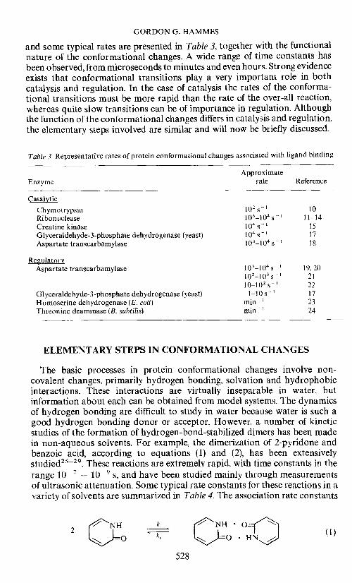
and some typical rates are presented in Table 3, together with the functionalnature of the conformational changes. A wide range of time constants hasbeen observed, from microseconds to minutes and even hours. Strong evidenceexists that conformational transitions play a very important role in bothcatalysis and regulation. In the case of catalysis the rates of the conforma-tional transitions must be more rapid than the rate of the over-all reaction,whereas quite slow transitions can be of importance in regulation. Althoughthe function of the conformational changes differs in catalysis and regulation,the elementary steps involved are similar and will now be briefly discussed.
Table 3. Representative rates of protein conformational changes associated with ligand binding
ApproximateEnzyme rate Reference
Catalytic
Chymrnrypsin 102s' 10Ribonuclease 103—104s 11—14
Creatine kinase i0 s_I 15
Glyceraldehyde-3-phosphate dehydrogenase (yeast) i0 s 17
Aspartate transcarbamylase 1 O—1 04 s 18
RegulatoryAspartate transcarbamylase 103—104s 1 19,20
102—103s 21
10—102s' 22
Glyceraldehyde-3-phosphate dehydrogenase (yeast) 1—10 s 1 171-lomoserine dehydrogenase (E. coli) rnin' 23
Threonine deaminase (B. subtilis) rnin I 24
ELEMENTARY STEPS IN CONFORMATIONAL CHANGES
The basic processes in protein conformational changes involve non-covalent changes, primarily hydrogen bonding, solvation and hydrophobicinteractions. These interactions are virtually inseparable in water, butinformation about each can be obtained from model systems. The dynamicsof hydrogen bonding are difficult to study in water because water is such agood hydrogen bonding donor or acceptor. However, a number of kineticstudies of the formation of hydrogen-bond-stabilized dimers has been madein non-aqueous solvents. For example, the dimerization of 2-pyridone andbenzoic acid, according to equations (1) and (2), has been extensivelystudied2 29 These reactions are extremely rapid, with time constants in therange iO — 1O s, and have been studied mainly through measurementsof ultrasonic attenuation. Some typical rate constants for these reactions in avariety of solvents are summarized in Table 4. The association rate constants
NH NHO2 0 UN (1)
528

ELEMENTARY STEPS IN ENZYME CATALYSIS AND REGULATION
2 OrCOH (2)
Table 4. Representative rate constants for hydrogen bond dimerization 2A r A2
Reactant Solvent109kf
(M' s1)l07kr(s') Reference
Benzoic acid Cd4CHC13Hexane
5.04.78.1
00730.750.022
252626
2-Pyridone CHC13Dioxane
1 H20-dioxaneCC14-dimethyl
sulphoxide (1.1 m)CC14-dimethyl
suiphoxide (5.Sm)
3.32.11.70.26
0.069
2.2131714.8
27
27282829
29
in all cases in Table 4, except for the last two entries, are approximately iOM 1 s
—1, which is essentially the value expected for a diffusion-controlled
reaction. The corresponding dissociation rate constants, on the other hand,vary considerably and roughly parallel the thermodynamic stability of thehydrogen bonds. The mechanism of these reactions can be schematicallywritten as follows:
A 1A D kAD tAD2 (3)D D A D A D---A
The first step in this mechanism represents the diffusion together of reactants;the second step represents the formation of the first hydrogen bond; and thethird step represents the formation of the second hydrogen bond. If all of theintermediates are assumed to be present in a steady state, which is suggestedby the fact that only a single time constant is found experimentally, theobserved forward and reverse rate constants can be written as
k = k1/{1 + (k_1/k2)(1 ± k_2/k3)} (4)
kr = k...3/{l + (k3/k...2)(1 + k2/k_1)} (5)
In order for kf to be equal to k1, as indicated by the experimental data, k2must be greater than k_1. In other words, desolvation of the solutes andformation of the first hydrogen bond must be faster than diffusion apart ofthe reactants. The value ofk_1 is about 1010 s, so that k2 must be 1011.4012s - , which is only 10—100 molecular vibrations. The observed dissociationrate constant under these conditions is kr = k.. 1(k...2/k2)(k_3/k3), and sincek...1 is essentially the same for all cases, the reverse rate constant is a directmeasure of the thermodynamic stability of solute—solute hydrogen bondsrelative to solute—solvent hydrogen bonds. In solvents containing appre-
529

GORDON G. HAMMES
ciable amounts of species forming strong hydrogen bonds, such as the lasttwo entries in Table 4, where high concentrations of dimethylsuiphoxide arepresent, the association rate is no longer diffusion-controlled, instead adetailed kinetic analysis indicates that desolvation of the solute, with aspecific rate constant of about 108s', is rate-determining29.
More direct measurements of desolvation rates have been made using bothultrasonic and n.m.r. techniques. Some typical rate constants are presentedin Table 5. The dissociation of H20 from NH3 is diffusion-controlled; how-
Table 5. Representative desolvation rate constants
Molecular species—
k(s 1) Reference
3030
NH3 H20(PhCH3)2NCH3' H20
2.22.7
x 1011x io
Dioxane(H20)2 2.8 x 10⁸ 31
(Dioxane)2(H20)2 1.0 x i0 31
Glycine, di, tn, glycine5 4 x 10⁸ 32
Only the sum of the solvation arid desolvation rate constants. i.e. the reciprocal relaxation time, could be determined.
ever, as hydrophobic groups are placed around the hydrogen bond acceptor,the rate of dissociation of water decreases considerably. This is probablydue to the fact that a sheath of strongly interacting water molecules formsaround the hydrophobic groups, which dissociate more slowly. The conclu-sions to be derived from these studies which are relevant to proteins are thatin a non-aqueous environment, such as might, for example, exist within aprotein, the elementary step of hydrogen bond formation has a specific rateconstant of 101 1.4012 s''. The specific rate constant for desolvation ofindividual protein groups, which probably is often rate-limiting in hydrogenbond formation in water, is about 108 s''. Both of these rate constantssuggest that the rate of conformational transitions in a protein should beconsiderably faster than those observed, and some additional studies withmodel systems suggest why this may be the case.
Ultrasonic measurements in aqueous polyethylene glycol solulions indi-cate that a relaxation process occurs with a reciprocal relaxation time of about108 s ' This relaxation process is due to solvation equilibria coupled tothe polymer chain motions or, in other words, the dynamics of hydrophobicand hydrogen bonding interactions involving solvent and polymer are beingobserved. The molecular weight dependence of the relaxation time is quitestriking: the relaxation time increases with increasing molecular weight untila molecular weight of about 4000 is reached and then remains essentiallyconstant (at 6 x 10" s) as the molecular weight is further increased34. Thisindicates that a molecular weight of about 4000 represents a maximum sizeunit for the relaxation process. Furthermore, with polymers of molecularweight greater than about 4000, but not with very small polymers, the relaxa-tion time decreases over a very narrow range of urea or guanidine concen-tration, which suggests that a cooperative change in solvent—polymerstructure is occurring35' 36, These results suggest that a minimum molecularsize (in this case about 4000 molecular weight) is required for cooperativity,and the ultrasonic relaxation time for the solvent—polymer system increases
530

ELEMENTARY STEPS IN ENZYME CATALYSIS AND REGULATION
as the degree of cooperativity increases. The obvious implication of thesefindings for proteins is that a possible rationale for the large size of proteins isto permit the occurrence of cooperative conformational transitions, andfurthermore the slowness of the conformational transitions in proteins,relative to the rates of the elementary steps involved, is due to the fact theyare highly cooperative. Both of these points are even more strongly illustratedby a second model system, polyglutamic acid. This polymer can exist ineither a helical or random coil configuration, and a cooperative transitionbetween these two states in aqueous solution can be triggered by small changesin pH. This cooperative transition is observed only with polymers containingmore than six residues37. Furthermore, the relaxation time for this process atthe midpoint of the transition is only about 1 p538 The rate constant for theelementary step in helix formation has been estimated from theory and experi-ment to be about 8 x iO s' 38 Although hydrogen bonding has usuallybeen assumed to be the dominant factor in helix formation, the magnitude ofthis rate constant suggests that desolvation is more likely to be the rate-determining step.
These model studies indicate that the practical limitation on the rateconstants for conformational changes in terms of the elementary steps in-volved in hydrogen bonding and solvation processes is about 10 s .Thefact that much slower conformational changes are observed (Table 3)suggests that highly cooperative transitions are occurring. Furthermore,highly cooperative phenomena require a large number of cooperativeelements or, in molecular terms, a macromolecule.
CATALYSIS
A molecular explanation of the tremendous catalytic efficiency of enzymesremains an elusive goal for chemists. The actual bond-breaking and bond-forming steps often involve acid—base catalysis, so that the elementary stepsare proton transfer reactions. Proton transfer reactions have been extensivelystudied, so that it is possible to predict the rates of protolytic reactions with agreat deal of certitude (cf. reference 39). For 'normal' acids and bases pro-tonation and deprotonation with hydroxyl ion are diffusion-controlledprocesses with typical rate constants of io'° M 1 s. These processes canbe written as
B+HBH (6)
BH+OHB+H2O (7)
By analogy with the earlier discussion of hydrogen bonding, the fact thatthese rates are diffusion-controlled implies that the actual proton transfer isfast compared with diffusion apart of the reactants—that is, the specific rateconstant for intramolecular proton transfer in water is about 1012 s ' Thisrate is very fast because of the rapid proton conduction which can occurthrough structured water. Marked deviations from diffusion control occur ifthe water structure is perturbed—for example, by internal hydrogen bondingor by an unusually high charge density.
In catalytic reactions the acid or base involved in catalysis must end up in
531

GORDON G. HAMMES
the same state of protonation as it starts in. Thus, for solvent-mediatedreactions, the cycle of equations (6) and (7) must occur. The rate constants forthe reverse reactions can be readily calculated from the ionization constant ofthe acid and the fact that both of the forward rate constants are approxi-mately 1010 M 1 s 1.The rate constant for the reverse of equation (6) is lObKa s , while that for equation (7) is 1010 Kw/Ka, where Ka is the acid ioniza-tion constant and K is the ionization constant of water. The maximumcatalytic rate then occurs when both of the rate constants for the reversereactions are maximized. This occurs with a PKa of about 7, which of course istypical of an imidazole residue. Imidazole has been implicated as being essen-tial for catalysis in many enzymatic reactions. These results indicate that themaximum rate constant for solvent-mediated acid—base catalysis is aboutiO s1; the maximum turnover numbers (catalytic rate constants) observeddo not exceed this value for most enzymes40.
Acid—base catalysis need not be mediated by water. Kinetic studies ofproton transfer between many different acids and bases have been made39.The over-all reaction can be written as
DH+AHA+D (8)
where D and A denote proton donor and acceptor, respectively. If the pK ofthe acceptor is much higher than that of the donor, the proton transfer to theacceptor is diffusion-controlled. The rate constant for the reverse reaction ofequation (8) is then proportional to the equilibrium constant for the reaction,the ratio of the ionization constants of acceptor and donor, KAJ'KD. In termsof the intramolecular proton transfer which occurs after the donor andacceptor have diffused together, the specific rate constant for proton transferin the forward direction must be about 1012 s_i (much larger than the rateconstant for diffusion apart of the reactants) and the rate constant for protontransfer for the reaction in the reverse direction must be approximately 1012KA/KD
Superficially, then, it would appear as though the upper limit for the maxi-mum catalytic rate of an enzymatic reaction were 1012 s — , but this is not thecase. First, a catalytic cycle requires both protonation and deprotonation, andthe rate cannot be maximal for both cases. Second, most substrates are verypoor proton acceptors or donors, so that proton transfer from or to ionizablegroups on the enzyme will be much slower than the maximum possible rateof proton transfer. Rates of proton transfer are considerably slower thannormal for carbon acids and bases because of changes in electronic structureaccompanying protonation and deprotonation39. The consequence of theselimitations for enzymatic reactions is considerable. For example, if the pKdifference between enzyme and substrate is seven pK units, the maximumproton transfer rate in the slowest direction would be about iO s. This isabout the maximum turnover number observed for enzymes. The concen-tration of the intermediate formed would be only iO of the enzyme con-centration: this requires that the specific rate constant for further reactionsof the intermediate must be greater than 1012 I, if an over-all turnovernumber of io s is to be achieved; furthermore, because of the low con-centration of the intermediate, it cannot be detected directly and its rate ofappearance and disappearance cannot be studied directly. The maximum
532

ELEMENTARY STEPS IN ENZYME CATALYSIS AND REGULATION
rate of an enzymatic reaction in this sample analysis is primarily determinedby the pK difference between substrate and enzyme group. For almost allcases this difference is greater than seven pK units and the substrate isfrequently a carbon acid or base, which further reduces the specific rateconstants. In order to explain the observed rates of enzymatic reactions withthis mechanism, the enzyme must considerably enhance the acidity orbasicity of the substrate through interactions with specific protein groups.
In summary. on the basis of the above considerations, it is unlikely that theturnover number for enzymes will exceed about i0 s 1, and it is also unlikelythat it will be possible to detect the intermediates in acid—base catalysisbecause they are present in very small concentrations. Experimental resultssupport these conclusions thus far.
A final mechanistic possibility which should be considered for acid—basecatalysis is concerted proton transfers—that is, simultaneous proton accep-tance and donation by the substrate. Unfortunately, the rate of such a processis difficult to estimate. The primary effect of a concerted process is to eliminatethe necessity of forming an unstable reaction intermediate in very low con-centrations. The upper bound for such a process can be taken as the directrate of proton transfer between the acid and base groups on the enzymeinvolved in the catalysis. A typical pK difference is about two units, so theupper bound for the rate constant is about 1010 s'. This is certainly un-realistically high, because of the generally poor acid—base properties of thesubstrate. In the most favourable model systems involving carbon acids andbases the proton transfer rates are reduced by three to four orders of magni-tude. Thus, a specific rate constant of 106 s_I is a reasonable upper bound forthe turnover number of enzymes involving concerted proton transfers.
For both concerted and sequential proton transfer mechanisms the observedturnover numbers for most enzymes are surprisingly close to the estimatedupper bounds of the rate constants for proton transfer reactions. Thus, theelementary steps of proton transfer appear to be proceeding at close to theirmaximum possible rates for most enzymes.
A number of studies have been made of enzyme mechanisms with fastreaction techniques, and some general conclusions can be derived (cf.references 8 and 9). First, as discussed above, the initial formation of enzyme--substrate complexes is generally quite specific and rapid (almost diffusion-controlled). Second. some type of cooperative isomerization or conforma-tional change very often occurs following the bimolecular formation ofenzyme—substrate complexes. This step probably involves reorienting thesubstrate (or enzyme) so as to produce effective catalysis. Third, a largenumber of reaction intermediates of comparable stability are frequentlyobserved in enzymatic reactions. Many of the observed interconversions ofreaction intermediates do not reflect the primary event of covalent bondformation or breakage. This implies that many of the chemical intermediatesare present in concentrations too small to be detected by available techniques,which is consistent with the previous discussion of proton transfer reactions.In fact, the reason the over-all reaction is slow compared with proton transferrates may be due to the reaction intermediates being present in very smallconcentrations.
The information discussed above can be used to form a plausible picture of533

GORDON G. HAMMES
how enzyme catalysis occurs. The enzyme appears to break down the cata-lysis into a number of steps, with the enzyme optimizing its configuration foreach step. The actual chemical events occur at close to their maximal possiblerates through. the entire catalytic cycle, and the enzyme adapts its configura-tion through cooperative conlormational changes, so that it can catalyse eachof the elementary reactions very efficiently. The flexible structure of theenzyme, which is due to its macromolecular nature, permits it to be a goodcatalyst for all reaction steps and may explain why all enzymes are macro-molecules.
RIBONUCLEASE
As an example of the elucidation of the elementary steps in enzyme catalysisthe mechanism of action of bovine pancreatic ribonuclease A is now con-sidered. This enzyme catalyses the breakdown of ribonucleic acid in twosteps, as shown in Figure 1. First, the diester linkage is broken and a pyrimi-
-O=P—O_
0
O=P—O-
Figure 1. The two-step hydrolysis of ribonucleic acid catalysedcreatic ribonuclease A
dine—2'3' cyclic phosphate is formed, and then the cyclic phosphate ishydrolysed to give the pyrimidine-3'-monophosphate and purine oligonu-cleotides with a terminal pyrimidine 3'-phosphate. Ribonuclease has beenextensively studied by many methods: the amino acid sequence isknown4 ', the three-dimensional structure is known44'45 and many otherchemical and physical studies have been carried out with this enzyme (cf.reference 46).
Kinetic studies generally have not employed ribonucleic acid itself as a534
O=P—O__ O==P—0
O 0
Py
0,p00CH2 CH2OH
NH2
Py =
OH
or
OH
CHH
by the enzyme bovine pan-
ELEMENTARY STEPS IN ENZYME CATALYSIS AND REGULATION
substrate, because the system becomes inhomogeneous as ribonucleic acidis degraded and the kinetic analysis is very complex. Instead model substratesof known structure, such as dinucleosides, pyrimidine-2',3' cyclic phosphatesand pyrimidine-3'-monophosphates, have been frequently used. The reactioncan be conveniently divided into three states, corresponding to the threetypes of model compounds. This is possible because the reactions separatingthe three substances occur slowly relative to the rates characterizing theenzyme—substrate interactions. However, at equilibrium essentially onlypyrimidine-3'-monophosphates are present47' 48
In the absence of substrates, a relaxation process is observed in solutions ofribonuclease having a relaxation time in the range of 0.1—1 ms49. This is dueto an isomerization of the enzyme. A simple mechanism consistent with thedata is
E'HEHE+H (9)
where E and E' represent different enzyme conformations and the protolyticequilibrium is assumed to equilibrate rapidly relative to the interconversionof E'H and ER Although the exact nature of this isomerization is not known,the associated rate constants are considerably smaller in D20 than in H20and the relaxation process is eliminated by modification of or binding atthe active site of the enzyme. Therefore, a conformational change associatedwith the active site, possibly involving hydrogen bonding seems likely.
A plausible explanation of this isomerization can be made in terms of thethree-dimensional structure of the enzyme. Ribonuclease is a compactkidney-shaped molecule with the active site located along a groove44'Inhibitors are bound to the enzyme near two histidine residues (numbers 12and 119 of the amino acid sequence). Chemical50' ' and n.m.r.52 evidencealso suggest that these residues are at the catalytic site. At the top of the 'hinge'of the groove is a third imidazole residue (histidine 48). The imidazole ring ispartially buried, and its environment could be altered by an opening andclosing of the groove associated with the active site. A possible interpretationis that this observed relaxation process is associated with an opening andclosing of the groove such that the imidazole residue is 'buried' in E'H andhas a pK of 6.1 when exposed in the EH isomer.
The interaction of dinucleosides, pyrimidine-2',3'-cyclic phosphates orpyrimidine-3'-phosphates with the enzyme is characterized by two relaxationprocesses, in addition to the process associated with the unligandedenzyme'114 In all cases the results obtained can be described by a two-stepmechanism: a bimolecular combination of enzyme and substrate followed byan isomerization or conformational change of the enzyme—substrate complex:
E+S='X,X2 (10)
The rate constants associated with the first step for a variety of substances areincluded in Table 2; the rate constants for the second step are approximatelyio—io s'.
Many of the rate constants have been determined as a funciion of pH andtemperature. The pH dependence of the bimolecular rate constant suggeststhat two ionizable groups on the enzyme are involved in the binding process,one in its basic form, the other in its acid form, with associated pK values of
535

GORDON G. HAMMES
approximately 5.4 and 6.411. This is consistent with pK values determinedfrom steady state kinetic studies48' The pH dependence of the rate ofthe conformational change of both liganded and unliganded enzymestrongly suggests that a pK of approximately 6 is of importance in the relaxa-tion process.
The simplest interpretation of these results is that the ionizable groups onthe enzyme influencing the bimolecular rate constant are the imidazolerings of histidines 12 and 119, since these have been directly implicated in thecatalytic mechanism by chemical studies. The third ionizable group, with apK of 6, is again probably the imidazole residue of histidine 48, and the con-formational change is probably similar in nature to that associated with theunliganded enzyme. Direct evidence supporting this role of histidine 48 isfound from n.m.r. studies which indicate that the binding of a 3' nucleotidei,erturbs the environment of this imidazole residue5 2 The suggestion alsohas been made that the conformational change associated with substratebinding swings lysine 41 near the substrate to aid in the catalytic reaction.
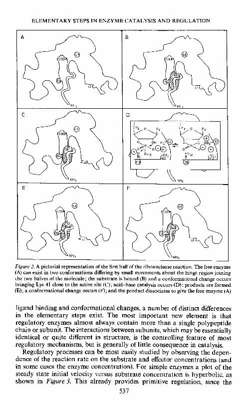
Thus, the over-all mechanism for the enzymatic reaction might be con-structed as follows. The enzyme exists in dynamic equilibrium between twoforms, differing in the structure of the active site groove. The substrate isbound at a rate almost as fast as that at which it can diffuse to the active site.(This is derived from the magnitudes of the bimolecular rate constants inTable 2.) When the substrate binds, the groove shape is altered and lysine 41swings over to the substrate to assist in the binding process, and the substrateis oriented very precisely so that the imidazole residues (histidines 12 and 119)can catalyse the chemical reaction through rapid proton transfer reactions.The conformational change is then reversed and the product dissociates.Both the transesterification and hydrolysis steps proceed in a similar manner.This mechanism is shown pictorially in terms of the enzyme structure inFigure 2.
Unfortunately, the elementary steps associated with the proton transferreactions cannot be studied: only the over-all rate of the conversion ofsubstrate to product (and vice versa) in the active site can be determined. Thisvaries from about 10 to i0 s 1 for a variety of substrates at their optimalpH48' The reason that the elementary steps cannot be observed isundoubtedly that the concentrations of the reaction intermediates are toosmall. Although the details of the proton transfer process remain to be eluci-dated, detailed consideration of the three-dimensional structure and stereo-chemical studies indicate that the mechanism probably involves a concertedproton transfer between two imidazole residues and the substrate such as thatshown in Figure 25558.
Thus, a combination of detailed kinetic, chemical, and structural studieshas led to a fairly complete picture of the catalytic process for ribonucleaseand has come close to resolving the entire time course of the reaction into itselementary steps.
REGULATION
Although the regulation of enzyme activity by switching the enzyme bet-ween active and inactive forms has features in common with catalysis, namely
536
ELEMENTARY STEPS IN ENZYME CATALYSIS AND REGULATION
Figure 2. A pictorial representation of the first half of the ribonuclease reaction. The free enzyme(A) can exist in two conformations differing by small movements about the hinge region joiningthe two halves of the molecule; the substrate is bound (B) and a conformational change occursbringing Lys 41 close to the active site (C); acid—base catalysis occurs (D); products are formed(E); a coriformational change occurs (F); and the product dissociates to give the free enzyme (A)
ligand binding and conformational changes, a number of distinct differencesin the elementary steps exist. The most important new element is thatregulatory enzymes almost always contain more than a single polypeptidechain or subunit. The interactions between subunits, which may be essentiallyidentical or quite different in structure, is the controlling feature of mostregulatory mechanisms, but is generally of little consequence in catalysis.
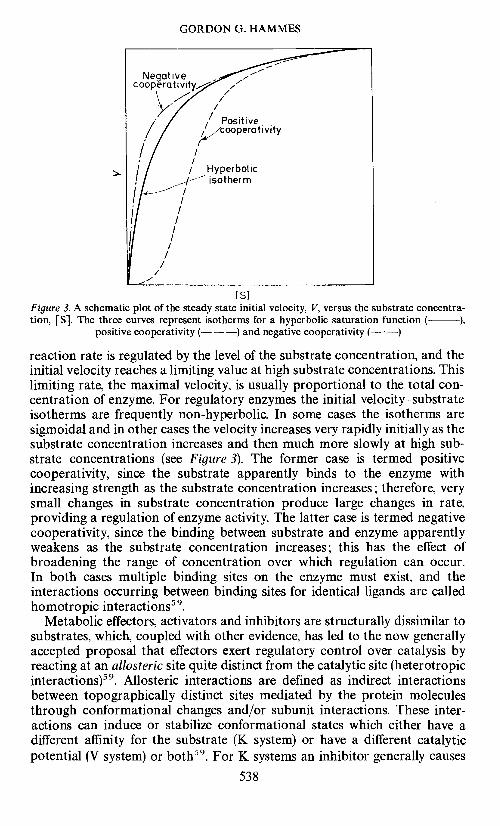
Regulatory processes can be most easily studied by observing the depen-dence of the reaction rate on the substrate and effector concentrations (andin some cases the enzyme concentration). For simple enzymes a plot of thesteady state initial velocity versus substrate concentration is hyperbolic, asshown in Figure 3. This already provides primitive regulation, since the
537
GORDON G. HAMMES
Figure 3. A schematic plot of the steady state initial velocity, V, versus the substrate concentra-tion, [S]. The three curves represent isotherms for a hyperbolic saturation function ( ),
positive cooperativity (— —) and negative cooperativity (— —)
reaction rate is regulated by the level of the substrate concentration, and theinitial velocity reaches a limiting value at high substrate concentrations. Thislimiting rate, the maximal velocity, is usually proportional to the total con-centration of enzyme. For regulatory enzymes the initial velocitysubstrateisotherms are frequently non-hyperbolic. In some cases the isotherms aresigmoidal and in other cases the velocity increases very rapidly initially as thesubstrate concentration increases and then much more slowly at high sub-strate concentrations (see Figure 3). The former case is termed positivecooperativity, since the substrate apparently binds to the enzyme withincreasing strength as the substrate concentration increases; therefore, verysmall changes in substrate concentration produce large changes in rate,providing a regulation of enzyme activity. The latter case is termed negativecooperativity, since the binding between substrate and enzyme apparentlyweakens as the substrate concentration increases; this has the effect ofbroadening the range of concentration over which regulation can occur.In both cases multiple binding sites on the enzyme must exist, and theinteractions occurring between binding sites for identical ligands are calledhomotropic interactions5 .
Metabolic effectors, activators and inhibitors are structurally dissimilar tosubstrates, which, coupled with other evidence, has led to the now generallyaccepted proposal that effectors exert regulatory control over catalysis byreacting at an allosteric site quite distinct from the catalytic site (heterotropicinteractions)5 . Allosteric interactions are defined as indirect interactionsbetween topographically distinct sites mediated by the protein moleculesthrough conformational changes and/or subunit interactions. These inter-actions can induce or stabilize conformational states which either have adifferent affinity for the substrate (K system) or have a different catalyticpotential (V system) or both59. For K systems an inhibitor generally causes
538
IS]

ELEMENTARY STEPS IN ENZYME CATALYSIS AND REGULATION
the initial velocity—substrate isotherm to become more sigmoidal, while anactivator causes it to be less sigmoidal, as illustrated in Figure 4. In both casesthe same maximal velocity is attained at sufficiently high substrate concen-trations, and the inhibitors and activators provide regulation only over arestricted range of substrate concentrations. For V systems the maximalvelocity is increased by an activator and decreased by an inhibitor.
A number of molecular models have been proposed to account for theregulation of enzyme activity. All of these models are based on the subunitstructure of proteins and alterations in subunit interactions and/or conforma-tions coupled to ligand binding. Two limiting molecular models are oftenused to describe the alteration of enzyme activity through conformationalchanges linked to ligand binding. One is due to Monod, Wyman and Changeux(MWC model)59; the other is due to Adair, Koshland, Nemethy and Filmer(AKNF model)60'61.
The MWC model is based on three postulates (1) the enzyme consists oftwo or more identical subunits, each containing a site for the substrate oreffector; (2) at least two different conformational states (usually designated asR and T states are in equilibrium and differ in their affinities for substrateand/or effector; and (3) the conformational changes of all subunits occur in aconcerted manner. A schematic illustration of the MWC model for a four-subunit enzyme is shown in Figure 5, where squares and circles are used toindicate different enzyme conformations. In the absence of substrate, theenzyme exists largely in T states (the square conformation), but substratesbind preferentially to the R states (the circular conformation), so that theconformational equilibrium is shifted to the R states by the binding of sub-strate. Quantitative analysis of this model indicates that sigmoidal bindingisotherms can be generated. Activators and inhibitors, by binding preferen-tially to the R and T states, respectively, can reduce or enhance the sigmoidi-city of the binding isotherms, exactly as often found for K systems (Figure 4).
-—- —— , —— /, / /
+ Activator / / /\ / / /\/ I // / / + Inhibitor
II /I / /II /I I 1'ControLII /I / /I/II/I/ 1/,//, .-.
Is]Figure 4. The effect of an activator and inhibitor on the initial velocity—substrate isotherm for a K
system with positive cooperativity
539

GORDON G. HAMMES
In the MWC model the subunits are all in the same conformation—that is,hybrid çonformational states containing both squares and circles cannotexist. An important limitation of this model is that only positive cooperati-vity can occur, so that a basis for negative cooperativity is not provided.
The basic assumptions of the AKNF model are that (1) two conformationalstates are available to each subunit, (2) only the subunit to which the ligandis bound changes its conformation and (3) the ligand-induced conformationalchange in one subunit alters its interactions with neighbouring subunits. Thestrength of the subunit interactions may be increased or decreased or remainthe same. A schematic illustration of this model for a four-subunit protein isincluded in Figure 5. Because each liganded state has different subunit inter-actions, it has a different effective binding constant for adding another ligand.Thus, this model can readily generate substrate binding isotherms displayingpositive or negative cooperativity, or even both. Activators and inhibitors
Concerted model (MWC)
4 S + + 45
s + [T1 + 35
i1' 'II'II —2 S + + 2 S
1t'
rs1S r 1 — cx
[sisi (X)
Simple sequential model (AKNF)
__®fl®®®® c®__t_HI_H®L1Figure5. Schematic representations of the MWC and AKNF models for a four-subunit enzyme.The squares and circles designate different subunit conformations and S is the substrate. The
free substrate has been omitted from the AKNF model for the sake of simplicity
540

ELEMENTARY STEPS IN ENZYME CATALYSIS AND REGULATION
can alter the effective substrate binding constants by changing the subunitinteractions. In contrast to the concerted nature of the MWC model, theAKNF model predicts a strictly sequential change of subunit conformationsexactly paralleling ligand binding. Clearly a more general model can begenerated by permitting both sequential and partially concerted conforma-tional changes. In practice these models are often very difficult to distinguishby experimental measurements.
The models discussed thus far are equilibrium models in that alterations inthe rates of enzyme catalysis are explained by changes in the equilibriumbinding characteristics of the enzyme. For K systems such an analysis isappropriate ii the binding steps and conformational changes are rapid relativeto the rate-determining step in catalysis. These models can also be used for Vsystems, with the additional postulate that each of the conformational statesof the enzyme has a different turnover number. The equilibrium assumptionappears to be valid for many systems. However, it should be noted thatapparent cooperativity in initial velocity-substrate isotherms can be genera-ted by parallel kinetic pathways and special relationships between the rateconstants6 264• In other words, complex mechanisms can lead to apparentcooperativity without postulating special conformational transitions. Al-though this possibility exists, thus far it has not been shown that this mech-anism is utilized by regulatory enzymes.
An extreme alteration of subunit interactions occurs in po1ymerization-depolymerization reactions, and polymerization equilibria probably playan important role in the regulation of some enzymes6 . If, for example, anenzyme exists in two polymeric states, each having a different affinity forsubstrate and effectors, a model is generated analogous to the MWC model,except that cooperativity in the binding isotherm is also dependent on theenzyme concentration66' 67 Again only positive cooperativity can be genera-ted with this model. Both K systems and V systems can be obtained with thismodel, exactly as previously discussed for conformational models, providedthe polymerization equilibria are adjusted rapidly relative to the rate-deter-mining step of catalysis.
Thus far the assumption has been made that allosteric enzymes respondrapidly to changes in ligand concentration. However, this is not required on afunctional basis. In fact, systems are known where ligands can induce changesin enzyme activity much more slowly than the rate of the over-all catalyticreaction. This causes a time lag in the response of the enzyme to changes inthe ligand concentrations. Such slowly responding enzymes are calledhysteretic'68. The molecular basis for this mode of regulation apparently isnot fundamentally different from previously discussed models: slow confor-mational changes, slow polymerization—depolymerization of enzymes andslow displacement of a tightly bound ligand have been proposed in specificcases.
Only the triggering of regulatory processes by ligand binding has beendiscussed. However, regulation can also occur through enzyme-catalysedcovalent modification of an enzyme—for example' by phosphorylation andadenylation69.
The elementary steps in the regulatory models discussed are not funda-mentally different from those generally involved in macromolecular confor-
541
GORDON G. HAMMES
mational transitions and the binding of small molecules by proteins. Thegreat range in the rates of regulatory processes must arise from differences inthe degree of cooperativity in the conformational transitions (cf. Table 3). Thefact that inter subunit conformational changes as well as intra subunit con-formational changes occur also is relevant. A new step which may be ofimportance is the polymerization—depolymerization of macromolecules.The basic interactions involved are hydrogen bonding, solvation, hydro-phobic, and electrostatic interactions, but essentially no quantitative rate dataare available for appropriate polymerization equilibria. The dissection ofenzyme regulatory processes into elementary steps is not quite as advanced asfor enzyme catalysis, but nevertheless useful molecular models are available(cf. references 65 and 69—71 for more extensive reviews).
ASPARTATE TRANSCARBAMYLASE
As mentioned earlier, aspartate transcarbamylase is a regulatory enzymewhich catalyses the reaction in equation (11). This is the first committed stepin the biosynthetic pathway for the synthesis of pyrimidines. The enzyme from
0 0 HII I + — I —
NH2C—01--O+
NH3—H—COO—
H2N—N—CH—COO+ P1
0 CH2 0 CF!2
coo coo (11)
Escherichia coli has been extensively studied by many workers, and a numberof reviews are available65' 7274 The binding of aspartic acid to the enzymein the presence of a saturating concentration of carbamyl phosphate, asmeasured by initial velocities, has a sigmoidal binding isotherm. The sigmoidi-city of this isotherm is increased by the feedback inhibitor CTP and de-creased by the activator ATP, exactly as shown in Figure 44 The maximumvelocity is not altered by effectors.
The enzyme can be resolved into two types of subunits: a catalyticallyactive subunit not subject to nucleotide control and a catalytically inactivesubunit that binds CTP strongly75. These two types of subunits can be readilyreconstituted to give an active enzyme subject to control by nucleotides. Thus,the allosteric nature of the control process is clearly established. The nativeenzyme contains six identical catalytic polypeptide chains and six identicalregulatory polypeptide chains with six regulatory and six catalytic sites76 81The catalytic subunit is a trimer and the regulatory subunit is primarily adimer. Electron microscopy and x-ray studies have established the generalnature of the three-dimensional structure of the enzyme: the two catalytictrimers are connected by the regulatory dimers, with no direct interactionoccurring between the catalytic trimers82' 83 The native molecule has athreefold and a twofold symmetry axis. A very schematic model of thisstructure is shown in Figure 6.
The binding of nucleotide effectors to the enzyme is complex. The bindingisotherms indicate negative cooperativity in binding to regulatory sites, aswell as binding to the catalytic sites which can be eliminated by millimolar
542

ELEMENTARY STEPS IN ENZYME CATALYSIS AND REGULATION
carbamyl phosphate79 81, 84—86 The cooperative unit is the regulatory dimer:binding of an effector molecule to one dimer site considerably reduces thebinding affinity for the second effector molecule. The inhibition and activationare directly proportional to the fraction of total regulatory sites occupied, sothat all regulatory sites participate equally in the regulatory function81' 86,The initial velocity—aspartate isotherm is sigmoidaI and the equilibriumbinding of succinate, an aspartate analogue, in the presence of carbamylphosphate also displays a sigmoidal binding isotherm87. Therefore, thisenzyme utilizes both positive and negative cooperativity in its regulatorymechanism.
Extensive kinetic studies have been made of the binding of ligands toaspartate transcarbamylase by temperature jump and stopped flow tech-niques. Effector molecules' studied include CTP20, 5-hromo-CTP19'865-bromo-CDP86, 5-bromo-CMP86 and the AlP analogue, 6-mercapto-9-3-D-rlbofuranosyl-purine-5'-triphosphate88; the kinetics of the binding of car-bamyl phosphate and the aspartate analogue, succinate, also have been stud-ied. Elementary steps associated with both catalysis and regulation have beenobserved. A summary of the results obtained is presented in Table 6; only the
Table 6. Elementary steps in catalysis and regulation for aspartate transcarbamylaae
Reactant Mechanism Function
Carbamyl phosphate Bimolecular association Binding(+ succinate) Conlormational change (stepwise)
Conlormational change (concerted)CatalysisRegulation
Succinate or L-malate(+ carbamyl phosphate)
Bimolecular associationConformational change (concerted)
BindingRegulation
CTP, ATP analogues Bimolecular association (stepwise) BindingRegulation
543
Figure 6. A pictorial representation of the structure of aspartate transcarbamylase. The light-coloured portions are catalytic subunits and the dark-coloured portions are regulatory subunits.

GORDON G. HAMMES
elementary steps of importance in the regulatory mechanism will be con-sidered here.
The equilibrium and kinetic data for the binding of all effector molecules areconsistent with a simple mechanism in which a rate-limiting conformationalchange follows a relatively rapid bimolecular reaction. The same conforma-tional change occurs at all classes of regulatory sites and alters the inter-actions between subunits. The negative cooperativity occurs in the initialrapid binding step. In terms of the molecular models discussed earlier, this is asimplified sequential type of model. The same two conformational states areutilized by both activators and inhibitors, since only a single relaxationprocess is observed with the enzyme in the presence of both an activator andinhibitor.
A simple regulatory mechanism accommodating all available data is thatthe binding of ATP and CTP causes the formation of two enzyme—effectorconformations, X1 and X2. The binding of CTP favours the formation of oneconformation (say X1), which can be regarded as the 'oft' state, while thebinding of ATP favours the formation of the other conformation (X2), whichcan be regarded as the 'on' state. As expected the binding of carbamylphosphate and succinate is also found to favour the formation of X2.
The binding of succinate to the enzyme in the presence of saturating con-centrations of carbamyl phosphate is quite complex. Two relaxation pro-cesses are observed with the isolated catalytic subunit which can be explainedin terms of a bimolecular binding step followed by a conformational changeof importance in catalysis1 s. These same relaxation processes are observedwith the native enzyme and are not altered by the binding of effectors. Inaddition a new relaxation process is observed with the native enzyme21. Theconcentration dependence of the associated relaxation time (which is in therange of about 2—20 ms) can be analysed quantitatively in terms of a con-certed conformational change, analogous to the MWC model. This con-formational transition is distinct from the conformational change associatedwith effector binding, since both transitions are found to occur simultan-eously. Moreover, activators and inhibitors alter the concentration depen-dence of the relaxation time exactly as predicted by the concerted model.Also, the kinetic parameters derived can be used to generate a sigmoidalequilibrium binding isotherm.
The interaction of carbamyl phosphate with the native enzyme at highconcentrations of succinate also is somewhat complex; a relatively slowrelaxation process, with a relaxation time of 10—100 ms, is found to accom-pany the binding of carbamyl phosphate to the native enzyme in the presenceof succinate, but is not observed with the catalytic subunit22. The simplestmechanism consistent with the data is a concerted conformational tran-sition, which appears to be distinct from the one associated with succinatebinding, since the concentration dependence is different, and the binding ofCTP alters the relaxation time differently in the two cases.
In addition to the kinetic data cited above, a large number of chemical andphysical studies have provided evidence that conformational changesaccompany ligand binding (cf. reference 73). The methods utilized includeultra-violet difference spectroscopy, optical rotatory dispersion, ultra-centrifugation, trypsin digestion of the enzyme, measurement of the rate
544

ELEMENTARY STEPS IN ENZYME CATALYSIS AND REGULATION
of reaction of enzyme sulphhydryl groups and sodium dodecyl sulphate in-activation of the enzyme. The results obtained indicate that the binding ofeffector molecules causes a different conformational change from that causedby the binding of substrates and that the binding of succinate to the nativeenzyme causes a much larger conlormational change than binding to theisolated catalytic subunit.
The over-all control mechanism for aspartate transcarbamylase can bedepicted as follows. The effector molecules, ATP and CTP, carry out theirfunction by altering a two-state conformational equilibrium, which occursroughly independently in each regulatory chain. The local conformationalchanges occurring in the regulatory chain alter its interaction with thecatalytic subunit, thereby altering the enzyme activity. Local conformationalchanges involved in the catalytic process also occur in the catalyticsubunit when carbamyl phosphate and succinate bind. The conforma-tional changes involved in control which are induced by carbamylphosphate and succinate binding appear to be quite distinct from each otherand from that induced by CTP and ATP binding. These two conformationaltransitions appear to be concerted in nature. Athough the structural basis ofthe concerted conformational transitions is not yet known, rotation of thecatalytic subunits with respect to each other around the threefold symmetryaxis may be involved (cf. Figure 6) 2 In any event, the over-all control mech-anism appears to be a combination of several different conformationaltransitions, each of which can lead to inhibition or enhancement of enzymicactivity. This multiplicity and coupling of conformational changes, whichprovides a sensitive and versatile control mechanism, is somewhat analogousto a mini-computer control of the enzymatic reaction with macromolecularconformational changes being utilized as interlocking switches.
CONCLUSION
The general approach to enzyme catalysis and regulation emphasized,namely the time resolution of the reaction mechanisms into their elementarysteps, provides insight into the molecular basis of the mechanisms. Ofnecessity, only a limited number of systems were discussed. This brief dis-cussion is intended to illustrate the potential of this approach and theinformation which can be obtained.
ACKNOWLEDGEMENT
This work was supported by a grant from the National Institutes of Health(GM 13292).
REFERENCES
C. Frieden and R. A. Alberty, J. Biol. Chem. 212, 859 (1955).2 L. T. Rozelle and R. A. Alberty, J. Phys. Chern. 61, 1637 (1957).
L. E. Erickson and R. A. Alberty. J. Phys. Chem. 63, 705 (1959).J. C. Gerhart and A. B. Pardee, J. Biol. Chem. 237, 891 (1964).J. V. Pasonneau and 0. H. Lowry, Biochem. Biophys. Res. Commun. 10, 7 (1962).
6 J, V. Pasonneau and 0. H. Lowry. Biochern. Biophys. Re,s. Commun. 13. 372 (1963).
545

GORDON G. HAMMES
Investigation of Rates and Mechanisms of Reactions. Vol. 6, Part II: 'Investigation of ele-mentary steps in solution and very fast reactions', G. G. Hammes, ed. in Techniques ofChemistry, A. Weissberger, series ed. Wiley-Interscience; New York (1974).G. G. Hammes, Ado. Prot. Chern. 23, 1(1968).G. G. Hammes and P. R. Schimmel, The Enzymes, 2, 67 (1970).'° G. P. Hess, J. McConn, E. Ku and G. McConkey, Phil. Trans. Roy. Soc. B, 257, 89 (1970).' G. G. Hammes and F. G. WaIz, Jr, J. Amer. Chem. Soc. 91,7197 (1969).
12 J. E. Erman and 0. G. Hammes, J. Amer. Chem. Soc. 88, 5067 (1966).13 E. J. del Rosario and G. G. Hammes, J. Amer. Chem. Soc. 92, 1750 (1970).14 J. E. Erman and G. 0. Hammes, J. Amer. Chem. Soc. 88, 5614 (1966).15 G. 0. Hammes and J. K. Hurst, Biochemistry, 8, 1083 (1969).' H. de A. Heck, J. Biol. Chem. 244, 4375 (1969).17 K. Kirschner, E. Gallego, I. Schuster and D. Goodall, J. Mol. Biol. 58. 29 (1971).18 G. G. Hammes, R. W. Porter and G. R. Stark, Biochemistry, 10, 1046 (1971).19 J Eckfeldt, G. G. Hammes, S. C. Mohr and C.-W. Wu, Biochemistry, 9. 3353 (1970.20 L. W. Harrison and G. G. Hammes, Biochemistry, 12, 1395 (1973).21 G. G. Hammes and C.-W, Wu, Biochemistry, 10, 1051 (1971).22 G. G. Hammes and C.-W. Wu, Biochemistry, 10, 2151 (1971).23 E. D. Barber and H. J. Bright, Proc. Nat. Acad. Sci. US, 60, 1370 (1968).24 G. W. Hatfield and H. E. Umbarger, J. Biol. Chem. 245, 1742 (1970).25 W. Maier, Z. Elektrochem. 64, 132 (1960).26 L. Borucki, Ber. Bunsenges. Physik. Chem. 71, 504 (1967).27 0. G. Hammes and A. C. Park, J. Amer. Chem. Soc. 91, 956 (1969).28 G. G. Hammes and H. 0. Spivey, J. Amer. Chem. Soc. 88, 1621 (1966).29 G. G. Hammes and P. 1. Lillford, J. Amer. Chem. Soc. 92, 7578 (1970).30 E. Grunwald and E. K. Ralph, III, J. Amer. Chern. Soc. 89, 4405 (19671.31 G. 0. Hammes and W. Knoche, J. Chem. Phys. 45, 4041 (1966).32 G. 0. Hammes and N. C. Pace, J. Phys. Chem. 72, 2227 (1968).
G. 0. Hammes and T. B. Lewis, J. Phys. Chem. 70, 1610 (1966).0. 0. Hammes and P. B. Roberts, J. Amer. Chem. Soc. 90, 7119 (1968).G. 0. Hammes and J. C. Swann, Biochemistry, 6, 1591 (1967).G. 0. Hammes and P. R. Schimmel, .1. Amer. Chem. Soc. 89, 442 (1967).J. Applequist and P. Doty, Abstracts, 135th Meeting of American Chemical Society, Boston,April 5, 1959.
38 A. F. Barksdale and J. E. Stuehr, J. Amer. Chem. Soc. 94, 3334 (1972).M. Eigen, Angew. Chem. 75, 489 (1963).
40 M. Eigen and G. G. Hammes, Ado. Enzymol. 25, 1 (1963).41 C. H. W. Hirs, S. Moore and W. H. Stein, J. Biol. Chem. 235, 633 (1960).42 J J Potts, A. Berger, J. Cooke and C. B. Anfinsen, I. Biol. Chem. 237, 1851 (1962).
D. J. Smith, W. H. Stein and S. Moore, J. Biol. Chem. 238, 227 (1963).G. Kartha, 3. Bello and D. Harker, Nature, 213, 862 (1967).H. W. Wyckoff, K. D. Hardman, N. M. Allewell, T. Ingami, L. N. Johnson and F. M. Richards,J. Biol. Chem. 242, 3984 (1967).
46 F. M. Richards and H. W. Wyckoff, The Enzymes, 4, 647 (1971).J. T. Babr, R. E. Cathou and G. G. Hammes, J. Biol. Chem. 240, 3372 (1965).
48 E. J. del Rosario and 0. G. Hammes, Biochemistry, 8, 1884 (1969).' T. C. French and G. G. Hammes, J. Amer. Chem, Soc. 87, 4669 (1965).° A. M. restfield, W. H. Stein and S. Moore, J. Biol. Chem. 238, 2421 (1963).' R. Heinrikson, W. H. Stein and S. Moore, J. Biol. Chem. 240, 2921 (1965),52 D. H. Meadows and 0. Jardetzky, Proc. Nat. Acad. Sci. US, 61, 406 (1968).
D. G. Herries, A. P. Mathias and B. R. Rabin, Biochem. J. 85, 127 (1962).H. Witzel, Frog. Nuc. Acid. Res. 2, 221 (1963)." D. A. Usher, D. I. Richardson Jr and F. Eckstein, Nature, 228, 663 (1970).
56 G. C. K. Roberts, E. A. Dennis, D. H. Meadows, J. S. Cohen and 0. Jardetzky, Proc. Nat.Acad. Sd. US, 62, 1151 (1969).D. Findlay, D. 0. Herries, A. P. Mathias, B. It Rabin and C. A. Ross, Biochem. J. 85, 152(1962).
58 D. A. Usher, E. S. Erenrich and F. Eckstein, Proc. Nat. Acad. Sd. US. 69, 115 (1972).J. Monod, 3. Wyman and 3.-P. Changeux, J. Mol. hot. 12, 88 (1965).
546

ELEMENTARY STEPS IN ENZYME CATALYSIS AND REGULATION
60 G. S. Adair, J. Biol. Chem. 63, 529 (1925).61 D. E. Koshland Jr, G. Nemethy and D. Filmer, Biochemistry, 5, 365 (1966).62 A. Worcel, S. Goldman and W. W. Cleland, J. Biol. Chem. 240, 3399 (1966).63 B. D. Sanwal and R. A. Cook, Biochemistry, 5, 886 (1966).64 J. R. Sweeny and J. R. Fisher, Biochemistry, 7, 561 (1968).65 G. G. Hammes and C-W. Wu, Adv. Biophys. Bioeng., 3, 1(1974).66 C. Frieden and R. Colman, J. Biol. Chem. 242, 1705 (1967).67 L. W. Nichol, W. J. H. Jackson and D. J. Winzor, Biochemistry, 6, 2449 (1967).68 C. Frieden, J. Biol. Chem. 245, 5788 (1970).69 E. R. Stadtman, The Enzymes, 1, 397 (1970).70 D. E. Koshland Jr, The Enzymes, 1, 341 (1970).71 K. Kirschner, Current Topics in Cellular Regulation, 3, 167 (1971).72 j C. Gerhart, Current Topics in Cellular Regulation, 2, 275 (1970).
0. R. Jacobson and G. R. Stark, The Enzymes, 9, 225 (1973).G. G. Hammes and C.-W. Wu, Science, 172, 1205 (1971).J. C. Gerhart and H. K. Schachman, Biochemistry, 4, 1054 (1965).
76 K. Weber, Nature, 218, 1116 (1968).0. G. Hammes, R. W. Porter and C.-W. Wu, Biochemistry, 9, 2292 (1970).
78 E. A. Meighen, V. Pigiet and H. K. Schachman, Proc. Nat. Acad. Sci. US,65, 234 (1970).C. C. Winiund and M. J. Chamberlin, Biochim. Biophys. Res. Commun. 40, 43 (1970).
80 J P. Rosenbusch and K. Weber, J. Biol. Chem. 246, 1644 (1971).S. Matsumoto and G. G. Hammes, Biochemistry, 12, 1388 (1973).
82 K. E. Richards and R. C. Williams, Biochemistry, 11, 3393 (1972).83 S. H. Warren, B. P. Edwards D. R. Evans, D. C. Wiley and W. N. Lipscomb, Proc. Nat. Acad.
Sci. US, 70, 1119 (1973).84 T. Buckman, Biochemistry, 9, 3255 (1970).85 C. W. Gray, M. J. Chamberlin and D. M. Gray, J. Biol. Chem. 248, 6071 (1973).86 C. Tondre and G. G. Hammes, Biochemistry, 13, 3131 (1974).87 J.-P. Changeux, J. C. Gerhart and H. K. Schachman, Biochemistry, 7, 538 (1968).88 cw Wu and G. 0. Hammes, Biochemistry, 12, 1400 (1973).
547