electrostatic effects in myoglobin. ph and ionic strength variations of ionization equilibria for...
TRANSCRIPT
S H I R E . H A h A b l A T 421D G U K D
Garner, M. H., Garner, W. H. , and Gurd, F. R. N . (1974), J . Biol. Chem. 249, 15 13.
Gillespie, J . M., Hapner, K. D., Hartzell, C. R., and Gurd, F. R . N. (l966), J . Mol. Biol. 21, 399.
Gurd, F. R. N . (1970), in Physical Principles and Techniques of Protein Chemistry, Part B, Leach, S., Ed., New York, N. Y . , Academic Press, p 383 1 .
Gurd, F. R. N., Lawson, P. J., Cochran, D. W., and Wenkert, E. (1971), J . Biol. Chem. 246, 3725.
Curd, F. R . IC., Morrow, J . S., Keim, P., Visscher, R . B., and Marshall, R. C . ( 1 974), in Protein-Metal Interactions, Friedman, M., Ed., New York, N . Y., Plenum, in press.
Hanania, G. I . H., and Irvine, D. H . (1970), J . Chem. SOC. A , 2389.
Hanania, G . I . H., and Nakhleh, E. T. (1970), 8th I n? . Congr. Biochem., Interlaken.
Hapner, K. D., Bradshaw, R. A., Hartzell, C.R., and Gurd, F. R. N . (1968), J . Biol. Chem. 243, 683.
Hartzell. C. R., Bradshaw, R. A,, Hapner, K. D., and Gurd, F. R. N . ( 1 968), J . Biol. Chem. 243, 690.
Hugli, T. E. (1968), Ph.D. Thesis, Indiana University, Bloom- ington. Ind.
Hugli, T . E., and Gurd, F. R. N. (1970), J . Biol. Chem. 245, 1939.
Janssen, L. H. M. (1970), Ph.D. Thesis, University of Nijmeg- en, Nijmegen, Netherlands.
Kendrew, J . C., and Parrish, R. G. (1957), Proc. Roy. Soc., Ser. A 238, 305.
Kirkwood, J. G . (1934), J . Chem. Phys. 2, 351. Kirkwood, J . G., and Westheimer, F. H. (1938), J . Chem.
Lee, B., and Richards, F. M. (1971). J . Mol. Biol. 55, 379. Linderstr$m-Lang, K. ( 1 924), C. R. Truv. Lab. Curlsberg 15,
Nakhleh, E. T. (1971), Ph.D. Thesis, American University of
Nigen, A. M., and Gurd, F. R. N . (1973), J . Biol. Chem. 248,
Nozaki, Y., and Tanford, C. ( 1 967), J . Biol. Chem. 242.473 1. Orttung, W. (1969), J . Amer. Chem. Soc. 91, 162. Orttung, W. (1970), Biochemistry 9, 2394. Schoenborn, B. P. ( 1 97 I ), Cold Spring Harbor Symp. Quant.
Shire, S. J., Hanania, G. I . H., and Curd, F. R . N. (1974),
Stryer, L., Kendrew, J . C., and Watson, H. C. ( 1 964), J . Mol.
Tanford, C. ( I 957a), J . Amer. Chem. Soc. 79, 5348. Tanford, C . (1957b), J . Amer. Chem. SOC. 79, 5340. Tanford, C. ( I96 I ), Physical Chemistry of Macromolecules,
Tanford, C . (1962), Advan. Protein Chem. 17, 69. Tanford, C., and Kirkwood, J. G. (1957), J . Amer. Chem. Soc.
Tanford, C . , and Roxby, R. (1972), Biochemistry 11, 2192. Watson, H. C. (1969), Progr. Stereorhem. 4, 299.
Phys. 6, 506.
7.
Beirut, Beirut, Lebanon.
3708.
Biol. 36, 569.
Biochemistry 13. 0000.
Biol. 8, 96.
Kew York, N. Y . , Wiley, Chapter 8.
79, 5333.
Electrostatic Effects in Myoglobin. pH and Ionic Strength Variations of Ionization Equilibria for Individual Groups in Sperm Whale Ferrimyoglobin?
S. J. Shire, G . I . H. Hanania,l and F. R. N . Gurd*
ABSTRACT: The variations with pH and ionic strength of the NH2-terminal valine, histidine, and iron-bound water ioniza- tion pK values for the major component of sperm whale fer- rimyoglobin were computed with a modified Tanford-Kirk- wood theory which includes a set of static solvent accessibility factors. Where possible experimental values were compared to the theoretical results and good agreement was obtained. Ex-
T h e preceding paper (Shire et ai., 1974) deals with the com- putation of titration curves of sperm whale myoglobin with the Tanford-Kirkwood treatment (Tanford and Kirkwood, 1957; Tanford and Roxby, 1972) adapted by the introduction of stat- ic solvent accessibility factors (Lee and Richards, 1971). The
t From the Department of Chemistry, Indiana University, Bloom- ington, Indiana 47401. Received March 11, 1974. This is the 60th paper in a series dealing with coordination complexes and catalytic properties of proteins and related substances. For the preceding paper, see Shire et ai. (1974). This work was supported by Public Health Ser- vice Research Grant HL-05556.
1 Permanent address: Department of Chemistry, American Univer- sity of Beirut, Beirut, Lebanon.
clusion of the newly introduced parameters yielded poor agree- ment with experiment for the ionic strength variation of the ionization pK value for the iron-bound water molecule. The computations with the modified theory were also performed for a minor sperm whale ferrimyoglobin component. The theoreti- cal ionic strength variation of pK value for the water molecule was again in agreement with experiment.
inclusion of the static solvent accessibiiity factors allows good overall fit to experimental data with the choice of individual in- trinsic pK values for various groups in myoglobin that are gen- erally reasonable in the light of other evidence.
The model allows computation in good agreement with ex- periment of the net charge as a function of pH for both the major component IV and the minor component I 1 of sperm whale ferrimyoglobin (Shire et al., 1974). Since the computa- tions also yield information about individual pK values, a fur- ther test of the theory is to compare the computed pK values with experimental ionization constants for individual groups in the protein. Experimental data of this kind are available for the NH2-terminal groups from the kinetics of the cyanate reaction
2974 B I O C H E M I S T R Y , V O L . 1 3 , N O . 1 4 , 1 9 7 4
I O N l Z A T l O U E Q U I L I B R I A O F I Y D I V I D U A L G R O U P S
(Garner et al., 1973) and for the titratable histidine residues from proton nuclear magnetic resonance titrations (Cohen et al., 1972).] In both cases, reasonable agreement between theo- ry and experiment is obtained (Shire et al., 1974).
The present paper examines in more detail the behavior of the individual groups in terms of the variation of pK with p H and ionic strength. Since the theory is based on electrostatic in- teractions and the interactions are a t a maximum a t zero ionic strength, a further test of the theory would be to investigate the ionic strength variation of pK values at low ionic strength for individual groups in the protein. Available experimental data on histidine residues and the NH2-terminal valine residue do not cover the appropriate range of low ionic strength. On the other hand, the iron-bound ionizing water molecule in ferrimy- oglobin offers an excellent probe for this kind of study. The dis- sociation constant for this ionization has been extensively stud- ied in a number of myoglobins covering a wide range of pH, protein concentration, and temperature, as well as of ionic strength variation (Hartzell et al., 1968; Hanania and Irvine, 1970; Nakhleh, 1971).
Experimental Section
The materials, including the preparation of sperm whale fer- rimyoglobin components, and the apparatus and procedure for potentiometric titrations, have been described (Shire et al., 1974). Ionization pK values for the dissociation of the iron- bound water were determined by the method of mixtures (Na- khleh, 1971), except that Good's buffers were used (Good et al., 1966). The stock buffer solutions a t I = 0.02 M were Mes2- NaOH-KCI (pH 6.0), Bicine-NaOH-KC1 (pH 8-10), and glycine-NaOH (pH 11.2). The buffers were diluted with boiled out deionized water to obtain the desired ionic strength.
Computations and treatment of data followed the methods of the preceding paper (Shire et al., 1974), and employed the parameters given therein. The results are expressed in terms of pK, for the ith group a t a given p H and given average charge state of the protein, derived by adding a computed electrostatic correction term to the intrinsic pK. Use is also made of pK1l2, the value of pK, corresponding to the half-titration of the group in question. The static solvent accessibility factors are derived from the values of Lee and Richards (1971) as adapted by Shire et al. ( I 974).
Results and Discussion
Iron-Bound Water. The ionization constant for the iron- bound water in sperm whale ferrimyoglobin has been investi- gated in detail (Nakhleh, 1971; Hanania and Irvine, 1970). In particular the variation with ionic strength was measured with care, permitting a detailed comparison with theory of the be- havior of a single dissociable group in the protein.
The intrinsic pK value was chosen such that the computed theoretical p K ~ p value (as defined in Shire et al., 1974) a t 25' and zero ionic strength was in close agreement with the experi- mental value, 8.93, obtained by extrapolation of experimental data at finite salt concentrations in the region of ionic strength I < 0.01 M . The computations were then repeated a t a series of finite ionic strength values for comparison with the observed results. All parameters were kept unchanged in each series of calculations. This in effect constitutes an evaluation of the Ci, terms in the Tanford-Kirkwood formulation (Tanford and
I L. H . Botelho, M. H. Garner, and G . I . H. Hnania, work in prog-
Abbreviations used are: Mes, 2-(N-morpholino)ethanesulfonic ress.
acid; Bicine, N,N-bis( 2-hydroxyethy1)glycine.
i
I I
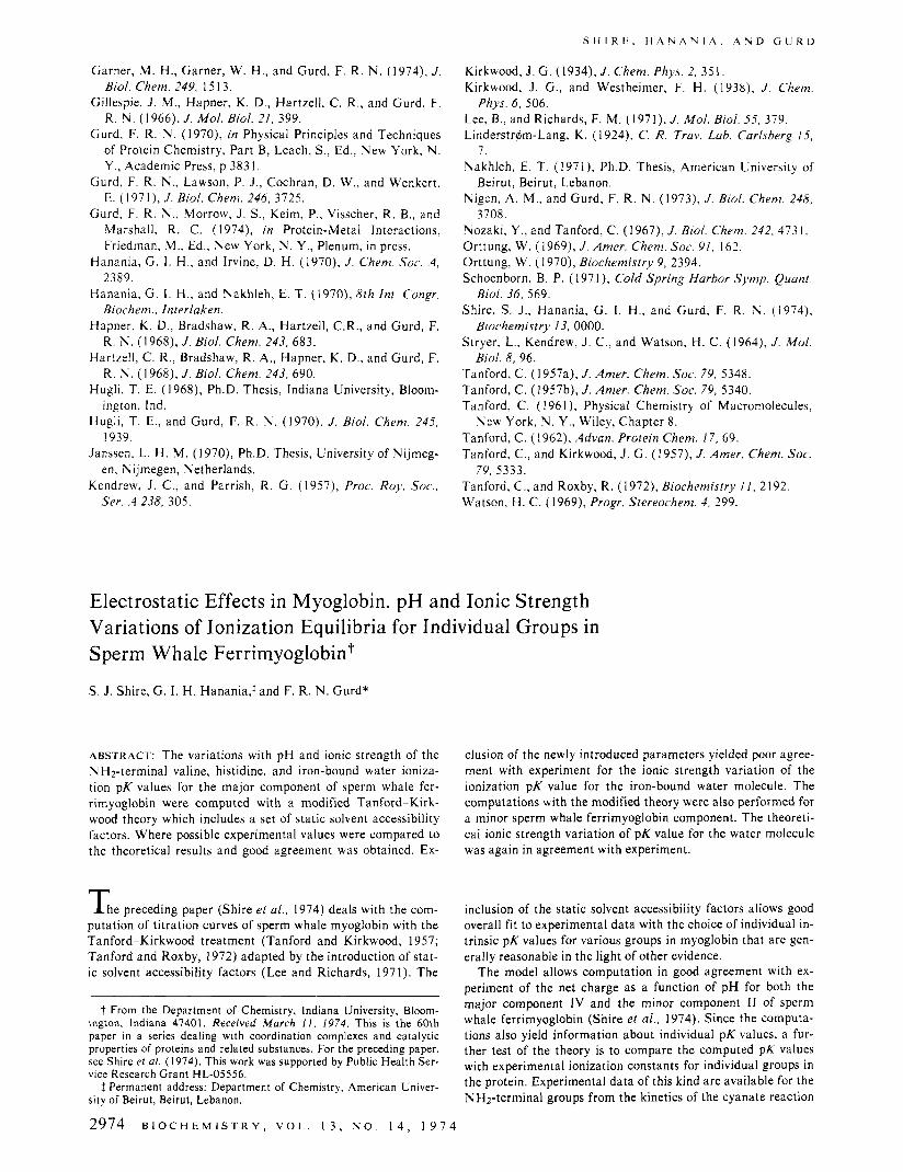
FIGURE I : Plots of the pK value for half-titration, pKi/2 against the ionic strength function, Ill2/( 1 + I l l 2 ) , for the ionization of the iron- bound water molecule in sperm whale ferrimyoglobin (major compo- nent I V ) at 25'. Experimental data are given in full circles, with an un- certainty of f 0 . 0 0 5 pH unit. Computations with solvent accessibility factors included, for d = 0 (-) and d = 1.0 8, (- - - -): solvent ac- cessibility factors not included, for d = 0 (- - -) and d = 1.0 8,
the text (- -). ( - - - -). , according to the Linderstrqm-Lang treatment described in
Kirkwood, 1957) which give the contribution due to finite salt concentration.
Four sets of calculations were done in this way, correspond- ing to the inclusion or exclusion of the static solvent accessibili- ty factors (eq 8 in Shire et ai. , 1974), and with the byrial pa- rameter d set at 0 or l .O A. The results are shown in Figure l , relating pK1/2 with the ionic strength function I1/z/(l + I l l 2 ) . Although the appropriate form of the extrapolation function is not known with certainty, it can be shown that the theory agrees with experiment when the experimental points and com- puted values are plotted using the same ionic strength function even if it differs from the one we chose. The reason for the par- ticular choice of function is given later in this paper. The filled circles are the experimental results (Nakhleh, 1971; Hanania and Irvine, 1970), which fit well the curves for either assump- tion for the value of d with the static solvent accessibility fac- tors included in the comparison, but not the curves obtained with the exclusion of the latter correction terms. The results clearly favor the use of the static solvent accessibility factors.
Included in Figure 1 as a dashed line is the variation of the pK value with ionic strength as calculated by the Linderstrdm- Lang equation based on a smeared charge model (Linder- strdm-Lang, 1924). The electrostatic correction parameter w was calculated from the structural parameters used in the dis- crete charge model computations, that is, b = 18 A and a = 20 A. The average charge 2 a t each ionic strength was obtained from the Tanford-Kirkwood calculations for d = 0 and includ- ing the static solvent accessibility factors. Agreement a t zero ionic strength is good, provided that a pK value computed with the modified Tanford-Kirkwood theory at zero net charge is used as the intrinsic pK. This result is not surprising since the Linderstrdm-Lang intrinsic pK is defined as the pK of the ion- izing group at zero net protein charge. Instead of a value of 8.93 a value of 8.90 was computed and thus for comparative purposes all the pK data a t the different ionic strengths were shifted up by a constant 0.03 value. Interestingly enough, the slope of the Linderstr&"ang plot is closer to experiment than the Tanford-Kirkwood plots without the static solvent ac- cessibility factors. This may be interpreted to mean that the ef- fect of spreading the charge on the surface of the sphere is to
B I O C H E M I S T R Y , V O L . 1 3 , N O . 1 4 , 1 9 7 4 2975
8.3k
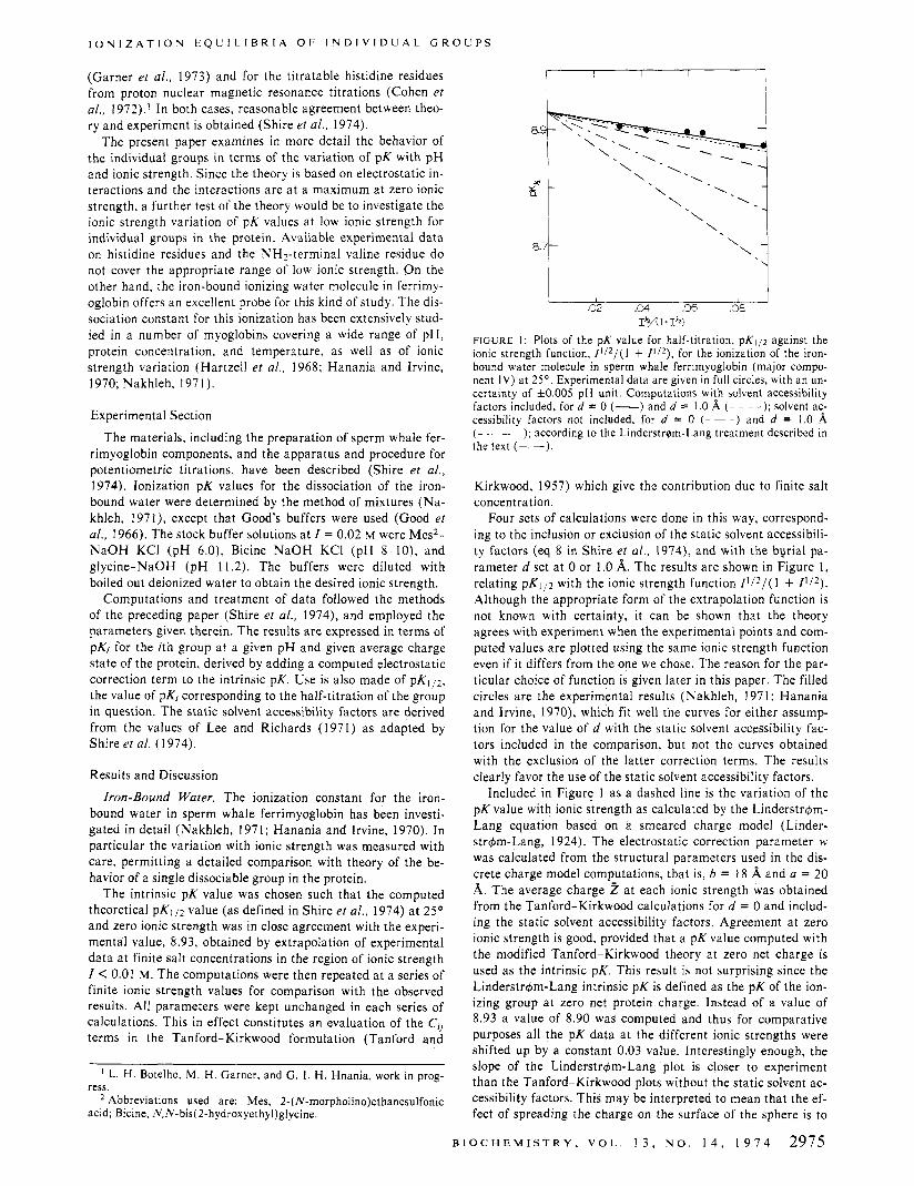
!:I, F l G L R E 2: Computed values for the ionization pK for iron-bound hater of sperm whale ferrimyoglobin (major component IV) . expressed as the value for half-titration, pK,,z. The described model was used with static solvent accessibility factors at 2S0 , and the ionic strength function f(/) = / ' , * / ( I + B a l ' / * ) . Reading from the top down the values of the product Ba are I , 3. 6 , and 12, respectively.
minimize the strong interactions of sites which are close to each other. The solvent accessibility factors perform the same minimizing function provided the sites under question are well exposed to solvent.
Choice of lonic Strength Function. It has been customary to plot the pK data vs. a general ionic strength function f ( / ) = / ] I 2 / ( 1 + B U / ' / ~ ) . Here B and a have their usual significance i n the Debye-Huckel treatment in which B is 0.329 X I O x cm-I for water a t 25' and a is the so-called distance of closest approach of counterions.
Approximating the protein molecule to a sphere allows one to estimate a from the diameters of the protein, counterions, and water molecules. This procedure has led to functions such as f ( / ) = /I':/(l + l2/ ' I2) for hemoglobin (Beetlestone and Ir- vine, 1968) andf(/) = P I 2 / ( 1 + 6/'!*) for myoglobin (Hanan- ia and Irvine, 1970). In the present work the parameters of the spherical model lead to Ba = 6.
The pKli2 value of the iron-bound water molecule is plotted in Figure 2 utilizing different values of Ba in the ionic strength function. It is evident from the plots that only the ionic strength functions / ' I 2 and I + / ] I 2 ) are anywhere near linearity up to / = 0.02 M . We have chosen to use the latter function.
This choice is supported by three observations. From the the-
1 I 8.3 49 5s 1'3.5
PL 1
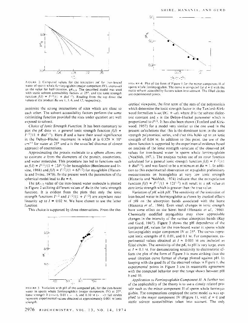
F l G L R E 3: Variation with pH of the computed pK, for the iron-bound water i n sperm whale ferrimyoglobin (major component IV) at 2 S 0 . Ionic strength 0 (-). 0.01 (- -), and 0.10 M ( - ~ -); full circles represent experimental values obtained a t approximately 0.001 M ionic strength.
2976 B I O C H E M I S T R Y , V O L . 1 3 , N O . 1 4 , 1 9 7 4
'\ 0
1
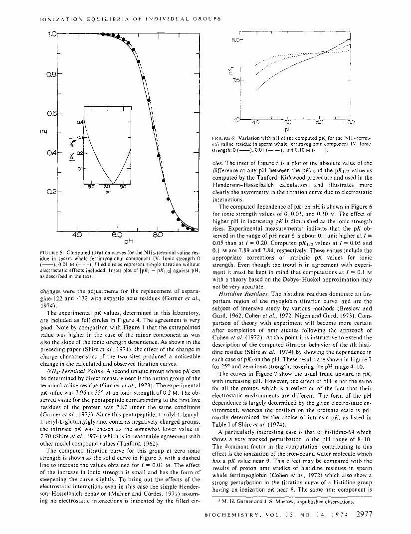
F I G L R E 4: Plot of the form of Figure I for the minor component I I of sperm whale ferrimyoglobin. The curve is computed for d = 0 with the static solvent accessibility factors taken into account. The filled circles a re experimental points.
oretical viewpoint, the first term of the sum of the polynomials which determine the ionic strength factor in the Tanford-Kirk- wood formalism is m / D ( 1 + K U ) , where D is the solvent dielec- tric constant and K is the Debye-Huckel parameter which is proportional to It has also been shown (Tanford and Kirk- wood, 1957) for a model very similar to the one used i n the present calculations that this is the dominant term i n the ionic strength polynomical series, and that this holds up to an ionic strength of 0.04 'M. In addition to this point, the use of the above function is supported by the experimental evidence based on analysis of the ionic strength variation of the observed pK values for iron-bound water in sperm whale ferrimyoglobin (Nakhleh, 1971). The analysis makes use of an error function calculated for a general ionic strength function f ( 1 ) = 1 + B U / ' / ~ ) , and was found to be a minimum a t Ba = 1. In addi- tion to this experimental observation on myoglobin preliminary measurements on hemoglobin a t verj low ionic strength (Hanania and Ilakhleh, 1974) indicate that the extrapolation function f ( / ) = / ( / 2 / ( I + 1 2 / 1 / 2 ) will result in a pK value a t zero ionic strength which is greater than the true value.
Variation of p K with p H . The sensitivity of the ionization of iron-bound water in ferrimyoglobin is shown by marked effects of pH on the absorption bands associated with the heme (Hanania et al., 1966). Even small changes in ionic strength have some effect on the Soret band (Hanania et a / . . 1966). Chemically modified myoglobins may show appreciable changes i n the intensity of the various absorption bands (Ray and Gurd, 1967). Figure 3 shows the pH dependence of the computed pK, values for the iron-bound water in sperm whale ferrimyoglobin major component IV a t 25'. The curves repre- sent ionic strengths of 0, 0.01, and 0.1 M . For comparison, ex- perimental values obtained a t / = 0.001 M are included as filled circles. The sensitivity of the pK, to pH is very large, even a t / = 0.1 M. For demonstrating sensitivity to electrostatic ef- fects the plot of the form of Figure 3 is more striking t h a n the usual titration curve format of charge plotted against pH. In keeping with the good fit of the observed values i n Figure I , the experimental points in Figure 3 are in reasonable agreement with the computed behavior over the range shown between pH 8 and 10.
.4pplication to Ferrimyoglobin Component I/. A further test of the applicability of the theory is to use a closely related pro- tein such as the minor component 1 1 of sperm whale ferrimyo- globin. The computations employed the same model as was ap- plied to the major component IV (Figure I ) , with d = 0 and static solvent accessibilities taken into account. The only
I O N I Z A T I O S E Q U I L I B R I A O F I N D l V l D L A L G R O U P S
I I I I 1 4.0 6.0 8.0
PH FIGURE 5 : Computed titration curves for the NHz-terminal valine res- idue in sperm whale ferrimyoglobin component 1V. Ionic strength 0 (-), 0.01 M (- - -); filled circles represent simple titration without electrostatic effects included. Inset: plot of IpKi - pKl/d against pH, as described in the text.
changes were the adjustments for the replacement of aspara- gine-122 and -132 with aspartic acid residues (Garner et al., 1974).
The experimental pK values, determined in this laboratory, are included as full circles in Figure 4. The agreement is very good. Note by comparison with Figure 1 that the extrapolated value was higher in the case of the minor component as was also the slope of the ionic strength dependence. As shown in the preceding paper (Shire et al., 1974), the effect of the change in charge characteristics of the two sites produced a noticeable change in the calculated and observed titration curves.
NH2- Terminal Valine. A second unique group whose pK can be determined by direct measurement is the amino group of the terminal valine residue (Garner et al., 1973). The experimental pK value was 7.96 a t 25' a t an ionic strength of 0.2 M. The ob- served value for the pentapeptide corresponding to the first five residues of the protein was , 7 3 7 under the same conditions (Garner et al . , 1973). Since this pentapeptide, L-valyl-L-leucyl- L-seryl-L-glutamylglycine, contains negatively charged groups, the intrinsic pK was chosen as the somewhat lower value of 7.70 (Shire et al . , 1974) which is in reasonable agreement with other model compound values (Tan ford, 1962).
The computed titration curve for this group a t zero ionic strength is shown as the solid curve in Figure 5, with a dashed line to indicate the values obtained for I = 0.01 M. The effect of the increase in ionic strength is small and has the form of steepening the curve slightly. T o bring out the effects of the electrostatic interactions even in this case the simple Hender- son-Hasselbalch behavior (Mahler and Cordes, 1971) assum- ing no electrostatic interactions is indicated by the filled cir-
I 1 I I I I I
7.O' 4.b 6.0 8.0 '3.3 Pt-
FIGURE 6 : Variation with pH of the computed pK, for the NH2-termi- nal valine residue in sperm whale ferrimyoglobin component I V . Ionic strength: 0 (-), 0.01 (- -), and 0.10 M (- - -).
cles. The inset of Figure 5 is a plot of the absolute value of the difference a t any pH between the pKi and the p K l p value as computed by the Tanford-Kirkwood procedure and used in the Henderson-Hasselbalch calculation, and illustrates more clearly the asymmetry in the titration curve due to electrostatic interactions.
The computed dependence of pK, on pH is shown in Figure 6 for ionic strength values of 0, 0.01, and 0.10 M. The effect of higher pH in increasing pK is diminished as the ionic strength rises. Experimental measurements3 indicate that the pK ob- served in the range of pH near 8 is about 0.1 unit higher a t I = 0.05 than a t I = 0.20. Computed p K l p values a t I = 0.05 and 0.1 M are 7.89 and 7.84, respectively. These values include the appropriate corrections of intrinsic pK values for ionic strength. Even though the trend is in agreement with experi- ment it must be kept in mind that computations a t I = 0.1 M
with a theory based on the Debye-Huckel approximation may not be very accurate.
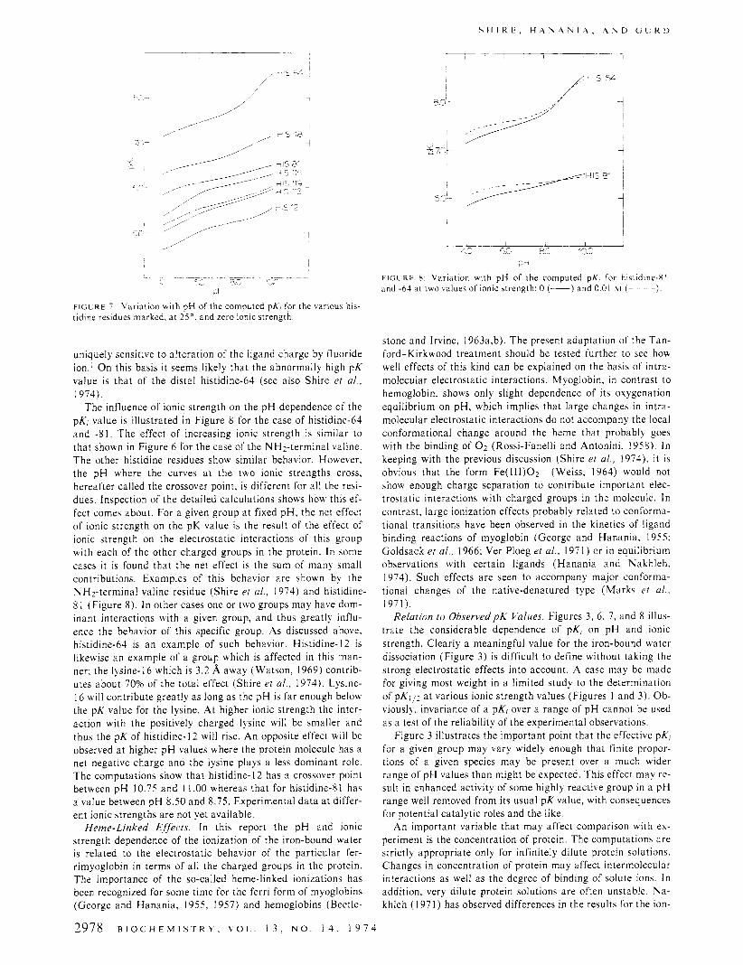
Histidine Residues. The histidine residues dominate an im- portant region of the myoglobin titration curve, and are the subject of intensive study by various methods (Breslow and Curd, 1962; Cohen et al., 1972; Nigen and Curd, 1973). Com- parison of theory with experiment will become more certain after completion of nmr studies following the approach of Cohen et al. (1972). At this point it is instructive to extend the description of the computed titration behavior of the ith histi- dine residue (Shire et al., 1974) by showing the dependence in each case of pKj on the pH. These results are shown in Figure 7 for 25' and zero ionic strength, covering the pH range 4-1 0.
The curves in Figure 7 show the usual trend upward in pK, with increasing pH. However, the effect of pH is not the same for all the groups, which is a reflection of the fact that their electrostatic environments are different. The form of the pH dependence is largely determined by the given electrostatic en- vironment, whereas the position on the ordinate scale is pri- marily determined by the choice of intrinsic pK, as listed in Table I of Shire el a / . (1974).
A particularly interesting case is that of histidine-64 which shows a very marked perturbation in the pH range of 8-10. The dominant factor in the computations contributing to this effect is the ionization of the iron-bound water molecule which has a pK value near 9. This effect may be compared with the results of proton nmr studies of histidine residues in sperm whale ferrimyoglobin (Cohen et a / . , 1972) which also show a strong perturbation in the titration curve of a histidine group having an ionization pK near 8. The same nmr component is
M. H . Garner and J. S. Morrow, unpublished observations
B I O C H E M I S T R Y , V O L . 1 3 , N O . 1 4 , 1 9 7 4 2977
S H I R E . H A Y A \ I A . A U D C i L R D
-- - - e.’ 8‘; d.
iJ
F I G L R E 7 Varidtion with pH of the computed pX, for the carioui his- tidine residues marked, a t 2 j 0 , and zero ionic strength
uniquely sensitive to alteration of the ligand charge by fluoride ion.’ On this basis it seems likely that the abnormally high pK value is that of the distal histidine-64 (see also Shire et al., 1974).
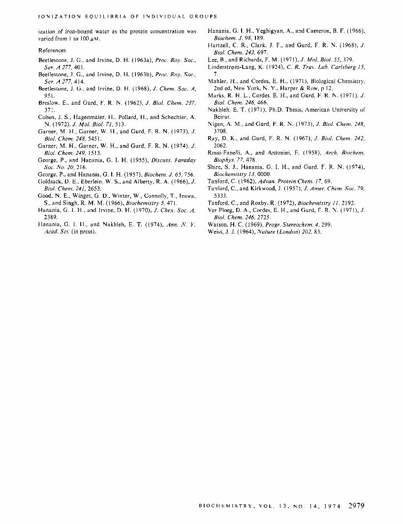
The influence of ionic strength on the pH dependence of the pKj value is illustrated in Figure 8 for the case of histidine-64 and -8 1 . The effect of increasing ionic strength is similar to that shown in Figure 6 for the case of the NHz-terminal valine. The other histidine residues show similar behavior. However, the pH where the curves a t the two ionic strengths cross, hereafter called the crossover point, is different for all the resi- dues. Inspection of the detailed calculations shows how this ef- fect comes about. For a given group a t fixed pH, the net effect of ionic strength on the pK value is the result of the effect of ionic strength on the electrostatic interactions of this group with each of the other charged groups in the protein. In some cases it is found that the net cffect is the sum of many small contributions. Examples of this behavior are shown by the NHz-terminal valine residue (Shire et al., 1974) and histidine- 8 1 (Figure 8). In other cases one or two groups may have dom- inant interactions with a given group, and thus greatly influ- ence the behavior of this specific group. As discussed above, histidine-64 is an example of such behavior. Histidine-] 2 is likewise an example of a group which is affected in this man- ner; the lysine-16 which is 3.2 A away (Watson, 1969) contrib- utes about 70% of the total effect (Shire er al., 1974). Lysine- I6 will contribute greatly as long as the pH is far enough below the pK value for the lysine. At higher ionic strength the inter- action with the positively charged lysine will be smaller and thus the pK of histidine- 12 will rise. An opposite effect will be observed a t higher pH values where the protein molecule has a net negative charge and the lysine plays a less dominant role. The computations show that histidine- 12 has a crossover point between pH 10.75 and 1 1 .OO whereas that for histidine81 has a value between pH 8.50 and 8.75. Experimental data a t differ- ent ionic strengths are not yet available.
Heme-Linked Effects. In this report the pH and ionic strength dependence of the ionization of the iron-bound water is related to the electrostatic behavior of the particular fer- rimyoglobin i n terms of all the charged groups in the protein. The importance of the so-called heme-linked ionizations has been recognized for some time for the ferri form of myoglobins (George and Hanania, 1955, 1957) and hemoglobins (Beetle-
2978 B I O C H E M I S T R Y , V O L . 1 3 . N O . 14, 1 9 7 4
~
I L-- --LA 13- n
- J.2 c.c a5 . i l l
2-3
F l C L R t 8 Variation with pH of the computed pK, for histidine-81 and -64 d t two calues of ionic strength 0 (-) and 0 01 w ( - )
stone and Irvine, 1963a,b). The present adaptation of the Tan- ford-Kirkwood treatment should be tested further to see how well effects of this kind can be explained on the basis of intra- molecular electrostatic interactions. Myoglobin, in contrast to hemoglobin. shows only slight dependence of its oxygenation equilibrium on pH, which implies that large changes in intra- molecular electrostatic interactions do not accompany the local conformational change around the heme that probably goes with the binding of 0 2 (Rossi-Fanelli and Antonini, 1958). In keeping with the previous discussion (Shire et al., 1974). it is obvious that the form Fe(1II)O2- (Weiss, 1964) would not show enough charge separation to contribute important elec- trostatic interactions with charged groups in the molecule. In contrast, large ionization effects probably related to conforma- tional transitions have been observed in the kinetics of ligand binding reactions of myoglobin (George and Hanania, 1955; Goldsack et al., 1966; Ver Ploeg et al., 197 1 ) or in equilibrium observations with certain ligands (Hanania and Nakhleh. 1974). Such effects are seen to accompany major conforma- tional changes of the native-denatured type (Marks et al.,
Relation to Observed p K Values. Figures 3, 6, 7, and 8 illus- trate the considerable dependence of pK, on pH and ionic strength. Clearly a meaningful value for the iron-bound water dissociation (Figure 3) is difficult to define without taking the strong electrostatic effects into account. A case may be made for giving most weight in a limited study to the determination of pKl,z a t various ionic strength values (Figures I and 3). Ob- viously, invariance of a pKj over a range of pH cannot be used as a test of the reliability of the experimental observations
Figure 3 illustrates the important point that the effective pK, for a given group may vary widely enough that finite propor- tions of a given species may be present over a much wider range of pH values than might be expected. This effect may re- sult i n enhanced activity of some highly reactive group in a pH range well removed from its usual pK value, with consequences for potential catalytic roles and the like.
An important variable that may affect comparison ui th ex- periment is the concentration of protein. The computations are strictly appropriate only for infinitely dilute protein solutions. Changes in concentration of protein may affect intermolecular interactions as well as the degree of binding of solute ions. I n addition, very dilute protein solutions are often unstable. Na- khleh (1971) has observed differences in the results for the ion-
1971).
I O N I Z A T I O N E Q U I L I B R I A O F I N D I V I D U A L G R O U P S
ization of iron-bound water as the protein concentration was varied from 1 to 100 w M .
References
Beetlestone, J . G., and Irvine, D. H. (1963a), Proc. Roy. Soc.,
Beetlestone, J. G., and Irvine, D. H. (1963b), Proc. Roy. Soc.,
Beetlestone, J. G., and Irvine, D. H. (1968), J . Chem. SOC. A,
Breslow, E., and Gurd, F. R. N . (1962), J . Biol. Chem. 237,
Cohen, J . S., Hagenmaier, H., Pollard, H., and Schechter, A.
Garner, M. H., Garner, W. H., and Curd, F. R. N . (1973), J .
Garner, M. H., Garner, W. H., and Curd, F. R. N . (1974), J .
George, P., and Hanania, G. I. H. (1955), Discuss. Faraday
George, P., and Hanania, G. I . H. (1957), Biochem. J . 65, 756. Goldsack, D. E., Eberlein, W. S., and Alberty, R. A. (1966), J .
Good, N. E., Winget, G. D., Winter, W., Connolly, T., Izawa,
Hanania, G. 1. H., and Irvine, D. H. (1970), J . Chex. SOC. A,
Hanania, G. I. H., and Nakhleh, E. T. (1974), Ann. N . Y.
Ser. A 277, 40 I .
Ser. A 277, 4 14.
951.
371.
N. (1972), J . Mol. Biol. 71, 513.
Biol. Chem. 248, 545 1.
Biol. Chem. 249, 1513.
SOC. No. 20, 2 16.
Biol. Chem. 241, 2653.
S., and Singh, R. M. M. (1966), Biochemistry 5, 471.
2389.
Acad. Sci. (in press).
Hanania, G. I . H., Yeghigyan, A., and Cameron, B. F. (1966),
Hartzell, C . R., Clark, J. F., and Curd, F. R. N. (1968), J .
Lee, B., and Richards, F. M. (1971), J . Mol. Biol. 55, 379. Linderstr4m-Lang K. (l924), C. R. Trav. Lab. Carlsberg 15,
Mahler, H., and Cordes, E. H., (1971), Biological Chemistry,
Marks, R. H. L., Cordes, E. H., and Curd, F. R. N . (1971), J .
Nakhleh, E. T. (1971), Ph.D. Thesis, American University of
Nigen, A. M., and Curd, F. R. N . (1973), J . Biol. Chem. 248,
Ray, D. K., and Gurd, F. R. N. (l967), J . Biol. Chem. 242,
Rossi-Fanelli, A., and Antonini, E. ( 1 958), Arch. Biochem.
Shire, S. J., Hanania, G. I. H., and Gurd, F. R. N. (1974),
Tanford, C. (1962), Advan. Protein Chem. 17, 69. Tanford, C., and Kirkwood, J . ( 1 957), J . Amer. Chem. SOC. 79,
Tanford, C., and Roxby, R. (1972), Biochemistry I / , 2192. Ver Ploeg, D. A,, Cordes, E. H., and Curd, F. R. N. (1971), J .
Watson, H. C. ( I 969), Progr. Stereochem. 4 , 299. Weiss, J. J. ( 1 964), Nature (London) 202, 8 3 .
Biochem. J . 98, 189.
Biol. Chem. 243, 697.
7.
2nd ed, New York, N. Y., Harper & Row, p 12.
Biol. Chem. 246, 466.
Beirut.
3708.
2062.
Biophys. 77, 478.
Biochemistry 13, 0000.
5333.
Biol. Chem. 246, 2725.
B I O C H E M I S T R Y , V O L . 1 3 , N O . 1 4 , 1 9 7 4 2979