electronically activated actin protein polymerization and alignment
TRANSCRIPT
Electronically Activated Actin Protein Polymerization andAlignment
Ian Y. Wong,† Matthew J. Footer,‡ and Nicholas A. Melosh*,†
Department of Materials Science & Engineering, Stanford UniVersity, Geballe Laboratory forAdVanced Materials, 476 Lomita Mall, Stanford, California 94305, Department of Biochemistry,
Stanford UniVersity School of Medicine, Beckman Center, 279 West Campus DriVe,Stanford, California 94305
Received November 27, 2007; E-mail: [email protected]
Abstract: Biological systems are the paragon of dynamic self-assembly, using a combination of spatiallylocalized protein complexation, ion concentration, and protein modification to coordinate a diverse set ofself-assembling components. Biomimetic materials based upon biologically inspired design principles orbiological components have had some success at replicating these traits, but have difficulty capturing thedynamic aspects and diversity of biological self-assembly. Here, we demonstrate that the polymerizationof ion-sensitive proteins can be dynamically regulated using electronically enhanced ion mixing and monomerconcentration. Initially, the global activity of the cytoskeletal protein actin is inhibited using a low-ionic strengthbuffer that minimizes ion complexation and protein-protein interactions. Nucleation and growth of actinfilaments are then triggered by a low-frequency AC voltage, which causes local enhancement of the actinmonomer concentration and mixing with Mg2+. The location and extent of polymerization are governed bythe voltage and frequency, producing highly ordered structures unprecedented in bulk experiments.Polymerization rate and filament orientation could be independently controlled using a combination of low-frequency (∼100 Hz) and high frequency (1 MHz) AC voltages, creating a range of macromoleculararchitectures from network hydrogel microparticles to highly aligned arrays of actin filaments with ∼750nm periodicity. Since a wide range of proteins are activated upon complexation with charged species, thisapproach may be generally applicable to a variety of biopolymers and proteins.
Introduction
Biological systems are dynamic entities that continuouslyreorganize their internal structural architectures in response toexternal stimuli. For instance, polymerization of actin ineukaryotic cells is highly regulated both spatially and temporally,enabling coordinated macroscopic behaviors such as cell growth,division, or motion.1,2 This behavior has inspired the develop-ment of “smart” biomimetic materials that are responsive toexternal stimuli such as temperature, light, pH, and ionconcentration.3–5 In particular, there has been great interest inion or pH-sensitive molecular components that assemble intospecific morphologies such as nanofiber networks,6,7 tubules,and membranes,8 and hydrogels.9–11 It is difficult however to
spatially regulate component assembly in these systems atnanometer to micrometer length scales, leading to the formationof relatively homogeneous bulk materials. As a result thesebiomimetic systems generally lack the sophistication andfunctionality of higher-order directed organization in truebiological systems. This also limits the use of these biomimeticsystems for the deterministic fabrication of discrete artificialnanostructures.
One approach for artificially controlling biomaterial assemblyis to globally inhibit activity by restricting access to activatingspecies, such as ligands, salt, or other monomers. Assemblycan then be initiated at a particular time and place through theintroduction of the requisite species, such as using microfluidicdelivery12 or photochemical release.13,14 This mechanism alsooccurs widely in biological systems, where transients in theconcentration of small molecules, proteins, or salt trigger proteincomplexation and function.15 Alternatively, locally enhancingthe concentration of reactive monomers can greatly acceleratenucleation and growth rates, leading to rapid polymerization.
† Stanford University.‡ Stanford University School of Medicine.
(1) Pollard, T. D.; Borisy, G. G. Cell 2003, 112, 453–465.(2) Rafelski, S. M.; Theriot, J. A. Annu. ReV. Biochem. 2004, 73, 209–
239.(3) Anseth, K. S.; Burdick, J. A. MRS Bull 2002, 27, 130–136.(4) Drury, J. L.; Mooney, D. J. Biomaterials 2003, 24, 4337–4351.(5) Mart, R. J.; Osborne, R. D.; Stevens, M. M.; Ulijn, R. V. Soft Matter
2006, 2, 822–835.(6) Hartgerink, J. D.; Beniash, E.; Stupp, S. I. Proc. Natl. Acad. Sci. U.S.A.
2002, 99, 5133–5138.(7) Ozbas, B.; Kretsinger, J.; Rajagopal, K.; Schneider, J. P.; Pochan, D. J.
Macromolecules 2004, 37, 7331–7337.(8) Zhang, S. G. Nat. Biotechnol. 2003, 21, 1171–1178.(9) Fogleman, E. A.; Yount, W. C.; Xu, J.; Craig, S. L. Angew. Chem.,
Int. Ed. 2002, 41, 4026–4028.
(10) Petka, W. A.; Harden, J. L.; McGrath, K. P.; Wirtz, D.; Tirrell, D. A.Science (Washington DC) 1998, 281, 389–392.
(11) Topp, S.; Prasad, V.; Cianci, G. C.; Weeks, E. R.; Gallivan, J. P. J. Am.Chem. Soc. 2006, 128, 13994–13995.
(12) Burdick, J. A.; Khademhosseini, A.; Langer, R. Langmuir 2004, 20,5153–5156.
(13) Liu, A. P.; Fletcher, D. A. Nano Lett 2005, 5, 625–628.(14) Marriott, G. Biochemistry 1994, 33, 9092–9097.(15) Berridge, M. J.; Lipp, P.; Bootman, M. D. Nat. ReV. Mol. Cell. Biol.
2000, 1, 11–21.
Published on Web 05/29/2008
10.1021/ja7103284 CCC: $40.75 2008 American Chemical Society7908 9 J. AM. CHEM. SOC. 2008, 130, 7908–7915
Here we investigate whether the activity of ion-sensitive proteinscan be similarly regulated by exploiting the enhanced ion andmonomer concentrations that exist within the diffuse-chargelayer surrounding a charged electrode.
When an electrical potential difference is applied, counterionsaccumulate in a diffuse-charge layer at the electrode surface toscreen the electric field.16,17 For low voltages the electricpotential ψ(x) from the surface can be approximated by theDebye-Huckel equation, ψ(x) ) ψ0 e-x/λD, where λD is theDebye screening length. Ion concentration profiles depend uponthe ion valence, z, and local potential as given by the Boltzmanndistribution, F(x) ) F∞ e-zeψx/kT. Within the diffuse charge layerthe ion concentration can be enhanced over several thousand-fold, and is particularly effective for divalent ions such as Ca2+
and Mg2+, because their +2 valence leads to an 8-foldenhancement relative to monovalent ions. For example, a Ca2+
concentration wave could be artificially replicated by theapplication of 50 mV in a 0.1 µM CaCl2 solution, leading to a55-fold increase in Ca2+ at the electrode surface. Since the localion concentration is considerably different for biomoleculessituated inside and outside the layer, this approach enableselectrical control localized within the nanometer scale Debyescreening length. These arguments are also applicable for thelocal enhancement of monomer concentration. This process doesnot rely upon Faradic processes or redox-active molecules andthus may be applicable to a broad spectrum of proteins andsynthetic molecules which are generally not electrochemicallyactive.
One of the most dynamic biopolymers is actin, responsiblefor cell motility and shape.18 Polymerized actin filaments (F-actin) are 8-nm diameter helical filaments that form bynucleation of a trimer of globular actin monomers (G-actin),and subsequently grow by the addition of G-actin to the filamenttermini. Polymerization of the 5 nm diameter G-actin intoF-actin is highly regulated in vivo through a large number ofactin-binding proteins.1,2 In vitro, reconstituted G-actin can bemaintained in its monomeric state by a low ionic strengthmedium and millimolar Ca2+ levels (Table 1).19 Inactivity inthis “G-buffer” is thought to be caused by Ca2+ binding in thehigh-affinity binding site of G-actin, which changes the structuralconformation20 and significantly increases the critical concentra-tion of monomer necessary for polymerization relative to Mg2+-bound G-actin.21 Polymerization of G-actin into F-actin can beinitiated by increasing the ionic strength to physiological levels
and adding millimolar concentrations of Mg2+, which promotesthe replacement of Ca2+ by Mg2+ in the high-affinity site.22–27
Since F-actin polymerization is an important, dynamic bio-chemical process that exhibits sensitivity to divalent ionconcentration but no redox activity, it is an ideal model systemwith which to investigate electrostatically activated self-as-sembly.
Actin polymerization falls into a broad class of proteinassembly which requires initial activation of the monomer(Mg2+ here), followed by nucleation and growth (-11e chargedG-actin). Since the activating and monomer species haveopposite charges and are freely diffusing in solution, it is notintuitive how these two species can be concentrated at the samelocation using electrostatics. The experiments below demonstratethat this class of polymerization reaction can indeed beelectrostatically actuated by a low-frequency AC bias, whichencourages both mixing and growth. Frequency-dependentmeasurements indicate the polymerization rate is controlled bya competition between mixing and nucleation, while the filamentmorphology is largely determined by electrokinetic effects.Polymerization was observed from 10-500 Hz, while higherfrequencies (1 kHz to 1 MHz) did not induce polymerizationbut could dielectrophoretically align long actin filaments.28
These separate regimes provide independent control of growthand orientation which can be exploited for formation of orderednanostructures. These results demonstrate that the uniqueelectrostatic conditions at the electrode surface can modulateself-assembly of free species in solution even without electro-chemical reactivity and provide excellent spatial and temporalcontrol.
Experimental Design
In these experiments G-actin was suspended in a low-salt (LS)buffer containing a small amount of Mg2+ formulated to inhibitglobal actin polymerization (Table 1), yet provide the necessaryionic conditions when concentrated in an electric field. The low-salt buffer had 0.20 mM Mg2+ and a Debye screening length of6.0 nm, intermediate between F-buffer and G-buffer. The LS buffersuccessfully inhibited F-actin polymerization, as control samplesof 5.0 µM G-actin in LS-buffer (including fluorescent phalloidindye) showed no polymerization after 2 h. In addition, a sedimenta-tion assay29 under these conditions also showed no polymerizationafter 24 h, consistent with previous measurements of G-actin criticalconcentration at low concentrations of Mg2+.30
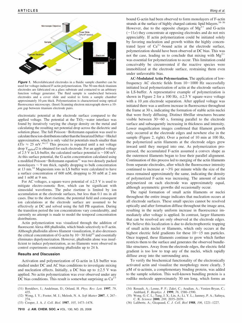
Electrostatically induced polymerization was investigated byplacing 5 µM Ca2+-complexed G-actin in LS-buffer on top of aglass wafer with opposing titanium electrodes with electrode gapsthat varied from 1-300 µm (Figure 1a). Titanium was chosen forits robust native oxide layer, which inhibits direct electron transfer.Electrolysis of water was observed at voltages of 3.5 V, so appliedpotentials were maintained at (2.5 V in all experiments. However,the TiO2 dielectric layer causes a considerable reduction in the
(16) Bard, A. J.; Faulkner, L. R. Electrochemical Methods: Fundamentalsand Applications, 2nd ed.; Wiley: Hoboken, NJ, 2001.
(17) Hunter, R. J., Foundations of Colloid Science, 2nd ed.; ClarendonPress, New York, 2001.
(18) Straub, F. B. Stud. Inst. Med. Chem. UniV. Szeged 1942, 2, 1–15.(19) Spudich, J. A.; Watt, S. J. Biol. Chem. 1971, 246, 4866–71.(20) Guan, J. Q.; Almo, S. C.; Reisler, E.; Chance, M. R. Biochemistry
2003, 42, 11992–12000.(21) Kinosian, H. J.; Selden, L. A.; Estes, J. E.; Gershman, L. C. Biochim.
Biophys. Acta 1991, 1077, 151–158.
(22) Carlier, M.-F.; Pantaloni, D.; Korn, E. D. J. Biol. Chem. 1986, 261,10778–10792.
(23) Cooper, J. A.; Buhle, E. L.; Walker, S. B.; Tsong, T. Y.; Pollard,T. D. Biochemistry 1983, 22, 2193–2202.
(24) Frieden, C. Proc. Natl. Acad. Sci. USA 1983, 80, 6513–6517.(25) Gershman, L. C.; Newman, J.; Selden, L. A.; Estes, J. E. Biochemistry
1984, 23, 2199–2203.(26) Pollard, T. D. Anal. Biochem. 1983, 134, 406–412.(27) Tobacman, L. S.; Korn, E. D. J. Biol. Chem. 1983, 258, 3207–3214.(28) Asokan, S. B.; Jawerth, L.; Carroll, R. L.; Cheney, R. E.; Washburn,
S.; Superfine, R. Nano Lett. 2003, 3, 431–437.(29) Sanders, M. C.; Way, M.; Sakai, J.; Matsudaira, P. J. Biol. Chem.
1996, 271, 2651–2657.(30) Tobacman, L. S.; Brenner, S. L.; Korn, E. D. J. Biol. Chem. 1983,
258, 8806–8812.
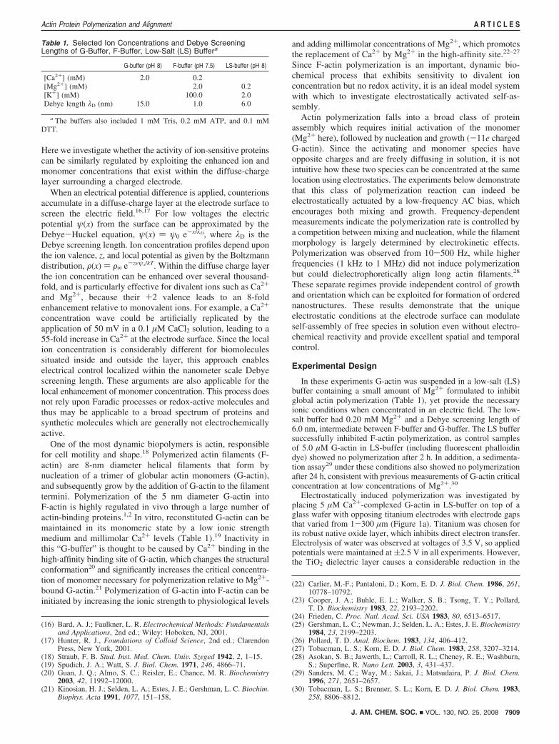
Table 1. Selected Ion Concentrations and Debye ScreeningLengths of G-Buffer, F-Buffer, Low-Salt (LS) Buffera
G-buffer (pH 8) F-buffer (pH 7.5) LS-buffer (pH 8)
[Ca2+] (mM) 2.0 0.2[Mg2+] (mM) 2.0 0.2[K+] (mM) 100.0 2.0Debye length λD (nm) 15.0 1.0 6.0
a The buffers also included 1 mM Tris, 0.2 mM ATP, and 0.1 mMDTT.
J. AM. CHEM. SOC. 9 VOL. 130, NO. 25, 2008 7909
Actin Protein Polymerization and Alignment A R T I C L E S
electrostatic potential at the electrode surface compared to theapplied voltage. The potential at the TiO2-water interface wasfound by iteratively varying the charge density on the metal andcalculating the resulting net potential drop across the dielectric andsolution phase. The full Poisson-Boltzmann equation was used tocalculatetheseion-distributionsratherthanthelinearizedDebye-Huckelapproximation, which is only valid for potentials much smaller thankT/e ≈ 25 mV.16,17 This process is repeated until a net voltagedrop Vapplied/2 is obtained for each electrode. For an applied voltageof 2.5 V in LS-buffer, the calculated surface potential is ∼100 mV.At this surface potential, the G-actin concentration calculated usinga modified Poisson-Boltzmann equation31 was two densely packedmonolayers ∼ 9 nm thick, rapidly dropping to bulk concentrationat 13 nm.32 Magnesium ions in LS buffer were calculated to havea surface concentration of 600 mM, dropping to 50 mM at 2 nmand 1 mM at 9 nm.
For AC voltages, a square-wave potential of (2.5 V is used tomitigate electro-osmotic flow, which can be significant withsinusoidal waveforms. The pulse risetime is limited by ionaccumulation at the electrode surfaces, but less than 0.1 ms in allcases. Due to the short risetime, the potential field and consequention calculations at the electrode surface are assumed to beeffectively at DC and solved using the full PB equation. Duringthe transition period the ion concentrations vary considerably, andcurrently no attempt is made to model the temporal concentrationdistributions.
Actin polymerization was visualized through the addition offluorescent Alexa 488 phalloidin, which binds selectively to F-actin.Although phalloidin allows filament visualization, it also decreasesthe critical concentration of G-actin by 10-30 fold33 and essentiallyeliminates depolymerization. However, phalloidin alone was insuf-ficient to induce polymerization, as no filaments were observed incontrol experiments containing phalloidin up to 24 h.
Results and Discussion
Activation and polymerization of G-actin in LS buffer wasstudied under DC and AC bias conditions to investigate mixingand nucleation effects. Initially, a DC bias up to (2.5 V wasapplied. No actin polymerization was ever observed under anyDC bias conditions. This result is somewhat surprising as Ca2+
bound G-actin had been observed to form monolayers of F-actinstrands at the surface of highly charged cationic lipid bilayers.34–36
However, due to the opposite charges of Mg2+ and G-actin(-11e) they concentrate at opposing electrodes and do not mixappreciably. If actin polymerization could be initiated solelyby favoring nucleation and growth within the highly concen-trated layer of Ca2+-bound actin at the electrode surface,polymerization should have been observed at DC bias. This wasnot the case, leading us to conclude that mixing with Mg2+
was essential for polymerization to occur. This limitation couldconceivably be circumvented if the reactive species wereimmobilized at the electrode surface, restraining them evenunder unfavorable bias.
AC-Modulated Actin Polymerization. The application of low-frequency AC electric fields from 10-1000 Hz successfullyinitiated local polymerization of actin at the electrode surfacesin LS-buffer. A representative example of polymerization isshown in Figure 2 for a 100 Hz, (2.5 V square-wave voltagewith a 10 µm electrode separation. After applied voltage wasinitiated there was a uniform increase in fluorescence throughoutthe frame at 30 s, indicating the formation of stable actin nucleithat were freely diffusing. Distinct fibrillar structures becamevisible between 30-60 s, forming parallel to the electrodesurface and subsequently increasing in thickness and intensity.Lower magnification images confirmed that filament growthonly occurred at the electrode edges and nowhere else in thesample (Figure 2, right). Over a period of 10 min at 100 Hz,the polymerized actin filaments at the electrode edges grewinward until they merged into one. As polymerization pro-gressed, the accumulated F-actin assumed a convex shape asthe outermost filaments began to lose their parallel alignment.Continuation of this process led to merging of the actin filamentsfrom separate electrodes, after which the fluorescence intensitycontinued to increase at ∼3% per minute while the size of themass remained approximately the same, indicating the densityof polymerized F-actin was increasing. The amount of actinpolymerized on each electrode was approximately equal,although asymmetric growths did occasionally occur.
The rapid formation of small actin filaments or nucleithroughout the entire image indicates that nucleation occurs atall electrode surfaces. These small species cannot be resolvedoptically and after formation diffuse throughout the image area,resulting in the nearly uniform increase in fluorescence im-mediately after voltage is applied. In contrast, larger filamentsthat can be resolved are only observed at the electrode edges.We believe this localization is due to dielectrophoretic trappingof small actin nuclei or filaments, which only occurs at thehighest electric field gradients for these 10-15 nm particles.Once trapped, these filaments continue to grow which furtherrestricts them to the surface and generates the observed bundle-like structures. Away from the electrode edges, the electric fieldgradient is too low to trap any of the nuclei, which rapidlydiffuse away into the surrounding area.
To verify the biochemical functionality of the electronicallyactivated actin and visualize the morphology more clearly, 5µM of R-actinin, a complementary binding protein, was addedto the sample solution. This well-known bundling protein is arodlike molecule approximately 30 nm long, which forms an
(31) Borukhov, I.; Andelman, D.; Orland, H. Phys. ReV. Lett. 1997, 79,435.
(32) Wong, I. Y.; Footer, M. J.; Melosh, N. A. Soft Matter 2007, 3, 267–274.
(33) Cooper, J. A. J. Cell. Biol. 1987, 105, 1473–1478.
(34) Renault, A.; Lenne, P. F.; Zakri, C.; Aradian, A.; Venien-Bryan, C.;Amblard, F. Biophys. J. 1999, 76, 1580–1590.
(35) Wong, G. C. L.; Tang, J. X.; Lin, A.; Li, Y. L.; Janmey, P. A.; Safinya,C. R. Science 2000, 288, 2035–2039.
(36) Laliberte, A.; Gicquaud, C. J. Cell. Biol 1988, 106, 1221–1227.
Figure 1. Microfabricated electrodes in a fluidic sample chamber can beused for voltage-induced F-actin polymerization. The 50-nm thick titaniumelectrodes are fabricated on a glass substrate and contacted to an arbitraryfunction voltage generator. The fluid sample is sandwiched betweenelectrodes and a cover slide and sealed to form a sample chamberapproximately 10-µm thick. Polymerization is characterized using opticalfluorescence microscopy. (Inset) Scanning electron micrograph shows a 10-µm gap between titanium electrode pairs.
7910 J. AM. CHEM. SOC. 9 VOL. 130, NO. 25, 2008
A R T I C L E S Wong et al.

antiparallel homodimer with actin-binding regions on eachend.37,38 Figure 3 shows discrete bundles characteristic ofR-actinin/actin that formed parallel to the electrode surface.These bundles were hundreds of micrometers long and con-centrated parallel to the electrode edges. These structures havetwo levels of hierarchical order: actin filaments bundled togetherby R-actinin and the bundles packed into a nematic configura-tion. The highly ordered actin configuration is unlike bulkexperiments, where R-actinin/actin form random isotropic gels.38
Some actin alignment has been previously observed at elevatedconcentrations of divalent ions (∼10 mM Mg2+) leading to salt-bridging bundles with diameters limited to ∼300 nm,39–42 orby actin polymerization within microfluidic channels43 withlower density.
The nematic ordering of these structures is consistent withactin filaments polymerizing along the electrode surface, thenbecoming displaced as new filaments begin to form. Thismechanism is distinct from bulk experiments, where the uniformdistribution of activated monomers allows filaments to grow at
any location and in any direction.26 We see no evidence thatfilaments continue to grow once displaced from the electrodesurface and the filament density is always highest at the electrodeedge. The high degree of nematic order is also apparentlymetastable as filaments begin to lose their alignment farther fromthe electrode surface. Similarly, if the electrode potential isturned off the filaments slowly reorient and exfoliate from thesurface. Exfoliation is slow (∼minutes), suggesting the filamentseven far from the electrode have limited mobility which mayarise from steric entanglements or salt bridges. Intriguingly, arecent study found nematic-phase actin filaments had decreasedlongitudinal diffusion at high divalent counterion concentra-tions,44 attributed to attractive salt-bridging effects. The highconcentration of Mg2+ at the electrode surface in our systemmay induce local salt-bridging, which gradually dissociates asthe filaments are displaced from the surface.
F-actin Entanglement. Actin gels can be formed if actinpolymerization is allowed to bridge the gap between electrodes,resulting in a mass of actin that remains intact even with theremoval of the AC voltage (Figure 4). After the voltage is turnedoff, these actin microgels typically swell in size by a factor of2, and diffuse away as a single object. This demonstrates thatthe actin filaments are not attached to the electrodes, and that
(37) Meyer, R. K.; Aebi, U. J. Cell. Biol 1990, 110, 2013–2024.(38) Wachsstock, D. H.; Schwarz, W. H.; Pollard, T. D. Biophys. J. 1993,
65, 205–214.(39) Tang, J. X.; Janmey, P. A. J. Biol. Chem. 1996, 271, 8556–8563.(40) Kwon, H. J.; Kakugo, A.; Shikinaka, K.; Osada, Y.; Gong, J. P.
Biomacromolecules 2005, 6, 3005–3009.(41) Wong, G. C. L.; Lin, A.; Tang, J. X.; Li, Y.; Janmey, P. A.; Safinya,
C. R. Phys. ReV. Lett. 2003, 91, 018103.(42) Lai, G. H.; Coridan, R.; Zribi, O. V.; Golestanian, R.; Wong, G. C. L.
Phys. ReV. Lett. 2007, 98, 187802.(43) Hirst, L. S.; Parker, E. R.; Abu-Samah, Z.; Li, Y.; Pynn, R.;
MacDonald, N. C.; Safinya, C. R. Langmuir 2005, 21, 3910–3914. (44) He, J.; Viamontes, J.; Tang, J. X. Phys. ReV. Lett. 2007, 99, 068103.
Figure 2. (left) F-Actin polymerizes at the edges of two electrodes spaced 10 µm apart during a 100 Hz, 5 Vpp square-wave bias. Actin filaments parallelto the electrodes are first observed at 60 s, subsequently increasing in intensity and width. (right) As F-actin continues to polymerize due to Mg2+-bindingin the applied field, the filaments grow from either electrode, eventually merging into one mass as polymerization progresses. Filaments are most concentratedat the electrode edge.
Figure 3. Visible actin bundles stacked with nematic ordering after 180min with the addition of 5 µM R-actinin. The electrode gap was 300 µm toenable clear visualization of the filaments.
Figure 4. Physically cross-linked hydrogel microparticles (“microgels”)of polymerized F-actin remain intact if the actin filaments from eitherelectrode coalesce, even after AC voltage is switched off. Successive imagesat 30 s intervals show an expansion in size before the mass drifts out of thefield of view.
J. AM. CHEM. SOC. 9 VOL. 130, NO. 25, 2008 7911
Actin Protein Polymerization and Alignment A R T I C L E S
the actin filaments experience appreciable dielectrophoretic forceeven at low applied frequencies. Such freestanding gel micro-structures have not been previously demonstrated using cyto-skeletal proteins, but similar physically cross-linked hydrogelmicroparticles (“microgels”) have technological applications asmicroreactors, biosensors, and drug delivery platforms.45
Gel-formation likely occurs as reoriented actin filaments atthe interface region undergo a crossover from a dilute semi-flexible polymer phase to an entangled gel phase, as the purelynematic structures were never stable. This critical filamentnumber density can be estimated by c** ≈ L-3�(Lp/L) in therodlike limit for filament contour lengths L much smaller thanthe persistence length Lp.46 For a persistence length Lp ) 17µm for phalloidin-bound actin47 and a typical experimentalcontour length L ≈ 2 µm, the transition filament number densityis c** ) 3.6 × 10¹⁷ m-3, which is equivalent to a bulk polymergel formed at a concentration of 0.44 µM G-actin. Thiscalculation is a lower bound as some filament anisotropy is likelyretained. An upper bound on the filament concentration isprovided by the isotropic to nematic transition, which wouldcause all the filaments to remain aligned. This transition can beestimated in the short-rod limit as cnem ) 4.3/(dL2).46 Usingthe F-actin diameter d ) 8 nm, cnem ) 1.5 × 10²⁰ m-3 whichwould correspond to a bulk polymer gel formed from 188 µMG-actin. The actual filament density in the entangled region isthus likely between 1017-1020 m-3, intermediate to the limitingcases of an isotropic entangled network (c**) and a fullyanisotropic nematic phase (cnem).
Actin Polymerization Kinetics and Mechanism. Insight intothe mechanisms that govern voltage-induced polymerization canbe gained by comparing the frequency-dependent polymerizationkinetics (Figure 5A) to bulk polymerization assays (Figure 5A,inset). In bulk solution polymerization proceeds through aninitial activation step where Ca2+ is exchanged for Mg2+ inthe tight binding site of G-actin.23,24 Subsequently, there is alag-time associated with the kinetically slow reaction of threeG-actin monomers to form stable trimeric F-actin nuclei,23,24,27
followed by a growth period where existing F-actin filamentselongate. At long times, the polymerization curve approachesa plateau as the bulk concentration of actin monomers is depletedand the reaction approaches steady-state.48,49
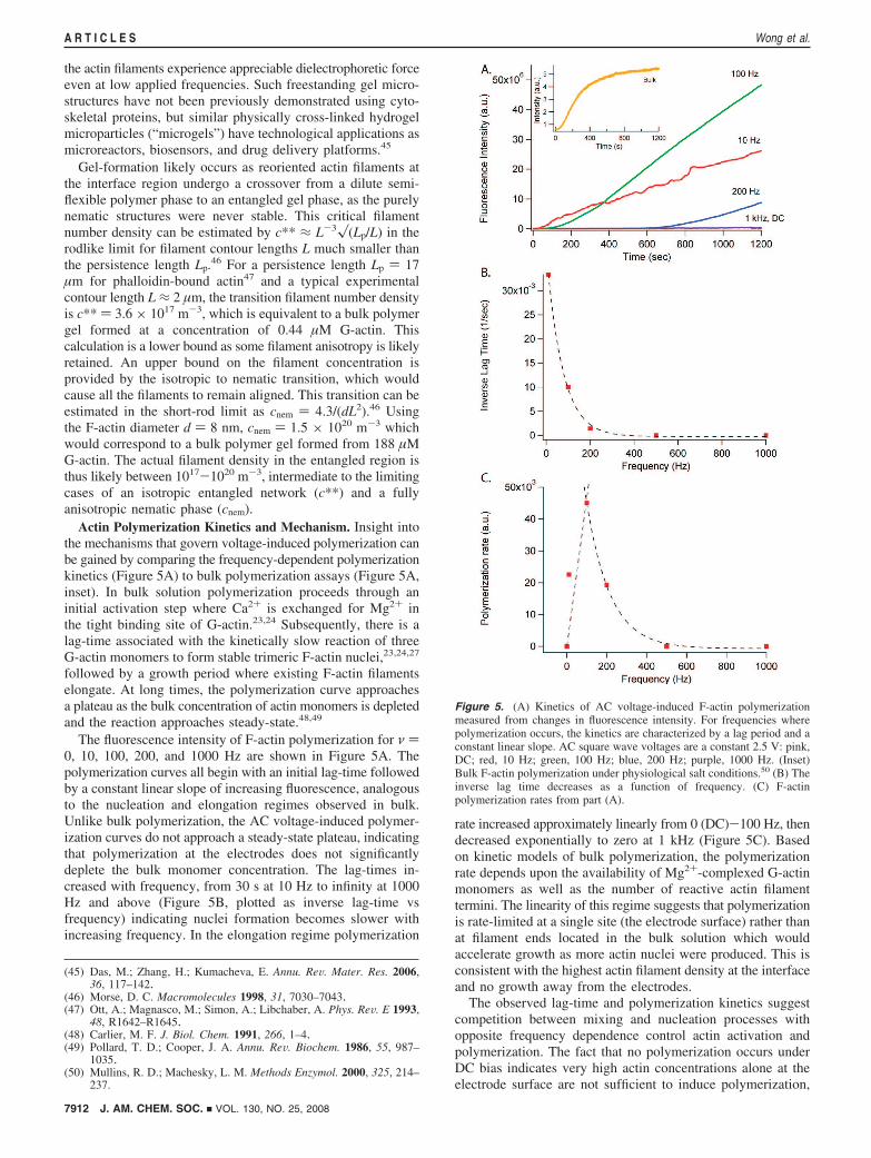
The fluorescence intensity of F-actin polymerization for ν )0, 10, 100, 200, and 1000 Hz are shown in Figure 5A. Thepolymerization curves all begin with an initial lag-time followedby a constant linear slope of increasing fluorescence, analogousto the nucleation and elongation regimes observed in bulk.Unlike bulk polymerization, the AC voltage-induced polymer-ization curves do not approach a steady-state plateau, indicatingthat polymerization at the electrodes does not significantlydeplete the bulk monomer concentration. The lag-times in-creased with frequency, from 30 s at 10 Hz to infinity at 1000Hz and above (Figure 5B, plotted as inverse lag-time vsfrequency) indicating nuclei formation becomes slower withincreasing frequency. In the elongation regime polymerization
rate increased approximately linearly from 0 (DC)-100 Hz, thendecreased exponentially to zero at 1 kHz (Figure 5C). Basedon kinetic models of bulk polymerization, the polymerizationrate depends upon the availability of Mg2+-complexed G-actinmonomers as well as the number of reactive actin filamenttermini. The linearity of this regime suggests that polymerizationis rate-limited at a single site (the electrode surface) rather thanat filament ends located in the bulk solution which wouldaccelerate growth as more actin nuclei were produced. This isconsistent with the highest actin filament density at the interfaceand no growth away from the electrodes.
The observed lag-time and polymerization kinetics suggestcompetition between mixing and nucleation processes withopposite frequency dependence control actin activation andpolymerization. The fact that no polymerization occurs underDC bias indicates very high actin concentrations alone at theelectrode surface are not sufficient to induce polymerization,
(45) Das, M.; Zhang, H.; Kumacheva, E. Annu. ReV. Mater. Res. 2006,36, 117–142.
(46) Morse, D. C. Macromolecules 1998, 31, 7030–7043.(47) Ott, A.; Magnasco, M.; Simon, A.; Libchaber, A. Phys. ReV. E 1993,
48, R1642–R1645.(48) Carlier, M. F. J. Biol. Chem. 1991, 266, 1–4.(49) Pollard, T. D.; Cooper, J. A. Annu. ReV. Biochem. 1986, 55, 987–
1035.(50) Mullins, R. D.; Machesky, L. M. Methods Enzymol. 2000, 325, 214–
237.
Figure 5. (A) Kinetics of AC voltage-induced F-actin polymerizationmeasured from changes in fluorescence intensity. For frequencies wherepolymerization occurs, the kinetics are characterized by a lag period and aconstant linear slope. AC square wave voltages are a constant 2.5 V: pink,DC; red, 10 Hz; green, 100 Hz; blue, 200 Hz; purple, 1000 Hz. (Inset)Bulk F-actin polymerization under physiological salt conditions.50 (B) Theinverse lag time decreases as a function of frequency. (C) F-actinpolymerization rates from part (A).
7912 J. AM. CHEM. SOC. 9 VOL. 130, NO. 25, 2008
A R T I C L E S Wong et al.
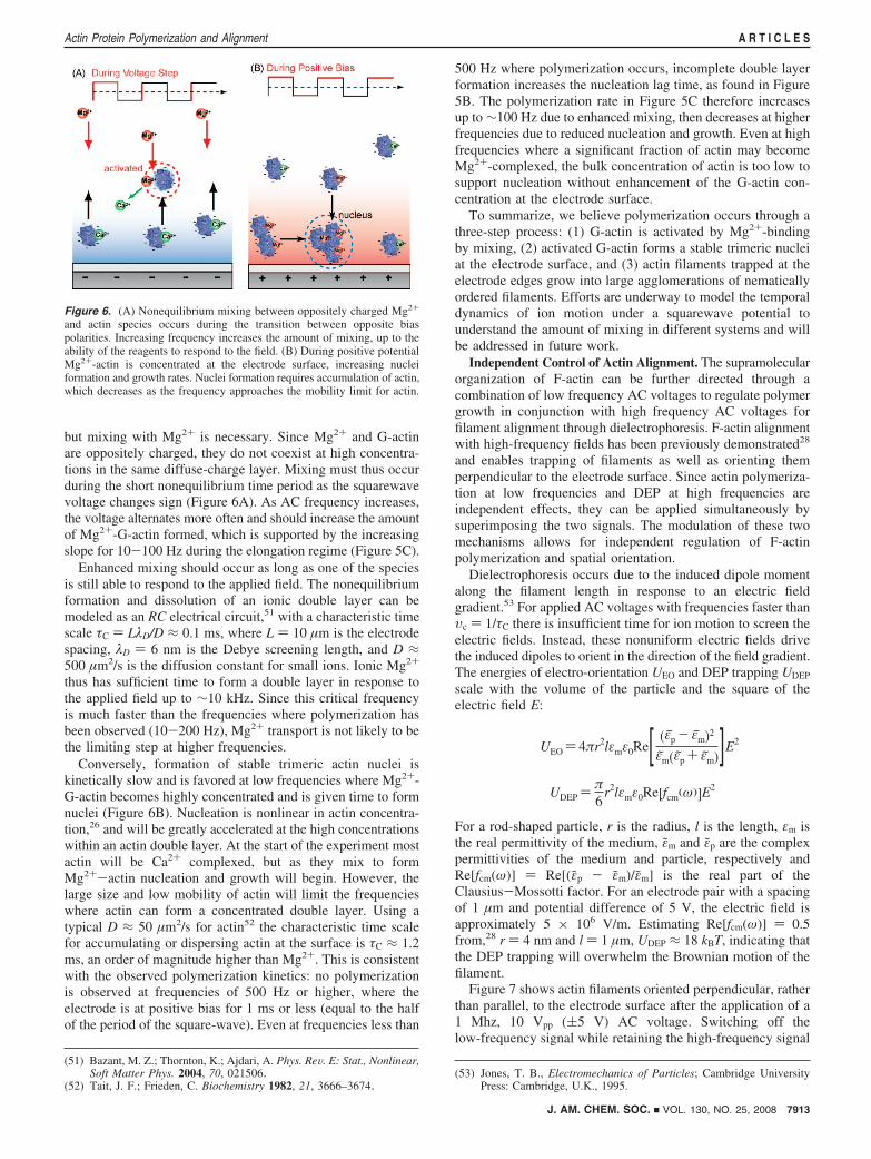
but mixing with Mg2+ is necessary. Since Mg2+ and G-actinare oppositely charged, they do not coexist at high concentra-tions in the same diffuse-charge layer. Mixing must thus occurduring the short nonequilibrium time period as the squarewavevoltage changes sign (Figure 6A). As AC frequency increases,the voltage alternates more often and should increase the amountof Mg2+-G-actin formed, which is supported by the increasingslope for 10-100 Hz during the elongation regime (Figure 5C).
Enhanced mixing should occur as long as one of the speciesis still able to respond to the applied field. The nonequilibriumformation and dissolution of an ionic double layer can bemodeled as an RC electrical circuit,51 with a characteristic timescale τC ) LλD/D ≈ 0.1 ms, where L ) 10 µm is the electrodespacing, λD ) 6 nm is the Debye screening length, and D ≈500 µm2/s is the diffusion constant for small ions. Ionic Mg2+
thus has sufficient time to form a double layer in response tothe applied field up to ∼10 kHz. Since this critical frequencyis much faster than the frequencies where polymerization hasbeen observed (10-200 Hz), Mg2+ transport is not likely to bethe limiting step at higher frequencies.
Conversely, formation of stable trimeric actin nuclei iskinetically slow and is favored at low frequencies where Mg2+-G-actin becomes highly concentrated and is given time to formnuclei (Figure 6B). Nucleation is nonlinear in actin concentra-tion,26 and will be greatly accelerated at the high concentrationswithin an actin double layer. At the start of the experiment mostactin will be Ca2+ complexed, but as they mix to formMg2+-actin nucleation and growth will begin. However, thelarge size and low mobility of actin will limit the frequencieswhere actin can form a concentrated double layer. Using atypical D ≈ 50 µm2/s for actin52 the characteristic time scalefor accumulating or dispersing actin at the surface is τC ≈ 1.2ms, an order of magnitude higher than Mg2+. This is consistentwith the observed polymerization kinetics: no polymerizationis observed at frequencies of 500 Hz or higher, where theelectrode is at positive bias for 1 ms or less (equal to the halfof the period of the square-wave). Even at frequencies less than
500 Hz where polymerization occurs, incomplete double layerformation increases the nucleation lag time, as found in Figure5B. The polymerization rate in Figure 5C therefore increasesup to ∼100 Hz due to enhanced mixing, then decreases at higherfrequencies due to reduced nucleation and growth. Even at highfrequencies where a significant fraction of actin may becomeMg2+-complexed, the bulk concentration of actin is too low tosupport nucleation without enhancement of the G-actin con-centration at the electrode surface.
To summarize, we believe polymerization occurs through athree-step process: (1) G-actin is activated by Mg2+-bindingby mixing, (2) activated G-actin forms a stable trimeric nucleiat the electrode surface, and (3) actin filaments trapped at theelectrode edges grow into large agglomerations of nematicallyordered filaments. Efforts are underway to model the temporaldynamics of ion motion under a squarewave potential tounderstand the amount of mixing in different systems and willbe addressed in future work.
Independent Control of Actin Alignment. The supramolecularorganization of F-actin can be further directed through acombination of low frequency AC voltages to regulate polymergrowth in conjunction with high frequency AC voltages forfilament alignment through dielectrophoresis. F-actin alignmentwith high-frequency fields has been previously demonstrated28
and enables trapping of filaments as well as orienting themperpendicular to the electrode surface. Since actin polymeriza-tion at low frequencies and DEP at high frequencies areindependent effects, they can be applied simultaneously bysuperimposing the two signals. The modulation of these twomechanisms allows for independent regulation of F-actinpolymerization and spatial orientation.
Dielectrophoresis occurs due to the induced dipole momentalong the filament length in response to an electric fieldgradient.53 For applied AC voltages with frequencies faster thanυc ) 1/τC there is insufficient time for ion motion to screen theelectric fields. Instead, these nonuniform electric fields drivethe induced dipoles to orient in the direction of the field gradient.The energies of electro-orientation UEO and DEP trapping UDEP
scale with the volume of the particle and the square of theelectric field E:
UEO ) 4πr2lεmε0Re[ (εp - εm)2
εm(εp + εm)]E2
UDEP )π6
r2lεmε0Re[fcm(ω)]E2
For a rod-shaped particle, r is the radius, l is the length, εm isthe real permittivity of the medium, εm and εp are the complexpermittivities of the medium and particle, respectively andRe[fcm(ω)] ) Re[(εp - εm)/εm] is the real part of theClausius-Mossotti factor. For an electrode pair with a spacingof 1 µm and potential difference of 5 V, the electric field isapproximately 5 × 10⁶ V/m. Estimating Re[fcm(ω)] ) 0.5from,28 r ) 4 nm and l ) 1 µm, UDEP ≈ 18 kBT, indicating thatthe DEP trapping will overwhelm the Brownian motion of thefilament.
Figure 7 shows actin filaments oriented perpendicular, ratherthan parallel, to the electrode surface after the application of a1 Mhz, 10 Vpp ((5 V) AC voltage. Switching off thelow-frequency signal while retaining the high-frequency signal
(51) Bazant, M. Z.; Thornton, K.; Ajdari, A. Phys. ReV. E: Stat., Nonlinear,Soft Matter Phys. 2004, 70, 021506.
(52) Tait, J. F.; Frieden, C. Biochemistry 1982, 21, 3666–3674.(53) Jones, T. B., Electromechanics of Particles; Cambridge University
Press: Cambridge, U.K., 1995.
Figure 6. (A) Nonequilibrium mixing between oppositely charged Mg2+
and actin species occurs during the transition between opposite biaspolarities. Increasing frequency increases the amount of mixing, up to theability of the reagents to respond to the field. (B) During positive potentialMg2+-actin is concentrated at the electrode surface, increasing nucleiformation and growth rates. Nuclei formation requires accumulation of actin,which decreases as the frequency approaches the mobility limit for actin.
J. AM. CHEM. SOC. 9 VOL. 130, NO. 25, 2008 7913
Actin Protein Polymerization and Alignment A R T I C L E S
immediately halts polymerization, but keeps the existing F-actinconfined in the electrode gap. Individual actin filaments can beresolved for low filament densities. Filament density can beactively controlled by adding a low-frequency AC voltage toinduce polymerization, or by decreasing the magnitude of the1 MHz field to release shorter trapped actin filaments. It shouldalso be noted that applying 1 MHz frequency alone never showsany actin growth or accumulation, ruling out field-inducedtrapping of pre-existing actin filaments within solution as thereason for the observed polymerization.
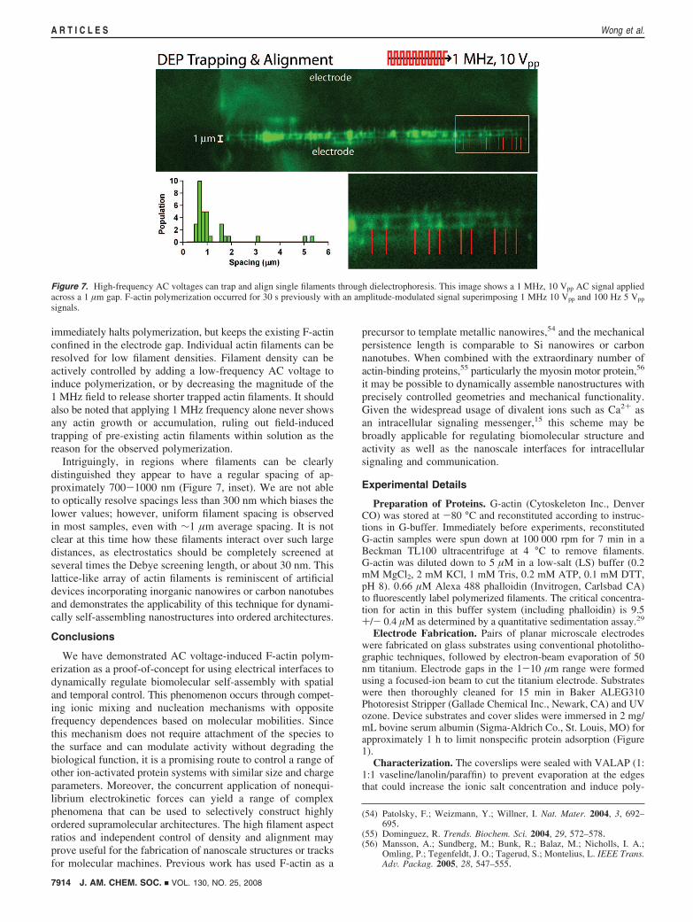
Intriguingly, in regions where filaments can be clearlydistinguished they appear to have a regular spacing of ap-proximately 700-1000 nm (Figure 7, inset). We are not ableto optically resolve spacings less than 300 nm which biases thelower values; however, uniform filament spacing is observedin most samples, even with ∼1 µm average spacing. It is notclear at this time how these filaments interact over such largedistances, as electrostatics should be completely screened atseveral times the Debye screening length, or about 30 nm. Thislattice-like array of actin filaments is reminiscent of artificialdevices incorporating inorganic nanowires or carbon nanotubesand demonstrates the applicability of this technique for dynami-cally self-assembling nanostructures into ordered architectures.
Conclusions
We have demonstrated AC voltage-induced F-actin polym-erization as a proof-of-concept for using electrical interfaces todynamically regulate biomolecular self-assembly with spatialand temporal control. This phenomenon occurs through compet-ing ionic mixing and nucleation mechanisms with oppositefrequency dependences based on molecular mobilities. Sincethis mechanism does not require attachment of the species tothe surface and can modulate activity without degrading thebiological function, it is a promising route to control a range ofother ion-activated protein systems with similar size and chargeparameters. Moreover, the concurrent application of nonequi-librium electrokinetic forces can yield a range of complexphenomena that can be used to selectively construct highlyordered supramolecular architectures. The high filament aspectratios and independent control of density and alignment mayprove useful for the fabrication of nanoscale structures or tracksfor molecular machines. Previous work has used F-actin as a
precursor to template metallic nanowires,54 and the mechanicalpersistence length is comparable to Si nanowires or carbonnanotubes. When combined with the extraordinary number ofactin-binding proteins,55 particularly the myosin motor protein,56
it may be possible to dynamically assemble nanostructures withprecisely controlled geometries and mechanical functionality.Given the widespread usage of divalent ions such as Ca2+ asan intracellular signaling messenger,15 this scheme may bebroadly applicable for regulating biomolecular structure andactivity as well as the nanoscale interfaces for intracellularsignaling and communication.
Experimental Details
Preparation of Proteins. G-actin (Cytoskeleton Inc., DenverCO) was stored at -80 °C and reconstituted according to instruc-tions in G-buffer. Immediately before experiments, reconstitutedG-actin samples were spun down at 100 000 rpm for 7 min in aBeckman TL100 ultracentrifuge at 4 °C to remove filaments.G-actin was diluted down to 5 µM in a low-salt (LS) buffer (0.2mM MgCl2, 2 mM KCl, 1 mM Tris, 0.2 mM ATP, 0.1 mM DTT,pH 8). 0.66 µM Alexa 488 phalloidin (Invitrogen, Carlsbad CA)to fluorescently label polymerized filaments. The critical concentra-tion for actin in this buffer system (including phalloidin) is 9.5+/- 0.4 µM as determined by a quantitative sedimentation assay.29
Electrode Fabrication. Pairs of planar microscale electrodeswere fabricated on glass substrates using conventional photolitho-graphic techniques, followed by electron-beam evaporation of 50nm titanium. Electrode gaps in the 1-10 µm range were formedusing a focused-ion beam to cut the titanium electrode. Substrateswere then thoroughly cleaned for 15 min in Baker ALEG310Photoresist Stripper (Gallade Chemical Inc., Newark, CA) and UVozone. Device substrates and cover slides were immersed in 2 mg/mL bovine serum albumin (Sigma-Aldrich Co., St. Louis, MO) forapproximately 1 h to limit nonspecific protein adsorption (Figure1).
Characterization. The coverslips were sealed with VALAP (1:1:1 vaseline/lanolin/paraffin) to prevent evaporation at the edgesthat could increase the ionic salt concentration and induce poly-
(54) Patolsky, F.; Weizmann, Y.; Willner, I. Nat. Mater. 2004, 3, 692–695.
(55) Dominguez, R. Trends. Biochem. Sci. 2004, 29, 572–578.(56) Mansson, A.; Sundberg, M.; Bunk, R.; Balaz, M.; Nicholls, I. A.;
Omling, P.; Tegenfeldt, J. O.; Tagerud, S.; Montelius, L. IEEE Trans.AdV. Packag. 2005, 28, 547–555.
Figure 7. High-frequency AC voltages can trap and align single filaments through dielectrophoresis. This image shows a 1 MHz, 10 Vpp AC signal appliedacross a 1 µm gap. F-actin polymerization occurred for 30 s previously with an amplitude-modulated signal superimposing 1 MHz 10 Vpp and 100 Hz 5 Vpp
signals.
7914 J. AM. CHEM. SOC. 9 VOL. 130, NO. 25, 2008
A R T I C L E S Wong et al.
merization. Samples were observed on a Zeiss Axioskop 2microscope with a 25× or 100× oil-immersion objective, epifluo-rescence optics and a Andor DV887 EMCCD camera. AC square-wave voltages of 10 Hz-10 kHz and 5 Vpp were generated usinga Tektronix AFG3251 Arbitrary Function Generator. Amplitude-modulated AC signals of 5 Vpp, 100 Hz (carrier) and 10 Vpp, 1MHz (modulation) were generated by externally triggering a Tenma72-7650 signal generator. The use of square-wave voltages weredesigned to minimize electro-osmotic flow effects, which were notobserved in video data. Fluorescence intensity was analyzed usingAndor iQ software at 10 s intervals.
Sedimentation Assay. Sedimentation assays with G-actin (5 µM)and Alexa 488 phalloidin (0.66 µM) in LS-buffer were performedfollowing established procedures.29 Briefly, samples were incubatedfor either 4 °C overnight or for 20 min at room temperature andwere spun down at 100 000 rpm for 7 min in a Beckman TL100ultracentrifuge at 4 °C to remove filaments. Both pellets andsupernatants from the centrifugation were analyzed by SDS-PAGE
and it was verified that final actin concentrations were consistentwith the initial sample concentration.
Bulk Polymerization Assay. Pyrene actin assays were conductedaccording to published methods.50 The final assay conditionsincluded 4.5 µM unlabeled actin and 0.5 µM pyrene-actin in buffer(10 mM Mops, 50 mM KCl, 2 mM MgCl2, 2 mM EGTA, 0.2 mMATP, 0.5 mM DTT at pH 7.0) at 24 C using an excitationwavelength of 357 nm and emission wavelength of 407 nm.
Acknowledgment. We thank V. B. Chu and S. Doniach forhelpful discussions, T. Carver and E. Yenilmez for assistance withfabrication, J. Sakata for careful reading of the manuscript, and ananonymous reviewer for insightful comments on electrokinetics.I.Y.W. was supported by an NSF Graduate Research Fellowshipand a Bio-X Interdisciplinary Initiatives Project.
JA7103284
J. AM. CHEM. SOC. 9 VOL. 130, NO. 25, 2008 7915
Actin Protein Polymerization and Alignment A R T I C L E S