electrochemical sensors and biosensors based on nanomaterials and nanostructures
TRANSCRIPT
Subscriber access provided by University of Cincinnati Libraries
Analytical Chemistry is published by the American Chemical Society. 1155 SixteenthStreet N.W., Washington, DC 20036Published by American Chemical Society. Copyright © American Chemical Society.However, no copyright claim is made to original U.S. Government works, or worksproduced by employees of any Commonwealth realm Crown government in the courseof their duties.
Review
Electrochemical Sensors and BiosensorsBased on Nanomaterials and NanostructuresChengzhou Zhu, Guohai Yang, He Li, Dan Du, and Yuehe Lin
Anal. Chem., Just Accepted Manuscript • DOI: 10.1021/ac5039863 • Publication Date (Web): 29 Oct 2014
Downloaded from http://pubs.acs.org on November 13, 2014
Just Accepted
“Just Accepted” manuscripts have been peer-reviewed and accepted for publication. They are postedonline prior to technical editing, formatting for publication and author proofing. The American ChemicalSociety provides “Just Accepted” as a free service to the research community to expedite thedissemination of scientific material as soon as possible after acceptance. “Just Accepted” manuscriptsappear in full in PDF format accompanied by an HTML abstract. “Just Accepted” manuscripts have beenfully peer reviewed, but should not be considered the official version of record. They are accessible to allreaders and citable by the Digital Object Identifier (DOI®). “Just Accepted” is an optional service offeredto authors. Therefore, the “Just Accepted” Web site may not include all articles that will be publishedin the journal. After a manuscript is technically edited and formatted, it will be removed from the “JustAccepted” Web site and published as an ASAP article. Note that technical editing may introduce minorchanges to the manuscript text and/or graphics which could affect content, and all legal disclaimersand ethical guidelines that apply to the journal pertain. ACS cannot be held responsible for errorsor consequences arising from the use of information contained in these “Just Accepted” manuscripts.

Electrochemical Sensors and Biosensors Based on Nano-materials and Nanostructures
Chengzhou Zhu,1,§ Guohai Yang, 1,§ He Li,1,§ Dan Du,1 and Yuehe Lin*,1,2
1School of Mechanical and Materials Engineering, Washington State University, Pullman, WA 99164 2Pacific Northwest National Laboratory, Richland, WA 99352
CONTENTS
Nonenzymatic Sensors
Glucose
Hydrogen Peroxide
Cation
Anion
Other Species
Electrochemical Enzyme-based Biosensors
Immobilization Strategies
Direct Electron Transfer
Other Papers of Interest
Genosensors
Nucleic Acid Assay
Rational Design of DNA Probe
Enzyme-Based Amplification
Nanomaterial-Enhanced Signal Amplification
Other Approaches for Signal Amplification
New Methods for DNA Detection
Electrochemical Detection of DNA Damage
Immunosensors
Voltammetry and Amperometry based Immunoassay
Cancer Biomarkers, Bacteria and Environment
Graphene
Other Nanomaterials
Electrochemiluminescence
Photoelectrochemistry
Other Papers of Interest
Cytosensors
Label-Free Cytosensing
Sandwich Cytosensing
Conclusions
Author Information
Biographies
Acknowledgment
References
Taking advantages of exceptional attributes, such as easy-to-operate, economical, sensitive, portable, and simple-to- construct, considerable attention has been devoted to the inte-gration of recognition elements with electronic elements to develop electrochemical sensors and biosensors. Recent dec-ades tremendous amounts of attention have been witnessed in this active area. Various electrochemical devices such as am-perometric sensors, electrochemical impedance sensors, elec-trochemical luminescence sensors as well as photoelectro-chemical sensors, provide wide applications of detecting chemical and biological targets in terms of electrochemical change of electrode interfaces.
With remarkable achievements in nanotechnology and na-noscience, nanomaterial-based electrochemical signal amplifi-cations have great potential of improving both in sensitivity and selectivity for electrochemical sensors and biosensors. First of all, it is well known that the electrode materials play a critical role in the construction of high-performance electro-chemical sensing platforms for detecting target molecules through various analytical principles. Furthermore, in addition to electrode materials, functional nanomaterials can not only produce a synergic effect among catalytic activity, conductivi-ty and biocompatibility to accelerate the signal transduction, but also amplify biorecognition events by specifically de-signed signal tags, leading to highly sensitive biosensing. Sig-nificantly, extensive research on construction of functional electrode materials, coupled with numerous electrochemical methods, is advancing wide applications of electrochemical devices. For example, Walcarius et al. highlighted the recent advances of nano-objects, nano-engineered and/or nanostruc-tured materials for the rational design of bio-functionalized electrodes and related (bio)sensing systems.1 The attractive-ness of such nanomaterials relies on their ability to act as ef-fective immobilization matrices, and their intrinsic and unique features as described above. These features combined with the functioning of biomolecules contribute to the improvement of bioelectrode performance in terms of sensitivity and specifici-ty. Our group recently presented a general overview of nano-material-enhanced paper-based biosensors including lateral-flow test-strip, and paper microfluidic devices.2With different kinds of nanoparticles (NPs), paper-based biosensor devices have shown great potential in the enhancement of sensitivity and specificity of disease diagnosis in developing countries.
This review focuses on recent advances in electrochemical sensors and biosensors based on nanomaterials and nanostruc-
Page 1 of 23
ACS Paragon Plus Environment
Analytical Chemistry
123456789101112131415161718192021222324252627282930313233343536373839404142434445464748495051525354555657585960
tures during 2013 to 2014. The aim of this effort is to provide the reader with a clear and concise view of new advances in areas ranging from electrode engineering, strategies for elec-trochemical signal amplification, and novel electroanalytical techniques used to the miniaturization and integration of the sensors. Moreover, the authors have attempted to highlight areas of latest and significant development of enhanced elec-trochemical nanosensors and nanobiosensors that inspire broader interests across various disciplines. Electrochemical sensors for small molecules, enzyme-based biosensors, genosensors, immunosensors and cytosensors are reviewed herein (Figure 1). Such novel advances are important for the development of electrochemical sensors as that open up new avenues and methods for future research. Readers interested in the general principles of electrochemical sensors and electro-chemical methods are recommended to refer to other excellent literature for a broad scope on this area.3-4 However, due to the explosion of publications in this active field, we do not claim that this review includes all of the published works in the past two years. Due to the large activity in this field we apologize to the authors of excellent work, which is unintentionally left out.
NONENZYMATIC SENSORS
The pursuit of electrochemical systems for bimolecular de-tection has received significant attention over the last two dec-ades.5 Electroanalysis toward small molecules is also of im-portance in a variety of areas. Enzymatic sensors possess high selectivity but suffer from limitations such as instability, com-plicated modification procedure, and critical micro-environmental factors. Such limitations stimulate the devel-opment of nonenzymatic electrochemical sensors with simple modification procedure and good stability. Enzyme-free elec-trochemical sensors have been widely used for determining the presence of hydrogen peroxide, glucose, and dopamine. The perspectives and current challenges of enzyme-free electro-chemical sensors were discussed by Chen et al.6 (142 refer-ences). Miao et al.7 recently reviewed electrocatalysis and electroanalysis of nickel, itsoxides, hydroxides and oxyhy-droxides toward small molecules (85 references). Following are some examples of nonenzymatic sonsors for the detection of small molecules.
Glucose
Glucose plays an important role in metabolism. Glucose bi-osensors have contributed significantly to clinical monitoring.8,
9With regard to the electrode materials, metal, metal oxide nanostructures and their hybrid nanocomposites are regarded to be the most promising materials currently used. Wang et al.
10 reviewed the progress made in the recent years in the field of direct and non-enzymatic electrochemical sensing of glu-cose (221 references). Tian et al.11 also reviewed the most recent advances in nonenzymatic glucose sensors based on varies of nanomaterials (125 references). Various nanomateri-als with different shape and composition were synthesized to construct novel nonenzymatic electrochemical sensors for glucose detection.
Cao et al.12 synthesized bimetallic PtCu nanochains through a water-based mild chemical route, compositions of which can be conveniently tuned at the mesoscopic scale by a facile deal-loying process. Electrochemical measurements demonstrate that the sensors made by these PtCu nanomaterials are very sensitive and specific for glucose detection due to the wiring of dispersed crystals, porous nanostructure, clean surface, and
synergetic electronic effects of the alloyed atoms. The resulted sensor performed very well in the detection of glucose in se-rum samples. Keerthy et al.13 produced reduced graphene ox-ide (rGO) modified with platinum nanocubes and copper oxide nanoflowers. Low cost screen printing technology was used to fabricate such electrodes for point-of-care (POC) glucose monitoring. These sensors were highly specific to glucose in the presence of commonly interfering species like ascorbic acid (AA), dopamine (DA), uric acid (UA) and acetaminophen. It was discussed that copper oxide catalyzes glucose oxidation and Pt NPs act as a co-catalyst to enhance the electron transfer during the oxidation of glucose.
Guo et al.14 constructed a Ni/CdS bifunctional Ti@TiO2 core-shell nanowire electrode through a hydrothermal and electrodeposition method. The resultant sensor based on the fabricated nanowire electrode exhibited great sensitivity in the electrochemical detection of glucose oxidation. The enhanced electrochemical performance on glucose sensing was attribut-ed to the high dispersity of Ni NPs and the ability to discrimi-nate the interfering species of CdS under the irradiation of visible light. The ability to combine the unique properties of individual nanomaterials provides a new and exciting frontier for the formation of novel electrodes.
Xu et al.15 prepared α-Fe2O3 cubes in the presence of a hy-drophobic iron-containing ionic liquid (IL) under hydrother-mal conditions and tested the photoelectrochemical properties by means of the transient photocurrent responses of ITO elec-trodes which were modified with the as-prepared α-Fe2O3. The photoelectrochemical approach in the application of glucose sensor was investigated. A glucose photoelectrochemical de-tection system based on non-enzyme catalytic oxidation reac-tion has shown promising results with high sensitivity and fast response.
Hydrogen Peroxide
The rapid and accurate detection of hydrogen peroxide (H2O2) has practical importance in the field of bioanalysis as well as food safety and environmental protection.16, 17 Nagaiah et al.
18 reported a H2O2 sensor based on electrochemical depo-sition of Pd-Pt and Pd-Au NPs on spectrographic graphite. The sensivity is originated from the codeposition of Pd with either Pt or Au enhancing electrocatalytic activity forH2O2reduction. Bai et al.19 developed a H2O2 sensor based on copper sulfide NPs-decorated rGO. They investigated the application of this sensor in detecting H2O2 content in human serum and urine samples, also H2O2 released from human cervical cancer cells. Their results have shown satisfactory recovery rates with good reproducibility, indicating potential applications in medical diagnosis.
A powerful enzymatic mimetics employing graphene oxide (GO), carbon nanotubes (CNTs) and Pt nanocatalysts was fabricated by Wang’s group.20 The GO-CNTs-Pt nanocompo-sites exhibited peroxidase-like catalysis activity and electroca-talysis activities, that tested in the colorimetric and electro-chemical detection of H2O2. Moreover, sandwiched electro-chemical immunoassays were demonstrated by using the GO-CNTs-Pt as catalytic tags. Such innovative nanostructure showed promise in the extensive catalysis applications in envi-ronmental, medical, industrial, and particularly aqueous bio-sensing fields.
Yang et al.21 reported the development of a microwave-assisted strategy for the synthesis of nitrogen and boron co-doped graphene with a hierarchical framework. The resultant
Page 2 of 23
ACS Paragon Plus Environment
Analytical Chemistry
123456789101112131415161718192021222324252627282930313233343536373839404142434445464748495051525354555657585960

sensor was able to selectively detect H2O2 in the presence of glucose and AA. Moreover, this electrode system could be easily functionalized with biomacromolecules to generate a cost-effective, highly sensitive and biocompatible sensor for a variety of applications. Tian et al.22 reported that ultrathin g-C3N4 nanosheets were fabricated by ultrasonication-assisted liquid exfoliation of bulk C3N4. They demonstrated using these nanosheets as an effective sensing platform for the detection of H2O2, which has been extended to the electrochemical de-tection of glucose both in buffer solution and human serum medium.
Wu et al.23 developed a novel nonenzymatic electrochemical sensor based on a p-methoxy zinc porphyrin-C60 derivative (ZnPp-C60), which was designed and synthesized by combin-ing p-methoxy porphyrin and C60 moieties with a flexible methylene chain. Combinded with theoretical information, the results showed that the ZnPp-C60would be a novel material for construction of a nonenzymatic electrochemical sensor for H2O2 and nitrite analysis in a relatively wide potential range with high sensitivity and stability.
Cation
The sensitive and selective detection of toxic heavy metals coupled with cost-effective and simple assaying procedure has paramount importance.24 Recently, Cui et al.25 developed a convenient and highly sensitive electrochemical detection platform for detecting copper. The sensor was fabricated on a glassy carbon electrode (GCE) through layer-by-layer (LBL) assembling modification with multi-wall carbon nanotubes (MWCNTs), poly (amidoamine) dendrimers and dithio-bis[succinimidyl-propionate] encapsulated Au NPs (DSP-Au NPs). The DSP modified sensor captures cysteamine (Cys) functionalized Ag NPs through the reaction between DSP and Cys. The presence of Cu2+ catalyzed cystocystamine regulated the assembly of Ag NPs on the sensor surface, resulting in the decrease of the electrochemical stripping signal of Ag NPs. With this strategy, the detection range of Cu2+ is 1.0-1000 nM with a detection limit of 0.48 nM.
In addition, Sadhukhan et al.26 synthesized two-dimensional C3N4 under microwave irradiation and used to modify GCE for the detection of Hg2+. Due to its graphene-like structure, this sensor detected Hg2+ down to 0.09 nM. For a real applica-tion, the device fabricated by Rattanarat’s group using screen-printing MWCNTs mixed carbon inks on polyester, and then modified with bi-nanoparticales and ferricyanide what sup-posed to enhance stripping signals and reduce Cu2+ interfer-ence.27 The top layer of the device contains five wax-defined channels extending outward from an open sample reservoir. In order to achieve high selectivity and sensitivity, each channel contains individual sample with pretreatment and detection zones. Under the optimized conditions, such paper-based de-vice can simultaneously detect Cd2+ and Pb2+ in the range of 5-150 µg L-1.
Anion
Madhu et al.28 prepared highly porous and heteroatom-enriched activated carbon (HAC) from banana stems. HAC was used to develop electrochemical sensors for the detection of nitrite in the application of monitoring environmental pollu-tion. HAC exhibits noteworthy performance in the highly sen-sitive detection of nitrite. Their method was tested to deter-mine nitrite in various water samples with acceptable results.
Conducting polymer-based modified electrodes have been extensively studied as sensing materials in the last decades.29 Yang et al.30 reported glassy carbon electrodes modified with polyaniline (PAni)-coated copper hexacyanoferrate were used as cyclic voltammetry sensor. The application of the sensor was investigated in the detection of sulfite, which is used as a preservative in a variety of food and pharmaceutical products to inhibit enzymatic and nonenzymatic browning, and also used in brewing industries as an antibacterial and antioxidizing agent.31 They showed that the synergistic effect of these struc-tures improved electrocatalytic activity towards detection.
Other Species
Most conducting polymer/graphene composites have excel-lent electrical conductivity. Gao et al.32 developed electro-chemically coated porous structure films of overoxidized polypyrrole/graphene (PPyox/GR) deposited onto GCE. This structure was successfully utilized as an efficient electrode material for the quantitative detection of adenine and guanine. With low background current, the permselective polymer coat-ing improved the selectivity and sensitivity of microelectrodes for the electropositive purine bases.
Lin et al.33 reported the hybridization of poly(luminol) (PLM) and poly(neutral red) (PNR) that is then enhanced by a conductive and steric hybrid nanotemplate using GO and MWCNTs. The PLM-PNR-MWCNT-GO mycelium-like nanocomposite is found to be electroactive, pH-dependent, and stable in the electrochemical system. This nanocompo-siteshowed electrocatalytic activity towards NADH with a high current response and low overpotential. It also exhibited a high sensitivity of 288.9 µA mM-1 cm-2 to NADH using am-perometry.
Changes in glutathione concentration at the cellular level may link to diseases such as pre-mature arteriosclerosis, leu-kemia, and diabetes. Lee et al.34 described a modified GCE through electropolymerization of caffeic acid onto the elec-trode surface in the presence of either CNTs or nanocarbon, affording a nanocomposite with a high concentration of o-quinone moiety onto the electrode. That can be used for the detection of glutathione through an electrocatalytic process. The detection concentrations as low as 500 nM was demon-strated.
Zhang et al.35 prepared a phosphomolybdate functionalized graphene nanocomposite for the detection of AA, in which polyoxometalates can irreversibly adsorb onto carbon materi-als to form carbon nanocomposite structures. The amperomet-ric signals are linearly proportional to the AA concentration in a wide concentration range from 1× 10⁻⁶ M to 8 × 10⁻³ M, with a detection limit of 0.5× 10⁻⁶ M.
Recently, layered transition metal dichalcogenides have been extensively studied due to their structural similarities with graphene and their interesting physicochemical properties, along with their diverse exotic electronic properties.36, 37 Nara-yanan et al.38 employed ultrathin MoS2 sheet-based electrodes for electrochemical detection of dopamine (DA) as an im-portant neurotransmitterin the presence of AA. The results showed that MoS2 is expected to be a good electrode material for electrochemical sensing applications. Sun et al.39 reported the Au NPs -decorated MoS2 nanosheets was synthesized by electrodeposition of Au NPs on MoS2 nanosheets, which pos-sess better properties than pure Au NPs and MoS2. The com-posite film modified electrode showed excellent electrocatalyt-ic activity toward the oxidation of AA, DA and UA with three
Page 3 of 23
ACS Paragon Plus Environment
Analytical Chemistry
123456789101112131415161718192021222324252627282930313233343536373839404142434445464748495051525354555657585960

well-resolved oxidation peaks. The peak potential separations were large enough to individually or simultaneously detecting AA, DA and UA.
Nitric oxide (NO) plays an important role in physiological processes. It has been reported that some human diseases are related to its biological function.40 Hunter et al.41 utilized standard photolithographic techniques and a nitric oxide (NO) selective xerogel polymer to fabricate an amperometric NO microfluidic sensor. The sensor detected NO levels in small sample volumes (∼250 µL) with low background noise. The detection sensitivity of 1.4 pA nM-1 was demonstrated along with the limit of detection (LOD) of 840 pM. This sensor ex-hibited excellent analytical performance in phosphate buffered saline. Moreover, the analytical performance of the device was investigated in simulated wound fluid and whole blood. The results showed that the sensor is able to measure NO in com-plicated biological samples. This proof of concept study demonstrated the feasibility of clinical application of this method. Metters et al.42 reported screen printed single-walled CNTs (SWCNTs) sensors which were fabricated upon flexible polyester substrates. The screen printed SWCNTs sensors are benchmarked using potassium ferrocyanide (II), DA, hydra-zine and capsaicin. By using this sensor, the detection of cap-saicin has been achieved at low micromolar levels. These elec-trodes hold potential in the development of disposable and highly reproducible sensors.
It is observed that sensors that exploit the unique properties of nanomaterials constitute the most rapidly expanding sensor research area. Moving forward several areas of research will enhance the nanostructured sensing platform. For example, researches in the storage and stability of the sensors will im-prove the shelf-life of functional biosensors.
ELECTROCHEMICALENZYME-BASED BIOSENSORS
Electrochemical enzyme-based biosensors, a subclass of chemical sensors, combine the high specificity of enzyme with the sensitivity of electrochemical transducers. Enzyme elec-trodes are electrochemical probes with a thin layer of immobi-lized enzyme on the surface of the working electrode. The enzyme is the most critical component of the enzyme elec-trode since it provides the selectivity for the sensor and cata-lyzes the formation of the electroactive product for detection. In the past two years, there were some review articles that focused on the development of various materials, techniques, and applications of electrochemical enzyme-based biosensors. Yu et al. described recent progress in electrochemical glucose biosensors and focused on some problems and bottlenecks in areas of enzymatic (glucose oxidase (GOx) based) am-perometric glucose sensing (240 citations).43 The focus of the review by Debora’s group is to present the current status and some trends in enzymatic nanoimmobilization.44 The recent advance of lactate biosensors45 and enzymatic uric acid46 bio-sensors were also systematic discussed. In addition, Clark et al. presented different immobilization strategies that have been used to create Cytochrome P450s (CYPs) biosensors, with particular emphasis on mammalian drug-metabolizing CYPs and characterization of CYP electrodes.47 Recent achieve-ments in this area have focused on the development of novel immobilization strategy and study of the direct electron trans-fer with the assistance of functional nanomaterials. Besides, some other papers of interest will also be addressed.
Immobilization Strategies
The immobilization of enzymes is one of the key steps in developing high-performance biosensors, since it will affect the loading as well as the bioactivity of the enzymes. To date, different methods have been studied to achieve efficient en-zyme immobilization, such as covalently binding enzymes onto substrate surface or incorporating enzymes into different matrixes. The development of the synthesis of nanomaterials provides enormous opportunities for tailoring their properties, thus enhancing their functions and application in immobiliza-tion of enzymes.
A great number of nanostructured materials with different sizes, shapes and compositions have been synthesized and utilized as novel electrode materials for immobilization of desired enzymes. Due to the homogenous spherical shape, high conductivity, and large surface area for biomolecular conjugation, graphite NPs, consisting of several stacked gra-phene sheets, were reported to construct an enzyme biosensor to detect glucose in real samples.48 After modification, GOx was linked with graphite NPs through amide bond. This cost-effective approach will be applied to other electrochemical biosensors. Wågberg reported on a novel sensing platform for H2O2 and glucose based on immobilization of Pd helical car-bon nanofiber (Pd-HCNF) hybrid nanostructures and GOx.49 Small size and homogeneous distribution of the Pd NPs in combination with good conductivity and large surface area of the HCNFs significantly reduce the overpotential and enhance the electron transfer rate and therefore lead to a high perfor-mance glucose sensing platform. Malhotra et al. synthesized a series of nanomaterials, such as NiFe2O4,
50 Tm2O351 and
Cu2O,52 which were utilized as electrode materials for immo-bilizing bienzyme (cholesterol esterase (ChEt) and cholesterol oxidase (ChOx). These fabricated bioelectrodes exhibit largely improved amperometric biosensing performance towards cho-lesterol.
Du et al. developed a series of robust organophosphorus pesticide (OPs) biosensors based on functional nanomaterials. As a typical example, a novel hydrolase biosensor, based on self-assembly of methyl parathion hydrolase (MPH) on the Fe3O4@Au nanocomposite, was developed for sensitive and selective detection of the OPs methyl parathion.53Several ad-vantages of this electrochemical biosensor were involved. First, the Fe3O4nanocomposite provides an easy way to con-struct the enzyme biosensor and renew the electrode surface simply by an external magnetic field. The hydrolase is not poisoned by OPs and thus is reusable for continuous meas-urement. Moreover, Au NPs not only provides a large surface area, high loading efficiency and fast electron transfer, but also stabilize the enzyme through electrostatic interactions. The MPH biosensor shows rapid response and high selectivity for detection of methyl parathion, with a linear range from 0.5 to 1000 ng mL−1 and a detection limit of 0.1 ng mL−1. A nano-hybrid of Au NPs, polypyrrole, and rGO (named as Au–PPy–rGO) was also prepared on the electrode, achieved by electro-chemical deposition of rGO with pyrrole and the introduction of Au NPs.54 Acetylcholinesterase (AChE) was further encap-sulated in a silica matrix and immobilized on the Au–PPy–rGO nanocomposite by co-deposition with (NH4)2SiF6. The obtained nanohybrid of Au–PPy–rGO not only increased the surface area of the modified electrode but also showed excel-lent conductivity. AChE molecules were protected by a bio-compatible 3D porous silica matrix to prevent them from leak-ing out and to retain their bioactivity. Furthermore, the fabri-cated AChE biosensor displayed high stability, excellent activ-
Page 4 of 23
ACS Paragon Plus Environment
Analytical Chemistry
123456789101112131415161718192021222324252627282930313233343536373839404142434445464748495051525354555657585960

ity and fast response to OPs. This assembly protocol is ex-pected to be used for the immobilization of various enzymes and proteins, leading to robust biosensors.
Zhang et al. reported a facile electrochemical biosensing in-terface for sensitive glucose determination based on Pt@BSA nanocomposite along with the covalent adsorption of GOx.55
The electrocatalytic activity toward oxygen reduction was significantly enhanced due to the excellent bioactivity of an-chored GOx and superior catalytic performance of interior Pt NPs. Upon the successive injection of glucose, the GOx-based biosensor catalyzed the oxidation of glucose to gluco-nolactone in the presence of oxygen, with the reduction peak current gradually decreased, making it suitable for glucose determination.
In addition to the simple modification of enzymes on the surface of electrode materials, embedding them within a dif-ferent matrix is widely reported. Cosnier’s group synthesized polypyrrolic bipyridine bis(phenantrolinequinone) Ru(II) complex/CNTs composites through electropolymerization, which was used for efficient enzyme entrapment.56 Its ability to oxidize NADH while immobilizing enzymes during its elec-trodeposition represents a straightforward technique to design functional bioelectrodes. In presence of NAD+, the resulting enzyme electrode exhibits high current densities for glucose oxidation with a detection limit of 1 µM of glucose. Kale et al. succeeded in immobilizing GOD in a mixture containing silica sol−gel and polyvinyl alcohol composite film.57 The sympa-thetic nature of polyvinyl alcohol and improved stabilizing effect of prehydrolyzed tetraethyl orthosilicate created in the matrix was favorable for high sensitivity. At the same time, the presence of Au NPs in the immobilization matrix not only offers a biocompatible microenvironment but also efficiently improved electron transfer between GOx/mediator and elec-trode surface.
LBL assembly has been selected as a reliable method to immobilize enzymes with preserved activity due to its simplic-ity and versatility. Based on the electrostatic interaction, poly(allylamine hydrochloride) (PAH) modified CNTs/Au composites were assembled with negatively charged enzyme, horseradish peroxidise (HRP) and ChOx, to fabricate a bien-zyme biosensor for the detection of cholesterol.58 The bien-zyme biosensor showed more highly efficient electrochemical signal transduction. In addition, CNTs and Au NPs could en-hance the electrochemical signal by catalyzing the response of H2O2, and effectively facilitate the electron transfer due to the good conductivity, further improving the detection sensitivity and stability. Considering the advantages in preserving en-zyme activity of poly(ethylene imine) (PEI), Ferreira et al. fabricated β-galactosidase (β-Gal) immobilized in LBL films with PEI and poly(vinyl sufonate) on an ITO electrode modi-fied with a layer of prussian blue (PB).59 Lactose could be detected with an amperometric method with sensitivity of 0.31 µA mmol−1 cm−2 and detection limit of 1.13 mmol L−1, which is sufficient for detecting lactose in milk and for clinical ex-ams.
Compared to the immobilization of enzymes onto a sub-strate surface, incorporation of enzymes into 3D matrix has the potential to increase the enzyme loading as well as to protect the enzyme from the surrounding environment. To increase the loading of GOx and simplify glucose biosensor fabrication, Yang et al. prepared hydrogel from Fc modified amino acid phenylalanine, which was utilized for the incorporation of
GOx.60 The resultant hydrogel featured good biocompatibility and contains a significant number of ferrocene (Fc) moieties, which can be considered as an ideal matrix to immobilize en-zymes for the preparation of mediator-based biosensors. Due to the improved enzyme loading and efficient electron transfer, the as prepared glucose biosensor exhibited good performance for the electrochemical detection of glucose, such as high sen-sitivity, wide linear range, short response time, and good sta-bility. Leopold et al. reported the xerogel biosensors contain-ing composite films of (3-mercaptopropyl)trimethoxy silane xerogel embedded with GOx, doped with Au NPs, monolayer protected clusters (MPCs), and coated with an outer polyure-thane layer.9 MPC network within the sol−gel acts as a 3D extension of the working electrode area that allows for the biosensor’s signal to have a decreased dependence on the dif-fusion of H2O2. It is found that the MPC-doped sol−gel glu-cose biosensors of this study equal or exceed comparable glu-cose biosensors reported previously. Similarly, 3D Pt NPs/ PAni hydrogel heterostructures were also used as novel matrix to load GOx and thus to construct highly sensitive glucose sensor (Figure 2).61 On the one hand, the Pt NPs/PAni hydro-gel heterostructure-based glucose sensor synergizes the ad-vantages of both the conducting hydrogel and the nanoparticle catalyst. On the other hand, the 3D porous structure of the PAni hydrogel favored the high density immobilization of the enzyme and the mass transfer of the glucose. The glucose en-zyme sensor based on this heterostructure exhibited unprece-dented sensitivity and low detection limit. Willner’s group utilized enzyme-capped relay-functionalized mesoporous car-bon NPs as effective bioelectrocatalytic 3D matrices to con-struct glucose electrochemical biosensor.62
Direct Electron Transfer (DET)
Enzyme-based electrochemical biosensors, especially the third-generation amperometric glucose biosensors, are fasci-nating because they function as the ideal biosensing model in the absence of mediators. The direct electrical communication of GOx can also contribute to the detection of glucose at low potentials slightly positive of the redox potential of GOx. Con-siderable attempts to overcome the long electron-tunneling distance were made to realize the direct electrochemistry of enzymes.63 Liu et al. employed whole-cell biocatalyst using a yeast surface displaying GOx and constructed GOx-yeast/CNTs electrochemical glucose sensing platform.64 Direct electrochemistry was achieved, suggesting that the host yeast cell did not have any adverse effect on the electrocatalytic property of the recombinant GOx. Ye et al. reported DET glu-cose biosensor based on GOx self-assembled on electrochemi-cally reduced carboxyl graphene.65 It was noted that the con-ductivity of the graphene was improved because most of the oxygen-containing groups were eliminated after electrochemi-cal reduction. Carboxylic acid groups remained and can effec-tively link with GOx. Well-defined and quasi-reversible redox peaks could be obtained. DET of GOx were also realized in bienzyme (glucoamylase and GOx) functionalized CNTs66 and MnO2 decorated rGO.67 Although well-defined voltammetric peaks of direct electrochemistry of GOx have been achieved in previous reports, the detection of glucose based on the direct electron transfer of GOx has been rarely realized. However, it is worth noting that the determination of glucose based on electroreduction of enzyme-consuming O2 at low potentials (close to the redox potential of GOx) should conceptually be-long to the first-generation amperometric glucose biosensors, rather than the third-generation ones.
Page 5 of 23
ACS Paragon Plus Environment
Analytical Chemistry
123456789101112131415161718192021222324252627282930313233343536373839404142434445464748495051525354555657585960
To address this issue, Gorski et al. systematically investi-gated the signal transduction and enzyme activity in biosen-sors based on the GOx and CNTs embedded in a bioadhesive film of chitosan (CHIT).68 This work focused on the on the role of DET in glucose sensing at a GOx/CNTs hybrid that was embedded in a CHIT on the electrode surface. Two main issues including the role of reactions relevant to the electro-chemical glucose sensing the effect of CNT on the retention and enzymatic activity of GOx in CHIT films and in aqueous suspensions were studied. The well-defined voltammetric peaks of direct electrochemistry of GOx were observed re-gardless of CHIT. However, the DET was not the mechanistic basis for glucose sensing at a GOx/CNT-based biosensor, in-dicating that GOx molecules that were within the electron tunneling distance from CNT were not enzymatically active toward glucose. The biosensor was sensitive to glucose in air-equilibrated solutions based on the O2-mediated enzymatic oxidation of glucose. The signal transduction relied on the net drop in a biosensor current that was caused by a decrease in a 4e− O2 reduction current and an increase in a 2e− H2O2 reduc-tion current. Moreover, they found that CNTs nearly doubled the retention of GOx in a biosensor, while CNTs significantly decreased the average enzymatic activity of GOx.
The third-generation biosensors based on enzyme DET were also reported in these two years. The typical example is that Cui et al. utilized functionalized planar boron-doped dia-mond (BDD) electrode as a biosensing platform for biomole-cule immobilization with GOx as a test model.69 In detail, BDD was treated with KOH and functionalized with 3-aminopropyltriethoxysilane (APTES). The free amino groups of GOx and APTES were cross-linked by glutaraldehyde (X), a bifunctional chemical to form a stable enzyme layer (GOx-XAPTES) on BDD. DET between the flavin adenine dinucleo-tide (FAD) center of GOx and the electrode was realized by using APTES-glutaraldehyde conjugate as a molecular wire to form electron tunneling between the FAD center and BDD. Amperometric responses of the GOx electrode to glucose were illustrated in both aerated and deaerated buffer solution to address whether the signal response to glucose can be attribut-ed to DET. Different from the other reports, the result con-firmed the bioelectrocatalytic activity of the electrical contact-ed GOx. In the presence of glucose, glucose is oxidized by GOx in concurrence with the biochemical reduction of the GOx FAD to FADH2.
Niwa et al. investigated the effects of a bare ITO film elec-trode surface structure on human cytochrome (CYP3A4) by using polycrystalline ITO and amorphous ITO film.70 They found DET from a human CYP layer or a CYP microsome adsorbed on ITO without any modification could be easily realized. Because of its larger surface area and negatively charged surface, the polycrystalline ITO film was a suitable electrode for the adsorption of CYP proteins while maintain-ing efficient DET and enzymatic activity. On this basis, the simple ITO interface was applied to drug metabolism and in-hibitor evaluation. Wang et al. for the first time employed small molecular hydrogel as a surrounding matrix to stabilize Cytochrome c (Cyt c), further facilitating electron transfer between redox enzyme and electrode.71 Significantly, the third-generation biosensors based on DET of Cyt c was successfully achieved to determine H2O2 at an optimized potential with high selectivity over other reactive oxygen species, oxygen, metal ions, AA, and so on, which provided a durable platform for real-time determination of H2O2 from live cells. In addition,
Zhao et al. investigated the effect of three kinds of nanostruc-tured silica−phytic acid (SiO2−PA) materials with diverse morphologies as electrode materials including spherical SiO2−PA (s-SiO2−PA), rod-like (r-SiO2−PA), and helical SiO2−PA (h-SiO2−PA) on the electrocatalytic towards DA detection based on laccase biosensor.72 Combining the direct bioelectrocatalytic, it was observed that that the laccase/h-SiO2−PA-modified electrode showed the best electrochemical performances because helical SiO2−PA could load more lac-case and provide more spatial freedom in its orientation and thus facilitate DET of laccase.
Other Papers of Interest
This two year period witnessed some novel electrochemical detection strategies and methods in developing new enzyme-based biosensors. Vagin et al. reported single-enzyme and membrane-free self-powered biosensor, in which both cathod-ic and anodic bioelectrocatalytic reactions are powered by cholesterol. Among them, ChOx was immobilized in a sol−gel matrix on both electrodes. Compared to either of the two indi-vidual electrodes, the self-powered sensor formed on high surface-area carbon cloth electrodes, resulted in enhanced sensitivity.73 To overcome the disadvantage of existing adeno-sine-5-triphosphate (ATP) biosensors, such as cascades of enzymatic reactions, Kucherenko’s group developed a biosen-sor system consisting of two biosensors.74 In detail, the first one was based on GOx and was designed to measure glucose concentration, and the other one was based on GOx and hexo-kinase and is sensitive toward both glucose and ATP. On this basis, simultaneously determination of glucose and ATP con-centrations by two independent bioselective elements hold great promise in novel sensing devices.
Reed et al. demonstrated electronic field-effect transistors (FETs) as sensitive devices. An Al2O3-passivated Si nanowire used to mimic transistor operation was created for measuring enzyme−substrate interactions via the monitoring of pH change.75 The change in pH can be measured by the nanorib-bon in solution in real time and is reflected in the change in drain current through the device. Urea in phosphate buffered saline (PBS), and penicillinase in PBS and urine can be effec-tively detected, at limits of detection of <200 µM and 0.02 units/mL, respectively. The enzyme kinetics can also be ana-lyzed to accurately determine the kinetic constant. This direct, rapid, and label-free detection method can be readily general-ized to many unrelated classes of substrates and enzymes. Schöning et al. reported a LBL nanofilm of polyamidoamine (PAMAM) dendrimer and CNTs on capacitive electrolyte-insulator-semiconductor (EIS) field-effect sensors for detect-ing urea.76 Through the optimization of the arrangements of a LBL film and the enzyme urease, adequate film architecture urease sandwiched between the LBL film and another CNT layer [EIS-(PAMAM/CNT)-urease-CNT] exhibited a superior output signal performance and higher sensitivity of about 33 mV/decade by means of capacitance−voltage (C/V) and dy-namic constant-capacitance measurements. It was indicated that the presence of the additional CNT layer was decisive to achieve a urea based EIS sensor with enhanced properties.
Development of a novel electrochemical interface along with functional materials and enzyme immobilization play a critical role for the rational design and construction bioelec-tronic devices. Wang et al. designed a facile and effective electrochemical sensing platform for the detection of glucose and urea in one sample without separation was developed us-
Page 6 of 23
ACS Paragon Plus Environment
Analytical Chemistry
123456789101112131415161718192021222324252627282930313233343536373839404142434445464748495051525354555657585960

ing CHIT-rGO/concanavalin A (Con A) as a sensing layer.77 In this system, the CHIT-rGO with a large specific surface area was introduced to immobilize a large amount of Con A, exhibiting nice pH-switchable behavior to Fe(CN)6
3−. The change of resistance to charge transfer or amperometric cur-rent in the presence of GOx or urease resulted from the change of glucose or urea concentration, thus realizing simultaneous detection of glucose and urea based on in situ pH-switchable enzyme-catalyzed reactions. Gorski et al. studied the nafion-induced current amplification in dehydrogenase-based biosen-sors.78 The fabricated biosensors were designed by sandwich-ing the enzyme−CHIT/CNTs film between an electrode and nafion film. The coating of such biosensors with nafion result-ed in the current increase by up to 1000%, depending on the enzyme. The increase in the biosensor current was attributed to the pH-driven increase in the enzyme activity inside the two-film interface. The combination of two-film interface with enzyme engineering to modify enzyme activity−pH profiles can lead to the enzyme-based biosensor devices with highly amplified current output. In addition, Liu et al. adopted in-site immobilizing method to embed GOx in copolymer involving N,N-diethylacrylamide and methyl acrylic acid.79 The effect of environmental stimuli, such as temperature, pH, the identity and concentration of anions, and the concentration of CO2 in solution on the voltammetric response of Fc dicarboxylic acid (Fc(COOH)2) at the film electrodes was investigated. This multiresponsive electrochemical behavior of the system could be further employed to maximize the electrochemical oxida-tion of glucose catalyzed by GOx entrapped in the films with Fc(COOH)2 as the mediator in solution.
Future efforts were aimed at further miniaturization and in-tegration of the electronic interface, further facilitating the development of advanced electroanalytical devices. For exam-ple, Wang et al. describes the first example of real-time non-invasive lactate sensing in human perspiration during exercise events using a flexible printed temporary-transfer tattoo elec-trochemical biosensor.80 This flexible tattoo lactate sensor consists of a mediated lactate oxidase (LOx) working elec-trode, prepared by functionalizing the surface of the printed tattoo electrode with tetrathiafulvalene and CNTs, followed by tethering the LOx enzyme, and a biocompatible CHIT over-layer. The lactate biosensor was used to electrochemical detec-tion of sweat lactate, thereby substantiating its utility for the noninvasive assessment of lactate levels and degree of physi-cal exertion. Mao et al. demonstrated a microfluidic chip-based online electrochemical system for in vivo continuous and simultaneous monitoring of glucose, lactate, and ascorbate in rat brain.6 Taking advantages of single-walled CNTs in fa-cilitating the electrochemical oxidation of ascorbate and dehy-drogenases to selectively catalyze the oxidation of glucose and lactate, microfluidic chip-based online electrochemical system allowed the integration of various detection units into a small device which is more suitable to establish a multicomponent analysis system with technical simplicity, near real-time nature, little crosstalk, and low cost.
GENOSENSORS
In electrochemical genosensors, single stranded DNA (ssDNA) fragments are immobilized onto the electrode surface as recognition probes for capturing the target DNA through hybridization. In the presence of hybrids, signals are generated via various mechanisms, and then be detected electrochemical-ly. According to electrochemical detection principle, several
important factors should be considered for the achievement of good sensitivity and selectivity in the bio-detection. The past two years has witnessed substantial advances toward the de-velopment of high performance electrochemical sensing plat-form for DNA detection. Recent review articles have focused on new methods and new signal amplification based on func-tional nanomaterials and enzymesin the DNA and RNA assays. Xu et al. recently evaluated the methods related to photoelec-trochemical DNA biosensors.81This kind of photoelectrochem-ical DNA biosensor provides excellent sensitivity due to the separation and the different energy forms of the excitation source and the detection signal. In the other review article, sandwich assay based on the biotechnologies and nanotech-nologies for nucleic acids was also introduced.82 Xu and coworkers summarized the latest developments in the applica-tion of nanomaterials as signal amplification elements in ultra-sensitive electrochemical detection of DNA (136 citations).83 From the point of their unique electrochemical properties, various nanomaterials with different signal amplification routes have been reviewed briefly. Meanwhile, some other new methods and related progress were also investigated in the development of electrochemical DNA sensors.84-87 For example, a paper analytical device for quantitative detection of DNA was reported.87 Here, we focus on the recent progresses in device fabrication, DNA probe design, enzyme-based am-plification, nanomaterial-enhanced signal amplification and other interesting methods.
Nucleic Acid Assay
Design of DNA Probe
Electrochemical DNA sensing platform consists of capture probes immobilized on the sensing surface for capturing tar-gets, and signal probes with electrochemical tags for signal generation. In the nucleic acid assays, good detection sensitivi-ty can be achieved by optimizing hybridization conditions and improving hybridization efficiency. High detection specificity relies on the design of specific probes and the elimination of non-specific binding on sensing surface.88,89 Under the circum-stances, design novel DNA probe was consider to be the effi-cient approach to enhance detection sensitivity. Due to the proper distance between the nucleobases, the rigid amido bonds, the high flexibility of the aminoethyl linkers, and in-tramolecular hydrogen bonding, the peptide nucleic acid (PNA) probe has great sequence specific affinity and stability and received great interest in DNA sensors. Based on the ad-vantage of the PNA-DNA hybridization, a rGO-based FET biosensor used for ultrasensitive label-free detection of DNA was reported.90 The detection limit as low as 100 fM was achieved, which is 1 order of magnitude lower than that of the previously reported graphene FET DNA biosensor based on DNA-DNA hybridization.
Fan et al. has demonstrated a new generation of electro-chemical DNA sensors for sensitive and specific detection of microRNA (miRNA).91 In their design, the use of DNA tetra-hedron ensures the stem-loop structure in well controlled den-sity with improved reactivity. The regulation of the thermody-namic stability of the stem-loop structure decreases the back-ground signal and increases the specificity as well. The at-tached enzymes bring the electrocatalytic signal to amplify the detection. The combination of these effects improves the sen-sitivity of the sensor and can apply to other miRNA detection. At the same time, DNA tetrahedral nanostructures contained a partially self-complementary region with a stem-loop hairpin
Page 7 of 23
ACS Paragon Plus Environment
Analytical Chemistry
123456789101112131415161718192021222324252627282930313233343536373839404142434445464748495051525354555657585960

structure was also innovativelydesigned.92 An electrochemical redox label was attached to the reconfigurable tetrahedron edge in such a way that reconfiguration of this edge changed the distance between the electrode and Fc in the presence of target DNA.
As mentioned above, the immobilization of the probe DNA on the surface of the electrode dictates the performance of the resulting sensor. However, it is very difficult to precisely con-trol the DNA spatial orientation and position on solid surfaces via formation of the self-assembled monolayer using thiolated ssDNA molecules. A bovine serum albumin-monolayer-based probe carrier platform has been reported to improve the per-formance in comparison to a conventional thiolated ssDNA probe self-assembled monolayer-based electrochemical DNA hybridization biosensor.93 Combining with enzyme-amplification method, a detection limit of 0.5 fM was achieved with high specificity and reproducible manner, which was primarily attributed to the enhanced spatial positioning range and accessibility of the probes on this novel platform.
Enzyme-Based Amplification
As well understood, the application of redox labels is the simplest way to produce an electrochemical signal. However, a redox label can only transfer one or a few electrons to or from the electrode surface. The limitation of the number of electrons transferred directly affects the sensitivity of the DNA sandwich assay. High sensitivity is highly desired since the DNA levels are low in some real problems, such as in patho-gen DNA detection and cancer or infectious disease DNA detection. Enzymes have been successfully used for detection of analytes providing both recognition, and amplification of the binding event with a detectable readout. In this enzyme-based amplification system, DNA sensors based on enzymatic catalytic reactions, such as HRP94,95 and alkaline phosphatase (ALP)96,97 were used as a substitute for the redox label, thus providing high, steady, and reproducible signal amplification.
Besides, loading HRP onto various nanomaterials has be-come a promising way to further amplify the detection signal and achieve a lower detection limit for the analyte. Our group demonstrated an ultrasensitive electrochemical DAN sensor amplified by CNTs-based labels for the detection of human acute lymphocytic leukemia (ALL)-related p185 BCR-ABL fusion transcript.98 Carboxylated CNTs were functionalized with HRP and target-specific detection probes to amplify the target hybridization signal. The activity of captured HRP was monitored by square-wave voltammetry measuring the elec-troactive enzymatic product in the presence of 2-aminophenol and hydrogen peroxide. The use of such labels greatly ampli-fies hybridization signals and enables the detection of full-length p185 BCR-ABL transcripts at sub-femtomole levels, which corresponds to picograms of target gene. The signal-amplified assay achieved a detection limit of 83 fM (5 × 10⁻¹⁸ mol in 60 µL) targets oligonucleotides and has a 4-order-wide dynamic range of target concentration. The resulting assay allowed robust discrimination between the perfect match and a three-base mismatch sequence. Ju et al. reported that noncova-lent π–π interaction led to a stable monolayer stacking of ferric porphyrin on both sides of the GO and demonstrated a simple and convenient pathway to fabricate a universal peroxidase mimic by this GO based nanocomposites.99 Combining with Au NPs–SWCNH modified electrode, the obtained trace label showed greatly enhanced peroxidase activity toward o-phenylenediamine (o-PD) oxidization in the presence of H2O2,
which recognize a biotinylated molecular beacon for specific electrochemical detection of DNA down to attomolar levels.
On the other hand, several novel electrochemical label-free methods using enzyme-amplification strategy have been re-ported. For example, Gao’s group developed a simple and ultrasensitive label-free miRNA biosensor, based on hybrid-ized miRNA-templated deposition of an insulating polymer film and electrochemical impedance spectroscopic detec-tion.100 Upon hybridization, the neutral surface of the biosen-sor was converted to anionic by the hybridized miRNA strands. The deposition of the insulating polymer film, poly(3,3-dimethoxybenzidine), was then carried out by the HRP-catalyzed polymerization of 3,3-dimethoxybenzidine in the presence of H2O2. Such a tool may open a new paradigm in routine miRNA analysis with a detection limit of 2.0 fM.
Besides this post-amplification strategy toward signal pro-duction by a hybridization event, there are target recycling and other strategies via nuclease. They have drawn more and more concerns owing to its striking improvement for the detection sensitivity toward target analytes.101-103 Dual signal amplifica-tion can be readily realized due to the introduction of the func-tional nanomaterials. Ju et al. combined circular strand-displacement polymerization with silver enhancement to achieve a dual signal amplification.104 After the molecular beacon hybridized with the target DNA and opened the loop part, the opened stem then hybridized with the primer assem-bled on Au NPs to initiate polymerization of the DNA strand, which led to the release of the target. The released target found another molecular beacon to trigger the polymerization cycle, resulting in the multiplication of the reporter Au NPs on the sensor surface. Sequentially, the Au NPs-promoted silver dep-osition afforded a signal trace for electrochemical stripping analysis of target DNA. This signal showed high selectivity and can be performed from10-16 to 10-12 mol L-1 with a detec-tion limit down to the sub-femto molar level.
Gao et al. described another amplification method for DNA detection by applying rolling circle amplification (RCA), which created a long ssDNA product and thus enhanced the electronic responses of Si nanowires FET significantly.105 Be-cause of the binding of an abundance of repeated sequences of RCA products, the fabricated nanosensor showed high sensi-tivity due to the enhanced signal-to-noise ratio (SNR). The biosensor has exhibited SNR > 20 for detection of 1 fM by employing the RCA amplification method, which exceeds the reported detection SNR by most previously reported DNA sensors.
Nanomaterial-Enhanced Signal Amplification
Given the limitation of the enzyme (i.e. poor stability and high cost), great attention has been paid to developing differ-ent kinds of functional nanomaterials, such as metal NPs, quantum dots (QDs), carbon-based nanomaterials, magnetic NPs and polymers to design advanced genosensors. Because of their biological compatibility, high surface area, chemical stability, nontoxicity, excellent catalytic activity and conduc-tivity, the introduction of nanomaterials has greatly improved the analytical performance, amplified detection signal and stabilized the recognition probes or biosensing interface.
It is well-known that the electrode materials as the key component are most widely used in electroanalytical investi-gations and play an important role in constructing high per-formance electrochemical sensing platforms to detect target molecules through different analytical principles. Jiao et al.
Page 8 of 23
ACS Paragon Plus Environment
Analytical Chemistry
123456789101112131415161718192021222324252627282930313233343536373839404142434445464748495051525354555657585960

synthesized graphene/poly(xanthurenic acid) nanocomposite via one-step a synchronous electrochemical method.106 Due to the synergistic effect, this graphene-based electrochemical platform showed an intrinsic advantage in highly sensitive impedimetric detection of DNA. They also synthesized sul-fonated PAni-GO, which can be used as novel electrode mate-rial to direct electrochemical detection of DNA.107A similar strategy was also reported to construct label-free electrochem-ical DNA biosensor based on water-soluble electroactive dye azophloxine-functionalized graphene.108 A sandwich-type DNA biosensor based on electrochemical co-reduction synthe-sis of graphene-three dimensional nanostructure gold nano-composite films was developed with high sensitivity due to its high active surface area and high conductivity.109 Other mate-rials such as CHIT–ionic liquid,110 mercury film/carbon nano-tubes111 as well as biocompatible nanostructured magnesium oxide-CHIT platform112 were also used as advanced electrode materials to design high performance genosensors.
Various nanomaterials, especially Au NPs and carbon na-nomaterials, have been used as excellent carriers for loading numerous signal elements such as enzymes, oligonucleotides and redox labels. Yu et al. adopted a hairpin sequence as the capture probe with a restriction site introduced into its stem segment.113As shown in Figure 3, with high efficiency and high fidelity of EcoRI, the enzymatic cleavage reaction only occurs on those probes that retain their stem−loop structure without capturing the target, leading to a reduced background signal. In contrast, the capture probe is opened by the target hybridization, deforming the restriction site and forcing the biotin tag away from the electrode. Au NPs modified with a large number of Fc-signaling probes are captured on the basis of the biotin−streptavidin complexation. Furthermore, Fc tags can be dragged in close proximity to the electrode surface via hybridization between the signaling probes and the capture probe residues after EcoRI treatment, facilitating interfacial electron transfer and further enhancing the signal. This sensor achieves an ultralow detection limit to the zeptomole region and a wide dynamic range over 7 orders of magnitude. Zhang et al. modified Au NPs with two types of signaling reporter DNAs, one probe is complementary to the target DNA, while the other is not. The presence of non-matched probe reduces the cross-reaction between target DNA and matched probe on the Au NPs, resulting in increased sensitivity of the sandwich-type DNA biosensor.114
The new developed nanomaterials can act as electroactive tracers for signal amplification by numerous signal species directly from themselves. Combined with effective methods for determination of nanotracers, ultrasensitive electrochemi-cal DNA-based assays have been easily developed. Liu et al. presented a novel strategy for simultaneous detection of multi-ple DNA targets based on the use of different encoding metal ions as tags. Metal ions bound to metallothionein molecules can be released after hybridization with DNA targets and then detected by stripping voltammetry.115 Remarkable electro-chemical properties of QD barcodes were also used asenhanc-ingspecies to improve signal. A novel dendritic QD nanoclus-ter was constructed and used as versatile electrochemilumi-nescence (ECL) and electrochemical probes for the detection of DNA and cancer cells.116 Dai et al. combined the high base-mismatch selectivity of ligase chain reaction and the remarka-ble voltammetric properties of QDs barcodes and provided the feasibility of sensitive multiplexed miRNA analysis detected by square wave voltammetry.117
Nanomaterials with enzyme-like characteristics were also used as a new method for signal amplification in genosensors. Huang et al. fabricated a sensitive gap-electrical biosensor based on self-catalytic growth of unmodified Au NPs as con-ductive bridges for amplifying DNA hybridization events.118 In the presence of target DNA, the obtained dsDNA product cannot adsorb onto the surface of Au NPs due to electrostatic interaction, which makes the unmodified Au NPs exhibit ex-cellent GOx-like catalytic activity. Such catalytic activity can enlarge the diameters of Au NPs in the glucose and HAuCl4
solution and result in a connection between most of the Au NPs and a conductive gold film formation with a dramatically increased conductance. In contrary, the catalytic activity sites of Au NPs are fully blocked by ssDNA due to the noncovalent interaction between nucleotide bases and Au NPs. It is of great significance to explore the interaction between functional ma-terials with electrochemical probes in a genosensor system, which provides wide opportunities to develop a novel sensing platform. Cui et al. found the ECL of ruthenium (II) complex functionalized GO (Ru−GO) could be effectively quenched by Fc−ssDNA absorbed on the Ru−GO nanosheets. The Ru−GO has good discrimination ability over ssDNA and dsDNA. Mu-tant ssDNA target responsible for the drug resistant tuberculo-sis can hybridize with Fc−ssDNA and release Fc−ssDNA from the Ru−GO surface, leading to the recovery of ECL.119 Zhang et al demonstrated that the oxygen groups at the surface of CNTs together with the intrinsic electron properties of CNTs were the major reason for the suppression of ECL of Ru(bpy)3
2+.120 Utilizing this essential quenching mechanism, a new signal-on DNA hybridization assay has been proposed on the basis of the CNTs modified electrode, where ssDNA la-beled with Ru(bpy)3
2+ derivatives probe (Ru-ssDNA) at the distal end was covalently attached onto the CNTs electrode. The quenched ECL signal returns in case of the presence of complementary ssDNA. Xu et al. constructed a sensitive DNA biosensor based on ECL resonance energy transfer between RuSi@Ru(bpy)3
2+ and Au@Ag2S NPs.121 According to the interaction between Au NPs and DNA immobilized on an electrode surface, Li et al. developed a novel DNA sensing method based on the ultrahigh charge-transfer efficiency of Au NPs.122 Moreover, Bonanni et al. used MoS2 nanoflakes as inherently electroactive labels to design a DNA sensor based on the differential affinity of MoS2 nanoflakes towards ssDNA and dsDNA.123
Other Approach for Signal Amplification
Zuo et al. demonstrated an ultrasensitive detection platform for miRNA by combining hybridization chain reaction (HCR) amplification and the tetrahedral DNA nanostructure probes.124 Among them, 3D tetrahedral DNA nanostructure as the scaffold to immobilize DNA recognition probes to increase the reactivity and accessibility, while DNA nanowire tentacles are used for efficient signal amplification by capturing multi-ple catalytic enzymes in a highly ordered way. The synergetic effect of DNA tetrahedron and nanowire tentacles has proven to greatly improve sensitivity for both DNA and miRNA de-tection. In fact, most of the reports on genosensors involve multiple magnification approaches mentioned above.
Tang et al. combined HCR amplification with silver nanotags to electrochemically monitor nucleic acids with high sensitivity.125 Due to the target-triggered long-range self-assembled DNA nanostructures and HCR, numerous silver nanotags were directly immobilized onto the long-range DNA
Page 9 of 23
ACS Paragon Plus Environment
Analytical Chemistry
123456789101112131415161718192021222324252627282930313233343536373839404142434445464748495051525354555657585960

nanostructures without the need of silver enhancement sub-strates and bioactive enzymes, each of which produces a strong electronic signal within the applied potentials. Under optimal conditions, the target-triggered long-range DNA nanostructures presented good electrochemical behavior for the detection of human immunodeficiency virus DNA at a concentration as low as 0.5 fM.
New Methods for DNA Detection
Nanopore analysis has emerged as the simplest single-molecule technique. Various ssDNA with similar lengths can slide through a-hemolysin (a-HL)-based protein nanopore at a bias voltage and lead to an indistinguishable signal for DNA detection. Kang et al. combined the DNA probe technique with nanopore detection and developed a new nanopore DNA biosensor.126 The DNA sensor relies on the hybridization reac-tion between the short HBV target strand and deliberately designed DNA probes. It was demonstrated that the target HBV DNA could be detected with high sensitivity and selec-tivity.
Chang et al. demonstrated an ion-exchange nanomembrane sensor for detection of DNA/RNA using the charge inversion phenomenon when negatively charged nucleic acids assemble on the surface of the positively charged membrane.127 Changes in current–voltage characteristics were used to identify and quantify targets that hybridize with specific complementary probes covalently functionalized on the membrane surface. Furthermore, the fabricated sensor is specific and able to dis-tinguish two base mismatches in the target sequence as well as capable of capturing and recording the target sequence from a heterogeneous mixture.
Electrochemical Detection of DNA Damage
DNA damage occurs frequently in organisms. Some endog-enous and exogenous chemicals have been found to induce structural damages to nuclear DNA by base oxidation or modi-fication. If unrepaired, these damaged DNA may lead to gene mutation and even tumor generation. Electrochemical genosensors are well qualified for the rapid screening of in-dustrial and environmental chemicals for their potential geno-toxicity.128-130 Specific type sensors for the identification and quantification of DNA damage products such as methylated DNA bases were addressed in these two years.
DNA aberrant methylation represses gene transcription, de-regulates gene expression, and causes various human diseases. Hence, detecting the DNA aberrant methylation level benefits the early diagnosis of some tumors and the epigenetic therapy for DNA methylation-related diseases. Ai’s group developed a series of electrochemical methods for DNA methylation detec-tion.131,132 For example, they constructed a photoelectrochemi-cal immunosensor for assay of DNA methylation, where Bi2S3 nanorods were used as photoelectric conversion material. In this system, the methyl-CpG-binding domain (MBD) proteins were captured on the electrode surface through the specifical interaction between MBD protein and symmetrical cytosine methylation in CpG region of dsDNA. Then, an anti-his tag antibody was captured to further inhibit the photocurrent and increase the detection sensitivity through the immunoreactions. This Bi2S3-based photoelectrochemical sensor possessed ex-cellent photoelectron property and presented high detection specificity, even distinguishing single-base mismatched se-quences.132
Xie et al. reported the highly sensitive detection of DNA methylation, methyltransferase activity and inhibitor screening based on DNA-Au NPs signal amplification.133 DNA hybrid methylated by DNA adenine methylation MTase can not be cleaved by Mbo I endounuclease and was loaded more interca-lated MB. It should be noted that MB was employed as elec-trochemical indicator and DNA-modified Au NPs were used as signal amplification unit because the DNA strands in this composite have strong adsorption ability for MB. Based on this principle, DNA methylation could be determined based on the voltammetric signal change of MB.
IMMUNOSENSORS
Immunoassays are the detection platforms based on specific antigen-antibody recognition. They are well established stand-ard bio-detection methods used in clinical laboratories for disease diagnosis, in food industry for food safety testing, and monitoring environmental contamination.134-138 Among im-munosensors, electrochemical immunosensors are attractive and have received considerable attention because of their great features of easy and economical mass production; excellent LOD with small volume of samples; and paper-based simple analytical platform. Two recent review articles summarize the recent trends of electrochemical immunosensors towards POC diagnostics.139, 140
Voltammetry and Amperometry-based Immunoassay
Voltammetry and amperometry, such as linear sweep, dif-ferential pulse, square-wave, stripping, are the most widely used electrochemical methods for immunoassay. Most of the strategies discussed below employ the sandwich immunoassay approach, in which the target antigen (Ag) is captured by its specific antibody (Ab1), and detected by labeled secondary antibody (Ab2).
Biomarkers and Bacteria Detection
The development of reliable, cost-effective, powerful detec-tion and monitoring strategies for cancer diagnosis is particu-larly important, due to the disease’s prevalence, high rates of recurrence, and potential lethality.141, 142
Zhang et al.143 designed a new anodic-stripping voltammet-ric immunoassay protocol for detection of IgG, by using CdS QDs LBL assembled hollow microspheres as molecular tags. In a sandwich-type immunoassay format, subsequent anodic-stripping voltammetric detection of cadmium released under acidic conditions from the coupled QDs was conducted at an in situ prepared mercury film electrode. The new immunoas-say is promising for enzyme-free and cost-effective analysis of low-abundance biomarkers.
Singh et al.144 presented a simple immunosensing scheme in which the incubation period was minimized without a large increase in the detection limit. This scheme was based on elec-trochemical-enzymatic redox cycling using GOx as an enzyme label, Ru(NH3)6
3+ as a redox mediator, and glucose as an en-zyme substrate. Fast electron mediation of Ru(NH3)6
3+ be-tween the electrode and the GOx label attached to the elec-trode allows high signal amplification. Benefiting from this, the detection limit for carbonhydrateantigen 125 (CA 125) was slightly higher than 0.1 U/mL.
Ren et al.145 described an electrochemical biogate for highly sensitive homogeneous electrochemical immunoassay by combining target-induced proximity hybridization with a mes-oporous silica nanoprobe. The electroactive MB was sealed in the inner pores of MSN with single-stranded DNA. More im-
Page 10 of 23
ACS Paragon Plus Environment
Analytical Chemistry
123456789101112131415161718192021222324252627282930313233343536373839404142434445464748495051525354555657585960
portantly, the in situ recycling of the proximate complex could be achieved with nicking endonuclease Nt.BbvCI to open more DNA biogates for release of more MB, thus amplifying the electrochemical signal. The proposed assay showed a wide detection range from 0.002 to 100 ng mL-1 with a detection limit of 1.3 pg mL-1 for prostate-specific antigen.
Parshetti et al.146 fabricated an ultrasensitive sandwich-type amperometric immunosensor for the detection of alpha-fetoprotein (AFP). Au/CHIT modified GCE and antibody-functionalized dumbbell-like Au-Fe3O4 heterostructures were used as the sensing platform and immuno-labels, respectively. The authors showed that the GCE modified with CHIT pro-duced high electrochemical response through the conjugation of more Au-Ab1 and the dumbbell-like Au-Fe3O4which served as a dual-probe to immobilize Ab2 onto Au, as well as to re-duce H2O2 by Fe3O4. That enhanced signal amplification.
Eletxigerra et al.147 reported an electrochemical magneto-immunosensor for detecting a biomarker of tumor necrosis factor alpha (TNFα). The sensor was constructed by using magnetic microbeads and disposable screen-printed carbon electrodes, with addition of hydroquinone as electron transfer mediator and H2O2 as the enzyme substrate. After a thorough optimization of the assay, extremely low limits of detection were achieved: 2.0 pg mL-1 (36 fM) and 5.8 pg mL-1 (105 fM) for standard solutions and spiked human serum, respectively.
Bhimji et al.148 developed the first electrochemical enzyme-linked immunosorbent assay to detect human immuno-deficiency virus-1 (HIV-1) and HIV-2 in clinical samples. Excellent performance relative to a commercial gold standard test was obtained, which was based on the surface functionali-zation of SU-8 and oxidation current of p-aminophenol. Be-cause of the heterogeneous nature of the assay, there is no interference by electroactive substances or electrode fouling.
Electrochemical immunoassay has also been used in other fields besides cancer biomarkers.149 Grewal et al.150 presented a method for label-free electrochemical detection of a protein from the enteric pathogen entamoeba histolytica using cell-free yeast embedded antibody-like fragments (yeast-scFv) as novel affinity agents. The key architectural improvements were made, including: (i) avoiding use of secondary antibodies and (ii) utilising yeast-scFv cell membrance fragments.
Tlili et al.151 integrated two complementary detection strate-gies for the identification and quantification of Escherichia coli based on bacteriophage T4 as a natural bioreceptor for living bacteria cells. One involves screening and viability as-says, employing bacteriophage as the recognition element in label-free electrochemical impedance spectroscopy. The other approach is a confirmation by loop-mediated isothermal am-plification to amplify specifically the E. coli Tuf gene after lysis of the bound E. coli cells, followed by detection using linear sweep voltammetry.
In another study,152 biotinylated full antibody-based im-munosensors have been optimized to enable the specific detec-tion of pathogenic bacteria S. pyogenes in human saliva. Elec-trodeposited polytyramine was used as a base layer for the conjugation of biotinyl antibodies via a biotin-Neutr Avidin bridge. The impedance-based electrochemical immunosensor showed linear response (100 cells/10 µL to 105 cells/10 µL) against S. pyogenes in cumulative incubation and 100 cells/10 µL to 104 cells/10 µL in single-shot incubation.
Our group153 proposed a new approach for detect-ing/screening OPs poisons by simultaneously providing the results of dual biomarkers of both enzyme inhibition and en-zyme adducts (Figure 4). Simultaneously, AChE enzyme ac-tivity of postexposure is also determined. The high detection sensitivity stems from enrichment of electroactive product, thiocholine, on the surface of Fe3O4/Au nanocomposites to-gether with the followed electrochemical oxidative desorption of thiocholine. In another work we154 presented the first report on the development of Fe3O4 at TiO2 magnetic NPs-based disposable electrochemical immunosensor with quantum dot-linked antibodies for sensitive and selective detection of OP-butyrylcholinesterase adduct in human plasma. Fe3O4 at TiO2 magnetic NPs not only selectively capture phosphorylated adduct by metal chelation but also directly separate it from biological matrices by simply exerting an external magnetic field.
Two new electrode functionalization strategies were devel-oped by Prieto-Simón et al.155 The first strategy relied on hy-drazide-phenyl diazonium salts that were electrografted onto the gold electrode surface. The second strategy involved the use of mono- and dithiolated self-assembled monolayers car-rying hydrazide functional groups. The immunosensors based on either a direct assay using electrochemical impedance spec-troscopy, or a sandwich-assay using differential pulse volt-ammetry for MS2 phage detection were investigated. Their results showed that both immobilization protocols efficiently controlled the orientation of antibodies and the permeability of the electroactive species in solution, resulting in strategies that can be easily tailored to prepare highly sensitive electrochemi-cal immunosensors.
Amaya-González et al.156 reported highly sensitive ap-proach for gluten analysis using aptamers as specific receptors, with the successful selection of aptamers for these water in-soluble prolamins that was achieved choosing the immu-nodominant apolar peptide from α2-gliadin as a target for se-lection. The excellent features of the biosensors make the pro-posed method a valuable tool for gluten detection in foods.
Graphene
Combination of nanomaterials and immunosensors shows great potential for monitoring biomolecules and sensitive de-tection of target analytes. Nanomaterials can be used as carri-ers to load signal markers or directly as signal reporters for sensitively detecting biomarkers, and they can accelerate elec-tron transfer when they are used as functional materials on electrode surfaces.157, 158 Nowadays, nanomaterials such as carbon materials,159, 160 colloidal nanocrystals (e.g., magnetic NPs, metal NPs, QDs) and other functional materials161-164 are being developed to increase the sensitivity of electrochemical detection of targets.
Graphene plays an important role in recent trends for im-munosensors fabrication. Immobilization of the bioactive spe-cies is crucial for proper detection. Graphene offers an easy way to protect and stabilize these species. In addition to other nanomaterials, graphene-based immunosensors have exhibited good analytical characteristics and shown great promise for clinical applications. Recently, Wu et al.165 introduced differ-ent approaches for the fabrication of graphene and the prepara-tion of graphene-modified electrodes for electrochemical sen-sors (119 citations). Zhu et al.166 provided an overview of elec-trochemical biosensing with graphene related materials, and discussed the role of graphene in different sensing protocols
Page 11 of 23
ACS Paragon Plus Environment
Analytical Chemistry
123456789101112131415161718192021222324252627282930313233343536373839404142434445464748495051525354555657585960
(123 citations). For recent examples, Lin et al.167 developed an electrochemical immunosensor for the detection of a cancer biomarker protein in serum at a low concentration with excel-lent signal noise ratio. This reusable biosensor utilized a mag-netic GO-modified gold electrode as the detection substrate. Fabrication method allows for reproducibility, ease of fabrica-tion and use, and storage stability enabling potential clinical use for rapid vascular endothelial growth factor detection. Gao et al.
168 constructed a sensitive sandwich-type immunosensor for detecting alpha-fetoprotein.poly(diallyldimethylammonium chloride) (PDDA) protected PB/Au NPs/IL functionalized rGO (IL-rGO-Au-PDDA-PB) nanocomposite was fabricated and used as a signal amplification label to enhance the electrochemical response. Lu et al.169 reported a novel, label-free and inherent electroac-tive redox biosensor based on ultrathin Au-Pt nanowire-decorated thionine/rGO (AuPtNWs/THI/rGO). The AuPt NWs/THI/rGO composites not only favor the immobilization of antibody but also facilitate the electron transfer. It is found that the resultant AuPtNWs/THI/rGO composites can be de-signed to act as a sensitive label-free electrochemical im-munosensor for the detection of carcinoembryonic-antigen.
Moreover, in recent years, the study of graphene-based de-rivative, such as heteroatom-doped graphene,170 GO171 and graphene nanoribbon,172 has been popular and extensive, par-ticularly with respect to electrochemical applications. Zhu’s group173 reported an approach to use the exceptional properties of the NG-based composite for the fabrication of an ultrasensi-tive electrochemical immunosensor based on a signal amplifi-cation strategy for the detection of matrix metalloproteinase-2. The design of the immunosensor also involved a polydopa-mine functionalized GO hybrid conjugated to horseradish pe-roxidase-secondary antibodies by covalent bonds as a multi-labeled and biocompatible probe to increase the electrochemi-cal response.
Other Nanomaterials
The biomolecule-nanomaterials hybrid system has excellent prospects for interfacing biological recognition events with electronic signal transduction so as to design a new generation of bioelectronic devices with high sensitivity.161-164 Den-drimers have good conductivity and large potential windows which are electrochemically important, and make them novel solvent (electrolyte) systems, holding great promise in many studies of electrochemical biosensing.174 Recently, a highly-sensitive electrochemical immunosensor based on den-drimers/Au NPs as a sensor platform and MWCNT-supported multiple bienzymes (Ab2/MWCNT/GOx/HRP) as labels was developed by Jeong’s group for the detection of carcinoem-bryonic-antigen (CEA) The linear dynamic range of the pro-posed immunosensor covered a five-order wide concentration range in which CEA detection can be made without dilution. The LOD of the proposed CEA immunosensor was much low-er than that of the conventional ELISA method.175
Li et al.176 synthesized a novel ionic liquid, 4-amino-1-(3-mercapto-propyl)-pyridine hexafluorophosphate (AMPPH), which was used as a functional monomer to fabricate AMPPH-modified Au NPs (AMPPH-AuNPs) via a one-pot synthesis method. Rabbit anti-human IgG was immobilized onto the nanointerface based on AMPPH-Au NPs, and used for human IgG immuosensing. The results indicate that AMPPH-Au NPs improved the immunosensing performance. Lou et al.177 proposed a novel competitive electrochemical
immunosensor by combining the electrochemical reduced GO-AuPd NPs platform with Ag NPs functionalized polystyrene bionanolabel for the sensitive detection of human interleukin-6. Meantime, they also introduced an electrically heated carbon electrode in the detection procedure of the immunosensor, and further improved the sensitivity. An ultrasensitive immunoas-say method based on the electrochemical measurement of PAni, which was catalytically produced by HRP-functionalized Au NPs (HRP-Au NPs) probe at an im-munosensor was developed by Lai et al. After performing a sandwich immunoreaction, the quantitatively captured HRP-Au NP nanoprobes could catalyze oxidation of aniline to pro-duce electroactive PAni on the immunosensor surface. The electrochemical measurement of PAni enabled a novel detec-tion strategy for HRP-based immunoassay.178
Electrochemiluminescence
In past two years, ECL has received much attention and be-came an important detection method.179, 180 Wang et al.181 syn-thesized a novel functionalized material using surface-decorated fullerene to encapsulate hollow and porous palladi-um nanocages. The resultant functionalized material has a high specific surface area, good electrocatalytic ability, and efficient photocatalytic activity, and has been used to construct an ECL immunosensor for the detection of Streptococcus suis Serotype 2. A wide linear detection range of 0.1 pg mL-1 to 100 ng mL-1 is acquired with a relatively low detection limit of 33.3 fg mL-1. Li et al.182 reported an ultrasensitive ECL pep-tide-based method for the determination of cardiac troponin I, incorporating amplification of signal reagent-encapsulated liposome. The principle was based on the idea of encompass-ing heavy labels in larger carriers and on polyvalent binding motifs, employing the Ru1-encapsulated liposome peptides and the magnetic capture peptides. Ju’s group183 prepared a hemin functionalized graphene sheet via the noncovalent as-sembly of hemin on nitrogen-doped graphene that actsas an oxygen reduction catalyst to produce sensitive ECL quenching of quantum dots through the annihilation of dissolved oxygen, the ECL coreactant, by its electrocatalytic reduction. With the use of the catalyst with high loading of hemin as a signal tag of the secondary antibody, a novel ultrasensitive immunoassay for the carcinoembryonic antigen detection was demonstrated.
Photoelectrochemistry
Photoelectrochemistry is a newly developed analytical method, and now attracts substantial research scrutiny in vari-ous fields.81, 184, 185 Tian et al.186 developed a photoelectro-chemical immunosensor by incorporating graphene quantum dots (GQDs) and highly oriented silicon nanowires (SiNWs) for the determination of microcystin-LR in water samples. The GQDs/SiNWs heterostructure was employed for signal trans-duction and biocompatible nanoscaffold for antibody immobi-lization. Zhang et al.187 developed a new photoelectrochemical biosensor for ultrasensitive detection of monoclonal antibodies anti-Tn, which was used against the breast tumor-associated carbohydrate antigen Tn. The detection sensitivity of1.0×10-13 g/mL was achieved. It should be noted that the CdSe QDs acted as both photosensitizer and an alternative multivalent presentation form for carbohydrate antigen with high binding affinity. This design would facilitate testing for disease-related sugar markers as well as evaluate the immunogenic properties of carbohydrate vaccine candidates.
Other Papers of Interest
Page 12 of 23
ACS Paragon Plus Environment
Analytical Chemistry
123456789101112131415161718192021222324252627282930313233343536373839404142434445464748495051525354555657585960

To achieve clinical or POC use, multiplexed electrochemi-cal targets detection have been investigated intensively in the last two years.188-191 Rusling192 reviewed multiplexed electro-chemical protein detection and that of translation to personal-ized cancer diagnostics (60 citiations).
Wu et al.193 reported the combination of signal amplifica-tion strategy with microfluidic paper-based analytical device for the quantitative analysis of four kinds of cancer biomarkers as model analytes, namely, alpha-fetoprotein, carcinoembry-onic antigen, cancer antigen 125, and carbohydrate antigen 153. Signal amplification was achieved through graphene modification of the immunodevice surface to accelerate the electron transfer and also the use of silica NPs as a tracing tag to label the signal antibodies. Using the horseradish peroxi-dase-O-phenylenediamine-H2O2 electrochemical detection system, the potential clinical applicability of this biosensor was demonstrated in the detection of four candidate cancer biomarkers in serum samples from cancer patients.
Yang et al.194 reported the design of a low-cost, portable in-telligent microscale electrochemical device that can automati-cally deliver multiple reagents in a controlled manner. The successful adaption of a bubble-based cartridge to the screen printed electrode system leads to automatic and rapid sample delivery at the electrode surface in one step with minimal user intervention. They have performed sensitive and selective detections of several biological targets, including tumor bi-omarkers and H1N1 split influenza vaccine.
Inorganic-organic (poly(3,4-ethylenedioxythiophene)-poly(styrene sulfonate)) heterostructured light-emitting diodes (LEDs) based on ZnO nanorods was presented by Zhang et al. As a proof-of-concept, an advanced multiplexed photoelectro-chemical immunosensor array was fabricated using the pre-pared LEDs as an excitation light source, and excellent per-formance for the detection of three different cancer biomarkers was achieved.195
Kong et al.196 proposed a branched electrode platform for label-free, reagentless and simultaneous tumor markers detec-tion based on MB and PB–two different redox substrates. The branched sensing electrode fabricated via photolithography on an ITO electrode consists of two separate circular areas (2 mm in diameter, named as W1 and W2), which joined to a rectan-gular area for the electrical contact. By equipping each branched sensing pad with different redox active substrates and different antibodies, the sensor can simultaneously re-spond to multiple targets in the sample.
Liu et al.197 reported on a protocol for a simultaneous com-petitive immunoassay for tetracycline (TC) and chlorampheni-col (CAP) on the same sensing interface. Monoclonal anti-TC and anti-CAP antibodies were conjugated onto CdS and PbS nanoclusters, respectively. Cd (II) and Pb (II) ions were re-leased from the surface of the corresponding nanoclusters by treatment with acid and detected by square wave anodic strip-ping voltammetry.
There has been substantial progress in the development of electrochemical immunosensors; however, a major challenge for electrochemical sensors is still simultaneous detection of multiple targets in complex biological samples with reliability, portability, low cost, rapid response, and excellent selectivity and sensitivity. Hence, the integration of electrochemical im-munoassays into a disposable format will have great potential in the applications of clinical diagnostics, particularly for POC.
CYTOSENSORS
Considering the vital role of cells in life science and human health, cell-related bio-assaying have become a hot research topic within the past decades.198-203 Thanks to the development of nanotechnology, many kinds of novel nanomaterials have emerged to anchor recognition units such as antibody, aptamer, receptors which can specifically and effectively capture cells, particularly cancer cells through binding the abnormal and overexpressed components such as proteins, glycans, and re-ceptors on the surfaces of cancer cells based on “target-binding” technology. Developing highly sensitive cytosensors will have a great impact in health care. Notably, electrochemi-cal cytosensing approaches play a more and more important role in the analysis and detection of target cells due to the in-herent advantages such as miniaturization, easy-to-operate, rapid response, satisfied sensitivity, high selectivity, afforda-ble, real-time, and nondestructive analysis.204-208 Recent ad-vances on electrochemical cytosensors have been investigated in the detection of cell type and number, cellular physiological parameters and crucial molecules on the cell surface or intra-cellular, pharmaceutical evaluation and screening. Different electrochemical methods have been used in the investigation, including common electrochemical methods, ECL method and photoelectrochemical method in label-free or sandwich as-says.209-213
Label-Free Cytosensing
For label-free electrochemical cytosensing, the key is to fabricate a biocompatible recognition interface onto the elec-trode, coupled with highly sensitive read-out electrical signal to report the cell-recognition events with the help of bio-functionalized nanomaterials. Jia’s group reported an ultrasen-sitive electrochemical cytosensor for quantification of carci-noembryonic antigen (CEA)-positive BXPC-3 cells.214 3D architecture Au@BSA microspheres were prepared via con-venient and “green” synthesis route. These microspheres were employed to develop the sensing layer with the conjugation of monoclonal anti-CEA antibody (anti-CEA). With its excellent conductivity, stability and biocompatibility, the 3D architec-ture Au@BSA microspheres were used to develop a label-free cytosensor. Highly specific detection of BXPC-3 cells with a broader detection range and a detection limit of 18 cells mL−1has been achieved Small molecules are also used for spe-cific recognition of cells. An effective cytosensor using car-boxymethyl CHIT-functionalized graphene (CMG-G) has been reported.209 The electrochemical cytosensor was fabricat-ed and functionalized with biocompatible CMC-G and a small molecule---folic acid. Electrochemical impedance spectrosco-py (EIS) detection results have showed that the detection limit for HL-60 cells was achieved at 500 cells per mL. In addition, aptamers can be useful molecular probes for cell detection. Quantitative determination of human colon cancer DLD-1 cells were performed by an electrochemical aptasensor.215 An effective biosensing interface between the MUC-1 aptamer anchored on biocompatible carbon nanospheres and Mucin 1 glycoprotein over-expressed on DLD-1 cells was investigated. Carbon nanospheres not only accelerated electron transfer but also supplied a highly stable matrix for the efficient immobili-zation of target MUC-1 aptamer, considerably amplifying the electrochemical signals and resulting sensitive detection per-formance towards DLD-1 cells. In order to increase the selec-tivity and sensitivity of cytosensors, Peng’s group employed a dual-aptamer recognition strategy for highly sensitive and
Page 13 of 23
ACS Paragon Plus Environment
Analytical Chemistry
123456789101112131415161718192021222324252627282930313233343536373839404142434445464748495051525354555657585960
specific detection of MEAR cells.216 Two types of cell-specific aptamers, ss-TLC1c and ds-TLS11a offered a unique nano-bio interfaces for cancer cell detection. The developed electro-chemical cytosensor showed great reliable performance with satisfied sensitivity and specificity in detecting single MEAR cancer cells in109 blood cells from WBC sample. Real-time intracellular Sensing is of great meaning for advancing fun-damental biological and clinical science. Competition strategy is another choice adopted for developing electrochemical cy-tosensors. It has been reported that ultrasensitive electrochem-ical detection of leukemia cells achieved a detection limit down to 10 cells.217 The aptamer–Fe3O4 MNP/cDNA–Au NPs nanoprobes were employed to complete the competitive bind-ing with leukemia cells. Due to a stronger binding affi-nity between the aptamer and targeting leukemia cells, the Fe3O4 MNP-bound aptamers preferred to form the aptamer–targeting cell complex and then dissociated cDNA–AuNPs nanoconju-gates in the presence of CCRF-CEM cells. Thereafter, the residual Au NPs on the nanoprobes will perform Au NP-catalyzed silver deposition for amplified signal read-out. Pio-neer work has been done by presenting a direct interface of vertically aligned single-walled carbon nanotubes (VASWCNTs) with eukaryotic cells (RAW 264.7 mouse mac-rophage cell line) for electrochemical study in intracellular environment.218 Combining the features of excellent high as-pect ratios, efficient electron transport and superior electrical conductivity, VASWCNTs were anchored onto an ITO sub-strate, then further wrapped by DNA, which will enter mouse macrophage cells through endocytosis. This allows real time monitoring intracellular events. Owing to the advantage of multiplexing ability, high through-output performance, low reagent consumption, microfluidic devices were used to study on-site real-time assay of the proliferation and apoptosis of HeLa cells.219
By virtue of its controllability and low background, ECL detection was widely used in cytosensing. A label-free ECL cytosensor was developed for specific determination of early apoptosis.220 Assembled L-cysteine-capped CdS-QDs/PAni nanofibers (PAni-NF) was used for the stable and high loading immobilization of Annexin V, which recognize apoptotic cells through the interaction between Annexin V and PS exposed on the cell membrane during the cell apoptosis process. That re-sulted in steric effect on the interaction between sensor and co-reactants. More captured cells would lead to lower ECL signal. Therapeutical effect could be evaluated by quantifying early apoptotic HepG2 cell induced by resveratrol. The results show that the label-free ECL cytosensor hold a great potential for rapid detection of cell apoptosis and drug screening. Photoe-lectrochemical assay is a new and promising analytical ap-proach for cytosensing. Low-toxic Ag2S quantum dots were studied in the photoelectrochemical detecton of cancer cells.213 As MCF-7 cells overexpress sialic acid (SA) on their mem-brane, boronic acid units were anchored onto Ag2S quantum dots based photoelectric interface that captures MCF-7 cells by virtue of the interaction between boronic acid and the ter-minal SA moiety on the membranes of MCF-7 cells. The pho-tocurrent decreased significantly after capturing MCF-7 cells. That because of the diffusion of the sacrificial electron donor to the surface of the electrode was blocked electron transfer after the immobilization of cell on the electrode surface and resulting in the decrease of photocurrent.
Sandwich Cytosensing
The performance of Sandwich cytosensing relies on the recognition probes specifically capturing the target cells and the signal probes specifically binding to captured target cells. Therefore, the key to fabricate high sensitive cytosensors is to develop high-performance recognition probes and signal probes, particularly, with the aid of emerging nanomaterials. Figure 5 illustrated a sandwich electrochemical cytosensor enhancing by robust nonenzymatic hybrid nanoelectrocata-lysts.205A kind of Fe3O4@ Ag−Pd bimetallic nanocage core-satellite hybrid NPs was found possessing significantly more robust electrocatalytic activities than the enzymatic peroxi-dase/H2O2 system for signal amplification in electrochemical cytosensing. The developed electrochemical cytosensor achieved detection limits of ~4 MCF-7 and ~5 T47D cells in 1 mL sample. Cho’s group reported an integrated multifunction-al platform based on biotin-doped conducting polymer nan-owires for cell electrochemical sensing.221 Conductive disul-fide-biotin doped polypyrrole nanowires with well-ordered three-dimensional structure effectively immobilize streptavi-din (SA) on the surface, and then biotin labeled monoclonal antibodies were attached to SA. Circulating tumor cells (CTCs) were captured by antibodies on the sensing surface. HRP/antibody-conjugated NPs were employed as signal probes for quantification of CTCs. The detection range of CTCs is 10 to 1×10⁴ cells, and a detection limit as low as 10 cells was reported. Zhu’s group further reported a multiplex electrochemical cytosensing platform for simultaneously de-tect and classify both acute myeloid leukemia cells (AML) and acute lymphocytic leukemia cells (ALL).222 In order to en-hance the specificity of bio-interection between recognition element and targeting cell, a multivalent recognition strategy has been proposed.207 Poly(amidomine) dendrimer modified rGO offered a multivalent recognition interface for the immo-bilization of Concanavalin A (Con A).That significantly en-hanced the cell capture efficiency and improving the sensitivi-ty of the cytosensing for cell surface glycan. Two different aptamer-functionalized graphene-Au NPs were integrated as recognition probe for capturing two different targeting cells. SBA-15 loaded with two kinds of redox-tags, HRP and cell-targeting aptamers were used as probes for specifically captur-ing the targeting cells, amplifying the electrochemical signals, and retaining distinguishable signals for multiplex cytosensing. An ECL cytosensor was configured for dynamic evaluation of cell surface N‑glycan expression.223 Due to the specific recog-nition of Con A with mannose and the core trimannoside fragment of N-glycan, Con A as cell recognition element was immobilized onto carboxylic group functionalized MWCNTs. Con A was doped onto Au NPs modified and Ru(bpy)3
2+-loaded silica nanoprobe serving as signal probe for capturing mannose and N-glycan expressed on K562 cell surface. There is an urgency to explore a portable and disposable device for cost-effective cytosensing. Yu’s group made good contribu-tions on developing lab-on-paper techniques for sensitive elec-trochemical or ECL detection of cells.224-225 The constructed microfluidic paper-based portable analytical devices (µ-PADs or lab-on-paper) showed great promise in the detection of cells or monitoring cell-related activities, such as drug screening and other biological research.
CONCLUSIONS
Inherent sensitivity, simplicity, speed, and cost benefits con-tinue to be strong driving forces for the development of elec-trochemical sensors and biosensors. In this Review, we have summarized remarkable advances in the development of novel
Page 14 of 23
ACS Paragon Plus Environment
Analytical Chemistry
123456789101112131415161718192021222324252627282930313233343536373839404142434445464748495051525354555657585960

ultrasensitive electrochemical assays based on nanomaterials and nanostructures.
There are thousands of sensor papers published during the past two years, where electrochemical sensors represent the most rapidly growing class. Compared to other methods, such as spectroscopy and chromatography, the electrochemical measurements are much cheaper and simpler, and easier for miniaturization, which makes it more suitable for POC detec-tion, particularly delivering benefits for resource-limited areas in both developed and developing countries. Besides that, a wide variety of strategies are used to improve the efficacy of sensing. Signal amplification for detection utilizing NPs used as carriers or tracers, catalysts and electronic conductors, pro-ducing a synergic effect among catalytic activity, conductivity and biocompatibility have been achieved.
New developments in nanotechnology and material science as well as in custom engineering of biorecognition compo-nents have advanced the development of useful and reliable electrochemical sensors and biosensors. The materials and biomaterials with rich nanostructures not only improve the electronic properties and increase the effective electrode sur-face for transferring electrochemical signal, but also produce detectable signals for indirect detection of targets. Thus the resulting methods possess high sensitivity and good specificity. The synergy of multifunctional materials, recognition ele-ments and electrochemical methods is improving the selectivi-ty, stability and reproducibility, thus promotes the develop-ment of sensors for assay and bioassay.
Significant advances have been made in several areas relat-ed to the design and application of electrochemical sensors and biosensors. However, there is still much effort needed to implement these ultrasensitive sensors and biosensors for real-world applications. The integration of electrochemical sensors into (paper-based) microfluidic formats with the incorporation of unique materials for detection needs to be extensively ex-plored in future. The development of these systems would also lead to significant advantages compared to the current analytic systems, in terms of simplicity, speediness, cost and automa-tion. We envision that a sensing device that can simultaneous-ly monitor the levels of cell, DNA, RNA, protein and small molecule-related markers in a single miniaturized and user-friendly format will offer the promise of practical applications.
AUTHOR INFORMATION
Corresponding Author
* E-mail: [email protected]. Tel: +1 509 335 8523.
Author Contributions
§These authors contributed equally.
ACKNOWLEDGMENT
This work was supported partially by Grant U01 NS058161 from the National Institutes of Health Office of the Director (NIH OD), and the National Institute of Neurological Disorders and Stroke (NINDS), Grant R01 OH008173-01 from the CDC/NIOSH, and Grant U54 ES016015-010003 from the National Institute of Envi-ronmental Health Sciences (NIH). Its contents are solely the re-sponsibility of the authors and do not necessarily represent the official views of the federal government. We would like to thank Dr. Wusi Maki for helpful discussion and editing the manuscript. Pacific Northwest National Laboratory is a multi-program nation-al laboratory operated for DOE by Battelle under Contract DE-AC05-76RL01830.
BIOGRAPHIES
Chengzhou Zhu graduated with a BS degree in chemistry from Shandong Normal University (P. R. China) in 2007. He joined the group of Prof. Shaojun Dong at the Changchun Institute of Ap-plied Chemistry and received his Ph.D. degree in January 2013. Since then he did postdoctoral work with Prof. Alexander Ey-chmüller supported by the Alexander von Humboldt Foundation in Dresden University of Technology. Currently, he is a postdoc-toral research associate in the School of Mechanical and Materials Engineering at Washington State University under the supervision of Prof. Yuehe Lin. He has coauthored over 48 peer-reviewed publications. His scientific interests focus on carbon and metal nanomaterials for electrochemical applications.
Guohai Yang received his B.S. degree from Lanzhou Universi-ty in China in 2008. He earned his Ph.D. degree in chemistry un-der the supervision of Prof. Junjie Zhu at Nanjing University in 2013. Currently, Dr. Yang is working as the postdoctoral fellow with Prof. Yuehe Lin in the School of Mechanical and Materials Engineering at Washington State University. His research inter-ests mainly focus on the development of electrochemical sensor arrays based on functional materials.
He Li is a professor at School of Biological Science and Tech-nology in University of Jinan. He received his Ph.D. degree from Chengdu Institute of Organic Chemistry, Chinese Academy of Sciences in 2004. In 2004, Dr. Li joined University of Jinan in China. He worked as research associate conducting research in biochemical analysis and nanomedicine in Professor Yuehe Lin’s group at Washington State University in 2014. His research inter-ests are mainly focused on developing various functional nano-materials for cancer diagnosis and therapy.
Dan Du received her Ph.D. in Analytical Chemistry from Nan-jing University in 2005. She joined Central China Normal Univer-sity in 2005 and was promoted to Full Professor in 2011. Current-ly she is a Research Professor in Washington State University. Her research interests include nanomaterials for biosensing and drug delivery, immunosensors for early detection of biomarkers, functional nanomaterials for bioanalysis. Dr. Du has published more than100 papers, with citations of ~2800, h-index 33.
Yuehe Lin is a professor at Washington State University and a Laboratory Fellow at Pacific Northwest National Laboratory. He has been actively working in the nanotechnology area, particularly in the development of new nanobioelectronic devices and nano-materials for biomedical diagnosis and drug delivery. His other research activities include synthesizing functional nanomaterials for energy and environmental applications. Dr. Lin has published ~300 papers, with total citations ~18,000, h-index 72.
REFERENCES
(1) Walcarius, A.; Minteer, S. D.; Wang, J.; Lin, Y.; Merkoci, A. J. Mater. Chem. B 2013, 1, 4878-4908. (2) Ge, X.; Asiri, A. M.; Du, D.; Wen, W.; Wang, S.; Lin, Y. TrAC- Trend Anal. Chem. 2014, 58, 31-39. (3) Ronkainen, N. J.; Halsall, H. B.; Heineman, W. R. Chem. Soc. Rev.2010, 39, 1747-1763. (4) Bakker, E.; Qin, Y. Anal. Chem. 2006, 78, 3965-3984. (5) Sage, A T.; Besant, J D.; Lam, B.; Sargent, E H.; Kelley, S O. Acc. Chem. Res. 2014, 47, 2417-2425. (6) Chen, X.; Wu, G.; Cai, Z.; Oyama, M.; Chen, X. Microchim. Acta 2014, 181, 689-705. (7) Miao, Y.; Ouyang, L.; Zhou, S.; Xu, L.; Yang, Z.; Xiao, M.; Ouyang, R. Biosens. Bioelectron. 2014, 53, 428-439. (8) Lin, Y.; Yu, P.; Hao, J.; Wang, Y.; Ohsaka, T.; Mao, L. Anal. Chem. 2014, 86, 3895-3901.
Page 15 of 23
ACS Paragon Plus Environment
Analytical Chemistry
123456789101112131415161718192021222324252627282930313233343536373839404142434445464748495051525354555657585960
(9) Freeman, M H.; Hall, J R.; Leopold, M C. Anal. Chem. 2013, 85, 4057-4065. (10) Wang, G.; He, X.; Wang, L.; Gu, A.; Huang, Y.; Fang, B.; Geng, B.; Zhang, X. Microchim. Acta 2014, 180, 161-186. (11) Tian, K.; Prestgard, M.; Tiwari, A. Mater. Sci. Eng. C 2014, 41, 100-118. (12) Cao, X.; Wang, N.; Jia, S.; Shao, Y. Anal. Chem.2013, 85, 5040-5046. (13) Keerthy, D.; John, S.; Ramachandran, T.; Bipin, G. N.; Satheesh Babu, T. G. Sens. Actuators, B 2014, 195, 197-205. (14) Guo, C.; Huo, H.; Han, X.; Xu, C.; Li, H. Anal. Chem. 2014, 86, 876-883. (15) Xu, L.; Xia, J.; Wang, L.; Qian, J.; Li, H.; Wang, G.; Sun, K.; He, M. Chem. Eur. J. 2014, 20, 2244-2253. (16) Wang, T.; Zhu, H.; Zhuo, J.; Zhu, Z.; Papakonstantinou, P.; Lubarsky, G.; Lin, J.; Li, M. Anal. Chem. 2013, 85, 10289-10295. (17) Li, W.; Kuai, L.; Qin, Q.; Geng, B. J. Mater. Chem. A 2013, 1, 7111-7117. (18) Nagaiah, T C.; Schäfer, D.; Schuhmann, W.; Dimcheva, N. Anal. Chem. 2013, 85, 7897-7903. (19) Bai, J.; Jiang, X. Anal. Chem. 2013, 85, 8095-8101. (20) Wang, H.; Li S.; Si, Y.; Zhang, N.; Sun, Z.; Wu, H.; Lin, Y. Nanoscale 2014,6, 8107-8116 (21) Yang, G.; Zhou, Y.; Wu, J.; Cao, J.; Li, L.; Liu, H.; Zhu, J. RSC Adv. 2013, 3, 22597-22604. (22) Tian, J.; Liu, Q.; Ge, C.; Xing, Z.; Asiri, A. M.; Al-Youbi, A. O.; Sun, X. Nanoscale 2013, 5, 8921-8924. (23) Wu, H.; Fan, S.; Jin, X.; Zhang, H.; Chen, H.; Dai, Z.; Zou, X. Anal. Chem. 2014, 86, 6285-6290. (24) Afkhami, A.; Soltani-Felehgari, F.; Madrakian, T.; Ghaedi, H.; Rezaeivala, M. Anal. Chim. Acta 2013, 771, 21-30. (25) Cui, L.; Wu, J.; Li, J.; Ge, Y.; Ju, H. Biosens. Bioelectron. 2014, 55, 272-277. (26) Sadhukhan, M.; Barman, S. J. Mater. Chem. A 2013, 1, 2752-2756. (27) Rattanarat, P.; Dungchai,W.; Cate, D. M.; Volckens, J.; Chailapakul, O.; Henry, C. S. Anal. Chem. 2014, 86, 3555-3562. (28) Madhu, R.; Veeramani, V.; Chen, S. Sci. Rep. 2014, 4, 4679. (29) Janáky, C.; Visy, C. Anal. Bioanal. Chem. 2013, 405, 3489-3511. (30) Yang, Y F, Yan, Y, Chen, X F, Zhai, W, Xu, Y H, Liu, Y Q. Electrocatalysis 2014, 5, 344-353. (31) Pundir, C S.; Rawal, R. Anal. Bioanal. Chem. 2013, 405, 3049-3062. (32) Gao, Y. S.; Xu, J. K.; Lu, L. M.; Wu, L. P.; Zhang, K. X.; Nie, T.; Zhu, X. F.; Wu, Y. Biosens. Bioelectron. 2014, 62, 261-267. (33) Lin, K. C.; Lai. S. Y.; Chen, S. M. Analyst 2014, 139, 3991-3998. (34) Lee, P. T.; Ward, K. R.; Tschulik, K.; Chapman, G.; Compton. R. G. Electroanalysis 2014, 26, 366-373. (35) Zhang, W. Y.; Du, D.; Gunaratne, D.; Colby, R.;Lin, Y.; Laskin, Electroanalysis 2014,26, 178-183. (36) Huang, X.; Zeng, Z.; Zhang, H. Chem. Soc. Rev. 2013, 42, 1934-1946. (37) Stephenson, T.; Li, Z.; Olsen, B.; Mitlin, D. Energy Environ. Sci. 2014, 7, 209-231. (38) Narayanan, T N.; Vusa, C S R.; Alwarappan, S. Nanotechnology 2014, 25, 335702. (39) Sun, H.; Chao, J.; Zuo, X.; Su, S.; Liu, X.; Yuwen, L.; Fan, C.; Wang, L. RSC Adv. 2014, 4, 27625-27629. (40) Xu, T.; Scafa, N.; Xu, L.; Su, L.; Li, C.; Zhou, S.; Liu, Y.; Zhang, X. Electroanalysis 2014, 26, 449-468. (41) Hunter, R A.; Privett, B J.; Henley, W. H.; Breed, E. R.; Liang, Z.; Mittal, R.; Yoseph, B. P.; McDunn, J. E.; Burd, E. M.; Cooper-smith, C. M.; Ramsey, J. M.; Schoenfisch, M. H. Anal. Chem. 2013, 85, 6066-6072. (42) Metters, J P.; Gomez-Mingot, M.; Iniesta, J.; Kadara, R O.; Banks, C E. Sens. Actuators, B 2013, 177, 1043-1052. (43) Chen, C.; Xie, Q.; Yang, D.; Xiao, H.; Fu, Y.; Tan, Y.; Yao, S. RSC Advances 2013, 3, 4473-4491. (44) Cipolatti, E. P.; Silva, M. J. A.; Klein, M.; Feddern, V.; Feltes, M. M. C.; Oliveira, J. V.; Ninow, J. L.; de Oliveira, D. J. Mol. Catal. B: Enzym. 2014, 99, 56-67.
(45) Rassaei, L.; Olthuis, W.; Tsujimura, S.; Sudholter, E. J. R.; van den Berg, A. Anal. Bioanal. Chem.2014, 406, 123-137. (46) Erden, P. E.; Kilic, E. Talanta 2013, 107, 312-323. (47) Schneider, E.; Clark, D. S. Biosens. Bioelectron. 2013, 39, 1-13. (48) Piao, Y.; Han, D. J.; Seo, T. S. Sens. Actuators, B 2014, 194, 454-459. (49) Jia, X.; Hu, G.; Nitze, F.; Barzegar, H. R.; Sharifi, T.; Tai, C.-W.; Wågberg, T. ACS Appl. Mater. Interfaces 2013, 5, 12017-12022. (50) Singh, J.; Roychoudhury, A.; Srivastava, M.; Chaudhary, V.; Prasanna, R.; Lee, D. W.; Lee, S. H.; Malhotra, B. D. J. Phys. Chem. C 2013, 117, 8491-8502. (51) Singh, J.; Roychoudhury, A.; Srivastava, M.; Solanki, P. R.; Lee, D. W.; Lee, S. H.; Malhotra, B. D. Nanoscale 2014, 6, 1195-1208. (52) Singh, J.; Srivastava, M.; Roychoudhury, A.; Lee, D. W.; Lee, S. H.; Malhotra, B. D. J. Phys. Chem. B 2013, 117, 141-152. (53) Zhao, Y.; Zhang, W.; Lin, Y.; Du, D. Nanoscale 2013, 5, 1121-1126. (54) Yang, Y.; Asiri, A. M.; Du, D.; Lin, Y. Analyst 2014, 139, 3055-3060. (55) Hu, C.; Yang, D.-P.; Zhu, F.; Jiang, F.; Shen, S.; Zhang, J. ACS Appl. Mater. Interfaces 2014, 6, 4170-4178. (56) Reuillard, B.; Le Goff, A.; Cosnier, S. Anal. Chem. 2014, 86, 4409-4415. (57) Lad, U.; Kale, G. M.; Bryaskova, R. Anal. Chem. 2013, 85, 6349-6355. (58) Cai, X. J.; Gao, X.; Wang, L. S.; Wu, Q.; Lin, X. F. Sens. Actuators, B 2013, 181, 575-583. (59) Campos, P. P.; Moraes, M. L.; Volpati, D.; Miranda, P. B.; Oliveira, O. N.; Ferreira, M. ACS Appl. Mater. Interfaces 2014, 6, 11657-11664. (60) Qu, F.; Zhang, Y.; Rasooly, A.; Yang, M. Anal. Chem. 2014, 86, 973-976. (61) Zhai, D.; Liu, B.; Shi, Y.; Pan, L.; Wang, Y.; Li, W.; Zhang, R.; Yu, G. ACS Nano 2013, 7, 3540-3546. (62) Trifonov, A.; Herkendell, K.; Tel-Vered, R.; Yehezkeli, O.; Woerner, M.; Willner, I. ACS Nano 2013, 7, 11358-11368. (63) Zhang, X. H.; Liao, Q. L.; Chu, M. M.; Liu, S.; Zhang, Y. Biosens. Bioelectron. 2014, 52, 281-287. (64) Wang, H.; Lang, Q.; Li, L.; Liang, B.; Tang, X.; Kong, L.; Mascini, M.; Liu, A. Anal. Chem. 2013, 85, 6107-6112. (65) Liang, B.; Fang, L.; Yang, G.; Hu, Y. C.; Guo, X. S.; Ye, X. S. Biosens. Bioelectron.2013, 43, 131-136. (66) Lang, Q. L.; Yin, L.; Shi, J. G.; Li, L.; Xia, L.; Liu, A. H. Bio-sens. Bioelectron. 2014, 51, 158-163. (67) Vilian, A. T. E.; Mani, V.; Chen, S.-M.; Dinesh, B.; Huang, S.-T. Ind. Eng. Chem. Res. 2014, 53, 15582-15589. (68) Wooten, M.; Karra, S.; Zhang, M.; Gorski, W. Anal. Chem. 2013, 86, 752-757. (69) Bai, Y.-F.; Xu, T.-B.; Luong, J. H. T.; Cui, H.-F. Anal. Chem. 2014, 86, 4910-4918. (70) Yoshioka, K.; Kato, D.; Kamata, T.; Niwa, O. Anal. Chem. 2013, 85, 9996-9999. (71) Zhou, J.; Liao, C.; Zhang, L.; Wang, Q.; Tian, Y. Anal. Chem. 2014, 86, 4395-4401. (72) Zhao, W.; Wang, K.; Wei, Y.; Ma, Y.; Liu, L.; Huang, X. Langmuir 2014, 30, 11131–11137. (73) Sekretaryova, A. N.; Beni, V.; Eriksson, M.; Karyakin, A. A.; Turner, A. P. F.; Vagin, M. Y. Anal. Chem.2014, 86, 9540-9547. (74) Kucherenko, I. S.; Didukh, D. Y.; Soldatkin, O. O.; Soldatkin, A. P. Anal. Chem.2014, 86, 5455-5462. (75) Mu, L.; Droujinine, I. A.; Rajan, N. K.; Sawtelle, S. D.; Reed, M. A. Nano Lett. 2014, 14, 5315-5322. (76) Siqueira, J. R.; Molinnus, D.; Beging, S.; Schöning, M. J. Anal.Chem. 2014, 86, 5370-5375. (77) Song, Y.; Liu, H.; Tan, H.; Xu, F.; Jia, J.; Zhang, L.; Li, Z.; Wang, L. Anal. Chem. 2014, 86, 1980-1987. (78) Karra, S.; Gorski, W. Anal. Chem. 2013, 85, 10573-10580. (79) Wang, P.; Liu, S.; Liu, H. J. Phys. Chem. B 2014, 118, 6653-6661.
Page 16 of 23
ACS Paragon Plus Environment
Analytical Chemistry
123456789101112131415161718192021222324252627282930313233343536373839404142434445464748495051525354555657585960
(80) Jia, W.; Bandodkar, A. J.; Valdés-Ramírez, G.; Windmiller, J. R.; Yang, Z.; Ramírez, J.; Chan, G.; Wang, J. Anal. Chem. 2013, 85, 6553-6560. (81) Zhao, W.-W.; Xu, J.-J.; Chen, H.-Y. Chem. Rev. 2014, 114, 7421-7441. (82) Shen, J.; Li, Y.; Gu, H.; Xia, F.; Zuo, X. Chem. Rev. 2014, 114, 7631-7677. (83) Wu, L.; Xiong, E. H.; Zhang, X.; Zhang, X. H.; Chen, J. H. Nano Today 2014, 9, 197-211. (84) Du, Y.; Lim, B. J.; Li, B.; Jiang, Y. S.; Sessler, J. L.; Ellington, A. D. Anal. Chem. 2014, 86, 8010-8016. (85) Labib, M.; Khan, N.; Ghobadloo, S. M.; Cheng, J.; Pezacki, J. P.; Berezovski, M. V. J. Am. Chem. Soc.2013, 135, 3027-3038. (86) Salamifar, S. E.; Lai R. Y. Anal. Chem. 2014, 86, 2849-2852. (87) Cunningham, J. C.; Brenes, N. J.; Crooks, R. M. Anal. Chem. 2014, 86, 6166-6170. (88) Riedel, M.; Kartchemnik, J.; Schöning, M. J.; Lisdat, F. Anal. Chem. 2014, 86, 7867-7874. (89) Topkaya, S. N.; Kosova, B.; Ozsoz, M. Clin. Chim. Acta 2014, 429, 134-139. (90) Cai, B.; Wang, S.; Huang, L.; Ning, Y.; Zhang, Z.; Zhang, G.-J. ACS Nano 2014, 8, 2632-2638. (91) Lin, M.; Wen, Y.; Li, L.; Pei, H.; Liu, G.; Song, H.; Zuo, X.; Fan, C.; Huang, Q. Anal. Chem. 2014, 86, 2285-2288. (92) Abi, A.; Lin, M.; Pei, H.; Fan, C.; Ferapontova, E. E.; Zuo, X. ACS Appl. Mater. Interfaces 2014, 6, 8928-8931. (93) Liu, Y.-H.; Li, H.-N.; Chen, W.; Liu, A.-L.; Lin, X.-H.; Chen, Y.-Z. Anal. Chem. 2013, 85, 273-277. (94) Paniel, N.; Baudart, J. Talanta 2013, 115, 133-142. (95) Martin-Fernandez, B.; Miranda-Ordieres, A. J.; Lobo-Castanon, M. J.; Frutos-Cabanillas, G.; de-los-Santos-Alvarez, N.; Lopez-Ruiz, B. Biosens. Bioelectron. 2014, 60, 244-251. (96) Bettazzi, F.; Hamid-Asl, E.; Esposito, C. L.; Quintavalle, C.; Formisano, N.; Laschi, S.; Catuogno, S.; Iaboni, M.; Marrazza, G.; Mascini, M.; Cerchia, L.; De Franciscis, V.; Condorelli, G.; Palchetti, I. Anal. Bioanal. Chem. 2013, 405, 1025-1034. (97) Peng, Y. L.; Jiang, J. H.; Yu, R. Q. Anal. Methods 2014, 6, 2889-2893. (98) Lee, A.-C.; Du, D.; Chen, B.; Heng, C.-K.; Lim, T.-M.; Lin, Y. Analyst 2014, 139, 4223-4230. (99) Wang, Q.; Lei, J.; Deng, S.; Zhang, L.; Ju, H. Chem. Commun.2013, 49, 916-918. (100) Gao, Z.; Deng, H.; Shen, W.; Ren, Y. Anal. Chem. 2013, 85, 1624-1630. (101 Liu, S.; Lin, Y.; Wang, L.; Liu, T.; Cheng, C.; Wei, W.; Tang, B. Anal. Chem. 2014, 86, 4008-4015. (102) Wang, M.; Fu, Z.; Li, B.; Zhou, Y.; Yin, H.; Ai, S. Anal. Chem. 2014, 86, 5606-5610. (103) Ren, Y.; Deng, H.; Shen, W.; Gao, Z. Anal. Chem.2013, 85, 4784-4789. (104) Gao, F. L.; Zhu, Z.; Lei, J. P.; Geng, Y.; Ju, H. X. Biosens. Bioelectron. 2013, 39, 199-203. (105) Gao, A.; Zou, N.; Dai, P.; Lu, N.; Li, T.; Wang, Y.; Zhao, J.; Mao, H. Nano Lett. 2013, 13, 4123-4130. (106) Yang, T.; Li, Q.; Meng, L.; Wang, X.; Chen, W.; Jiao, K. ACS Appl. Mater. Interfaces 2013, 5, 3495-3499. (107) Yang, T.; Meng, L.; Wang, X.; Wang, L.; Jiao, K. ACS Appl. Mater. Interfaces 2013, 5, 10889-10894. (108) Guo, Y. X.; Guo, Y. J.; Dong, C. Electrochim. Acta 2013, 113, 69-76. (109) Liu, A. L.; Zhong, G. X.; Chen, J. Y.; Weng, S. H.; Huang, H. N.; Chen, W.; Lin, L. Q.; Lei, Y.; Fu, F. H.; Sun, Z. L.; Lin, X. H.; Lin, J. H.; Yang, S. Y. Anal. Chim. Acta. 2013, 767, 50-58. (110) Erdem, A.; Muti, M.; Mese, F.; Eksin, E. Colloid Surf. B-Biointerfaces 2014, 114, 261-268. (111) Serpi, C.; Voulgaropoulos, A.; Girousi, S. Electroanalysis 2013, 25, 1256-1262. (112) Patel, M. K.; Ali, M. A.; Zafaryab, M.; Agrawal, V. V.; Rizvi, M. M. A.; Ansari, Z. A.; Ansari, S. G.; Malhotra, B. D. Biosens. Bioelectron. 2013, 45, 181-188.
(113) Qiu, L.; Qiu, L.; Wu, Z.-S.; Shen, G.; Yu, R.-Q. Anal. Chem. 2013, 85, 8225-8231. (114) Wang, Z. J.; Zhang, J.; Zhu, C. F.; Wu, S. X.; Mandler, D.; Marks, R. S.; Zhang, H. Nanoscale 2014, 6, 3110-3115. (115) Zheng, L. C.; Li, X. Y.; Liu, P. P.; Wu, G. F.; Lu, X. Q.; Liu, X. H. Biosens. Bioelectron.2014, 52, 354-359. (116) Jie, G. F.; Zhang, J.; Jie, G. X.; Wang, L. Biosens. Bioelectron. 2014, 52, 69-75. (117) Zhu, W. Y.; Su, X. P.; Gao, X. Y.; Dai, Z.; Zou, X. Y. Biosens. Bioelectron. 2014, 53, 414-419. (118) Zhang, J.; Nie, H.; Wu, Z.; Yang, Z.; Zhang, L.; Xu, X.; Huang, S. Anal. Chem. 2013, 86, 1178-1185. (119) Li, F.; Yu, Y.; Li, Q.; Zhou, M.; Cui, H. Anal. Chem. 2014, 86, 1608-1613. (120) Tang, X.; Zhao, D.; He, J.; Li, F.; Peng, J.; Zhang, M. Anal. Chem. 2013, 85, 1711-1718. (121) Wu, M.-S.; He, L.-J.; Xu, J.-J.; Chen, H.-Y. Anal. Chem. 2014, 86, 4559-4565. (122) Yang, Y.; Li, C.; Yin, L.; Liu, M.; Wang, Z.; Shu, Y.; Li, G. ACS Appl. Mater. Interfaces 2014, 6, 7579-7584. (123) Loo, A. H.; Bonanni, A.; Ambrosi, A.; Pumera, M. Nanoscale 2014, 6, 11971-11975. (124) Ge, Z.; Lin, M.; Wang, P.; Pei, H.; Yan, J.; Shi, J.; Huang, Q.; He, D.; Fan, C.; Zuo, X. Anal. Chem. 2014, 86, 2124-2130. (125) Zhuang, J. Y.; Fu, L. B.; Xu, M. D.; Yang, H. H.; Chen, G. N.; Tang, D. P. Anal. Chim. Acta 2013, 783, 17-23. (126) Yao, F.; Zhang, Y.; Wei, Y.; Kang, X. Chem. Commun. 2014, 50, 13853-13856. (127) Senapati, S.; Slouka, Z.; Shah, S. S.; Behura, S. K.; Shi, Z. G.; Stack, M. S.; Severson, D. W.; Chang, H. C. Biosens. Bioelectron. 2014, 60, 92-100. (128) Song, B.; Pan, S.; Tang, C.; Li, D.; Rusling, J. F. Anal.Chem. 2013, 85, 11061-11067. (129) Wei, X.; Ma, X.; Sun, J.-j.; Lin, Z.; Guo, L.; Qiu, B.; Chen, G. Anal. Chem. 2014, 86, 3563-3567. (130) Wu, Y.; Zhang, B.; Guo, L.-H. Anal. Chem. 2013, 85, 6908-6914. (131) Yin, H. S.; Zhou, Y. L.; Xu, Z. N.; Chen, L. J.; Zhang, D.; Ai, S. Y. Biosens. Bioelectron. 2013, 41, 492-497. (132) Yin, H. S.; Sun, B.; Zhou, Y. L.; Wang, M.; Xu, Z. N.; Fu, Z. L.; Ai, S. Y. Biosens. Bioelectron.2014, 51, 103-108. (133) Jing, X. Y.; Cao, X. Q.; Wang, L.; Lan, T.; Li, Y. Y.; Xie, G. M. Biosens. Bioelectron. 2014, 58, 40-47. (134) Li, Z.; Wang, Y.; Wang, J.; Tang, Z.; Pounds, J.G.; Lin, Y. Anal. Chem. 2010, 82, 7008-7014. (135) Zou, Z.; Du, D.; Wang, J.; Smith, J.N.; Timchalk, C.; Li, Y.; Lin, Y. Anal. Chem. 2010, 82, 5125-5133 (136) Zhang, W.; Asiri, A M.; Liu, D.; Du, D.; Lin, Y. TrAC-Trends Anal. Chem. 2014, 54, 1-10. (137) Kang, J-H.; Korecka, M.; Toledo, J. B.; Trojanowski, J. Q.; Shaw, L. M. Clin. Chem. 2013, 59, 903-916. (138) Wang, H.; Wang, J.; Timchalk, C.; Lin, Y. Anal. Chem. 2008, 80, 8477-8484 (139) Wan, Y.; Su, Y.; Zhu, X. H.; Liu, G.; Fan, C. H. Biosens. Bioelectron. 2013, 47, 1-11. (140) Liu, B.; Du, D.; Hua, X.; Yu, X.; Lin, Y. Eletroanalysis 2014, 26, 1214-1223. (141) Taleat, Z.; Cristea, C.; Marrazza, G.; Mazloum-Ardakani, M.; Săndulescu, Robert. J. Electroanal. Chem. 2014, 717-718, 119-124. (142) Vilela, D.; Orozco, J.; Cheng, G.; Sattayasamitsathit, S.; Galarnyk, M.; Kan, C.; Wang, J.; Escarpa, A. Lab Chip 2014, 14, 3505-3509. (143) Zhang, B.; Tang, D.; Goryacheva, I. Y.; Niessner, R.; Knopp, D. Chem. Eur. J. 2013, 19, 2496-2503. (144) Singh, A.; Park, S.; Yang, H. Anal. Chem.2013, 85, 4863-4868. (145) Ren, K.; Wu, J.; Zhang, Y.; Yan, F.; Ju, H. Anal. Chem. 2014, 86, 7494-7499. (146) Parshetti, G. K.; Lin, F-H.; Doong, R-A. Sens. Actuators, B 2013, 186, 134-143.
Page 17 of 23
ACS Paragon Plus Environment
Analytical Chemistry
123456789101112131415161718192021222324252627282930313233343536373839404142434445464748495051525354555657585960

(147) Eletxigerra, U.; Martinez-Perdiguero, J.; Merino, S.; Villalonga, R.; Pingarrón, J. M.; Campuzano, S. Anal. Chim. Acta. 2014, 838, 37-44. (148) Bhimji, A.; Zaragoza, A A.; Live, L S.; Kelley, O S. Anal. Chem. 2013, 85, 6813-6819. (149) Evtugyn, G.; Porfireva, A.; Sitdikov, R.; Evtugyn, V.; Stoikov, I.; Antipin, I.; Hianik, T. Electroanalysis 2013, 25, 1847-1854. (150) Grewal, Y. S.; Shiddiky, M. J. A.; Gray, S. A.; Weigel, K M.; Cangelosi, G. A.; Trau, M. Chem. Commun. 2013, 49, 1551-1553. (151) Tlili, C.; Sokullu, E.; Safavieh, M.; Tolba, M.; Ahmed, M. U.; Zourob, M. Anal. Chem. 2013, 85, 4893-4901. (152) Ahmed, A.; Rushworth, J. V.; Wright, J. D.; Millner, P. A. Anal. Chem. 2013, 85, 12118-12125. (153) Ge, X.; Tao, Y.; Zhang, A.; Lin, Y.; Du, D. Anal. Chem. 2013, 85, 9686-9691. (154) Zhang, X.; Wang, H.; Yang, C.; Du, D.; Lin, Y. Biosens. Bioe-lectron. 2013, 41, 669-674. (155) Prieto-Simón, B.; Saint, C.; Voelcker, N. H. Anal.Chem. 2014, 86, 1422-1429. (156) Amaya-González, S.; de-los-Santos-Álvarez, N.; Miranda-Ordieres, A. J.; Lobo-Castañón, M. J. Anal. Chem. 2014, 86, 2733-2739. (157) Pumera, M. Electrochem.Commun. 2013, 36, 14-18. (158) Suginta, W.; Khunkaewla, P.; Schulte, A. Chem. Rev. 2013, 113, 5458-5479. (159) Yang, Y.; Asiri, A. M.; Tang, Z.; Du, D.; Lin, Y. Mater. Today 2013, 16, 365-373. (160) Münzer, A. M.; Michael, Z. P.; Star, A. ACS Nano2013, 7, 7448-7453. (161) Kleijn, S. E. F.; Lai, S. C. S.; Koper, M. T. M.; Unwin, P. R. Angew. Chem. Int. Ed. 2014, 53, 3558-3586. (162) Zhang, Y.; Guo, Y.; Xianyu, Y.; Chen, W.; Zhao, Y.; Jiang, X. Adv. Mater. 2013, 25, 3802-3819. (163) Lei, R.; Guo, C.; Xiong, H.; Dong, C.; Zhang, X.; Wang, S. Electroanalysis 2014, 26, 1004-1012. (164) Cheng, Q.; Li, J-F.; Zhang, L.; Liu, L. Anal. Lett. 2014, 47, 592-602. (165) Wu, S.; He, Q.; Tan, C.; Wang, Y.; Zhang, H. Small 2013, 9, 1160-1172. (166) Zhu, C.; Dong, S. Electroanalysis 2014, 26, 14-29. (167) Lin, C. W.; Wei, K. C.; Liao, S-S.; Huang, C-Y.; Sun, C-Y.; Wu, P-J.; Lu, Y-J.; Yang, H-W.; Ma, C-C. M. Biosens. Bioelectron.2014, DOI:10.1016/j.bios.2014.08.080 (168) Gao, Q.; Liu, N.; Ma, Z. Anal. Chim. Acta. 2014, 829, 15-21. (169) Lu, W.; Ge, J.; Tao, L.; Cao, X.; Dong, X.; Qian, W. Electrochim. Acta 2014, 130, 335-343. (170) Zhao, L.; Li, S.; He, J.; Tian, G.; Wei, Q.; Li, H. Biosens. Bioe-lectron. 2013, 49, 222-225. (171) Park, S.; Singh, A.; Kim, S.; Yang, H. Anal. Chem. 2014, 86, 1560-1566. (172) Martín, A.; Escarpa, A. TrAC-Trends Anal. Chem. 2014,56, 13-26. (173) Yang, G.; Li, L.; Rana, R. K.; Zhu, J. J. Carbon 2013, 61, 357-366. (174) Hasanzadeh, M.; Shadjou, N.; Eskandani, M.; Soleymani, J.; Jafari, F.; de la Guardia, M. TrAC-Trends Anal. Chem. 2014, 53, 137-149. (175) Jeong, B.; Akter, R.; Han, O. H.; Rhee, C. K.; Rahman, M. D. A. Anal. Chem. 2013, 85, 1784-1791. (176) Li, R.; Wu, K.; Liu, C.; Huang, Y.; Wang, Y.; Fang, H.; Zhang, H.; Li, C. Anal. Chem. 2014, 86, 5300-5307. (177) Lou, Y.; He, T.; Jiang, F.; Shi, J. J.; Zhu, J. J. Talanta 2014, 122, 135-139. (178) Lai, G.; Zhang, H.; Tamanna, T.; Yu, A. Anal. Chem. 2014, 86, 1789-1793. (179) Muzyka, K. Biosens.Bioelectron.2014, 54, 393-407. (180) Zhang, Y.; Su, M.; Ge, L.; Ge, S.; Yu, J.; Song, X. Carbon 2013, 57, 22-33. (181) Wang, H.; Bai, L.; Chai, Y.; Yuan, R. Small 2014, 10, 1857-1865.
(182) Qi, H. L.; Qiu, X. Y.; Xie, D. P.; Ling, C.; Gao, Q.; Zhang, C. X. Anal. Chem. 2013, 85, 3886-3894. (183) Deng, S.; Lei, J.; Huang, Y.; Cheng, Y.; Ju, H. Anal. Chem. 2013, 85, 5390-5396. (184) Ladomenou, K.; Kitsopoulos, T. N.; Sharma, G. D.; Coutsolelos. A. G. RSC Adv. 2014, 4, 21379-21404. (185) Hu, C.; Zheng, J.; Su, X.; Wang, J.; Wu, W.; Hu, S. Anal. Chem. 2013, 85, 10612-10619. (186) Tian, J.; Zhao, H.; Quan, X.; Zhang, Y.; Yu, H.; Chen, S. Sens. Actuators, B 2014, 196, 532-538. (187) Zhang, X.; Liu, M.; Mao, Y.; Xu, Y.; Niu, S. Biosens. Bioelec-tron. 2014, 59, 21-27. (188) Luo, X.; Xu, Q.; James, T.; Davis, J. J. Anal. Chem. 2014, 86, 5553-5558. (189) Gunda, N. S. K.; Mitra, S. K. J. Electrochem. Soc. 2014, 161, B3167-B3172. (190) Paroloa, C.; Merkoçi, A. Chem. Soc. Rev. 2013, 42, 450-457. (191) Neves, M. M. P. S.; González-García, M. B.; Delerue-Matos, C.; Costa-García, A. Sens. Actuators, B 2013, 187, 33-39. (192) Rusling, J. F. Anal. Chem. 2013, 85, 5304-5310. (193) Wu, Y.; Xue, P.; Kang, Y.; Hui, K. M. Anal. Chem. 2013, 85, 8661-8668. (194) Yang, F.; Zuo, X.; Li, Z.; Deng, W.; Shi, J.; Zhang, G.; Huang, Q.; Song, S.; Fan, C. Adv. Mater. 2014, 26, 4671-4676. (195) Zhang, Y.; Ge, L.; Li, M.; Yan, M.; Ge, S.; Yu, J.; Song, X.; Cao, B. Chem. Commun. 2014, 50, 1417-1419. (196) Kong, F. Y.; Xu, B. Y.; Du, Y.; Xu, J. J.; Chen, H. Y. Chem. Commun.2013, 49, 1052-1054. (197) Liu, B.; Zhang, B.; Chen, G.; Tang, D. Microchim. Acta 2014, 181, 257-262. (198) Im, H.; Shao, H.; Park, Y.I.; Peterson, V.M.; Castro, C.M.; Wiessleder, R.; Lee, H. Nat. Biotechnol. 2014, 32, 490-495. (199) Lee, K.; Cui, Y.; Lee, L. P.; Irudayaraj, J. Nat. Nanotechnol. 2014, 9, 474-480. (200) Huber, F.; Lang, H. P.; Backmann, N.; Rimoldi, D.; Gerber, Ch. Nat. Nanotechnol. 2013, 8, 125-129. (201) Yoon, H. J.; Kim, T.H.; Zhang, Z.; Azizi, E.; Pham, T. M.; Paoletti, C.; Lin, J.; Ramnath, N.; Wisha, M. S.; Hayes, D.F.; Simeone D. M.; Nagrath, S. Nat. Nanotechnol.2013, 8, 735-741. (202) Biffi, G.; Tannahill, D.; McCafferty, J.; Balasubramanian, S. Nat. Chem. 2013, 5, 182-186. (203) Wang, Y.; Tang, L. H.; Li, Z. H.; Lin, Y. H.; Li, J. H. Nat. Protocols 2014, 9, 1945-1955. (204) Shen, J. W.; Li, Y. B.; Gu, H. S.; Xia, F.; Zuo, X. L. Chem. Rev. 2014, 114, 7631-7677. (205) Zheng, T. T.; Zhang, Q. F.; Feng, S.; Zhu, J. J.; Wang, Q.; Wang, H. J. Am. Chem. Soc. 2014, 136, 2288-2291. (206) Wu, Y. F.; Xue, P.; Kang, Y. J.; Hui, K. M. Anal. Chem.2013, 85, 3166-3173. (207) Chen, X. J.; Wang, Y. Z.; Zhang, Y. Y.; Chen, Z. H.; Liu. Y.; Li, Z. L.; Li, J. H. Anal. Chem. 2014, 86, 4278-4286. (208) Liu, J. Y.; Qin, Y. N.; Li, D.; Wang, T. S.; Liu, Y. Q.; Wang, J.; Wang, E. K. Biosens. Bioelectron. 2013, 41, 436-441. (209) Yang, G. H.; Cao, J. T.; Li, L. L.; Rana, R. K.; Zhu, J. J. Carbon 2013, 51, 124-133. (210) Wang, H.; Cai, H. H.; Zhang, L.; Cai, J. Y.; Yang, P. H.; Chen, Z. W. Biosens. Bioelectron. 2013, 50, 167-173. (211) Wang, Y.Z.; Chen, Z.H.; Liu, Y.; Li, J.H. Nanoscale 2013, 5, 7349-7355. (212) Zheng, T. T.; Fu, J. J.; Hu, L. H.; Fan, Q.; Hu, M. J.; Zhu, J. J.; Hua, Z. C.; Wang, H. Anal. Chem. 2013, 85, 5609-5616. (213) Zhang, X. R.; Liu, M. S.; Liu, H.X., Zhang, S. S. Biosens. Bioelectron. 2014, 56, 307-312. (214) Hu, C. Y.; Yang, D. P.; Wang, Z. Y.; Yu, L. L.; Zhang, J. L.; Jia, N. Q. Anal. Chem. 2013, 85, 5200-5206. (215) Cao, H. M.; Ye, D. X.; Zhao, Q. Q.; Luo, J.; Zhang, S.; Kong, J. L. Analyst 2014, 139, 4917-4923. (216) Qu, L. M.; Xu, J. H.; Tan, X. F.; Liu, Z.; Xu, L. G.; Peng, R. ACS Appl. Mater. Interfaces2014, 6, 7309-7315. (217) Zhang, K.; Tan, T. T.; Fu, J. J.; Zheng, T. T.; Zhu, J. J. Analyst 2013, 138, 6323-6330.
Page 18 of 23
ACS Paragon Plus Environment
Analytical Chemistry
123456789101112131415161718192021222324252627282930313233343536373839404142434445464748495051525354555657585960

(218) Rawson, F. J.; Yeung, C. L.; Jackson, S. K.; Mendes, P. M. Nano Lett. 2013, 13, 1-8. (219) Cao, J. T.; Zhu, Y. D.; Rana, R. K.; Zhu, J. J. Biosens. Bioelectron. 2014, 51, 97-102. (220) Zhang, L.; Jiang, J. H.; Luo, J. J.; Zhang, L.; Cai, J. Y.; Teng, J. W.; Yang, P.H. Biosens. Bioelectron. 2013, 49, 46-52. (221) Hong, W.Y.; Jeon, S.H.; Lee, E.S.; Cho, Y. Biomaterials 2014, 35, 9573-9580.
(222) Zheng, T. T.; Tan, T. T.; Zhang, Q. F.; Fu, J. J.; Wu, J. J.; Zhang, K.; Zhu, J. J.; Wang, H. Nanoscale 2014, 5, 10360-10368. (223) Chen, Z. H.; Liu, Y.; Wang, Y. Z.; Zhao, X.; Li, J. H. Anal. Chem.2013, 85, 4431-4438. (224) Su, M.; Ge, L.; Kong, Q. K.; Zhang, X. X.; Ge, S.G.; Li, N. Q.; Yu, J.H.; Yan, M. Biosens. Bioelectron.2015, 63, 232-239. (225) Wu, L. D.; Ma, C.; Ge, L.; Kong, Q. K.; Yan, M.; Ge, S. G.; Yu, J. H. Biosens. Bioelectron.2015, 63, 450-457.
Page 19 of 23
ACS Paragon Plus Environment
Analytical Chemistry
123456789101112131415161718192021222324252627282930313233343536373839404142434445464748495051525354555657585960

Figure 1. Schematic illustration of electrochemical sensors and biosensors based on nanomaterials and nanostructures, in which electrochemical sensor for small molecular, enzyme-based biosensors, genosensors, immunosensors and cytosensors are signifi-cantly demonstrated.
Figure 2. (a) Schematic representation of the 3D heterostructure of the Pt NP/PAni hydrogel, in which the PAni hydrogel acts as a matrix for the immobilization of the GOx enzyme and homogeneous loading of Pt NPs. (b) A 2D scheme showing the reaction mechanism of the glucose sensor based on the Pt NP/PAni hydrogel heterostructure. Reprinted from ref 61.Copyright 2013 American Chemical Society.
Figure 3. (A) Design of the cooperative amplification-based electrochemical sensor for the zeptomole detection of DNA. Reprinted from ref 113. Copyright 2013 American Chemical Society.
Page 20 of 23
ACS Paragon Plus Environment
Analytical Chemistry
123456789101112131415161718192021222324252627282930313233343536373839404142434445464748495051525354555657585960

21
Figure 4. (A) Schematic illustration of the preparation of core−shell Fe3O4/Au magnetic nanocomposites and the electrochemical oxidative desorption process of thiocholine; (B) Schematicillustration of measurement of AChE activity via the reactivation from OP-exposed sam-ple; (C) Parallel measurement of AChE activity in postexposure sample with and without reactivation. Reprinted from ref 153. Copyright 2013 American Chemical Society.
Page 21 of 23
ACS Paragon Plus Environment
Analytical Chemistry
123456789101112131415161718192021222324252627282930313233343536373839404142434445464748495051525354555657585960

22
Figure 5. Schematic illustration of the fabrication of Fe3O4@Ag−Pdhybrid NPs (A) and the cytosensor assembly process (B). Reprinted from ref 205. Copyright 2014 American Chemical Society.
Page 22 of 23
ACS Paragon Plus Environment
Analytical Chemistry
123456789101112131415161718192021222324252627282930313233343536373839404142434445464748495051525354555657585960
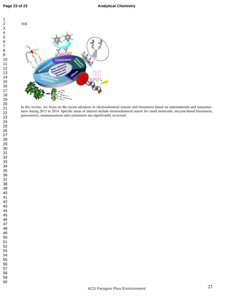
23
TOC
In this review, we focus on the recent advances in electrochemical sensors and biosensors based on nanomaterials and nanostruc-tures during 2013 to 2014. Specific areas of interest include electrochemical sensor for small molecular, enzyme-based biosensors, genosensors, immunosensors and cytosensors are significantly reviewed.
Page 23 of 23
ACS Paragon Plus Environment
Analytical Chemistry
123456789101112131415161718192021222324252627282930313233343536373839404142434445464748495051525354555657585960