elbow & forearm - mccc
TRANSCRIPT

H O W V I T A L I S T H E E L B O W T O O U R D A I L Y L I V E S ?
Elbow & Forearm

Clarification of Terms
The elbow includes: 3 bones (humerus, radius, and ulna)
2 joints (humeroulnar and humeroradial)
Allows for elbow flexion and extension
The forearm includes: 2 bones (radius and ulna)
2 joints (proximal radioulnar joint and distal radioulnar joint)
Allows for forearm pronation and supination
The interaction among the 4 joints enables the hand to be placed in a nearly infinite number of positions, greatly enhancing the functional potential of the entire UE
Mansfield, p91-92

Osteology of the Elbow & Forearm (Bones)
4 bones relate to the function of the elbow and forearm:
Scapula
Humerus
Ulna
Radius
Mansfield, p92

Osteology of the Elbow & Forearm (Bones)…cont
Scapula:
Infraglenoid tubercle
Supraglenoid tubercle
Coracoid process
Lippert, p149


Osteology of the Elbow & Forearm (Bones)…cont
Humerus:
Trochlea
Capitulum
Medial Epicondyle
Lateral Epicondyle
Lateral Supracondylar Ridge
Olecranon Fossa
Lippert, p149-150

Osteology of the Humerus


Osteology of the Elbow & Forearm (Bones)…cont
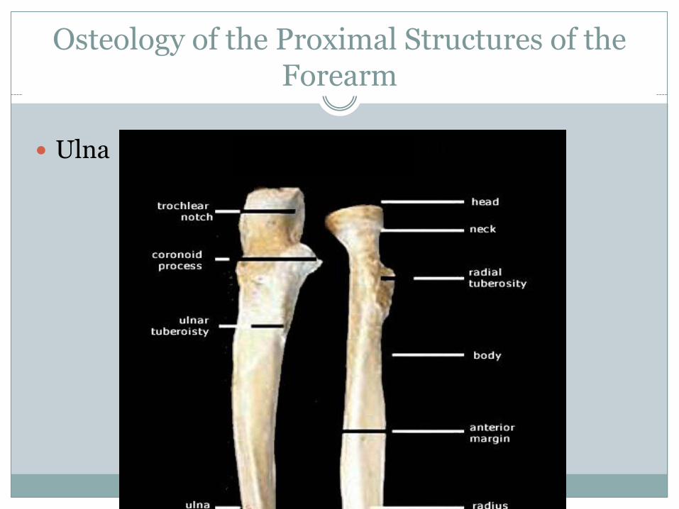
Ulna:
Olecranon Process
Trochlear Notch
Coronoid Process
Radial Notch
Ulnar Tuberosity
Styloid Process
Head
Lippert, p150


Osteology of the Elbow & Forearm (Bones)…cont
Radius:
Head
Radial tuberosity
Styloid process
Fovea
Lippert, p150

Osteology of the Proximal Structures of the Forearm
Ulna Radius

Osteology of the Distal Structures of the Forearm
Ulna Radius

Joint Structure of the Elbow
Humeroulnar Joint
Provides most of the structural stability to the elbow through the jaw-like trochlear notch of the ulna interlocking with the spool-shaped trochlea of the humerus
This hinge-like joint limits motion of the elbow to ___________ and __________________
Humeroradial Joint
Formed by the ball-shaped capitulum of the humerus with the bowl-shaped fovea of the radius
This permits continuous contact between the radial head and the capitulum during pronation and supination, as the radius spins about its own axis
Mansfield, p95

Joint Structure of the Elbow…cont
Carrying Angle: With the forearm supinated and elbow fully extended, the forearm
projects laterally about 15-20o relative to the humerus. This is normal, but tends to be greater in females.
Lippert, p148-149

Joint Movement of the Elbow
Osteokinematics:
Flexion
extension
Arthrokinematics:
Mansfield, p97

Supporting Structures of the Elbow
Anterior Capsule:
Thin connective tissues encloses the humeroulnar joint, humeroradial joint and proximal radioulnar joint
Medial Collateral Ligament:
Attaches proximally to the medial epicondyle and distally to the coronoid and olecranon processes, providing stability by resisting valgus forces
Lateral Collateral Ligament:
Originates on lateral epicondyle and attaches to the lateral aspect of the proximal forearm, providing stability by resisting varus forces
Mansfield, p96

Joint Structure of the Forearm
Proximal Radioulnar Joint:
The head of the radius articulates with the radial notch of the ulna
Distal Radioulnar Joint:
The distal end of the radius rotates around the distal end of the ulna
Functionally, they are considered one joint
The radioulnar joint is a uniaxial pivot joint allowing only pronation and supination of the forearm
Lippert, p148

Joint Movement of the Forearm
Osteokinematics:
Pronation
Supination

Joint Movement of the Forearm…cont
Arthrokinematics:
The distal radius rotates around the ulna which is stationary
The distal radius is larger and broader than the distal ulna
Lippert, p148

Joint Movement of the Forearm…cont
Supination & Pronation
Shoulder rotation can often be functionally substituted for each motion
But not if the humerus is held tight against the thorax and the elbow is in 90o of flexion

Supporting Structures of the Forearm
Annular Ligament:
Thick circular band of connective tissue that wraps around the radial head and attaches it to either side of the radial notch of the ulna.
This ring-like structure holds the radial head firmly against the ulna, allowing it to spin freely during pronation/supination.
Distal Radioulnar Joint Capsule:
Provides stability to the distal radioulnar joint
Interosseous Membrane:
Helps bind the radius to the ulna; serves as a site for muscle attachments, and acts as a mechanism to transmit forces proximally through the forearm
Mansfield, p99

Supporting Structures of the Forearm…cont

Supporting Structures of the Forearm…cont
Interosseous Membrane

Myology of the Elbow & Forearm (Muscles)
The Muscles of the Elbow & Forearm:
Biceps
Brachialis
Brachioradialis
Triceps
Anconeus
Supinator
Pronator teres
Pronator quadratus
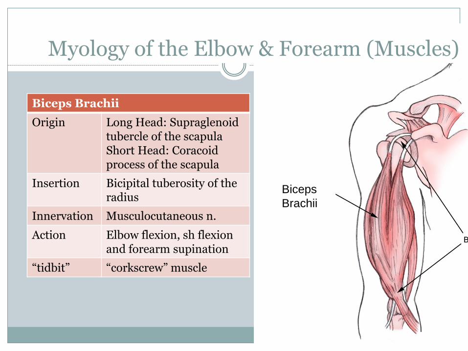
Biceps Brachii
Origin Long Head: Supraglenoid tubercle of the scapula Short Head: Coracoid process of the scapula
Insertion Bicipital tuberosity of the radius
Innervation Musculocutaneous n.
Action Elbow flexion, sh flexion and forearm supination
“tidbit” “corkscrew” muscle
Biceps Brachii
Muscle Bellies
Biceps
Brachii
Tendons
Myology of the Elbow & Forearm (Muscles)
Biceps
Brachii

Biceps brachii
How do we stretch the biceps brachii?
How do we strengthen the biceps brachii concentrically?
Eccentrically?
Isometrically?
Closed chain?
Open chain?

Brachialis
Origin Anterior aspect of the distal humerus
Insertion Coronoid process of the ulna
Innervation Musculocutaneous n.
Action Elbow flexion
“tidbit” “workhorse” for elbow flexion
Myology of the Elbow & Forearm (Muscles)

Brachioradialis
Origin Lateral supracondylar ridge of the humerus
Insertion Near the styloid process of the distal radius
Innervation Radial n.
Action Elbow flexion, Pronation or supination of the forearm to the neutral position
Myology of the Elbow & Forearm (Muscles)
Brachialis & Brachioradialis
How do we stretch the brachialis & brachioradialis?
How do we strengthen them concentrically?
Eccentrically?
Isometrically?

Myology of the Elbow & Forearm (Muscles)
Triceps Brachii
Origin Long Head: infraglenoid tubercle of the scapula Lateral Head: posterior aspect of the superior humerus, lateral to the radial groove Medial Head: posterior aspect of the superior humerus, medial to the radial groove
Insertion Olecranon process of the ulna
Innervation Radial n.
Action Elbow extension Sh extension: Long head only

Triceps brachii
How do we stretch the triceps brachii?
How do we strengthen the triceps brachii concentrically?
Eccentrically?
Isometrically?
Closed chain?
Open chain?

Myology of the Elbow & Forearm (Muscles)
Anconeus
Origin Posterior aspect of the laterals epicondyle of the humerus
Insertion Olecranon process of the ulna
Innervation Radial n.
Action Elbow extension?
“tidbit” Believed to “clear” the joint space of soft tissue to permit full elbow extension. Too small to create torque for elbow extension.

Myology of the Elbow & Forearm (Muscles)
Supinator
Origin Lateral epicondyle of the humerus and supinator crest of the ulna
Insertion Lateral surface of the proximal radius
Innervation
Radial n.
Action Forearm supination,

Supinator
How do we stretch the supinator?
Strengthen it concentrically? Eccentrically?
Myology of the Elbow & Forearm (Muscles)
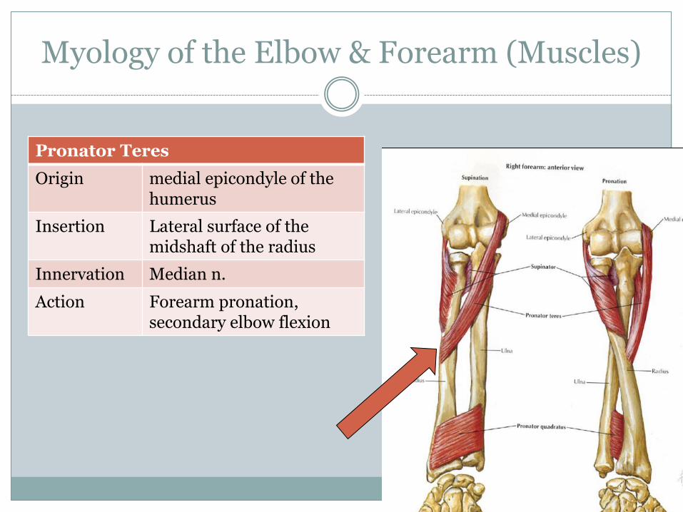
Pronator Teres
Origin medial epicondyle of the humerus
Insertion Lateral surface of the midshaft of the radius
Innervation Median n.
Action Forearm pronation, secondary elbow flexion

Myology of the Elbow & Forearm (Muscles)
Pronator Quadratus
Origin Anterior surface of the distal ulna
Insertion Anterios surface of the distal radius
Innervation
Median n.
Action Forearm pronation
Pronators
How do we stretch the pronator muscles?
How do we strengthen them concentrically? Eccentrically? Isometrically?

Myology of the Elbow & Forearm (Muscles)
Anatomical Relationships:
Muscle bellies of biceps, brachialis, and triceps are proximal to the elbow joint, while muscle bellies of brachioradialis, pronator teres, pronator quadratus, and supinator are distal to the elbow joint.
Anteriorly lies the biceps, brachialis, brachioradialis, pronator teres and pronator quadratus
The brachialis is deep to the biceps, except at the distal humerus where it can be palpated on either side of the biceps tendon
The brachioradialis and pronator teres are located superficially
The pronator quadratus is located deep to several wrist and hand tendons
Lippert, p155

Myology of the Elbow & Forearm (Muscles)
Anatomical Relationships continued:
Posteriorly, the triceps makes up the entire posterior arm proximal to the elbow joint
The long and lateral heads are superficial and the medial head is deep
The anconeus is very small and is located superficially on the posterior elbow, just distal to the triceps insertion
The supinator lies deep to the wrist extensors and the brachialis
Lippert, p156

Myology of the Elbow & Forearm (Muscles)
Prime Movers:
Action Muscle
Elbow Flexion Biceps, brachialis, brachioradialis
Elbow Extension Triceps
Forearm Pronation Pronator teres, pronator quadratus
Forearm Supination Biceps, supinator
Lippert, p157

Myology of the Elbow & Forearm (Muscles)
Summary of Muscle Innervation:
Muscle Nerve Spinal Segment
Brachialis Musculocutaneous C5, C6
Biceps Musculocutaneous C5, C6
Brachioradialis Radial C5, C6
Triceps Radial C6, C7
Anconeus Radial C7, C8
Pronator Teres Median C6, C7
Pronator Quadratus Median C8, T1
Supinator Radial C6
Lippert, p158

Redundancy is a fact of life/function
Innervation
The musculocutaneous n.
Supplies the elbow flexors EXCEPT the brachioradialis
The radial n.
Supplies the elbow extensors
The median n.
Supplies all the pronators of the forearm

Redundancy is a fact of life/function
The elbow flexors are innervated by 3 different nerves*
Preservation of “hand to mouth” activities
The likelihood of all 3 nerves
being injured is “slim”
*musculoskeletal n.
radial n.
median n.
Mansfield, p103

Common Pathologies
Lateral Epicondylitis (Tennis Elbow)
Overuse of common wrist extensor tendon where it inserts due to repetitive wrist extension activities
Medial Epicondylitis (Golfer’s Elbow)
Inflammation and overuse of the common flexor tendon at its insertion site due to repetitive wrist flexion activities
Little League Elbow
Overuse injury at medial epicondyle due to a repetitive throwing motion, creates valgus stress at elbow
Nursemaid’s Elbow
Radial head subluxation due to being picked up by one hand
Lippert, p156-157

Triceps brachii
Anconeus
Teres Major
Teres Minor
Infraspinatus
Identify!
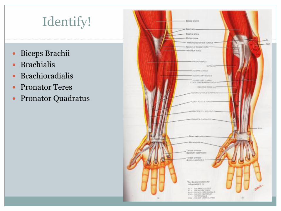
Identify!
Biceps Brachii
Brachialis
Brachioradialis
Pronator Teres
Pronator Quadratus

References
Lippert, L.S. (2011). Clinical Kinesiology and Anatomy, 5th ed. Philadelphia, PA: F.A. Davis.
Mansfield, P.J., & Neumann, D.A. (2009). Essentials of Kinesiology for the Physical Therapist Assistant. St. Louis, MO: Mosby Elsevier.