effects of negative pressure wound therapy on...
TRANSCRIPT

LUND UNIVERSITY
PO Box 117221 00 Lund+46 46-222 00 00
Effects of Negative Pressure Wound Therapy on Perivascular Groin Infections afterVascular Surgery. Wound Healing, Cost-Effectiveness and Patient-Reported Outcome.
Monsen, Christina
Published: 2016-01-01
Link to publication
Citation for published version (APA):Monsen, C. (2016). Effects of Negative Pressure Wound Therapy on Perivascular Groin Infections after VascularSurgery. Wound Healing, Cost-Effectiveness and Patient-Reported Outcome. Lund: Lund University, Faculty ofMedicine
General rightsCopyright and moral rights for the publications made accessible in the public portal are retained by the authorsand/or other copyright owners and it is a condition of accessing publications that users recognise and abide by thelegal requirements associated with these rights.
• Users may download and print one copy of any publication from the public portal for the purpose of privatestudy or research. • You may not further distribute the material or use it for any profit-making activity or commercial gain • You may freely distribute the URL identifying the publication in the public portal

Take down policyIf you believe that this document breaches copyright please contact us providing details, and we will removeaccess to the work immediately and investigate your claim.
Download date: 04. Jul. 2018
Lund University, Faculty of Medicine Doctoral Dissertation Series 2016:107
ISBN 978-91-7619-333-4ISSN 1652-8220
Printed by Media-Tryck, Lund U
niversity 2016 Nordic Ecolabel 3041 0903
978
9176
1933
34
Ch
ristin
a M
on
sen
Effects of N
egative Pressure Wound Th
erapy on Perivascular Groin Infections after V
ascular Surgery
107
Effects of Negative Pressure Wound Therapy on Perivascular Groin Infections after Vascular Surgery Wound Healing, Cost-Effectiveness and Patient-Reported Outcome
Christina Monsen
DepartMent of CliniCal sCienCes, MalMö | lunD university 2016Christina Monsen was born and raised in Malmö, Sweden’s third largest city, located in the south of the country. She has practised as a registered nurse for the past 30 years. She is currently working at the Vascular Centre, Malmö, Skåne University Hospital, and her area of expertise is wounds and wound treatment.


1
Effects of Negative Pressure Wound Therapy on Perivascular Groin Infections after Vascular Surgery
Wound Healing, Cost-Effectiveness
and Patient-Reported Outcome

2

3
Effects of Negative Pressure Wound Therapy on Perivascular Groin Infections
after Vascular Surgery
Wound Healing, Cost-Effectiveness and Patient-Reported Outcome
Christina Monsen
DOCTORAL DISSERTATION which, by due permission of the Faculty of Medicine at Lund University, Sweden,
will be defended in Jubileums Aulan, Jan Waldenströmsg 5, Skåne University Hospital, Malmö, on Friday 14th October 2016, at 9.30 a.m.
Faculty Opponent MD PhD Lena Blomgren, Institutionen för molekylär medicin och kirurgi,
Karolinska institutet, Stockholm.

4
Organization: LUND UNIVERSITY DOCTORAL DISSERTATION
Faculty of Medicine. Dept. of Clinical Sciences,Malmö. Division of Vascular Surgery
Date of issue: 2016-10-14
Author: Christina Monsen Sponsoring organization: --
Title: EFFECTS OF NEGATIVE PRESSURE WOUND THERAPY ON PERIVASCULAR GROIN INFECTION AFTER VASCULAR SURGERY. Wound Healing, Cost-Effectiveness and Patient-Reported Outcome
AbstractBackground: Surgical site infection (SSI) in the groin after vascular surgery is common and deep perivascular infection leads to long periods of hospitalization, sometimes to amputation and/or death. Negative pressure wound therapy (NPWT) is increasingly used for treating wounds such as deep perivascular groin infections after vascular surgery, but there is no scientific evidence supporting its benefit over traditional wound therapy.Aims: To study the effect of NPWT on wound healing, complications, resource use, quality of life, cost-effectiveness, and to explore the experiences of patients with deep perivascular groin infections after vascular surgery undergoing NPWT at home.Methods: A retrospective study was performed on consecutive patients undergoing NPWT between 2004 and 2006, and a randomized controlled trial was conducted between 2007 and 2011, where patients undergoing NPWT were compared to those treated with a traditional alginate dressing. Finally, a qualitative interview study was conducted between 2013 and 2014, in which patients undergoing NPWT in the outpatient setting were interviewed 7-14 days after discharge.Results: Twenty-eight patients/33 groins were studied in Study I, ten patients in each group in Studies II & III and 15 patients in Study IV. The median wound healing time was 55 days in Study I, 57 days in the NPWT group compared to 104 in the alginate group (p=0.026) in Studies II & III and 58 days in Study IV. The graft preservation rate in NPWT patients was 83%, 86% and 85% in Studies I, II/III and IV, respectively. Bacterial clearance from the wound was the same in the NPWT and alginate group in Studies II &III. One patient in the NPWT and one in the alginate group in Studies II &III had a severe bleeding from the femoral artery reconstruction site. Nine (43%) out of 21 groins with synthetic graft infections in Study I had an infection-related complication, compared to 0 (0%) out of 12 groins in those that did not have a synthetic graft infection (p=0.012), and non-healing wounds were associated with amputation (p=0.005) and death (p<0.001). A median of 21 (IQR 15-30) dressing changes were performed in the NPWT group, compared to 73 (IQR 51-98) (p<0.001) in the alginate group in Studies II & III. Compared to alginate therapy, NPWT saved the nurses 4.5 hours of work the first week after surgical revision in Study III. The total costs for the NPWT and alginate group in Study III were the same, of which 87% and 83%, respectively, were attributed to in-hospital costs. In Study III, estimation of Euroqol 5 Dimensions instrument and Brief Pain Inventory showed no differences at respective time points between the two groups. In Study IV an overall theme emerged from the descriptions of the experiences of patients with deep perivascular groin infection after vascular surgery undergoing NPWT at home, namely that it meant a transition from being a dependent patient to a person who needs to be involved and have self-care competence. A need to feel prepared for this before discharge from hospital was expressed. Lack of information and feelings of uncertainty prolonged the time before feeling confident in managing the treatment. The informants gradually accepted the need to be tied to a machine, became competent in its management, and found solutions to perform everyday tasks. Overall, it was a relief to be treated at home.Conclusion: NPWT in patients with deep perivascular SSI after vascular surgery is superior to traditional alginate therapy in terms of wound healing and cost-effectiveness. Patients expressed several benefits of treatment withNPWT at home. However, they experienced unnecessary stress and anxiety due to lack of information on the treatment and instruction concerning the equipment. Therefore, adequate information and education must be provided
Key words: Negative Pressure Wound Therapy. Vacuum Assisted Closure. Surgical Site Infection. Groin. Quality of Life. Pain. Resource Use. Cost-effectiveness. Content Analysis.
Classification system and/or index terms (if any)
Supplementary bibliographical information: Language: English
ISSN and key title 1652-8220 ISBN:978-91-7619-333-4
Recipient’s notes Number of pages: Price
Security classification
I, the undersigned, being the copyright owner of the abstract of the above-mentioned dissertation, hereby grant to all reference sources permission to publish and disseminate the abstract of the above-mentioned dissertation.
Signature Date

5
Effects of Negative Pressure Wound Therapy on perivascular groin infections
after vascular surgery
Wound Healing, Cost-Effectiveness and Patient-Reported Outcome
Christina Monsen
Malmö 2016 Lund University, Faculty of Medicine,
Department of Clinical Sciences, Malmö Division of Vascular Surgery

6
Cover photo by Sue Harden Mugelli
Copyright: Christina Monsen
Lund University, Faculty of Medicine Doctoral Dissertation Series 2016:107 ISBN 978-91-7619-333-4 ISSN 1652-8220 Printed in Sweden by Media-Tryck, Lund University Lund 2016

7
To my husband, Atle, and our children, Rasmus and Anna
Remember, every day is a gift

8
Contents
Abstract 10 List of publications 12 Abbreviations 13
Introduction 15
Background 17 Wounds 17 Surgical wounds 20 Wound treatment 30 Negative pressure wound therapy in open wounds 32 Cost 38 NPWT in the out-patient care setting 39 Patient-reported outcomes 40 Patients’ experiences of NPWT 42 The transition from hospital care to outpatient care 43
Thesis at a glance 45
Aims 47
Methods 49 Ethics 49 Setting 49 Definition of wound healing time 50 Study design 50 Participants 51 Data collection 52 Analysis 54

9
Results 57 Patient characteristics 57 Wound healing 57 Severity of wound infection and microbiology 57 Complications following wound therapy 58 Resource and cost 59 Health-related quality of life and pain 60 Patients’ experiences of NPWT at home 60
Discussion 63 The findings 63 Methodological considerations 70
Conclusions 77
Future research 79 Advanced NPWT 79 Measurement of wound cavities 79 Challenges associated with NPWT in the outpatient setting 79
Clinical implications 81
Populärvetenskaplig sammanfattning 83
Acknowledgements 85
References 87

10
Abstract
Background: Surgical site infection (SSI) in the groin after vascular surgery is common and deep perivascular infection leads to long periods of hospitalization, sometimes to amputation and/or death. Negative pressure wound therapy (NPWT) is increasingly used for treating wounds such as deep perivascular groin infections after vascular surgery, but there is no scientific evidence supporting its benefit over traditional wound therapy.
Aims: To study the effect of NPWT on wound healing, complications, resource use, quality of life, cost-effectiveness, and to explore the experiences of patients with deep perivascular groin infections after vascular surgery undergoing NPWT at home
Methods: A retrospective study was performed on consecutive patients undergoing NPWT between 2004 and 2006, and a randomized controlled trial was conducted between 2007 and 2011, where patients undergoing NPWT were compared to those treated with a traditional alginate dressing. Finally, a qualitative interview study was conducted between 2013 and 2014, in which patients undergoing NPWT in the outpatient setting were interviewed 7-14 days after discharge.
Results: Twenty-eight patients/33 groins were studied in Study I, ten patients in each group in Studies II & III and 15 patients in Study IV. The median wound healing time was 55 days in Study I, 57 days in the NPWT group compared to 104 in the alginate group (p=0.026) in Studies II & III and 58 days in Study IV. The graft preservation rate in NPWT patients was 83%, 86% and 85% in Studies I, II/III and IV, respectively. Bacterial clearance from the wound was the same in the NPWT and alginate group in Studies II &III. One patient in the NPWT and one in the alginate group in Studies II &III had a severe bleeding from the femoral artery reconstruction site. Nine (43%) out of 21 groins with synthetic graft infections in Study I had an infection-related complication, compared to 0 (0%) out of 12 groins in those that did not have a synthetic graft infection (p=0.012), and non-healing wounds were associated with amputation (p=0.005) and death (p<0.001). A median of 21 (IQR 15-30) dressing changes were performed in the NPWT group, compared to 73 (IQR 51-98) (p<0.001) in the alginate group in Studies II & III. Compared to alginate therapy, NPWT saved the nurses 4.5 hours of work the first week after surgical revision in Study III. The total costs for the NPWT and alginate group in Study III were the same, of which 87% and 83%, respectively, were attributed to in-hospital costs. In Study III, estimation of Euroqol 5 Dimensions instrument and Brief Pain Inventory showed no differences at respective time points between the two groups. In Study IV an overall theme emerged from the descriptions of the experiences of patients with deep perivascular groin infection after vascular surgery undergoing NPWT at home,

11
namely that it meant a transition from being a dependent patient to a person who needs to be involved and have self-care competence. A need to feel prepared for this before discharge from hospital was expressed. Lack of information and feelings of uncertainty prolonged the time before feeling confident in managing the treatment. The informants gradually accepted the need to be tied to a machine, became competent in its management, and found solutions to perform everyday tasks. Overall, it was a relief to be treated at home.
Conclusion: NPWT in patients with deep perivascular SSI after vascular surgery is superior to traditional alginate therapy in terms of wound healing and cost-effectiveness. Patients expressed several benefits of treatment with NPWT at home. However, they experienced unnecessary stress and anxiety due to lack of information on the treatment and instruction concerning the equipment. Therefore, adequate information and education must be provided.
12
List of publications
This thesis is based on the following publications, which will be referred to in the text by their roman numerals.
I. Svensson S, Monsen C, Kölbel T, Acosta S
Predictors for Outcome after Vacuum Assisted Closure Therapy of Peri-vascular Surgical Site Infection in the Groin Eur J Endovasc Surg. 2008; 36: 84-89
II. Monsen C, Wictorsson C, Wann Hansson C, Acosta S Vacuum-assisted wound closure versus alginate for the treatment of deep perivascular wound infections in the groin after vascular surgery J Vasc Surg. 2014; 59: 145-151
III. Monsen C, Acosta S, Mani K, Wann Hansson C A randomized study of NPWT closure versus alginate dressings in peri-vascular groin infections: quality of life, pain and cost J Wound Care. 2015; 24, 252,254-256,258-260
IV. Monsen C, Acosta S, Kumlien C Patients experiences of Negative Pressure Wound Therapy at home for the treatment of deep perivascular groin infection after vascular surgery Submitted
Papers are reproduced with permission from the respectively publisher, Papers I and II, Elsevier, and Paper III, J Wound Care.

13
Abbreviations
APSP Acute post-surgical pain
ASA American Society of Anesthesiologists
BPI Brief Pain Inventory
CDC Center for Disease Control and Prevention (USA)
CLI Critical limb ischemia
CFUs Colony-forming units
CRP C-reactive protein
EQ-5D 3L EuroQol 5 - Dimensions instrument, 3 levels
FDA Food and Drug Administration (USA)
HRQoL Health-related quality of life
IQR Interquartile range
NPWT Negative pressure wound therapy
PROM Patient-reported outcome measures
PU Polyurethane
PVA Polyvinyl alcohol
QoL Quality of life
RCT Randomized controlled trial
SBU Statens beredning för medicinsk och social utvärdering
SF-36 The Short Form (36) Health Survey
SSI Surgical site infection
VAS Visual analogue scale
WBP Wound bed preparation

14

15
Introduction
The most common complication among hospital patients is healthcare-associated infections (Burke, 2003). The most frequent healthcare-associated infections in Europe are respiratory tract infections (23.5%), followed by surgical site infections (SSIs) (19.6%) and urinary tract infections (19.0%) (Suetens et al., 2013). Deep postoperative wound infections, especially in the groin after vascular surgery, are a major problem in the care of patients who have undergone vascular surgery. The infection of a vascular graft in the groin may lead to catastrophic consequences for the patient; a long hospital stay, increased risk of morbidity (Exton & Galland, 2007), amputation or mortality (Szilagyi et al., 1972; Mayer et al., 2011; Turtiainen & Hakala, 2014), as well as increased costs to society (Mayer et al., 2011). Negative pressure wound therapy (NPWT) is a treatment modality for wound complications that is being used increasingly for deep SSIs, both for subcutaneous infection, Szilagyi grade II (Mayer et al., 2011), and perivascular Szilagyi grade III infections (Dosluoglu et al., 2010; Mayer et al., 2011; Berger et al., 2012; Verma et al., 2015). NPWT has become a popular and attractive treatment alternative for infections in groin incisions after vascular surgery (Szilagyi et al., 1972), but there is no scientific evidence supporting its benefit over traditional wound therapy. No randomized controlled trials have been carried out to compare NPWT with traditional wound treatment. Furthermore, very few studies have been conducted including patient-reported outcomes such as quality of life and patients’ experiences of NPWT, and no studies could be found on NPWT in the outpatient care setting in patients with deep perivascular infection in the groin after vascular surgery. There is thus a need for knowledge and scientific evidence on the efficacy and safety of NPWT in treating vascular SSIs in the groin, in particular, to evaluate wound healing time, wound complications, resource use, costs, cost-effectiveness, and patient suffering and experiences.

16

17
Background
Wounds
Wounds are categorized as acute, chronic or complicated (Velnar et al., 2009). Acute wounds following surgical incision or trauma heal within a limited time frame and in a specific manner. A chronic wound develops when the wound does not heal, or does not heal as expected (Stadelmann, 1998; Enoch & Leaper, 2008; Velnar et al., 2009). Complicated wounds are characterized by infection or loss of tissue caused, for instance, by the resection of tissue after cancer surgery (Velnar et al., 2009). The wound healing process is the same, regardless of the type of wound, and involves four phases: inflammation, reconstruction, epithelialization and maturation (Johnstone et al., 2005).
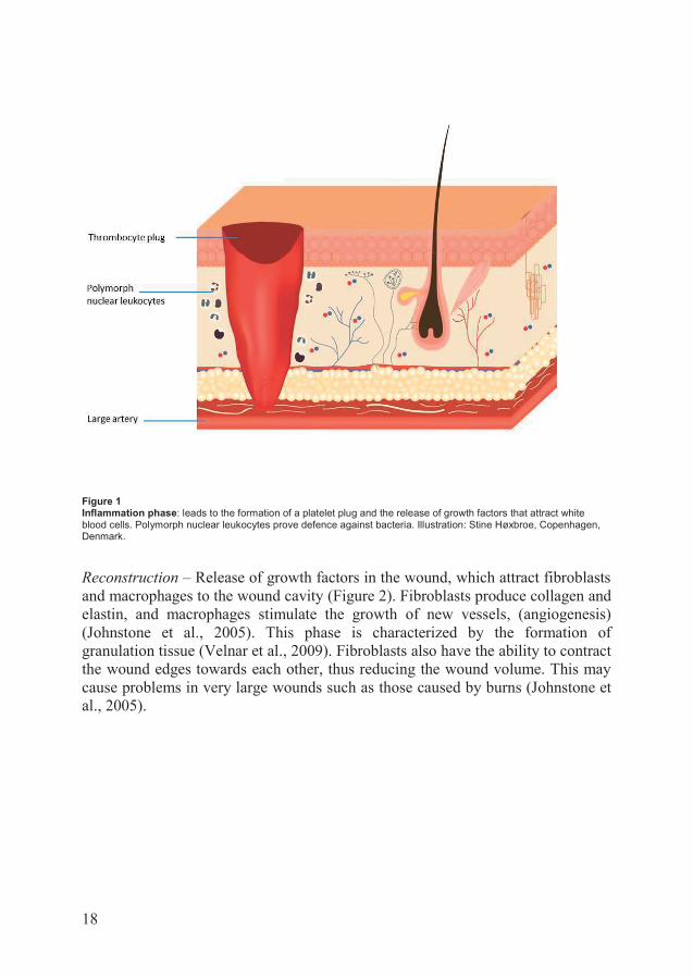
Inflammation – In the case of bleeding, vasoconstriction and activation of the haemostatic system occur, which result in the aggregation of platelets and the formation of a platelet plug (Johnstone et al., 2005) (Figure 1). Fibrin strings are also formed to strengthen the platelet plug. Growth factors are directed to the wound, attracting white blood cells. Polymorph nuclear leukocytes (also called granulocytes) provide defence against bacteria and remove dead tissue (Stadelmann, 1998). In the case of infected, necrotic or chronic wounds, wound healing may be prolonged by an extended period of inflammation (Hanson et al., 2005; Johnstone et al., 2005). Prolonged inflammation leads to increased levels of matrix metalloproteinases, decreased levels of growth factors (Schultz et al., 2003; Milne, 2015) and decreased release of cytokines, all of which impair wound healing (Johnstone et al., 2005).
18
Figure 1 Inflammation phase: leads to the formation of a platelet plug and the release of growth factors that attract white blood cells. Polymorph nuclear leukocytes prove defence against bacteria. Illustration: Stine Høxbroe, Copenhagen, Denmark.
Reconstruction – Release of growth factors in the wound, which attract fibroblasts and macrophages to the wound cavity (Figure 2). Fibroblasts produce collagen and elastin, and macrophages stimulate the growth of new vessels, (angiogenesis) (Johnstone et al., 2005). This phase is characterized by the formation of granulation tissue (Velnar et al., 2009). Fibroblasts also have the ability to contract the wound edges towards each other, thus reducing the wound volume. This may cause problems in very large wounds such as those caused by burns (Johnstone et al., 2005).

19
Figure 2 Reconstruction phase: the release of growth factors attracts fibroblasts and macrophages. Illustration: Stine Høxbroe, Copenhagen, Denmark.
Epithelialization – Macrophages release epidermal growth factors which stimulate the formation of new epithelial cells from the wound edges or from sweat glands and hair follicles (Johnstone et al., 2005). Epithelialization ceases when the wound edges join (Velnar et al., 2009). This process is delayed by necrotic tissue or slough in the wound (Johnstone et al., 2005).
Maturation – The wound is strengthened by the remodelling of collagen, which becomes more organised and solid (Johnstone et al., 2005). The healed wound has approximately 80% of the solidity of the original intact tissue (Velnar et al., 2009). The wound healing and remodelling process may be impaired in patients suffering from malnutrition. The maturation phase may last for several months for closed wounds and for several years in open wounds (Johnstone et al., 2005).

20
Surgical wounds
Incisions are made in the skin during surgery to expose underlying structures, tissues and organs prior to surgical procedures (Toon et al., 2015a). After surgery, the wound is usually closed by drawing the wound edges together with sutures, staples, clips or tissue glue (Toon et al., 2015b). This is termed primary wound closure (Toon et al., 2015a). An infected wound may be left open to allow wound cleansing, and secondary wound closure usually takes place 10–14 days after primary surgery.
Definition of surgical site infection
SSIs are categorized as superficial, deep or organ/space according to the U.S. Center for Disease Control and Prevention (CDC) (CDC SSI, 2016).
Superficial incisional SSI o Occurs within 30 days after any operative procedure o Involves only the skin and subcutaneous tissue
And – at least of one of the following: o Purulent drainage from the incision o Identified organism from an aseptically obtained culture of fluid
or tissue from the superficial tissue o Superficial incision opened by a surgeon, the attending physician
or other designee and culture- or non-culture-based testing is not performed And – at least one of the following symptoms of infection:
Pain or tenderness Localized swelling Erythema Heat
o A superficial incision infection diagnosed by a surgeon, the attending physician or other designee
Deep incisional SSI o Occurs within 30 days or 90 days after an operative procedure
depending on the type of procedure: in vascular surgery within 30 days: abdominal aortic
aneurysm repair in vascular surgery within 90 days: peripheral vascular
bypass surgery o Involves deep tissue (muscle and fascial layer)
21
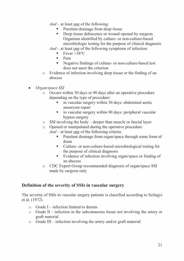
And – at least one of the following: Purulent drainage from deep tissue Deep tissue dehiscence or wound opened by surgeon.
Organism identified by culture- or non-culture-based microbiologic testing for the purpose of clinical diagnosis
And – at least one of the following symptoms of infection: Fever >38°C Pain Negative findings of culture- or non-culture-based test
does not meet the criterion o Evidence of infection involving deep tissue or the finding of an
abscess
Organ/space SSI o Occurs within 30 days or 90 days after an operative procedure
depending on the type of procedure: in vascular surgery within 30 days: abdominal aortic
aneurysm repair in vascular surgery within 90 days: peripheral vascular
bypass surgery o SSI involving the body – deeper than muscle or fascial layer o Opened or manipulated during the operative procedure
And – at least one of the following criteria: Purulent drainage from organ/space through some form of
drain Culture- or non-culture-based microbiological testing for
the purpose of clinical diagnosis Evidence of infection involving organ/space or finding of
an abscess o CDC Expert Group recommended diagnosis of organ/space SSI
made by surgeon only
Definition of the severity of SSIs in vascular surgery
The severity of SSIs in vascular surgery patients is classified according to Szilagyi et al. (1972).
o Grade I – infection limited to dermis o Grade II – infection in the subcutaneous tissue not involving the artery or
graft material o Grade III – infection involving the artery and/or graft material

22
Risk factors for SSI
Risk factors for SSI are divided into patient-, procedure- and environmentally related factors (Table 1). Low serum albumin, high age, critical limb ischaemia (CLI), diabetes mellitus, smoking and overweight are general patient-related risk factors for vascular SSI (Cheadle, 2006; Neumayer et al., 2007; Owens & Stoessel, 2008). Procedure-related risk factors include factors such as inadequate preoperative showers, poor surgical technique and prolonged operation time (Cheadle, 2006; Neumayer et al., 2007; Owens & Stoessel, 2008). An operation time that exceeds four hours is associated with an increased risk of developing SSI (Cheadle, 2006). Patients classified as having an increased risk of perioperative complications according to the American Society of Anesthesiologists (ASA) classification, ASA class ≥ 2, or those undergoing acute surgery have an increased risk of developing SSI (Neumayer et al., 2007). SSIs are often caused by the patients’ own bacterial flora, but may also be caused by exogenous sources such as the surgical team, operating theatre, or the flow of personnel to and from the operating theatre (Erichsen Andersson et al., 2012b).
Patients undergoing vascular surgery may often have multiple patient- and procedure-related risk factors for SSI. Some of the most prevalent patient-related risk factors in vascular surgery patients are diabetes mellitus, smoking, coronary arterial disease, CLI, remote infection of a foot ulcer, hypertension, obesity and chronic obstructive pulmonary disease (Wiseman et al., 2015). The most common procedure-related factors are lengthy operations ≥ four hours, infrainguinal groin incisions, acute or reoperation and hypothermia due to shock (Inui & Bandyk, 2015).
The incidence of vascular SSI is highly dependent on the type of surgical procedure performed. The incidence of SSI after carotid artery surgery has been reported to be 0.2% (Greenstein et al., 2007). The rate of SSI after open repair of abdominal aortic aneurysm has been found to be 2.4%, and is higher in overweight patients, 6.3% (Giles et al., 2010). Most importantly, the incidence of SSI in patients with CLI in the lower extremities after vascular surgery has been reported to be 27% (Turtiainen et al., 2010).

23
Table 1 Risk factors for vascular SSI
Patient-related
Procedure-related
Environmental factors
Prolonged preoperative hospitalization Obesity Female gender Malnutrition Smoking Diabetes mellitus Critical limb ischaemia with foot ulcer End-stage renal disease Vitamin K antagonist therapy Prior irradiation at the incision site High dose of corticosteroids Chemotherapy for malignant disease Nasal carriage of Staphylococcus aureus
Groin incision Short time between multiple groin incisions Vein harvest incision Biomaterial implant Reoperation Prolonged operative time Hypothermia Hyperglycaemia
Poor operating theatre ventilation Poor environmental surface cleaning Poor instrument and vascular implant sterility Poor sterile operative technique
SSI in the groin after vascular surgery
The infrainguinal groin area is a contaminated area prone to vascular SSI. (Turtiainen et al., 2012; Turtiainen & Hakala, 2014; Inui & Bandyk, 2015). Reported rates of SSI after vascular procedures involving groin incisions are summarized in Table 2. If the fascia has been cut and opened to expose the common femoral artery there is an increased risk that a superficial SSI may develop into a deep perivascular SSI (Szilagyi grade III) (van der Slegt et al., 2014). The risk of deep SSI has also been reported to increase after surgical revision. A high Rutherford class of the severity of CLI was found to be a significant risk factor for both superficial and deep SSI (van der Slegt et al., 2014). The groin area harbours a high burden of bacteria in a warm moist environment, and often has multiple skin folds. All these factors are favourable for bacterial growth. There are also a number of lymphatic vessels in the groin, which may be cut inadvertently, leading to lymphorrhea or lymphocele (Ploeg et al., 2009; Pejkic et al., 2014). Lymphocele may also favour bacterial growth. Lymphorrhea may sometimes be copious and uncontrolled, resulting in dampness of the patient and bedclothes, requiring the bedclothes to be changed several times per day, which further increases the risk of wound infection. The groin area is also a highly mobile area, and too much movement may hamper the wound healing process after surgery. In particular, traditional wound dressings tend to attach poorly in this area.

24
Table 2 Reported rates of SSI in vascular procedures
Vascular surgical procedure Reference Rate of SSI
Vascular surgery (lower limb revascularization and open aortic surgery)
Turtiainen (2010) 26.6% of patients
Lower limb revascularization Turtiainen (2012) 21.9% of patients
Groin infections after vascular surgery
Hasselmann (2015b) Ratio of endovascular aneurysm repair to open vascular surgery = 64%:36%
19.0% of patients
Matatov (2013) Ratio of endovascular aneurysm repair to open vascular surgery = 24%:76%
30.2% of groins
Endovascular aortic aneurysm repair
Hasselmann (2015a) 4.4% of groins
Signs of wound infection may range from redness, swelling and localized pain in superficial SSIs, to purulent wound secretion and false arterial aneurysm (pseudo-aneurysm) in deep perivascular infections (Engin et al., 2005) (Figure 3).
Figure 3 Female patient with an infected synthetic femoro-popliteal by-pass prosthesis. Note the pus in the right groin (long arrow) and the cutaneous fistula further down (short arrow). The patient underwent complete removal of the by-pass with severe bleeding as a consequence, and died postoperatively due to acute myocardial infarction. Photo: © S. Acosta

25
It is important to be aware of the possible presence of a biofilm in an infected wound, particularly in synthetic graft infections. A biofilm is composed of a matrix of bacteria and is formed to protect the bacteria against the conditions in the environment, and may thus promote bacterial growth. Biofilms are resistant to antibiotics and most other forms of antibacterial treatment. Thorough surgical revision, ensuring the mechanical removal of any biofilm, is therefore very important in deep SSIs (Wilkins et al., 2014). The often extensive revisions in deep perivascular infections lead to large wounds requiring secondary healing (Figure 4). It is important to take tissue biopsies for bacterial cultures when performing revision since swabs of the surface area have been shown to be insufficient to diagnose the bacteria responsible for the infection (Høiby et al., 2015).
Figure 4 Extensive surgical revision under local anaesthesia. Note the considerable amount of debris in the small bowl. Photo: © S. Acosta
Deep perivascular groin infections after vascular surgery are associated with higher rates of morbidity (Exton & Galland, 2007), amputation and death (Szilagyi et al., 1972; Mayer et al., 2011; Turtiainen & Hakala, 2014). The risk of infection is increased when synthetic material is used compared to autogenous vein material

26
(Turtiainen & Hakala, 2014). It may be necessary to remove the infected graft to enable wound healing. This is a dangerous surgical procedure that exposes the patient to the risk of severe bleeding complications and death. If no reconstruction is possible after graft removal, major amputation is often required and survival is poor (Turtiainen & Hakala, 2014).
Prevention of SSI
Preoperative skin antisepsis Preoperative bathing or showering with chlorhexidine solution is common practice before surgical procedures (Webster & Osborne, 2015). The area is also washed with antiseptic solution in the operating theatre immediately prior to surgical incisions (Dumville et al., 2015a). The aim of both these procedures is to remove as many of the microorganisms present on the skin as possible, and thereby prevent SSI. It is currently recommended that patients take three to five pre-operative showers with antiseptic solutions to reduce the risk of SSI (Jakobsson et al., 2011). However, in a recent review, Webster & Osborne (2015) found no clear evidence of any benefits of preoperative baths or showers with chlorhexidine, compared to other washing products, in reducing SSI. Some evidence has been found that preoperative skin antisepsis in the operating theatre is beneficial. A preparation containing 0.5% chlorhexidine in methylated spirits was found to be associated with a lower rate of SSI than alcohol-based povidone iodine in clean surgery (Dumville et al., 2015a). This suggests that in everyday clinical work practitioners should consider the cost and potential side effects when choosing routines for skin antisepsis (Dumville et al., 2015a).
Preoperative hand antisepsis Healthcare professionals involved in surgical procedures routinely carry out surgical hand antisepsis to reduce the risk of SSI. However, there is no clear evidence showing which is the most effective method of hand antisepsis. Studies have shown that soap including chlorhexidine gluconate may reduce the number of colony-forming units (CFUs) on the hands compared with povidone iodine (Tanner et al., 2016). The time taken to wash the hands may also be an important factor. Surgical hand antisepsis with chlorhexidine gluconate for 3 minutes has been found to reduce the number of CFUs more than a 2-minute hand scrub. However, the quality of the studies was considered low (Tanner et al., 2016). Hygiene guidelines in Swedish healthcare are prescribed by the Swedish National Board of Health and Welfare regarding basic hygiene in the healthcare and welfare services (SOSFS 2015:10), while regulations for healthcare professionals in the operating theatre are laid out in the Swedish Handbook for Healthcare (Handbook for Healthcare, 2016).

27
Environment in the operating theatre The air quality in the operating theatre is influenced by personnel entering and leaving, the number of people in the operating theatre, and the length of the opera-tion. An explorative study on air quality using air sampling and determination of the number of CFUs per m3 during implant surgery following orthopaedic trauma, showed that a high flow of personnel has a strong negative impact on the operating room environment, and interventions aimed at preventing SSI by reducing traffic flow were recommended (Erichsen Andersson et al., 2012b).
Antibiotic prophylaxis Antibiotic prophylaxis has played a central role in the prevention of SSIs after vascular surgery since the 1970s (Kaiser et al., 1978). In a recent systematic review on the prevention of SSI it was concluded that antibiotic prophylaxis may still be the most important factor in reducing SSI after peripheral vascular surgery (Turtiainen et al., 2014). Timely administration of antibiotics, in relation to the start of surgery, is important so as to afford the maximum preventive effect. In a recent study on patients undergoing orthopaedic surgery, it was found that the timing of the administration of intravenous antibiotic prophylaxis was often neglected and that only 49% were given antibiotic prophylaxis within the recommended time interval of 30 minutes before the start of surgery (Erichsen Andersson et al., 2012a).
Impact of surgical techniques on SSI Atraumatic surgical technique is important in reducing SSI. Great care must be taken when performing incisions and anatomic dissections to avoid lymphatic wound complications (Sandmann, 2016). Care should also be taken to stop bleeding before closure of the wound. Haematomas should be avoided, since bacterial growth. An expanding haematoma may also cause pressure necrosis of the overlying skin, particularly the closed wound edges, which may lead to secondary wound infection (Bandyk, 2008). Great care should be taken when separating the wound edges during surgery, and forceful pulling of the wound edges by retractors may result not only in skin ischaemia and wound edge necrosis, but also in damage to the lymphatic ducts (Sandmann, 2016). Extensive use of diathermy may lead to tissue necrosis (Bandyk, 2008). Groin incisions may be performed vertically or transversely depending on whether or not extensive exposure of the femoral arteries is needed. Vertical incisions are associated with higher wound complication rates than transverse, probably due to the larger dissection area, the longer exposure time of the vessels, longer operation time and poorer adaptation of the wound edges after skin closure (Swinnen et al., 2010).
28
Pain All surgical procedures result in acute post-surgical pain (APSP), triggered by nociceptive, inflammatory or neuropathic stimulation of the peripheral nerve fibres. This process usually resolves within a reasonable postoperative period (Glass et al., 2015). Both under-treatment and over-treatment of APSD can lead to several postoperative problems and serious consequences for the patient (Agroff, 2014). A patient with severe APSD has a higher risk of suffering from complica-tions such as a decrease in alveolar ventilation, hypertension, myocardial ischaemia and poor wound healing (Vadivelu, 2010). A correlation has also been found between APSP and the risk of developing persistent pain (Kehlet et al., 2006; Meeks et al., 2015). Therefore, the treatment of APSP could be improved by identifying patients with a high risk of developing APSP, and ensuring they receive adequate treatment.
Nutrition It is important to consider the patient’s nutritional status before a undertaking sur-gical procedure since it has been demonstrated that malnutrition is associated with increased infection rates and delayed wound healing (Stechmiller, 2010). For example, patients with hypoalbuminaemia undergoing total joint arthroplasty have been reported to have higher risks of SSI, pneumonia, long hospital stay and readmission than patients with normal albumin levels (Bohl et al., 2016). Risk assessment to identify patients at risk of malnutrition and preoperative nutritional optimization are simple strategies to prevent adverse outcomes due to malnutrition (de Luis et al., 2014; Golladay et al., 2016). If it is not possible to meet nutritional needs with oral nutrition, which is the most safe, cheap, and best method, then forced feeding with enteral or parenteral nutrition should be considered (de Luis et al., 2014).
Care of surgical incisions Almost all surgical procedures result in wounds that heal by primary healing. Gauze or cotton absorbent dressings and adhesive tape or adhesive dressings are usually used to cover the surgical incision. Several different kinds of dressings are available for surgical wounds. However, there is still no evidence to suggest that a particular wound dressing is more effective than another in preventing SSI or reducing the rate of SSI (Dumville et al., 2014). The Cochrane review by Dumville et al. (2014) recommend, therefore, that decisions on wound dressing should be based on a combination of cost and the ability of the dressing to meet the needs in each case, e.g. the absorption of exudate.
Neither is there any clear guidance regarding when patients can take showers or baths postoperatively, or when the dressing should be removed. If bathing or showering is delayed postoperatively, sweat and dirt will accumulate on the body. The collection of large amounts of exudate will increase the risk of skin

29
maceration, which in turn can have negative effects on wound healing. No conclusive evidence has been found regarding the advantages or disadvantages of early (within 12 hours) versus delayed (>48 hours) postoperative showering or bathing in preventing SSI (Toon et al., 2015b), or with regard to early removal of the dressing (Toon et al., 2015a), indicating that randomized controlled trials of high scientific quality are needed in these areas (Toon et al. 2015a; Toon et al. 2015b).
Incisional negative pressure wound therapy The results of two recent systematic reviews, involving meta-analysis of NPWT on closed surgical incisions, suggest that incisional NPWT may be able to reduce the rate of SSI (Semsarzadeh et al., 2015; Hyldig et al., 2016). The reason for this reduction appears to be the combined effects of the airtight dressing, minimizing the risk of contamination of the wound from bacteria in the perineal and groin area, the reduction of dead space, leaving the closed incision dry, counteracting the lateral retraction forces drawing the wound edges together (Wilkes et al., 2012), and the clearance of haematoma and/or seroma and oedema through the lymphatic drainage system (Kilpadi et al., 2011). One retrospective study has been carried out on SSI in the groin after vascular surgery including a comparative historical control group, in which it was found that NPWT was superior to standard wound treatment (Matatov et al., 2013). However, no randomized controlled trial (RCT) has yet been carried out on vascular surgery patients. Evidence-based data on incisional NPWT will be available in the near future. At the time of printing (September 2016) 51 RCTs were registered at ClinicalTrial.gov (http://clinicaltrial.gov) with the status “recruiting” or “completed”, of which eight studies include patients undergoing lower limb vascular surgery. to support recommendation in wound care and its role in reduction in wound complications, SSI, seroma/hematoma formation and wound dehiscence and improvement insurgical scar quality. There is an ongoing A clinical RCT is ongoing at our department to investigate whether NPWT of closed surgical incisions in the groin after vascular surgery can reduce the occurrence of SSI (Hasselmann et al., 2015a). Hopefully, the results of these studies will elucidate the role of NPWT in reducing wound complications, SSI and seroma/haematoma formation, allowing recommendations for wound care after vascular surgery to be formulated.
Treatment of SSI in the groin Patients with graft infection or infection of the native femoral artery can be treated in several ways, from conservative treatment (Mayeret al., 2011) to aggressive vascular reconstructive surgery with graft excision and extra-anatomic bypass (van der Slegt et al., 2014).
Patients with wound infections and graft exposure may be treated in the following ways.

30
1. Local debridement(s), antibiotics and local wound treatment 2. Attempts to preserve the vascular reconstruction by sartorius or rectus
femoris muscle flap coverage (Ger, 1976; Fischer et al., 2013) 3. Removal of synthetic material and replacement with autogenous vein
material 4. Removal of the infected reconstruction and replacement with an
antibiotic-impregnated graft (Inui & Bandyk, 2015) 5. Removal of the infected reconstruction and bypass using an extra-
anatomic conduit (Engin et al., 2005; Inui & Bandyk, 2015) 6. Removal of the infected reconstruction with or without subsequent leg
amputation (Inui & Bandyk, 2015)
A survey of current practice in the treatment of deep groin infections after vascular surgery at Scandinavian university hospitals was performed. The first strategy of wound treatment included NPWT (Table 3).
Table 3 First strategy in the treatment of deep groin infections after vascular surgery in Scandinavian university hospitals
Hospital Graft preservation Wound dressing Muscle flap coverage
Umeå Synthetic material is replaced by vein
NPWT (not always) Sometimes
Uppsala Yes NPWT Liberal use when grafts are exposed
Stockholm, Karolinska Institute
Synthetic material is sometimes replaced by vein
NPWT Seldom
Örebro Yes NPWT Seldom
Gothenburg Yes NPWT Standard with synthetic graft
Linköping Synthetic material is replaced by vein
NPWT Seldom
Malmö Yes NPWT Seldom
Helsinki (Finland) Yes NPWT Seldom
Trondheim (Norway) Yes NPWT Seldom
Copenhagen (Denmark) Yes NPWT Seldom
Wound treatment
Wound treatment today is based on moist wound healing and wound bed preparation. Moist wound healing reduces fibrosis, soreness and pain. Wound bed preparation (WBP) is an approach intended optimize wound treatment based on the specific needs in each case, e.g. debridement and management of exudate and

31
bacteria or bacterial imbalance (Schultz et al., 2003). The TIME structure (see below) has been developed to provide a framework for WBP (Leaper et al., 2014).
T = Tissue – assessment of the tissue and the need for debridement for the removal of necrotic tissue, exudate, biofilm and other slough from the surface of the wound.
I = Infection/inflammation – assessment of the aetiology of the wound, signs of infection and the need for topical antiseptic or/and systematic antibiotic treatment.
M = Moisture imbalance – assessment of the aetiology and management of wound exudate and adjustment of wound treatment for an optimal moist wound environment.
E = Edge of wound – assessment of the status of the skin surrounding the wound and advancing or undermined wound edges.
The TIME framework can also be used to guide the documentation of wound appearance and the effects of wound treatment (Leaper et al., 2014). The measurement of the area of the wound and documentation of changes are important in the evaluation of wound treatment (Flanagan, 2003), and complement TIME. This is usually done by measuring the length, width and depth of the wound using a tape measure. However, these measures provide incomplete information on the wound area, depending on the wound edges are often undermined and irregular, and the wound may have cavities that cannot be easily measured. Different ways of measuring the area of the wound (Gethin & Cowman, 2006; Mayrovitz & Soontupe, 2009) and wound volume (Körber et al., 2006; Kecelj-Leskovec et al., 2007) have been evaluated in previous studies. The success of these method varies, and the techniques are often complicated and expensive. VisitrakTM (Smith & Nephew, Hull, UK) allows standardized assessment of the wound area. The Visitrak device makes use of a transparent film (Visitrak Grid) on which the outline of the wound is drawn. The film is then placed on the Visitrak device, which is a portable tablet, for calculation of the length and width of the wound and its area. A reduction in the area of the wound of 20-40% during the first two to four weeks after the first appearance of the wound is associated with wound healing within 12 weeks (Flanagan, 2003). Therefore, the aim of any form of wound treatment is to reduce the size and depth of the wound as quickly as possible.
Alginate dressings
Alginate dressings are absorbent wound care products that contain sodium and calcium fibres that absorb wound exudate and blood (Lindholm & Grauers, 2011). As the dressing absorbs exudate or blood it become larger and more gel-like, and

32
an exchange of ions takes place between the calcium in the dressing and the sodium in the wound exudate. Alginate dressings are best suited to wounds with a large amount of exudate (Lindholm, 2012).
Negative pressure wound therapy in open wounds
NPWT is often instigated in open wounds after surgical revision of an infected wound. The wound cavity is then filled with a wound filler such as polyurethane (PU) foam or gauze. The wound is then covered with an adhesive PU film. A hole is cut in the plastic film and a suction adapter is placed above the hole, while the other end of the tube is connected to a vacuum device. The desired level of negative pressure is set, and NPWT is then induced. The exudate from the wound is collected and measured in a canister, which is an integrated part of the vacuum device (Figure 5).
Figure 5. NPWT equipment and physiological effects of NPWT. Illustration by Stine Høxbroe, Copenhagen, Denmark

33
History of negative pressure in wound healing
Negative pressure was used in some form already before the Roman Empire was established, more than 2000 years ago. The fluids in infected wounds were sucked out by so-called “mouth suckers”, who were considered indispensable in the Roman army. However, mouth sucking of wounds was not always successful. According to some historians, the last active pharaoh of Ptolemaic Egypt, Cleopatra, either killed herself, or was murdered, by the bite of an Egyptian cobra. Legend has it that mouth suckers were called to try to save her life, but to no avail. Cupping therapy is another ancient form of healing, dating from 3000 BC, in which local suction is created by applying heated cups over wounds to extract wound fluid (Harris, 1838). Dr G Bier developed the method at the end of the 18th century using glass cups of varying sizes and shapes, to treat wounds in different parts of the body (Miller, 2013). In 1952, Raffl (1952) described a temporary suction method to evacuate wound fluid by placing a drainage catheter connected to a vacuum source in the wound cavity in a patient undergoing breast reconstruc-tion with skin flaps after radical mastectomy. This drainage method substantially improved the after-care of mastectomy patients. During the war between the Soviet Union and Afghanistan (1979–89) further developments were made in surgical techniques and wound care, and in 1985, a surgeon from the Soviet Union started to treat infected war wounds with foam sponges and negative pressure (Miller 2013). At the beginning of the 1990s, Dr Argenta and Dr Morykwas, from Wake Forest University in the USA, developed a NPWT system with PU sponges together with a medical company, KCI, in San Antonio, USA (Argenta & Morykwas, 1997). This was called vacuum-assisted closure, and the VAC system is widely used today to treat different kinds of wounds. Several commercial NPWT systems are currently available (Glass & Nanchahal, 2012). The positive clinical effects of NPWT have led to refinements of the method, and the indications have been extended (Vig et al., 2011; Birke-Sorensen et al., 2011).
Physiological effects of NPWT
Oedema has negative effects on wound healing due to increased interstitial pressure, decreased blood flow and reduced oxygen tension in the wound. The decrease in nutrition to the wound edges leads to poorer resistance to infection and delayed wound healing. NPWT reduces the oedema by active suction drainage. The wound cavity will still be moist due to the closed environment (Borgquist et al. 2011a). Experimental studies have demonstrated that NPWT induces a decrease in microvascular blood flow 0.5 cm from the wound edges, and an increase in blood flow 2.5 cm from the wound edges (Borgquist et al., 2010b; Borgquist et al., 2011a; Malmsjö & Ingemansson, 2011). The negative pressure distributes the mechanical forces in the wound, leading to mechanical stress/contraction (Figure

34
5). Angiogenesis is directly influenced by this mechanical stress (Banwell & Musgrave, 2004). NPWT causes contraction of the wound edges, which promotes wound healing (Borgquist et al., 2011b).
Wound fillers
Different types of wound fillers, such as PU or polyvinyl alcohol (PVA) foam, and gauze, are used with NPWT. The manufacturer KCI uses different colours to code their sponges: black PU foam has large air pores (400–600 μm), grey foam, which is used in automated topical wound exudate distribution and removal (VAC VeraFloTM), has a pore size of 133–600 μm, while white PVA foam has a smaller pore size of 60–270 μm. The black PU foam is more permeable to fluid and blood products than the white PVA foam, however, ingrowth of the adjacent tissue is higher in the black foam than in white foam or in gauze (Malmsjö et al., 2012). The black foam is mainly used when there is rich fluid secretion from the wound, and when rapid granulation of tissue is considered possible. The white foam and gauze are mainly used when sensitive structures such as tendons, nerves and blood vessels are exposed in the wound, to avoid damage to these structures, and to allow easier and more pain-free removal of the dressings during dressing changes (Malmsjö et al., 2012) (Christensen et al., 2013). In an experimental study in larger wounds, foam was found to enable a higher degree of wound contraction than gauze (Malmsjö & Ingemansson, 2011). However, no difference was found in wound contraction or wound healing time between PU foam and gauze in smaller wounds. No difference was observed between the two wound fillers in the microcirculation when measuring the blood flow 2.5 cm from the wound edges, whereas PU foam led to a more pronounced decrease in microcirculation 0.5 cm from the wound edges than gauze (Malmsjö & Ingemansson, 2011). It has also been reported that PU foam leads to a thick layer of granulation tissue, whereas PVA foam and gauze lead to thinner granulation tissue.
Pressure level
The standard negative pressure in NPWT is usually -125 mm Hg when using commercially available equipment. This recommendation is based on the original experimental work performed by the inventors (Morykwas et al., 1997), where a significant increase in granulation tissue was found at -125 mm Hg compared to no negative pressure at all. In a later study, no difference in tissue granulation was seen between -25 mm Hg and 0 mm Hg, whereas -500 mm Hg had harmful effects on the granulation tissue (Morykwas et al., 2001). The recommended pressure level today varies between -40 and -150 mm Hg, depending on the wound and the patient. If the wound is located in the foot and the patient has peripheral arterial

35
insufficiency, higher pressure levels should be avoided. Lower pressure levels are also recommended If NPWT causes pain. In general, rich secretion of fluid from the wound requires higher pressure levels for better wound control and healing.
NPWT may be delivered in the continuous, variable (cycles with high and low negative pressure) or intermittent mode (on-off). A commonly used cycle in the intermittent mode is five minutes of NPWT and two minutes without. Several experimental studies have shown that intermittent therapy leads to faster growth of granulation tissue than continuous therapy (Morykwas et al., 1997; Malmsjö et al., 2012). Wound contraction is more pronounced following intermittent and variable NPWT than continuous NPWT (Malmsjö et al., 2012). Morykwas et al. (1997) also reported that intermittent NPWT led to a higher rate of granulation tissue formation than continuous mode, and that both intermittent and continuous mode led to a significantly higher rate of granulation tissue formation than a wet-to-moist saline gauze dressing. However, intermittent NPWT suffers from the drawback that some patients experience pain every time suction is applied. Another problem associated with intermittent therapy is that the dressing may be filled with wound exudate during the off period, which may lead to detachment of the adhesive film over time, leakage and the need for re-dressing (Birke-Sorensen et al., 2011). In another study on intermittent NPWT, cycles of high and low pressure (-80 mm Hg and -10 mm Hg) were found to have similar effects to on-off mode (-80 mm Hg and 0 mm Hg) (Borgquist et al., 2010a).
NPWT and biofilm burden
There is conflicting evidence regarding the effect of NPWT on the biofilm burden. Experimental studies have shown a NPWT-mediated reduction in Staphylococcus epidermidis and Staphylococcus aureus (Banwell & Musgrave, 2004; Birke-Sorensen et al., 2011), whereas clinical studies have shown the opposite (Birke-Sorensen et al., 2011; Glass & Nanchahal, 2012). The current recommendation is that NPWT can be used as an adjunctive therapy to systemic antibiotics and debridement of necrotic and infected wounds (Birke-Sorensen et al., 2011).
Clinical indications of NPWT in open wounds
NPWT is widely used in acute wounds in the management of trauma, SSI, diabetic foot ulcers, skin grafts, open abdomen wounds, pressure ulcers and venous leg ulcers (Banwell & Musgrave, 2004; Birke-Sorensen et al., 2011; Borgquist et al., 2011b). NPWT may be used to achieve a clean granulated wound suitable for secondary suture, split-thickness skin grafting or reconstructive procedures with free muscle flaps, or simply allowed to granulate to full skin epithelialization (Birke-Sorensen et al., 2011; Vig et al., 2011).

36
Contraindications for NPWT
NPWT should not be applied over a tissue with necrosis and slough/debris, untreated osteomyelitis, non-enteric and unexplored fistulas, exposed vessels or nerves, anastomotic sites, bone or tendons (FDA, 2016). Wounds resulting from the removal of malignant tumours should not be treated with NPWT due to the theoretical risk of seeding and spreading of malignant cells. NPWT can be used in selected palliative cases if it is believed that better local control of the malignant wound can be achieved (Banwell & Musgrave, 2004).
NPWT in vascular surgery patients with open wounds
The most common application of NPWT within vascular surgery is in the treat-ment of SSIs, fasciotomy wounds and open abdomen wounds (Cheng et al., 2014; Acosta et al., 2016). NPWT may be used in SSIs in groin and leg incisions after peripheral vascular surgery and in groin incisions after aortic endovascular surgery (Mayer et al., 2011). NPWT seems to be useful in the treatment of fasciotomy wounds after ischaemia–reperfusion syndrome in vascular surgery patients (Saziye et al., 2011). If split-thickness skin grafting is necessary in fasciotomy, NPWT may help achieve a better skin graft take (Nguyen et al., 2015).
NPWT should be used with caution in patients with CLI with foot wounds (Wallin et al., 2011). Wounds may not heal if revascularization is not performed prior to the start of NPWT, but if revascularization not is possible or has failed, NPWT may still be attempted (Vig et al., 2011).
NPWT has been reported to be useful in a hybrid approach for infected vascular reconstructions (Thorbjørnsen et al., 2016) that consist of:
1. relining the infected reconstruction with a stent graft (Figure 6)
2. surgical revision without clamping the reconstruction, and
3. NPWT to permit granulation and secondary delayed healing (Figure 7).

37
Figure 6 Wound in a patient who had previously undergone patch reconstruction of the left common femoral artery. Patch infection and pseudoaneurysm developed. An endovascular procedure was then performed, and access was gained to the arterial circulation through the right common femoral artery. A stent graft was placed over the whole length of the patch, and NPWT was started after surgical wound revision (EndoVAC therapy; Thorbjørnsen et al., 2016). Photo: © S. Acosta
Figure 7 The same patient as in figure 6, at 2-year follow-up. No signs of infection were present, neither clinically nor at CT angiography. The stent graft was patent and the ankle-brachial index was normal (1.0). Photo: © S. Acosta

38
Cost
The main cost drivers in wound treatment is the length of hospital stay, followed by the frequency of dressing changes, the time taken for the wound to heal and possible complications, and not the wound dressing material (Marsh et al., 2012).
SSIs result in prolonged hospital stays and high care costs (de Lissovoy et al., 2009). It has been reported that, on average, SSIs extended the length of hospital stay by ten days, while increasing costs by 20 000 US dollars per admission (de Lissovoy et al., 2009). More economic analysis, including the length of hospital stay, complications, the need for surgery, personnel resource use, wound dressing material and wound healing time, is warranted in patients treated with NPWT compared to traditional methods (Birke-Sorensen et al., 2011).
Cost-effectiveness
The cost of treatment should be considered in relation to the clinical outcome of wound healing. If the clinical outcome is superior but the cost for wound treatment is high, is it highly likely that the treatment is cost-effective (Marsh et al., 2012) (Figure 8).
Figure 8 Model of cost-effectiveness presented by Professor C Phillips (2016)

39
NPWT in the out-patient care setting
The duration of hospital stay has decreased in Sweden during recent decades due to limited financial and personnel resources. This poses considerable challenges to both healthcare organisations and the individual patient (Government Office of Sweden, 2015). This means that patients are discharged, often with residual care needs, to the home environment, where they not only require support from informal carers and/or home care services, but are expected to manage their care themselves.
Technical advancements have made it possible for patients to continue advanced treatment in their own home, and a higher number of patients are now being treated with NPWT at home. However, since NPWT treatment of deep perivascular wound infections is off-label use, there are currently no general national or international clinical practice guidelines although local guidelines have been developed. The clinical practice guidelines for NPWT in patients with deep perivascular wound infections state that patients should be treated in hospital as long as a vascular graft, a native artery or synthetic graft is exposed. Once the artery or synthetic graft is covered with granulation tissue the patient can be discharged and their treatment continued at home with a portable vacuum device. However, the patient’s ability to manage the equipment at home must be assessed before discharge (Figure 9).
Figure 9 Patient with a portable vacuum device at discharge.

40
Patient-reported outcomes
Patient-reported outcome measures (PROM) is a comprehensive term covering patient-reported tools that can provide insight into the patients’ perspective of health, and their perceptions of how disease and treatment affect their quality of life (QoL). PROM instruments can be designed to provide information about important areas such as health status, health-related quality of life (HRQoL), QoL, wellbeing, satisfaction with treatment, symptoms and functioning (Meadows, 2011).
Knowledge about how disease and/or treatment affect patients’ daily life is important. Therefore, the patients’ perspective should also be included in epidemi-ological, clinical and health economic research. The concept of HRQoL is not only intended to recognise subjective factors, but also plays an important role in the evaluation of healthcare due to the increasing prevalence of chronic health conditions requiring long-term treatment and care (Bullinger & Quitmann, 2014). It is important to choose the appropriate instrument in a specific area of interest, and to carry out the survey at the right time. Timely studies may increase the motivation of the respondents to answer the questions and the commitment of those performing the data collection (McGrail et al., 2012).
Quality of life
A patient suffering from a medical condition such as an SSI, will experience a degree of suffering, which may have a negative influence on their quality of life (QoL). QoL is a complex concept, and was introduced by The World Health Organization (WHO) (The WHOQOL Group, 1995). The WHO defines quality of life as “an individual’s perception of their position in life in the context of the culture and value systems in which they live and in relation to their goals, expectations, standards and concerns” (WHO, 1997). To meet the needs of clinical medicine and clinical trials, the term health-related quality of life (HRQoL) is often used. It is generally agreed that relevant aspects of HRQoL include general health, physical functioning, physical symptoms, and emotional, cognitive and social functioning (Fayer & Machin, 2016). It is important to consider the patient’s own perception of their QoL since studies have shown that observers are poor judgers of patients’ opinions. The perceptions of both relatives and healthcare professionals have proven to differ from those obtained from patients when completing QoL questionnaires (Fayer & Machin, 2016). A major part of QoL research has focused on the development of instruments. Two different approaches are used in QoL instruments: generic and disease-specific. These instruments are multi-dimensional and attempt to assess various dimensions from the patient’s point of view (Bullinger, 2002).

41
Quality of life measurements
Several generic QoL instruments are available, such as the EuroQol 5- Dimensions instrument (EQ-5D) and the Short-Form (36) Health Survey (SF 36) (Bullinger, 2002). Generic instruments are used irrespective of the diagnosis or condition, and provide a description of an individual’s general health profile, including physical, psychological and social aspects (Fayer & Machin, 2016). Generic instruments allow comparisons between different diseases and populations. However, this broad approach may be less sensitive to changes associated with a specific diagnosis or treatment, leading to reduced responsiveness (Rabin & de Charro, 2001; Bullinger, 2002). Depending on the aim of the study, the general recommendation is that a generic instrument should be used in combination with a disease-specific instrument. Disease-specific instruments are used in a specific single area of disease, but they cannot be used to compare health outcomes in groups of patients with different diagnoses or treatments (Bullinger, 2002).
Studies have demonstrated that pressure ulcers have a social, physical and psychological impact on patients’ HRQoL (Gorecki et al., 2009). Factors that have been shown to affect HRQoL among patients with chronic leg ulcers are pain, decreased mobility, wound exudate and odour (Green et al., 2014). A number of wound-specific instruments exist. A systematic review of QoL instruments used to measure the impact of venous leg ulceration showed that the available instruments failed to discriminate between different causes of leg ulceration and to detect changes in QoL related to ulcer healing (Palfreyman et al., 2010). Few wound-specific instruments have been developed and psychometrically tested for Swedish conditions, and at the time this work started, no such instrument existed. Today, the wound-specific instrument, Cardiff Wound Impact Schedule, has been translated into Swedish and has been shown to have good psychometric properties in Swedish patients with diabetic foot ulcers, and hard-to-heal leg ulcers (Fagerdahl et al., 2014).
Pain
Patients with SSIs often suffer from pain (Erichsen Andersson et al., 2010), which can lead to higher levels of psychological stress (Matsuzaki & Upton, 2013). Stress, in turn, has been shown to be related to delayed wound healing (White, 2009). Pain causes considerable levels of suffering and reduces QoL (González-Consuerga & Verdú, 2011). For example, in a previous review it was found that pain was the most significant factor affecting HRQoL in patients suffering from venous leg ulcers. Furthermore, unrelieved pain increases the risk of complications and can evolve into chronic pain syndrome (Brennan et al., 2007).

42
Pain measurement
The Declaration of Montreal says that “access to pain management is a fundamen-tal human right” (Cousins & Lynch, 2011). Valid, reliable and regularly performed pain assessment is vital for effective pain management (Barrett, 2007). However, the patient’s experience of pain should always be considered. Studies have shown that healthcare professionals tend to underestimate the patient’s pain (Idvall et al., 2005). There are several pain assessment scales, and one of the most common is the numeric rating scale, on which patients rate their pain from 0-10, where 0 means no pain and 10 is the worst pain imaginable (Breivik, 2008). A more comprehensive pain assessment instrument is the Brief Pain Inventory (BPI) which, besides measuring pain severity, also measures the degree of interference with functions in everyday life (Cleeland, 2009).
Patients’ experiences of NPWT
Some qualitative studies have been carried out to investigate patients’ experiences of NPWT; the majority of which include patients with both acute and chronic wounds (Abbotts, 2010; Moffatt et al., 2011; Bolas & Holloway, 2012; Ottosen & Pedersen, 2013; Upton & Andrews, 2013). It has been found that patients reported anxiety and worries about the technical equipment, and that NPWT affects their daily life (Bolas & Holloway, 2012; Fagerdahl et al., 2013; Upton & Andrews, 2013). In addition, pain has been demonstrated to be a problem especially during dressing change and directly after the dressing has been changed (Abbotts, 2010; Fagerdahl et al., 2013; Upton & Andrew, 2013). Having to carry the NPWT equipment led to decreased physical mobility because of the tube (Abbotts, 2010; Bolas & Holloway, 2012; Ottosen & Pedersen, 2013; Upton & Andrews, 2013), and a feeling of being tied to the equipment (Fagerdahl et al., 2013; Ottosen & Pedersen, 2013). Furthermore, patients described an increased feeling of illness as they were reminded about the wound when they were carrying around the NPWT equipment (Bolas & Holloway, 2012). It has also been found previously that patients were embarrassed by the odour during NPWT (Abbotts, 2010; Ottosen & Pedersen, 2013), and that they longed for the return of normality to their life (Abbotts, 2010; Fagerdahl et al., 2013). Some studies revealed that the patients felt there was a lack of communication with healthcare professionals (Moffatt et al., 2011; Ottosen & Pedersen, 2013), which in turn resulted in feelings of anxiety and worries concerning loss of control of the situation (Ottosen & Pedersen, 2013). However, information on the treatment and education about the treatment and on how to use the equipment seem to have a positive influence on patients’ experience of NPWT positively (Moffatt et al., 2011; Fagerdahl et al., 2013; Ottosen & Pedersen, 2013).
43
The transition from hospital care to outpatient care
Before a patient can be discharged from hospital, they, their significant others, primary healthcare workers and/or municipal carers need to be properly prepared, which is a challenge in both the short and long term (Neiterman et al., 2015). Inadequate preparation and information result in feelings of insecurity and concern among patients (Boughton & Halliday, 2009). Furthermore, Hinami et al. (2014) reported an association between poor care coordination and higher risk of readmission. Thus, patients’ social needs are as important as their medical needs at the time of discharge (Neiterman et al., 2015). Patients are worried about what is going to happen in the next step of their care. They are worried about being discharged too early, leading to feelings of fear and anxiety (Ho et al., 2015). It has also been reported that patients who had undergone fast-track colonic cancer surgery experienced new unfamiliar symptoms that affected their everyday lives after discharge. These patients applied a “wait-and-see” strategy and did not contact the healthcare services until the situation became intolerable (Krogsgaard et al., 2014).
Shorter hospital stays have become a hallmark of the healthcare system of today, and the number of patients being treated with advanced methods at home will, therefore, probably increase. Evidence-based guidelines and solutions are necessary to meet the challenges this will pose. Furthermore, the majority of patients with perivascular infections after vascular surgery are elderly and frail with comorbidities, which means that they may need combined care efforts over an extended period.
Suffering from an SSI after a surgical procedure means that patient must undergo an intensive period of wound treatment, during which they experience turbulence, discomfort, limitations in everyday activities and uncertainty (Erichsen Andersson et al., 2010). The treatment of SSIs with NPWT has become more common, and patients are thus more dependent on healthcare services during wound treatment. Patients must also be involved in their own treatment to regain control of their life.
The change from health to illness and dependence on healthcare, and then regaining health within illness can be seen in the light of Meleis’ transition theory (Meleis, 2007).
A process of transition takes place when a person goes through a change in their life, their relationships, their health or their environment. Transitions are both the result of a change, and result in changes in life (Meleis et al., 2000). Individuals undergoing such a transition tend to be vulnerable to risks that may in turn affect their health and wellbeing. Gaining an understanding of this transition process may facilitate the identification of these risks (Meleis et al., 2000).
44
Studies have shown that patients experience a transition from one hospital to another, or between wards, as unpredictable, frightening and stressful, but also as a step towards recovery and relief (Uhrenfeldt et al., 2013). This may also be the case upon discharge from hospital. Healthcare professionals must therefore focus on patients’ outcome of transfers as safe, predictable and individual (Uhrenfeldt et al., 2013).
To be able to recognise a health–illness transition, healthcare professionals must consider the patterns of all transitions in a person’s life, rather than concentrating on only one specific type of transition (Meleis et al., 2000). To understand how patients experience the transition resulting from discharge from hospital, it is necessary to identify the factors that facilitate or impede progress towards achieving a healthy transition. Personal, environmental, community, cultural or societal conditions may influence the processes of a healthy transition and the outcome of such a transition. The transition process is complex, and involves several essential properties such as awareness, engagement, change and difference, time span and critical points and events (Meleis et al., 2000).

45
Thesis at a glance
Study
Participants
Methods/design
Data collected
Paper I Predictors of the outcome of vacuum-assisted closure of peri-vascular surgical site infection in the groin
28 patients (33 groins) undergoing NPWT therapy
Retrospective study Clinical data
Paper II Vacuum-assisted wound closure versus alginate for the treatment of deep peri-vascular wound infections in the groin after vascular surgery
10 patients randomized to NPWT 10 patients randomized to alginate dressings
Prospective randomized controlled trial
Wound surface area Wound cultures Blood samples Time to full skin epithelialization
Paper III A randomised study of NPWT closure versus alginate dressings in peri-vascular groin infections: quality of life, pain and cost
10 patients randomized to NPWT 10 patients randomized to alginate dressings
Prospective randomized controlled trial
QoL – EQ-5D 3L Pain – BPI Resources use Cost
Paper IV Patients experiences of negative pressure wound therapy at home for the treatment of deep perivascular groin infection after vascular surgery
15 SSI patients treated with NPWT at home
Explorative qualitative interview study Content analysis
Individual interviews

46

47
Aims
The overall aim of this thesis was to study the effect of NPWT on wound healing, complications, resource use, quality of life, cost-effectiveness, and to explore the experiences of patients with deep perivascular groin infections after vascular surgery undergoing NPWT at home
The specific aims were: to assess wound healing, amputation and mortality and factors associated
with complications after NPWT in patients with perivascular surgical site infection in the groin
to compare wound healing time with NPWT vs best other treatment (alginate wound dressing) in patients with deep perivascular infection in the groin after vascular surgery
to compare NPWT with alginate wound dressing in terms of QoL, pain, resource use, cost and cost-effectiveness in patients with deep perivascular groin infection after vascular surgery
to explore experiences of NPWT at home, in patients with deep perivascular groin infection after vascular surgery, and management in daily life

48

49
Methods
Ethics
Study I was performed within the framework of clinical follow-up study, and ethical approval was therefore not sought. Ethical approval was obtained for Studies II and III from the Regional Ethics Committee (No. 2006/616). Studies II and III were planned and initiated before the formal requirement for the registration of interventional studies in the “Clinical Trial Register” and this was therefore not done. An application for ethical approval was made and granted for Study IV (No. 2013/102).
The patients included in Studies II-IV were given written and verbal information about the studies, and it was emphasized that participation was voluntary. They were informed that they could withdraw from the study at any time without any consequences for their continuing care. Written informed consent was obtained for Studies II-IV.
After the inclusion of twenty patients for Studies II and III, the ethics of the studies were reconsidered. An interim analysis of the data clearly showed that NPWT was a superior from of treatment with regard to wound healing time, and it was thus considered unethical to continue the study using the alternative form of treatment.
In Study IV, individual interviews were conducted with patients treated with NPWT at home. There is a risk that interviews may be perceived as intrusive, and the researcher must therefore ensure that the questions are no more intrusive than necessary. Furthermore, the informants may feel that they are in a state of depend-ence. However, the interviewer was only present in the role of researcher, and was not involved in the care and wound treatment of the participating patients.
Setting
The four studies included in this thesis were performed on patients with deep perivascular infections in the groin after vascular surgery. The studies are carried out at Vascular Centre, Malmö, Skåne University Hospital in the southern part of

50
Sweden. Patients with deep infections in the groin underwent thorough surgical revision under local or general anaesthesia.
Definition of wound healing time
The wound healing time is the interval between surgical revision and full skin epithelialization by secondary delayed healing.
Study design
Study I was a retrospective study on consecutive patients from 2004 to 2006 with perivascular inguinal SSI receiving NPWT. All SSI were registered in the clinical register of postoperative surgical infections. The vacuum devices used in Study I were a VAC ATS during hospital treatment and a portable VAC Freedom unit upon discharge from hospital (both from KCI Medical, San Antonio, Texas, USA). During the period of this study, dressings were changed routinely three times a week both in hospital and at the outpatient setting.
Studies II and III were prospective RCTs conducted between 2007 and 2011 on the same patients. Based on the results obtained in Study I (where the median complete wound healing time was found to be 55 days in 27 groins), it was hypothesized that wound healing time with alginate wound dressing treatment would be 30 days longer. To be able to show that NPWT is superior to treatment with alginate dressings with 90% power and a 5% level of significance, 42 patients would have been required, 21 in each group (Altman, 1991). All patients underwent surgical revision before the start of the study. Multiple wound biopsies and swabs were usually obtained for cultures, and one sample was always taken near the graft or arteries. On the day after surgical revision, patients were randomized to either NPWT or alginate therapy, considered to be the best alternative treatment, using sealed opaque envelops. NPWT was commenced either immediately or the day after surgical revision, and PU foam sponges were used as wound fillers together with a continuous negative pressure of 125 mm Hg using the VAC ATS vacuum device. Wound dressings were routinely changed three times per week. If an exposed vascular graft or native artery was visible, a non-adhesive silicone-based dressing (Mepitel®, Mölnlycke Health Care AB, Göteborg, Sweden) was first applied to cover and protect the exposed vessels, to minimize the risk of injury to the vessels and subsequent bleeding. The patients were treated in hospital as long as the graft or native artery was visible, without coverage by granulation tissue during dressing changes. Upon discharge from

51
hospital, the vacuum device was replaced by a portable ActiVAC® unit (KCI Medical). Subsequent dressing changes were performed at hospital, in the outpatient clinic near the ward for vascular surgery patients, by specialized nurses. When the wound had become small and shallow, NPWT was discontinued and alginate dressings were used.
In the patients randomized to the alginate dressing group, the dressings were used as wound fillers. These were covered by another absorptive dressing and fixed with adhesive tape. Two kinds of alginate dressings were used during the study period: Sorbalgon (Hartmann ScandiCare AB, Anderstorp, Sweden) and Melgisorb (Mölnlycke Health Care AB, Gothenburg, Sweden). Dressings were changed when they had become completely soaked (clinically indicated). Patients were not discharged if the graft or arteries were visible in the wound bed. After discharge from hospital, dressings were changed by district nurses, either at primary care facilities or at the patient’s home.
Study IV was an explorative qualitative interview study. The collected data were analysed using manifest and latent content analysis. All patients had a deep peri-vascular groin infection after vascular surgery, and were being treated with either the KCI ActiVAC system or an Avance NPWT unit (Mölnlycke Health Care), both of which are portable. The wound dressings were changed twice a week in the vascular surgical outpatient clinic at the hospital.
Participants
In Study I, 28 patients with deep perivascular groin infections being treated with NPWT were included. Five patients had bilateral deep perivascular groin infections, resulting in a total of 33 groin incisions in the study.
In Studies II and III, ten patients were randomized to NPWT and ten to treatment with alginate dressings. Patients were excluded if they did not consent, had any kind of cognitive impairment, or if it was anticipated that they would not be able to complete the study, for example, planned transfer to another hospital. During the study period, 46 patients were excluded. Nineteen patients did not fulfil the inclusion criteria, eleven did not want to participate, and sixteen were excluded due to the surgeon’s preference regarding treatment (i.e. they were not suitable for randomization to the two forms of treatment).
Fifteen patients treated with NPWT in their home were included in Study IV. The patients were invited to participate in the study the day before, or the day of, discharge from hospital. Exclusion criteria were inability to participate due to dementia, difficulties in understanding Swedish, transferral to another hospital, or living too far away from the hospital.

52
Data collection
Data collection for Study I was carried out between 1st August 2004 and 31st December 2006. Data were collected according to a predefined study protocol, including information on indications for operation/intervention, wound culture results, antibiotics, wound healing time, wound dressing changes and complica-tions related to NPWT. Data collection started in January 2007.
Data collection for Studies II and III was carried out between February 2007 and November 2011. The patients were monitored each week by the nurse responsible for the study (CM) or by a nurse connected to the study, working at the outpatient clinic. At weekly check-ups the area of the wound area was measured using VisitrakTM (Sugama et al., 2007) (Smith & Nephew, Hull UK), and the depth with a Visitrak depth indicator. Samples were collected for wound cultures and blood samples were collected for the analysis of C-reactive protein (CRP) and leukocytes (Table 4).
Table 4 Data collection schedule for Studies II & III
Study start
Every week
After
4 weeks
Full skin
epithelialization
At the event
Wound surface area X X Wound depth X X Body mass index X Wound culture X X Antibiotics X X Blood sampling: CRP, leukocytes
X X
Number dressing changes X X Wound dressing material X X Pain (BPI) X X HRQoL (EQ-5D 3L) X X Complications, e.g. bleeding, amputation
X
Surgical debridement X NPWT completed and transition to traditional wound dressing for NPWT group
X
In Study IV the interviews were performed 7-14 days after discharge from hospital during the period September 2013 to November 2014. Patients were able to choose the place for the interview, either at home or when visiting the vascular outpatient clinic. All interviews except one were performed in the participants’ home. The same researcher (CM) performed all the interviews using a semi-structured inter-view guide. The interview started by the researcher asking the participant to describe their experience of having NPWT at home. This was followed by questions on how the treatment affected their everyday life, the information they received before discharge and the functioning of the equipment. The interviews were recorded and transcribed verbatim.
53
Health-related quality of life and pain
In Study III, HRQoL was measured at the start of the study and when the wound had healed, using the generic instrument EQ-5D 3L (Lach et al., 1999; Burström 2002). EQ-5D 3L consists of five items with a three-response -scale (no problems, some problems and extreme problems). Each item represents a dimension: mobil-ity, usual activities, pain/discomfort, and anxiety/depression. Besides the individ-ual self-reported health status, a weighted index-value, a health profile was calcu-lated. The second part of EQ-5D 3L includes a visual analogue scale (EQ-VAS), where the individuals estimate their health between 0 (worst health) and 100 (best health) (Rabin & de Charro, 2001). Data were lacking from four patients in both groups, who did not fill in the EQ-5D 3L questionnaire on the second occasion when their wound had healed. EQ-5D 3L was developed by a network of researchers from several countries, including Sweden (EuroQol Group, 1990). The instrument has been translated into several languages, and is widely used (Rabin & de Charro, 2001).
Pain was measured in Study III using the BPI. This instrument consists of 11 items: four items concerning the severity of pain, and seven items on how pain interferes with everyday life. The pain severity items (1-4) and six of the seven interference in everyday life items (6-11) are answered on a numeric scale from 0 (none) to 10 (severe). In item 5, the patient estimates the effect of pain relief on a scale from 0-100 (0 not at all relieved, 100 completely relieved). The items concerning how pain interfered with the enjoyment of life were however, excluded to limit the number of questions. Furthermore, this was considered difficult to answer while being treated in hospital. The BPI was evaluated at the start of the study and after four weeks of treatment. If the patient’s wound had healed before four weeks, the questionnaire was administered at the time the wound healed. This instrument has been used and psychometrically tested in both Sweden and several other countries (Carleson 2009; Cleeland 2009).
Resources use and cost
In Study III, the resources use and the cost, based on the length of treatment, the number of wound dressing changes/week and time saved per nurse during the first week of wound treatment, were analysed for the two study groups. Data were collected using the study protocol and from the patients’ medical records (Table 4).
The time required to change the wound dressing differed for the two treatments compared in Study III. The time required to change the NPWT dressing was estimated to be 40 minutes for a registered nurse, and 20 minutes with the assistance of an assistant nurse. The estimated time required to change the alginate

54
wound dressing for an assistant nurse was 30 minutes. Registered nurses performed all NPWT dressing changes.
The costs presented in Study I are given in euros (EUR) based on the exchange rate on 28th August 2007 (100 SEK=9.5 EUR, www.forex.se). The healthcare costs presented in Study III were calculated according to the cost level in 2009 (midway through the study), using an exchange rate of 100 SEK = 9.4 EUR (www.onda.com).
Analysis
Quantitative statistical analysis
Statistical analysis was performed with the help of SPSS for Windows, version 14.0 in Study I, and version 20.0 in Studies II and III (SPSS Inc., Chicago, Illinois, USA). Continuous variables were expressed as medians and interquartile range (IQR) or range, and the Mann–Whitney U test was used for group comparisons. The Wilcoxon signed-rank test was used for the comparison of continuous variables within a group. The chi-squared test or Fisher’s exact test was used for the comparison of nominal variables between groups. A p-value <0.05 was consid-ered to indicate a statistically significant difference. The comparison of cost data between groups was performed with bootstrap simulations. Comparisons between groups were based on a 95% confidence interval calculated for each cost category, with 10,001 bootstrap simulation resampling.
Qualitative analysis
The data collected in Study IV were analysed with the research question in focus using qualitative content analysis (Berg, 2004). Content analysis focuses on communication with attention to the content or contextual meaning of a text (Hsieh & Shannon, 2005). Furthermore, content analysis is useful to identify special characteristics of messages (Berg, 2004). Manifest and latent content analysis, as described by Graneheim & Lundman (2004), was used to analyse the transcribed interviews. Manifest analysis describes the obvious and visible compo-nents in a text, while latent analysis involves the interpretation of the components and deals with the deep underlying structural meaning of a text (Berg, 2004; Graneheim & Lundman, 2004).
The researcher (CM) and one of the co-authors (CK) independently read the transcribed text several times to become familiar with the text. Words or phrases

55
carrying a meaning of importance concerning the patients’ experience of NPWT at home and management of the equipment were identified. The meaning units were then condensed into a description close to the text, and thereafter abstracted to codes. The codes identified by the two authors were compared and discussed (CM, CK) and, with the aim of the study in mind, a model for the analysis was decided upon. The codes were then transferred into subcategories, and at this stage the analysis was discussed and validated by the third author (SA). Finally, similar subcategories were grouped into categories, and an overall theme emerged (Table 5). During the whole analysis process, the authors reflected on and discussed their findings, taking their preunderstanding into consideration.
Table 5 Overview of categories and subcategories
Subcategories Examples Category
Need for support and knowledge
“First time the alarm went was when the canister was full. It was four o´clock in the morning, and I was a bit afraid to start fiddling with it by myself, so we went to the hospital at six o´clock in the morning so they could change it. Now I´ve watched – I´ve seen it done – but sometimes it goes so fast”
Need to feel prepared
Dealing with new technology
“It depends on what kind of person you are - I felt very anxious when I was at home because I had little idea about what to do with the machine if something went wrong. I felt anxious when it began to beep. Oh no – what is it now?”
Restraints on everyday life
“I don’t like to go to the shops and things with the machine, and tube and stuff. I prefer to send my husband, which I’ve never done before. I have been indoors a lot of the time this week….”
Handling with to be tied up to a machine
Discomfort resulting from the treatment
“It’s the sound - this duck - it quacks all the time. I don’t know if it’s a good sign or not, but it goes on continuously.”
Adjusting and finding new solutions
“That noise when you’re trying to sleep. It depends on what kind of person you are, I usually fall asleep easily. I take a box and the biggest quilt I can find, and wrap it up and push it into the box and put some pillows over it – then I can sleep.”
“I can live a fairly normal life. I can drive my car and go anywhere, go shopping and go out into the garden. I can’t do much, but the treatment doesn’t make much difference, because I have difficulty bending down anyway”
Relief to be at home

56

57
Results
Patient characteristics
The median age of the patients in the studies ranged from 69 to 75 years. The proportions of women in the studies ranged from 35 to 40%. In Studies I and II/III the proportion of patients treated with NPWT at home was 48 and 70%, respectively. The proportion of synthetic graft infections was 40-64% of the treated groin wounds in the studies (Table 6).
Wound healing
The median wound healing time was 55 days in Study I, and 57 days for the NPWT patients in Studies II and III, both of which were significantly shorter than the 104 days in the alginate therapy group (p=0.026) (Table 8) (The median wound healing time for the 46 patients excluded from Studies II/III who were treated with NPWT was 64 days). In Study IV the median wound healing time was 58 days (Table 8). The graft preservation rate in NPWT patients was 83%, 86% and 85% in Studies I, II/III and IV, respectively, while relapse of local clinical infection after NPWT in the groin occurred in 12%, 10% and 0%, respectively (Table 6). End follow-up was on 14th June 2016 for patients included in Studies II/III and IV.
Severity of wound infection and microbiology
Study I included patients with 21 synthetic graft infections and three vein graft infections. In Studies II/III four patients in both the NPWT and the alginate group had synthetic graft infections. All the patients in Studies I and II/III, and 13 out of the 15 patients in study IV, had a perivascular Szilagyi grade III infection. Two patients in Study IV had Szilagyi grade II infections (Table 7). Intestinal bacterial flora was present in 88% (29/33) of the wound cultures obtained at surgical revision in Study I and, according to the antibiotic resistance pattern, 55% (18/33)

58
had been given inappropriate antibiotic therapy during the initial treatment period. In Studies II/III, there was no statistically significant difference in the number of patients with positive cultures of intestinal bacterial flora at the first surgical revision (NPWT=9; alginate=7), and the number of positive cultures decreased equally slowly during the following weeks in both groups (NPWT=8; alginate = 6 at 21 days).
Table 6 Patient characteristics and outcome in the studies
Characteristic
Study I
Studies II & III
Study IV
Number of patients 28 20 15
Number of groins treated for deep infection 33 20 15
Median age (range) 75 (48 – 88) 72 (60 – 84) 69 (61 – 84)
Proportion of women (%) 10/28 (36) 7/20 (35) 6/15 (40)
Proportion of wounds treated at home with the initially selected mode of therapy (%) 16/33 (48) 14/20 (70) 15/15 (100)
Diabetes mellitus/wounds (%) 14/33 (42) 11/20 (55) 5/15 (33)
Lower limb revascularization/limb (%) 23/33 (70) 13/20 (65) 10/15 (67)
Synthetic graft infection/wounds (%) 21/33 (64) 4/10 (40) NPWT
4/10 (40) Alginate 6/15 (40)
Graft preservation rate* (%) 20/24 (83) 6/7 (86) NPWT
5/6 (83) Alginate 11/13 (85)
Relapse of local clinical infection (%) 4/33 (12) 1/10 (10) NPWT
1/10 (10) Alginate 0/15 (0)
*Graft preservation rate – synthetic-, vein-, arterial- or stent graft
Complications following wound therapy
Two patients experienced severe arterial bleeding from arterial reconstruction in the groin during NPWT in hospital in Study I. One patient in the NPWT group and one patient in the alginate group had arterial bleeding from arterial reconstruction in the groin during treatment in Studies II/III, and these patients underwent two and four reoperations, respectively. Nine out of 21 groins (43%) with synthetic graft infections in Study I had an infection-related complication, compared to 0 (0%) out of 12 groins in those that did not have a synthetic graft infection (p=0.012). Non-healing wounds were associated with amputation (p=0.005) and

59
death (p<0.001). Amputation was associated with death (p=0.034). In Studies II/III one patient in the NPWT group underwent femur amputation, and one patient in the alginate group died as a consequence of SSI.
Table 7 Wound healing, duration of hospitalization and outpatient care
Study 1 33 groins
Studies II & III NPWT (n=10)
Studies II & III
Alginate (n=10)
Study IV n=15
Median time to full skin epithelialization, days (range)
55 (25-217) 57 (25-115)
(n=9)
104 (57-175)
(n=7)
58 (34-104)
Fascia suture Szilagyi II (Fascia suture closure of artery; fascia above vessels intact)
0 0 0 2
Szilagyi III(disrupted fascia after arterial surgery)
33 10 10 13
Median duration of wound treatment in hospital, days (range)
23 (3-82) 13 (5-93) 20 (6-76) 6 (2-58)
Median duration of outpatient wound treatment , days (range)
Not documented
42 (18-81)
79 (32-171)
54 (15-74)
Failure of wound treatment*, n (%) 6 (18) 1 (10) 5 (50) – *Non-healed wound, visible graft after 1 month of treatment, arterial bleeding
Resource and cost
In Study I, the median duration of hospital stay was 23 days (range 3-82). The median total cost of treating 28 patients with NPWT in hospital was 26 022 EUR (range 4 078-136 330), and including the outpatient NPWT, the median cost was 1 299 EUR per patient (210-7416) [The cost of NPWT in hospital accounted for 2.7% (range 0.6-9.9%) of the total hospital cost in patients who completed their treatment in hospital.
In Study III, the median number of wound dressing changes was significantly lower in the NPWT group than in the alginate group (21 vs. 73, p<0.001). During the first week of treatment the time saved by personnel due to fewer wound dressing changes in the NPWT group was 4.5 hours per nurse per week, compared to the alginate group. The median number of wound dressing changes in the outpatient clinic was also significantly lower in the NPWT group than in the alginate group (20 vs. 48, p=0.004). The major share of the total cost in both groups was the cost of hospital care; 87% for the NPWT group and 83% for the alginate group. The wound dressing material in the NPWT group was significantly

60
more expensive than in the alginate group (792 vs. 260 EUR; p 0.001). However, outpatient wound care was significantly more expensive in the alginate group than in the NPWT group (2236 vs. 733 EUR; p=0.004). There was, however, no difference in the total cost between the two groups.
Health-related quality of life and pain
Study III revealed no difference in HRQoL between the two groups, according to EQ-5D and EQ-VAS, either at the start of the study or after wound healing. Significant improvements were seen in the NPWT group in the EQ-5D domains, self-care (p=0.034), usual activities (p=0.046), EQ-5D index value (p=0.046) and in EQ-VAS (p=0.028) after wound healing, compared to the start of the study. No such differences were found in the alginate dressing group.
No differences were found in pain intensity or pain interference in everyday life between patients in the NPWT group and those in the alginate group, either at the start of the study or after four weeks’ treatment. The BPI scores improved significantly in both groups between the two time points with regard to “worst pain” (NPWT p=0.007; alginate p=0.039), “general activity affected” (NPWT p=0.047; alginate p=0.02) and “mood affected” (NPWT p=0.04; alginate p=0.024). Patients in the NPWT group reported less pain affecting their relations with other people (p=0.04) and sleep (p=0. 024) after four weeks compared to the start of the study.
Patients’ experiences of NPWT at home
The descriptions of the experiences of the informants in Study IV revealed an overall theme. Having a deep perivascular groin infection and being treated with NPWT at home meant “a transition from being a dependent patient to a person who needs to be involved and have self-care competence”. The overall theme was composed of three categories: the Need to feel prepared, Handling to be tied up to a machine, and Relief to be at home. These were related to each other by internal variations in five subcategories (Table 5).
The informants described how they needed to feel prepared when they were discharged from hospital. They needed support and knowledge about the treatment to feel safe and secure upon returning home. It was important to have support from the healthcare services and to know where to turn if problems arose. They expres-sed a lack of knowledge on how the treatment actually worked, and uncertainty about the amount of wound exudate. Worries and anxiety about how to deal with
61
the new technology were revealed. The informants described how they lacked information and education on how to operate the equipment, for example, what to do if the alarm sounded. They sometimes managed to solve problems by themselves, and sometimes had to go to the hospital for help. They described that they thought that they should have been able to solve some problems by themselves, but lacked the necessary knowledge about how to do it.
The informants were connected to the NPWT equipment 24 hours a day, and they described how they were handling to being tied to a machine. This resulted in physical, emotional and practical restraints on everyday life. One physical restrain was a decrease in reach, as the tube was considered too short. The equipment was also experienced to be heavy. Limitations on social life were also expressed, and an unwillingness to be seen with the equipment in public. The informants described different kinds of discomfort resulting from the treatment, such as pain and skin irritations due to the adhesive drape. Another problem was the noise made by the equipment, which was most disturbing at night. However, the reported degree of discomfort varied from none at all to considerable. The informants also described how they were adjusting to the situation and finding new solutions to allow them to carry out everyday activities. For example, they used walking frames, shoulder bags, backpacks and chairs when moving around with the heavy equipment. They also devised various ways of reducing the noise using blankets and boxes.
Despite all their worries and initial feelings of insecurity, the informants said it was a relief to be at home. Although they felt tired and weak during their first few days at home, being at home, being able to sleep in their own bed, use their own bathroom and do what they wanted, outweighed the disadvantages. Those who lived a long way from the hospital did not consider it an inconvenience; it was still better to be at home.

62

63
Discussion
The findings
Advantages of NPWT
Wound healing time NPWT led to faster wound healing than alginate therapy in patients with deep perivascular wound infections in the groin, of Szilagyi grade III, after vascular surgery. Despite the fact that only nine patients were evaluable in the NPWT group and seven in the alginate dressing group, wound healing was shown to be statistically significantly faster in the NPWT group (Studies II/III). The median time to full skin epithelialization was 57 days in the NPWT group. This time interval, between initial debridement and full skin epithelialization after NPWT, was a consistent finding. The median times for wound healing in the non- randomized patients in Studies II/III, the consecutive patients in Study I, and the patients treated with NPWT at home (Study IV), were 64, 55 and 58 days, respec-tively. In comparison, the median time required for full skin epithelialization in the alginate group was 104 days. Since the present RCT is based on only a small number of patients, larger studies with a higher level of evidence should be performed to confirm the present results. Studies II/III constitute the only RCT performed on deep perivascular wound infections in the groin after vascular surgery up to date (Cheng et al., 2014; Dumville et al., 2015b).
NPWT-mediated positive effects Besides improved wound healing, the results of Study III showed clear benefits of NPWT in terms of less frequent dressing changes and reduced demands on the time of personnel. Although not studied in the present work, NPWT reduces contamination of the wound from surrounding areas, and the spread of bacteria to other patients and personnel. Experimental studies suggest that faster wound healing is achieved with NPWT with foam due to the reduction in wound volume and greater macro-contraction, than when gauze dressings are used in NPWT (Malmsjö & Ingemansson, 2011). The reduction of oedema and the prevention of fluid leakage to the surrounding skin, causing secondary skin damage, may be

64
critical factors that make NPWT superior to alginate therapy in terms of wound healing, although this was not studied in the present work.
Patient comfort NPWT is a closed system that provides active wound therapy. Fluid leakage out-side the wound is avoided as the exudate is collected in a canister, where the amount can be monitored. As shown in Study III, this form of treatment reduces the number of dressing changes compared to conventional wound treatment, and this could be an important advantage for patients since dressing changes are often painful.
Patients who develop SSIs normally suffer significantly from pain (Erichsen Andersson et al., 2010). However, the results of Studies III and IV indicate that pain was a relatively mild problem among the patients in both groups. Upton & Andrews (2013) reported similar results regarding pain in relation to NPWT, where the majority of patients reported that pain had a minimal to moderate impact on a typical day. The most painful episodes associated with NPWT were the removal of the dressing from the surrounding skin, where hair adhered to the dressing tape. The patients in Study IV said that the pain was less a short while after dressing changes. This suggests that NPWT is, at least, not more painful than other forms of wound treatment. Furthermore, pain could be reduced by instilling topical lidocaine or warm saline into the wound prior to dressing change (Christensen et al., 2013). Alternatively, dressings could be changed more frequently to avoid tissue ingrowth, thus reducing pain (Birke-Sorensen et al., 2011).
In contrast to other studies (Abbotts, 2010; Ottosen & Pedersen, 2013), the patients in Study IV did not mention any problems associated with odour from the wound or wound treatment. This could be attributed to meticulous debridement and the removal of infected and necrotic tissue before starting NPWT.
Disadvantages of NPWT
NPWT-associated bleeding Serious life-threatening bleeding from the groin may occur during NPWT. Two patients in Study I and one patient in Studies II/III suffered from severe bleeding during NPWT. In addition, another patient randomized to the alginate group in Studies II/III was transferred to NPWT due to the need for frequent wound dress-ing changes as a result of fluid leakage, and subsequently suffered from NPWT-associated bleeding. The author is well aware that NPWT over exposed vessels is contra-indicated by the manufacturers of NPWT equipment and the U.S. Food and Drug Administration (FDA), and any therapy given in such cases is considered to be off-label use. However, bearing in mind alternative treatment options, NPWT
65
was considered worthwhile in these frail patients with cardiovascular disease, and severe bleeding during hospital care in a few patients was therefore considered acceptable. There are several case reports on NPWT-associated bleeding (Brehm, 2006; Verma et al., 2015; Končar et al., 2016). However, caution should be exercised regarding the cause of such bleeding, since some bleeding events are likely to be related to the SSI and not NPWT (Verma et al., 2015). The number of cases of NPWT-associated bleeding in patients with SSI after vascular surgery is probably greatly underreported, and fatal NPWT-associated bleeding does occur (Verma et al., 2015). However, it is important that exposed vessels are covered with a silicone dressing to avoid direct contact with the foam or gauze. This will minimize the ingrowth of tissue in the dressing, which could cause tearing of the vessel when the dressing is removed. Furthermore, all patients with infected and exposed vessels in the groin should be monitored in hospital until the vessels are completely covered by granulation tissue, before discharge can be considered. It should be remembered that regardless of the type of wound dressing or treatment, there is always an increased risk of bleeding from vascular reconstructions in deep perivascular SSI (Szilagyi, 1972).
Inconvenience to patients Disturbing noise from the vacuum device, especially at night, was stated to be an inconvenience by the patients who were treated with NPWT at home (Study IV). They described the noise as being like “quacking ducks”, “coffee machines” and “beeping”. Disturbing noise was also reported in the studies by Upton & Andrews study (2013) and Abbotts (2010), however, this has not been mentioned in other studies. Air leakage through the drape could be one reason for noise. The wounds studied in this work were in the groin, where it is generally difficult to apply wound dressings. Therefore, care should be taken to ensure there is no air leakage before the patients leave the hospital or outpatient clinic. Furthermore, healthcare professionals could give instructions and advice to patients on how they can check for leakage themselves, and what they should do to remedy this.
The equipment was stated as causing inconvenience both in this work (Study IV) and in previous studies. It was said to be too heavy, and the shortness of the tube resulted in limitations in everyday life (Bolas & Holloway, 2012; Fagerdahl et al., 2013). Moreover, patients were embarrassed to be seen in public with the equip-ment, which restricted their social life. This has also been reported in other quali-tative studies on NPWT (Abbotts, 2010; Moffatt et al., 2011; Bolas & Holloway, 2012; Ottosen & Pedersen, 2013). Problems associated with the equipment and restrictions on social life may be a general problem of NPWT, and are not specific to NPWT for perivascular wounds in the groin. It is important that industry collab-orates with the healthcare sector in future product development, taking patients’ experiences of NPWT into consideration. One desire expressed by the patients was a more practical bag or a backpack to carry the vacuum device, to facilitate activi-

66
ties in everyday life. New smaller and lighter vacuum devices, such as the PICO (Smith & Nephew, Hull, UK) have recently become available. However, these devices are of limited use in the treatment of larger wound cavities with abundant wound exudate.
Bacterial clearance from the wound
The acceleration of wound healing in NPWT has been attributed to several factors. It is not clear whether the clearance of bacteria from the wound plays an important role, as evidence is conflicting. Pinocy et al. (2003) found that NPWT reduced the number of positive smears from wounds, with total elimination of bacteria within 14 days of the start of NPWT. In a study on adult patients with osteomyelitis, it was found that NPWT were able to resulted in significantly more negative smears than conventional wound management (Tan et al., 2011). However, a much slower reduction in the number of positive smears was found in both groups in the present work, with no significant difference between the groups (Study II). Furthermore, no difference was seen in the reduction of CRP with time between the two groups. In a retrospective study on 25 patients undergoing NPWT of open wounds where serial quantitative cultures were performed, no consistent effect of bacterial clearance was found, and in fact bacterial colonization increased during NPWT (Weed et al., 2004).
While wound cultures were often obtained from tissue biopsies at initial debride-ment in Studies I and II, samples for wound cultures were collected with swabs on the ward. The sample collection technique on the ward may have varied between healthcare personnel, which may result in unreliable results. It is highly likely that the results of some wound cultures were due to bacterial contamination, unrelated to the bacteria involved in the SSI.
To determine whether NPWT provides faster elimination of bacteria from the infected wound, an RCT with primary bacterial endpoints should be conducted. An experimental RCT in a more controlled environment in collaboration with microbiologists may advance our understanding of the mechanisms of NPWT. For instance, the insertion of a synthetic interposition graft in the femoral arteries in both groins in a porcine model, could be followed by inoculation of a certain type and amount of bacteria (Gao et al., 2010). NPWT could then be applied to one groin and conventional treatment to the other. Dressing changes would be per-formed under general anaesthesia and bacterial from the graft surface and perigraft tissue biopsies could be cultured in a well-controlled way to determine the quantities of bacteria present as a function of time.

67
Graft preservation and recurrence of local infection
The graft preservation rate was high, and the rate of re-occurrence of the local infection was low in the present studies. Similar results have been reported previously (Dosluoglu et al., 2010; Mayer et al., 2011). Berger et al. (2012) reported a graft preservation rate of 100%. It seems to be possible to preserve the majority of infected grafts without recurrence of the local infection for several years (Mayer et al., 2011; Verma et al., 2015). In Study I four patients suffered relapses resulting in the recurrence of local infections. In the NPWT group in Studies II/III one patient relapsed showing a superficial local infection that was managed by antibiotics alone after review of the medical records 5-9 years after NPWT. Hence, major reconstructive vascular surgery seems to be unnecessary in patients who have undergone NPWT. Some authors have proposed that local NPWT on open wounds should be used as a first-line therapy for the treatment of Szilagyi grade III vascular infections (Mayer et al., 2011). The few cases of bleeding complications may be offered the durable EndoVAC hybrid repair solution at endovascular centres (Thorbjørnsen et al., 2016).
Cost-effectiveness of NPWT
The overwhelming cost of the treatment of patients is the cost of hospitalization. SSIs are associated with prolonged hospital stays (de Lissovoy et al., 2009), and the length of hospitalization is thus the main cost driver, while the cost of wound dressing has very little impact on the total cost (Marsh et al., 2012). The total costs for NPWT and treatment with alginate dressings did not differ in the present work (Study III). One possible explanation of this may be the small sample sizes in the two groups, resulting in a type II statistical error. Another contributing factor may be the occurrence of two deaths before the wound healed in the alginate group, thus reducing the cost in this group. The median duration of hospitalization for the NPWT patients in Study I was 23 days, compared to 13 days for the NPWT patients in Studies II/III. The rate of outpatient care in patients treated with NPWT increased from 48% in Study I to 60% in Studies II/III. It is probable that increased experience with NPWT among hospital staff contributed to a reduction in hospital stay and increased the rate of outpatient NPWT care. Future studies may show a further decrease in hospital stay among patients undergoing NPWT, resulting in lower costs for NPWT patients.
The results of Study III show a reduction in the time required for wound dressing changes in the patients treated with NPWT, compared to alginate dressings. If wound healing is more rapid with NPWT, the treatment can be considered cost-effective, despite the fact that the dressing material is more expensive. Furthermore, the results of Study IV show that outpatient NPWT is well accepted

68
and tolerated by the patients, which will allow earlier discharge from hospital and a reduction in the overall cost.
The transition from hospital care to outpatient care with NPWT
The overall theme identified in Study IV illustrates how the patients’ role changed from being a patient in hospital care, dependent on help from healthcare profes-sionals, to a person who needed to be involved and responsible for managing NPWT on their own, at home, after discharge. This change in role is consistent with the process of the health–illness transition described by Meleis (2000). Transitions are defined as “the passage or movement from one state, condition or place to another” (Meleis. 2000). The process is complex, and consists of more than one type of concurrent transition (Davies, 2005), examples of which are organisational, developmental, situational or health–illness transitions (Meleis, 2007). The rationale for considering this to be important is that people undergoing transitions tend to be vulnerable, and often in experience a period of confusion, instability and distress. This period is followed by a new period of stability, and finally an end point illustrated by three domains: the nature of transitions, transition conditions, and patterns of response (Meleis 2000). The nature of the transition of a patient with a perivascular wound infection in the groin, being treated with NPWT reflects a health–illness transition. This include the properties of transition; awareness, engagement, change and difference, time span and critical points and events. First the informants gain awareness of their responsibility to manage NPWT at home. They had to feel engagement in their own care and treatment, and finding their own new routines and solutions that was working in everyday life. In doing so, they regained control over their life in a changed life situation. However, the initial feelings of stress and anxiety made the time required for this process unnecessarily long.
Several critical points and events could be identified during this process, for example, discharge from the hospital, their first days at home and the triggering of alarms by the equipment when they did not know where to turn for help. A number of factors influence transition, including personal and cultural beliefs and attitudes, socioeconomic status, preparation and knowledge, and community and sociocultural conditions. Among the patients with perivascular groin infections, it was clear that, apart from personal factors, preparation before discharge and knowledge of how NPWT works had a major influence on the transition process. The results of Study IV showed that these patients felt a need to be properly prepared for their new situation, and that inadequate support and knowledge before discharge resulted in unnecessary feelings of anxiety and insecurity. Indicators that facilitated a healthy transition for the patients were identified as feelings of like-mindedness with the healthcare professionals at the vascular outpatient clinic. Staff at the clinic answered their questions and gave advice and

69
support. Another dimension that influenced the transition process was the extent to which the patients developed confidence in handling the NPWT equipment by themselves. It is important that nurses and other healthcare professionals support through times of transition, and Meleis’ theory of nursing transitions is useful to in understanding the impact of different changes in life (Davies, 2005).
During the study period, 2004-2014, NPWT has become a more common mode of treatment, and knowledge about the practical management of the materials and equipment involved has increased. This has resulted in shorter hospital stays for patients being treated with NPWT. However, the results of Study IV indicate that, despite existing routines, patients must be better prepared for discharge. To avoid unnecessary anxiety, healthcare professionals must be more aware that patients need more information about NPWT and the management of the equipment. The need for patient information and education in relation to NPWT has also been highlighted in previous studies (Moffatt et al., 2011; Fagerdahl et al., 2013). It is therefore suggested that patients should to be involved when new advanced technical devices are introduced in healthcare. Healthcare professionals working in primary healthcare or in community care must also have access to this knowledge to be able to care and support patients being treated with NPWT at home. In previous studies, patients described that the nursing staff who cared for them at home were initially unfamiliar with NPWT, and were not able to answer all their questions about the treatment (Abbotts, 2010; Moffatt et al., 2011). This was, however, not the case in the present work, since the nurses at the vascular outpatient clinic at the hospital were responsible for the care of the patients. These nurses are trained in handling NPWT and, where possible, the same nurse cared for the same patient during the whole period of treatment. Continuity has been shown to be an important factor for success in wound treatment, and is of great importance to both the patient and the healthcare professional (SBU, 2014).
As more patients are being discharged and continuing NPWT at home, organised patient information and instruction are of the utmost importance. This will not only increase patient safety, but also facilitate the possibilities to ensure a healthy transition from hospital to the home, avoiding stress and anxiety (Meleis et al., 2000; Uhrenfeldt et al., 2013; Ho et al., 2015). However, despite the problems and discomfort described in Study IV, the patients expressed relief at being home again. In summary, NPWT in the outpatient care appears to be accepted and appreciated by the patients, although standardised discharge routines must exist.

70
Methodological considerations
Variability in study design
This thesis is based on methodological diversity, including retrospective and pro-spective studies, and quantitative and qualitative analytical methods. The studies were performed in chronological order, from I to IV.
Study I was a retrospective study on consecutive patients treated with NPWT between 2004 and 2006. The shortcoming of this study is missing or inaccurate information regarding some variables in the patient case records, leading reduced validity of the study. Furthermore, there was no control group. The implementation of a wound surveillance register during the period of the study increased the reproducibility of the retrieval of patients with wound infections.
The study described in Papers II & III was an RCT, where the power calculation was based on the results in Study I. The main end point was wound healing time. Randomization to NPWT or the control group (alginate dressings) minimized the risk of confounding. RCTs have good internal validity, whereas the external validity is not considered as good. It was not possible to implement a single- or double-blinded design of the RCT to further reduce the risk of bias.
A predefined study protocol was used in Studies II & III, increasing the reproduci-bility of the study variables.
Subjective self-reported data were collected in Study III, using the generic instrument, EQ-5D 3L to assess HRQoL and BPI for pain measurement. EQ-5D 3L and BPI were chosen since they are well established, valid, reliable, and easy to use.
Study IV was an explorative qualitative interview study with the aim of describing patients’ experiences of NPWT at home, and its management in everyday life. This is a well-tested procedure used to obtain findings as trustworthy data (Lincoln & Guba, 1985; Graneheim & Lundman 2004).
Number of patients
The RCT described in Studies II & III is based on a small sample size. Scientifically, it would have been desirable to have a larger sample of patients, as the risk of type II errors increases with small sample sizes. However, it was possible to show that NPWT was superior to alginate dressings in achieving faster healing, despite the small sizes of the two groups. Furthermore, the wound healing times among the NPWT patients in Study I, the non-randomized patients in

71
Studies II/III and the patients in Study IV, were similar to those of the randomized patients in Studies II/III.
Recruiting patients for a randomized controlled trial
Only 20 out of 66 eligible patients (30.3%) could be included during this long, 4.5 year, study period, implying that there were difficulties in the enrolment of patients. The CONSORT diagram indicated that some patients did not meet the inclusion criteria, while 16 patients were not included due to logistic problems or due to the surgeon’s preference for a particular form of treatment, meaning that the potential study candidate could not be randomized to one treatment or the other. Both surgeons and patients were probably more reluctant to use the alternative treatment (alginate dressings) than NPWT at the end of the study period compared to the start of the study. It is known to be difficult to recruit a high proportion of randomized patients in clinical trials in surgery among those eligible (Blazeby, 2012). However, since the results of NPWT for perivascular groin infections were perceived as very good in Study I, and the treatment became accepted as the gold standard by clinicians, and preferred by healthcare personnel, the proportion of randomized patients must be considered reasonable.
Assessment of the risk of bias in the RCT
The RCT design minimizes the risk of selection and information bias, and confounding. Patient randomization is performed to make the two groups as simi-lar as possible in terms of patient characteristics. However, no information was available on nutritional status, steroid use or the use of analgesics, and it is thus impossible to know whether there were any differences in these parameters between the two groups. Although the treatment of the patients in Studies II/III was selected by randomization, there is a certain bias (Cochrane Handbook, 2011). Allocation by sealed opaque envelops is considered to carry a low risk of bias. However, randomization by computer-generated sequences is considered to offer an even lower risk of bias. The fact that the participants, healthcare personnel and outcome assessors not were blinded to the allocation constitutes a high risk of bias.
The follow-up rate and reporting of the main outcome (≥ 70% reporting), wound healing time, in Study II were considered to have a low risk of bias. In the NPWT group, one patient who experienced NPWT-related bleeding from the site of vascular anastomosis had a complicated course of wound healing, and a skin transplant was performed after 59 days of NPWT in the groin. In the alginate group, two patients died due to lower limb ischaemia and wound-infection-related complications (unhealed wounds after 129 and 67 days of alginate treatment). Wound healing in another patient in the alginate group was considered a failure

72
after 30 days of alginate treatment due to poor wound healing in the presence of an interposition synthetic graft, and secondary wound closure was performed. HRQoL was not accessed with EQ-5D 3L in Study III due to missing information on four patients in each group, which constitutes an unclear risk of bias.
Methodological issues in the clinical setting when conducting RCTs
The long patient enrolment time, 4.5 years was a negative factor in the quality of Studies II/III. One reason for the low inclusion rate was the surgeons’ own beliefs of the superiority of NPWT. Some patients could not be included in the study since the doctor in charge applied the NPWT dressing directly after initial debridement. Medical personnel were, unfortunately, not always neutral in their professional role in relation to the study. Many of them had their own beliefs favouring NPWT, and their opinions were often transferred to other personnel and patient. The few inclusions per year resulted in low awareness of the study among personnel and even study investigators. Very few patients were included in the summer months between June and August due to staff vacations. It would have been better if a half-time/part-time dedicated research nurse had been connected to the study.
Reduction in frequency of dressing changes in NPWT patients
The routine for dressing changes in NPWT patients changed prior to the start of Study IV. During the course of Studies I, II & III, dressings were changed three times per week, while this was reduced to twice per week during Study IV. This change of routine, which persists today, was probably initiated to decrease the workload on nursing staff. Whether this is of benefit to the patient or compromises patient safety is unclear. The reduction in the frequency of dressing changes affects resource utilization, cost and cost-effectiveness.
Estimation of health-related quality of life
The generic instrument EQ-5D 3L was the chosen for measurements of HRQoL, as this was the only tool available in Swedish at the start of this work. This version has only three response alternatives, and may therefore be less sensitive to minor changes in health status. During recent years, this instrument has been developed, and a version with a five-level response scale now exists, which is considered to be more sensitive and responsive (Janssen et al., 2013). Nevertheless, EQ-5D 3L identified a significant decrease in quality of life score in a cohort of patients undergoing elective infrainguinal arterial surgery, 2 weeks after wound complica-

73
tions, compared to those without wound complications, while no such effect was seen 4 weeks postoperatively (McGillicuddy et al., 2016). This suggests that the 3L version of EQ-5D is valid and reliable in this group of patients.
The QoL measures were not the main end point in the RCT, and the EQ-5D results did not differ between the two groups. It is highly likely that the absence of a significant difference between the QoL in the two groups could be attributed to the small numbers of patients in each group, and thus the prevalence of a type II statistical error. In addition, the second measurement of QoL took place when the wound had healed, which may have been too long after the start of wound therapy. It would probably have been better to apply the EQ-5D 3L questionnaire after the first week of wound therapy, to better reflect the QoL during the most difficult period after initial wound debridement, when there was a need for frequent dressing changes in the alginate group.
According to the study protocol, it was intended that the EQ-5D 3L questionnaire should be filled in at an extra outpatient visit, when the wound had completely healed. A total of eight patients did not fill in this questionnaire. Two patients died before the wound healed, and two underwent secondary wound closure, one with skin transplantation and one with sutures. In the other four patients, it proved difficult to complete the EQ-5D 3L questionnaire after discharge from hospital. Two of these patients were readmitted for severe heart failure, and were hospitalized for long periods at other hospitals. The other two patients failed to complete the questionnaire due to logistical reasons, probably due to long vacations among the study personnel. It would therefore have been desirable to have studied QoL during the hospital stay, to ensure that as many patients as possible completed the EQ-5D 3L questionnaire.
Inability to assess pain
Pain assessment NRS before, directly after, and 30 minutes after, wound dressing changes was included in the study protocol in Studies II/III. However, this was not possible for various logistic reasons. It would have been interesting to study this since there are reports that dressing changes in NPWT patients are more painful than changes of conventional dressings (Upton & Andrews, 2015). Estimation of pain is an important issue, as increased pain may be associated with prolonged wound healing (White, 2009; Matsuzaki & Upton, 2013).
According to the study protocol, BPI measurements were repeated four weeks after the start of the study. BPI data were missing from three patients due to complicated courses and failure of wound therapy.

74
Inability to measure frequency of wound dressing changes
Although not included in the study protocol, it became obvious that the frequency of dressing changes was important. Patients in the alginate group needed to have their dressing changed several times per day during the first week after debride-ment. An attempt was also made to monitor the frequency of patient clothing and bedclothes changes due to fluid leakage from the wound. However, this had to be abandoned since it not was possible to monitor this 24 hours a day.
Inability to find a simple method for wound volume measurement
Measurement of the wound volume would have been an appropriate method of following wound healing. However, the validity and reproducibility of all the methods have been questioned, especially simple cost-effective methods for routine clinical use (Langemo et al., 2001). It was, therefore, not possible to establish a feasible bedside method of measuring the volume of the wound cavity in the groin. Wound surface area and wound depth were thus measured as separate variables during the first three weeks. It was not possible to show that early wound contraction (Borgquist et al., 2011a), in terms of reduction in wound surface area, was a mechanism in faster wound healing in the NPWT group in Study II.
Trustworthiness in qualitative studies
The concepts credibility, transferability, dependability and confirmability have been used in qualitative research to describe various aspects of trustworthiness (Lincoln & Guba, 1985). Previous qualitative studies have included different type of wounds, both acute and chronic (Abbotts, 2010; Moffatt et al., 2011; Ottosen & Pedersen, 2013). Credibility and transferability were strengthened by the fact that only patients with a specific type of wound, i.e. deep perivascular groin infection after vascular surgery treated with NPWT, were included. To further, strengthen the credibility, the patients were free to choose the time and place of the interviews. All interviews except one were conducted in the patient’s own home, which probably made them feel safe and secure. To strengthen the dependability, two authors (CM and CK) immersed themselves in the data independently by reading and rereading the texts. New insights and interpretations were discussed, and a preliminary finding was discussed and further validated by the third author (SA). Representative quotations from the transcribed text were used to make the findings understandable.
There is sometimes a risk that the researchers know too much about the phenom-enon under study. Therefore, the researchers systematically and critically reflected upon their preunderstanding during the whole analysis process. During the study
75
period, CM was a doctoral student, and was not involved in the treatment of the patients, and met the patients only as a researcher. CK is an experienced qualita-tive researcher with experience of vascular surgical nursing, but was not working at the Vascular Centre during the course of the study. SA was medically respons-ible for some of the included patients, but was only involved in the analysis after anonymization of the texts. However, all researchers have knowledge of NPWT, which could have affected the analysis.

76

77
Conclusions
Study I: The treatment of synthetic vascular graft infections in the groin with NPWT increased the risk of developing infection-related complications. Non-healing SSIs after NPWT were associated with amputation and death.
Study II: NPWT induced faster wound healing than alginate therapy in patients with deep perivascular wound infection in the groin after vascular surgery.
Study III: NPWT of perivascular groin infections resulted in faster wound healing when compared to alginate therapy, and personnel-intensive healthcare resources were reduced. No difference in QoL and pain was detectable between groups. From a health-economic perspective, NPWT in patient with deep peri-vascular groin infection can regarded as the cost-effective strategy due to improved clinical outcome with equal cost and QoL measures.
Study IV: The informants expressed several benefits of NPWT at home. However, they experienced unnecessary stress and anxiety during the first few days after discharge due to a lack of understanding about both the treatment and the equipment. The short hospitalization durations of today place high demands on healthcare organisations to ensure patient safety, and make patients feel secure when treatment continuous at home. Therefore, adequate information and education are required to facilitate the transition from being a patient to a person with self-care competence who has the ability to manage NPWT at home.

78
79
Future research
Advanced NPWT
It appears that it is very difficult to perform large RCTs comparing NPWT with treatment using conventional dressings in infected groin wounds after vascular surgery. It may be more feasible to study different wound fillers in combination with NPWT in an RCT (Skrinjar et al., 2016). NPWT with instillation and dwell time is an adjunctive treatment modality for selected complex wounds, and an interesting approach for the treatment of infected perivascular wounds after arterial reconstruction (Kim et al., 2015). NPWT with instillation could be compared to NPWT alone in an RCT. Factors such as different dwell times and instillation solutions could be also be studied in an RCT comparing two different NPWT instillation methods.
Measurement of wound cavities
Routine wound measurements is one important part in the follow-up of wound treatment and wound healing (Flanagan, 2003). Therefore, it would be desirable to have a standardized method that is easy to use and measures both the wound area and volume. This is especially important in undermined wound cavities with irregular wound edges. Simply measuring the length and depth of a wound does not give accurate information on the wound volume. Standardized wound volume measurements would also increase the possibility of structured documentation and the evaluation of wound treatment.
Challenges associated with NPWT in the outpatient setting
Clearly, patients for whom continuing NPWT at home is planned must receive adequate information and education before discharge from hospital. Healthcare organisations must develop routines and guidelines for this. The information and

80
education should include facts about NPWT, and how it works. Instruction and advice must be given on how the equipment works, and how it should be managed in everyday life. This should include charging the batteries, changing the canister, interpreting the alarm, and where to turn if problems arise. Both practical instruction and simple, written instructions are needed. One important factor in ensuring that patients feel safe and secure is that healthcare professionals themselves have the required knowledge and competence to manage NPWT. Healthcare organisations are responsible for ensuring that their staff have the required competence. NPWT is considered an advanced form of wound treatment, and it is therefore suggested that healthcare professionals should be certified to handle NPWT. When responsibility for wound treatment with NPWT is delegated from the hospital specialist to another caregiver, there may be a risk that other caregivers lack the necessary competence. This can threaten patient safety and lead to insecurity among patients. One way to increase competence among healthcare professionals is to develop a web-based course on NPWT. This course should contain both theory and practical instructions on how to perform dressing changes. Web-based material for patients and their relatives, providing practical advice to facilitate the management of the equipment at home should also be developed.

81
Clinical implications
Infrainguinal vascular surgery is associated with a high rate of SSI. This fact justifies additional preventive measures to avoid SSI, such as closed incisional NPWT. The work presented in this thesis has shown that NPWT in open perivascular infected groin wounds after vascular surgery is associated with favourable clinical outcome in terms of shorter wound healing time, lower demands on resources, greater cost-effectiveness and better patient comfort. The long patient inclusion time of 4.5 years in the randomized controlled trial is an indirect indicator of the superior treatment associated with NPWT, since surgeons felt reluctant to allow patients to be included in the study, due to their own positive experience of NPWT. It would be impossible to conduct such a study today due to ethical reasons and unwillingness among medical staff. Patients are increasingly being discharged from hospital for continued NPWT at home, which means that healthcare professionals must be aware of the worries and needs of individual patients undergoing NPWT at home, and they must be able to support the transition to a healthy state. To reduce anxiety and stress, improved routines are needed regarding written information and instructions on how to manage the equipment. It should also be made clear who the patient should contact if problems arise. Companies supplying NPWT equipment, or hospital clinics, should provide simple written instructions for patients and their relatives.

82
83
Populärvetenskaplig sammanfattning
Inom sjukvården finns risk för patienter att drabbas av vårdrelaterade infektioner (VRI), som är den vanligaste komplikationen när patienter vårdas på sjukhus. Patienter som genomgår någon form av kirurgiskt ingrepp kan drabbas av infektion i operationssåret, vilket är den andra vanligaste VRIn. Att drabbas av en djup sårinfektion efter ett kärlkirurgiskt ingrepp innebär ofta förlängd sjukhusvistelse, flera återinläggningar på sjukhus, ökade kostnader, amputation och död. För patienter som opererats med kärlkirurgi är detta ett extra allvarligt tillstånd speciellt om en syntetisk kärlprotes använts.
En sårinfektion kan sprida sig djupt ner i vävnaden i operationssåret vilket inte sällan leder till att såret måste öppnas och rensas upp från infekterad och död vävnad. Efter upprensning lämnas såret öppet och det kan då vara ett sår som är stort både i längd, bredd och djup. Den här typen av sår läcker ofta en stor mängd sårvätska, framförallt i början av sårbehandlingen. En relativt ny form av sårbehandlingsmetod är undertrycksbehandling. Den används i allt större utsträckning vid sårinfektion efter kirurgiskt ingrepp. Undertrycksbehandling är ett slutet system som bygger upp ett undertryck i såret. Behandlingen skapar en fuktig sårmiljö, tar hand om överflödig sårvätska som samlas i en behållare, som byts när den är full. Undertrycksbehandlingen innebär också att det blir en mekanisk sammandragning av sårytorna. Syftet med denna behandlingsmetod är att erhålla en snabbare sårläkning. Det finns dock inget starkt vetenskapligt belägg för att undertrycksbehandling är bättre jämfört med traditionell sårbehandling när det gäller sårläkningstid, komplikationer och kostnader. Det finns även få studier om hur patienter upplever undertycksbehandling.
Denna avhandling bygger på fyra studier med syfte att utvärdera effekten av undertrycksbehandling vid djup sårinfektion i ljumsksår efter kärlkirurgi avseende sårläkningstid, komplikationer som amputation och dödlighet. Studierna syftar även till att utvärdera hur behandlingen påverkar utnyttjandet av resurser och kostnader samt hur patienter upplever att behandlas med undertrycksbehandling i hemmet och hur behandlingen hanteras i deras dagliga liv.
I den första studien genomfördes en journalgenomgång av 28 patienter med totalt 33 ljumsksår som hade behandlats med undertrycksbehandling under tidsperioden 2004-2007. Studie 2 och 3 bygger på en gemensam datainsamling. I dessa två studier jämfördes undertrycksbehandling med traditionell sårbehandling som vid

84
planeringen av studien var alginat kompresser. Tjugo patienter fördelades slumpmässigt mellan dessa två studiegrupper men hjälp av lottning. I studie 4 genomfördes femton intervjuer med patienter som behandlades med undertrycksbehandling i det egna hemmet. Intervjuerna genomfördes 7-14 dagar efter att de skrivits ut från sjukhuset, 14 av dem utfördes i patienternas egen bostad och en i samband med såromläggning på mottagningen. De fyra studierna genomfördes på en kärlkirurgisk enhet i Södra Sverige.
Sårläkningstiden var i genomsnitt 55 dagar för patienter i studie 1. I studie 2 och 3 var sårläkningstiden halverad för dem som behandlades med undertrycks-behandling, 57 dagar jämfört med 104 för dem som behandlades med alginat kompresser. Denna skillnad är statistiskt säkerställd. I delstudie 4 var sårläkningstiden 58 dagar.
Det finns en risk att drabbas av blödning från en artär i botten av sårområdet i samband med djup sårinfektion efter kärlkirurgi. I delstudie 1 drabbades 2 patienter, och i studie 2 och 3 var det en i vardera studiegrupp som drabbades av blödning till följd av sårinfektionen. Under delstudiernas uppföljningsperiod var det 4 patienter delstudie 1 och en i vardera studiegrupp i delstudie 2 och 3 och ingen i delstudie 4 som fått återfall av sin sårinfektion efter att såret läkts, enligt journalgenomgång som genomfördes maj 2016.
För patienter som behandlas med undertryck så har behandlingstiden inom slutenvården på sjukhus blivit allt kortare. Slutenvårdstiden var 23 dagar i delstudie 1, 13 dagar för undertrycksgruppen och 20 dagar för alginat gruppen i delstudie 2 och 3 och 6 dagar i den fjärde delstudien.
I delstudie 3 undersöktes kostnadseffektiviteten med undertrycksbehandling. Det gjordes färre såromläggningar i gruppen som behandlades med undertryck (21 såromläggningar) jämfört med gruppen som behandlades med alginat kompresser (73 såromläggningar) under hela behandlingsperioden. Denna skillnad är statistisk säkerställd. Tidsbesparing för omläggning utförd av sjukvårdspersonal beräknades till 4.5 timmar under den första veckans sårbehandling för gruppen med undertrycksbehandling jämfört med gruppen som behandlades med alginat. Det påvisades ingen statistisk säkerställd skillnad i totalkostnader mellan grupperna.
I den fjärde studien framkom det ett övergripande tema som innebär att patienterna genomgår en övergång från att vara en beroende patient till att bli en person som har egenvårdsförmåga och är involverad i sin egen vård. De beskriver att de behöver förberedas inför utskrivning från sjukhus. Brist på information och känslan av osäkerhet kring hantering av utrustningen för undertrycksbehandling leder till oro och rädsla. Succesivt över tid så utvecklar patienterna kunskap om hanteringen och de finner sina egna rutiner och lösningar som fungerar i vardagen. Den övergripande känslan var lättnad av att kunna få behandling i hemmet.

85
Acknowledgements
This has been the biggest challenge in my life so far, and I wouldn’t have succeeded without the help of many people around me.
Professor Stefan Acosta, my main supervisor: First of all, thank you for believing in me. I am so grateful for your interminable patience, with both the randomized controlled trial and with me. Thank you for your support and your guidance through this process. You pushed me to do more than I thought I could, and I am forever grateful.
Professor Christine Kumlien, my co supervisor: Thank you for all your stimulating questions and comments, which encouraged me to do better, and for all your time and support.
Susanne Svensson, Catharina Wictorsson, my co-author and Rebecka Striberger nurse at the Vascular Centre: Thank you for your help with the inclusion and monitoring of the patients in the randomized controlled trial and Catharina for your valuable support throughout this process.
I would also like to thank the rest of the staff at the Vascular Centre for helping me recruit the patients in Studies II, III and IV.
Anna Björk: Thanks for all your support during the ups and downs of this work, it was so valuable.
Associate professor Kevin Mani, my co-author: Thank you for your valuable help with Study III.
Marie Strömbäck, head of the vascular ward: Thank you for believing in me, for your support and valuable commitment, and for finding solutions that made time for my research studies, despite several reorganisations.
Ulla Pressing Jeppsson: Thank you for your belief in me and for your support during this journey.
Sue Harden Mugelli & Annica Rosvall, two of my best and most loyal supporters, who believed in me and who listened to and encouraged me when I needed it. Sue - your help and advice in technical issues were invaluable, and thank you for your work on the cover picture – I am so pleased with it.

86
Thanks also to Professor Eva Idvall, at the Department of Care Sciences, Malmö University, who let me participate in the doctoral seminars, and the PhD students and senior lecturers at the Department, for reading and commenting on my work at the seminars – your help improved manuscripts.
I am grateful to all the patients who participated in these studies, making my research possible.
To all my friends, no one mentioned, no one forgotten, for being patient with me when I perhaps neglected you over the past few years – now I hope to be able to catch up.
Camilla Jönsson, my sister, who believed in me and encouraged me when I needed it. I am grateful for your dedication to our parents when I was short of time.
P-O Jönsson, my brother, whom I called several times for urgent help with the computer or printer.
My parents, Kerstin and Kjell: Thank you for your support and for believing in me. I am grateful for your patience with me over the past few years, when I did not have much time for you.
My children, Rasmus and Anna, the loves of my life. Your love and support helped me get through this process.
My beloved husband, Atle, you are one of my biggest supporters, and you have been patient with me during the ups and downs of this journey. You have shown me that I can do things I thought I couldn’t. Without you this journey would not have been possible – I love you.
I want to thank Vårdakademin, Skåne University Hospital, for awarding me two years of funding for my PhD studies, which was very valuable.
I would also like to thank the Faculty of Medicine at Lund University, for giving me the opportunity to write this thesis.

87
References
Abbotts, J. Patients’ view on topical negative pressure: ‘effective but smelly´. Br J Nurs. 2010;19,37-41.
Acosta, S., Wanhainen, A., Björk, M. Temporary abdominal closure after abdominal aortic aneurysm repair: A systematic review of contemporary observational studies. Eur J Vasc Endovasc. 2016;51,371-378.
Agroff, C. Recent management advances in acute postoperative pain. Pain Pract. 2014;14,477-487.
Altman, DG. Practical statistics for medical research. London: Chapman and Hall. 1991. First CRC Press Reprint 1999.p.456, Nomogram Fig 15.2.
Argenta, L.C & Morykwas, M.J. Vacuum-assisted closure: a new methods for wound control and treatment: clinical experience. Ann Plast Surg. 1997;38,563-577.
Bandyk, D. Vascular surgical site infection: Risk factors and preventive measures. Semin Vasc Surg. 2008;21,119-123.
Banwell, P & Musgrave, M. Topical negative pressure therapy: mechanism and indications. Int Wound J. 2004;1,95-106.
Barrett, S. Heal not hurt: a piloting an initiative on wound pain assessment. Br J Community Nurs. 2007;12,18-21.
Berg, BL. Qualitative research methods for the social science. Allyn and Bacon, Boston. Fifth edition. 2004.
Berger, P., de Bie, D., Moll, FL., Borst, GJ. Negative pressure wound therapy on exposed prosthetic vascular graft in the groin. J Vasc Surg. 2012;56,714-720.
Birke-Sorensen, H., Malmsjo, M., Rome, P., Hudson, D., Krug, E., Berg, L., Bruhin, A., Caravaggi, C., Chariker, M., Depoorter, M., Dowsett, C., Dunn, R., Duteille, F., Ferreira, F., Francos Martinez, J.M., Grudzien, G., Ichioka, S., Ingemansson, R., Jeffery, S., Lee, C., Vig, S., Runkel, N., International expert panel on negative pressure wound therapy (NPWT.EP)., Martin, R., Smith, J. J Plast Reconstr Aesthet Surg. 2011;64,1-16.
Blazeby, JM. Recruiting patients into randomized clinical trials in surgery. Br J Surg. 2012;99,30-308.
Bohl, D., Shen, M., Kayupov, E., Della Valle, J. Hypoalbuminemia independently predicts surgical site infection, pneumonia, length of stay and readmission after total joint arthroplasty. J Arthroplasty. 2016;31,15-21.
Bolas, N & Hollaway, S. Negative pressure wound therapy: a study on patient perspectives. Br J Community Nurs. 2012;Suppl,30-35.

88
Borgquist, O., Anesäter, E., Hedström, E., Lee, CK., Ingemansson, R., Malmsjö, M. Measurement of wound edge microvascular blood flow during negative pressure wound therapy using thermodiffusion and transcutaneous and invasive laser Doppler velocimetry. Wound Repair Regen. 2011a;19,727-733.
Borgquist, O., Ingemansson, R., Malmsjö, M. Individualizing the use of negative pressure wound therapy for optimal wound healing: a focused review of the literature. Ostomy Wound Manage. 2011b;57,44-54.
Borgquist, O., Ingemansson, R., Malmsjö, M. The effect of intermittent and variable negative pressure wound therapy on wound edge microvascular blood flow. Ostomy Wound Manage. 2010a;56,60-67.
Borgquist, O., Ingemansson, R., Malmsjö, M. Wound edge microvascular blood flow during negative-pressure wound therapy: examining effects of pressure from -10 to – 175 mmHg. Plast Reconstr Surg. 2010b;125,502-509.
Bougthon, M & Halliday, L. Home alone: Patient and carer uncertainty surrounding discharge with continuing clinical care needs. Contempt Nurse. 2009; 33,30-40.
Brehm. V. Regarding “Preservation of infected and exposed vascular grafts using vacuum assisted closure without muscle flap coverage”. J Vasc Surg. 2006;44,226.[Letters to the editor
Breivik, H., Borchgrevink, P.C., Allen, S.M., Rosseland, L.A., Romundstad; L., Breivik Hals, E.K., Kvarstein, G., Stubhaug, A. Assessment of pain. B J Anaesth. 2008;101,17-24.]
Brennan, F., Carr, D., Cousins, M. Pain management: A fundamental human right. Anesth Analg. 2007;105,205-221.
Bullinger, M. Assessing health related quality of life in medicine. An overview over concepts, methods and applications in international research. Restor Neurolo Neurosci. 2002;20,93-101.
Bullinger, M & Quitmann, J. Quality of life as patient-reported outcomes: principles of assessment. Dialogues Clin Neurosci. 2014;16,137-145.
Burke, J. Infection control - A problem for patient safety. N Engl J Med. 2003;348,651-656.
Burström, K. Hälsorelaterad livskvalitet mätt med EQ-5D. Beskrivning av instrumentet samt resultat från befolkningsundersökning i Stockholms län. Samhällsmedicin, Stockholm läns landsting. 2002. (In Swedish)
Carlesson, B. Kvalitetsindikatorer för bedömning av patienter med cancerrelaterad smärta. In. Idvall, E (eds). Kvalitetsindikatorer inom omvårdnad. Gothia förlag, Stockholm. 2009. (In Swedish)
CDC, Center for disease control and prevention 2016-04-15. http://www.cdc.gov/nhsn/PDFs/pscManual/9pscSSIcurrent.pdf
Cheadle, W. Risk factors for surgical site infection. Surg Infect. 2006;7,7-11. Cheng, H-T., Hsu, Y-C., Wu, C-I. Efficacy and safety of negative pressure wound therapy
for Szilagyi grade III peripheral vascular graft infection. Interact Cardiovasc Thorac Surg. 2014;19,1048-1052.

89
Christensen, TJ., Thorum, T., Kubiak, EN. Lidocaine analgesia for removal of wound vacuum-assisted closure dressings: A randomized double-blinded placebo-controlled trial. J Orthop Trauma. 2013;27,107-112.
Cleeland, C.S. The Brief Pain Inventory. User Guide. The University of Texas, MD Andersson Cancer Center. 2009.
Cochrane for Systematic reviews of intervention (http://training.cochrane.org/handbook) 2011.
Cousin, M.J & Lynch, M.E. The declaration Montreal: Access to pain management is a fundamental human right. Pain. 2011;152,2673-2674.
Davies, S. Meleis’s theory of nursing transitions and relatives’ experiences of nursing home entry. J Adv Nurs. 2005;52,658-671.
de Lissovoy, G., Fraeman, K., Hutchins, V., Murphy, D., Song, D., Vaughn, B. Surgical site infection: Incidence and impact on hospital utilization and treatment cost. Am Infec Control. 2009;37,387-397.
de Luis, D., Culebras, J., Aller, R., Eiros-Bouza, J-M. Surgical infection and malnutrition. Nutr Hosp. 2014;30,509-513.
Dosluoglu, HH., Loghmanee, C., Lall, P., Cherr, G., Harris, L., Dryjski, M. Management of early (<30 days) vascular groin infections using vacuum assisted closure alone without muscle flap coverage in a consecutive patient series. J Vasc Surg. 2010;51,1160-1166.
Dumville, JC., Gray, TA.,Walter, CJ., Sharp, CA., Page, T. Dressing for the prevention of surgical site infection. Cochrane Database Syst Rev. 2014;9,1-81.
Dumville, JC., McFarlane, E., Edwards, P., Lipp, A., Holmes, A., Liu, Z. Preoperative skin antiseptics for preventing surgical wound infection after clean surgery. Cochrane Database Syst Rev. 2015a;4,1-64.
Dumville, JC., Owen, GL., Crosbie, EJ., Peinemann, F., Liu, Z. Negative pressure wound therapy for treating surgical wound healing by secondary intention (Review). Cochrane Database Syst Rev. 2015b;6,1-37.
Engin, C., Posacioglu, H., Ayik, F., Apaydin, AZ. Management of vascular infection in the groin. Tex Heart Inst J. 2005;32,529-534.
Enoch, S & Leaper, D.J. Basic science of wound healing. Surgery (Oxford). 2008,2,31-37. Erichsen Andersson, A., Bergh, I., Karlsson, J., Nilsson, K. Patients’ experiences of
acquiring a deep surgical site infection. An interview study. Am J Infect Control. 2010;38,711-717.
Erichsen Andersson, A., Bergh, I., Karlsson, J., Eriksson, B., Nilsson, K. The application of evidence-based measures to reduce surgical site infections during orthopedic surgery - report of a single-center experience in Sweden. Patient Saf Surg. 2012a;6,1-8.
Erichsen Andersson, A., Bergh, I., Karlsson, J., Eriksson, B., Nilsson, K. Traffic flow in the operating room: An explorative and descriptive study on air quality during orthopedic trauma implant surgery. Am J Infect Control. 2012b;40,750-755.
EuroQol Group. EuroQol – a new facility for the measurement of health-related quality of life. Health Policy. 1990;16,199-208.

90
Exton, RJ & Galland, RB. Major groin complications following the use of synthetic grafts. Eur J Vasc Endovasc Surg. 2007;34,188-190.
Fagerdahl, A-M., Boström, L., Ottosson, C., Ulfvarson, J. Patients experience of advanced wound treatment – A qualitative study. Wounds. 2013;25,205-211.
Fagerdahl, A-M., Boström, L., Ulfvarson, J., Bergström, G., Ottosson, C. Translation and validation of the wound-specific quality of life instrument Cardiff Wound Impact Schedule in a Swedish population. Scand J Caring Sci. 2014;28,398-404.
Fayers, P & Machin, D. Quality of life. The assessment, analysis and reporting of patient-reported outcomes. Third edition. John Wiley & Sons, Ltd. UK. 2016.
FDA – U.S. Food and drug administration. Guidance for industry and FDA staff – class II special controls guidance document: Non-powered suction apparatus device intended for negative pressure wound therapy. 2016-05-06http://www.fda.gov/MedicalDevices/DeviceRegulationandGuidance/GuidanceDocuments/ucm233275.htm
Fischer, J., Mirzabeigi, M., Sieber, B., Nelson, J., Wu, L., Kovach, S., Low, D., Serletti, J., Kanchwala, S. Outcome analysis of 244 consecutive flaps for managing complex groin wounds. J Plast Reconstr Aesthet Surg. 2013;66,1396-1404.
Flanagan, M. Wound measurement: can it help us to monitor progression to healing. Journal Wound Care. 2003;12,189-194.
Gao, H., Sandermann, J., Prag, J., Lund, L., Lindholt, J.S. Prevention of primary vascular graft infection with silver-coated polyester graft in porcine model. Eur J Vasc Endovasc Surg. 2010;39,472-477.
Gehtin, G & Cowman, S. Wound measurement comparing the use of acetate tracing and VisitrakTM digital planimetry. J Clin Nurs. 2006;15,422-427.
Ger, R. The coverage of vascular repairs by muscle transposition. J Trauma. 1976;16,974-978.
Giles, KA., Wyers, MC., Pomposelli, FB., Hamdan, AD., Ching, YA., Schermerhorn, ML. The impact of body mass index on perioperative outcomes of open and endovascular abdominal aortic aneurysm repair from the National Surgical Quality Improvement Program, 2005-2007. J Vasc Surg. 2010;52,1471-1477.
Glass, G & Nanchahal, J. The methodology of negative pressure wound therapy. J Plast Reconstr Aesthet Surg. 2012;65,989-1001.
Glass, J., Hardy, L., Meeks, N., Carroll, B. Acute pain management in dermatology. Risk assessment and treatment. J Am Acad Dermatol. 2015;73,543-560.
Golladay, G., Satpathy, J., Jiranek, W. Patient optimization – strategies that work: malnutrition. J Arthroplasty. 2016;8,1-4.
González-Consuerga, RV.& Verdú, J. Quality of life in people with venous leg ulcers: an integrative review. J Adv Nurse. 2011;67,926-944.
Gorecki, C., Brown, J., Nelson, A., Briggs, M., Schoonhoven, L., Dealey, C., Defloor, T., Nixon, J., On behalf of the European Quality of Life Pressure Project group. Impact of pressure ulcer on quality of life in older patients. A systematic review. J Am Geriatr Soc. 2009;57,1175-1183.

91
Government Office of Sweden. Trygg och effektiv utskrivning från slutenvård. Betänkande av utredning om betalningsansvar. Statens offentliga utredningar SOU 2015:20. ISBN 978-01-38-24250-6. (In Swedish)
Graneheim, U H & Lundman, B. Qualitative content analysis in nursing research:concepts, procedures and measures to achieve trustworthiness. Nurse Educ Today. 2004;24,105-112.
Green, J., McKinley, R., Pooler, A. The impact of chronic venous leg ulcers: a systematic review. J Wound Care. 2014;23,601-612.
Greenstein, AJ., Chassin, MR., Wang, J., Rockman, CB., Riles, TS., Tuhrim, S., Halm, EA. Association between minor and major surgical complications after carotid endarterectomy: results of the New York Carotid Artery Surgery study. J Vasc Surg. 2007;46,1138-1144.
Handbook for Healthcare. Personalföreskrifter på operationsavdelning. http://www.vardhandboken.se/Texter/Operationsvard/Personalforeskrifter-pa-operationsavdelning/ 2016-05-23 (In Swedish)
Hanson, D., Langemo, D., Thompson, P., Anderson, J., Hunter, S. Understanding wound fluid and phases of healing. Adv Skin Wound Care. 2005;18,360-362.
Harris, T. Poisoned wounds. The Medical Examiner. 1838;1,250-254.Hasselmann, J., Kühme, T., Björk, J., Acosta, S. Incisional negative pressure wound
therapy in prevention of surgical site infection after vascular surgery with inguinal incisions: Rationale and design of a randomized controlled trial (INVIPS-Trial). Surgical Science. 2015a;6,562-571.
Hasselmann, J., Kühme, T., Acosta, S. Antibiotic prophylaxis with trimethoprim/sulfamethoxazole instead of coxacillin fails to improve inguinal surgical site infection rate after vascular surgery. Vasc Endovasc Surg. 2015b;49,129-134.
Hinami, K., Bilimoria, K., Kallas, P., Simons, Y., Christensen, N., Williams, M. Patient experiences after hospitalizations for elective surgery. Am J Surg. 2014;207,855-862.
Ho, J., Kuluski, K., Gill, A. A patient-centered transitions framework for persons with complex chronic conditions. Care Manage J. 2015;16:3,159-169.
Hsieh, HF & Shannon, S. Three approaches to qualitative content analysis. Qual Health Res. 2005;15,1277-1288.
Hylding, N., Birke-Sorenssen, H., Kruse, M., Vinter, C., Joergensen, JS, Sorensen, JA., Mogensen, O., Lamont, RF., Bille, C. Meta-analysis of negative-pressure wound therapy for closed surgical incisions. Br J Surg. 2016;103,477-486.
Høiby, N., Bjarnsholt, T., Moser, C., Bassi, GL., Coenye, T., Donelli, G., Hall-Stodley, L., Holá, V., Imbert, C., Kirketerp-Møller, K., Lebeux, D., Oliver, A., Ullman, AJ., Williams, C., for the ESMID Study group for Biofilms (ESGB) and consulting external expert Werner Zimmerli. ESCMID guideline for the diagnosis and treatment of biofilm infections. Clin Microbiol Infect. 2015;21,1-25.
Idvall, E., Berg, K., Unosson, M., Brudin, L. Differences between nurse and patient assessment on postoperative pain management in two hospitals. J Eval Clin Pract. 2005;11,444-451.

92
Inui, T & Bandyk, D. Vascular surgical site infections: risk factors and preventive measures. Semin Vasc Surg. 2015;28,201-207.
Jakobsson, J., Perlkvist, A., Wann-Hansson, C. Searching for evidence regarding using postoperative disinfection showers to prevent surgical site infections: A systematic review. Worldviews Evid Based Nurse. 2011;8,143-152.
Janssen, MF., Pickard, S., Golicki, d., Gudex, C., Niewada, M., Scalone, L., Swinburn, P., Busschbach, J. Measurement properties of EQ-5D-5L compared to the EQ-5D-3L across eight patient groups: a multi-country study. Qual Life Res. 2013;22,1717-1727.
Johnstone, CC., Farley, A., Hendry, C. The physiological basics of wound healing. Nurs Stand; 2005;19,59-65.
Kaiser, A., Clayson, K., Mulherin, J., Roach, A., Allen, T., Edwards, W., Dale, A. Antibiotic prophylaxis in vascular surgery. Ann Surg. 1978;188,283-288.
Kecelj-Lesovec, N., Jezeršek, M., Možina, J., Pavlović, M., Lunder, T. Measurement of venous leg ulcers with laser-based three dimensional method: Comparison to computer planimetry with photography. Wound Repair Regen. 2007;15,767-771.
Kehlet, H., Jensen, TS., Wolf, CJ. Persistent postsurgical pain: risk factors and prevention. Lancet. 2006;367,1618-1625.
Kilpadi, DV & Cunningham, MR. Evaluation of closed incision management with negative pressure wound therapy (CIM): hematoma/seroma and involvement of the lymphatic system. Wound Repair Regen. 2011;19,588-596.
Kim, PJ., Attinger, CE., Olawoye, O., Crist, BD., Gabriel, A., Galiano, RD., Gupta, S., Lantis li, JC., Lavery, L., Lipsky, BA., Teot, L. Negative pressure wound therapy with instillation: review of evidence and recommendations. Wounds. 2015;27,2-19.
Končar, I., Cvetković, S., Dragaš, M., Pejkić, S., Lazović, G., Banzić, I., Žuvela, M., Marković, M., Davidović, L. Vacuum-assisted wound closure in vascular surgery – clinical and cost benefits in a developing country. Vojnosanit Pregl. 2016;73,9-15.
Krogsgaard, M., Dreyer, P., Egerod, I., Jarden, M. Post-discharge symptoms following fast-track colonic cancer surgery: a phenomenological hermeneutic study. SpringerPlus. 2014;3,276.
Körber, A., Rietkötter, J., Grabbe, S., Dissemond, J. Three-dimensional documentation of wound healing: First results of a new objective method for measurement. J Dtsch Dermatol Ges. 2006;4,848-854.
Lach, B., Hedman, M., Schiaffino, A. Determining correspondence between scores on the EQ-5D “thermometer” and 5-point categorical scale. Med Care. 1999;37,671-677.
Langemo, D., Melland, H., Olsson, B., Hansson, D., Hunter, S., Henley, S., Thompson, P. Comparison of 2 wound volume measurement methods. Adv Skin Wound Care. 2001;14,190-196.
Leaper, D., Schultz, G., Carville, K., Fletcher, J., Swanson, T., Drake, R. Extending the TIME concept: what have we learned in the past 10 years? Int Wound J. 2014;9,1-19.
Lincoln, Y S & Guba, E G. Naturalistic inquiry. Sage Publication Inc. Newbury Park. CA, USA. 1985.

93
Lindholm C & Grauers M. Sårbehandling. Katalog över sårprodukter. Gothia förlag. Stockholm. 2011. (In Swedish)
Lindholm C. Sår. Studentlitteratur. Lund. 2012. (In Swedish) Malmsjö, M & Ingemansson, R. Effects of green, black foam and gauze on contraction,
blood flow and pressure delivery to the wound bed in negative pressure wound therapy. J Plast Reconst Aesthet Surg. 2011;64,289-296.
Malmsjö, M., Gustafsson, L., Lindstedt, S., Gesslein, B., Ingemansson, R., The effects of variable, intermittent, and continuous negative pressure wound therapy, using foam or gauze, on wound contraction, granulation tissue formation, and ingrowth into the wound filler. Eplasty. 2012;12,e289-296.
Marsh, K., Phillips, C., Fordham, R., Bertraou, E., Hale, J. Estimating cost-effectiveness in public health: a summary of modelling and valuation methods. Health Econ Rev. 2012;2,1-6.
Matatov T., Reddy, K., Doucet L., Zhao C., Zhang W. Experience with negative pressure incision management system in prevention of groin wound infection in vascular patients. J Vasc Surg. 2013;57,791-795.
Matsuzaki, K & Upton, D. Wound treatment and pain management: a stressful time. Int Wound J. 2013;10,638-644.
Mayer, D., Hasse, B., Koelliker, J., Enzler, M., Veith, F., Rancic, Z., Lachat, M. Long-term results of vascular graft and artery preserving treatment with negative pressure wound therapy in Szilagyi grad III infections justify paradigm shift. Ann Surg. 2011;254,1-7.
Mayrovitz, H & Soontupe, L. Wound areas by computerized planimetry of digital images: Accuracy and reliability. Adv Skin Wound Care. 2009;22,222-229.
McGillicuddy, E., Ozaki, K., Shah, S., Belkin, M., Hamdan, A., Barshes, N., The impact of vascular surgery wound complications on quality of life. J Vasc Surg. 2016 doi:10.1016/j.jvs.2016.05.068 (In press).
McGrail, K., Bryan, S., Davis, J. Let´s all go to PROM: The case for routine patient-reported outcome measurement in Canadian healthcare. Healthc Pap. 2012;11,8-18.
Meadows, K. Patient-reported outcome measures: an overview. Br J Community Nurs. 2011;16,146-151.
Meeks, N., Glass, J., Carroll, B. Acute pain management in dermatology. Mechanism and pathways. J Am Acad Dermatol. 2015;73,533-540.
Meleis, AI., Sawer, LM., Im, EO., Hilfinger Messias, DK., Schumacher, K. Experiencing transitions: An emerging middle-range theory. Adv Nurs Sci. 2000;23,12-28.
Meleis, AI. Theoretical nursing: development and progress. 4th ed. Lippincott Williams & Wilkins, Philadelphia. 2007.
Miller, C. The history of negative wound pressure therapy (NPWT): from “Lip service” to the modern vacuum system. J Am Coll Clin Wound Spec. 2013; 4, 61-62.
Milne, J. Wound-bed preparation: the importance of rapid and effective desloughing to promote healing. Br J Nurs. 2015;24,52-58.

94
Moffatt, C.J., Mapplebeck, L., Muttay, S., Morgan, P.A. The experience of patient with complex wounds and the use of NPWT in home-care setting. J Wound Care. 2011;20,512,514,516,518,520,522-524,526-527.
Morykwas, M., Argenta, L., Shelton-Brown, E., Mc Guirt, W. Vacuum-assisted closure: a new method for wound control and treatment: animal studies and basic foundation. Ann Plast Surg. 1997;38,553-562.
Morykwas, M., Faler, B., Pearce, D., Argenta, L. Effects on varying levels of subatmospheric pressure on the granulation tissue formation in experimental wounds in swine. Ann Plast Surg. 2001;47,547-551.
Negyun, TQ., Franczyk, M., Lee, JC., Greives, MR., O´Connor, A., Gottlieb, LJ. Prospective randomized controlled trial comparing two methods of securing skin graft using negative pressure wound therapy: vacuum-assisted closure and gauze suction. J Burn Care Res. 2015;36,324-328.
Neiterman, E., Wodchis, W., Bourgeault, I.L. Experiences of older adults in transition from hospital to community. Can J Aging. 2015;34,90-99.
Neumayer, L., Hosokawa, P., Itani, K., El-Tamer, M., Henderson, W., Khuri, S. Multivariable predictors of postoperative surgical site infection after general and vascular surgery: results from the patient safety in surgery study. J Am Coll Surg. 2007;204,1178-1187.
Ottosen, B & Pedersen, B.D. Patients’ experiences of NPWT in outpatient setting in Denmark. J Wound Care. 2013;22,197-206.
Owens, C.D & Stoessel, K. Surgical site infections: epidemiology, microbiology and prevention. J Hosp Infect. 2008;70,3-10.
Palfreyman, S., Tod, A., Brazier, J., Michaels, J. A systematic review of health-related quality of life instruments used for people with venous ulcers: an assessment of their suitability and psychometric properties. J Clin Nurs. 2010;19,2673-2703.
Pejkić, S., Dragaš, M., Ilić, N., Konćar, I., Opaćić, D., Maksimović, Ž., Davidović, L. Incidence and prevalence of groin incisional complication after aortobifemoral bypass grafting. Annal Vasc Surg. 2014;28,1832-1839.
Phillips, C. The principal of cost burden. EWMA conference 11-13/5-2016. 11/5, 15.45-16.00, session room 3, Bremen; Germany.
Pinocy, J., Albes, JM., Wicke, C., Ruck, P., Ziemer, G. Treatment of periprosthetic soft tissue infection of the groin following vascular surgical procedures by means of a polyvinyl alcohol-vacuum sponge system. Wound Repair Regen. 2003:11,104-109.
Ploeg, A., Lardenoye, J-W., Vranken-Peeters, M-P., Hamming, J., Breslau, P. Wound complications at the groin after peripheral arterial surgery sparing lymphatic tissue: a double –blind randomized clinical trial. Am J Surg. 2009;197,747-751.
Rabin, R & de Charro, F. EQ-5D: a measure of health status from the EuroQol Group. Ann Med. 2001;33,337-343.
Raffl, A. The use of negative pressure under skin flaps after radical mastectomy. Ann Surg. 1952;136,1048.
Sandmann, W. How to avoid lymphatic wound complications after vascular groin surgery. Eur J Vasc Endovasc Surg. 2016;52,63.

95
Saziye, K., Mustafa, C., Ilker, U., Afksendyios, K. Comparison of vacuum-assisted closure device and conservative treatment for fasciotomy wound healing in ischemia reperfusion syndrome: preliminary results. Int Wound J. 2011;8,229-236.
SBU (Swedish Council on Health Technology Assessment) Svårläkta sår hos äldre.Prevention och behandling. (Chronic ulcers in elderly-prevention and treatment). 2014. SBU-rapport nr 226. ISBN 978-91-85413-67-6. (In Swedish)
Schultz, G., Sibbald, G., Falanga, V., Ayello, E., Dowsett, C., Harding, K., Romanelli, M., Stacey, M., Vanscheidt, W. Wound bed preparation: a systematic approach to wound management. Wound Repair Regen. 2003;11,1-28.
Semsarzadeh, N., Tadsina, K., Maddox, J., Chopra, K., Singh, D. Closed incision negative pressure wound therapy is associated with decreased surgical site infections: A Meta-analysis. Plast Reconstr Surg. 2015;136,592-602.
Skinjar, E., Duschek, N., Bayer, G., Assadian, O., Koulas, S., Hirsch, K., Basic, J., Assadian, A. Randomized controlled trial comparing the combination of polymeric membrane dressing plus negative pressure wound therapy against negative pressure wound therapy alone: The WICVAC study. Wound Repair Regen. 2016;Jun16. Doi10.1111/wrr.12457.
Stadelmann, W., Digenis, A., Tobin, G. Physiology and healing dynamics of chronic cutaneous wounds. Am J Surg. 1998;176,26-38.
Stechmiller, J. Understanding the Role of nutrition and wound healing. Nutr Clin Pract. 2010;25,61-68.
Suetens, C., Hopkins, S., Kolman, J., Diaz Högberg, L. ECDC surveillance report. Point prevalence survey of healthcare-associated infection and antimicrobial use in European acute care hospitals 2011-2012. 2013; © European Centre for Disease Prevention and Control. Stockholm: ISBN 978-92-9193-485-0 doi 10.2900/86011 http://ecdc.europa.eu/en/publications/Publications/healthcare-associated-infections-antimicrobial
Sugama J., Matsui, Y., Sanada., Konya, A., Okuwa M., Kitagawa A. A study of the efficiency and convenience of an advanced portable wound measurement system (VISITRAKTM). J Clin Nurs. 2007;16,1265-1269.
Swinnen, J., Chao, A., Tiwari, A., Crozier, J., Vicaretti, M., Fletcher, J. Vertical or transverse incisions for access to the femoral artery: a randomized control study. Ann Vasc Surg. 2010;24,336-341.
Szilagyi, E., Smith, R., Elliott, J., Vrandecic, M. Infection in arterial reconstruction with synthetic grafts. Ann Surg. 1972;176,321-333.
Tan, Y., Wang, X., Li, H., Zheng, Q., Li, J., Feng, G., Pan, Z. The clinical efficacy of the vacuum-assisted closure therapy in management of adult osteomyelitis. Arch Orthop Trauma Surg. 2011;131,255-259.
Tanner, J., Dumville, JC., Norman, G., Fortnam, M. Surgical hand antisepsis to reduce site infections. Cochrane Database Syst Rev. 2016;1,1-101.
The National Board of Health and Welfare (Socialstyrelsen). Basal hygien i vård och omsorg. (Basic hygiene in care and welfare). SOSFS 2015:10

96
The WHOQOL Group. The World Health Organization Quality of Life assessment (WHOQOL): a position paper from the World Health Organization. Soc Sci Med. 1995;41,1403-1409.
Thorbjørnsen, K., Djivani Gidlund, K., Björk, M., Kragterman, B., Wanhainen, A. Long-term outcome after EndoVAC hybrid repair of infected vascular reconstructions. Euro J Vasc Endovasc. 2016;51,724-732.
Toon, CD., Luksuku, C., Ramamoorthy, R., Davidson, BR., Gurusamy, KS. Early versus delayed dressing removal after primary closure of clean and clean-contaminated surgical wounds. Cochrane Database Syst Rev. 2015a;9,1-23.
Toon, CD., Sinha, S., Davidson, BR., Gurusamy, KS. Early versus delayed post-operative bathing or showering to prevent wound complications. Cochrane Database Syst Rev. 2015b;7,1-23.
Turtiainen, J & Hakala, T. Surgical wound infections after peripheral vascular surgery. Scan J Surg. 2014;103,226-231.
Turtiainen, J., Saimanen, E., Mäkinen, K., Nykänen, A., Venermo, M., Uurto, I., Hakala, T. Effect of Triclosan-coated sutures on the incidence of surgical wound infection after lower limb revascularization surgery: a randomized controlled trial. Wound J Surg. 2012;36,2528-2534.
Turtiainen, J., Saimanen, E., Partio, T., Kärkkäinen, J., Kiviniemi, V., Mäkinen, K., Hakala, T. Surgical wound infections after vascular surgery: prospective multicenter observational study. Scan J Surg. 2010;99,167-172.
Uhrenfeldt, L., Aagaard, H., Hall, E., Fegran, L., Spliid Ludvigsen, M., Meyer, G. A qualitative meta-synthesis of patients´ experiences of intra- and inter – hospital transitions. J Adv Nurs. 2013;69,1678-1690.
Upton. D & Andrew, A. Negative pressure wound therapy: improving the patient experience Part 3 of 3. J Wound Care. 2013;22,671-680.
Upton, D & Andrews, A. Pain and trauma in negative pressure wound therapy: a review. Int Wound J. 2015;12,100-105.
Vadivelu, N., Mitra, S., Narayan, D. Recent advances in postoperative pain management. Yale J Biol Med. 2010;83,11-25.
van der Slegt, J., Kluytmans, J., Mulder, P., Veen, E., Ho, G., van der Laan, L. Surgical site infection after multiple groin incisions in peripheral vascular surgery. Surg Infect. 2014;15,752-756.
Velnar, T., Bailey, T., Smrkolj. The wound healing process: an overview of the cellular and molecular mechanisms. J Int Med Res. 2009;37,1528-1542.
Verma, H., Ktenidis, K., George, R., Tripathi, R. Vacuum-assisted closure therapy for vascular graft infection (Szilagyi grade III) in the groin – a 10 year multi-center experience. Int Wound J. 2015;12,317-321.
Vig, S., Dowsett, C., Berg, L., Caravaggi, C., Rome, P., Birke-Sorensen, H., Bruhin, A., Chariker, M., Depooter, M., Dunn, R., Duteille, F., Ferreira, F., Francos Martínez, JM., Grudzien, G., Hudson, D., Ichioka, S., Ingemansson, R., Jeffery, S., Krug, E., Lee, C., Malmsjö, M., Runkel, N., International expert panel on negative pressure wound therapy ( NPWT) Martin, R., Smith, J. Evidence-based recommendation for

97
the use of negative pressure wound therapy in chronic wounds: steps towards an international consensus. J Tissue Viability. 2011;20,1-18.
Wallin, A-M., Boström, L., Ulfvarson, J., Ottosson, C. Negative pressure wound therapy – a descriptive study. Ostomy Wound Manage. 2011;57,22-29.
Webster, J & Osborne, S. Preoperative bathing or showering with skin antiseptics to prevent surgical site infection. Cochrane Database Syst Rev. 2015;2,1-49.
Weed, T., Ratliff, C., Drake, D. Quantifying bacterial burden during negative pressure wound therapy. Does the wound VAC enhance bacterial clearance? Ann Plast Surg. 2004;52,276-280.
White, R.J. wound infection associated pain. J Wound Care. 2009;18:6,245-249. Wilkes, RP., Kilpadi, DV., Zhao, Y., Kazala, R., McNulty, A. Closed incision
management with negative pressure wound therapy (CIM): biomechanics. Surg Innov. 2012;19,67-75.
Wilkins, M., Hall-Stoodley, L., Allan, R., Faust, S. New approaches to the treatment of bio-film-related infections. J Infect. 2014;69,47-52.
Wiseman, J., Fernandes-Taylor, S., Barnes, M., Saunders, S., Saha, S., Havlena, J., Rathouz, P., Kent, C. Predictors of surgical site infection after hospital discharge in patients undergoing major vascular surgery. J Vasc Surg. 2015;62,1023-1031.
World Health Organization. WHOQOL Measuring Quality of Life. The world health organization quality of life instruments. (The WHOQOL 100 and the WHOQOL-BREF). 1997: 1-15.


Lund University, Faculty of Medicine Doctoral Dissertation Series 2016:107
ISBN 978-91-7619-333-4ISSN 1652-8220
Printed by Media-Tryck, Lund U
niversity 2016 Nordic Ecolabel 3041 0903
978
9176
1933
34
Ch
ristin
a M
on
sen
Effects of N
egative Pressure Wound Th
erapy on Perivascular Groin Infections after V
ascular Surgery
107
Effects of Negative Pressure Wound Therapy on Perivascular Groin Infections after Vascular Surgery Wound Healing, Cost-Effectiveness and Patient-Reported Outcome
Christina Monsen
DepartMent of CliniCal sCienCes, MalMö | lunD university 2016Christina Monsen was born and raised in Malmö, Sweden’s third largest city, located in the south of the country. She has practised as a registered nurse for the past 30 years. She is currently working at the Vascular Centre, Malmö, Skåne University Hospital, and her area of expertise is wounds and wound treatment.