effectiveness of chest physiotherapy in infants hospitalized with
TRANSCRIPT
HAL Id: inserm-00701095http://www.hal.inserm.fr/inserm-00701095
Submitted on 24 May 2012
HAL is a multi-disciplinary open accessarchive for the deposit and dissemination of sci-entific research documents, whether they are pub-lished or not. The documents may come fromteaching and research institutions in France orabroad, or from public or private research centers.
L’archive ouverte pluridisciplinaire HAL, estdestinée au dépôt et à la diffusion de documentsscientifiques de niveau recherche, publiés ou non,émanant des établissements d’enseignement et derecherche français ou étrangers, des laboratoirespublics ou privés.
Effectiveness of chest physiotherapy in infantshospitalized with acute bronchiolitis: a multicenter,
randomized, controlled trial.Vincent Gajdos, Sandrine Katsahian, Nicole Beydon, Véronique Abadie, Loïc
De Pontual, Sophie Larrar, Ralph Epaud, Bertrand Chevallier, SylvainBailleux, Alix Mollet-Boudjemline, et al.
To cite this version:Vincent Gajdos, Sandrine Katsahian, Nicole Beydon, Véronique Abadie, Loïc De Pontual, et al..Effectiveness of chest physiotherapy in infants hospitalized with acute bronchiolitis: a multicenter,randomized, controlled trial.. PLoS Medicine, Public Library of Science, 2010, 7 (9), pp.e1000345.<10.1371/journal.pmed.1000345>. <inserm-00701095>

Effectiveness of Chest Physiotherapy in InfantsHospitalized with Acute Bronchiolitis: A Multicenter,Randomized, Controlled TrialVincent Gajdos1,2,3*, Sandrine Katsahian4,5, Nicole Beydon6, Veronique Abadie7,8, Loıc de Pontual9,10,
Sophie Larrar3,11, Ralph Epaud12,13, Bertrand Chevallier14,15, Sylvain Bailleux1, Alix Mollet-Boudjemline1,
Jean Bouyer2,3, Sylvie Chevret4,5, Philippe Labrune1,3
1 Assistance Publique – Hopitaux de Paris (APHP), Pediatric Department, Hopital Antoine Beclere, Clamart, France, 2 Inserm, CESP Centre for Research in Epidemiology and
Population Health, U1018, Reproduction and Child Development Team, Villejuif, France, 3 Universite Paris Sud 11, Paris, France, 4 APHP, Biostatistic Department, Hopital
Saint Louis, Paris, France, 5 Inserm UMRS U717, Paris, France, 6 APHP, Pulmonology Unit, Pediatric Department, Robert Debre Hospital, Paris, France, 7 Pediatric
Department, Hopital Necker Enfants Malades, Paris, France, 8 Universite Paris 5, Paris, France, 9 APHP, Pediatric Department, Hopital Jean Verdier, Bondy, France,
10 Universite Paris Nord, Bobigny, France, 11 APHP, Pediatric Emergency Department, Necker Enfants Malades, Paris, France, 12 APHP, Pediatric Department, Hopital
Armand Trousseau, Paris, France, 13 Universite Paris 6, Paris, France, 14 APHP, Pediatric Department, Hopital Ambroise Pare, Boulogne, France, 15 Universite Versailles,
Saint Quentin en Yvelines, France
Abstract
Background: Acute bronchiolitis treatment in children and infants is largely supportive, but chest physiotherapy is routinelyperformed in some countries. In France, national guidelines recommend a specific type of physiotherapy combining theincreased exhalation technique (IET) and assisted cough (AC). Our objective was to evaluate the efficacy of chestphysiotherapy (IET + AC) in previously healthy infants hospitalized for a first episode of acute bronchiolitis.
Methods and Findings: We conducted a multicenter, randomized, outcome assessor-blind and parent-blind trial in sevenFrench pediatric departments. We recruited 496 infants hospitalized for first-episode acute bronchiolitis between October2004 and January 2008. Patients were randomly allocated to receive from physiotherapists three times a day, either IET + AC(intervention group, n = 246) or nasal suction (NS, control group, n = 250). Only physiotherapists were aware of theallocation group of the infant. The primary outcome was time to recovery, defined as 8 hours without oxygensupplementation associated with minimal or no chest recession, and ingesting more than two-thirds of daily foodrequirements. Secondary outcomes were intensive care unit admissions, artificial ventilation, antibiotic treatment,description of side effects during procedures, and parental perception of comfort. Statistical analysis was performed on anintent-to-treat basis. Median time to recovery was 2.31 days, (95% confidence interval [CI] 1.97–2.73) for the control groupand 2.02 days (95% CI 1.96–2.34) for the intervention group, indicating no significant effect of physiotherapy (hazard ratio[HR] = 1.09, 95% CI 0.91–1.31, p = 0.33). No treatment by age interaction was found (p = 0.97). Frequency of vomiting andtransient respiratory destabilization was higher in the IET + AC group during the procedure (relative risk [RR] = 10.2, 95% CI1.3–78.8, p = 0.005 and RR = 5.4, 95% CI 1.6–18.4, p = 0.002, respectively). No difference between groups in bradycardia withor without desaturation (RR = 1.0, 95% CI 0.2–5.0, p = 1.00 and RR = 3.6, 95% CI 0.7–16.9, p = 0.10, respectively) was foundduring the procedure. Parents reported that the procedure was more arduous in the group treated with IET (meandifference = 0.88, 95% CI 0.33–1.44, p = 0.002), whereas there was no difference regarding the assessment of the child’scomfort between both groups (mean difference = 20.07, 95% CI 20.53 to 0.38, p = 0.40). No evidence of differencesbetween groups in intensive care admission (RR = 0.7, 95% CI 0.3–1.8, p = 0.62), ventilatory support (RR = 2.5, 95% CI 0.5–13.0, p = 0.29), and antibiotic treatment (RR = 1.0, 95% CI 0.7–1.3, p = 1.00) was observed.
Conclusions: IET + AC had no significant effect on time to recovery in this group of hospitalized infants with bronchiolitis.Additional studies are required to explore the effect of chest physiotherapy on ambulatory populations and for infantswithout a history of atopy.
Trial registration: ClinicalTrials.gov NCT00125450
Please see later in the article for the Editors’ Summary.
PLoS Medicine | www.plosmedicine.org 1 September 2010 | Volume 7 | Issue 9 | e1000345
Citation: Gajdos V, Katsahian S, Beydon N, Abadie V, de Pontual L, et al. (2010) Effectiveness of Chest Physiotherapy in Infants Hospitalized with AcuteBronchiolitis: A Multicenter, Randomized, Controlled Trial. PLoS Med 7(9): e1000345. doi:10.1371/journal.pmed.1000345
Academic Editor: Rosalind Louise Smyth, University of Liverpool, United Kingdom
Received April 15, 2010; Accepted August 19, 2010; Published September 28, 2010
Copyright: � 2010 Gajdos et al. This is an open-access article distributed under the terms of the Creative Commons Attribution License, which permitsunrestricted use, distribution, and reproduction in any medium, provided the original author and source are credited.
Funding: This work was supported by a grant from French Health Ministry (PHRC AOM 03/123) and by a grant from the Association des Reseaux Bronchiolites(ARB). The funders had no role in study design, data collection and analysis, decision to publish, or preparation of the manuscript.
Competing Interests: All authors declare that (1) no financial support for the submitted work from anyone other than their employer; (2) no financialrelationships with commercial entities that might have an interest in the submitted work in the previous 3 years; (3) no spouses, partners, or children withfinancial relationships that may be relevant to the submitted work; and (4) no non-financial interests that may be relevant to the submitted work.
Abbreviations: AC, assisted cough; CI, confidence interval; HR, hazard ratio; IET, increased exhalation technique; IQR, interquartile range; NS, nasal suction; PICU,pediatric intensive care unit; RR, relative risk; RSV, respiratory syncytial virus; SD, standard deviation
* E-mail: [email protected]
Chest Physiotherapy in Acute Bronchiolitis
PLoS Medicine | www.plosmedicine.org 2 September 2010 | Volume 7 | Issue 9 | e1000345
Introduction
Acute viral bronchiolitis is generally a self-limiting condition
and is most commonly associated with respiratory syncytial virus
(RSV) infection. It is the most common lower respiratory tract
infection in the first years of life, and it is usually a mild to
moderate disease. However, some infants develop severe disease
and hospitalization is necessary in about 1% of previously healthy
infected infants [1–3]. Recent population-based studies have
shown that mean annual hospitalization rates for conditions
related to RSV infection were between 5 and 17 per 1,000
children under 12 mo of age [4,5].
The treatment of bronchiolitis in infants is largely supportive,
with oxygen supplementation, minimal handling of the infant, and
the use of enteral feeding, intravenous fluids, or ventilatory support
when necessary. Respiratory symptoms result from bronchiolar
obstruction due to inflammatory edema in the small airways, with
an accumulation of mucus and cellular debris secondary to the
epithelial necrosis caused by the virus [6–8].
Chest physiotherapy is routinely used in children with chronic
respiratory diseases, such as cystic fibrosis and primary ciliary
dyskinesia, or in children with neuromuscular disease, to facilitate
the clearance of tracheobronchial secretions. Chest physiotherapy
aims to clear airway obstruction, thereby decreasing airway
resistance, improving gas exchange, and making breathing easier.
International recommendations do not recommend chest physio-
therapy for the management of bronchiolitis [9,10]. These
recommendations are based on a recent Cochrane review analyzing
three clinical trials evaluating chest physiotherapy in infants with
bronchiolitis [11]; however, these studies have several limitations.
Most importantly, these studies were based on percussion and
vibration techniques, which are very different from the increased
exhalation technique (IET) with assisted cough (AC) that might be
more appropriate in this context. IET is designed to clear the distal
airways, whereas AC is known to facilitate large-airway clearance
[12,13]. During IET, the manual compression of the infant’s thorax
is aimed at achieving distal airways flow limitation at low lung
volume (as during the rapid thoracic compression lung function
technique [14–16]) to facilitate mucus clearance.
A French consensus panel recommended the use of IET + AC
in infants with bronchiolitis [17]. Despite the very large use of IET
+ AC in outpatients with bronchiolitis in France (82.5%–99%)
[18], the efficacy of this technique has so far not been evaluated.
Thus, we conducted a multicenter, randomized, outcomes
assessor-blind and parent-blind trial to evaluate the efficacy of
chest physiotherapy by IET + AC in previously healthy infants,
hospitalized for a first episode of acute bronchiolitis.
Methods
Ethics StatementThe Saint Germain en Laye ethics committee approved the
study.
PatientsWe conducted the study in seven French pediatrics departments
in the Parisian area (Antoine Beclere, Clamart; Jean Verdier,
Bondy; Ambroise Pare, Boulogne; Kremlin Bicetre, Le Kremlin
Bicetre; and Paris, Robert Debre, Necker Enfants Malades, and
Armand Trousseau) during bronchiolitis outbreaks from October
2004 through January 2008 (Texts S1 and S2). Planned start and
end dates were identical for all centers.
We included infants between the ages of 15 d and 24 mo
hospitalized with a first episode of wheezing diagnosed as
bronchiolitis. Bronchiolitis was diagnosed on the basis of a history
of upper respiratory tract infection and clinical findings consistent
with bronchiolitis, including wheezing or wheezing with crackles
and respiratory distress. Infants were eligible within 24 h of
hospitalization if they presented at least one of the following on
admission: toxic aspect; history of apnea or cyanosis; respiratory
rate .60/min; pulse oxymetry ,95%; alimentary intake ,2/3 of
needs. A maximum of two chest physiotherapy procedures since
admission was allowed before inclusion.
Infants with severe respiratory distress necessitating immediately
admission to the pediatric intensive care unit (PICU), with cardiac
disease, with a previous significant respiratory condition, or
premature (,34 wk) were not eligible. Infants were not included
if they had contraindications for the intervention (IET + AC):
thrombocytopenia, prolonged corticosteroid treatment, rickets,
bone diseases, known rib fracture.
The parents were informed about the study, its aims, and
design. In particular, they were informed that they could not stay
with their children during treatment (to respect blinding). Both
parents gave written informed consent.
Clinical bronchiolitis was confirmed at enrollment and
clinicians determined the duration of symptoms before hospital-
ization, clinical respiratory score, and clinical variables (respiratory
and heart rates, temperature, and oxygen saturation whilst
breathing ambient air). Medical history was obtained from parents
or guardians, on a standardized data-collection form including
questions about personal history of eczema, family history of
asthma or eczema in parents and siblings, and tobacco smoke in
the home environment.
RandomizationRandomization involved the chest physiotherapist opening a
sealed sequentially numbered envelope containing a random
allocation computer generated with SAS (SAS Inc.) software
packages in advance by the biostatistician. Randomization was
stratified according to center and according to age (,2 mo, $2
mo) at each center, using permutation blocks with a block size of
four that was not mentioned to the physicians involved in the
patient recruitment.
All pediatric department staff, parents, and guardians were
blind to treatment assignment. Randomization codes were kept
secure until data entry was complete. Thus, those involved in the
evaluation of primary outcome or in the decision of the
cointerventions were blinded to group assignment.
Study InterventionThe treatment, either intervention or control, was performed by
the physiotherapist staying alone with the infant, in a room with a
covered window pane, to ensure that clinicians and parents could
not observe treatment, thus preserving blinding in the trial. All
infants received treatment three times daily. In each center, four to
six physiotherapists, specially trained to carry out chest physio-
therapy in children, participated in the study. The therapists were
not involved in the evaluation of time to recovery.
Intervention group. The intervention was defined as the
IET followed by AC, with gentle nasal suction (NS).
Just before the start of the study, a senior physiotherapist
presented this technique at each center and all physiotherapists
received formal training in these techniques. During the study, a
referent physiotherapist at each center ensured that chest
physiotherapy was consistent and standardized.
IET involved the generation of synchronized thoracic-abdom-
inal movement by the hands of the physiotherapist at the
beginning of expiration with one hand on the thorax, meanwhile,
Chest Physiotherapy in Acute Bronchiolitis
PLoS Medicine | www.plosmedicine.org 3 September 2010 | Volume 7 | Issue 9 | e1000345

with the other on the abdomen, centered on the umbilicus, the
physiotherapist applied an abdominal counter-weight. This
maneuver allowed to create a passive expiratory flow. The
maneuver began at the end of the inspiratory plateau and was
pursued until the end of expiration, according to the infant’s
thoraco-pulmonary compliance and up to his or her chest wall and
lung resistance limits. The resulting dynamic compression of the
respiratory system increased expiratory airflow. The procedure
was repeated until meeting auscultation-efficacy criteria (decrease
or disappearance of wheezing and/or increase of ronchi), but did
not last longer than 10 to 15 min. The procedure was stopped in
the case of respiratory status aggravation. If no spontaneous
coughing occurred, coughing could be triggered by pressure on the
suprasternal notch. Gentle NS with a flexible probe was used to
remove mucous secretions at the end of the procedure. All patients
were closely monitored by continuous pulse oxymetry during chest
physiotherapy.
Control group. Similarly to the intervention group, the
control group also spent 10–15 min in a room alone with
the therapist three times daily. In this group of infants, the
physiotherapists performed only gentle NS to remove mucous
secretions for few minutes and stayed inside the room for the
remaining time without performing any maneuver on the infants.
Other TreatmentsAll children enrolled in this study followed the same clinical
treatment pathway to ensure consistent care and minimal
variability of the results. Guidelines for the use and termination
of oxygen supplementation and orogastric feeding or intravenous
fluids were followed.
Oxygen supplementation was administered if oxygen saturation
was below 95% when awake and 92% when asleep. It was stopped
when oxygen saturation was consistently above 95% when awake
and 92% when asleep. Nurses interrupted oxygen supplementa-
tion three times per day to assess saturation in room air.
Enteral feeding was administered when possible, with orogastric
feeding offered to infants spontaneously ingesting less than two-
thirds of their daily needs or with significant signs of chest
recession, tachypnea (.60/min) or hypoxemia, or a worsening of
respiratory signs during feeding. Intravenous fluids were preferred
over oral feeding if respiratory conditions did not improve with
orogastric feeding or oral feeding was insufficient (particularly if
vomiting occurred during orogastric feeding). The use of
intravenous fluids stopped when the infant was able to tolerate
oral feeding. A physician and nurses reevaluated the need for
orogastric feeding and intravenous fluids twice daily.
Other treatments, such as bronchodilators, corticosteroids, and
antibiotics, are not recommended in national and international
guidelines [2,3,10,19], but could be prescribed freely if physicians
felt it appropriate. Data concerning deviations from the clinical
treatment pathway, including drug treatments, were recorded.
Outcome MeasurePrimary outcome. The primary outcome was time from
randomization to recovery. An infant was considered to be cured if
no oxygen supplementation had been given for 8 h and the child
had minimal or no chest recession and was ingesting more than
two-thirds of daily needs. The nursing staff recorded respiratory
and heart rates, oxygen saturation, and signs of chest recession
when the patient was quiet, at least once every 8 h. Evaluation was
based on a clinical score that could be recorded reliably, every 8 h,
by any doctor, nurse, or physiotherapist [20].
Secondary outcomes. First, physiotherapists reported side
effects during procedures: bradycardia (,80/min) without
desaturation, bradycardia with desaturation (SpO2,85%),
vomiting, transient respiratory destabilization, or bouts of
hypotonia requiring the interruption of the procedure.
Upon discharge from the hospital, parents answered a question-
naire regarding their perception of their child’s comfort and were
invited to give their opinion on the efficiency of physiotherapy for
their own child. As parents were not allowed to be present during the
physiotherapy procedure, their opinion was a global one, based on
their observations before and after each procedure, and on their
global evaluation of their child. Parents answered several questions: (1)
During hospitalization, how would you evaluate the comfort of your
child? Analogical visual scale ranging from 0 (very bad) to 10
(excellent); (2) How would you evaluate how arduous the physiother-
apy procedure was for your child? Analogical visual scale ranging
from 0 (not arduous at all) to 10 (very arduous); (3) Do you think that
the physiotherapy procedure has really worsened, rather worsened,
not changed anything, rather improved, or really improved the
comfort of your child (choose one answer)?; and (4) Do you think that
the physiotherapy procedure has really worsened, rather worsened,
not changed anything, rather improved, or really improved your
child’s breathing (choose one answer).
Finally, secondary PICU admission and artificial ventilation,
antibiotic treatment were recorded. The parents were contacted
by telephone within 30 d of discharge to identify cases of relapse
and rehospitalization.
Computation of Sample SizeNo accurate data for mean time to recovery were available from
the literature. Therefore, to determine the sample size, we used the
duration of hospitalization for bronchiolitis recorded in the study
hospitals during previous years (mean duration of hospitalization,
6.5 d, and standard deviation [SD], 3.5 d). For a type I error of
0.05 and a power of 0.80, for detecting a 20% decrease in time to
recovery in the IET + AC group, we needed to include 228 infants
(114 infants in each group). One of the aims of this trial was to
investigate possible interactions with age. We therefore set up two
groups of 228 children (under and over the age of 2 mo), giving
456 children in total. We planned to include an additional 10% of
patients to ensure that we had sufficient subjects for analysis (due
to potential study dropouts or consent withdrawals). We therefore
planned to enrol 500 infants in this trial.
Statistical AnalysisAnalysis was performed on an intent-to-treat basis and all
patients included in the study were analyzed, including the two lost
to follow-up (one in each group).
Baseline demographic data were expressed as number and
percentage for binary or ordinary data, and means 6SD for
continuous data unless skewed, where median and interquartile
range (IQR) were reported. Time failure data were summarized as
medians and 95% confidence interval (CI) [21].
We first tested treatment by age group (,2 mo and $2 mo)
interaction on the primary outcome by fitting Cox models in each age
group, then testing for quantitative interaction with the Gail and
Simon test [22]. No treatment by age interaction was found (p = 0.97),
making it possible to perform the analysis on the pooled sample. Thus,
survival curves for time to recovery were estimated on the whole
cohort using the Kaplan-Meier method, then compared across
randomized groups by using the log-rank test stratified by age group.
We additionally adjusted survival analyses for prognostic
baseline covariates (personal eczema or history of atopy, age in
months, hypoxemia at randomization, need for IV fluids at
randomization, atelectasia at randomization, duration of symp-
toms, use of mucolytics before randomization, RSV infection),
Chest Physiotherapy in Acute Bronchiolitis
PLoS Medicine | www.plosmedicine.org 4 September 2010 | Volume 7 | Issue 9 | e1000345
using a Cox model. The center effect, that is, difference in baseline
risk between centers, was analyzed using frailty models [23].
For secondary outcomes, we compared adverse events frequen-
cy using the Fisher test. The need for PICU admission or
ventilation, lung atelectasia, relapse, and the need for antibiotic
treatment or secondary hospitalization were compared between
the two groups using the chi square test stratified on age. Data
from analogical visual scales were compared using Wilcoxon test.
Finally, we tested treatment by covariate interactions on the primary
outcome with personal eczema or history of atopy (history of atopy was
defined as eczema or asthma in first-degree relatives), hypoxemia
(SpO2,95%) at randomization, and RSV infection. These analyses
were not prespecified and were identified by post hoc analysis. All these
quantitative interactions were tested with the Gail and Simon test [22].
Measures of treatment effect were either hazard ratio (HR) for
survival data, relative risk (RR) for binary data, mean differences
for continuous data, all given with 95% CIs. For continuous
skewed variables, 95% CIs were obtained by the bootstrap method
[24]. All tests were two-tailed, with p-values of 0.05 or less
considered as statistically significant.
Statistical analysis was carried out with R version 2.10.11 (The
R Foundation for Statistical Computing; http://www.R-project.
org) and SAS version 9.2 (SAS Inc.).
Results
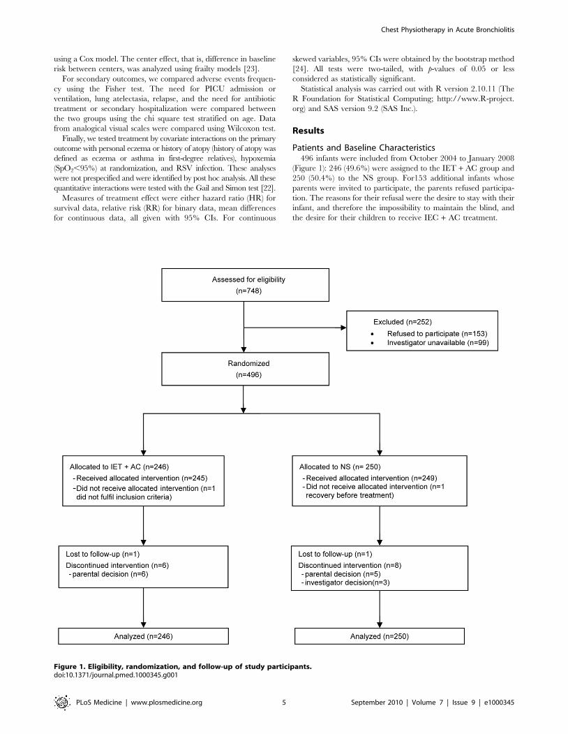
Patients and Baseline Characteristics496 infants were included from October 2004 to January 2008
(Figure 1): 246 (49.6%) were assigned to the IET + AC group and
250 (50.4%) to the NS group. For153 additional infants whose
parents were invited to participate, the parents refused participa-
tion. The reasons for their refusal were the desire to stay with their
infant, and therefore the impossibility to maintain the blind, and
the desire for their children to receive IEC + AC treatment.
Figure 1. Eligibility, randomization, and follow-up of study participants.doi:10.1371/journal.pmed.1000345.g001
Chest Physiotherapy in Acute Bronchiolitis
PLoS Medicine | www.plosmedicine.org 5 September 2010 | Volume 7 | Issue 9 | e1000345

At randomization, there were no marked differences between
the two randomized groups for demographic variables, percentage
of infants with hypoxemia or feeding difficulties, percentage of
infants with nasal aspirate positive for RSV, and duration of
respiratory symptoms before hospitalization (Table 1). The
proportion of cases of lung atelectasia diagnosis on X-ray was
higher in the NS group (12.9% versus 7.6%).
Before randomization, 36 (14.4%) infants in the NS group and
47 (19.1%) infants in the IET + AC group received bronchodilator
inhalations (salbutamol). 34 (13.6%) infants in the NS group and
25 (10.2%) infants in the IET + AC group were treated with oral
corticosteroids (betamethasone).
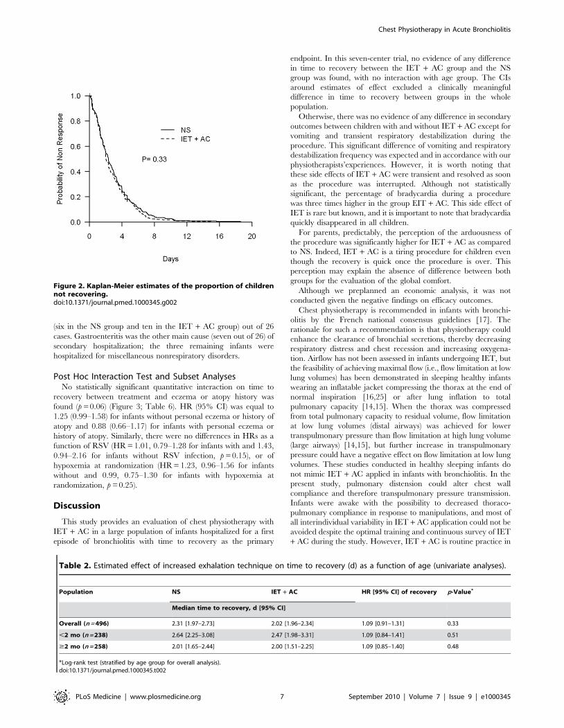
Primary End PointThe physiotherapy intervention (IET + AC) had no significant
effect on time to recovery. The median (95% CI) time to recovery
was 2.31 (1.97–2.73) d for the NS group and 2.02 (1.96–2.34) d for
the IET + AC group (HR [95% CI] = 1.09, 0.91–1.31, p = 0.33;
Figure 2; Table 2). Significant heterogeneity across centers in
baseline hazards was observed (p = 0.001), reflecting a difference
between centers in time to recovery independently of the
treatment effect. After controlling for prognostic baseline covar-
iates, the random effect Cox regression model, that takes into
account the center effect, showed that the effect of IET + AC on
time to recovery remained not significant (HR = 1.21, 0.97–1.49,
p = 0.09). Of note, the center effect persisted, though erased by the
handling of baseline prognostic covariate differences (p = 0.03).
Secondary End PointsTable 3 summarizes side effects during procedures reported by
physiotherapists in both groups: there were no significant
difference between groups in the proportion of children who
experienced one episode of bradycardia with desaturation (RR
[95% CI] = 1.0, 0.2–5.0, p = 1.00) or without desaturation
(RR = 3.6, 0.7–16.9, p = 0.10). Conversely, the proportion of
children who either had transient respiratory destabilization or
vomited during the procedure was significantly higher in the group
treated with IET + AC than in the group treated with NS (RR
= 10.2, 1.3–78.8, p = 0.005 and RR = 5.4, 1.6–18.4, p = 0.002,
respectively).
We obtained 371 (74.8%) responses to parental questionnaire
(187 in the NS group and 184 in the IET + AC group). Table 4
summarizes parental answers regarding the influence of the
physiotherapist visit on both the child’s comfort and respiratory
status. According to the parents, the procedure was significantly
more arduous in the group treated with IET (mean difference
[95% CI] = 0.88, 0.33–1.44, p = 0.002), whereas there was no
significant difference regarding the comfort of the child between
both groups (Difference of mean = 20.07, 20.53 to 0.38,
p = 0.40). Finally, there was no significant difference in the way
the parents rated the influence of physiotherapy on respiratory
status (RR = 0.99, 0.90–1.08, p = 0.89 or comfort (RR = 0.99,
0.94–1.05, p = 0.84).
17 (3.4%) infants, including seven (1.4%) requiring ventilatory
support, were admitted to the PICU. There was no evidence of
any difference between groups in the number of infants requiring
intensive care admission (RR = 0.7, 0.3–1.8, p = 0.62) or ventila-
tory support (RR = 2.5, 0.5–13.0, p = 0.29). 69 (28.5%) infants in
the NS group and 67 (28.6%) in the IET + AC group received
antibiotics (RR = 1.0, 0.7–1.3, 1.00). 53 infants in each group
(31.0% in the IET + AC group) relapsed within 1 mo after
discharge (RR = 1.1, 0.8–1.5, p = 0.73). 26 patients were admitted
to the hospital within 1 mo after discharge (12 in the NS group and
14 in the IET + AC group; RR = 1.2, 0.6–2.6, p = 0.68) (Table 5).
Respiratory symptoms were responsible for rehospitalization in 16
Table 1. Demographic characteristics of the infants on admission to the hospital.
Characteristic NS (n = 250) IET + AC (n = 246)
Age (mo), median [IQR] 2.0 [1.3–4.0] 2.1 [1.3–3.8]
Male gender, n (%) 141 (56.4) 134 (54.5)
Gestation (wk), mean ± SD 39.161.65 39.161.67
Environmental tobacco smokea, n (%) 69 (29.0) 65 (26.9)
Personal eczema or history of atopya,b, n (%) 100 (40.7) 97 (39.8)
Kindergartena, n (%) 37 (15.0) 29 (12.0)
Bronchodilators before randomizationa, n (%) 36 (14.4) 47 (19.1)
Corticosteroids before randomizationa, n (%) 34 (13.6) 25 (10.2)
Feeding difficulties before randomizationa, n (%) 222 (89.2) 207 (84.8)
Duration of respiratory symptoms at randomization in daysa, median [IQR] 3.0 [2.0–4.0] 3.0 [2.0–4.0]
SpO2,95% at randomization, n (%) 110 (44.2) 106 (44.2)
Atelectasia at randomizationc, n (%) 31 (12.9) 18 (7.6)
Supplementary oxygen and intravenous feeding, n (%)
No oxygen 141 (56.4) 135 (54.9)
Oxygen only 86 (34.4) 86 (34.9)
Oxygen and intravenous feeding 23 (9.2) 25 (10.2)
RSV +, n (%) 152 (76.4) 137 (73.3)
Temperature at randomization (uC), mean ± SD 37.260.7 37.360.6
aThe data were obtained by parental reporting.bHistory of atopy was defined as eczema or asthma in first-degree relatives.c480 (96.7%) had X-ray at admission.doi:10.1371/journal.pmed.1000345.t001
Chest Physiotherapy in Acute Bronchiolitis
PLoS Medicine | www.plosmedicine.org 6 September 2010 | Volume 7 | Issue 9 | e1000345
(six in the NS group and ten in the IET + AC group) out of 26
cases. Gastroenteritis was the other main cause (seven out of 26) of
secondary hospitalization; the three remaining infants were
hospitalized for miscellaneous nonrespiratory disorders.
Post Hoc Interaction Test and Subset AnalysesNo statistically significant quantitative interaction on time to
recovery between treatment and eczema or atopy history was
found (p = 0.06) (Figure 3; Table 6). HR (95% CI) was equal to
1.25 (0.99–1.58) for infants without personal eczema or history of
atopy and 0.88 (0.66–1.17) for infants with personal eczema or
history of atopy. Similarly, there were no differences in HRs as a
function of RSV (HR = 1.01, 0.79–1.28 for infants with and 1.43,
0.94–2.16 for infants without RSV infection, p = 0.15), or of
hypoxemia at randomization (HR = 1.23, 0.96–1.56 for infants
without and 0.99, 0.75–1.30 for infants with hypoxemia at
randomization, p = 0.25).
Discussion
This study provides an evaluation of chest physiotherapy with
IET + AC in a large population of infants hospitalized for a first
episode of bronchiolitis with time to recovery as the primary
endpoint. In this seven-center trial, no evidence of any difference
in time to recovery between the IET + AC group and the NS
group was found, with no interaction with age group. The CIs
around estimates of effect excluded a clinically meaningful
difference in time to recovery between groups in the whole
population.
Otherwise, there was no evidence of any difference in secondary
outcomes between children with and without IET + AC except for
vomiting and transient respiratory destabilization during the
procedure. This significant difference of vomiting and respiratory
destabilization frequency was expected and in accordance with our
physiotherapists’experiences. However, it is worth noting that
these side effects of IET + AC were transient and resolved as soon
as the procedure was interrupted. Although not statistically
significant, the percentage of bradycardia during a procedure
was three times higher in the group EIT + AC. This side effect of
IET is rare but known, and it is important to note that bradycardia
quickly disappeared in all children.
For parents, predictably, the perception of the arduousness of
the procedure was significantly higher for IET + AC as compared
to NS. Indeed, IET + AC is a tiring procedure for children even
though the recovery is quick once the procedure is over. This
perception may explain the absence of difference between both
groups for the evaluation of the global comfort.
Although we preplanned an economic analysis, it was not
conducted given the negative findings on efficacy outcomes.
Chest physiotherapy is recommended in infants with bronchi-
olitis by the French national consensus guidelines [17]. The
rationale for such a recommendation is that physiotherapy could
enhance the clearance of bronchial secretions, thereby decreasing
respiratory distress and chest recession and increasing oxygena-
tion. Airflow has not been assessed in infants undergoing IET, but
the feasibility of achieving maximal flow (i.e., flow limitation at low
lung volumes) has been demonstrated in sleeping healthy infants
wearing an inflatable jacket compressing the thorax at the end of
normal inspiration [16,25] or after lung inflation to total
pulmonary capacity [14,15]. When the thorax was compressed
from total pulmonary capacity to residual volume, flow limitation
at low lung volumes (distal airways) was achieved for lower
transpulmonary pressure than flow limitation at high lung volume
(large airways) [14,15], but further increase in transpulmonary
pressure could have a negative effect on flow limitation at low lung
volumes. These studies conducted in healthy sleeping infants do
not mimic IET + AC applied in infants with bronchiolitis. In the
present study, pulmonary distension could alter chest wall
compliance and therefore transpulmonary pressure transmission.
Infants were awake with the possibility to decreased thoraco-
pulmonary compliance in response to manipulations, and most of
all interindividual variability in IET + AC application could not be
avoided despite the optimal training and continuous survey of IET
+ AC during the study. However, IET + AC is routine practice in
Figure 2. Kaplan-Meier estimates of the proportion of childrennot recovering.doi:10.1371/journal.pmed.1000345.g002
Table 2. Estimated effect of increased exhalation technique on time to recovery (d) as a function of age (univariate analyses).
Population NS IET + AC HR [95% CI] of recovery p-Value*
Median time to recovery, d [95% CI]
Overall (n = 496) 2.31 [1.97–2.73] 2.02 [1.96–2.34] 1.09 [0.91–1.31] 0.33
,2 mo (n = 238) 2.64 [2.25–3.08] 2.47 [1.98–3.31] 1.09 [0.84–1.41] 0.51
$2 mo (n = 258) 2.01 [1.65–2.44] 2.00 [1.51–2.25] 1.09 [0.85–1.40] 0.48
*Log-rank test (stratified by age group for overall analysis).doi:10.1371/journal.pmed.1000345.t002
Chest Physiotherapy in Acute Bronchiolitis
PLoS Medicine | www.plosmedicine.org 7 September 2010 | Volume 7 | Issue 9 | e1000345

France and physiotherapists were substantially experienced and
trained. Therefore, the study reflects the effectiveness of
physiotherapy in infants with bronchiolitis in real life practice as
recommended by guidelines.
Previous studies on chest physiotherapy included smaller
numbers of subjects, with physician-assessed clinical scores as the
primary outcome and nonrelevant variables as secondary
outcomes (durations of oxygen supplementation and hospitaliza-
tion duration stay) [26–28]. Duration of oxygen supplementation
was clearly not a relevant outcome because it was required in only
56.5% of the infants studied. The results of any study including
only infants requiring oxygen supplementation should therefore
not be extrapolated to infants not requiring oxygen.
We tried to determine the time to recovery as accurately as
possible. It was therefore necessary to assess the conditions of the
infants around the clock, and not only when physicians were
present. Nurses recorded respiratory signs, heart rate, and SpO2 at
least every 8 h, as previously performed [29]. Thus, the 0.3-d
(which is almost equivalent to 8 h) difference observed between the
two randomized groups in median time to recovery could be
related to the study design, which hampered the detection of a
smaller difference. However, a smaller gain of healing time would
not have any clinical relevance. Otherwise, we have previously
shown that the clinical signs used for monitoring (respiratory rates
and retraction signs) were recorded similarly by all physicians,
nurses, and physiotherapists, with a high level of interobserver
reproducibility [20]. We compared IET + AC with NS, because
NS provides temporary relief for nasal congestion and is routinely
used.
Age, which was thought to be a possible determinant of IET
efficacy, did not influence treatment effect. We used a cut-off point
of 2 mo to allow stratifying randomization according to previous
recommendations that set the age limit for severe bronchiolitis
between 6 and 8 wk. Indeed, although such a dichotomization
achieved some loss of information, the effectiveness of chest
physiotherapy could have been decreased in the youngest owing to
their highest risk of severe disease. Airway size increases with age,
and we thought that it would be difficult to increase the clearance
of bronchial secretions from very small airways by physiotherapy.
The lack of documented specific mechanisms of pulmonary
change after IET makes it difficult to draw firm conclusions
concerning the age-related efficacy of physiotherapy.
Post hoc analyses showed no significant quantitative interaction
between IET + AC effect on the main endpoint and personal
Table 3. Side effects reported by physiotherapists during procedures.
Side Effect NS (n = 250) IET + AC (n = 246) Relative Risk [95% CI] p-Valuea
Bradycardia with desaturation, n (%) 3 (1.2%) 3 (1.2%) 1.0 [0.2–5.0] 1.00
Bradycardia without desaturation, n (%) 2 (0.8%) 7 (2.8%) 3.6 [0.7–16.9] 0.10
Vomiting during procedure 1 (0.4%) 10 (4.1%) 10.2 [1.3–78.8] 0.005
Respiratory destabilization 3 (1.2%) 16 (6.5%) 5.4 [1.6–18.4] 0.002
Hypotonia 0 (0.0%) 2 (0.8%) NA 0.24
aFischer exact test.doi:10.1371/journal.pmed.1000345.t003
Table 4. Parental opinions regarding the comfort of their child and the consequences of the procedure on this parameter and onthe respiratory status.
Parental Opinion NS (n = 187) IET + AC (n = 184)Mean Difference[95% CI]
Relative Risk[95% CI] p-Value
Evaluation de the comfort of your child duringhospitalization, median (IQR)
7.8 [5.7–9.0] 7.5 [6.2–8.7] 20.07 [20.53 to 0.38] — 0.40a
Evaluation of the procedure arduous, median (IQR) 4.3 [2.0–6.3] 5.0 [3.0–7.1] 0.88 [0.33–1.44] — 0.002a
Influence of the physiotherapist visit on thecomfort of your baby, n (%)
— 0.99 [0.90–1.08]b 0.89c
Worsening 5 (2.7%) 12 (6.5%) — — —
No influence 25 (13.4%) 19 (10.3%) — — —
Improvement 157 (83.9%) 153 (83.2%) — — —
Influence of the physiotherapist visit on therespiratory status of your baby, n (%)
— 0.99 [0.94–1.05]b 0.84c
Worsening 4 (2.1%) 1 (0.5%) — — —
No influence 8 (4.3%) 12 (6.6%) — — —
Improvement 175 (93.6%) 170 (92.9%) — — —
371 couples of parents (74.8%) completed the questionnaire. Percentages were calculated for the population of respondents (n = 371).aWilcoxon test.bRelative risk was computed for improvement versus (no influence + worsening).cFischer Exact test.doi:10.1371/journal.pmed.1000345.t004
Chest Physiotherapy in Acute Bronchiolitis
PLoS Medicine | www.plosmedicine.org 8 September 2010 | Volume 7 | Issue 9 | e1000345

atopy or history of atopy in first-degree relatives, the type of virus,
or the presence of baseline hypoxemia. Although exploratory, our
findings for infants without personal atopy or history of atopy in
first-degree relatives and for those without hypoxemia were close
to statistical significance as shown by the CIs around estimate of
effect for these subgroups (respectively [0.99–1.58] and [0.96–
1.56]) and deserve further investigations.
Infants included in the present study represent a spectrum of
bronchiolitis phenotype including infants prone or not prone to
experience repeated wheezing episodes. Reynolds et al. [30]
suggested that there might be two different groups of patients:
those with obstructive disease resulting entirely from infection,
thickening of the bronchiolar walls, and intrabronchial secretions,
and those with a predisposition to asthma, who develop
obstruction because of both inflammation and bronchospasm. It
is still currently impossible to determine which of the infants
hospitalized for bronchiolitis will subsequently develop asthma,
even though family history of atopy and atopic dermatitis in the
infant have been shown to be predictive of asthma [31]. It is
possible that future studies will lead to recommendations for
bronchiolitis treatment according to the phenotype of the infant,
rather than merely being based on current disease diagnosis alone
[32]. Our study was not designed to address this issue and atopic
status was not found to influence the effect of IET + AC.
The physiopathology of bronchiolitis remains unclear, but the
need for oxygen supplementation is considered as a marker of
considerable ventilation heterogeneity, due to a large number of
units with poor ventilation (obstructed) and good perfusion or
overdistension and poor perfusion. In the absence of segmental or
lobar lung atelectasia, ventilation heterogeneity is a peripheral
phenomenon unlikely to be affected by physiotherapy. Infants
requiring oxygen supplementation may not benefit at all from
physiotherapy, but the benefits of physiotherapy for nonhypoxe-
mic infants did not reach significance (Figure 3; Table 5). Studying
a mixture of patients may provide unclear or misleading results.
This perspective raises questions about limiting studies to
homogeneous populations, making it impossible to extrapolate
the results obtained to patients with a different clinical status. The
heterogeneity in concerned patient populations may contribute to
the difficulty of definitively evaluating physiotherapy in clinical
trials and in making clear recommendations on its benefit in
practice.
The ‘‘interphysiotherapist’’ variability is an important issue in
our study. Despite having taken great care in optimizing the
training, some variability in the way compression was applied may
remain and is difficult to evaluate. We deliberately chose to have
more than one physiotherapist by center to mimic as closely as
possible real-life conditions, and to be able to extrapolate our
results, should they have been positive. The problem of
practitioner variability for complex interventions is common in
nonpharmacological trials. We standardized the cessation of the
intervention on the same auscultation-efficacy criteria in order to
reduce interphysiotherapist variability.
In summary, this large, randomized, controlled, outcomes
assessor-blind and parent-blind trial of three daily chest physio-
therapy sessions with the IET technique in infants hospitalized for
a first episode of bronchiolitis provided no evidence that this
treatment shortened time to recovery. Our results did not support
the recommendation that chest physiotherapy be routinely
performed in hospitalized infants with acute bronchiolitis. Our
conclusions in infants with severe bronchiolitis cannot necessarily
be extrapolated to infants with mild or moderate bronchiolitis.
Further studies would be required to assess chest physiotherapy in
outpatients and for infants without history of atopy.
Table 5. Secondary outcomes.
Secondary Outcome NS (n = 250) IET + AC (n = 246) Relative Risk [95% CI] p-Value a
PICU admission, n (%) 10 (4.1%) 7 (2.9%) 0.7 [0.3–1.8] 0.62
Ventilation, n (%) 2 (0.8%) 5 (2.0%) 2.5 [0.5–13.0] 0.29
Antibiotics 69 (28.5%) 67 (28.6%) 1.0 [0.7–1.3] 1.0
Relapse 53/182 (29.1%) 53/171 (31.0%) 1.1 [0.8–1.5] 0.73
New hospitalization 12/182 (6.6%) 14/171 (8.2%) 1.2 [0.6–2.6] 0.68
Data obtained from the parents by telephone interview 30 d after discharge. We obtained 353 responses (71.2%) and percentages were calculated for the population ofrespondents (n = 353).aFischer exact test for percentage comparison.doi:10.1371/journal.pmed.1000345.t005
Figure 3. HRs and 95% CIs for healing in the group receivingIET + AC, as compared with the NS group, as a function ofbaseline prognostic factors. History of atopy was defined as thepresence of eczema or asthma in first-degree relatives.doi:10.1371/journal.pmed.1000345.g003
Chest Physiotherapy in Acute Bronchiolitis
PLoS Medicine | www.plosmedicine.org 9 September 2010 | Volume 7 | Issue 9 | e1000345

Supporting Information
Alternative Language Text S1 French translation of the article
by VG.
Found at: doi:10.1371/journal.pmed.1000345.s001 (0.34 MB
PDF)
Text S1 Trial protocol.
Found at: doi:10.1371/journal.pmed.1000345.s002 (0.30 MB
PDF)
Text S2 CONSORT checklist.
Found at: doi:10.1371/journal.pmed.1000345.s003 (0.11 MB
PDF)
Acknowledgments
We thank our Independent Safety Monitoring Board, namely, P. Blanc
(Pediatric Department, Poissy, Saint Germain Hospital), L. Chevret
(Pediatric Intensive Care Unit, Kremlin Bicetre Hospital, Universite Paris
Sud 11), C. Delacourt (Pediatric Pulmonology Department, Necker,
Enfants Malades, Universite Paris 5, Rene Descartes), and P. Hubert
(Pediatric Intensive Care Unit, Necker, Enfants Malades, Universite Paris
5, Rene Descartes). We thank C. Perez, clinician psychologist, for her help
for parental perception recording. We thank the project manager, M.
Legrand (APHP, Research Direction), and the research assistants A.
Doutres, and A. Traore. We thank all local physicians, physiotherapists,
the nursing staff, all patients, and their parents who participated in this
study. None of those listed received any financial compensation for their
contributions.
Author Contributions
ICMJE criteria for authorship read and met: VG SK NB VA LdP SL RE
BC SB AMB JB SC PL. Agree with the manuscript’s results and
conclusions: VG SK NB VA LdP SL RE BC SB AMB JB SC PL. Designed
the experiments/the study: VG NB VA RE SB SC PL. Analyzed the data:
VG SK VA. Collected data/did experiments for the study: VG NB VA
AMB. Enrolled patients: VG NB VA LdP SL RE BC. Wrote the first draft
of the paper: VG NB VA. Contributed to the writing of the paper: VG SK
NB VA LdP SL RE BC SB AMB JB SC PL. Supervised the intervention of
physiotherapists: SB.
References
1. Glezen P, Denny FW (1973) Epidemiology of acute lower respiratory disease in
children. N Engl J Med 288: 498–505.
2. Bush A, Thomson AH (2007) Acute bronchiolitis. BMJ 335: 1037–1041.
3. Smyth RL, Openshaw PJM (2006) Bronchiolitis. Lancet 368: 312–322.
4. Hall CB, Weinberg GA, Iwane MK, Blumkin AK, Edwards KM, et al. (2009)
The burden of respiratory syncytial virus infection in young children.
N Engl J Med 360: 588–598.
5. Koehoorn M, Karr CJ, Demers PA, Lencar C, Tamburic L, et al. (2008)
Descriptive epidemiological features of bronchiolitis in a population-based
cohort. Pediatrics 122: 1196–1203.
6. Wohl ME, Chernick V (1978) State of the art: bronchiolitis. Am Rev Respir Dis
118: 759–781.
7. Aherne W, Bird T, Court SD, Aherne W, Bird TM, et al. (1970) Pathological
changes in virus infections of the lower respiratory tract in children. J Clin Pathol
23: 7–18.
8. Hall CB (1998) Respiratory syncytial virus. In: Feigin RD, Cherry JD, eds.
Textbook of pediatric infectious diseases. Philadelphia: WB Saunders. pp
2084–2111.
9. SIGN (2006) Bronchiolitis in children: a National Clinical Guideline. Available:
http://www.sign.ac.uk/pdf/sign91.pdf. Accessed 8 August 2010..
10. Subcommittee on Diagnosis and Management of Bronchiolitis (2006) Diagnosis
and management of bronchiolitis. Pediatrics 118: 1774–1793.
11. Perrotta C, Ortiz Z, Roque M (2007) Chest physiotherapy for acute bronchiolitis
in paediatric patients between 0 and 24 months old. Cochrane Database Syst
Rev. CD004873.
12. King M, Brock G, Lundell C (1985) Clearance of mucus by simulated cough.
J Appl Physiol 58: 1776–1782.
13. Sivasothy P, Brown L, Smith IE, Shneerson JM (2001) Effect of manually
assisted cough and mechanical insufflation on cough flow of normal subjects,
patients with chronic obstructive pulmonary disease (COPD), and patients with
respiratory muscle weakness. Thorax 56: 438–444.
14. Feher A, Castile R, Kisling J, Angelicchio C, Filbrun D, et al. (1996) Flow
limitation in normal infants: a new method for forced expiratory maneuvers
from raised lung volumes. J Appl Physiol 80: 2019–2025.
15. Hayden MJ, Sly PD, Devadason SG, Gurrin LC, Wildhaber JH, et al. (1997)
Influence of driving pressure on raised-volume forced expiration in infants.
Am J Respir Crit Care Med 156: 1876–1883.
16. Marsh MJ, Fox GF, Hoskyns EW, Milner AD (1994) The Hering-Breuer
deflationary reflex in the newborn infant. Pediatr Pulmonol 18: 163–169.
17. (2001) Consensus conference on the management of infant bronchiolitis. Paris,
France, 21 September 2000. Proceedings. Arch Pediatr 8 Suppl 1: 1s–196s.
18. Beauvois E (2001) Role of respiratory therapy in the treatment acute
bronchiolitis in infants. Arch Pediatr 8 Suppl 1: 128S–131S.
19. Yanney M, Vyas H (2008) The treatment of bronchiolitis. Arch Dis Child 93:
793–798.
20. Gajdos V, Beydon N, Bommenel L, Pellegrino B, de Pontual L, et al. (2009) Inter-
observer agreement between physicians, nurses, and respiratory therapists for
respiratory clinical evaluation in bronchiolitis. Pediatr Pulmonol 44: 754–762.
21. Bland M (2000) An introduction to medical statistics. Oxford: Oxford University
Press.
Table 6. Search for treatment by covariate interactions on the main outcome measure, time to recovery.
Prognostic Baseline Covariate NS IET + AC HR [95% CI] of Recovery p-Valuea
Median Time to Recovery, d [95% CI]
Personal eczema or history of atopyb,c 0.06
Yes (n = 197) 1.96 [1.36–2.73] 2.30 [1.73–3.07] 0.88 [0.66–1.17]
No (n = 293) 2.42 [2.04–2.85] 2.02 [1.92–2.33] 1.25 [0.99–1.58]
RSVd 0.15
Positive (n = 289) 2.34 [1.97–2.99] 2.33 [1.94–2.88] 1.01 [0.79–1.28]
Negative (n = 97) 2.33 [1.35–3.32] 1.92 [1.29–2.08] 1.43 [0.94–2.16]
Hypoxemia at randomization 0.25
Yes (n = 216) 2.73 [2.30–3.32] 2.47 [2.02–3.17] 0.99 [0.75–1.30]
No (n = 273) 1.90 [1.36–2.52] 1.96 [1.51–2.08] 1.23 [0.96–1.56]
ap-Value refers to the statistics of Gail and Simon’s quantitative interaction test (1df-chi-squared test).bThe data were obtained from the parents.cHistory of atopy was defined as eczema or asthma in first-degree relatives.dRSV was tested in only 386 infants.doi:10.1371/journal.pmed.1000345.t006
Chest Physiotherapy in Acute Bronchiolitis
PLoS Medicine | www.plosmedicine.org 10 September 2010 | Volume 7 | Issue 9 | e1000345

22. Gail M, Simon R (1985) Testing for qualitative interactions between treatment
effects and patient subsets. Biometrics 41: 361–372.23. Aalen OO (1988) Heterogeneity in survival analysis. Stat Med 7: 1121–1137.
24. Efron B, Tibshirani R (1998) An introduction to the bootstrap. Boca Raton.
London: Chapman & Hall/CRC xvi: 436.25. Hoskyns EW, Milner AD, Hopkin IE (1987) Validity of forced expiratory flow
volume loops in neonates. Arch Dis Child 62: 895–900.26. Bohe L, Ferrero ME, Cuestas E, Polliotto L, Genoff M (2004) [Indications of
conventional chest physiotherapy in acute bronchiolitis]. Medicina (B Aires) 64:
198–200.27. Nicholas KJ, Dhouieb MO, Marshall TG, Edmunds AT, Grant MB (1999) An
evaluation of chest physiotherapy in the management of acute bronchiolitis.Physiotherapy 85: 669–674.
28. Webb MS, Martin JA, Cartlidge PH, Ng YK, Wright NA (1985) Chest
physiotherapy in acute bronchiolitis. Arch Dis Child 60: 1078–1079.
29. Wainwright C, Altamirano L, Cheney M, Cheney J, Barber S, et al. (2003) A
multicenter, randomized, double-blind, controlled trial of nebulized epinephrine
in infants with acute bronchiolitis. N Engl J Med 349: 27–35.
30. Reynolds EO, Cook CD (1963) The treatment of bronchiolitis. J Pediatr 63:
1205–1207.
31. Castro-Rodriguez JA, Holberg CJ, Wright AL, Martinez FD (2000) A clinical
index to define risk of asthma in young children with recurrent wheezing.
Am J Respir Crit Care Med 162: 1403–1406.
32. Frey U, von Mutius E (2009) The challenge of managing wheezing in infants.
N Engl J Med 360: 2130–2133.
Chest Physiotherapy in Acute Bronchiolitis
PLoS Medicine | www.plosmedicine.org 11 September 2010 | Volume 7 | Issue 9 | e1000345

Editors’ Summary
Background. Bronchiolitis, which is usually caused by therespiratory syncytial virus (RSV), is the commonest infectionof the lower respiratory tract (the lungs and the passagesthrough which air enters the lungs) in infants. A third of allchildren have bronchiolitis during their first year of life. Theillness begins with stuffiness, a runny nose, a mild cough,and mild fever. Then, as the smallest airways in the lung (thebronchioles) become inflamed (swell) and blocked withmucus, the cough worsens, and the infant may develop awheeze, shallow breathing, and a rapid heartbeat. Mostcases of bronchiolitis are mild and clear up within two weekswithout any treatment but some infants develop severedisease. Such infants struggle to get enough air into theirlungs, drawing in their chest with each breath (chestrecession). They have trouble eating and drinking, and theoxygen level in their blood can drop dangerously low. About1% of previously healthy infants need hospitalizationbecause of severe bronchiolitis. These severely affectedinfants are not normally given any medications but, wherenecessary, they are given oxygen therapy, fed through atube into their stomach, and given fluids through a vein.
Why Was This Study Done? In some countries, chestphysiotherapy is routinely given to infants with bronchiolitiseven though this is not a recommended treatment inter-nationally. In France, for example, virtually all outpatientswith bronchiolitis receive a form of chest physiotherapyknown as increased exhalation technique with assistedcough (IET + AC). IET—manual chest compression—is de-signed to clear mucus from the bronchioles whereas AC—coughing triggered by applying pressure to the top of thebreastbone—facilitates clearance of the large airways. But isIET + AC an effective treatment for bronchiolitis? In thisstudy, the researchers undertook a multicenter, randomized,controlled trial to answer this question. A randomized trial isa study in which patients are randomly allocated to receiveeither the treatment under study or a control treatment.Usually in such trials, noone is aware of the treatmentallocations until the trial has been completed. This is calledblinding and avoids unconscious biases being introducedinto the results. In this trial, although the parents, caregivers,and outcome assessors were blinded, the physiotherapistsand the infants were aware of treatment allocations. Thephysiotherapists were not involved in patient assessment,however, and the infants were sufficiently young that theirknowledge of their treatment was unlikely to bias the results.
What Did the Researchers Do and Find? The researchersenrolled nearly 500 children aged 15 days to 2 years whowere admitted to seven French hospitals for a first episode ofacute bronchiolitis. They randomly allocated the patients toreceive IET + AC (intervention group) or nasal suction(control group) three times a day from a physiotherapist
working alone in a room with blacked-out windows. Theprimary outcome of the trial was the patients’ time torecovery. Infants were judged to have recovered if they hadnot had oxygen therapy or showed signs of chest recessionfor 8 hours and had ingested more than two-thirds of theirdaily food requirement. Infants in the control group took anaverage of 2.31 days to recover whereas those in theintervention group took 2.02 days. However, this differencein recovery time was not statistically significant. That is, itcould have happened by chance. The researchers alsorecorded several secondary outcomes such as admission toan intensive care unit, help with breathing, antibiotictreatment, and parental perceptions of their child’scomfort. There were no significant differences between thetwo treatment groups for any of these secondary outcomes,although the parents did report that the IET + AC treatmentwas harder on their children than nasal suction while notreducing their overall comfort.
What Do These Findings Mean? These findings showthat IET + AC had no significant effect on the time torecovery of a large population of French infants admitted tohospital with severe bronchiolitis. These results cannot beextrapolated, however, to infants with mild or moderatebronchiolitis, and further studies are needed to assesswhether chest physiotherapy is of any benefit in anoutpatient setting. Three small trials of a different form ofchest physiotherapy have also previously failed to find anyeffect of chest physiotherapy on recovery time. Thus, none ofthe currently available results support the routine use ofchest physiotherapy in infants admitted to a hospital forsevere bronchiolitis.
Additional Information. Please access these Web sites viathe online version of this summary at http://dx.doi.org/10.1371/journal.pmed.1000345
N The UK National Health Service Choices Web site providesdetailed information on all aspects of bronchiolitis
N Kidshealth, a resource maintained by the NemoursFoundation (a not-for-profit organization for children’shealth) provides information for parents on bronchiolitisschizophrenia and on respiratory syncytial virus (in Englishand Spanish)
N The British Lung Foundation also provides information onbronchiolitis schizophrenia and on respiratory syncytialvirus
N The MedlinePlus encyclopedia has a page on bronchiolitisschizophrenia (in English and Spanish)
N The US Centers for Disease Control and Prevention hasdetailed information on respiratory syncytial virus
Chest Physiotherapy in Acute Bronchiolitis
PLoS Medicine | www.plosmedicine.org 12 September 2010 | Volume 7 | Issue 9 | e1000345