effective radiation dose of three diagnostic tests in cardiology: single photon emission computed...
TRANSCRIPT
Rev Port Cardiol. 2013;32(12):981---986
Revista Portuguesa de
CardiologiaPortuguese Journal of Cardiology
www.revportcardiol.org
ORIGINAL ARTICLE
Effective radiation dose of three diagnostic tests incardiology: Single photon emission computedtomography, invasive coronary angiography andcardiac computed tomography angiography
Pedro de Araújo Goncalvesa,b,c,∗, Pedro Jerónimo Sousaa,c, Rita Caléd,Hugo Marquese, Miguel Borges dos Santosa, André Dias f, Hélder Doresa,Maria Salomé Carvalhoa, António Ventosaa, Teresa Martins f,Rui Campante Telesa, Manuel Almeidaa, Miguel Mendesa
a Servico de Cardiologia, Hospital de Santa Cruz --- Centro Hospitalar de Lisboa Ocidental, Lisboa, Portugalb CEDOC --- Chronic Diseases Research Center --- Faculdade de Ciências Médicas-UNL, Lisboa, Portugalc Centro Cardiovascular, Hospital da Luz, Lisboa, Portugald Servico de Cardiologia, Hospital Garcia de Orta, Almada, Portugale Centro de lmagiologia, Hospital da Luz, Lisboa, Portugalf Servico de Medicina Nuclear, Hospital de Santa Cruz --- Centro Hospitalar de Lisboa Ocidental, Lisboa, Portugal
Received 30 January 2013; accepted 29 May 2013Available online 25 November 2013
KEYWORDSIonizing radiation;Single photonemission computedtomography;Invasive coronaryangiography;Cardiac computedtomography;Obesity
AbstractIntroduction: Diagnostic tests that use ionizing radiation play a central role in cardiology andtheir use has grown in recent years, leading to increasing concerns about their potential stochas-tic effects.
The aims of this study were to compare the radiation dose of three diagnostic tests: sin-gle photon emission computed tomography (SPECT), invasive coronary angiography (ICA) andcardiac computed tomography (cardiac CT) and their evolution over time, and to assess theinfluence of body mass index on radiation dose.Methods: We assessed consecutive patients included in three prospective registries (SPECT, ICAand cardiac CT) over a period of two years. Radiation dose was converted to mSv and comparedbetween the three registries. Differences over time were evaluated by comparing the first with
the fourth semester. Results: A total of 6196 exams were evaluated: 35% SPECT, 53% ICA and 22% cardiac CT. Meanradiation dose was 10.7±1.2 mSv for SPECT, 8.1±6.4 mSv for ICA, and 5.4±3.8 mSv for cardiacCT (p<0.001 for all). With regard to the radiation dose over time, there was a very small∗ Corresponding author.E-mail address: [email protected] (P. de Araújo Goncalves).
0870-2551/$ – see front matter © 2013 Sociedade Portuguesa de Cardiologia. Published by Elsevier España, S.L. All rights reserved.http://dx.doi.org/10.1016/j.repc.2013.05.005
982 P. de Araújo Goncalves et al.
reduction in SPECT (10.7 to 10.5 mSv, p=0.004), a significant increase (25%) in ICA (7.0 to 8.8mSv; p<0.001), and a significant reduction (29%) in cardiac CT (6.5 to 4.6 mSv, p<0.001). Obesitywas associated with a significantly higher radiation dose in all three exams.Conclusions: Cardiac CT had a lower mean effective radiation dose than invasive coronaryangiography, which in turn had a lower mean effective dose than SPECT.
There was a significant increase in radiation doses in the ICA registry and a significant decreasein the cardiac CT registry over time.© 2013 Sociedade Portuguesa de Cardiologia. Published by Elsevier España, S.L. All rightsreserved.
PALAVRAS-CHAVERadiacão;Cintigrafia deperfusão miocárdica;Coronariografiainvasiva;Tomografiacomputorizadacardíaca;Obesidade
Dose efetiva de radiacão de três exames de diagnóstico em cardiologia: cintigrafiade perfusão miocárdica, coronariografia invasiva e tomografia computorizadacardíaca
ResumoIntroducão: Os exames diagnósticos que usam radiacão ionizante têm um papel central nacardiologia e a par do seu uso crescente, tem aumentado a preocupacão pelos seus potenciaisefeitos estocásticos.
Os objetivos deste estudo foram: 1) Comparar a dose de radiacão de três exames: Cintigrafiade perfusão miocárdica (SPECT), coronariografia invasiva (CAT) e tomografia computorizadacardíaca (AngioTC) e a sua evolucão temporal. 2) Avaliar o impacto do índice de massa corporalna dose de radiacão.Métodos: Doentes consecutivos incluídos em três registos prospetivos (SPECT, CAT e AngioTC)durante dois anos. A dose de radiacão foi convertida a mSv e comparada entre os três registos.A evolucão temporal foi avaliada por comparacão do 1.◦ e 4.◦ semestres.Resultados: Foram avaliados 6196 exames: 35% SPECT, 53% CAT e 22% AngioTC. A dose deradiacão foi: 10,7 ± 1,2 mSv para o SPECT; 8,1 ± 6,4 mSv para o CAT; 5,4 ± 3,8 mSv paraa AngioTC (p < 0,001 todas comparacões).
Evolucão temporal da dose de radiacão: reducão muito ligeira no SPECT (10,7 para 10,5mSv; p = 0,004); aumento significativo (25%) no CAT (7,0 para 8,8 mSv; p < 0,001); reducãosignificativa (29%) na AngioTC (6,5 para 4,6 mSv; p < 0,001). A obesidade associou-se a níveisde radiacão significativamente mais elevados nos três exames.Conclusão: O exame associado a uma menor dose de radiacão foi a AngioTC, seguida do CATque, por sua vez, foi menor que a do SPECT. Houve um aumento significativo da dose de radiacãono registo CAT e uma reducão significativa no registo da AngioTC ao longo do tempo.© 2013 Sociedade Portuguesa de Cardiologia. Publicado por Elsevier España, S.L. Todos osdireitos reservados.
I
Iuicicp
(lo
tfqat
ddw
ba
List of abbreviations
BMI Body mass indexCAD coronary artery diseaseCT computed tomographyICA invasive coronary angiographySPECT single photon emission computed tomography
ntroduction
n recent years, the development of imaging techniquessing ionizing radiation has resulted in considerable progressn the diagnosis and treatment of heart disease. Three
ommonly used diagnostic modalities that involve ioniz-ng radiation are used for assessing patients with possibleoronary artery disease (CAD): single photon emission com-uted tomography (SPECT), cardiac computed tomographytIa
cardiac CT) and invasive coronary angiography (ICA), theatter being considered the gold standard for the diagnosisf CAD.1
Different radiation doses have been reported for each ofhese exams, ranging from 5 to 10 mSv for ICA, 6 to 15 mSvor SPECT, and 4 to 21 mSv for cardiac CT.2---5 With more fre-uent use of these exams, there have been growing concernsbout the radiation’s potential secondary effects, especiallyhe stochastic effects of high cumulative doses over time.6,7
We have previously reported on the effective radiationose associated with cardiac CT in a single-center registry,ocumenting a significant decrease in dose over time, andere able to identify the predictors of higher dose.8
New scanners and acquisition protocols have recentlyeen developed which lead to significant reductions in radi-tion dose associated with cardiac CT.9,10
The aims of this study were to evaluate and comparehe radiation dose used in three diagnostic tests --- SPECT,CA and cardiac CT --- and their evolution over time, and tossess the influence of body mass index on radiation dose.
Effective radiation dose of three diagnostic tests in cardiology 983
Table 1 Demographic and clinical characteristics of the study population.
Cardiac CT (n=1344) ICA (n=3267) SPECT (n=1585)
Age (years, mean ± SD) 59±12 66±12 64±9Male (%) 60% 61% 63%BMI (kg/m2) 27.3±4.3 27.3±4.2 27.5±4.4Diabetes (%) 16% 29% N/AHypertension (%) 57% 72% N/ADyslipidemia (%) 54% 57% N/ASmoking (%) 27% 31% N/APrevious MI (%) 3% 17% N/APrevious PCI (%) 7% 18% N/APrevious CABG (%) 3% 7% N/A
Values are means (SD) or percentages. BMI: body mass index; CABG: coronary artery bypass grafting; CT: computed tomography; ICA:ot av
R
Dp(i
wIi
ttpmi(
The factors associated with the 25% increase in meaneffective radiation dose with ICA from the first to the fourthsemester were the higher proportions of positive exams,radial vascular access and exams performed by fellows in
1210.7
8.1
5.4
10
8
6
4
2
0SPECT
mSvICA
p<0.001all comparisons
Cardiac CT
invasive coronary angiography; MI: myocardial infarction; N/A: nphoton emission computed tomography.
Methods
From three prospective registries of SPECT, ICA and cardiacCT, we selected for this analysis the exams performed dur-ing a two-year period (October 1, 2008 to September 30,2010) in which the indication was assessment of possibleCAD.
The exams were performed with an SMV DST-XL gammacamera using 99m Tc-tetrofosmin with stress/rest orrest/stress protocols (SPECT registry), a Siemens CoroskopTOP/ARTIS dFC system (ICA registry), and a SiemensSomatom Definition dual-source scanner (cardiac CT reg-istry). The effective radiation dose was converted to mSv inaccordance with current literature and the manufacturer’sproduct information and compared between the registries.Briefly, a factor of 0.014 mSv/Gy cm was used for theconversion of cardiac CT dose-length product,9,11 a factor of0.183 mSv/Gy cm2 was used for the conversion of ICA dose-area product,12,13 and factors of 0.0060 mSv/MBq−1 (afterexercise) and 0.0071 mSv/MBq−1 (at rest) were used forthe conversion of injected activity in SPECT.14---16 To eval-uate the evolution of radiation doses over time, the studyperiod was divided into four semesters according to the dateof the exam and effective radiation dose was comparedbetween the first and last semesters in each registry. Allprospectively collected variables in the respective registrieswere analyzed, looking for predictors of dose change overtime.
Statistical analysis
Continuous variables are presented as mean ± standarddeviation (unless otherwise specified), and categoricalvariables as number (n) or frequency (%).
Continuous variables were analyzed using theMann---Whitney or Kruskal---Wallis nonparametric tests.The chi-square test was used to assess differences infrequencies.
Statistical significance was accepted for two-sided p val-ues <0.05.
The statistical analysis was performed using SPSS Statis-tics 17.0 for Windows.
Fsap
ailable; PCI: percutaneous coronary intervention; SPECT: single
esults
uring the two-year period of this analysis, 6196 exams wereerformed: 3267 (52.7%) ICA, 1585 (25.6%) SPECT and 134421.7%) cardiac CT. The demographic and clinical character-stics of the study population are presented in Table 1.
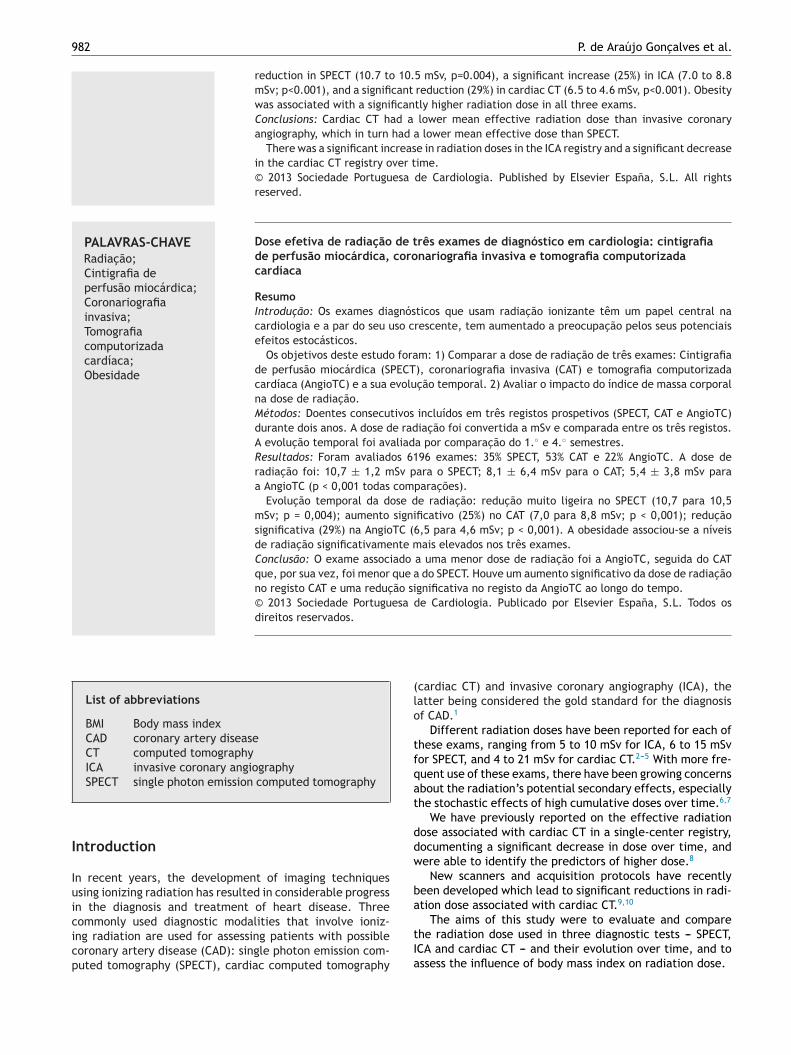
Mean effective radiation dose was 8.2±5.6 mSv for thehole population, 10.7±1.2 mSv for SPECT, 8.1±6.4 mSv for
CA and 5.4±3.8 mSv for cardiac CT (p<0.001 for all compar-sons, Figure 1).
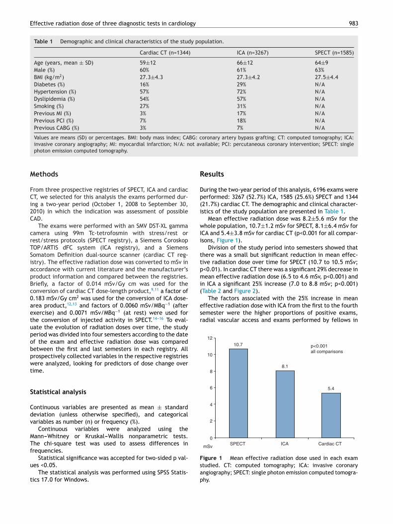
Division of the study period into semesters showed thathere was a small but significant reduction in mean effec-ive radiation dose over time for SPECT (10.7 to 10.5 mSv;<0.01). In cardiac CT there was a significant 29% decrease inean effective radiation dose (6.5 to 4.6 mSv, p<0.001) and
n ICA a significant 25% increase (7.0 to 8.8 mSv; p<0.001)Table 2 and Figure 2).
igure 1 Mean effective radiation dose used in each examtudied. CT: computed tomography; ICA: invasive coronaryngiography; SPECT: single photon emission computed tomogra-hy.
984 P. de Araújo Goncalves et al.
Table 2 Mean effective radiation dose for each exam over the four semesters.
1st semester 2nd semester 3rd semester 4th semester p (1st vs. 4th)
SPECT 10.7 ± 1.1 10.7 ± 1.4 10.7 ± 1.3 10.5 ± 0.9 0.004ICA 7.0 ± 6.0 7.6 ± 5.6 9.0 ± 6.9 8.7 ± 6.9 <0.001Cardiac CT 6.5 ± 3.7 6.2 ± 4.2 5.0 ± 4.1 4.6 ± 3.0 <0.001
CT: computed tomography; ICA: invasive coronary angiography; SPECT: single photon emission computed tomography.
12
10
10.7
7.0
6.5 6.2
5.04.6
7.6
8.9 8.8
10.7 10.7 10.5p=0.004(1st vs. 4th)
p<0.001(1st vs. 4th)
p<0.001(1st vs. 4th)
8
6
4
2
01 2 3 4
Time (semesters)mSv
SPECT ICA Cardiac CT
Figure 2 Time trends in mean effective radiation dose used ineach exam. CT: computed tomography; ICA: invasive coronaryap
tgfRI4roapfrtda
12
10.1
6.3
8.3
10.2
8.0
4.45.1
10.7
11.4
p<0.01 for all(<25 vs ≥30 kg/m
2)10
6
8
4
2
0
mSv25-30
BMI (kg/m2)<25 ≥30
SPECT ICA Cardiac CT
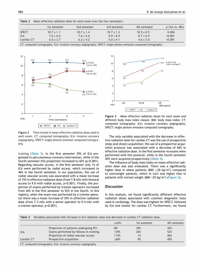
Figure 3 Mean effective radiation doses for each exam anddifferent body mass index classes. BMI: body mass index; CT:cS
t(sep4
ahtp
D
ngiography; SPECT: single photon emission computed tomogra-hy.
raining (Table 3). In the first semester 39% of ICA pro-ressed to percutaneous coronary intervention, while in theourth semester this proportion increased to 42% (p<0.001).egarding vascular access, in the first semester only 1% ofCA were performed by radial access, which increased to6% in the fourth semester. In our population, the use ofadial vascular access was associated with a mean increasef 15% in effective radiation dose (from 7.8 mSv with femoralccess to 9.0 with radial access, p<0.001). Finally, the pro-ortion of exams performed by trainee operators increasedrom 26% in the first semester to 52% in the fourth. In this
egistry, when the exam was performed by a trainee opera-or there was a mean increase of 29% in effective radiationose (from 7.3 mSv with a senior operator to 9.4 mSv withtrainee operator, p<0.001).
Irub
Table 3 Variables associated with increase in ICA radiation dose
ICAProportion of patients undergoing PCI
Exams performed by fellows in training
Proportion of radial vascular access
Cardiac CT Prospective acquisition
CT: computed tomography; ICA: invasive coronary angiography.
omputed tomography; ICA: invasive coronary angiography;PECT: single photon emission computed tomography.
The only variable associated with the decrease in effec-ive radiation dose for cardiac CT was the use of prospectivestep-and-shoot) acquisition: the use of a prospective acqui-ition protocol was associated with a decrease of 60% inffective radiation dose. In the first semester no exams wereerformed with this protocol, while in the fourth semester5% were acquired prospectively (Table 3).
The influence of body mass index on mean effective radi-tion dose was also evaluated. There was a significantlyigher dose in obese patients (BMI ≥30 kg/m2) comparedo overweight patients, which in turn was higher that inatients with normal weight (BMI <25 kg/m2) (Figure 3).
iscussion
n this analysis, we found significantly different effectiveadiation doses associated with common diagnostic testssed in cardiology. The dose was highest for SPECT, followedy ICA and lowest for cardiac CT. Furthermore, we found
and decrease in cardiac CT radiation dose.
�mSv 1st semester 4th semester
ND 39% 42%↑29% 26% 52%↑15% 1% 46%↓60% 0% 45%
gy
dtttipfCs
C
IdClcarsOd
E
PdwcM
Cftsi
Rosi
C
T
R
Effective radiation dose of three diagnostic tests in cardiolo
some time trends in the mean effective radiation dose asso-ciated with ICA and cardiac CT related to particular clinicaland procedural methodologies.
The biological effects of ionizing radiation are relatedto the cumulative effective dose, and doses above 100 mSvhave been linked to stochastic effects including the develop-ment of cancer, while the effects of lower radiation levels,common in diagnostic X-ray imaging, are much less clear.4,17
Although other theoretical models based on dose-thresholdand hormetic effects have been proposed, the more con-servative linear no-threshold model, which assumes that nolevel of radiation is without risk, is widely accepted.4,17
On this basis, procedures that use ionizing radiationshould be performed in accordance with the ‘‘as low asreasonably achievable’’ philosophy, and physicians orderingand performing cardiac imaging diagnostic tests should befamiliar with the associated radiation doses and with waysin which they can be minimized.
The mean effective radiation dose we found for eachexam is in agreement with previous studies.3,4,6,18 Further-more, we confirmed that certain variables influence theeffective radiation dose delivered by these exams. For ICA,the effective radiation dose increased with the use of radialaccess and with less experienced operators, which is in linewith published data.13,19 The higher radiation dose in theICA registry over time was also associated with a higherproportion of positive exams; although we did not quan-tify the difference between positive and negative ICA, wecan assume that positive tests needed more cine angiogramsof the coronary arteries, with a consequent increase in theradiation dose used.
For cardiac CT, the introduction and increasingly fre-quent use of a prospective protocol during the study periodwas associated in our experience with a significant decreasein the effective radiation dose for this exam, as has beendemonstrated by other authors.20---22 Finally, for SPECT, thedose change over time was very small, which is to beexpected since there were no changes in protocol duringthe study period.
It is worth noting that during the same period, doses asso-ciated with stress-only and rest-only SPECT studies weresignificantly lower (with mean effective doses of 2.3±0.9mSv and 5.8±1.0 mSv, respectively) but they were not con-sidered for the purpose of this study, and the small numberof patients involved (n=49 and n=63, respectively) would nothave had a significant impact on the overall SPECT radiationdose.
Mean effective radiation doses were significantly higherfor obese patients in all the exams analyzed. This was espe-cially true for cardiac CT and ICA, with an almost two-foldincrease in radiation dose compared to their normal-weightcounterparts. In the SPECT registry, the effect of BMI wasless pronounced. This should be taken in consideration whenselecting the appropriate diagnostic exam, especially forthose at higher risk from radiation exposure, like womenand younger patients.23 In line with this, particular atten-tion should be paid to cardiac CT dose, since patients in ourregistry undergoing cardiac CT were significantly younger
than those in the ICA and SPECT registries.Although the present study focuses on comparison of theradiation dose between three different diagnostic exams,other features should be taken into account when comparing
985
ifferent imaging modalities. As cardiac CT and ICA requirehe administration of iodinated contrast, care should beaken in the presence of impaired renal function or his-ory of allergies; likewise, the probability of CAD is also anmportant factor, as SPECT and ICA are more appropriate foratients with higher probability of CAD.24,25 Thus, all theseeatures (radiation dose, need for iodinated contrast andAD probability) should be taken into consideration whenelecting the most appropriate exam for each patient.
onclusions
n these registries of diagnostic tests commonly used in car-iology, the mean effective radiation dose used in cardiacT was lower than that used in ICA, which in turn was
ower than the doses used in SPECT. There was a signifi-ant increase over time in the mean effective radiation dosessociated with ICA, mainly related to the increased use ofadial access, and a decrease in cardiac CT doses as a con-equence of the implementation of a prospective protocol.besity was associated with a significantly higher radiationose in all three exams.
thical disclosures
rotection of human and animal subjects. The authorseclare that the procedures followed were in accordanceith the regulations of the relevant clinical research ethicsommittee and with those of the Code of Ethics of the Worldedical Association (Declaration of Helsinki).
onfidentiality of data. The authors declare that they haveollowed the protocols of their work center on the publica-ion of patient data and that all the patients included in thetudy received sufficient information and gave their writtennformed consent to participate in the study.
ight to privacy and informed consent. The authors havebtained the written informed consent of the patients orubjects mentioned in the article. The corresponding authors in possession of this document.
onflicts of interest
he authors have no conflicts of interest to declare.
eferences
1. Task Force on Myocardial Revascularization of the EuropeanSociety of Cardiology (ESC), the European Association forCardio-Thoracic Surgery (EACTS), European Association forPercutaneous Cardiovascular Intervention (EAPCI), Wijns W,et al. Guidelines on myocardial revascularization. Eur Heart J.2010;31:2501---55.
2. Hirshfeld Jr JW, Balter S, Brinker JA, et al. ACCF/AHA/HRS/SCAIclinical competence statement on physician knowledge tooptimize patient safety and image quality in fluoroscopically
guided invasive cardiovascular procedures: a report of theAmerican College of Cardiology Foundation/American HeartAssociation/American College of Physicians Task Force on Clin-ical Competence and Training. Circulation. 2005;111:511---32.
9
1
1
1
1
1
1
1
1
1
1
2
2
2
2
2
86
3. Einstein AJ, Moser KW, Thompson RC. Radiation doseto patients from cardiac diagnostic imaging. Circulation.2007;116:1290---305.
4. Gerber TC, Carr JJ, Arai AE, et al. Ionizing radiation in car-diac imaging: a science advisory from the American HeartAssociation Committee on Cardiac Imaging of the Council onClinical Cardiology and Committee on Cardiovascular Imagingand Intervention of the Council on Cardiovascular Radiology andIntervention. Circulation. 2009;119:1056---65.
5. Fazel R, Shaw LJ. Radiation exposure from radionuclide myocar-dial perfusion imaging: concerns and solutions. J Nucl Cardiol.2011;18:562---5.
6. Fazel R, Krumholz HM, Wang Y, et al. Exposure to low-dose ion-izing radiation from medical imaging procedures. N Engl J Med.2009;361:849---57.
7. Almeida AG. Cardiac computed tomography and radiation:balancing benefit and risk. Rev Port Cardiol. 2010;29:1677---82.
8. Sousa PJ, Goncalves PA, Marques H, et al. Radiation in cardiacCT: predictors of higher dose and its reduction over time. RevPort Cardiol. 2010;29:1655---65.
9. Achenbach S, Marwan M, Ropers D, et al. Coronary computedtomography angiography with a consistent dose below 1 mSvusing prospectively electrocardiogram-triggered high-pitch spi-ral acquisition. Eur Heart J. 2010;31:340---6.
0. Duarte R, Bettencourt N, Costa JC, et al. Coronary computedtomography angiography in a single cardiac cycle with a meanradiation dose of approximately 1 mSv: initial experience. RevPort Cardiol. 2010;29:1667---76.
1. Hausleiter J, Meyer T, Hermann F, et al. Estimated radi-ation dose associated with cardiac CT angiography. JAMA.2009;301:500---7.
2. Betsou S, Efstathopoulos EP, Katritsis D, et al. Patient radiationdoses during cardiac catheterization procedures. Br J Radiol.1998;71:634---9.
3. Neill J, Douglas H, Richardson G, et al. Comparison of radia-tion dose and the effect of operator experience in femoral andradial arterial access for coronary procedures. Am J Cardiol.
2010;106:936---40.4. Cerqueira MD, Allman KC, Ficaro EP, et al. Recommendations forreducing radiation exposure in myocardial perfusion imaging.J Nucl Cardiol. 2010;17:709---18.
2
P. de Araújo Goncalves et al.
5. Hesse B, Tagil K, Cuocolo A, et al. EANM/ESC procedural guide-lines for myocardial perfusion imaging in nuclear cardiology. EurJ Nucl Med Mol Imaging. 2005;32:855---97.
6. Myoview package insert. European Prescribing Information.Available at http://www.gehealthcare.com/euen/molecular-imaging/congress/pdf/myoview-pi.pdf
7. Einstein AJ. Effects of radiation exposure from cardiac imaging:how good are the data? J Am Coll Cardiol. 2012;59:553---65.
8. Kaufmann PA, Knuuti J. Ionizing radiation risks of car-diac imaging: estimates of the immeasurable. Eur Heart J.2011;32:269---71.
9. Jolly SS, Yusuf S, Cairns J, et al. Radial versus femoral access forcoronary angiography and intervention in patients with acutecoronary syndromes (RIVAL): a randomised, parallel group, mul-ticentre trial. Lancet. 2011;377:1409---20.
0. Gopal A, Mao SS, Karlsberg D, et al. Radiation reduction withprospective ECG-triggering acquisition using 64-multidetectorcomputed tomographic angiography. Int J Cardiovasc Imaging.2009;25:405---16.
1. Stolzmann P, Leschka S, Scheffel H, et al. Dual-source CT instep-and-shoot mode: noninvasive coronary angiography withlow radiation dose. Radiology. 2008;249:71---80.
2. Ferreira AM, Lopes R, Correia Mda G, et al. Low-dose cardiacCT. Rev Port Cardiol. 2010;29:459---62.
3. Einstein AJ, Henzlova MJ, Rajagopalan S. Estimating risk of can-cer associated with radiation exposure from 64-slice computedtomography coronary angiography. JAMA. 2007;298:317---23.
4. Taylor AJ, Cerqueira M, Hodgson JM, et al. ACCF/SCCT/ACR/AHA/ASE/ASNC/NASCI/SCAI/SCMR 2010 appropriate use crite-ria for cardiac computed tomography. A report of the AmericanCollege of Cardiology Foundation Appropriate Use Criteria TaskForce, the Society of Cardiovascular Computed Tomography, theAmerican College of Radiology, the American Heart Associa-tion, the American Society of Echocardiography, the AmericanSociety of Nuclear Cardiology, the North American Society forCardiovascular Imaging, the Society for Cardiovascular Angiog-raphy and Interventions, and the Society for CardiovascularMagnetic Resonance. J Am Coll Cardiol. 2010;56:1864---94.
5. NICE clinical guideline 95. Chest pain of recent onset:assessment and diagnosis of recent onset chest pain ordiscomfort of suspected cardiac origin. Available from:http://www.nice.org.uk/guidance/CG95