effect of bisphosphonates sodium alendronate on shedding ... research.pdf · most of the crowns of...
TRANSCRIPT
International Dental & Medical Journal of Advanced Research ● Vol. 1 ● 2015 1
International Dental & Medical Journal of Advanced Research (2015), 1, 1–8
O R I G I N A L A R T I C L E
Effect of bisphosphonates sodium alendronate on shedding of deciduous molars in rabbits (histological and histochemical study)Afaf N. Aboalrejal1, Amal M. El-Motayam1, Amira E. Fares1, Soheer Y. Saleh2
1Department of Oral Biology, Faculty of Oral and Dental Medicine, Cairo University, Egypt, 2Department of Physiology, Faculty of Veterinary Medicine, Cairo University, Egypt
AbstractAim: The purpose of this study was to investigate the effects of bisphosphonate (Bps) (sodium alendronate) on root resorption of deciduous lower molars in a rabbit.Materials and Methods: This study was carried out using 40 newborns healthy New Zealand white rabbits of average body weight 50-55 g, their nutrition was provided maternally, and a commercial pelleted diet of 16.7% crude protein, 13.7% crude fiber, and 2590 kcal of digestible energy per kg would be added. The animals were classified into two main groups: Group 1 (control group): This group is consisted of 20 rabbits which were subjected to oral dose of 2.5 mg/kg of body wt./day normal saline by using oropharengeal tube. The rabbits were divided into two sub groups: Group 2 (Bps group): This group is consisted of 20 rabbits, which were subjected to daily oral dose of 2.5 mg/kg of body wt./day sodium alendronate using oropharengeal tube were divided into two subgroups: At the end of the experimental period; the mandibles were removed and hemisected. The hemisected mandibles were prepared for light microscope examination using hematoxyline and eosin and tartrate-resistant acid phosphatase (TRAP) (special histochemical stain).Results: (1) Histological results: In control subgroup A, the deciduous molars of all animals have been fully erupted with considerable amounts of roots resorption. In control subgroup B, the deciduous molars of all animals have been completely shed. In Bps group, subgroup A showed delayed eruption of the deciduous molars and some dental abnormalities. In Bps group subgroup B, the deciduous molars still present with some degree of root resorption. Most of the crowns of the underlying pre-molars were formed with developing of their roots. Cementum-like tissue was found between the deciduous molars and erupting pre-molars, (2) Histochemical results: The control subgroup A showed large number of positively stained multinucleated odontoclasts on the resorbed dentin surface of deciduous molars with formation of Howship’s lacunae. The control subgroup B showed negative stain in all layers of the erupting pre-molars (no odontoclasts were seen in this subgroup). The Bps group subgroup A showed negative stain in all layers of the deciduous tooth germs that appeared in the late bell stage. The Bps group subgroup B showed negative stain in all layers of the erupting pre-molars as well as in all layers of the deciduous molars.Conclusions: Treatment with sodium alendronate (2.5 mg/kg body wt./day) from 1st day of birth resulted in delayed eruption of deciduous molars and some dental abnormalities as enlargements of pre-dentin layer, discontinuity of odontoblastic layer, defective dentin mineralization and appearance of denticle-like structure in the pulp. TRAP histochemical results revealed that the treatment with Bps result in decrease numbers of odontoclasts and osteoclasts as their apoptosis and separation from Howship’s lacunae.
Keywords Deciduous teeth shedding, rabbit, sodium alendronate and tartrate-resistant acid phosphatase stain
Correspondence Afaf Noman Aboalrejal, Department of Oral Biology, Faculty of Oral and Dental Medicine, Cairo University, Egypt. Email: [email protected]
Received 27 September 2015: Accepted 29 October 2015
doi: 10.15713/ins.idmjar.31
Bisphosphonates and shedding of deciduous teeth Aboalrejal, et al.
2 International Dental & Medical Journal of Advanced Research ● Vol. 1 ● 2015
Introduction
Rabbits are small mammals fall into the order Lagomorpha (Lagomorpha means “hare-shaped”), family Leporidae, and genus and species Oryctolagus cuniculus. There are many species of rabbits, the rabbits are herbivores (plant eating) and have a high reproduction rate. The teeth of the rabbit have some characteristic mammalian features; they are heterodont and diphyodont (i.e., two successive dentition, deciduous and permanent dentition).[1] The rabbit has a large mandible with a deceptively small oral cavity. The dental formula of deciduous teeth of rabbit is:
2 I C M� , , ]21
00
32
16=
The dental formula of the permanent teeth of rabbit is:
2 Pm M 28I� � ,��
21
00
32
33
=
The digit 2 in front of the formula indicates the left and right sides of the mouth. The letter I stand for incisor teeth; rabbits have two incisors for upper and one for the lower jaw. The letter C stands for canine teeth in which rabbits normally do not have. The letter pm indicates for the pre-molar cheek teeth; three pre-molars for the upper jaw and two for the lower. The letter m is the molar cheek teeth, and there are three on both the upper and the lower jaws.[2]
Shedding of teeth is the physiological elimination of the deciduous teeth due to their root resorption prior to the eruption of their permanent successors at specific ages. This process is accompanied by resorption of calcified tissues; bone, cementum, and dentin by the resorbing cells osteoclasts or odontoclasts.
Like any physiological process, shedding is affected by local or systemic factors. Administration of drugs that can interfere with the process of hard tissue resorption may affect normal shedding. Among drugs that interfere with the activity of osteoclasts and odontoclasts are bisphosphonates (Bps). During shedding of the teeth both bone formation and resorption take place, it is a biological event that offers an excellent model for studying the possible effects of drugs that interfere with bone metabolism.
Bps are a class of drug analogs of pyrophosphate with high affinity to hydroxyapatite. Alendronate sodium (Fosamax) is a commonly used anti-resorptive agent effective for reducing bone resorption, increasing bone density, and decreasing fracture incidence.
They have the ability to inhibit osteoclast-mediated bone resorption and are widely used in prevention and treatment of osteoporosis and other bone disorders. The pronounced selectivity of Bps for bone rather than other tissues is the basis for their value in clinical practice. Their preferential uptake by and adsorption to mineral surfaces in bone brings them into close contact with osteoclasts and probably some osteocyte.[3,4]
Tartrate-resistant acid phosphatase (TRAP) is one of the few enzymes that are histochemically detectable on the formalin-fixed
paraffin-embedded tissue. TRAP has been for many years used as a histochemical marker of the osteoclast, but neither its biological function nor its natural substrate is fully understood. Expression of TRAP is associated with the activation and differentiation of osteoclasts, as well as certain macrophages and other cells of a monohistiocytic phenotype such as the Gaucher cell.[5]
The aim of this study was to investigate the effects of Bps on physiological root resorption of deciduous molars. Moreover, the presence of osteoclasts in the alveolar bone was examined.
Materials and Methods
This study was carried out using 40 newborns healthy New Zealand white rabbits of average body weight 50-55 g, their nutrition was provided maternally and a commercial pelleted diet of 16.7% crude protein, 13.7% crude fiber, and 2590 kcal of digestible energy per kg would be added. The animals were classified into two main groups:
Group 1 (control group)
This group is consisted of 20 rabbits which were subjected to an oral dose of 2.5 mg/kg of body wt./day normal saline by using oropharengeal tube. The rabbits were divided into two subgroups.
Subgroup AIt is consisted of 10 animals, which were sacrificed by CO2 inhalation at the 10th day after birth.
Subgroup BIt is consisted of 10 animals, which were sacrificed by CO2 inhalation at the 20th day after birth.
Group 2 (Bps group)
This group is consisted of 20 rabbits, which were subjected to daily oral dose of 2.5 mg/kg of body wt./day sodium alendronate using oropharengeal tube[6] were divided into two subgroups:
Subgroup AIt is consisted of 10 animals which were sacrificed by CO2 inhalation at the 10th day after birth.
Subgroup BIt is consisted of 10 animals which were sacrificed by cervical dislocation at the 20th day after birth. At the end of the experimental period, the animals were sacrificed by CO2 inhalation; the mandibles removed and were hemisected.
The hemisected mandibles were prepared for light microscope examination using hematoxyline and eosin (H and E) and TRAP (special histochemical stain).
Histological examination (H and E stain)[7]
The specimens were fixed immediately in 10% calcium formol for 12 h in 4°C, The calcium formal was prepared by mixing 10 g
Aboalrejal, et al. Bisphosphonates and shedding of deciduous teeth
International Dental & Medical Journal of Advanced Research ● Vol. 1 ● 2015 3
calcium chloride, 100 ml formaline, and 900 ml distilled water, the specimens were rinsed well by water, then the specimens were decalcified with 10% ethylene diamine tetra-acetic acid (EDTA), pH - 7.3 at 4°C,[8] solution was being changed every 48 h for a month. After decalcification, the specimens were then dehydrated by transferring them through different concentrations in ascending grades of ethyl alcohol (EtOH) (50, 60, 80, 90, 96% and absolute alcohol 100%). Specimens were then transferred to xylol to be cleared from alcohol, then the specimens were embedded in paraffin wax and were embedded in the center of paraffin wax blocks, the embedded specimens were sectioned by microtome with longitudinal sections of 5-7 µ-thick, these sections were transferred in different concentrations of alcohol decreasing gradually 96% and 70% and then distilled water, sections were obtained and mounted on clean glass slides and stained with H and E for light microscopic examination.
Histochemical examination method (TRAP)
After decalcification with (EDTA) detection of odontoclast was carried out by using TRAP b5 stain. TRAP special stain was used for the demonstration of acid phosphatase activity an azo dye-coupling technique was used modified from[9] this stain requires four buffer solutions.
ProcedureTwo coplin jars were filled each with 50 ml basic stock incubation solution of Buffer I Solution (sodium acetate anhydrous 9.2 g/L, tartaric acid 11.4 g/L and glacial acetic acid 2.8 ml; pH = 4.7-5.0), containing 30 mg of Fast Red TR salt (F-8764;Sigma) and were preheated (leaved in 37°C) oven while slides were going through deparaffinization/rehydration, slides were deparaffinized and rehydrated to distilled water, 1½ ml of Buffer II Solution (20 g/L naphthol phosphate in ethylene glycol monoethyl) was added to one coplin jar, slides were placed in this coplin jar and was incubated at 37°C for 1 h.[10] A few minutes before incubation time is up, 1 ml of Buffer III Solution (sodium nitrite 50 g/L) and 1 ml of Buffer IV Solution (2 g/L pararosaniline chloride) were mixed. The mixture is gently mixed in hand for 30 s, and then was let sit for 2 min, this solution was added to the second preheated coplin jar of basic stock incubation solution, then was mixed well and the incubated slides were transferred from the first jar to the second jar without rinsing. For desired intensity of red in osteoclasts was developed at 37°C for 5-12 min. If tissue shows, any non-specific red stain should be checked periodically under the microscope during development and proceed immediately to next step. The slides were rinsed with distilled water, counterstained with Mayer’s hematoxylin and fast green 0.02%.[11] The slides were rinsed with distilled water, dehydrating through 3 changes of 95% ethanol (EtOH) and 2 changes of 100% EtOH (45 s each), clearance in 3 changes of xylene and mounted.
Results
I-Histological results
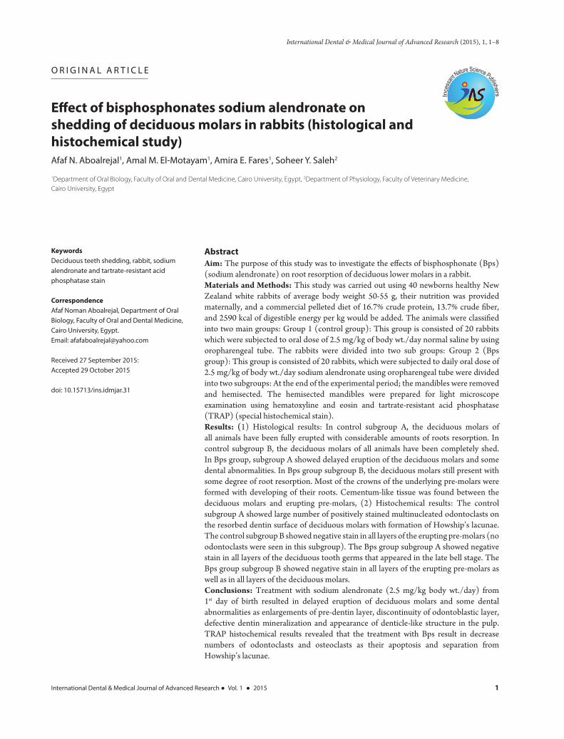
In control subgroup A, the deciduous molars of all animals have been fully erupted with considerable amounts of roots resorption [Figure 1a].
In control subgroup B, the deciduous molars of all animals have been completely shed, and the first pre-molars in almost all cases have been erupted, While the second pre-molars were found to be about to erupt [Figure 1b and c].
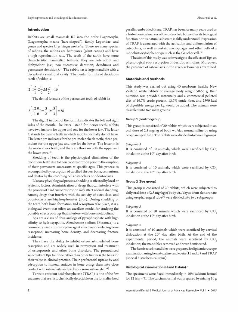
In Bps group subgroup A showed delayed eruption of the deciduous molars and some dental abnormalities as enlargement of pre-dentin layer, discontinuity of the odontoblastic layer, defective dentin mineralization and appearance of denticle–like structures in the pulp [Figures 1d and 2a].
In Bps group subgroup B, the deciduous molars still present with some degree of root resorption. Most of the crowns of the underlying pre-molars were formed with developing of their roots. Cementum-like tissue was found between the deciduous molars and erupting pre-molars.
In Bps groups, the alveolar bone showed increased density; decreased number of osteoclasts and their detachment from the alveolar bone surface [Figure 2b-d].
II-Histochemical results
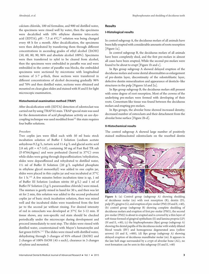
The control subgroup A showed large number of positively stained multinucleated odontoclasts on the resorbed dentin
Figure 1: (a) Control group (subgroup A) showing shedding of deciduous molar (m) with root resorption (R), dentin (D), pulp (P), gingiva (G), and eruption of pre-molar (PM) (H and E, ×40), (b) control group (subgroup B) showing complete shedding of deciduous molars and eruption of first pre-molar (PM1), the second pre-molar (PM2) is about to erupted and is covered by a thin layer of soft tissue formed of gingival epithelium (E) and lamina propria (LP) (H and E, ×40), (c) the bisphosphonates (Bps) group (subgroup A) showing the dental papilla of the deciduous molar with widely dilated blood vessels (BV) and homogenous degenerated area (yellow arrows) (H and E, ×400), (d) Bps group (subgroup A) showing delayed eruption of deciduous mandibular molar (m). It appears in the late bell stage surrounded by a crypt of alveolar bone (AL), no root formation can be seen in this subgroup (H and E, ×40)
dc
ba
Bisphosphonates and shedding of deciduous teeth Aboalrejal, et al.
4 International Dental & Medical Journal of Advanced Research ● Vol. 1 ● 2015
surface of deciduous molars with the formation of Howship’s lacunae [Figure 3a-c].
The control subgroup B showed negative stain in all layers of the erupting pre-molars (no odontoclasts were seen in this
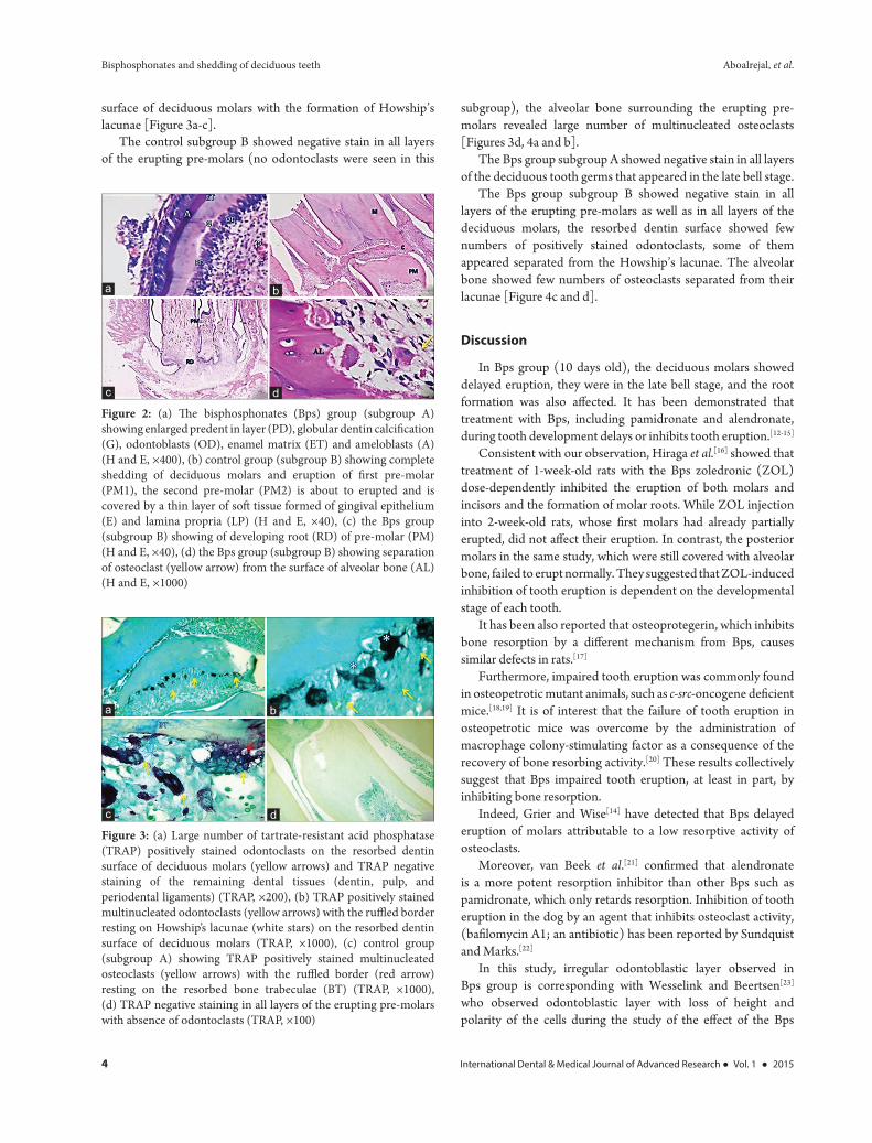
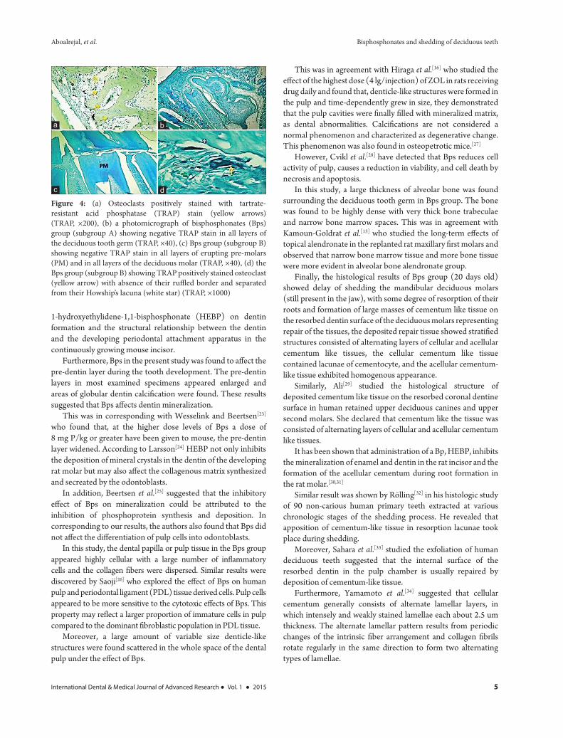
subgroup), the alveolar bone surrounding the erupting pre-molars revealed large number of multinucleated osteoclasts [Figures 3d, 4a and b].
The Bps group subgroup A showed negative stain in all layers of the deciduous tooth germs that appeared in the late bell stage.
The Bps group subgroup B showed negative stain in all layers of the erupting pre-molars as well as in all layers of the deciduous molars, the resorbed dentin surface showed few numbers of positively stained odontoclasts, some of them appeared separated from the Howship’s lacunae. The alveolar bone showed few numbers of osteoclasts separated from their lacunae [Figure 4c and d].
Discussion
In Bps group (10 days old), the deciduous molars showed delayed eruption, they were in the late bell stage, and the root formation was also affected. It has been demonstrated that treatment with Bps, including pamidronate and alendronate, during tooth development delays or inhibits tooth eruption.[12-15]
Consistent with our observation, Hiraga et al.[16] showed that treatment of 1-week-old rats with the Bps zoledronic (ZOL) dose-dependently inhibited the eruption of both molars and incisors and the formation of molar roots. While ZOL injection into 2-week-old rats, whose first molars had already partially erupted, did not affect their eruption. In contrast, the posterior molars in the same study, which were still covered with alveolar bone, failed to erupt normally. They suggested that ZOL-induced inhibition of tooth eruption is dependent on the developmental stage of each tooth.
It has been also reported that osteoprotegerin, which inhibits bone resorption by a different mechanism from Bps, causes similar defects in rats.[17]
Furthermore, impaired tooth eruption was commonly found in osteopetrotic mutant animals, such as c-src-oncogene deficient mice.[18,19] It is of interest that the failure of tooth eruption in osteopetrotic mice was overcome by the administration of macrophage colony-stimulating factor as a consequence of the recovery of bone resorbing activity.[20] These results collectively suggest that Bps impaired tooth eruption, at least in part, by inhibiting bone resorption.
Indeed, Grier and Wise[14] have detected that Bps delayed eruption of molars attributable to a low resorptive activity of osteoclasts.
Moreover, van Beek et al.[21] confirmed that alendronate is a more potent resorption inhibitor than other Bps such as pamidronate, which only retards resorption. Inhibition of tooth eruption in the dog by an agent that inhibits osteoclast activity, (bafilomycin A1; an antibiotic) has been reported by Sundquist and Marks.[22]
In this study, irregular odontoblastic layer observed in Bps group is corresponding with Wesselink and Beertsen[23] who observed odontoblastic layer with loss of height and polarity of the cells during the study of the effect of the Bps
Figure 2: (a) The bisphosphonates (Bps) group (subgroup A) showing enlarged predent in layer (PD), globular dentin calcification (G), odontoblasts (OD), enamel matrix (ET) and ameloblasts (A) (H and E, ×400), (b) control group (subgroup B) showing complete shedding of deciduous molars and eruption of first pre-molar (PM1), the second pre-molar (PM2) is about to erupted and is covered by a thin layer of soft tissue formed of gingival epithelium (E) and lamina propria (LP) (H and E, ×40), (c) the Bps group (subgroup B) showing of developing root (RD) of pre-molar (PM) (H and E, ×40), (d) the Bps group (subgroup B) showing separation of osteoclast (yellow arrow) from the surface of alveolar bone (AL) (H and E, ×1000)
dc
ba
Figure 3: (a) Large number of tartrate-resistant acid phosphatase (TRAP) positively stained odontoclasts on the resorbed dentin surface of deciduous molars (yellow arrows) and TRAP negative staining of the remaining dental tissues (dentin, pulp, and periodental ligaments) (TRAP, ×200), (b) TRAP positively stained multinucleated odontoclasts (yellow arrows) with the ruffled border resting on Howship’s lacunae (white stars) on the resorbed dentin surface of deciduous molars (TRAP, ×1000), (c) control group (subgroup A) showing TRAP positively stained multinucleated osteoclasts (yellow arrows) with the ruffled border (red arrow) resting on the resorbed bone trabeculae (BT) (TRAP, ×1000), (d) TRAP negative staining in all layers of the erupting pre-molars with absence of odontoclasts (TRAP, ×100)
dc
ba
Aboalrejal, et al. Bisphosphonates and shedding of deciduous teeth
International Dental & Medical Journal of Advanced Research ● Vol. 1 ● 2015 5
1-hydroxyethylidene-1,1-bisphosphonate (HEBP) on dentin formation and the structural relationship between the dentin and the developing periodontal attachment apparatus in the continuously growing mouse incisor.
Furthermore, Bps in the present study was found to affect the pre-dentin layer during the tooth development. The pre-dentin layers in most examined specimens appeared enlarged and areas of globular dentin calcification were found. These results suggested that Bps affects dentin mineralization.
This was in corresponding with Wesselink and Beertsen[23] who found that, at the higher dose levels of Bps a dose of 8 mg P/kg or greater have been given to mouse, the pre-dentin layer widened. According to Larsson[24] HEBP not only inhibits the deposition of mineral crystals in the dentin of the developing rat molar but may also affect the collagenous matrix synthesized and secreated by the odontoblasts.
In addition, Beertsen et al.[25] suggested that the inhibitory effect of Bps on mineralization could be attributed to the inhibition of phosphoprotein synthesis and deposition. In corresponding to our results, the authors also found that Bps did not affect the differentiation of pulp cells into odontoblasts.
In this study, the dental papilla or pulp tissue in the Bps group appeared highly cellular with a large number of inflammatory cells and the collagen fibers were dispersed. Similar results were discovered by Saoji[26] who explored the effect of Bps on human pulp and periodontal ligament (PDL) tissue derived cells. Pulp cells appeared to be more sensitive to the cytotoxic effects of Bps. This property may reflect a larger proportion of immature cells in pulp compared to the dominant fibroblastic population in PDL tissue.
Moreover, a large amount of variable size denticle-like structures were found scattered in the whole space of the dental pulp under the effect of Bps.
This was in agreement with Hiraga et al.[16] who studied the effect of the highest dose (4 lg/injection) of ZOL in rats receiving drug daily and found that, denticle-like structures were formed in the pulp and time-dependently grew in size, they demonstrated that the pulp cavities were finally filled with mineralized matrix, as dental abnormalities. Calcifications are not considered a normal phenomenon and characterized as degenerative change. This phenomenon was also found in osteopetrotic mice.[27]
However, Cvikl et al.[28] have detected that Bps reduces cell activity of pulp, causes a reduction in viability, and cell death by necrosis and apoptosis.
In this study, a large thickness of alveolar bone was found surrounding the deciduous tooth germ in Bps group. The bone was found to be highly dense with very thick bone trabeculae and narrow bone marrow spaces. This was in agreement with Kamoun-Goldrat et al.[13] who studied the long-term effects of topical alendronate in the replanted rat maxillary first molars and observed that narrow bone marrow tissue and more bone tissue were more evident in alveolar bone alendronate group.
Finally, the histological results of Bps group (20 days old) showed delay of shedding the mandibular deciduous molars (still present in the jaw), with some degree of resorption of their roots and formation of large masses of cementum like tissue on the resorbed dentin surface of the deciduous molars representing repair of the tissues, the deposited repair tissue showed stratified structures consisted of alternating layers of cellular and acellular cementum like tissues, the cellular cementum like tissue contained lacunae of cementocyte, and the acellular cementum-like tissue exhibited homogenous appearance.
Similarly, Ali[29] studied the histological structure of deposited cementum like tissue on the resorbed coronal dentine surface in human retained upper deciduous canines and upper second molars. She declared that cementum like the tissue was consisted of alternating layers of cellular and acellular cementum like tissues.
It has been shown that administration of a Bp, HEBP, inhibits the mineralization of enamel and dentin in the rat incisor and the formation of the acellular cementum during root formation in the rat molar.[30,31]
Similar result was shown by Rölling[32] in his histologic study of 90 non-carious human primary teeth extracted at various chronologic stages of the shedding process. He revealed that apposition of cementum-like tissue in resorption lacunae took place during shedding.
Moreover, Sahara et al.[33] studied the exfoliation of human deciduous teeth suggested that the internal surface of the resorbed dentin in the pulp chamber is usually repaired by deposition of cementum-like tissue.
Furthermore, Yamamoto et al.[34] suggested that cellular cementum generally consists of alternate lamellar layers, in which intensely and weakly stained lamellae each about 2.5 um thickness. The alternate lamellar pattern results from periodic changes of the intrinsic fiber arrangement and collagen fibrils rotate regularly in the same direction to form two alternating types of lamellae.
Figure 4: (a) Osteoclasts positively stained with tartrate-resistant acid phosphatase (TRAP) stain (yellow arrows) (TRAP, ×200), (b) a photomicrograph of bisphosphonates (Bps) group (subgroup A) showing negative TRAP stain in all layers of the deciduous tooth germ (TRAP, ×40), (c) Bps group (subgroup B) showing negative TRAP stain in all layers of erupting pre-molars (PM) and in all layers of the deciduous molar (TRAP, ×40), (d) the Bps group (subgroup B) showing TRAP positively stained osteoclast (yellow arrow) with absence of their ruffled border and separated from their Howship’s lacuna (white star) (TRAP, ×1000)
dc
ba
Bisphosphonates and shedding of deciduous teeth Aboalrejal, et al.
6 International Dental & Medical Journal of Advanced Research ● Vol. 1 ● 2015
At 20 days, old Bps resulted in the separation of some odontoclasts from their lacunae. This was in corresponding with Watanabe et al.[35] who observed that after the administration of Bps to 6 days old rabbits, many odontoclasts detached from the dentine surface of the deciduous teeth. The same observation appeared in the alveolar bone surrounding the erupting pre-molars at 20 days Bps group. Some osteoclasts appeared separated from the resorbed bone surface, and other osteoclasts appeared apoptotic with the loss of their ruffled borders.
Direct inhibition of osteoclast activity is suggested by in vivo observations of the disappearance of the ruffled border, which is the convoluted osteoclast membrane opposed to bone, associated with osteoclast activity.[36]
In agreement with our study, Watanabe et al.[35] observed that osteoclasts exhibit cytological alteration after treatment with Bps, which induces apoptosis of osteoclasts and loss of their ruffled borders.
Bps directly or indirectly induces apoptosis in osteoclasts, little is known about mediators of apoptosis in osteoclasts, which are difficult to culture. All Bps induce the caspase-dependent formation of pyknotic nuclei.[37]
In this study control specimens stained with TRAP histochemical stain showed a large number of positively stained multinucleated odontoclasts on the resorbed dentin surface of deciduous molars. While Bps specimens stained with TRAP histochemical stain showed few numbers of positively stained odontoclasts, some of them appeared separated from the Howship’s lacunae.
In addition, the histochemical results of Sahara et al.[38] showed TRAP-positive mononuclear cells, supposed to be pre-odontoclasts, first appeared near the blood vessels in the connective tissues, pulp or dental follicle. Then they came in contact with the tooth surface, dentin or cementum, by elongation of their cell processes. After attachment to the tooth surface, they fused with each other and formed multinucleate odontoclasts.
According to Sahara,[39] in the case of the internal resorption of human deciduous teeth, the odontoblasts on the pre-dentin surface had mostly degenerated, atrophied or even disappeared from the pre-dentin surface before the arrival of the pre-odontoclasts.
Similar results were reported by Bradaschia-Correa et al.[15] who suggested that alendronate treated specimens showed few TRAP-positive multinucleated cells in the root surface of deciduous molar. However, these cells exhibited latent phenotype because they were not adhered to the mineralized surfaces. The presence of these cells suggests that all the molecules and factors of the signaling eruptive cascade have been triggered and act to stimulate odontoclast/osteoclast formation.[40]
The alveolar bone surrounding the erupting pre-molars in the control group of this study exhibited large number of TRAP positively stained multinucleated osteoclasts distributed all over the bone surface. While in Bps group the alveolar bone showed very few to negative TRAP positively stained osteoclasts, some of them were detached from the bone surface. This was in
agreement with Lee et al.[41] who examined the effect of Bps on bone remodeling of rat sagittal suture after rapid expansion. They reported that the multinuclear giant cells and TRAP activity were reduced in the Bps injected group compared with the saline injection group.
In addition, Myoung et al.[42] demonstrated that in TRAP specific staining, osteoclastic activity was lower in the Bps treated group than in the control group after autogenous free-bone grafting in rats.
Moreover, Altundal and Güvener[43] indicated that topical application of Bps inhibited experimental tooth movement in rats and decreased a number of multinuclear giant cells.
Overall, the histological results of this study were coincide with the TRAP histochemical results suggesting that the multinucleated bone resorbing cell (osteoclasts) as well as tooth resorbing cell (odontoclasts) are the ultimate target of Bp action. According to Rodan[44] different compounds of Bps act differently on the osteoclast. The following effects have been described: (a) Inhibition of osteoclast formation/recruitment, (b) inhibition of osteoclast activation, (c) inhibition of the activity of mature osteoclasts, and (d) reduction of the osteoclast life span by induction of apoptosis.
Conclusion
Our results suggest that treatment with Bps during tooth development has the potential to retard tooth eruption (due to inhibition of alveolar bone resorption) and induce several types of dental abnormalities. However, clinical evidence to support the results is limited at the moment. Extensive human clinical studies have to be performed to discuss the implications of Bps therapy for tooth development in pediatric populations.
Within the limitations of this study, it can be concluded that:• 10 days old rabbits of the control group revealed eruption
of the deciduous molars with considerable amount of root resorption.
• Both external and internal resorption could be detected during shedding of the deciduous molars of the control group of rabbits.
• 20 days old rabbits of the control group revealed complete shedding of deciduous molars and beginning of eruption of pre-molars.
• Treatment with sodium alendronate (2.5 mg/kg body wt./day) for 10 days from 1st day of birth resulted in delayed eruption of deciduous molars.
• Treatment with sodium alendronate (2.5 mg/kg body wt./day) for 10 days resulted in some dental abnormalities as enlargements of pre-dentin layer, discontinuity of odontoblastic layer, defective dentin mineralization and appearance of denticle-like structure in the pulp.
• Treatment with sodium alendronate (2.5 mg/kg body wt./day) for 20 days resulted in delayed shedding of deciduous molars, delayed eruption of pre-molars and formation of cementum like tissue between them.
Aboalrejal, et al. Bisphosphonates and shedding of deciduous teeth
International Dental & Medical Journal of Advanced Research ● Vol. 1 ● 2015 7
• Alveolar bone abnormalities as a result of Bps treatment were observed in form of increased density, decrease numbers of osteoclast and their detachments from the alveolar bone surface.
References
1. Zoba A, Rabab M. Histomorphological study of dentine pulp complex of continuously growing teeth in the rabbits. Life Sci J 2012;9:1554-64.
2. Hirschfeld Z, Weinreb MM, Michaeli Y. Incisors of the rabbit: Morphology, histology, and development. J Dent Res 1973;52:377-84.
3. Russell RG. Bisphosphonates: From bench to bedside. Ann N Y Acad Sci 2006;1068:367-401.
4. Roelofs AJ, Thompson K, Ebetino FH, Rogers MJ, Coxon FP. Bisphosphonates: Molecular mechanisms of action and effects on bone cells, monocytes and macrophages. Curr Pharm Des 2010;16:2950-60.
5. Hayman AR, Bune AJ, Bradley JR, Rashbass J, Cox TM. Osteoclastic tartrate-resistant acid phosphatase (Acp 5): Its localization to dendritic cells and diverse murine tissues. J Histochem Cytochem 2000;48:219-28.
6. Massa LF, Bradaschia-Correa V, Arana-Chavez VE. Immunocytochemical study of amelogenin deposition during the early odontogenesis of molars in alendronate-treated newborn rats. J Histochem Cytochem 2006;54:713-25.
7. Bancroft JD, Gamble M. Theory and Practice of Histological Techniques. New York: Elsevier Health Sciences; 2008.
8. Warshawsky H, Moore G. A technique for the fixation and decalcification of rat incisors for electron microscopy. J Histochem Cytochem 1967;15:542-9.
9. Sakakura Y, Yajima T, Tsuruga E. Confocal laser scanning microscopic study [corrected] of tartrate-resistant acid phosphatase-positive cells in the dental follicle during early morphogenesis of mouse embryonic molar teeth. Arch Oral Biol 1998;43:353-60.
10. Cole AA, Walters LM. Tartrate-resistant acid phosphatase in bone and cartilage following decalcification and cold-embedding in plastic. J Histochem Cytochem 1987;35:203-6.
11. Fuenzalida M, Illanes J, Lemus R, Guerrero A, Oyarzún A, Acuña O, et al. Microscopic and histochemical study of odontoclasts in physiologic resorption of teeth of the polyphyodont lizard, Liolaemus gravenhorsti. J Morphol 1999;242:295-309.
12. Hodgson B. More about bisphosphonates. J Am Dent Assoc 2009;140:829-30.
13. Kamoun-Goldrat A, Ginisty D, Le Merrer M. Effects of bisphosphonates on tooth eruption in children with osteogenesis imperfecta. Eur J Oral Sci 2008;116:195-8.
14. Grier RL 4th, Wise GE. Inhibition of tooth eruption in the rat by a bisphosphonate. J Dent Res 1998;77:8-15.
15. Bradaschia-Correa V, Massa LF, Arana-Chavez VE. Effects of alendronate on tooth eruption and molar root formation in young growing rats. Cell Tissue Res 2007;330:475-85.
16. Hiraga T, Ninomiya T, Hosoya A, Nakamura H. Administration of the bisphosphonate zoledronic acid during tooth development inhibits tooth eruption and formation and induces dental abnormalities in rats. Calcif Tissue Int 2010;86:502-10.
17. Kostenuik P. Inhibition of bone resorption by OPG or alendronate significantly reduced bone growth, molar eruption and tooth root development in neonatal rats. Bone 2009;45:S50.
18. Helfrich MH. Osteoclast diseases and dental abnormalities. Arch Oral Biol 2005;50:115-22.
19. Soriano P, Montgomery C, Geske R, Bradley A. Targeted disruption of the c-src proto-oncogene leads to osteopetrosis in mice. Cell 1991;64:693-702.
20. Kodama H, Yamasaki A, Nose M, Niida S, Ohgame Y, Abe M, et al. Congenital osteoclast deficiency in osteopetrotic (op/op) mice is cured by injections of macrophage colony-stimulating factor. J Exp Med 1991;173:269-72.
21. van Beek ER, Cohen LH, Leroy IM, Ebetino FH, Löwik CW, Papapoulos SE. Differentiating the mechanisms of antiresorptive action of nitrogen containing bisphosphonates. Bone 2003;33:805-11.
22. Sundquist KT, Marks SC Jr. Bafilomycin A1 inhibits bone resorption and tooth eruption in vivo. J Bone Miner Res 1994;9:1575-82.
23. Wesselink PR, Beertsen W. The influence of 1-hydroxyethylidene 1,1-bisphosphonate (HEBP) on dental root resorption in the mouse. Calcif Tissue Int 1989;45:104-10.
24. Larsson A. The short-term effects of high doses of ethylene-1-hydroxy-1, 1-diphosphonate upon early dentin formation. Calcif Tissue Res 1974;16:109-27.
25. Beertsen W, Niehof A, Everts V. Effects of 1-hydroxyethylidene-1, 1-bisphosphonate (HEBP) on the formation of dentin and the periodontal attachment apparatus in the mouse. Am J Anat 1985;174:83-103.
26. Saoji N. Effect of Bisphosphonate on Osteogenic Differentiation of Pulp and PDL Cells Masterdegree Thesis, University of Alabama at Birmingham; 2008.
27. Ida-Yonemochi H, Noda T, Shimokawa H, Saku T. Disturbed tooth eruption in osteopetrotic (op/op) mice: Histopathogenesis of tooth malformation and odontomas. J Oral Pathol Med 2002;31:361-73.
28. Cvikl B, Agis H, Stögerer K, Moritz A, Watzek G, Gruber R. The response of dental pulp-derived cells to zoledronate depends on the experimental model. Int Endod J 2011;44:33-40.
29. Ali ZH. Evaluation of the mineralized tissue in the pulp of retained human deciduous teeth (histological and immunohistochemicalstudy). J Am Sci 2012;8:398-407.
30. Ten Cate AR, Anderson RD. An ultrastructural study of tooth resorption in the kitten. J Dent Res 1986;65:1087-93.
31. Sasaki T, Shimizu T, Watanabe C, Hiyoshi Y. Cellular roles in physiological root resorption of deciduous teeth in the cat. J Dent Res 1990;69:67-74.
32. Rölling I. Histomorphometric analysis of primary teeth during the process of resorption and shedding. Scand J Dent Res 1981;89:132-42.
33. Sahara N, Okafuji N, Toyoki A, Ashizawa Y, Yagasaki H, Deguchi T, et al. A histological study of the exfoliation of human deciduous teeth. J Dent Res 1993;72:634-40.
34. Yamamoto T, Li M, Liu Z, Guo Y, Hasegawa T, Masuki H, et al. Histological review of the human cellular cementum with special reference to an alternating lamellar pattern. Odontology 2010;98:102-9.
35. Watanabe J, Amizuka N, Noda T, Ozawa H. Cytochemical and ultrastructural examination of apoptotic odontoclasts
Bisphosphonates and shedding of deciduous teeth Aboalrejal, et al.
8 International Dental & Medical Journal of Advanced Research ● Vol. 1 ● 2015
induced by bisphosphonate administration. Cell Tissue Res 2000;301:375-87.
36. Sato M, Grasser W, Endo N, Akins R, Simmons H, Thompson DD, et al. Bisphosphonate action. Alendronate localization in rat bone and effects on osteoclast ultrastructure. J Clin Invest 1991;88:2095-105.
37. Reszka AA, Halasy-Nagy JM, Masarachia PJ, Rodan GA. Bisphosphonates act directly on the osteoclast to induce caspase cleavage of mst1 kinase during apoptosis. A link between inhibition of the mevalonate pathway and regulation of an apoptosis-promoting kinase. J Biol Chem 1999;274:34967-73.
38. Sahara N, Toyoki A, Ashizawa Y, Deguchi T, Suzuki K. Cytodifferentiation of the odontoclast prior to the shedding of human deciduous teeth: An ultrastructural and cytochemical study. Anat Rec 1996;244:33-49.
39. Sahara N. Cellular events at the onset of physiological root resorption in rabbit deciduous teeth. Anat Rec 2001;264:387-96.
40. Wise GE, Frazier-Bowers S, D’Souza RN. Cellular, molecular, and genetic determinants of tooth eruption. Crit Rev Oral Biol Med 2002;13:323-34.
41. Lee K, Sugiyama H, Imoto S, Tanne K. Effects of bisphosphonate on the remodeling of rat sagittal suture after rapid expansion. Angle Orthod 2001;71:265-73.
42. Myoung H, Park JY, Choung PH. Effects of a bisphosphonate on the expression of bone specific genes after autogenous free bone grafting in rats. J Periodontal Res 2001;36:244-51.
43. Altundal H, Güvener O. The effect of alendronate on resorption of the alveolar bone following tooth extraction. Int J Oral Maxillofac Surg 2004;33:286-93.
44. Rodan GA. Mechanisms of action of bisphosphonates. Annu Rev Pharmacol Toxicol 1998;38:375-88.
How to cite this article: Aboalrejal AN, El-Motayam AM, Fares AE, Saleh SY. Effect of bisphosphonates sodium alendronate on shedding of deciduous molars in rabbits (histological and histochemical study). Int Dent Med J Adv Res 2015;1:1-8.