easl clinical practice guidelines on the prevention ... · gallstones or cholelithiasis are a major...
TRANSCRIPT
EASL Clinical Practice Guidelines on the prevention, diagnosisand treatment of gallstonesq
European Association for the Study of the Liver (EASL) ⇑
Introduction
Gallstones or cholelithiasis are a major public health problem inEurope and other developed countries and affect up to 20% of thepopulation. Gallstone disease is the most common gastrointesti-nal disorder for which patients are admitted to hospitals inEuropean countries [1]. The interdisciplinary care for patientswith gallstone disease has advanced considerably during recentdecades thanks to a growing insight into the pathophysiologicalmechanisms and remarkable technical developments in endo-scopic and surgical procedures. In contrast, primary preventionfor this common disease is still in its infancy.
The EASL Clinical Practice Guidelines (CPG) on the prevention,diagnosis and therapy of gallstones aim to provide currentrecommendations on the following issues:
1. Prevention of gallstones2. Diagnosis of gallbladder stones3. Medical therapy of gallbladder stones4. Surgical therapy of gallbladder stones5. Diagnosis of bile duct stones6. Endoscopic and surgical therapy of bile duct stones7. Diagnosis and therapy of intrahepatic stones8. Therapy of gallstones during pregnancy
The EASL CPG on gallstone disease define the use of preven-tive, diagnostic and therapeutic modalities, including medical,endoscopic and surgical procedures, in the management ofpatients with gallstones. They are intended to assist physiciansand other professional healthcare workers as well as patientsand interested individuals in the clinical decision making processby describing a range of generally accepted approaches for theprevention, diagnosis and treatment of gallstone disease.
These guidelines have been produced using evidence fromPubMed and Cochrane database searches until September 2015.The evidence and recommendations in these guidelines havebeen graded on the strength of the supporting evidence accordingto the Grading of Recommendations Assessment Development
and Evaluation (GRADE) [2–5]. We considered within-study riskof bias (methodological quality), directness of evidence, hetero-geneity, precision of effect estimates, and risk of publication bias.Each recommendation has been qualified by giving the grade ofevidence underlying the recommendation. The evidence isgraded as follows: (A) high quality evidence: further research isvery unlikely to change our confidence in the estimate of effect(randomized trials or double-upgraded observational studies);(B) moderate quality evidence: further research is likely to havean important impact on our confidence in the estimate of effectand may change the estimate (downgraded randomized trialsor upgraded observational studies); (C) low quality evidence:further research is very likely to have an important impact onour confidence in the estimate of effect and is likely to changethe estimate (observational studies or double-downgradedrandomized trials); and (D) very low quality evidence: we arevery uncertain about the estimate (case series/case reports,downgraded observational studies, triple-downgraded random-ized trials). The strength of the recommendations is based onboth the aggregate evidence quality and an assessment of theanticipated benefits and harms. A strong recommendation hasbeen made when there is certainty about the various factors thatdetermine the strength of a recommendation, and most or all ofthe individuals in the relevant population will benefit by follow-ing the recommendation; a weak recommendation has beengiven when there is uncertainty about the various factors thatdetermine the strength of a recommendation.
Prevention of gallstones
Primary prevention of gallstones
Both cholesterol and pigment gallstone diseases originate fromthe complex interaction of genetic, environmental, local, systemicand metabolic abnormalities [6]. In Western populationscholesterol gallstones account for 90–95% of all gallstones. Blackpigment stones are the major stone type in patients with chronichaemolytic disorders or cirrhosis, although most patients withblack pigment stones have neither of these conditions. Choles-terol and black pigment stones are nearly always formed in thegallbladder, whereas brown pigment stones develop primarilyin the main bile duct. In Western subjects brown pigment stonesare usually found in the bile ducts following cholecystectomy andin patients with sclerosing cholangitis, whereas in Orientalpatients they occur in association with chronic infectious
Journal of Hepatology 2016 vol. 65 j 146–181
Received 9 March 2016; accepted 9 March 2016q Clinical Practice Guideline Panel: Frank Lammert (Chairman), Monica Acalovschi,Giorgio Ercolani, Karel J. van Erpecum, Kurinchi S. Gurusamy, Cees J. vanLaarhoven, Piero Portincasa.⇑ Corresponding author. Address: European Association for the Study of the Liver(EASL), The EASL Building – Home of European Hepatology, 7 rue Daubin, CH1203 Geneva, Switzerland. Tel.: +41 (0) 22 807 03 60; fax: +41 (0) 22 328 07 24.E-mail address: [email protected].
Clinical Practice Guidelines
cholangitis [7]. Sludge is not a cause of gallstone formation andarises with stasis and reduced enterohepatic bile circulation,although stasis itself will contribute to gallstone formation. Sincegallstone disease is one of the most prevalent and costly digestivediseases in Western countries [8], primary non-pharmacologicalprevention would be desirable in the general population [9]. Sev-eral risk factors exist for cholesterol, pigment and mixed gall-stones. For some non-genetic risk factors, general or specificprimary preventive measures are conceivable.
Lifestyle
Can gallstones be prevented?
Healthy lifestyle and food, regular physical activity and maintenance of an ideal body weight might prevent cholesterol gallbladder stones and symptomatic gallstones (low quality evidence; weak recommendation)
Comment: Lifestyle affects the pathogenesis of cholesterol gall-stones acting on one or more factors belonging to the metabolicsyndrome, namely obesity, diabetes mellitus, and insulin resistance[10–16]. Obesity predisposes to gallstone formation [17] andincreases the risk of cholecystectomy by increasing the risk ofsymptomatic gallstones [18–28]. Thus, increased body mass index(BMI) is a definitive risk factor for gallstone growth [6,20,26,29],and increased BMI per se is also a causal risk factor for symptomaticgallstone disease, particularly in women [30]. The risk of symp-tomatic gallstones has been reported to rise with increasing BMIand waist circumference as well as serum triglycerides [31].
Additional obesity-associated factors facilitating cholesterol gall-stone formation include gallbladder stasis [32–35], insulin resis-tance, dyslipidemia (reduced high density lipoproteins, HDL [31]and hypertriglyceridemia), sedentary lifestyle [30,36], hormonereplacement therapy [30], and fast food consumption [30]. Prospec-tive cohort studies [31,37,38] rather than case-control [28,39,40]and cross-sectional studies [19,41–43] are of value when investigat-ing serum lipids and their association with gallstone disease andobesity. Appropriate lifestyle interventions should therefore focuson ideal weight maintenance and weight reduction among over-weight and obese individuals in the general population [30]. Insulinresistance and diabetes mellitus type 2 are also strongly associatedwith cholesterol gallstones independently of obesity [44]. Such con-ditions represent additional targets for the prevention of gallstones.
Physical activityQuestionnaire-based surveys found that physical activity appearsto protect against gallstone formation [36,45–48] and to cut therisk of symptomatic stones by about 30% [36,45,49–51]. In arecent prospective cohort study (European prospective investiga-tion into cancer (EPIC)-Norfolk) using a validated questionnaireagainst energy expenditure and cardio-respiratory fitness [52],a total of 25,639 volunteers, aged 40–74 years, were ranked intofour groups of increasing physical activity and monitored over14 years for symptomatic gallstones. After 5 and 14 years, 135(uncomplicated) and 290 (complicated) incident cases of sympto-matic gallstones were recorded, respectively (68% women). Thehighest level of physical activity (equivalent to exercising for1 h a day in a sedentary job, or 30 min a day in a standing job,or heavy manual job without any additional activity) was
associated with a 70% decreased risk of symptomatic gallstonesin both sexes; a likely causal effect was particularly seen after5 years. The potential beneficial effects of physical activity on gall-stone formation and associated complications are supported bypathogenic mechanisms. Hyperinsulinemia promotes hepaticuptake of cholesterol [53] predisposing to increased secretion of bil-iary cholesterol [54] and decreased secretion of bile acids (both con-ditions predispose to cholesterol supersaturated lithogenic bile)[55]. By contrast, regular exercise reduces insulin levels [56], insulinresistance [57], triglyceridemia [58], and fatty acid-dependenthypersecretion of gallbladder mucin [59]. Also, during physicalactivity serum HDL-cholesterol levels increase [60,61] as marker ofincreasing reverse cholesterol transport to the liver [62]. Notably,HDL-cholesterol is the precursor of bile acids [63], which contributeto decreased biliary cholesterol saturation, and indeed HDL-choles-terol levels are inversely related to gallstone prevalence [41]. Anadditional effect of physical activity involves the prokinetic effecton the intestine [64] and cholecystokinin-dependent gallbladdercontraction [65]. The importance of maintaining an ideal bodyweight and regular physical activity should therefore be reinforcedin the general population [45], since the overall beneficial effectsof physical activity on cardiovascular health extend beyond the pro-tective effect on gallstone formation [29].
DietLarge population-based, long-term, prospective epidemiologicalstudies aiming to identify the protective value of dietary compo-nents are hampered by difficulties in estimating the precisequantity and ingestion pattern of nutrients. Nevertheless, high-fiber and high-calcium diets reduce biliary hydrophobic bileacids, whereas a regular eating pattern decreases gallbladderstasis by increasing regular gallbladder emptying [45]. Both aspectsplay a preventive role for cholesterol cholelithiasis. The likelihoodof gallstone disease is increased by consumption of typicalWesternized hypercaloric diets [66], including meat intake [48].Reducing total caloric intake might therefore prove useful [67].
Fruit and vegetables [68] might be protective againstgallstone disease, but data on the benefits of vegetarian dietsremain controversial. Although protection might be conferredby a lower BMI [69], and regular use of vegetable oils and vitaminC [46,70], studies on different populations have shown either aprotective effect [71–75] or a lack of a protective effect ofvegetarian diets on gallstones [47,76].
Poly- and monounsaturated fats [77], and specifically nutconsumption [77,78], might protect against gallstone disease,possibly as part of a healthy diet.
Data regarding coffee intake are controversial: caffeine intake(sources: coffee, black tea, and caffeinated soft drinks) and coffeein particular, are reportedly protective in some [79–84], but notall epidemiological studies [47]. Geographical, cultural and drink-ing patterns of coffee might explain discrepant results [47].Besides the potential effect on hepatobiliary secretion of choles-terol and intestinal motility, additional mechanisms of actioncaffeine or coffee intake are still poorly understood.
Although prospective epidemiological studies reported protec-tive effects of alcohol consumption on gallstone formation[31,78,79], and multifactorial analysis indicated that Danish patientswith symptomatic gallstones consume less alcohol as compared tothose with asymptomatic stones [30], the findings are controversial[72,81–83,85,86], and due to its negative effects on overall health,alcohol cannot be recommended for the prevention of gallstones.
JOURNAL OF HEPATOLOGY
Journal of Hepatology 2016 vol. 65 j 146–181 147
Regular vitamin C supplementation or regular use of vitaminC-enriched diet might have a protective effect on gallstone for-mation. In fact, cholesterol conversion to bile acids requires 7a-hydroxylation and an appropriate content of vitamin C in thehepatocyte [87,88]. In humans, vitamin C deficiency might there-fore increase the risk of cholesterol gallstone formation [70]. Ingallstone patients, vitamin C supplementation (500 mg � 4 timesa day) changed biliary bile acid composition, increased phospho-lipids, and proved to be protective by prolonging the crystalliza-tion time of biliary cholesterol [89]. Furthermore, observationalstudies have identified an association between low vitamin Cconsumption and risk of gallstones/gallbladder disease[48,70,90] or cholecystectomy [91]. In a German observationalpopulation-based study (n = 2129 subjects aged 18–65 years),gallstone prevalence by ultrasonography was 4.7% vs. 8.2% inpatients reporting regular use of vitamin C (n = 232) or not usingvitamin C (n = 1897), respectively [92].
Prevention of gallstones in the general population
Is a pharmacological treatment advisable for the prevention of gallstones in the general population?
Pharmacological prevention of gallstones is not advisable in the general population (very low quality evidence; weak recommendation)
Comment: There is no indication for administeringursodeoxycholic acid (UDCA) as a preventive drug for gallstonedisease in the general population, apart from high risk groups(see section Primary prevention of gallstones in high risk groups).Similarly, there is not enough evidence to embark on gallstone/sludge prophylaxis with UDCA in pregnancy (because gallstonemay be transient in this situation) or with omega-3 fatty acidsupplementation [93].
Conflicting results are available on the protective effect ofstatins alone or with UDCA on gallstone disease. The use ofstatins was evaluated in two population-based case-controlstudies. A decreased risk of gallstone disease and cholecystec-tomy emerged with regular use of statins [94,95], a trendconfirmed in the Nurses’ Health Study evaluating the use of statinsover a period of 10 years [96]. A case-controlled study confirmedthe protective effect of statin use on the risk of cholecystectomy[97]. Although results appear promising, the protective effect oflovastatin [98–101], pravastatin alone [102–106] or with UDCA[107], simvastatin alone [103,108–112] or with UDCA [113,114]and fluvastatin [115] on biliary cholesterol saturation, biliary lipidcomposition, cholesterol crystallization, gallstone formation, andstone dissolution is weak and not always confirmed. In a recentmeta-analysis involving a total of 622868 participants from sixstudies (four case-control studies, one cohort study and onecross-sectional study), current statin use was associated with alower risk of cholecystectomy as compared with non-use. Theeffect was significantly more evident for moderate and high statinuse than low statin use (i.e. 1–4 prescriptions) [116]. A Finnishcase-control study matched 272 patients using statins with 272patients not using statins by age and sex to investigate the influ-ence of statin use on complicated gallstone disease at gallbladdersurgery. While patients using statins did not have worse outcomes
after cholecystectomy than non-users, statin treatment wasassociated with a shorter operation time for laparoscopic cholecys-tectomy [117]. So far, however, better controlled studies arerequired to confirm such findings, and statins cannot be proposedfor the prevention of gallstones [118,119].
Ezetimibe is a selective cholesterol absorption inhibitor actingon the intestinal Niemann–Pick C1-like 1 (NPC1L1) protein.Murine studies based on lithogenic diets have shown beneficialeffects of ezetimibe on biliary lipid composition, intestinalcholesterol absorption and biliary cholesterol secretion andsaturation, crystal aggregation, gallstone formation, bile flow,gallbladder motility function, and cholecystosteatosis[120–123]. In the hamster model on a lithogenic diet, ezetimibeprevented the increase of biliary cholesterol and cholesterolaccumulation in the liver [124]. The translational value of sucheffects of ezetimibe was confirmed in a pilot study in cholesterolgallstone patients: ezetimibe reduced biliary cholesterol satura-tion and retarded cholesterol crystallization [120]. However, ina small retrospective, case-control study ezetimibe did notappear to influence the prevalence of gallstones [125]. Morerecently, in a large Danish study involving 67,385 participants,it was shown that genetic variation in NPC1L1, mimicking theeffect of ezetimibe monotherapy, was indeed associated with adose-dependent reduction of serum low-density lipoprotein(LDL) cholesterol concentrations and the risk of ischemic vasculardisease. However, the cumulative incidence of symptomaticgallstone disease increased (sample of 3,886 subjects) [126].The possibility exists that in humans (who express NPC1L1 inintestine and liver) genetically reduced activity of NPC1L1 causeslower uptake rates of cholesterol from both the intestine intoenterocytes and from bile into hepatocytes. The latter effectmight increase the risk of gallstone disease. Gallbladder-relatedadverse effects, however, were not associated with ezetimibetreatment in the IMPROVE-IT trial with a minimum follow-upof 2.5 years (comparing patients treated with ezetimibe plusstatin to patients treated with statin alone) [127]. Overall,these data indicate that the use of ezetimibe for the preventionof cholesterol gallstones warrants further investigation[118,119,128,129]. This therapeutic approach should be put inperspective when confronted with groups of patients displayingmetabolic abnormalities and high cardiovascular risk, the use ofcombined lipid-lowering therapy (statins/ezetimibe), gender-specific gallstone risk (higher in women than in men), and theoverall duration of ezetimibe treatment.
Finally, aspirin is currently not accepted for the prevention ofgallstones [6].
Primary prevention of gallstones in high risk groups
Rapid weight loss
When can ursodeoxycholic acid be used to prevent gallstones in obese patients?
In situations that are associated with rapid weight loss (e.g. very-low-calorie diet, bariatric surgery), temporary ursodeoxycholic acid (at least 500 mg per day until body weight has stabilized) may be recommended (moderate quality evidence; weak recommendation)
Clinical Practice Guidelines
148 Journal of Hepatology 2016 vol. 65 j 146–181
Comment: Increased BMI and female gender are definitiverisk factors for gallstone growth [6,20,26,29]. Increased BMI isalso a causal risk factor for symptomatic gallstone disease [30].Obesity will influence most pathogenic mechanisms for gallstoneformation, i.e. supersaturation of bile with cholesterol, increasedpropensity to cholesterol crystallization, stone aggregation, anddefective gallbladder emptying [6,26,130–134]. However, the riskof gallstones also increases significantly during rapid weightloss (>1.5 kg/week) due to a weight reduction programme[131,135–137] and decreases at approximately 2 years whenbody weight stabilizes [138,139]. Weight cycling is also a modestindependent risk factor for gallstone formation [48,82,132,140].By contrast, progressive reduction of body weight at moderatespeed (max. 1.5 kg/week) [136,141,142] in obese subjectsdecreases excessive de novo biosynthesis and biliary excretionof cholesterol, with decreased risk of gallstone formation. Arecent study performing a multivariate analysis in 171 patientsreported that factors associated with gallstone formation afterbariatric surgery are higher rate of weight loss, progressivedecrease in percentage of gallbladder emptying, prolonged over-night fasting, and reduced intake of calories and fibers [143].
Rapid weight loss can be achieved by very-low-calorie diets(i.e. diets containing less than 800 kcal per day [139,144–147]or bariatric surgery, such as Roux-en-Y gastric bypass (RYGB))[81,131,137–139,148–152]. Although the majority of newlyformed gallstones remains asymptomatic following rapid weightloss, the risk of both uncomplicated and complicated gallstonedisease and cholecystectomy is still increased and is 3-foldgreater in very-low-calorie than in low-calorie diets [139].Appropriate fat content (at least 7 g/day) in very-low-caloriediets might improve gallbladder motility and decrease the riskof symptomatic gallstones, as shown in recent controlled studies[139,153,154]. Patients undergoing rapid weight loss are morelikely to become symptomatic for gallstones, with incidencereaching 28% to 71% after gastric bypass [27,150,151,155,156].Cholecystectomy is indicated in up to one-third of patients by3 years after surgery [27,150]. After bariatric surgery, the risk ofdeveloping gallstone disease increases to 48% for weight lossgreater than 25% of original weight, especially after gastric bypassor sleeve gastrectomy [157–162]. The same trend is observed inobese patients using hypocaloric diets postoperatively [137].
In obese patients undergoing rapid weight loss either withvery-low-calorie diets or bariatric surgery without cholecystec-tomy, the litholytic hydrophilic UDCA prevents cholesterolgallstone formation following rapid weight reduction. However,the costs of chronic treatment and patient compliance have tobe considered [137,138,148–151,163]. A meta-analysis of 13randomised control trials (RCTs) on the protective effect of UDCAduring weight loss (1,791 patients, 1,217 randomized to UDCAand 574 randomized to placebo) confirmed that UDCA (range300–1,200 mg/day) can prevent gallbladder stone formation dur-ing dieting or after bariatric surgery [164]. Treatment with UDCAshould last until body weight is stabilized at a dose (range500–600 mg/day) that is lower than for litholysis [150]. Indeed,treatment efficacy is best during the period of weight loss, sincethe risk of developing stones decreases once the weight hasstabilized [150]. A decision tree analysis shows that gallstoneprevention with UDCA lowers costs [165]. UDCA has becomethe standard prophylactic treatment for cholesterol cholelithiasisin obese patients following very-low-calorie diets or afterbariatric surgery. Patients undergoing either vertical banded
gastroplasty or adjustable gastric banding were randomized toplacebo or 500 mg UDCA/day. Incidence of gallstone formationat 12 and 24 months was 22% and 30% (placebo group) and 3%and 8% (UDCA), respectively. Cholecystectomy rate was 12%and 5% in placebo and UDCA groups, respectively [138]. In thestudy of Wudel et al. [151], gallstones developed in 71% ofpatients within 12 months of gastric bypass; 41% of thesegallstone patients became symptomatic, and 67% of symptomatic,patients were cholecystectomized. UDCA was effective inpreventing gallstone formation as compared to placebo, but amajor concern was the poor therapeutic outcome due to lack ofcompliance. Further studies are required to confirm that acombined intervention (e.g. diet plus UDCA) has thepotential to improve stone prevention during weight loss[143,166].
The beneficial effect of fish oil (n-3) polyunsaturated fattyacids on biliary crystallization was confirmed in a randomizeddouble-blind placebo-controlled trial in obese women duringrapid weight loss with a hypocaloric diet (1200 kcal/day), andcompared with UDCA (1200 mg/day) [93].
No severe side effect can be expected with UDCA at the dosageemployed in previous studies (i.e. 300–1200 mg/day)[137,138,147,148,151]. Sugerman et al. [150] noted that somepatients on UDCA dropped out because of ‘‘vomiting or skinrashes”, but similar rates were observed in the placebo group.Similar adverse events between UDCA and placebo, unrelatedto the dose of UDCA, were reported by Shiffman et al. [147] (i.e.constipation, headache, diarrhea, dizziness, and upper respiratoryinfections, ranging from 16% to 30% of patients).
No indication exists for aspirin use to prevent gallstone recur-rence [167].
Should prophylactic cholecystectomy be performed during bariatric surgery in obese subjects undergoing rapid weight loss?
Prophylactic cholecytectomy is not routinely indicated during bariatric surgery (very low quality evidence; weak recommendation)
Comment: Gallstone-related complications after bariatric sur-gery generally appear within 7–18 months [168–172]. During amedian follow-up of 3 years, almost 20% of patients undergoinglaparoscopic RYGB with an intact gallbladder became symp-tomatic and required cholecystectomy. The estimated 5-yeargallbladder disease-free survival was low (77.4%) [173]. Anothertheoretical advantage of prophylactic cholecystectomy wouldbe prevention of future bile duct stones, which can be difficultto remove endoscopically after RYGB, due to altered anatomy.Based on such estimates, concurrent prophylactic cholecystec-tomy during RYGB has previously been recommended, based onthe rationale that the conversion rate to open surgery is notincreased, neither is operative time nor hospital stay [173].Further studies, however, have suggested that most patientsremain asymptomatic [156,157,160,168,169,174–178] and neverrequire further interventions following RYGB. Thus, concurrent(prophylactic) cholecystectomy during laparoscopic bypasssurgery is no longer routinely performed [168,169,174–176,179].
JOURNAL OF HEPATOLOGY
Journal of Hepatology 2016 vol. 65 j 146–181 149
Essentially, cholecystectomy is reserved for the subgroup ofpatients with symptomatic gallstones or abnormal gallbladder find-ings (e.g. chronic cholecystitis, tumor-like lesions) [152,171,173,180,181]. This assumption stands despite the fact that post-RYGBcholecystectomy in symptomatic gallstone patients becomesmore difficult and endoscopic retrograde cholangiopancreatogra-phy (ERCP) may not be feasible for anatomical reasons [182].
With areas of uncertainties concerning the most cost-effectivestrategy for gallbladder management in patients undergoingRYGB, a recent decision model was developed on the US Healthsystem background [183]. Three possible options werecompared: prophylactic concurrent cholecystectomy, RYGB withpreserved gallbladder (with or without postoperative UDCAtherapy), and selective cholecystectomy only for patients withgallstones identified by ultrasonography. The most cost-effectivestrategy was RYGB without cholecystectomy, provided that therisk of post-surgical gallstone complications remains low [180]and UDCA is not used. UDCA treatment appears a too expensiveoption after RYGB, and in this case concurrent cholecystectomybecomes less costly. Another limitation with UDCA use is thevariable compliance to daily prescription, ranging from 40% to85% [150,151,158,168,172].
Long-term therapy with somatostatin or analogues
Is primary prevention of gallstones with ursodeoxycholic acid of indicated in patients on somatostatin or analogue treatment?
In patients on long-term therapy with somatostatin or analogues, concomitant treatment with ursodeoxycholic acid can be considered to prevent cholesterol gallstone formation (low quality evidence; weak recommendation)
Comment: Patients requiring long-term therapy with somato-statin or various analogues (e.g. patients with neuroendocrineneoplasms) exhibit prolongation of intestinal transit, severelyimpaired gallbladder emptying despite preserved postprandialcholecystokinin (CCK) release [184], and several lithogenicchanges in bile [185–188]. Despite the frequent occurrence ofgallstones, they infrequently become symptomatic or promptacute surgery [189]. Careful follow-up of these patients withrespect to cholelithogenic changes is recommended, and con-comitant treatment with UDCA might be considered[186,187,190].
Total parenteral nutrition
Is primary prevention of gallstones indicated during total parenteral nutrition?
Patients on total parenteral nutrition are at increased risk of gallbladder sludge formation but no recommendation for prevention can be given (very low quality evidence; weak recommendation)
Comment: Biliary sludge is often found incidentally in condi-tions of increased gallbladder stasis and/or concurrent change ofbiliary composition, e.g. prolonged fasting (especially during totalparenteral nutrition, TPN) [191]. Due to transient changes ofgallbladder kinetics and biliary composition, both sludge andsmall gallstones might disappear after restoration of oral diet(e.g. three meals a day with sufficient fat to improve gallbladderemptying and sludge clearance) [192–196]. Patients on TPNshould be shifted to enteral nutrition whenever possible. Contro-versial data exist concerning the stimulation of the gallbladder inTPN with CCK (either by daily exogenous CCK or by fast infusionof high doses of crystalline amino acids) [192,193,197–199]. Inone study a mixed soybean/medium chain triglyceride/olive/fishoil emulsion used for long-term parenteral nutrition was associ-ated with both disappearance and decreased size of gallstonesafter 3 and 2 months, respectively in two children continuingUDCA at 15 mg/kg/day [200]. The overall results of such studies,albeit convincing, are hampered by the low number of cases.Furthermore, there is no indication for prophylactic treatment withUDCA in patients with sludge after TPN has been interrupted [190].Use of omega-3 fatty acid-enriched TPN likely increased omega-3fatty acids content in biliary phosphatidylcholines and decreasedbiliary supersaturation in cholesterol [201] with a mechanism alsoinvolving biliary mucin suppression [202].
Hormone therapy
Is there an indication for pharmacological or surgical prevention of gallstones during hormone replacement therapy?
Physicians who prescribe hormone replacement therapy should be aware of the increased risk for gallstones. Currently there is no indication for pharmacological orsurgical stone prevention during hormone replacement therapy (very low quality evidence; weak recommendation)
Comment: Hormone therapy is widely used for controllingmenopausal symptoms and has also been used for the manage-ment and prevention of cardiovascular disease, osteoporosisand dementia in older women. A recent Cochrane meta-analysis[203] compared the effects of hormone therapy by oral, transder-mal, subcutaneous or intranasal routes (oestrogen-only andcombined continuous with or without progestogens) withplacebo for 3 to 7 years. From 23 randomized double-blind studies(involving 42,830 women aged 26 to 91 years, mainly from theHeart and Estrogen-progestin Replacement Study (HERS) 1998and the Women’s Health Initiative (WHI) 1998 study) resultsshowed a significantly increased risk of gallbladder disease withoestrogen-only (absolute risk increase from 26 to 45 per 1000,95% confidence interval (CI = 36–57), with combined continuoustreatment (absolute risk from 27 to 47 per 1000, 95% CI = 38–60),including postmenopausal women with cardiovascular disease[204,205]. The risk started to increase in the active group in thefirst year. Caution is therefore recommended in prescribingdifferent types of continuous hormone therapy for controllingmenopausal symptoms. While carefully evaluating potentialsevere health hazards, treatment should be reserved for groups
Clinical Practice Guidelines
150 Journal of Hepatology 2016 vol. 65 j 146–181
at low risk of cardiovascular disease, venous thrombo-embolism, orbreast cancer. The risk of gallbladder disease is well established,but medical gallstone prophylaxis has not been addressed in ran-domized trials thus far.
Prevention of recurrent bile duct stones
Are there effective strategies to prevent recurrent bile duct stones?
No general recommendation can be given for the pharmacological prevention of recurrent bile duct stones (very low quality evidence; weak recommendation)
Comment: Recurrent bile duct stones are observed in 5–20%of patients after endoscopic sphincterotomy [206–211] and canusually be removed endoscopically. Currently, there are no vali-dated prophylactic measures. No consistent benefit of pharmaco-logical secondary prevention has been observed, and data on thepotential effects of UDCA [212] have not been validated in ran-domized controlled trials [212,213].
Patients with mutations of the gene encoding the phos-phatidylcholine floppase ABCB4 have a monogenic predispositionfor low phospholipid-associated cholelithiasis (LPAC). Due to lowbiliary phospholipid concentrations, cholesterol gallstone diseasedevelops before the age of 40 years with intrahepatic bile ductand gallbladder cholesterol stones and recurrent biliary symp-toms after cholecystectomy [214–217]. The diagnosis is basedon medical history, clinical findings, and imaging. Microscopicexamination of duodenal bile or hepatic bile obtained duringERCP for crystals and microliths (and chemical analysis) can con-tribute to patient management in this setting. Whereas diagnos-tic clues are provided by the family history of cholelithiasis infirst-degree relatives and recurrent bile duct stones [218], genetictesting via sequence analysis of the ABCB4 gene may provideadditional information but is not necessary to make the diagnosisof LPAC. The majority of LPAC patients benefit from prophylacticor long-term therapy with UDCA (15 mg/kg body weight per day)to be initiated in young adults to prevent the occurrence or therecurrence of stones as well as related complications [216].
Diagnosis of gallbladder stones
Biliary colic
When should symptomatic gallbladder stones be suspected?
The characteristic symptoms of gallbladder stones, i.e. episodic attacks of severe pain in the right upper abdominal quadrant or epigastrium for at least 15-30 minutes with radiation to the right back or shoulder and a positive reaction to analgesics, should be identified by medical history and physical examination (very low quality evidence; weak recommendation)
Comment: Gallbladder stones are present in 10–20% of Wes-tern populations but the incidence increases with age and ishigher in women. In about 80% of carriers they are asymptomatic.The natural history of asymptomatic gallstones suggests thatmost remain asymptomatic throughout life. Symptoms developwith a rate of 1–4% per year, 20% becoming symptomatic within20 years of diagnosis [219–222]. Complications occur with a rateof 1–3% per year after the first colic episode, and 0.1–0.3% inasymptomatic patients [219,223].
Only three symptoms are significantly associated with thepresence of gallstones: biliary colic (Odds Ratio (OR) = 2.6; 95%CI = 2.4–2.9), radiating pain (OR = 2.8; 95% CI = 2.2–3.7) and theuse of analgesics (OR = 2.0; 95% CI = 1.6–2.5) [224]. Although bil-iary pain has a positive likelihood ratio of 1.34, the positive pre-dictive value of biliary symptoms is very low (0.25) [225]. Nauseaand vomiting may be present. Pain is severe (intensity higherthan 5 on a 0–10 pain visual analogue scale) and begins abruptlyor increases progressively in intensity before stabilizing. Thisresults from gallbladder distention after acute and usually tran-sient obstruction of the cystic duct by a stone or sludge. Mostattacks resolve spontaneously. Irregular periodicity of the pain,onset at approximately 1 h after meals, onset during the eveningor at night, awakening the patient from sleep, and duration ofmore than 1 h are all highly suggestive of biliary pain[226,227]. Duration longer than 5 h indicates most often acutecholecystitis. Complications of gallstones are preceded by at leastone ‘‘warning” episode of biliary colic in over half of the patients[228,229].
In about 50% of patients the pain episodes recur after a firstbiliary attack [219,223,230]. Symptoms such as dyspepsia, heart-burn, bloating, flatulence are often present in these patients. Theyare not characteristic of gallstone disease, as they can also occurin individuals without stones and might indicate disorders suchas functional dyspepsia, gastroesophageal reflux disease, irritablebowel syndrome, or cardiac disease. If present in patients withgallstones, they usually persist after cholecystectomy [226,231–233]. Alternative causes of upper abdominal pain should be con-sidered in the differential diagnosis of biliary pain.
Laboratory tests do not contribute to the diagnosis of uncom-plicated symptomatic gallbladder stones, since they show normalvalues in the large majority of patients.
Imaging
Which imaging modality is most appropriate to diagnose gallstones?
In a patient with a recent history of biliary pain, abdominal ultrasound should be performed (high quality evidence; strong recommendation)
In case of strong clinical suspicion of gallbladder stones and negative abdominal ultrasound, endoscopic ultrasound (or magnetic resonance imaging) may be performed (low quality evidence; weak recommendation)
Comment: Abdominal ultrasonography is the imaging ofchoice in patients with upper abdominal quadrant pain. Itsaccuracy for detecting gallbladder stones is in excess of 95%[234–236]. Older patients with atypical abdominal pain,
JOURNAL OF HEPATOLOGY
Journal of Hepatology 2016 vol. 65 j 146–181 151
immunocompromised patients with unclear site of infection, orpatients with bacteremia suspicious for an abdominal septicfocus may also be evaluated by abdominal ultrasound for thepresence of (complicated) gallstones.
In abdominal ultrasound, gallstones appear as echogenic fociwith a hypoechoic distal shadow. Mobility differentiates stonesfrom polyps and should be proven by examining the patient indifferent positions such as supine, left lateral decubitus orupright. Biliary sludge is also detected by ultrasound as sand-likesmall echogenic foci [237].
Endoscopic ultrasound (EUS) has a high sensitivity of 94–98%to detect cholecystolithiasis in patients with biliary pain but nor-mal abdominal ultrasound [238]. The procedure might be partic-ularly helpful in patients with unexplained acute and acuterecurrent pancreatitis, which might be caused by biliary sludge[239–242]. Magnetic resonance imaging (MRI) has been recom-mended when ultrasound findings are inconclusive [243,244].Computed tomography (CT) is less useful for diagnosis of gall-bladder stones.
Acute cholecystitis
What are the appropriate investigations to diagnose acute cholecystitis?
Acute cholecystitis should be suspected in a patient with fever, severe pain located in the right upper abdominal quadrant lasting for several hours, and right upper abdominal pain and tenderness on palpation (Murphy’s sign) (moderate quality evidence; strong recommendation)
In case of strong clinical suspicion of acute cholecystitis, a computerised tomography scan may be performed (very low quality evidence; weak recommendation)
Comment: Acute cholecystitis is the most common complica-tion of gallstone disease, occurring in about 10% of the patientswith symptomatic gallstones [245]. Acute inflammation of thegallbladder wall is usually due to obstruction of the cystic ductby a stone. Acute cholecystitis is present in 3–9% of all patientswith acute abdominal symptoms who present in the emergencyroom, and about 45–80% of the patients report previous attacksof biliary pain [223,229]. Patients with acute cholecystitis havesevere and worsening pain lasting for several (usually more than5) hours, irradiating in the interscapular area or right shoulder,accompanied by fever and often by nausea and vomiting. Painin the right (but not the left) upper abdominal quadrant associ-ated with tenderness on palpation (Murphy’s sign) is highlyspecific and sensitive for the diagnosis [246]. Fever and elevatedinflammatory parameters (white blood count, C-reactive protein)are usually present. To assess the severity of acute cholecystitis,which guides further monitoring and treatment decisions, theevaluation of blood urea nitrogen, creatinine, albumin and arte-rial blood gas analysis may be required [247].
Abdominal ultrasound accurately detects gallstones, a dis-tended gallbladder, thickened (>4 mm) gallbladder wall, peric-holecystic fluid and a sonographic Murphy’s sign (intensifiedpain upon probe pressure directly over the gallbladder). Ultra-sound has lower sensitivity for detecting stones in the settingof acute cholecystitis [243], but the combination of gallbladder
stones with either a sonographic Murphy’s sign or a thickenedgallbladder wall has a positive predictive value of 92% and 95%,respectively, for acute cholecystitis [248].
Although CT for acute cholecystitis is still underevaluated, itcan accurately visualize gallbladder distention and wall thicken-ing and identify complications of acute cholecystitis such as gall-bladder wall emphysema, abscess formation, and perforation[249,250]. Thus, it is often used preoperatively in emergencyroom settings.
Radioisotope cholescintigraphy (Tc-HIDA scan) detects cysticduct obstruction by failure of the gallbladder to fill after intra-venous injection of the tracer. It has very high sensitivity forthe diagnosis of acute cholecystitis [250–252], but the lack ofgallstone visualization and the ionizing radiation make ultra-sound the preferred imaging modality in Europe [244]. Althoughin a recent meta-analysis there were no significant differences inspecificity among abdominal ultrasound (83%; 95% CI = 74–89%),MR imaging (81%; 95% CI = 69–90%) and cholescintigraphy (90%;95% CI = 86–93%) [250], the latter two modalities are less suitablefor the acute settings.
Medical therapy of gallstones
Bile acid dissolution therapy
Should gallbladder stones be dissolved with bile acids taken orally?
Litholysis using bile acids alone or in combination with extracorporeal shock wave lithotripsy is not recommended for gallbladder stones (moderate quality evidence; strong recommendation)
Comment: Although meta-analysis of studies on litholysisusing UDCA [253] showed acceptable therapy success in patientswith small non-calcified stones in a functioning gallbladder (63%of patients free from stones after >6 months), there is a lack ofeffectiveness in preventing symptoms and complications thatsubsequently occur as there is a high long-term recurrence rate(25–64% after 5 years and 49–80% after 10 years) [254–265]. Evi-dence from randomized controlled trials, systematic reviews andcohort studies show that extracorporeal shock wave lithotripsy(ESWL), similar to bile acid dissolution therapy with UDCA alone,has a low rate of cure, with only 55% of carefully selected patientsremaining free of stones [266].
The majority of recurring stones are symptomatic, and a thirdof patients have to undergo cholecystectomy after an average of3 years [267]. Over 3 months, only 26% of patients remained freeof colic after treatment with UDCA compared with 33% after pla-cebo, and about 2% of patients had gallstone complications aftertreatment with UDCA, which is similar to the annual rate of com-plications in those not taking the drug [253,264,265,268–272].
The results of a Japanese cohort analysis that showed a lithol-ysis independent reduction of the risk of biliary pain or acutecholecystitis [273] were not confirmed in a subsequent Dutchstudy, in which UDCA did not reduce biliary symptoms inhighly symptomatic patients on the cholecystectomy waiting list[268].
Clinical Practice Guidelines
152 Journal of Hepatology 2016 vol. 65 j 146–181
Therapy of biliary colic
How is a patient with biliary colic treated?
Biliary colic should be treated with nonsteroidal anti-inflammatory drugs (e.g. diclofenac, indomethacin) (moderate quality evidence; weak recommendation)
In addition, spasmolytics (e.g. butylscopolamine) and for severe symptoms, opioids (e.g. buprenorphine) may be indicated (low quality evidence; strong recommendation)
Comment: When treating acute biliary colic one must differ-entiate between immediate drug therapy against pain and causaltherapy, i.e. cholecystectomy. Based on evidence from only onetrial, early laparoscopic cholecystectomy within 24 h after thediagnosis of biliary colic provides causal therapy and decreasesthe morbidity on the cholecystectomy waiting list [274], but fur-ther RCTs are needed before this approach can be recommendedin the setting of short waiting times [275].
For the analgesic treatment of biliary colic analgesics in com-bination with spasmolytics are commonly used. Nonsteroidalanti-inflammatory drugs (NSAIDs) such as diclofenac (e.g. 50–75 mg I.M.), ketoprofen (e.g. 200 mg I.V.) or indomethacin (e.g.50 mg I.V. or 2 � 75 mg suppositories) have analgesic effects onbiliary colic [276–278]. Recent RCTs illustrate that their adminis-tration reduces the risk of developing acute cholecystitis duringthe course of biliary colic [278–280]. In comparison with otherdrugs NSAIDs are more efficacious in controlling pain than spas-molytic drugs [278]. Contraindications such as a history of hyper-sensitivity/severe allergic reactions to an NSAID as well asimpairment of renal function and gastrointestinal complicationshave to be considered. Weaker analgesics such as metamizol[281] or paracetamol might be sufficient in individual cases. Inaddition, biliary colic caused by gallbladder stones has also beensuccessfully treated with nitroglycerin [282].
For severe symptoms, stronger analgesically active opioids areadministered, although there was no difference between NSAIDsand opioids in RCTs [278]. Best suited might be buprenorphine,because it appears to contract the sphincter Oddi less than mor-phine [283–285]. The efficacy of different drug combinations (e.g.NSAIDs + opioids) has not been sufficiently evaluated.
Antibiotics
Are antibiotics generally indicated in acute cholecystitis?
Antibiotics in mild acute cholecystitis, i.e. without cholangitis, bacteriemia/sepsis, abscess or perforation, are not recommended at all times (very low quality evidence; weak recommendation)
Comment: Initial therapy for acute cholecystitis is directedtowards general support for the patient, including fluid and elec-trolyte replacement as well as the correction of metabolic imbal-ances. Antimicrobial therapy is usually empirical in patients withacute cholecystitis. However, no correlation between the severityof symptoms, gallbladder description, or positive gallbladder
culture and the use of antibiotics postoperatively has beenobserved [286]. Recently a small randomized controlled trialcould not demonstrate that intravenous antibiotic treatmentwith amoxicllin/clavulanate or a combination of ciprofloxacinand metronidazole improves early outcome of hospitalcourse in patients with mild acute cholecystitis [287]. Immuno-compromised patients with complicated cholecystitis (acutecholangitis, bacteremia/sepsis, perforation, abscess) commonlyreceive antibiotics. Initial therapy should cover the Enterobacte-riaceae, in particular Escherichia coli. Coverage of anaerobes, inparticular Bacteroides spp., is warranted in patients in seriousclinical condition [288]. In prospective series, age P70 years, dia-betes as comorbidity and distended gallbladder at admissionwere associated with failure of conservative treatment; persis-tent leukocytosis and tachycardia were found to be predictorsfor the need of cholecystectomy at 24 and 48 h follow-up [289].
Surgical therapy of gallbladder stones
Patients with symptomatic gallstones
What is the treatment for symptomatic gallbladder stones?
Cholecystectomy is the preferred option for treatment of symptomatic gallbladder stones (moderate quality evidence; strong recommendation)
Comment: Depending on the intensity and the number ofsymptomatic episodes, a cholecystectomy should be performedfor symptomatic cholecystolithiasis because approximatelyhalf of the patients have recurring colic [268]. The risk ofcomplications such as acute cholecystitis, biliary pancreatitis,obstructive jaundice, and cholangitis is 0.5–3% per year[219,221,230,290,291]. The alternatives for surgery include bileacid dissolution therapy with UDCA and ESWL but such treat-ments cannot be recommended because of the low rate of cure,high rate of recurrence of gallstones, and the lack of effectivenessin preventing symptoms and complications after medical treat-ment. The rate of cure is only 27% after UDCA and only 55% afterESWL in carefully selected patients and the rate of recurrent gall-stones was >40% after complete dissolution of stones or ESWLwithin a period of 4 years. Furthermore, approximately 30% ofpatients had symptoms within 3 months irrespective of whetherUDCA was used and the annual rate of complications after UDCAwas approximately 2%, which is similar to the annual rate of com-plications in those not taking UDCA [190,253,264,265,268,292].Cholecystectomy prevents gallstone complications but may notbe necessary if biliary colic symptoms have not occurred withinthe last 5 years or after just one episode of biliary colic (withan approximately 50% chance of another colic within 1 year)[221]. While the recurrence of biliary colic does not increasethe rate of complications associated with cholecystectomy, it isdifficult to predict the patients that will develop complicationssuch as acute cholecystitis, pancreatitis, obstructive jaundice,and cholangitis, all of which increase the risk of conversion toan open procedure and prolong hospital stay after cholecystec-tomy. Abdominal symptoms persist in every third to fourth
JOURNAL OF HEPATOLOGY
Journal of Hepatology 2016 vol. 65 j 146–181 153
patient after cholecystectomy [231,232,293–297]; whereassymptoms are often not very specific, individualized decisionmaking as towards cholecystectomy is mandatory to reduceredundant operations.
Indications in patients with asymptomatic gallbladder stones
Should patients with asymptomatic gallstones be treated?
Routine treatment is not recommended for patients with asymptomatic gallbladder stones (very low quality evidence; weak recommendation)
Comment: There have been no RCTs assessing the benefit ofcholecystectomy in asymptomatic patients. Neither comprehen-sive clinical observations nor detailed analyses of prospectivestudies on the clinical course of asymptomatic cholecystolithiasisprove the efficacy of cholecystectomy in asymptomatic patientswith stones. Approximately, 0.7–2.5% of patients with asymp-tomatic gallstones develop symptoms related to gallstones everyyear. The annual incidence of complications such as acutecholecystitis, acute pancreatitis, obstructive jaundice, or cholan-gitis is 0.1–0.3% [219,221]. The treatment (open or laparoscopiccholecystectomy) of asymptomatic patients with gallbladderstones does not increase their life expectancy, because the riskof surgery (mortality and morbidity) outweighs the probabilityof complications [223,298]. Furthermore, costs are lower forpatients with asymptomatic gallstones if one waits untilsymptoms or complications occur rather than prophylacticcholecystectomy or litholysis (see recommendation: Shouldgallbladder stones be dissolved with bile acids taken orally?)[299]. In Western countries with a low gallbladder carcinomaprevalence [300], the slight but still very low risk of gallbladdercancer in asymptomatic cholecystolithiasis does not justify itstreatment [301,302]. Diabetics also do not need prophylactictherapy [303,304].
Exceptions
Is cholecystectomy indicated in patients with porcelain gallbladder?
Asymptomatic patients with porcelain gallbladder may undergo cholecystectomy (very low quality evidence; weak recommendation)
Comment: A relatively high percentage of patients developgallbladder carcinoma without prophylactic cholecystectomy.According to earlier studies with porcelain gallbladder mainlydiagnosed on abdominal X-ray, carcinomas are found in up to20% of all calcified gallbladders [305]. This connection wasnot confirmed in all series [306] and a causative relationshipbetween porcelain gallbladder and gallbladder cancer has notbeen established [307]. Differentiation between homogeneouswall calcification (carcinoma rate very low) and spottycalcification (carcinoma rate 7%) should also be made [308]. A
cholecystectomy may be avoided in patients with homogeneouswall calcifications [304]. A porcelain gallbladder is currentlymainly diagnosed with ultrasound, with selection of a differentpopulation compared to earlier studies. A confirmation using CTis recommended before surgery.
An association between gallbladder carcinoma and gallstoneshas been noted in several studies [309–312]. However, given thecomplications related to cholecystectomy including the risk ofbile duct injury (see section Bile duct injuries), there is consider-able uncertainty in benefits of prophylactic cholecystectomy inthis patient group. Depending on additional risk factors between67 and 769 cholecystectomies have to be performed to prevent agallbladder tumor [313].
Is surgery indicated for gallbladder polyps?
Cholecystectomy should be performed in patients with gallbladder polyps ≥1 cm without or with gallstones regardless of their symptoms (moderate quality evidence; strong recommendation). Cholecystectomy should also be considered in patients with asymptomatic gallbladder stones and gallbladder polyps 6-10 mm and in case of growing polyps (very low quality evidence; weak recommendation)
Cholecystectomy may be recommended for asymptomatic patients with primary sclerosing cholangitis and gallbladder polyps irrespective of size (low quality evidence; weak recommendation)
Cholecystectomy is not indicated in patients with asymptomatic gallbladder stones and gallbladder polyps ≤5 mm (moderate quality evidence; strong recommendation)
Comment: The prevalence of gallbladder polyps in the generalpopulation is between 1% and 7% [314–318]. The prevalence ofadenomas (which are considered to be premalignant) in peoplewith gallbladder polyps is under 5% [315,319]. In several largestudies polyps that were P1 cm in diameter had a clearlyincreased probability of adenomas. Since up to 50% of polypsP1 cm in diameter carry carcinoma [315,316,320–323], patientsshould undergo cholecystectomy.
Given the complications associated with cholecystectomy (seesection Bile duct injuries), there is considerable uncertainty inbenefits of prophylactic cholecystectomy in patients with asymp-tomatic gallbladder stones and gallbladder polyps with a size of6–10 mm. A systematic review based on 10 observational studiesnoted that the rate of growth of polyp may not be a good predic-tor of a neoplastic polyp [324]. However, the same review notedthat some malignant neoplastic polyps were less than 1 cm (butP5 mm), although the vast majority of intermediate polyps(6–10 mm) show a benign natural course [325]. Gallbladderpolyps can be demonstrated more precisely with endosonogra-phy than with transcutaneous sonography (87–97% vs. 52–76%)[326,327]. Therefore, endosonography may be helpful to differen-tiate gallbladder polyps of 6–10 mm in size that are suspicious ofgallbladder cancer on transcutaneous sonography.
In patients with primary sclerosing cholangitis (PSC),gallbladder mass lesions are frequently malignant and the inci-dence of intraepithelial neoplasia is high [328–330], therefore it
Clinical Practice Guidelines
154 Journal of Hepatology 2016 vol. 65 j 146–181
is reasonable to offer cholecystectomy to PSC patients with gall-bladder polyps or other mass lesions, even if these are <1 cm indiameter.
For polyps >18–20 mm an open cholecystectomy should pri-marily be considered because of the significant malignancy risk[319,323,331,332]. Although there is no high quality evidence,polyps 6–10 mm in size not being removed could be followed-up by ultrasound (in non-obese patients) or endosonographyperformed initially every 3 to 6 months and later annually, ifpolyp size does not increase [316,323]. It seems reasonablenot to follow-up asymptomatic polyps 65 mm, generallydetected as incidental finding. During ultrasound, gallbladderpolyps may be differentiated from gallbladder stones by chang-ing the patient’s position. The presence of more than one polypfavours a diagnosis of cholesterol polyps rather than adenomas.The depiction of vessels in the polyp base that are typical foradenomas is occasionally successful using color duplex sonogra-phy [333].
Is cholecystectomy recommended to patients undergoing other surgery?
Cholecystectomy is not routinely recommended for patients with asymptomatic stones during abdominal surgery including bariatric surgery and in those undergoing kidney, lung or pancreas transplantation (very low quality evidence; weak recommendation)
In patients in the early phase after heart or lung transplantation with symptomatic gallbladder stones, cholecystectomy should be deferred whenever possible (very low quality evidence; weak recommendation)
Comment: The risk of gallbladder stones becoming symp-tomatic and of complications developing after malabsorptive/restrictive obesity surgery is 10–15% [157,172]. Consideringthat patients will subsequently require a major operation ifthey develop symptoms related to gallstones and the factthat no evidence of increased complications because of con-comitant cholecystectomy during major abdominal opera-tions exists [171], prophylactic cholecystectomy may beoffered to patients with asymptomatic gallstones under-going major abdominal operations, although not generallyrecommended.
The incidence of asymptomatic cholecystolithiasis rises inthe first 2 years after heart, pulmonary, kidney and pancreastransplantation, with an increased incidence of complications.Prophylactic cholecystectomy reduces mortality and was calcu-lated to be cost-effective in asymptomatic patients with gall-bladder stones after heart transplantation [334,335], but is notcost-effective in patients undergoing kidney or lung transplanta-tion [334]. Because the mortality of cholecystectomy before andimmediately after heart or lung transplantation is markedlyincreased, it is preferable to delay the surgery after transplanta-tion whenever possible [336–339]. In contrast, the risk oftreatment of gallbladder stones in patients who had solidtransplantation is comparable to the general population[340].
Should prophylactic cholecystectomy be offered to patients with hereditary spherocytosis or sickle cell disease?
Cholecystectomy should be considered in patients with hereditary spherocytosis and sickle cell disease and concomitant asymptomatic gallstones at the time of splenectomy. In patients with sickle cell disease and asymptomatic gallstones, an additional reason for prophylactic cholecystectomy during other abdominal surgery is to avoid diagnostic uncertainty in case of sickle cell crises (very low quality evidence; weak recommendation)
Comment: Hereditary spherocytosis belongs to a group ofheterogeneous inherited anemias characterized by spherical-shaped erythrocytes (spherocytes) on the peripheral blood smear.Common complications include haemolytic episodes, aplasticcrises and (as the most common outcome) formation of pigment(bilirubinate) cholelithiasis [341,342]. Gallstone prevalenceincreases from 5% to 40–50% by 10 and >50 years of age respec-tively, with a 4 to 5-fold increased risk in the presence of Gilbert’ssyndrome. Splenectomy is an important therapeutic option, curesthe majority of patients with hereditary spherocytosis, and pre-vents hemolytic-dependent cholelithiasis. Indeed, preventivemeasures are required in this special group of patients to avoidgallstone-related complications before splenectomy is per-formed. Prophylactic (laparoscopic) cholecystectomy is advisablein asymptomatic gallstone patients [157,343] at the time ofsplenectomy [342].
Patients with sickle cell disease have high risk of pigmentstone formation. Haemolysis and infections can be preventedby early recognition of sickle cell disease, taking appropriatemeasures for the prevention of sickling crises. Prophylactic chole-cystectomy during abdominal surgery for other reasons isadvised in patients with sickle cell disease and asymptomaticgallstones as to avoid diagnostic uncertainty in case of sickle cellcrises [157,343].
Preoperative investigations
What additional investigations are necessary before elective cholecystectomy?
In addition to abdominal ultrasound for confirming the presence of gallstones (moderate quality evidence; strong recommendation), no routine tests are necessary. Liver biochemical tests may be performed in individually selected cases (very low quality evidence; weak recommendation)
Comment: The preoperative work up in patients scheduled forcholecystectomy includes physical examination, abdominalultrasound, laboratory tests, and other radiologic examinations.Before elective cholecystectomy, at least one abdominalultrasound should confirm the presence of gallbladder stones;however it does not need to be repeated immediatelypreoperatively if already performed. Routine esophago-gastro-duodenoscopy (EGD) in patients referred for cholecystectomy is
JOURNAL OF HEPATOLOGY
Journal of Hepatology 2016 vol. 65 j 146–181 155
not recommended. Despite the diagnostic sensitivity of endo-scopic yield, a meta-analysis of 12 cohort studies comprising6,317 patients reported that its value as a tool to prevent surgeryis limited, hence preoperative EGD should only be consideredselectively [344].
Although a routine electrocardiogram (ECG) is often per-formed preoperatively, only 2% of the anaesthesiologists reportedthat they changed preoperative management of patients in lightof ECG findings [345]. Preoperative ECG should be considered inpatients who have cardiac risk factors, but should not be recom-mended routinely for patients who have no risk factors and arescheduled for low risk surgery such as cholecystectomy [346].
Two randomized studies and one review compared asymp-tomatic patients receiving or not receiving chest X-ray and theydid not find any difference in delays or cancellation of surgery[347–349]. It can be concluded that in young patients both ECGand chest X-ray may not be required; they can be ordered inthe elderly patients or in selected cases with high risk of postop-erative complications [350,351].
Laboratory tests include white blood count which may be use-ful in evaluating the postoperative outcome, in particular inpatients with complications such as infections, or to check theevolution of leukocytosis when surgery is performed for acutecholecystitis [352]. Since the risk of transfusions is 0.7% forlaparoscopic cholecystectomy (slightly higher in case of ‘‘openprocedure”), even the routine preoperative evaluation of haemo-globin and haematocrit is not considered mandatory [352,353].Platelets count, international normalized ratio and partial throm-boplastin time are often screened and requested to evaluatecoagulation factors in patients scheduled for surgery. However,coagulation tests are not recommended (unless there are specificrisk factors for bleeding in the patient’s history) [350], but in allpatients bleeding history has to be taken adequately.
Liver biochemical tests such as bilirubin, alanine aminotrans-ferase (ALT), aspartate aminotransferase (AST), c-glutamyltranspeptidase (c-GT) and alkaline phosphatase (AP) can be use-ful for predicting the presence of bile duct obstruction or otherhepatic disease [353–356] but there is currently no evidence thatthese tests are mandatory. The absence of common bile duct(CBD) dilation and lack of biochemical alterations makes thediagnosis of choledocholithiasis highly unlikely [355,357]. In thisgroup of patients, preoperative EUS or magnetic resonancecholangiopancreatography (MRCP), or intraoperative cholangiog-raphy are not required.
Type of cholecystectomy
Should cholecystectomy be performed by open or laparoscopic access routinely?
Laparoscopic cholecystectomy is the standard method of cholecystectomy for symptomatic gallbladder stones including acute calculous cholecystitis (high quality evidence; strong recommendation)
Comment: Worldwide the laparoscopic cholecystectomy hasbecome a standard intervention. Today more than 93% of allcholecystectomies are started laparoscopically. The conversionrate to an open cholecystectomy is 4–8% [358–361]. The current
meta-analysis [362] of RCTs that compared both procedures[363,364–397] shows an identical complication rate for thelaparoscopic cholecystectomy with an average hospital stay of3 days shorter and a 3 weeks shorter convalescence period. Thisreflects the cost analyses that demonstrate an 18% cost reductionfor the inpatient treatment using the laparoscopic procedurecompared to the open cholecystectomy [398]. Even in historiccomparisons, the current complication rates (bile leak 0.4–1.5%,wound infection 1.3–1.8%, pancreatitis 0.3%, bleeding 0.2–1.4%)are also lower than for the open cholecystectomy [358,361]. Alarge meta-analysis in 1996 [399] still suggested a trend towardsmore bile duct injuries. Today the major bile duct injury rate aftercholecystectomy is low (0.2–0.4%) and independent of whetherthe procedure is carried out by open or laparoscopic access[361,362,400,401]. Also at meta-analysis level, the ‘small-incisioncholecystectomy’ has proved to be equivalent to laparoscopiccholecystectomy and may serve as a valuable alternative [362].
As shown by RCTs, patients with acute cholecystitis can alsobe operated laparoscopically [402–404]. However, the operatingtime, the risks, and the conversion rates are higher for laparo-scopic cholecystectomy in the acute-phase than for electivecholecystectomy after resolution of acute cholecystitis.
If there is strong suspicion of (advanced) gallbladder carci-noma, an open cholecystectomy should be performed instead oflaparoscopic cholecystectomy. If Mirizzi’s syndrome is present(if it was diagnosed preoperatively), it is not a contraindicationfor the laparoscopic method per se. However, particularly forMirizzi II (fistula between gallbladder and hepatic duct), thephysician should be prepared for conversion [405,406].
Should an open or a laparoscopic cholecystectomy be performed in patients with cirrhosis?
Laparoscopic cholecystectomy is the preferred method of cholecystectomy for symptomatic gallbladder stones in patients with Child-Pugh A or Child-Pugh B liver cirrhosis (moderate quality evidence; strong recommendation)
Comment: For patients with Child-Pugh A or Child-Pugh Bliver cirrhosis, laparoscopic cholecystectomy is associated withfewer complications than open cholecystectomy and hence isthe preferred option [407]. However, the complication rates ofcholecystectomy are high regardless of the laparoscopic or openaccess for Child-Pugh C patients [408], and most series reporthigher morbidity and conversion rates in patients with preoper-ative Model for End-Stage Liver Disease (MELD) scores >13[409,410]. Where strong contraindications for cholecystectomyexist, as in end-stage liver disease, and severe symptomatic gall-bladder stones, endoscopic cholecystoduodenal stenting has beenreported in small retrospective series [411].
Is there an alternative to laparoscopic cholecystectomy for the treatment of patients with symptomatic gallstones?
Mini-laparotomy-cholecystectomy (laparotomy <8 cm) is the alternative to laparoscopic cholecystectomy (high quality evidence; strong recommendation)
Clinical Practice Guidelines
156 Journal of Hepatology 2016 vol. 65 j 146–181
Comment: RCTs that compared the laparoscopic cholecystec-tomy to the mini-laparotomy-cholecystectomy (laparotomy<8 cm) found no difference between both procedures withrespect to complication rates, duration of hospital stay, andconvalescence periods [362,366,412–422] and hence themini-laparotomy-cholecystectomy is a suitable alternative tolaparoscopic cholecystectomy.
Method of laparoscopic cholecystectomy
Number and size of ports
What is the number and size of ports that should be used for performing laparoscopic cholecystectomy?
Currently laparoscopic cholecystectomy should be performed using 4 ports with 2 ports of at least 10 mm in size and 2 ports of at least 5 mm in size (very low quality evidence; weak recommendation)
Comment: There is considerable uncertainty regarding theclinical benefits of mini-laparoscopic cholecystectomy or singleport laparoscopic cholecystectomy vs. standard laparoscopiccholecystectomy [423,424]. Their safety has yet to be establishedand they cannot be routinely recommended [423–425].
Prophylactic antibiotic use
Is routine antibiotic prophylaxis necessary prior to elective laparoscopic cholecystectomy?
Routine antibiotic prophylaxis is not necessary prior to elective laparoscopic cholecystectomy (very low quality evidence; weak recommendation)
Comment: A systematic review of RCTs showed that therewere no significant differences in the proportion of people whodeveloped surgical site infections (approximately 3% with orwithout prophylactic antibiotic use) or extra-abdominal infec-tions (approximately 1.4% with or without prophylactic antibioticuse) [426]. An RCT demonstrated that there is no need for routineantibiotic prophylaxis even in patients in whom gallbladder isperforated during surgery [427].
Intraoperative cholangiography
Is routine or selective intraoperative cholangiography necessary during cholecystectomy in patients at low risk of common bile duct stones?
Routine or selective intraoperative cholangiography is not necessary during cholecystectomy in patients at low risk of common bile duct stones (low quality evidence; weak recommendation)
Comment: A systematic review of RCTs comparing routine orselective intraoperative cholangiography vs. no cholangiographyshowed that there were no significant differences in the propor-tion of people who had bile duct injury (no bile duct injury incholangiography group vs. approximately 0.2% bile duct injuryin the no cholangiography group), in the proportion of peoplewith retained common bile duct stones, or in mortality risk[428]. The complications in patients receiving routinecholangiography were higher as compared to no cholangiographyduring the open cholecystectomy era [428]. The operating timewas also longer in the routine cholangiography group than nocholangiography group [428] (which is expected because ofadditional procedure). Because of the lack of significant benefitafter routine cholangiography and increased operating time afterroutine cholangiography, there is currently no evidence torecommend routine cholangiography during cholecystectomy.However, a retrospective database review showed that theincidence of bile duct injury was lower in patients undergoingintraoperative cholangiography compared to those not undergo-ing intraoperative cholangiography [429]. So, considerableuncertainty surrounds the issue of routine intraoperative cholan-giography. In a recently published RCT, symptomatic gallstonepatients with intermediate risk for choledocholithiasis wererandomized to either immediate cholecystectomy with intraop-erative cholangiography or to preoperative EUS, followed, ifrequired, by ERCP and thereafter, laparoscopic cholecystectomywith intraoperative cholangiography. Patients who had immedi-ate cholecystectomy as first step exhibited shorter hospital stayand fewer CBD investigations, without differences in morbidityor quality of life between both groups [430]. Nevertheless, thepercentage of patients with detected common bile duct stoneswas approximately 20% in both groups only, possibly related tothe relatively low specificity for bile duct stones of the inclusioncriteria that were used to define intermediate risk (i.e. amino-transferase activities twice the upper limit of normal in thepresence of at least one other modified liver biochemical test).Also, the experience of many surgeons with intraoperativecholangiography could be limited nowadays.
Intraoperative loss of gallstones
Is conversion to open cholecystectomy indicated in patients in whom gallbladder stones have spilled into the intra-peritoneal cavity and have not been retrieved?
Intraoperative loss of gallbladder stones is not a reason for conversion to open surgery (very low quality evidence; weak recommendation)
Comment: Gallbladder perforations resulting in spillage ofgallstones into the peritoneal cavity can occur in 4–19% of laparo-scopic cholecystectomies [431–433]. If these stones are notretrieved, the patients may develop sequels such as pain, port-site abscess, intra-abdominal abscess, internal fistula such ascolonic fistula, external fistula (intra-abdominal abscesses whichdrain spontaneously to the exterior) or wound sinus in 0–15% ofpatients [431–433]. Therefore, every attempt should be made toretrieve these stones by washing out the peritoneal cavity.
JOURNAL OF HEPATOLOGY
Journal of Hepatology 2016 vol. 65 j 146–181 157
However, if the stones cannot be retrieved, conversion to opensurgery is not necessary solely for the purpose of retrieving thesestones.
Day-surgery laparoscopic cholecystectomy
Is day-surgery safe in laparoscopic cholecystectomy?
Day-surgery may be as safe as overnight stay laparoscopic cholecystectomy in patients without systemic disease (moderate quality evidence; weak recommendation)
Comment: A systematic review of RCTs reported that day-sur-gery appears just as safe as overnight stay surgery in laparoscopiccholecystectomy [434]. However, day-surgery does not seem toresult in improvement in any patient-oriented outcomes suchas return to normal activity or earlier return to work, althoughsignificant savings in terms of costs support better utilization ofthe limited health care resources.
Timing of cholecystectomy
Patients with uncomplicated biliary colic
When should laparoscopic cholecystectomy be performed in patients with uncomplicated biliary colic?
Cholecystectomy should be performed as early as possible for patients with uncomplicated biliary colic (low quality evidence; weak recommendation)
Comment: The major reason for delaying surgery in patientswith uncomplicated biliary colic is the delay on the waiting list,i.e., there are no medical reasons for delaying surgery in an anaes-thetically fit patient with uncomplicated biliary colic. Delayingsurgery, on the other hand, exposes the patient to the risk of gall-stone complications. Based on evidence from a single trial with ahigh risk of bias [274], early laparoscopic cholecystectomy (lessthan 24 h after diagnosis of biliary colic) decreases morbidityduring the waiting period for elective laparoscopic cholecystec-tomy (mean waiting time 4 months), the hospital stay, andoperating time. Therefore, early laparoscopic cholecystectomy ispreferable.
Patients with acute cholecystitis
How should patients with acute cholecystitis be treated?
Early laparoscopic cholecystectomy (preferably within 72 h of admission) should be performed by surgeons with adequate expertise in patients with acute cholecystitis(high quality evidence; strong recommendation)
Comment: Acute cholecystitis is the most common complica-tion of gallstone disease. A systematic review of RCTs comparing
early laparoscopic cholecystectomy performed within 1 week ofonset of symptoms vs. delayed laparoscopic cholecystectomy per-formed after at least 6 weeks of resolution of symptoms showedthat early laparoscopic cholecystectomy shortens the total lengthof hospital stay by about 4 days [435]. Early laparoscopic chole-cystectomy does not increase the rate of serious complications,as compared to late cholecystectomy (6.5% vs. 5.0%, respectively)[435]. The conversion rate for acute cholecystitis is approxi-mately 20% in both the early and delayed groups [435] and is,thus, much higher than for the elective operation after uncompli-cated biliary colic. The operation should be performed as soon asany anaesthetic or surgical issues are resolved. The reason is thatthe conversion rate is lower and the duration of hospital stay isshorter the earlier the operation takes place [436]. Conservativetreatment of acute cholecystitis without routine cholecystectomyis possible. However, if treatment is merely conservative withoutroutine cholecystectomy, over one-third of the patients havecomplications or are admitted as emergencies because of biliarypain. For 30% of the patients, a cholecystectomy is eventuallynecessary [437,438]. If the patient cannot have early elective sur-gery within 1 week because of late diagnosis or other medicalreasons (high risk of surgery) [439], cholecystectomy shouldnot be performed within the following 6 weeks since evidencefrom an RCT showed that morbidity after laparoscopic cholecys-tectomy between 7 and 45 days was approximately 2–3 timesthat of surgery performed early or after the 6 weeks interval[440].
In 10–30% of patients with acute cholecystitis severe compli-cations such as gallbladder gangrene, empyema, or perforationdevelop [403,441,442]. A preoperative CT scan may providehelpful information in these situations. Fistulas between thegallbladder and the gastrointestinal tract develop in less than1% of all gallstone patients. Clinically a bilioenteric fistulamanifests by ascending cholangitis or bile acid loss syndrome.Approximately 60% of cholecystoduodenal fistulas are asymp-tomatic. If larger stones pass through the fistula, gallstone ileuscan result [443]. Aerobilia in the absence of previous surgery orendoscopic procedures can indicate the presence of fistula andfurther investigations with MRI, with MRCP and ERCP may con-firm the diagnosis.
Patients with simultaneous gallbladder and bile duct stones
When should cholecystectomy be performed in patients with gallbladder stones after endoscopic removal of bile duct stones?
In patients with simultaneous gallbladder and bile duct stones, early laparoscopic cholecystectomy should beperformed within 72 h after preoperative ERCP for choledocholithiasis (moderate quality evidence; strong recommendation)
Comment: In a randomized trial to evaluate timing oflaparoscopic cholecystectomy after endoscopic sphincterotomy,laparoscopic cholecystectomy within 72 h after ERCP leads tosignificantly less recurrent biliary events as compared to delayedlaparoscopic cholecystectomy (after 6–8 weeks); there are nodifferences in conversion rate, operation time or surgical compli-cations [444]. Same-day but separate ERCP and cholecystectomy
Clinical Practice Guidelines
158 Journal of Hepatology 2016 vol. 65 j 146–181
are not recommended, since this may impede complication man-agement. No studies have compared intervals of 2–4 weeks with6–8 weeks.
Cholecystectomy in the elderly and in patients with high anaestheticrisk
Should cholecystectomy be performed in elderly patients and in patients with high anaesthetic risk?
Cholecystectomy should be performed in the elderly and in patients with high anaesthetic risk with gallstone complications (such as acute cholecystitis, gallstone pancreatitis, or obstructive jaundice) as soon as the generalstatus allows surgery (low quality evidence; weak recommendation). Laparoscopic cholecystectomy should not be withheld on the basis of chronological age alone (very low quality evidence; weak recommendation)
Comment: In elderly patients with symptomatic gallstones,cholecystectomy should be performed if possible. Althoughdeferral of cholecystectomy after endoscopic sphincterotomy isan option in patients with biliary pancreatitis, evidence from asystematic review of RCTs showed that deferral of cholecystec-tomy was associated with higher mortality, recurrent biliarypain, jaundice or cholangitis, and further investigations wererequired [445]. However, most trials on elderly patients in thissystematic review excluded those unfit for surgery. The trial thatincluded only high risk patients (based on one or more of the fol-lowing criteria: age over 70; high cardiac risk index (Goldmancardiac risk index >13); chronic pulmonary disease; liver cirrho-sis Child-Pugh stages B or C; neurologic deficit or joint diseaseassociated with severely impaired mobility; BMI >30 kg/m2)found that the benefits following routine cholecystectomy com-pared to cholecystectomy deferral were similar to low riskpatients [445].
In patients with severe acute cholecystitis or difficult anatomyof the biliary system, subtotal cholecystectomy (laparoscopic oropen) or percutaneous cholecystostomy followed by cholecystec-tomy later are possible options [446,447]. In particular, percuta-neous cholecystostomy represents a treatment alternative in highrisk patients with acute cholecystitis [448,449]. Chang et al. [450]who removed the drain after a median time of 23 ± 16 daysexperienced recurrence of cholecystitis or cholangitis in 12% oftheir patients, but higher rates have been reported in other stud-ies [451]. The issue of whether definitive treatment by cholecys-tectomy is needed in high risk surgical patients with acutecholecystitis after a percutaneous cholecystostomy is unsolvedas there have been no RCTs addressing this issue. However, chole-cystectomy should be considered, since patients whose medicalcondition improves after percutaneous cholecystostomy mightworsen during follow-up without definitive surgical treatment[437].
Endoscopic gallbladder drainage may have a potential as analternative drainage method in acute cholecystitis. In a system-atic review, the technical and clinical success rates as well asthe frequency of adverse events in endoscopic nasogallbladderdrainage or gallbladder drainage by transpapillary stent were81% and 96%, 75% and 88%, and 3.6% and 6.3%, respectively[452].
Age limitations for performing laparoscopic cholecystectomyhave not been defined. Some studies have shown that laparo-scopic cholecystectomy can be performed safely even in thoseover the age of 75–80 years [453,454] while other studies haveshown that the rate of conversion to open cholecystectomy, therate of complications, and the length of hospital stay were higherin patients older than 65–70 years [455,456].
Finally, in a small randomized trial, ERCP with sphinctero-tomy was superior to conservative therapy in elderly patientswith acute cholecystitis, considered at high risk for cholecystec-tomy [457].
Bile duct injuries
Diagnosis of bile duct injuries
How are bile duct injuries diagnosed after surgery?
Suspected bile duct injury after surgery warrants urgent investigation including laboratory tests (white blood count, bilirubin, liver enzymes) and imaging (abdominal ultrasound, contrast-enhanced CT, magnetic resonance cholangiopan-creatography) to detect bile leak and/or intra-abdominal fluid while the patient is kept in hospital under close observation (low quality evidence; weak recommendation)
Comment: Bile duct injury is defined as any damage includingleakage of the bile duct system with negative impact on thepatient. Risk factors include impacted cystic duct stones, Mirizzisyndrome, impacted Hartmann’s gallbladder pouch stone,inflammatory alterations, or anatomical anomalies of the intra-hepatic ducts [458]. Bile duct injury is a complication with poten-tially major consequences for the patients, since they have asignificantly higher 1 and 2-year mortality compared with thosewithout such an injury [429,459]. Both, for diagnosing and classi-fying of bile duct injuries MRCP, contrast-enhanced CT, ERCP and/or percutaneous transhepatic cholangiography may be used[460–464]. In hospitals with little experience in bile duct injuries,MRCP is the diagnostic tool of choice, if available. MRCP withgadoxetic acid disodium identifies a bile leak with a sensitivityof 76–100% and specificity of up to 100% [465–468] and maybe used as a non-invasive test to detect bile leak. SubsequentERCP establishes the nature of bile duct injury in at least 90%[469,470].
Only around 40% of the lesions are recognized during primarycholecystectomy. Intraoperative cholangiography allows theearly identification of bile duct injury in 70% of patients[469,470]. Since the introduction of laparoscopic cholecystec-tomy, the literature points to an increased number of bile ductinjuries, as compared to the era of open cholecystectomy [471].A study of more than 50,000 unselected patients from the Swed-ish Registry for Gallstone Surgery and ERCP (GallRiks) revealedthat 1.5% of patients undergoing cholecystectomy between2005 and 2010 developed bile duct injury, but only a fifth ofthese injuries (0.3%) involved partial or complete transectionof the bile duct [429]. The reported incidence of bile ductinjuries in laparoscopic cholecystectomy varies between0.04–1.5% [362,472–481]. The incidence in open cholecystectomyranges from 0–0.5% [459,473,480,482,483]. However, a system-atic review of RCTs comparing laparoscopic cholecystectomy vs.
JOURNAL OF HEPATOLOGY
Journal of Hepatology 2016 vol. 65 j 146–181 159
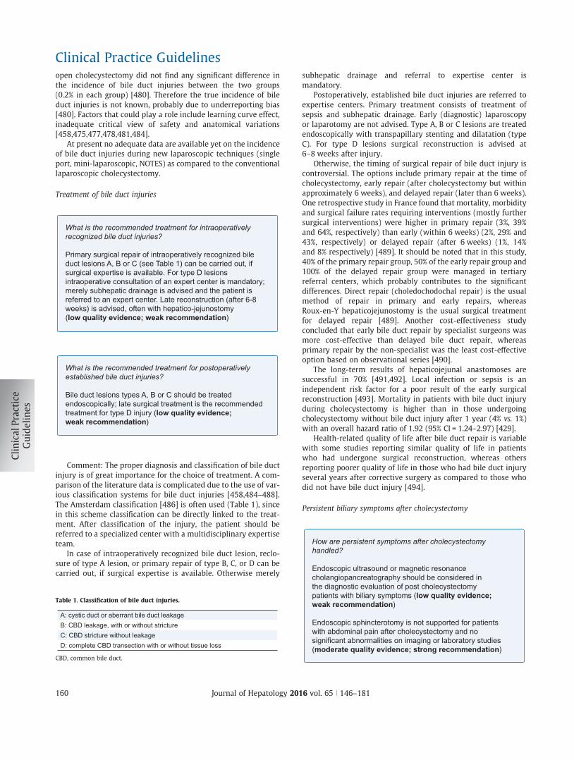
open cholecystectomy did not find any significant difference inthe incidence of bile duct injuries between the two groups(0.2% in each group) [480]. Therefore the true incidence of bileduct injuries is not known, probably due to underreporting bias[480]. Factors that could play a role include learning curve effect,inadequate critical view of safety and anatomical variations[458,475,477,478,481,484].
At present no adequate data are available yet on the incidenceof bile duct injuries during new laparoscopic techniques (singleport, mini-laparoscopic, NOTES) as compared to the conventionallaparoscopic cholecystectomy.
Treatment of bile duct injuries
What is the recommended treatment for intraoperatively recognized bile duct injuries?
Primary surgical repair of intraoperatively recognized bile duct lesions A, B or C (see Table 1) can be carried out, if surgical expertise is available. For type D lesions intraoperative consultation of an expert center is mandatory; merely subhepatic drainage is advised and the patient is referred to an expert center. Late reconstruction (after 6-8 weeks) is advised, often with hepatico-jejunostomy (low quality evidence; weak recommendation)
What is the recommended treatment for postoperatively established bile duct injuries?
Bile duct lesions types A, B or C should be treated endoscopically; late surgical treatment is the recommended treatment for type D injury (low quality evidence; weak recommendation)
Comment: The proper diagnosis and classification of bile ductinjury is of great importance for the choice of treatment. A com-parison of the literature data is complicated due to the use of var-ious classification systems for bile duct injuries [458,484–488].The Amsterdam classification [486] is often used (Table 1), sincein this scheme classification can be directly linked to the treat-ment. After classification of the injury, the patient should bereferred to a specialized center with a multidisciplinary expertiseteam.
In case of intraoperatively recognized bile duct lesion, reclo-sure of type A lesion, or primary repair of type B, C, or D can becarried out, if surgical expertise is available. Otherwise merely
subhepatic drainage and referral to expertise center ismandatory.
Postoperatively, established bile duct injuries are referred toexpertise centers. Primary treatment consists of treatment ofsepsis and subhepatic drainage. Early (diagnostic) laparoscopyor laparotomy are not advised. Type A, B or C lesions are treatedendoscopically with transpapillary stenting and dilatation (typeC). For type D lesions surgical reconstruction is advised at6–8 weeks after injury.
Otherwise, the timing of surgical repair of bile duct injury iscontroversial. The options include primary repair at the time ofcholecystectomy, early repair (after cholecystectomy but withinapproximately 6 weeks), and delayed repair (later than 6 weeks).One retrospective study in France found that mortality, morbidityand surgical failure rates requiring interventions (mostly furthersurgical interventions) were higher in primary repair (3%, 39%and 64%, respectively) than early (within 6 weeks) (2%, 29% and43%, respectively) or delayed repair (after 6 weeks) (1%, 14%and 8% respectively) [489]. It should be noted that in this study,40% of the primary repair group, 50% of the early repair group and100% of the delayed repair group were managed in tertiaryreferral centers, which probably contributes to the significantdifferences. Direct repair (choledochodochal repair) is the usualmethod of repair in primary and early repairs, whereasRoux-en-Y hepaticojejunostomy is the usual surgical treatmentfor delayed repair [489]. Another cost-effectiveness studyconcluded that early bile duct repair by specialist surgeons wasmore cost-effective than delayed bile duct repair, whereasprimary repair by the non-specialist was the least cost-effectiveoption based on observational series [490].
The long-term results of hepaticojejunal anastomoses aresuccessful in 70% [491,492]. Local infection or sepsis is anindependent risk factor for a poor result of the early surgicalreconstruction [493]. Mortality in patients with bile duct injuryduring cholecystectomy is higher than in those undergoingcholecystectomy without bile duct injury after 1 year (4% vs. 1%)with an overall hazard ratio of 1.92 (95% CI = 1.24–2.97) [429].
Health-related quality of life after bile duct repair is variablewith some studies reporting similar quality of life in patientswho had undergone surgical reconstruction, whereas othersreporting poorer quality of life in those who had bile duct injuryseveral years after corrective surgery as compared to those whodid not have bile duct injury [494].
Persistent biliary symptoms after cholecystectomy
How are persistent symptoms after cholecystectomy handled?
Endoscopic ultrasound or magnetic resonance cholangiopancreatography should be considered in the diagnostic evaluation of post cholecystectomy patients with biliary symptoms (low quality evidence; weak recommendation)
Endoscopic sphincterotomy is not supported for patients with abdominal pain after cholecystectomy and no significant abnormalities on imaging or laboratory studies (moderate quality evidence; strong recommendation)
Table 1. Classification of bile duct injuries.
A: cystic duct or aberrant bile duct leakageB: CBD leakage, with or without strictureC: CBD stricture without leakageD: complete CBD transection with or without tissue loss
CBD, common bile duct.
Clinical Practice Guidelines
160 Journal of Hepatology 2016 vol. 65 j 146–181
Comment: Symptoms may persist or return in some patientsafter cholecystectomy. In fact, 10–40% of patients have persistentcomplaints after cholecystectomy, sometimes after a briefinterval without symptoms [495,496]. ‘‘Post cholecystectomysyndrome” is an old term that is nowadays replaced by morespecific causes. Importantly, presence of bile duct stones shouldbe excluded. In the rare subgroup of patients with LPACsyndrome caused by ABCB4 gene mutations, symptoms recurafter cholecystectomy due to the presence of intrahepatic sludgeand microliths, or recurrent bile duct stones (see section Preven-tion of recurrent bile duct stones) [216].
In an RCT [497], 118 patients with post cholecystectomy painwere screened, and crystals were detected microscopically induodenal bile from 12 patients. When using UDCA for a fewmonths, the biliary-type abdominal pain significantly improvedor resolved. This study provides evidence that microlithiasismay be a cause for post cholecystectomy pain. Microlithiasiscan be confirmed by the microscopic examination of duodenalbile or hepatic bile obtained during ERCP [498].
More probable, since gallstone symptoms are relativelyunspecific, alternative preexisting causes should be considered,such as functional dyspepsia, irritable bowel syndrome, (duo-deno-) gastro-oesophageal reflux, and (rarely) sphincter of Oddidysfunction [496]. As demonstrated in a large RCT in patientswith abdominal pain after cholecystectomy undergoing ERCPwith manometry, sphincterotomy did not reduce disabilitydue to pain in comparison to a sham procedure [499]. These find-ings do not support endoscopic sphincterotomy for thesepatients.
Diagnosis of bile duct stones
Medical history and physical examination
When should common bile duct stones be looked for?
Common bile duct stones should be searched for in patients with jaundice, acute cholangitis or acute pancreatitis (high quality evidence; strong recommendation)
Comment: CBD stones are present in 3–16% (depending onage) of patients with stones in the gallbladder [500–507]. Theyeither occur by migration from the gallbladder (secondarystones), or less often develop de novo in the bile duct, forexample in case of CBD dilation with stasis (primary stones).In contrast to gallbladder stones, CBD stones are asymptomaticin only 5–12% of cases [508]. The natural history of asymp-tomatic CBD stones is not well known, but it seems to be morebenign than that of symptomatic CBD stones. In a small seriesof patients, CBD stones remained asymptomatic during a 5-year follow-up [509].
The common presentation of symptomatic CBD stones isacute biliary pain, caused by distention of the CBD followingits partial or complete obstruction. The pain is located in theright upper abdominal quadrant or the epigastrium, lasts more
than 30 min and up to several hours, and does not depend onthe body position. It might be challenging to differentiate thepain from that caused by gallbladder stones. Spontaneouspassage of the stones into the duodenum, in case of smallstones [510], or backwards into the distended duct may relievethe pain. Especially small stones impacted in the sphincter ofOddi may cause distal obstruction and trigger acute pancreati-tis. Larger stones cause more often proximal obstruction. Bileduct obstruction is often followed by complications, such asjaundice and cholangitis.
Laboratory diagnosis and imaging
Are laboratory tests included in the diagnostic work up of common bile duct stones?
The evaluation of patients with suspected common bile duct stones commonly includes serum liver biochemical tests (low quality evidence; weak recommendation)
Comment: Patients with symptomatic CBD stones often havealtered liver biochemical tests. The initial evaluation of suspectedCBD stones includes serum bilirubin concentrations as well ALT,AST, c-GT and AP activities [511,512]. In particular in the first72 h of biliary obstruction, serum aminotransferase activitiesare markedly elevated, followed by a more gradual rise in theAP and bilirubin levels if the obstruction persists [513]. If liverbiochemical tests show normal values in the first 24 h followingonset of pain, and if CBD dilation is absent on ultrasound, theprobability of a CBD stone is very low [355,357]. In contrast,the positive predictive values for abnormal bilirubin, AP orc-GT range from 25–50% only [357,512,514,515]. These lattercholestatic liver biochemical tests progressively increase withthe duration and severity of biliary obstruction. For example inone study, a serum bilirubin concentration of at least 1.7 mg/dL(29 lmol/L) portended a specificity of 60% for CBD stones,whereas the specificity increased to 75% at a cutoff of 4 mg/dl(68 lmol/L); however, only one-third or less of patients withcholedocholithiasis display such marked hyperbilirubinemia[512,514].
What imaging modality should be used to detect CBD stones?
Abdominal ultrasound should be the first imaging method when CBD stones are suspected (low quality evidence; weak recommendation). Stones in the gallbladder, a dilated CBD, acute cholangitis and hyperbilirubinemia are strong predictors for CBD stones (high quality evidence; strong recommendation)
Patients with an intermediate probability of CBD stones should undergo further evaluation with endoscopic ultrasound (or magnetic resonance cholangiopancreatography) (moderate quality evidence; strong recommendation)
JOURNAL OF HEPATOLOGY
Journal of Hepatology 2016 vol. 65 j 146–181 161
Comment: Abdominal ultrasound detects CBD dilation withhigh sensitivity, which is an indirect sign for the presence ofCBD stones. In fact, stones may be directly visualized in thedilated CBD. The ultrasound sensitivity for CBD stones is con-siderably lower than that for gallbladder stones [516,517],but can be as high as 80% for experienced operators [518].Ultrasonographic evidence of bile duct stone, CBD dilation,signs of acute cholangitis and jaundice are the best predictorsfor CBD stones [516]. A negative ultrasound result does noteliminate the diagnosis of CBD stones, if suspected; however,when liver biochemical tests are also normal, the probabilityis very low.
In patients with intermediate probability of CBD stones andinconclusive abdominal ultrasound, EUS is a valuable alternative.EUS and MRCP detect CBD stones >5 mm with similar accuracy,but EUS is more cost-effective than MRCP [519–527]. Accordingto a recent systematic review [528], the sensitivity of EUS is95% with a specificity of 97%, whereas the sensitivity of MRCPis 93% with a specificity of 96%.
CT imaging has a high sensitivity for CBD dilation [529,530]. Italso evaluates other possible causes of upper abdominal pain andgallstone complications but is associated with a significant radi-ation dose. ERCP detects CBD stones with a very high sensitivity[520,530,531]. However, it is a procedure with radiation exposureand only recommended as first diagnostic step for patients withhigh probability of CBD stones, in whom concomitant endoscopictherapy is envisaged.
Diagnosis of acute cholangitis
How is acute cholangitis diagnosed?
In patients with fever and a history of chills, with abdominal pain and/or jaundice, white blood cells, C-reactive protein and liver biochemical tests should be determined and abdominal ultrasound should be performed as the initial investigations (moderate quality evidence; strong recommendation)
Comment: Acute cholangitis can be diagnosed by the presenceof the Charcot’s triad, i.e. pain and tenderness in the right upperquadrant, high spiking fever, often with rigors, and jaundice.Charcot’s triad has high specificity but low sensitivity [532]. Painmay be the single symptom in a minority of patients, and can beabsent, especially in elderly patients. Jaundice is present in 60–70% and fever in 90% of the patients with acute cholangitis[532–536].
The biochemical signs of acute cholangitis are leucocytosiswith a left shift and increased serum C-reactive protein (CRP)concentrations. Aminotransferase activities and cholestaticparameters often increase within the first hours following thepain attack. Abdominal ultrasound often demonstrates CBDdilation, though it is less sensitive for CBD stones, and theexamination of the distal bile duct is even more difficult in thesetting of acute inflammation. There is currently sufficient evi-dence that EUS is superior to MRCP in this setting [522,524]
and to CT [524] in detecting CBD stones in patients withobstructive jaundice.
Diagnosis of acute biliary pancreatitis
How is acute biliary pancreatitis diagnosed?
The diagnosis of acute biliary pancreatitis is based on the presence of upper abdominal pain and altered pancreatic and liver biochemical tests in patients with gallbladder and/or common bile duct stones (moderate quality evidence; strong recommendation)
The exclusion of bile duct stones by endoscopic ultrasound (or magnetic resonance cholangiopancreatography) may prevent the potential risks of endoscopic retrograde cholangiopancreatography in patients with acute biliary pancreatitis and suspected bile duct stones (low quality evidence; weak recommendation)
Comment: In 4–8% of the patients with gallbladder stones,stones migrate into the main bile duct causing acute pancreatitisas they pass into the duodenum or impact in the sphincter ofOddi [537,538]. It is outside the scope of this guideline to discussacute pancreatitis in detail, and for further information we referto the current acute pancreatitis guideline of the InternationalAssociation of Pancreatology [539].
Gallstone migration, even of small stones, is often preceded bya period of biliary obstruction [540]. A warning pain is absent in50% of cases [228,229]. Biochemical tests indicate hyperlipasemiaor hyperamylasemia (>3 times the upper limit of normal),elevated aminotransferase activities and cholestatic parameters,leucocytosis and increased CRP concentrations. In the absenceof alcohol abuse or known pre-existent abnormal liver biochem-istry, ALT activities >150 U/ml indicate the biliary cause ofpancreatitis with a positive predictive value exceeding 85%[541–544]. Biliary crystals can be detected microscopically in duode-nal or hepatic bile obtained during ERCP in patients with idiopathicacute pancreatitis, indicating the biliary cause [241,242,498,545].
Ultrasound is the first investigation usually performed.Patients with pancreatitis or obstructive jaundice have moreand smaller gallbladder stones than those with acute cholecysti-tis or uncomplicated gallstone disease [240]. Ultrasoundfrequently visualizes CBD dilation but is less accurate in detectinggallstones in acute pancreatitis. EUS or MRCP can be performed inthis situation, when the biliary aetiology is not clear or whenERCP is considered [356].
MRCP is reasonably accurate to detect bile duct stones inpatients with biliary pancreatitis [546,547] but might miss smallstones. EUS is superior to all other methods in detecting stones<5 mm, i.e. those that often cause acute pancreatitis. Its sensitiv-ity reaches 100% and specificity is 95%, resulting in an accuracy of97% for the diagnosis of CBD stones [548]. In patients with milddisease in whom laparoscopic cholecystectomy is ultimatelyplanned ERCP and sphincterotomy is not indicated unless thereare bile duct stones present, which should first be confirmed byMRCP or EUS [521].
Clinical Practice Guidelines
162 Journal of Hepatology 2016 vol. 65 j 146–181
Endoscopic and surgical therapy of bile duct stones
Treatment of bile duct stones without complications
What is the recommended treatment for bile duct stones?
Endoscopic sphincterotomy and stone extraction is a recommended treatment of bile duct stones (moderate quality evidence; weak recommendation). Intraoperative endoscopic retrograde cholangiopancreatography or laparoscopic bile duct exploration in combination with cholecystectomy are alternatives when adequate expertise is available (moderate quality evidence; strong recommendation)
In case of failed standard stone extraction, extracorporeal shock wave, electrohydraulic or laser lithotripsy may be performed (low quality evidence; weak recommendation). In the case of altered anatomy (e.g. previous Roux-en-Yanastomosis, bariatric surgery) percutaneous or endoscopic(balloon endoscopy-assisted) treatment of bile duct stones can be considered (low quality evidence; weak recommendation). In the case of failed endoscopic therapy, cholecystectomy combined with bile duct exploration or intraoperative endoscopic retrograde cholangiopancreatography should be performed (low quality evidence; weak recommendation)
Comment: Choledocholithiasis is a relative frequent phe-nomenon in patients with symptomatic gallbladder stones(prevalence 3–16% of cases). Although there may be spontaneouspassage to the small bowel in many cases, there is a significantrisk of biliary pain and complications such as jaundice, cholangi-tis and pancreatitis. Therefore, the general consensus is thatsymptomatic choledocholithiasis should be treated. The naturalhistory of asymptomatic choledocholithiasis appears morebenign. Nevertheless, more than 25% of patients appear todevelop (often serious) complications during follow-up[509,504,549]. The choice of therapy depends on time of diagno-sis (before, during or after cholecystectomy) and local expertise[550,551]. In the last decades, there has been an expanding rolefor the endoscopic treatment (sphincterotomy and stone extrac-tion) of bile duct stones. Nevertheless, ERCP is associated with arisk of complications (especially pancreatitis) and in recent years,experience and volume of the endoscopist has been a topic ofconsiderable debate. Performance of at least 100 proceduresannually is associated with better outcome, whereas patientage is not related to complication risk [552].
Endoscopic papillary balloon dilatation with a large diameterballoon (12–20 mm) represents an option to facilitate the extrac-tion of large stones [553,554]. A meta-analysis including 6 RCTswith 835 patients [555] reported fewer overall complication ratesand lower risk of perforation with no difference in post-ERCpancreatitis, infection, or bleeding.
Currently preoperative ERCP and laparoscopic cholecystec-tomy is the preferred option in the management of patients withsimultaneous gallbladder and CBD stones, although there isevidence that intraoperative ERCP results in lower incidence ofERCP-related pancreatitis and shorter hospital stay, and is
cost-effective in comparison to the splitting of procedures[549,556–558]. According to recent meta-analyses, in cases offailed endoscopic therapy, laparoscopic or open cholecystectomycombined with transcystic stone extraction or CBD exploration orintraoperative ERCP are alternatives, with comparable stoneclearance rates, morbidity and mortality as primary endoscopicapproach [559,560]. In high risk cases (see recommendation: Isprimary closure preferred over T-tube drainage during surgicalbile duct exploration?), T-tube insertion remains the safestoption [561,562].
Surgical experience with open CBD exploration has decreaseddramatically in the last decades, and the number of surgeonsexperienced in laparoscopic common bile duct exploration islimited. Therefore, endoscopic stone removal is currently the pre-ferred approach in most countries. The timing of sphincterotomy,however, remains controversial. Two trials (one in patients withgallstone pancreatitis) indicate fewer endoscopic proceduresand shorter hospital stay without increased morbidity withinitial cholecystectomy (and postoperative ERCP) vs. initial endo-scopic assessment of the CBD and subsequent cholecystectomy[430,563].
What are the best forms of treatment for bile duct stones when detected intraoperatively or postoperatively?
In case of intraoperative detection of bile duct stones, bile duct exploration, transcystic stone extraction or endoscopic clearance represent alternative treatment options (moderate quality evidence; weak recommendation). Upon postoperative diagnosis of bile duct stones, endoscopic sphincterotomy and stone extraction are recommended (low quality evidence; weak recommendation)
Comment: When bile duct stones are detected during theoperation, transcystic stone extraction can be attempted, if thesurgeon is experienced in this procedure. Transcystic stoneextraction is safe, and success rate is approximately 75%. Laparo-scopic stone extraction can have a high success rate, but has a rel-atively high complication rate and should not be performedexcept in centers of expertise [564–566]. Upon postoperativediagnosis of bile duct stones, endoscopic sphincterotomy andstone extraction are the common measures.
Is primary closure preferred over T-tube drainage during surgical bile duct exploration?
In case of surgical bile duct exploration, primary closure may be preferred over T-tube drainage in low risk cases (low quality evidence; weak recommendation)
Comment: Systematic reviews and meta-analysis on random-ized controlled trials on primary closure vs. T-tube drainagedemonstrated that T-tube drainage prolongs operating time andhospital stay compared to primary closure without any evidenceof benefit after open or laparoscopic common bile duct explo-ration in low risk cases [561,567]. In high risk cases involving
JOURNAL OF HEPATOLOGY
Journal of Hepatology 2016 vol. 65 j 146–181 163
patients with recurrent bile duct stones, acute cholangitis ormultiple bile duct stones where ERCP has failed, T-tube insertionor alternative procedures such as choledochoduodenostomyrepresent safe options [561,562].
When should cholecystectomy be performed in patients with gallbladder stones after endoscopic removal of bile duct stones?
In patients with simultaneous gallbladder and bile duct stones, early laparoscopic cholecystectomy should be performed within 72 h after preoperative ERCP for choledocholithiasis (moderate quality evidence;strong recommendation)
Comment: See section: Patients with simultaneous gallblad-der and bile duct stones
Treatment of acute cholangitis
How should patients with acute cholangitis be treated?
Treatment of cholangitis should include immediate broad spectrum antibiotics and biliary decompression (moderate quality evidence; strong recommendation)
Timing of biliary decompression depends on severity of thecholangitis and effects of medical therapy including antibiotics and may preferably be performed within 24 h; urgent decompression should be considered in case of severe cholangitis not responding to fluid resuscitation and intravenous antibiotics (low quality evidence; weak recommendation)
Endoscopic treatment with sphincterotomy is the preferred mode of biliary decompression; in the presence of contraindications for sphincterotomy, biliary stenting with stone removal at a later stage should be performed (low quality evidence; weak recommendation)
In case of failed endoscopic decompression or contraindications to endoscopic therapy, percutaneous bile duct drainage is the procedure of choice (low quality evidence; weak recommendation)
Comment: Cholangitis is a serious complication of gallstones,with significant morbidity and mortality, especially in the elderly[568]. First line treatment should include general supportivemeasures including adequate intravenous hydration and antibi-otics, which are required in the first hour after hospital admissionin case of sepsis [569]. Considering the polymicrobial content ofinfected bile, broad spectrum antibiotics should be applied. Thechoice of antibiotic coverage depends on cholangitis severityand local antimicrobial resistance patterns. Enteric gram-nega-tive bacteria are usually cultured from bile of patients with acutecholangitis, especially E. coli and Klebsiella species. Nevertheless,
the microbiological profile has changed in the last decades, dueto increased instrumentation of the bile ducts and frequent useof antibiotics in the population. Polymicrobial bile cultures areoften found. Anaerobic bacteria are usually isolated in conjunc-tion with aerobic bacteria, rather than a sole isolate from bile,and often in the setting of previous bile duct instrumentationand a more severe clinical condition. There is a clear differencebetween results of bile cultures compared to associated bloodcultures [570]. Bile cultures are positive in 80–100%, and bloodcultures in 20–60% of patients with cholangitis. Streptococcusand Enterococcus species are infrequently and anaerobic bacteriaare rarely cultured from blood. One of the main goals of antibi-otics is to control bacteremia and sepsis. Most antibiotics (withthe exception of quinolones) are not or are less well excreted intobile in case of biliary obstruction. Empirical antibiotic therapythat includes coverage of the aerobic gram-negative bacteriaand anaerobic bacteria should be considered until the results ofbile cultures and blood cultures are available. The duration ofantibiotic therapy will depend on severity of the clinical condi-tion at presentation, whether blood cultures were positive andrecovery after biliary drainage.
Most cholangitis patients will respond satisfactorily to initialconservative therapy with broad spectrum antibiotics. Althoughthese patients could get elective biliary decompression and stoneremoval, it appears wise to achieve biliary decompression in allcholangitis patients at the earliest time point possible, preferablywithin 24 h, since up to 20% of patients will run a progressivecourse with severe deterioration [571]. Urgent decompressionshould be considered in case of severe cholangitis not respondingto fluid resuscitation and intravenous antibiotics. Consensus cri-teria for defining severity of cholangitis have been published[572].
Biliary decompression can be achieved by ERCP, percuta-neous drainage or surgery. Results of endoscopic therapy foracute gallstone cholangitis were superior to surgical treatment,both in retrospective and prospective randomized trials[269,573]. Also, in a non-randomized study comparing percuta-neous transhepatic biliary drainage with ERCP in elderlycholangitis patients, endoscopic drainage yielded significantlylower morbidity and mortality [574]. Therefore, ERCP is nowconsidered the treatment of choice for acute cholangitis dueto gallstones. Percutaneous transhepatic drainage should beconsidered when ERCP is impossible or has failed in experthands, whereas surgery should be avoided. It is wise to aspiratebile after bile duct cannulation before contrast injection, inorder to avoid increased bile duct pressure and bacteremias.Aspirated bile should be sent for culture. In stable patients,sphincterotomy with stone extraction can be performed duringthe initial procedure. Even if bile duct stones are not detected,sphincterotomy performed during endoscopic decompressionleads to faster reconvalescence and shorter hospital stay [575].In case of significant coagulation disturbances, large andmultiple stones, or unstable patients, nasobiliary drain place-ment or biliary endoprostheses are preferred as initial treat-ment. Nasobiliary and endoprosthesis are equally effectiveunder these circumstances [576]. Nevertheless, endoprosthesisshould be preferred since this is less uncomfortable for thepatient and is associated with less dislocation [576,577].Definite stone removal can then be performed at a later stage,after recovery from the acute episode.
Clinical Practice Guidelines
164 Journal of Hepatology 2016 vol. 65 j 146–181
Treatment of acute biliary pancreatitis
Which patients with acute biliary pancreatitis should undergo endoscopic retrograde cholangiopancreatography?
For biliary pancreatitis with suspected coexistent acute cholangitis antibiotics should be initiated, and endoscopic retrograde cholangiopancreatography with sphincterotomy and stone extraction should be performed, with timing depending on the severity of cholangitis but preferably within 24 h (high quality evidence; strong recommendation)
An endoscopic retrograde cholangiopancreatography is probably indicated in patients with biliary pancreatitis and obstructed bile duct (low quality evidence; weak recommendation)
An early endoscopic retrograde cholangiopancreatography is probably not indicated in patients with predicted severe biliary pancreatitis in the absence of cholangitis or obstructed bile duct (low quality evidence; weak recommendation)
An early endoscopic retrograde cholangiopancreatography is not indicated in patients with predicted mild biliary pancreatitis in the absence of cholangitis or obstructed bile duct (moderate quality evidence; strong recommendation)
In patients with suspected biliary pancreatitis without cholangitis, endoscopic ultrasound (or magnetic resonance cholangiopancreatography) may prevent potential endoscopic retrograde cholangiopancreatography and prevent its risks if no stones are detected (low quality evidence; weak recommendation)
Comment: It is outside the scope of this guideline to discussacute pancreatitis in detail, but some aspects of endoscopic treat-ment are mentioned. For further information we refer to theguideline Acute Pancreatitis 2013 of the International Associationof Pancreatology [539]. The advice in the current guideline is inline with the International Association of Pancreatology (IAP)/American Pancreatic Association (APA) guideline.
If concomitant cholangitis is suspected, an endoscopic inter-vention is recommended, preferably within 24 h [538,578–580].Urgent ERCP should be considered in case of severe cholangitisnot responding to fluid resuscitation and intravenous antibiotics.The role of ERCP in predicting severe pancreatitis withoutcholestasis/cholangitis is controversial. A meta-analysis of sevenRCTs with 757 patients in total did not support ERCP in patientswith biliary pancreatitis without cholangitis or biliary obstruc-tion, regardless of the predicted severity of the pancreatitis[581]. However, in case of predicted severe pancreatitis, the num-ber of included patients in the meta-analysis was too small todraw definite conclusions. The meta-analysis supported ERCP inpatients with biliary obstruction without cholangitis. It shouldbe realised that in the early stage of biliary pancreatitis, the pre-diction of bile duct stones based on liver biochemistry, abdominalultrasound or CT scan is highly unreliable. The explanation is thatnot only bile duct stones, but also the peripancreatic edema cancause biliary obstruction [356]. Nevertheless, the course of
laboratory parameters during the first 48 h after admission canpredict to some extent clinical course and persistent bile ductstones, which are associated with the severity of acute pancreati-tis and worse outcome [582,583]. Of note, MRCP or EUS can pre-vent a proportion of (negative) ERCP procedures that may beconsidered because of suspected bile duct obstruction in theabsence of cholangitis. Although MRCP is non-invasive and lessoperator-dependent, the disadvantage in comparison to EUS isthe lower sensitivity for small (<5 mm) CBD stones [519,522–524,531,547,584–614]. In fact, patients with biliary pancreatitisoften exhibit such small stones [241,615].
The best timing for EUS, MRCP and ERCP in patients with bil-iary pancreatitis and biliary obstruction in absence of cholangitisis not clear (IAP pancreatitis guideline 2013). In a post-hoc anal-ysis, the meta-analysis [581] found no significant effect of ERCPtiming on mortality. ERCP is not necessary for mild biliary pan-creatitis in absence of cholangitis or biliary obstruction[538,578,581,616,617]. A preoperative ERCP before cholecystec-tomy does not have to be carried out routinely, since small bileduct stones generally pass spontaneously with normalization ofthe laboratory parameters [618,619].
What is the best time to perform cholecystectomy after acute biliary pancreatitis?
Cholecystectomy during the same hospital admission is the preferred option in patients with mild acute biliary pancreatitis (high quality evidence; strong recommendation)
Comment: In patients with mild acute biliary pancreatitis,early laparoscopic cholecystectomy is preferable to laparoscopiccholecystectomy performed on the routine waiting list to avoidrecurrent gallstone-related complications [620,621]. Whilelaparoscopic cholecystectomy has generally been performed afterthe acute symptoms resolve and the serum amylase activitiesreturn to near normal levels, recent RCTs confirm that performinglaparoscopic cholecystectomy during the same hospital admis-sion results not only in shorter hospital stay [622] but alsoreduces the rate of recurrent gallstone-related complications(recurrent pancreatitis, cholecystitis, choledocholithiasis needingERCP, gallstone colic) from 17% to 5% [621]. There have been con-cerns about performing the surgery very early because of the riskof predicted severe pancreatitis [623]. Waiting up to 72 h allowsthe pancreatitis to be confirmed to be mild and perform any addi-tional investigations and treatments such as MRCP, EUS or ERCP ifindicated [624] and hence may overcome the issue of predictedsevere pancreatitis.
Considerable uncertainty exists regarding the timing of chole-cystectomy in patients with severe acute biliary pancreatitis andno definite recommendations can be made regarding the timingof cholecystectomy in patients with acute severe pancreatitis,since there are no RCTs on this issue [624]. With open cholecys-tectomy, early cholecystectomy (within 6 weeks of index admis-sion) resulted in increased complication rates (includingincreased risk of infected peripancreatic collections) and lengthof hospital stay in observational studies [625,626]. Delayedlaparoscopic cholecystectomy may decrease the conversion toopen cholecystectomy since the inflammation and fluid
JOURNAL OF HEPATOLOGY
Journal of Hepatology 2016 vol. 65 j 146–181 165
collections associated with severe pancreatitis are likely to settledown or become well-defined pseudocysts during the waitingtime. Disadvantages of delayed laparoscopic cholecystectomyare potential recurrence of biliary symptoms and prolonged hos-pital stay [627]. Nevertheless, it appears wise to postpone chole-cystectomy in patients with severe biliary pancreatitis withperipancreatic collections until these collections are dissolvedor in case of persistent collections, until at least 6 weeks afterpancreatitis onset.
Diagnosis and therapy of intrahepatic bile duct stones
What is the preferred diagnostic method for intrahepatic bile duct stones?
If intrahepatic bile duct stones are suspected, abdominal ultrasound is the first method of choice and magnetic resonance cholangiopancreatography the second (very low quality evidence; weak recommendation)
Comment: Intrahepatic bile duct stones (hepatolithiasis) typ-ically occur in the setting of bile duct strictures and are seen afterbile duct injury, in patients with primary or secondary sclerosingcholangitis, or recurrent pyogenic cholangitis (‘‘oriental cholangi-tis”) [628–631]. Ascending cholangitis is a frequent acute compli-cation associated with hepatolithiasis, and chronic complicationsinclude secondary biliary cirrhosis, segmental or lobar atrophy,liver abscess, and cholangiocarcinoma.
Abdominal ultrasound has an advantage over diagnostic ERCP,because it is non-invasive and can identify bile ducts that areobstructed by non-calcified intrahepatic bile duct stones. MRCPis also to be preferred over ERCP for the diagnosis of hepatolithi-asis (sensitivity 97% vs. 59%, respectively) and can reliably detectbile duct strictures (specificity 97%, sensitivity 93%) as well aslesions proximal of the obstruction and outside of the bile ducts[606,632–634]. Although stones are often not directly visibleusing CT, dilated ducts and strictures as well as liver abscessescan be demonstrated [632].
Should asymptomatic intrahepatic bile duct stones be treated?
Asymptomatic intrahepatic bile duct stones do not always have to be treated. The treatment decision should be made individually for each patient and interdisciplinarily for symptomatic intrahepatic bile duct stones (very low quality evidence; weak recommendation)
Comment: During the course of 15 years, asymptomatic intra-hepatic bile duct stones became symptomatic only in 11.5% ofpatients after a mean of 3.4 years [633]. The most common symp-toms are colic, jaundice and fever due to cholangitis or liverabscesses, and rarely cholangiocarcinoma [633]. Therefore, it isjustified to use a wait-and-see approach. An interdisciplinarytreatment plan is useful for symptomatic stones. For planningthe subsequent treatment, both ERCP and percutaneous
transhepatic cholangiography are primarily important. Surgicalresection should be considered for patients with unilateral stonedisease, particularly if biliary strictures and/or lobar atrophy arealso present [635,636]. Partial hepatectomy is associated withstone clearance rates higher than 80% and fewer recurrences thanendoscopic modalities [637–639].
The other treatment options for hepatolithiasis includeperoral cholangioscopic lithotripsy (POCSL) or percutaneoustranshepatic cholangioscopic lithotripsy (PTCSL), which may beuseful for diffusely distributed intrahepatic bile duct stones[632,636,640–643]. In a series of POCSL for hepatolithiasis, therate of complete stone removal was 64% [644]. For PTCSL, higherrates of complete stone clearance have been reported (80–85%)[645–647]. However, both POCSL and PTCSL are limited by highrates of recurrent stones on long-term follow-up (22–50%).
Patients with LPAC syndrome caused by ABCB4 mutations (seesection Prevention of recurrent bile duct stones) are prone todevelop intrahepatic bile duct stones (alone or in combinationwith bile duct and gallbladder stones) [216]. Cholecystectomyis indicated in the case of symptomatic gallbladder stones orsludge [216]. Biliary drainage or partial hepatectomy may beindicated in the case of symptomatic intrahepatic bile ductdilation filled with stones. LPAC patients with end-stage liverdisease may be candidates for liver transplantation.
Therapy of gallstones during pregnancy
Therapy of gallbladder stones during pregnancy
How are symptomatic gallbladder stones treated in pregnancy?
Laparoscopic cholecystectomy can be performed during pregnancy if the indication is urgent, regardless of trimester (low quality evidence; weak recommendation)
Patients with gallbladder and bile duct stones who are asymptomatic after bile duct clearance should undergo cholecystectomy post partum (very low quality evidence; weak recommendation)
Comment: Gallbladder sludge or gallstones develop in 5% ofpregnant women each, but only 1.2% of the women with sludgeor stones presented with biliary pain in a large prospective studyin 3,254 pregnancies [648]. Of note, a randomized intervention toincrease physical activity (from 15.7 to 18.6 in the first and 10.2to 12.1 MET-hours per week in the third trimester) did notdecrease the incidence of gallbladder sludge or stones duringpregnancy [649]. Sludge is associated with gallbladder hypo-motility during pregnancy and is not an indication for interven-tion. There is no indication for treating pregnant women withsludge or stones with UDCA. Asymptomatic pregnant patientswith stones are not treated. However, in many of these patientsa cholecystectomy becomes necessary in the first year afterpregnancy [650].
Pregnancy is not a general contraindication for cholecystec-tomy [651–653]. In fact, cholecystectomy is the second mostcommon nonobstetric antenatal surgical procedure [654].
Clinical Practice Guidelines
166 Journal of Hepatology 2016 vol. 65 j 146–181
Surgical management of pregnant patients with symptomaticstones is supported by studies showing recurrent symptoms in92%, 64% and 44% of patients in the first, second and third trime-ster, respectively [655,656], and 23–39% of patients develop pan-creaticobiliary complications [657,658]. However comparingconservative and surgical treatment of symptomatic cholelithia-sis, no significant differences with respect to the frequency ofpreterm delivery or fetal mortality were detected in 6 studieswith a total of 310 patients [655]. The second trimester is thesafest trimester for cholecystectomy. The current data and expe-rience show that safe laparoscopic cholecystectomy is also possi-ble for urgent indications in the first trimester [658–660]. In thethird trimester the indication is more restrictive because of thecrowded abdomen and the danger of inducing labour. Theintra-abdominal pressure should be kept below 12–15 mmHg,and the fetus should be monitored during the operation[661,662].
Treatment of bile duct stones during pregnancy
How are symptomatic bile duct stones treated in pregnancy?
During pregnancy symptomatic bile duct stones should be treated by endoscopic sphincterotomy and stone extraction by an experienced endoscopist (low quality evidence; weak recommendation). The use of x-rays is not contraindicated provided care is taken to minimize radiation exposure (very low quality evidence; weak recommendation)
Comment: Several studies have confirmed the safety of ERCPduring pregnancy [663–666]. The examination should be per-formed by an experienced endoscopist, since pregnancy is anindependent risk factor for post-ERCP pancreatitis [666,667].Radiation exposure during cholangiography has been estimatedto be 2–10 rads with variable conceptus doses [656,668]. The flu-oroscopy times and doses should be limited as much as possibleand no hardcopy X-ray films with additional radiation exposureshould be taken. Image guidance by ultrasound or bile aspirationcan be applied to confirm successful biliary cannulation andreduce radiation [669,670]. The pregnant patient generally lieson her left side during ERCP to minimize compression of aortaand vena cava. During endoscopic sphincterotomy, the uterusshould not be placed between the sphincterotome and thegrounding pad.
Future perspectives
According to the CPG panel members, the following future areasof research should be supported to further improve the preven-tion and treatment of gallstones:
- Research into strategies for primary prevention of gallstones- Research on the pathogenesis of gallstones in patients withfatty liver disease
- Implementation of genetic and exogenous lithogenic risk fac-tors in novel prevention strategies
- Cost-effectiveness analyses of the course of silent or mildlysymptomatic gallbladder stones with respect to laparoscopiccholecystectomy
- Care research on long-term results of cholecystectomy andregionally different frequencies of operations
- Research into alternative treatments of gallstones, particularlyfor patients at high risk of surgery
- Studies on the risk for biliary colic and complications (in par-ticular gallbladder cancer) in carriers of asymptomatic gall-stones or gallbladder sludge
- Pathogenesis and prevention of recurrent common and intra-hepatic bile duct stones
- Studies on the bile microbiome and inflammation of the bileducts
Conflict of interest
The authors declared that they do not have anything to discloseregarding funding or conflict of interest with respect to thismanuscript.
Acknowledgements
This guideline has been prepared with the endorsement of theEuropean Society of Clinical Investigation (ESCI). The authors wouldlike to thank Caroline S. Stokes (Homburg) and Leonilde Bonfrate(Bari) for systematic literature reviews and careful assistance.We would like to thank the reviewers of this Clinical PracticeGuideline for their time and critical reviewing: Guido Costam-agna, John P. Neoptolemos, Tilman Sauerbruch.
References
[1] Farthing M, Roberts SE, Samuel DG, Williams JG, Thorne K, Morrison-Rees S,et al. Survey of digestive health across Europe: final report. Part 1: Theburden of gastrointestinal diseases and the organisation and delivery ofgastroenterology services across Europe. UEG J 2014;2:539–543.
[2] Atkins D, Best D, Briss PA, Eccles M, Falck-Ytter Y, Flottorp S, et al. Gradingquality of evidence and strength of recommendations. BMJ 2004;328:1490.
[3] Schunemann HJ, Jaeschke R, Cook DJ, Bria WF, El-Solh AA, Ernst A, et al. Anofficial ATS statement: grading the quality of evidence and strength ofrecommendations in ATS guidelines and recommendations. Am J Respir CritCare Med 2006;174:605–614.
[4] Guyatt GH, Oxman AD, Kunz R, Vist GE, Falck-Ytter Y, Schunemann HJ, et al.What is ‘‘quality of evidence” and why is it important to clinicians? BMJ2008;336:995–998.
[5] Guyatt GH, Oxman AD, Vist GE, Kunz R, Falck-Ytter Y, Alonso-Coello P, et al.GRADE: an emerging consensus on rating quality of evidence and strengthof recommendations. BMJ 2008;336:924–926.
[6] Portincasa P, Moschetta A, Palasciano G. Cholesterol gallstone disease.Lancet 2006;368:230–239.
[7] Neoptolemos JP, Hofmann AF, Moossa AR. Chemical treatment of stones inthe biliary tree. Br J Surg 1986;73:515–524.
[8] Liver Disease Subcommittee of the Digestive Disease Interagency Coordi-nating Committee. Action plan for liver disease research. Bethesda: U.S.Department of Health and Human Services, National Institutes of Health;2004, p. 145–150.
[9] Hofmann AF. Primary and secondary prevention of gallstone disease:implications for patient management and research priorities. Am J Surg1993;165:541–548.
[10] Krawczyk M, Wang DQ, Portincasa P, Lammert F. Dissecting the geneticheterogeneity of gallbladder stone formation. Semin Liver Dis2011;31:157–172.
[11] Biddinger SB, Haas JT, Yu BB, Bezy O, Jing E, Zhang W, et al. Hepatic insulinresistance directly promotes formation of cholesterol gallstones. Nat Med2008;14:778–782.
JOURNAL OF HEPATOLOGY
Journal of Hepatology 2016 vol. 65 j 146–181 167
[12] Grundy SM. Cholesterol gallstones: a fellow traveler with metabolicsyndrome? Am J Clin Nutr 2004;80:1–2.
[13] Chen LY, Qiao QH, Zhang SC, Chen YH, Chao GQ, Fang LZ. Metabolicsyndrome and gallstone disease. World J Gastroenterol 2012;18:4215–4220.
[14] Ruhl CE, Everhart JE. Association of diabetes, serum insulin, and C-peptidewith gallbladder disease. Hepatology 2000;31:299–303.
[15] Haffner SM, Diehl AK, Mitchell BD, Stern MP, Hazuda HP. Increasedprevalence of clinical gallbladder disease in subjects with non-insulin-dependent diabetes mellitus. Am J Epidemiol 1990;132:327–335.
[16] Misciagna G, Guerra V, Di Leo A, Correale M, Trevisan M. Insulin and gallstones: a population case control study in southern Italy. Gut2000;47:144–147.
[17] Friedman GD, Kannel WB, Dawber TR. The epidemiology of gallbladderdisease: observations in the Framingham Study. J Chronic Dis 1966;19:273–292.
[18] The epidemiology of gallstone disease in Rome Italy. Part I. Prevalence datain men. The Rome Group for Epidemiology and Prevention of Cholelithiasis(GREPCO). Hepatology 1988;8:904–906.
[19] Völzke H, Baumeister SE, Alte D, Hoffmann W, Schwahn C, Simon P, et al.Independent risk factors for gallstone formation in a region with highcholelithiasis prevalence. Digestion 2005;71:97–105.
[20] Maclure KM, Hayes KC, Colditz GA, Stampfer MJ, Speizer FE, Willett WC.Weight, diet, and the risk of symptomatic gallstones in middle-agedwomen. N Engl J Med 1989;321:563–569.
[21] Tsai CJ, Leitzmann MF, Willett WC, Giovannucci EL. Central adiposity,regional fat distribution, and the risk of cholecystectomy in women. Gut2006;55:708–714.
[22] Klein S, Wadden T, Sugerman HJ. AGA technical review on obesity.Gastroenterology 2002;123:882–932.
[23] Willett WC, Dietz WH, Colditz GA. Guidelines for healthy weight. N Engl JMed 1999;341:427–434.
[24] Scragg RK, McMichael AJ, Baghurst PA. Diet, alcohol, and relative weight ingall stone disease: a case-control study. BMJ 1984;288:1113–1119.
[25] Jorgensen T. Prevalence of gallstones in a Danish population. Am JEpidemiol 1987;126:912–921.
[26] Stampfer MJ, Maclure KM, Colditz GA, Manson JE, Willett WC. Risk ofsymptomatic gallstones in women with severe obesity. Am J Clin Nutr1992;55:652–658.
[27] Amaral JF, Thompson WR. Gallbladder disease in the morbidly obese. Am JSurg 1985;149:551–557.
[28] Scragg RK, Calvert GD, Oliver JR. Plasma lipids and insulin in gall stonedisease: a case-control study. BMJ 1984;289:521–525.
[29] Stinton LM, Myers RP, Shaffer EA. Epidemiology of gallstones. GastroenterolClin North Am 2010;39:157–169.
[30] Stender S, Nordestgaard BG, Tybjaerg-Hansen A. Elevated body mass indexas a causal risk factor for symptomatic gallstone disease: a mendelianrandomization study. Hepatology 2013;58:2133–2141.
[31] Banim PJ, Luben RN, Bulluck H, Sharp SJ, Wareham NJ, Khaw KT, et al. Theaetiology of symptomatic gallstones quantification of the effects of obesity,alcohol and serum lipids on risk. Epidemiological and biomarker data froma UK prospective cohort study (EPIC-Norfolk). Eur J Gastroenterol Hepatol2011;23:733–740.
[32] Di Ciaula A, Wang DQ, Portincasa P. Gallbladder and gastric motility inobese newborns, pre-adolescents and adults. J Gastroenterol Hepatol2012;27:1298–1305.
[33] Vezina WC, Paradis RL, Grace DM, Zimmer RA, Lamont DD, Rycroft KM,et al. Increased volume and decreased emptying of the gallbladder in large(morbidly obese, tall normal, and muscular normal) people. Gastroenterol-ogy 1990;98:1000–1007.
[34] Portincasa P, Di Ciaula A, Wang HH, Palasciano G, van Erpecum KJ,Moschetta A, et al. Coordinate regulation of gallbladder motor function inthe gut-liver axis. Hepatology 2008;47:2112–2126.
[35] Portincasa P, Di Ciaula A, Palmieri V, Van Berge-Henegouwen GP,Palasciano G. Effects of cholestyramine on gallbladder and gastric emptyingin obese and lean subjects. Eur J Clin Invest 1995;25:746–753.
[36] Leitzmann MF, Rimm EB, Willett WC, Spiegelman D, Grodstein F, StampferMJ, et al. Recreational physical activity and the risk of cholecystectomy inwomen. N Engl J Med 1999;341:777–784.
[37] Boland LL, Folsom AR, Rosamond WD. Atherosclerosis Risk in CommunitiesStudy I. Hyperinsulinemia, dyslipidemia, and obesity as risk factors forhospitalized gallbladder disease. A prospective study. Ann Epidemiol2002;12:131–140.
[38] Festi D, Dormi A, Capodicasa S, Staniscia T, Attili AF, Loria P, et al. Incidenceof gallstone disease in Italy: results from a multicenter, population-based
Italian study (the MICOL project). World J Gastroenterol 2008;14:5282–5289.
[39] Thijs C, Knipschild P, Brombacher P. Serum lipids and gallstones: a case-control study. Gastroenterology 1990;99:843–849.
[40] Marks JW, Cleary PA, Albers JJ. Lack of correlation between serumlipoproteins and biliary cholesterol saturation in patients with gallstones.Dig Dis Sci 1984;29:1118–1122.
[41] Petitti DB, Friedman GD, Klatsky AL. Association of a history of gallbladderdisease with a reduced concentration of high-density-lipoprotein choles-terol. N Engl J Med 1981;304:1396–1398.
[42] Attili AF, Capocaccia R, Carulli N, Festi D, Roda E, Barbara L, et al. Factorsassociated with gallstone disease in the MICOL experience. MulticenterItalian Study on Epidemiology of Cholelithiasis. Hepatology 1997;26:809–818.
[43] Jorgensen T. Gallstones and plasma lipids in a Danish population. Scand JGastroenterol 1989;24:916–922.
[44] Weikert C, Weikert S, Schulze MB, Pischon T, Fritsche A, Bergmann MM,et al. Presence of gallstones or kidney stones and risk of type 2 diabetes. AmJ Epidemiol 2010;171:447–454.
[45] Leitzmann MF, Giovannucci EL, Rimm EB, Stampfer MJ, Spiegelman D, WingAL, et al. The relation of physical activity to risk for symptomatic gallstonedisease in men. Ann Intern Med 1998;128:417–425.
[46] Misciagna G, Centonze S, Leoci C, Guerra V, Cisternino AM, Ceo R, et al. Diet,physical activity, and gallstones – A population-based, case-control studyin southern Italy. Am J Clin Nutr 1999;69:120–126.
[47] Walcher T, Haenle MM, Mason RA, Koenig W, Imhof A, Kratzer W. The effectof alcohol, tobacco and caffeine consumption and vegetarian diet ongallstone prevalence. Eur J Gastroenterol Hepatol 2010;22:1345–1351.
[48] Ortega RM, Fernandez-Azuela M, Encinas-Sotillos A, Andres P, Lopez-Sobaler AM. Differences in diet and food habits between patients withgallstones and controls. J Am Coll Nutr 1997;16:88–95.
[49] Kriska AM, Brach JS, Jarvis BJ, Everhart JE, Fabio A, Richardson CR, et al.Physical activity and gallbladder disease determined by ultrasonography.Med Sci Sports Exerc 2007;39:1927–1932.
[50] Storti KL, Brach JS, FitzGerald SJ, Zmuda JM, Cauley JA, Kriska AM. Physicalactivity and decreased risk of clinical gallstone disease among post-menopausal women. Prev Med 2005;41:772–777.
[51] Kato I, Tominaga S. Factors associated with levels of physical activity atwork and during leisure time. Nihon Koshu Eisei Zasshi 1992;39:822–829.
[52] Banim PJ, Luben RN, Wareham NJ, Sharp SJ, Khaw KT, Hart AR. Physicalactivity reduces the risk of symptomatic gallstones: a prospective cohortstudy. Eur J Gastroenterol Hepatol 2010;22:983–988.
[53] Chait A, Bierman EL, Albers JJ. Low-density lipoprotein receptor activity incultured human skin fibroblasts. Mechanism of insulin-induced stimula-tion. J Clin Invest 1979;64:1309–1319.
[54] Nepokroeff CM, Lakshmanan MR, Ness GC, Dugan RE, Porter JW. Regulationof the diurnal rhythm of rat liver beta-hydroxy-beta-methylglutarylcoenzmye A reductase activity by insulin, glucagon, cyclic AMP andhydrocortisone. Arch Biochem Biophys 1974;160:387–396.
[55] Subbiah MT, Yunker RL. Cholesterol 7 alpha-hydroxylase of rat liver: aninsulin sensitive enzyme. Biochem Biophys Res Commun 1984;124:896–902.
[56] Kirwan JP, Kohrt WM, Wojta DM, Bourey RE, Holloszy JO. Enduranceexercise training reduces glucose-stimulated insulin levels in 60- to 70-year-old men and women. J Gerontol 1993;48:M84–M90.
[57] Seals DR, Hagberg JM, Hurley BF, Ehsani AA, Holloszy JO. Effects ofendurance training on glucose tolerance and plasma lipid levels in oldermen and women. JAMA 1984;252:645–649.
[58] Tran ZV, Weltman A, Glass GV, Mood DP. The effects of exercise on bloodlipids and lipoproteins: a meta-analysis of studies. Med Sci Sports Exerc1983;15:393–402.
[59] Mingrone G, Greco AV, Finotti E, Passi S. Free fatty acids: a stimulus formucin hypersecretion in cholesterol gallstone biles. Biochim Biophys Acta1988;958:52–59.
[60] Leon AS, Sanchez OA. Response of blood lipids to exercise training alone orcombined with dietary intervention. Med Sci Sports Exerc 2001;33:S502–S515.
[61] Baker TT, Allen D, Lei KY, Willcox KK. Alterations in lipid and proteinprofiles of plasma lipoproteins in middle-aged men consequent to anaerobic exercise program. Metabolism 1986;35:1037–1043.
[62] Gupta AK, Ross EA, Myers JN, Kashyap ML. Increased reverse cholesteroltransport in athletes. Metabolism 1993;42:684–690.
[63] Halloran LG, Schwartz CC, Vlahcevic ZR, Nisman RM, Swell L. Evidence forhigh-density lipoprotein-free cholesterol as the primary precursor for bile-acid synthesis in man. Surgery 1978;84:1–7.
Clinical Practice Guidelines
168 Journal of Hepatology 2016 vol. 65 j 146–181
[64] Koffler KH, Menkes A, Redmond RA, Whitehead WE, Pratley RE, Hurley BF.Strength training accelerates gastrointestinal transit in middle-aged andolder men. Med Sci Sports Exerc 1992;24:415–419.
[65] Philipp E, Wilckens T, Friess E, Platte P, Pirke KM. Cholecystokinin, gastrinand stress hormone responses in marathon runners. Peptides 1992;13:125–128.
[66] Tsunoda K, Shirai Y, Hatakeyama K. Prevalence of cholesterol gallstonespositively correlates with per capita daily calorie intake. Hepatogastroen-terology 2004;51:1271–1274.
[67] Mendez-Sanchez N, Zamora-Valdes D, Chavez-Tapia NC, Uribe M. Role ofdiet in cholesterol gallstone formation. Clin Chim Acta 2007;376:1–8.
[68] Tsai CJ, Leitzmann MF, Willett WC, Giovannucci EL. Fruit and vegetableconsumption and risk of cholecystectomy in women. Am J Med2006;119:760–767.
[69] Key TJ, Davey GK, Appleby PN. Health benefits of a vegetarian diet. ProcNutr Soc 1999;58:271–275.
[70] Simon JA, Hudes ES. Serum ascorbic acid and gallbladder disease preva-lence among US adults: the Third National Health and Nutrition Examina-tion Survey (NHANES III). Arch Intern Med 2000;160:931–936.
[71] Biss K, Ho KJ, Mikkelson B, Lewis L, Taylor CB. Some unique biologiccharacteristics of the Masai of East Africa. N Engl J Med 1971;284:694–699.
[72] Kratzer W, Kron M, Hay B, Pfeiffer MM, Kachele V. Prevalence ofcholecystolithiasis in South Germany - an ultrasound study of 2,498persons of a rural population. Z Gastroenterol 1999;37:1157–1162.
[73] Kratzer W, Kachele V, Mason RA, Hill V, Hay B, Haug C, et al. Gallstoneprevalence in Germany: the Ulm Gallbladder Stone Study. Dig Dis Sci1998;43:1285–1291.
[74] Pixley F, Mann J. Dietary factors in the aetiology of gall stones: a casecontrol study. Gut 1988;29:1511–1515.
[75] Pixley F, Wilson D, McPherson K, Mann J. Effect of vegetarianism ondevelopment of gall stones in women. BMJ 1985;291:11–12.
[76] Nervi F, Covarrubias C, Bravo P, Velasco N, Ulloa N, Cruz F, et al. Influence oflegume intake on biliary lipids and cholesterol saturation in young Chileanmen. Identification of a dietary risk factor for cholesterol gallstoneformation in a highly prevalent area. Gastroenterology 1989;96:825–830.
[77] Tsai CJ, Leitzmann MF, Willett WC, Giovannucci EL. The effect of long-termintake of cis unsaturated fats on the risk for gallstone disease in men: aprospective cohort study. Ann Intern Med 2004;141:514–522.
[78] Leitzmann MF, Tsai CJ, Stampfer MJ, Rimm EB, Colditz GA, Willett WC, et al.Alcohol consumption in relation to risk of cholecystectomy in women. Am JClin Nutr 2003;78:339–347.
[79] Leitzmann MF, Willett WC, Rimm EB, Stampfer MJ, Spiegelman D, ColditzGA, et al. A prospective study of coffee consumption and the risk ofsymptomatic gallstone disease in men. JAMA 1999;281:2106–2112.
[80] Leitzmann MF, Stampfer MJ, Willett WC, Spiegelman D, Colditz GA,Giovannucci EL. Coffee intake is associated with lower risk of symptomaticgallstone disease in women. Gastroenterology 2002;123:1823–1830.
[81] La Vecchia C, Negri E, D’Avanzo B, Franceschi S, Boyle P. Risk factors forgallstone disease requiring surgery. Int J Epidemiol 1991;20:209–215.
[82] Jorgensen T. Gall stones in a Danish population. Relation to weight, physicalactivity, smoking, coffee consumption, and diabetes mellitus. Gut1989;30:528–534.
[83] Misciagna G, Leoci C, Guerra V, Chiloiro M, Elba S, Petruzzi J, et al.Epidemiology of cholelithiasis in southern Italy. Part II: Risk factors. Eur JGastroenterol Hepatol 1996;8:585–593.
[84] Ruhl CE, Everhart JE. Association of coffee consumption with gallbladderdisease. Am J Epidemiol 2000;152:1034–1038.
[85] Kratzer W, Kachele V, Mason RA, Muche R, Hay B, Wiesneth M, et al.Gallstone prevalence in relation to smoking, alcohol, coffee consumption,and nutrition. The Ulm Gallstone Study. Scand J Gastroenterol 1997;32:953–958.
[86] Basso L, McCollum PT, Darling MR, Tocchi A, Tanner WA. A descriptivestudy of pregnant women with gallstones. Relation to dietary and socialhabits, education, physical activity, height, and weight. Eur J Epidemiol1992;8:629–633.
[87] Ginter E. Cholesterol: vitamin C controls its transformation to bile acids.Science 1973;179:702–704.
[88] Ginter E. Chenodeoxycholic acid, gallstones and vitamin C. N Engl J Med1976;295:1260–1261.
[89] Gustafsson U, Wang FH, Axelson M, Kallner A, Sahlin S, Einarsson K. Theeffect of vitamin C in high doses on plasma and biliary lipid composition inpatients with cholesterol gallstones: prolongation of the nucleation time.Eur J Clin Invest 1997;27:387–391.
[90] Simon JA, Hudes ES. Serum ascorbic acid and other correlates of gallbladderdisease among US adults. Am J Public Health 1998;88:1208–1212.
[91] Simon JA, Grady D, Snabes MC, Fong J, Hunninghake DB. Ascorbic acidsupplement use and the prevalence of gallbladder disease. Heart &Estrogen-Progestin Replacement Study (HERS) Research Group. J ClinEpidemiol 1998;51:257–265.
[92] Walcher T, Haenle MM, Kron M, Hay B, Mason RA, Walcher D, et al. VitaminC supplement use may protect against gallstones: an observational studyon a randomly selected population. BMC Gastroenterol 2009;9:74.
[93] Mendez-Sanchez N, Gonzalez V, Aguayo P, Sanchez JM, Tanimoto MA,Elizondo J, et al. Fish oil (n-3) polyunsaturated fatty acids beneficially affectbiliary cholesterol nucleation time in obese women losing weight. J Nutr2001;131:2300–2303.
[94] Bodmer M, Brauchli YB, Krahenbuhl S, Jick SS, Meier CR. Statin use and riskof gallstone disease followed by cholecystectomy. JAMA 2009;302:2001–2007.
[95] Erichsen R, Froslev T, Lash TL, Pedersen L, Sorensen HT. Long-term statinuse and the risk of gallstone disease: a population-based case-controlstudy. Am J Epidemiol 2011;173:162–170.
[96] Tsai CJ, Leitzmann MF, Willett WC, Giovannucci EL. Statin use and the riskof cholecystectomy in women. Gastroenterology 2009;136:1593–1600.
[97] Merzon E, Weiss NS, Lustman AJ, Elhayani A, Dresner J, Vinker S. Statinadministration and risk of cholecystectomy: a population-based case-control study. Expert Opin Drug Saf 2010;9:539–543.
[98] Duane WC. Effects of lovastatin and dietary cholesterol on bile acid kineticsand bile lipid composition in healthy male subjects. J Lipid Res1994;35:501–509.
[99] Logan GM, Duane WC. Lovastatin added to ursodeoxycholic acid furtherreduces biliary cholesterol saturation. Gastroenterology 1990;98:1572–1576.
[100] Mitchell JC, Logan GM, Stone BG, Duane WC. Effects of lovastatin on biliarylipid secretion and bile acid metabolism in humans. J Lipid Res 1991;32:71–78.
[101] Glasinovic JC, Scrivanti M, Mege RM, Raddatz O, Alvarado A, Valdivia MT,et al. Gallstone dissolution in an adult female population: trial withlovastatin therapy. Rev Med Chil 1996;124:793–798.
[102] Hoogerbrugge-vd Linden N, de Rooy FW, Jansen H, van Blankenstein M.Effect of pravastatin on biliary lipid composition and bile acid synthesis infamilial hypercholesterolaemia. Gut 1990;31:348–350.
[103] Smit JW, Van Erpecum KJ, Portincasa P, Renooij W, Erkelens DW, VanBerge-Henegouwen GP. Effects of simvastatin and cholestyramine on bilelipid composition and gall bladder motility in patients with hypercholes-terolaemia. Gut 1995;37:654–659.
[104] Smit JW, van Erpecum KJ, Stolk MF, Geerdink RA, Cluysenaer OJ, ErkelensDW, et al. Successful dissolution of cholesterol gallstone during treatmentwith pravastatin. Gastroenterology 1992;103:1068–1070.
[105] Horiuchi I, Ohya T, Tazuma S, Mizuno T, Takizawa I, Kajiyama G. Effects ofpravastatin (CS-514) on biliary lipid metabolism in patients with hyper-lipidemia. Metabolism 1991;40:226–230.
[106] Sharma BC, Agarwal DK, Baijal SS, Saraswat VA, Choudhuri G, Naik SR.Endoscopic management of acute calculous cholangitis. J GastroenterolHepatol 1997;12:874–876.
[107] Hillebrant CG, Nyberg B, Gustafsson U, Sahlin S, Bjorkhem I, Rudling M,et al. Effects of combined treatment with pravastatin and ursodeoxycholicacid on hepatic cholesterol metabolism. Eur J Clin Invest 2002;32:528–534.
[108] Duane WC, Hunninghake DB, Freeman ML, Pooler PA, Schlasner LA,Gebhard RL. Simvastatin, a competitive inhibitor of HMG-CoA reductase,lowers cholesterol saturation index of gallbladder bile. Hepatology1988;8:1147–1150.
[109] Chapman BA, Burt MJ, Chisholm RJ, Allan RB, Yeo KH, Ross AG. Dissolutionof gallstones with simvastatin, an HMG CoA reductase inhibitor. Dig Dis Sci1998;43:349–353.
[110] Allescher HD. Differentialtherapie der akuten und chronischen Cholangitis.Bay Internist 1996;16:29–36.
[111] Miettinen TE, Kiviluoto T, Taavitsainen M, Vuoristo M, Miettinen TA.Cholesterol metabolism and serum and biliary noncholesterol sterols ingallstone patients during simvastatin and ursodeoxycholic acid treatments.Hepatology 1998;27:649–655.
[112] Smith JL, Roach PD, Wittenberg LN, Riottot M, Pillay SP, Nestel PJ, et al.Effects of simvastatin on hepatic cholesterol metabolism, bile lithogenicityand bile acid hydrophobicity in patients with gallstones. J GastroenterolHepatol 2000;15:871–879.
[113] Pauletzki J, Holl J, Sackmann M, Neubrand M, Klüppelberg U, Sauerbruch T,et al. Gallstone recurrence after direct contact dissolution with methyl tert-butyl ether. Dig Dis Sci 1995;40:1775–1781.
[114] Mazzella G, Parini P, Festi D, Bazzoli F, Aldini R, Roda A, et al. Effect ofsimvastatin, ursodeoxycholic acid and simvastatin plus ursodeoxycholic
JOURNAL OF HEPATOLOGY
Journal of Hepatology 2016 vol. 65 j 146–181 169
acid on biliary lipid secretion and cholic acid kinetics in nonfamilialhypercholesterolemia. Hepatology 1992;15:1072–1078.
[115] Porsch-Ozcurumez M, Hardt PD, Schnell-Kretschmer H, von Bergmann K,Darui C, Nonhoff J, et al. Effects of fluvastatin on biliary lipids in subjectswith an elevated cholesterol saturation index. Eur J Clin Pharmacol2001;56:873–879.
[116] Kan HP, Guo WB, Tan YF, Zhou J, Liu CD, Huang YQ. Statin use and risk ofgallstone disease: a meta-analysis. Hepatol Res 2014. http://dx.doi.org/10.1111/hepr.12433, [epub].
[117] Pulkkinen J, Eskelinen M, Kiviniemi V, Kotilainen T, Poyhonen M,Kilpelainen L, et al. Effect of statin use on outcome of symptomaticcholelithiasis: a case-control study. BMC Gastroenterol 2014;14:119.
[118] Di Ciaula A, Wang DQ, Garruti G, Wang HH, Grattagliano I, de Bari O, et al.Therapeutic reflections in cholesterol homeostasis and gallstone disease: areview. Curr Med Chem 2014;21:1435–1447.
[119] Lioudaki E, Ganotakis ES, Mikhailidis DP. Lipid lowering drugs andgallstones: a therapeutic option? Curr Pharm Des 2011;17:3622–3631.
[120] Wang HH, Portincasa P, Mendez-Sanchez N, Uribe M, Wang DQ. Effect ofezetimibe on the prevention and dissolution of cholesterol gallstones.Gastroenterology 2008;134:2101–2110.
[121] Zuniga S, Molina H, Azocar L, Amigo L, Nervi F, Pimentel F, et al. Ezetimibeprevents cholesterol gallstone formation in mice. Liver Int 2008;28:935–947.
[122] Mathur A, Walker JJ, Al-Azzawi HH, Lu D, Swartz-Basile DA, Nakeeb A, et al.Ezetimibe ameliorates cholecystosteatosis. Surgery 2007;142:228–233.
[123] de Bari O, Wang HH, Portincasa P, Paik CN, Liu M, Wang DQ. Ezetimibeprevents the formation of oestrogen-induced cholesterol gallstones inmice. Eur J Clin Invest 2014;44:1159–1168.
[124] Valasek MA, Repa JJ, Quan G, Dietschy JM, Turley SD. Inhibiting intestinalNPC1L1 activity prevents diet-induced increase in biliary cholesterol inGolden Syrian hamsters. Am J Physiol Gastrointest Liver Physiol 2008;295:G813–G822.
[125] Stein A, Hermoni D, Elis A, Konikoff FM. Effect of ezetimibe on theprevalence of cholelithiasis. World J Gastroenterol 2012;18:5789–5792.
[126] Lauridsen BK, Stender S, Frikke-Schmidt R, Nordestgaard BG, Tybjaerg-Hansen A. Genetic variation in the cholesterol transporter NPC1L1, ischaemicvascular disease, and gallstone disease. Eur Heart J 2015;36:1601–1608.
[127] Cannon CP, Blazing MA, Giugliano RP, McCagg A, White JA, Theroux P, et al.Ezetimibe added to statin therapy after acute coronary syndromes. N Engl JMed 2015;372:2387–2397.
[128] DiCiaula A, Wang DQ, Wang HH, Bonfrate L, Portincasa P. Targets forcurrent pharmacologic therapy in cholesterol gallstone disease. Gastroen-terol Clin North Am 2010;39:245–264.
[129] Wang HH, Portincasa P, de Bari O, Liu KJ, Garruti G, Neuschwander-TetriBA, et al. Prevention of cholesterol gallstones by inhibiting hepaticbiosynthesis and intestinal absorption of cholesterol. Eur J Clin Invest2013;43:413–426.
[130] Tsai CJ, Leitzmann MF, Willett WC, Giovannucci EL. Weight cycling and riskof gallstone disease in men. Arch Intern Med 2006;166:2369–2374.
[131] Yang H, Petersen GM, Roth MP, Schoenfield LJ, Marks JW. Risk factors forgallstone formation during rapid loss of weight. Dig Dis Sci 1992;37:912–918.
[132] Portincasa P, Wang DQ. Intestinal absorption, hepatic synthesis, and biliarysecretion of cholesterol: where are we for cholesterol gallstone formation?Hepatology 2012;55:1313–1316.
[133] Weinsier RL, Ullmann DO. Gallstone formation and weight loss. Obes Res1993;1:51–56.
[134] Portincasa P, Wang DQ. Gallstones. In: Podolsky KD, Camilleri M, Fitz JG,Kalloo AN, Shanahan F, Wang TC, editors. Yamada’s textbook of gastroen-terology. Hoboken: Wiley-Blackwell; 2015. p. 1808–1834.
[135] Everhart JE. Contributions of obesity and weight loss to gallstone disease.Ann Intern Med 1993;119:1029–1035.
[136] Weinsier RL, Wilson LJ, Lee J. Medically safe rate of weight loss for thetreatment of obesity: a guideline based on risk of gallstone formation. Am JMed 1995;98:115–117.
[137] Worobetz LJ, Inglis FG, Shaffer EA. The effect of ursodeoxycholic acidtherapy on gallstone formation in the morbidly obese during rapid weightloss. Am J Gastroenterol 1993;88:1705–1710.
[138] Miller K, Hell E, Lang B, Lengauer E. Gallstone formation prophylaxis aftergastric restrictive procedures for weight loss: a randomized double-blindplacebo-controlled trial. Ann Surg 2003;238:697–702.
[139] Johansson K, Sundstrom J, Marcus C, Hemmingsson E, Neovius M. Risk ofsymptomatic gallstones and cholecystectomy after a very-low-calorie dietor low-calorie diet in a commercial weight loss program: 1-year matchedcohort study. Int J Obes 2014;38:279–284.
[140] Syngal S, Coakley EH, Willett WC, Byers T, Williamson DF, Colditz GA. Long-term weight patterns and risk for cholecystectomy in women. Ann InternMed 1999;130:471–477.
[141] Heshka S, Spitz A, Nunez C, Fittante AM, Heymsfield SB, Pi-Sunyer FX.Obesity and risk of gallstone development on a 1200 kcal/d (5025 Kj/d)regular food diet. Int J Obes Relat Metab Disord 1996;20:450–454.
[142] Al-Jiffry BO, Shaffer EA, Saccone GT, Downey P, Kow L, Toouli J. Changes ingallbladder motility and gallstone formation following laparoscopic gastricbanding for morbid obestity. Can J Gastroenterol 2003;17:169–174.
[143] Festi D, Villanova N, Colecchia A. Risk factors for gallstone formation duringweight loss. Clin Gastroenterol Hepatol 2015;13:613.
[144] Tsai AG, Wadden TA. Systematic review: an evaluation of major commer-cial weight loss programs in the United States. Ann Intern Med 2005;142:56–66.
[145] Kamrath RO, Plummer LJ, Sadur CN, Adler MA, Strader WJ, Young RL, et al.Cholelithiasis in patients treated with a very-low-calorie diet. Am J ClinNutr 1992;56:255S–257S.
[146] Liddle RA, Goldstein RB, Saxton J. Gallstone formation during weight-reduction dieting. Arch Intern Med 1989;149:1750–1753.
[147] Shiffman ML, Kaplan GD, Brinkman-Kaplan V, Vickers FF. Prophylaxisagainst gallstone formation with ursodeoxycholic acid in patients partici-pating in a very-low-calorie diet program. Ann Intern Med 1995;122:899–905.
[148] Broomfield PH, Chopra R, Sheinbaum RC, Bonorris GG, Silverman A,Schoenfield LJ, et al. Effects of ursodeoxycholic acid and aspirin on theformation of lithogenic bile and gallstones during loss of weight. N Engl JMed 1988;319:1567–1572.
[149] Williams C, Gowan R, Perey BJ. A double-blind placebo-controlled trial ofursodeoxycholic acid in the prevention of gallstones during weight lossafter vertical banded gastroplasty. Obes Surg 1993;3:257–259.
[150] Sugerman HJ, Brewer WH, Shiffman ML, Brolin RE, Fobi MA, Linner JH, et al.A multicenter, placebo-controlled, randomized, double-blind, prospectivetrial of prophylactic ursodiol for the prevention of gallstone formationfollowing gastric-bypass-induced rapid weight loss. Am J Surg 1995;169:91–96.
[151] Wudel LJ, Wright JK, Debelak JP, Allos TM, Shyr Y, Chapman WC. Preventionof gallstone formation in morbidly obese patients undergoing rapid weightloss: results of a randomized controlled pilot study. J Surg Res 2002;102:50–56.
[152] Schauer PR, Burguera B, Ikramuddin S, Cottam D, Gourash W, Hamad G,et al. Effect of laparoscopic Roux-en Y gastric bypass on type 2 diabetesmellitus. Ann Surg 2003;238:467–485.
[153] Festi D, Colecchia A, Orsini M, Sangermano A, Sottili S, Simoni P, et al.Gallbladder motility and gallstone formation in obese patients followingvery low calorie diets. Use it (fat) to lose it (well). Int J Obes Relat MetabDisord 1998;22:592–600.
[154] Gebhard RL, Prigge WF, Ansel HJ, Schlasner L, Ketover SR, Sande D, et al. Therole of gallbladder emptying in gallstone formation during diet-inducedrapid weight loss. Hepatology 1996;24:544–548.
[155] Shiffman ML, Sugerman HJ, Kellum JM, Brewer WH, Moore EW. Gallstoneformation after rapid weight loss: a prospective study in patients under-going gastric bypass surgery for treatment of morbid obesity. Am JGastroenterol 1991;86:1000–1005.
[156] Schmidt JH, Hocking MP, Rout WR, Woodward ER. The case for prophylacticcholecystectomy concomitant with gastric restriction for morbid obesity.Am Surg 1988;54:269–272.
[157] O’Brien PE, Dixon JB. A rational approach to cholelithiasis in bariatricsurgery: its application to the laparoscopically placed adjustable gastricband. Arch Surg 2003;138:908–912.
[158] Villegas L, Schneider B, Provost D, Chang C, Scott D, Sims T, et al. Is routinecholecystectomy required during laparoscopic gastric bypass? Obes Surg2004;14:206–211.
[159] Li VK, Pulido N, Martinez-Suartez P, Fajnwaks P, Jin HY, Szomstein S, et al.Symptomatic gallstones after sleeve gastrectomy. Surg Endosc 2009;23:2488–2492.
[160] D’Hondt M, Sergeant G, Deylgat B, Devriendt D, Van Rooy F, VansteenkisteF. Prophylactic cholecystectomy, a mandatory step in morbidly obesepatients undergoing laparoscopic Roux-en-Y gastric bypass? J GastrointestSurg 2011;15:1532–1536.
[161] Moon RC, Teixeira AF, DuCoin C, Varnadore S, Jawad MA. Comparison ofcholecystectomy cases after Roux-en-Y gastric bypass, sleeve gastrectomy,and gastric banding. Surg Obes Relat Dis 2014;10:64–68.
[162] Tsirline VB, Keilani ZM, El Djouzi S, Phillips RC, Kuwada TS, Gersin K, et al.How frequently and when do patients undergo cholecystectomy afterbariatric surgery? Surg Obes Relat Dis 2014;10:313–321.
Clinical Practice Guidelines
170 Journal of Hepatology 2016 vol. 65 j 146–181
[163] Uy MC, Talingdan-Te MC, Espinosa WZ, Daez ML, Ong JP. Ursodeoxycholicacid in the prevention of gallstone formation after bariatric surgery: ameta-analysis. Obes Surg 2008;18:1532–1538.
[164] Stokes CS, Gluud LL, Casper M, Lammert F. Ursodeoxycholic acid and dietshigher in fat prevent gallbladder stones during weight loss: a meta-analysisof randomized controlled trials. Clin Gastroenterol Hepatol2014;12:1090–1100.
[165] Shoheiber O, Biskupiak JE, Nash DB. Estimation of the cost savings resultingfrom the use of ursodiol for the prevention of gallstones in obese patientsundergoing rapid weight reduction. Int J Obes Relat Metab Disord1997;21:1038–1045.
[166] Stokes CS, Lammert F. Risk factors for gallstone formation during weightloss. Reply. Clin Gastroenterol Hepatol 2015;13:614.
[167] Adamek HE, Buttmann A, Weber J, Riemann JF. Can aspirin preventgallstone recurrence after successful extracorporeal shockwave litho-tripsy? Scand J Gastroenterol 1994;29:355–359.
[168] Papasavas PK, Gagne DJ, Ceppa FA, Caushaj PF. Routine gallbladderscreening not necessary in patients undergoing laparoscopic Roux-en-Ygastric bypass. Surg Obes Relat Dis 2006;2:41–47.
[169] Patel JA, Patel NA, Piper GL, Smith DE, Malhotra G, Colella JJ. Perioperativemanagement of cholelithiasis in patients presenting for laparoscopic Roux-en-Y gastric bypass: have we reached a consensus? Am Surg2009;75:470–476.
[170] Kim JJ, Schirmer B. Safety and efficacy of simultaneous cholecystectomy atRoux-en-Y gastric bypass. Surg Obes Relat Dis 2009;5:48–53.
[171] Tucker ON, Fajnwaks P, Szomstein S, Rosenthal RJ. Is concomitantcholecystectomy necessary in obese patients undergoing laparoscopicgastric bypass surgery? Surg Endosc 2008;22:2450–2454.
[172] Swartz DE, Felix EL. Elective cholecystectomy after Roux-en-Y gastricbypass: why should asymptomatic gallstones be treated differently inmorbidly obese patients? Surg Obes Relat Dis 2005;1:555–560.
[173] Tarantino I, Warschkow R, Steffen T, Bisang P, Schultes B, Thurnheer M. Isroutine cholecystectomy justified in severely obese patients undergoing alaparoscopic Roux-en-Y gastric bypass procedure? A comparative cohortstudy. Obes Surg 2011;21:1870–1878.
[174] Patel KR, White SC, Tejirian T, Han SH, Russell D, Vira D, et al. Gallbladdermanagement during laparoscopic Roux-en-Y gastric bypass surgery:routine preoperative screening for gallstones and postoperative prophy-lactic medical treatment are not necessary. Am Surg 2006;72:857–861.
[175] Ellner SJ, Myers TT, Piorkowski JR, Mavanur AA, Barba CA. Routinecholecystectomy is not mandatory during morbid obesity surgery. SurgObes Relat Dis 2007;3:456–460.
[176] Portenier DD, Grant JP, Blackwood HS, Pryor A, McMahon RL, DeMaria E.Expectant management of the asymptomatic gallbladder at Roux-en-Ygastric bypass. Surg Obes Relat Dis 2007;3:476–479.
[177] Shiffman ML, Sugerman HJ, Kellum JH, Brewer WH, Moore EW. Gallstonesin patients with morbid obesity. Relationship to body weight, weight lossand gallbladder bile cholesterol solubility. Int J Obes Relat Metab Disord1993;17:153–158.
[178] Fakhry SM, Herbst CA, Buckwalter JA. Cholecystectomy in morbidly obesepatients. Am Surg 1987;53:26–28.
[179] Plecka Ostlund M, Wenger U, Mattsson F, Ebrahim F, Botha A, Lagergren J.Population-based study of the need for cholecystectomy after obesitysurgery. Br J Surg 2012;99:864–869.
[180] Worni M, Guller U, Shah A, Gandhi M, Shah J, Rajgor D, et al. Cholecys-tectomy concomitant with laparoscopic gastric bypass: a trend analysis ofthe nationwide inpatient sample from 2001 to 2008. Obes Surg2012;22:220–229.
[181] Escalona A, Boza C, Munoz R, Perez G, Rayo S, Crovari F, et al. Routinepreoperative ultrasonography and selective cholecystectomy in laparo-scopic Roux-en-Y gastric bypass. Why not? Obes Surg 2008;18:47–51.
[182] Fobi MA, Chicola K, Lee H. Access to the bypassed stomach after gastricbypass. Obes Surg 1998;8:289–295.
[183] Benarroch-Gampel J, Lairson DR, Boyd CA, Sheffield KM, Ho V, Riall TS.Cost-effectiveness analysis of cholecystectomy during Roux-en-Y gastricbypass for morbid obesity. Surgery 2012;152:363–375.
[184] van Liessum PA, Hopman WP, Pieters GF, Jansen JB, Smals AG, RosenbuschG, et al. Postprandial gallbladder motility during long term treatment withthe long-acting somatostatin analog SMS 201–995 in acromegaly. J ClinEndocrinol Metab 1989;69:557–562.
[185] Moschetta A, Stolk MF, Rehfeld JF, Portincasa P, Slee PH, Koppeschaar HP,et al. Severe impairment of postprandial cholecystokinin release and gall-bladder emptying and high risk of gallstone formation in acromegalicpatients during Sandostatin LAR. Aliment Pharmacol Ther 2001;15:181–185.
[186] Avila NA, Shawker TH, Roach P, Bradford MH, Skarulis MC, Eastman R.Sonography of gallbladder abnormalities in acromegaly patients followingoctreotide and ursodiol therapy: incidence and time course. J ClinUltrasound 1998;26:289–294.
[187] Montini M, Gianola D, Pagani MD, Pedroncelli A, Caldara R, Gherardi F, et al.Cholelithiasis and acromegaly: therapeutic strategies. Clin Endocrinol1994;40:401–406.
[188] Roti E, Minelli R, Gardini E, Salvi M, Bianconi L, Balducci L, et al. Chronictreatment with a long-acting somatostatin analogue in a patient withintestinal carcinoid tumor: occurrence of cholelithiasis. J Endocrinol Invest1990;13:69–72.
[189] Attanasio R, Mainolfi A, Grimaldi F, Cozzi R, Montini M, Carzaniga C, et al.Somatostatin analogs and gallstones: a retrospective survey on a largeseries of acromegalic patients. J Endocrinol Invest 2008;31:704–710.
[190] Venneman NG, van Erpecum KJ. Gallstone disease: primary and secondaryprevention. Best Pract Res Clin Gastroenterol 2006;20:1063–1073.
[191] Maringhini A, Ciambra M, Baccelliere P, Raimondo M, Orlando A, Tine F,et al. Biliary sludge and gallstones in pregnancy: incidence, risk factors, andnatural history. Ann Intern Med 1993;119:116–120.
[192] Sitzmann JV, Pitt HA, Steinborn PA, Pasha ZR, Sanders RC. Cholecystokininprevents parenteral nutrition induced biliary sludge in humans. SurgGynecol Obstet 1990;170:25–31.
[193] Zoli G, Ballinger A, Healy J, O’Donnell LJ, Clark M, Farthing MJ. Promotion ofgallbladder emptying by intravenous aminoacids. Lancet1993;341:1240–1241.
[194] Quigley EM, Marsh MN, Shaffer JL, Markin RS. Hepatobiliary complicationsof total parenteral nutrition. Gastroenterology 1993;104:286–301.
[195] Marks JW, Stein T, Schoenfield LJ. Natural history and treatment withursodiol of gallstones formed during rapid loss of weight in man. Dig DisSci 1994;39:1981–1984.
[196] Valdivieso V, Covarrubias C, Siegel F, Cruz F. Pregnancy and cholelithiasis:pathogenesis and natural course of gallstones diagnosed in early puer-perium. Hepatology 1993;17:1–4.
[197] Doty JE, Pitt HA, Porter-Fink V, Denbesten L. Cholecystokinin prophylaxis ofparenteral nutrition-induced gallbladder disease. Ann Surg 1985;201:76–80.
[198] Dawes LG, Muldoon JP, Greiner MA, Bertolotti M. Cholecystokinin increasesbile acid synthesis with total parenteral nutrition but does not preventstone formation. J Surg Res 1997;67:84–89.
[199] Wu ZS, Yu L, Lin YJ, Jun ZJ, Min WS, Jun Y, et al. Rapid intravenousadministration of amino acids prevents biliary sludge induced by totalparenteral nutrition in humans. J Hepatobiliary Pancreat Surg2000;7:504–509.
[200] Diamanti A, Papa RE, Panetta F. Disappearance of the gallstones underSMOFlipid: true or coincidental association? Clin Nutr 2013;32:150–151.
[201] Berr F, Holl J, Jungst D, Fischer S, Richter WO, Seifferth B, et al. Dietary N-3polyunsaturated fatty acids decrease biliary cholesterol saturation ingallstone disease. Hepatology 1992;16:960–967.
[202] Kim JK, Cho SM, Kang SH, Kim E, Yi H, Yun ES, et al. N-3 polyunsaturatedfatty acid attenuates cholesterol gallstones by suppressing mucin produc-tion with a high cholesterol diet in mice. J Gastroenterol Hepatol2012;27:1745–1751.
[203] Marjoribanks J, Farquhar C, Roberts H, Lethaby A. Long term hormonetherapy for perimenopausal and postmenopausal women. CochraneDatabase Syst Rev 2012;7:CD004143.
[204] Cirillo DJ, Wallace RB, Rodabough RJ, Greenland P, LaCroix AZ, LimacherMC, et al. Effect of estrogen therapy on gallbladder disease. JAMA2005;293:330–339.
[205] Simon JA, Hunninghake DB, Agarwal SK, Lin F, Cauley JA, Ireland CC, et al.Effect of estrogen plus progestin on risk for biliary tract surgery inpostmenopausal women with coronary artery disease. The Heart andEstrogen/progestin Replacement Study. Ann Intern Med 2001;135:493–501.
[206] Kratzer W, Mason RA, Grammer S, Preclik G, Beckh K, Adler G. Difficult bileduct stone recurrence after endoscopy and extracorporeal shockwavelithotripsy. Hepatogastroenterology 1998;45:910–916.
[207] Testoni PA, Tittobello A. Long-term efficacy of endoscopic papillo-sphinc-terotomy for common bile duct stones and benign papillary stenosis. SurgEndosc 1991;5:135–139.
[208] Tanaka M, Takahata S, Konomi H, Matsunaga H, Yokohata K, Takeda T, et al.Long-term consequence of endoscopic sphincterotomy for bile duct stones.Gastrointest Endosc 1998;48:465–469.
[209] Sugiyama M, Atomi Y. Pancreatic juice can reflux into the bile duct inpatients without anomalous pancreaticobiliary junction. J Gastroenterol2004;39:1021–1022.
[210] Bergman JJ, van der Mey S, Rauws EA, Tijssen JG, Gouma DJ, Tytgat GN, et al.Long-term follow-up after endoscopic sphincterotomy for bile duct stones
JOURNAL OF HEPATOLOGY
Journal of Hepatology 2016 vol. 65 j 146–181 171
in patients younger than 60 years of age. Gastrointest Endosc 1996;44:643–649.
[211] Duh YC, Lai HS, Chen WJ. Accessory hepatic duct associated with acholedochal cyst. Pediatr Surg Int 1997;12:54–56.
[212] Caddy GR, Tham TC. Gallstone disease: symptoms, diagnosis and endo-scopic management of common bile duct stones. Best Pract Res ClinGastroenterol 2006;20:1085–1101.
[213] Katsinelos P, Kountouras J, Paroutoglou G, Chatzimavroudis G, Zavos C.Combination of endoprostheses and oral ursodeoxycholic acid or placeboin the treatment of difficult to extract common bile duct stones. Dig LiverDis 2008;40:453–459.
[214] Rosmorduc O, Hermelin B, Poupon R. MDR3 gene defect in adults withsymptomatic intrahepatic and gallbladder cholesterol cholelithiasis. Gas-troenterology 2001;120:1459–1467.
[215] Rosmorduc O, Hermelin B, Boelle PY, Parc R, Taboury J, Poupon R. ABCB4gene mutation-associated cholelithiasis in adults. Gastroenterology2003;125:452–459.
[216] Rosmorduc O, Poupon R. Low phospholipid associated cholelithiasis:association with mutation in the MDR3/ABCB4 gene. Orphanet J Rare Dis2007;2:29.
[217] Poupon R, Rosmorduc O, Boelle PY, Chretien Y, Corpechot C, ChazouilleresO, et al. Genotype-phenotype relationships in the low-phospholipid-associated cholelithiasis syndrome: a study of 156 consecutive patients.Hepatology 2013;58:1105–1110.
[218] Condat B, Zanditenas D, Barbu V, Hauuy MP, Parfait B, El Naggar A, et al.Prevalence of low phospholipid-associated cholelithiasis in young femalepatients. Dig Liver Dis 2013;45:915–919.
[219] Attili AF, De Santis A, Capri R, Repice AM, Maselli S. The natural history ofgallstones: the GREPCO experience. The GREPCO Group. Hepatology1995;21:655–660.
[220] Gracie WA, Ransohoff DF. The natural history of silent gallstones: theinnocent gallstone is not a myth. N Engl J Med 1982;307:798–800.
[221] Friedman GD, Raviola CA, Fireman B. Prognosis of gallstones with mild orno symptoms: 25 years of follow-up in a health maintenance organization.J Clin Epidemiol 1989;42:127–136.
[222] Gibney EJ. Asymptomatic gallstones. Br J Surg 1990;77:368–372.[223] Ransohoff DF, Gracie WA. Treatment of gallstones. Ann Intern Med
1993;119:606–619.[224] Berger MY, van der Velden JJ, Lijmer JG, de Kort H, Prins A, Bohnen AM.
Abdominal symptoms: do they predict gallstones? A systematic review.Scand J Gastroenterol 2000;35:70–76.
[225] Berger MY, Olde Hartman TC, van der Velden JJ, Bohnen AM. Is biliary painexclusively related to gallbladder stones? A controlled prospective study.Br J Gen Pract 2004;54:574–579.
[226] Berhane T, Vetrhus M, Hausken T, Olafsson S, Sondenaa K. Pain attacks innon-complicated and complicated gallstone disease have a characteristicpattern and are accompanied by dyspepsia in most patients: the results ofa prospective study. Scand J Gastroenterol 2006;41:93–101.
[227] Thistle JL, Longstreth GF, Romero Y, Arora AS, Simonson JA, Diehl NN, et al.Factors that predict relief from upper abdominal pain after cholecystec-tomy. Clin Gastroenterol Hepatol 2011;9:891–896.
[228] Glasgow RE, Cho M, Hutter MM, Mulvihill SJ. The spectrum and cost ofcomplicated gallstone disease in California. Arch Surg 2000;135:1021–1027.
[229] Besselink MG, Venneman NG, Go PM, Broeders IA, Siersema PD, GooszenHG, et al. Is complicated gallstone disease preceded by biliary colic? JGastrointest Surg 2009;13:312–317.
[230] Ransohoff DF, Gracie WA. Management of patients with symptomaticgallstones: a quantitative analysis. Am J Med 1990;88:154–160.
[231] Weinert CR, Arnett D, Jacobs D, Kane RL. Relationship between persistenceof abdominal symptomes and successful outcome after cholecystectomy.Arch Intern Med 2000;160:989–995.
[232] Vetrhus M, Berhane T, Soreide O, Sondenaa K. Pain persists in manypatients five years after removal of the gallbladder: observations from tworandomized controlled trials of symptomatic, noncomplicated gallstonedisease and acute cholecystitis. J Gastrointest Surg 2005;9:826–831.
[233] LambertsMP, LugtenbergM, RoversMM, RoukemaAJ, Drenth JP,Westert GP,et al. Persistent and de novo symptoms after cholecystectomy: a systematicreview of cholecystectomy effectiveness. Surg Endosc 2013;27:709–718.
[234] McIntosh DM, Penney HF. Gray-scale ultrasonography as a screeningprocedure in the detection of gallbladder disease. Radiology 1980;136:725–727.
[235] Shea JA, Berlin JA, Escarce JJ, Clarke JR, Kinosian BP, Cabana MD, et al.Revised estimates of diagnostic test sensitivity and specificity in suspectedbiliary tract disease. Arch Intern Med 1994;154:2573–2581.
[236] Ahmed M, Diggory R. The correlation between ultrasonography andhistology in the search for gallstones. Ann R Coll Surg Engl 2011;93:81–83.
[237] Jungst C, Kullak-Ublick GA, Jungst D. Gallstone disease: microlithiasis andsludge. Best Pract Res Clin Gastroenterol 2006;20:1053–1062.
[238] Thorboll J, Vilmann P, Jacobsen B, Hassan H. Endoscopic ultrasonography indetection of cholelithiasis in patients with biliary pain and negativetransabdominal ultrasonography. Scand J Gastroenterol 2004;39:267–269.
[239] Ros E, Navarro S, Bru C, Garcia-Puges A, Valderrama R. Occult microlithiasisin ’idiopathic’ acute pancreatitis: prevention of relapses by cholecystec-tomy or ursodeoxycholic acid therapy. Gastroenterology 1991;101:1701–1709.
[240] Venneman NG, Buskens E, Besselink MG, Stads S, Go PM, Bosscha K, et al.Small gallstones are associated with increased risk of acute pancreatitis:potential benefits of prophylactic cholecystectomy? Am J Gastroenterol2005;100:2540–2550.
[241] Lee SP, Nicholls JF, Park HZ. Biliary sludge as a cause of acute pancreatitis. NEngl J Med 1992;326:589–593.
[242] Coyle WJ, Pineau BC, Tarnasky PR, Knapple WL, Aabakken L, Hoffman BJ,et al. Evaluation of unexplained acute and acute recurrent pancreatitisusing endoscopic retrograde cholangiopancreatography, sphincter of Oddimanometry and endoscopic ultrasound. Endoscopy 2002;34:617–623.
[243] Park MS, Yu JS, Kim YH, Kim MJ, Kim JH, Lee S, et al. Acute cholecystitis:comparison of MR cholangiography and US. Radiology 1998;209:781–785.
[244] Yarmish GM, Smith MP, Rosen MP, Baker ME, Blake MA, Cash BD, et al. ACRappropriateness criteria right upper quadrant pain. J Am Coll Radiol2014;11:316–322.
[245] Friedman GD. Natural history of asymptomatic and symptomatic gall-stones. Am J Surg 1993;165:399–404.
[246] Trowbridge RL, Rutkowski NK, Shojania KG. Does this patient have acutecholecystitis? JAMA 2003;289:80–86.
[247] Miura F, Takada T, Strasberg SM, Solomkin JS, Pitt HA, Gouma DJ, et al. TG13flowchart for the management of acute cholangitis and cholecystitis. JHepatobiliary Pancreat Sci 2013;20:47–54.
[248] Ralls PW, Colletti PM, Lapin SA, Chandrasoma P, Boswell WD, Ngo C, et al.Real-time sonography in suspected acute cholecystitis. Prospective evalu-ation of primary and secondary signs. Radiology 1985;155:767–771.
[249] Bennett GL, Rusinek H, Lisi V, Israel GM, Krinsky GA, Slywotzky CM, et al.CT findings in acute gangrenous cholecystitis. Am J Roentgenol2002;178:275–281.
[250] Kiewiet JJ, Leeuwenburgh MM, Bipat S, Bossuyt PM, Stoker J, BoermeesterMA. A systematic review and meta-analysis of diagnostic performance ofimaging in acute cholecystitis. Radiology 2012;264:708–720.
[251] Kalimi R, Gecelter GR, Caplin D, Brickman M, Tronco GT, Love C, et al.Diagnosis of acute cholecystitis: sensitivity of sonography, cholescintigra-phy, and combined sonography-cholescintigraphy. J Am Coll Surg2001;193:609–613.
[252] Yokoe M, Takada T, Strasberg SM, Solomkin JS, Mayumi T, Gomi H, et al.TG13 diagnostic criteria and severity grading of acute cholecystitis. JHepatobiliary Pancreat Sci 2013;20:35–46.
[253] May GR, Sutherland LR, Shaffer EA. Efficacy of bile acid therapy forgallstone dissolution: a meta-analysis of randomized trials. AlimentPharmacol Ther 1993;7:139–148.
[254] Janssen J, Johanns W, Weickert U, Rahmatian M, Greiner L. Long-termresults after successful extracorporeal gallstone lithotripsy: outcome of thefirst 120 stone-free patients. Scand J Gastroenterol 2001;36:314–317.
[255] Lanzini A, Jazrawi RP, Kupfer RM, Maudgal DP, Joseph AE, Northfield TC.Gallstone recurrence after medical dissolution. An overestimated threat? JHepatol 1986;3:241–246.
[256] Petroni ML, Jazrawi RP, Goggin PM, Lanzini A, Facchinetti F, Heaton KW,et al. Characteristics of recurrent gallstones following non-surgical treat-ment: implications for retreatment. Eur J Gastroenterol Hepatol 1991;3:473–478.
[257] Sackmann M, Pauletzki J, Aydemir U, Holl J, Sauerbruch T, Hasford J, et al.Efficacy and safety of ursodeoxycholic acid for dissolution of gallstonefragments: comparison with the combination of ursodeoxycholic acid andchenodeoxycholic acid. Hepatology 1991;14:1136–1141.
[258] Sackmann M, Niller H, Klüppelberg U, von Ritter C, Pauletzki J, Holl J, et al.Gallstone recurrence after shock-wave therapy. Gastroenterology1994;106:225–230.
[259] Pelletier G, Raymond JM, Capdeville R, Mosnier H, Caroli-Bosc FX. Gallstonerecurrence after successful lithotripsy. J Hepatol 1995;23:420–423.
[260] Cesmeli E, Elewaut AE, Kerre T, De Buyzere M, Afschrift M, Elewaut A.Gallstone recurrence after successful shock wave therapy: the magnitudeof the problem and the predictive factors. Am J Gastroenterol 1999;94:474–479.
Clinical Practice Guidelines
172 Journal of Hepatology 2016 vol. 65 j 146–181
[261] Tsumita R, Sugiura N, Abe A, Ebara M, Saisho H, Tsuchiya Y. Long-termevaluation of extracorporeal shock-wave lithotripsy for cholesterol gall-stones. J Gastroenterol Hepatol 2001;16:93–99.
[262] Rabenstein T, Radespiel-Troger M, Hopfner L, Benninger J, Farnbacher M,Greess H, et al. Ten years experience with piezoelectric extracorporealshockwave lithotripsy of gallbladder stones. Eur J Gastroenterol Hepatol2005;17:629–639.
[263] Venneman NG, van Berge-Henegouwen GP, Portincasa P, Stolk MF, Vos A,Plaisier PW, et al. Absence of apolipoprotein E4 genotype, good gallbladdermotility and presence of solitary stones delay rather than prevent gallstonerecurrence after extracorporeal shock wave lithotripsy. J Hepatol 2001;35:10–16.
[264] O’Donnell LD, Heaton KW. Recurrence and re-recurrence of gall stones aftermedical dissolution: a longterm follow up. Gut 1988;29:655–658.
[265] Carrilho-Ribeiro L, Pinto-Correia A, Velosa J, Carneiro De Moura M. A ten-year prospective study on gallbladder stone recurrence after successfulextracorporeal shock-wave lithotripsy. Scand J Gastroenterol 2006;41:338–342.
[266] Gurusamy KS, Davidson BR. Gallstones. BMJ 2014;348:g2669.[267] Adamek HE, Rochlitz C, von Bubnoff AC, Schilling D, Riemann JF. Predictions
and associations of cholecystectomy in patients with cholecystolithiasistreated with extracorporeal shock wave lithotripsy. Dig Dis Sci 2004;49:1938–1942.
[268] Venneman NG, Besselink MG, Keulemans YC, van Berge Henegouwen GP,Boermeester MA, Broeders IA, et al. Ursodeoxycholic acid exerts nobeneficial effect in patients with symptomatic gallstones awaiting chole-cystectomy. Hepatology 2006;43:1276–1283.
[269] Lai EC, Mok FP, Tan ES, Lo CM, Fan ST, You KT, et al. Endoscopic biliarydrainage for severe acute cholangitis. N Engl J Med 1992;326:1582–1586.
[270] Glambek I, Arnesjo B, Soreide O. Correlation between gallstones andabdominal symptoms in a random population. Results from a screeningstudy. Scand J Gastroenterol 1989;24:277–281.
[271] Kennedy TM, Jones RH. Epidemiology of cholecystectomy and irritablebowel syndrome in a UK population. Br J Surg 2000;87:1658–1663.
[272] Thistle JL, Cleary PA, Lachin JM, Tyor MP, Hersh T. The natural history ofcholelithiasis: the National Cooperative Gallstone Study. Ann Intern Med1984;101:171–175.
[273] Tomida S, Abei M, Yamaguchi T, Matsuzaki Y, Shoda J, Tanaka N, et al. Long-term ursodeoxycholic acid therapy is associated with reduced risk ofbiliary pain and acute cholecystitis in patients with gallbladder stones: acohort analysis. Hepatology 1999;30:6–13.
[274] Salman B, Yuksel O, Irkorucu O, Akyurek N, Tezcaner T, Dogan I, et al.Urgent laparoscopic cholecystectomy is the best management for biliarycolic. A prospective randomized study of 75 cases. Dig Surg 2005;22:95–99.
[275] Gurusamy KS, Koti R, Fusai G, Davidson BR. Early versus delayedlaparoscopic cholecystectomy for uncomplicated biliary colic. CochraneDatabase Syst Rev 2013;6:CD007196.
[276] Thornell E, Jansson R, Svanvik J. Indomethacin intravenously – a new wayfor effective relief of biliary pain: a double-blind study in man. Surgery1981;90:468–472.
[277] Thornell E, Nilsson B, Jansson R, Svanvik J. Effect of short-termindomethacin treatment on the clinical course of acute obstructivecholecystitis. Eur J Surg 1991;157:127–130.
[278] Colli A, Conte D, Valle SD, Sciola V, Fraquelli M. Meta-analysis: nonsteroidalanti-inflammatory drugs in biliary colic. Aliment Pharmacol Ther2012;35:1370–1378.
[279] Akriviadis EA, Hatzigavriel M, Kapnias D, Kirimlidis J, Markantas A,Garyfallos A. Treatment of biliary colic with diclofenac: a randomized,double-blind, placebo-controlled study. Gastroenterology 1997;113:225–231.
[280] Kumar A, Deed JS, Bhasin B, Kumar A, Thomas S. Comparison of the effect ofdiclofenac with hyoscine-N-butylbromide in the symptomatic treatment ofacute biliary colic. ANZ J Surg 2004;74:573–576.
[281] Schmieder G, Stankov G, Zerle G, Schinzel S, Brune K. Observer-blind studywith metamizole versus tramadol and butylscopolamine in acute biliarycolic pain. Arzneimittelforschung 1993;43:1216–1221.
[282] Hassel B. Treatment of biliary colic with nitroglycerin. Lancet 1993;342:1305.
[283] Staritz M, Poralla T, Manns M, Meyer zum Büschenfelde KH. Effect ofmodern analgesic drugs (tramadol, pentazocine, and buprenorphine) onthe bile duct sphincter in man. Gut 1986;27:567–569.
[284] Cuer JC, Dapoigny M, Ajmi S, Larpent JL, Lunaud B, Ferrier C, et al. Effects ofbuprenorphine on motor activity of the sphincter of Oddi in man. Eur J ClinPharmacol 1989;36:203–204.
[285] Wu SD, Zhang ZH, Jin JZ, Kong J, Wang W, Zhang Q, et al. Effects of narcoticanalgesic drugs on human Oddi’s sphincter motility. World J Gastroenterol2004;10:2901–2904.
[286] Kanafani ZA, Khalife N, Kanj SS, Araj GF, Khalifeh M, Sharara AI. Antibioticuse in acute cholecystitis: practice patterns in the absence of evidence-based guidelines. J Infect 2005;51:128–134.
[287] Mazeh H, Mizrahi I, Dior U, Simanovsky N, Shapiro M, Freund HR, et al. Roleof antibiotic therapy in mild acute calculus cholecystitis: a prospectiverandomized controlled trial. World J Surg 2012;36:1750–1759.
[288] Westphal JF, Brogard JM. Biliary tract infections: a guide to drug treatment.Drugs 1999;57:81–91.
[289] Barak O, Elazary R, Appelbaum L, Rivkind A, Almogy G. Conservativetreatment for acute cholecystitis: clinical and radiographic predictors offailure. Isr Med Assoc J 2009;11:739–743.
[290] McSherry CK, Ferstenberg H, Calhoun WF, Lahman E, Virshup M. Thenatural history of diagnosed gallstone disease in symptomatic andasymptomatic patients. Ann Surg 1985;202:59–63.
[291] Festi D, Reggiani ML, Attili AF, Loria P, Pazzi P, Scaioli E, et al. Naturalhistory of gallstone disease: expectant management or active treatment?Results from a population-based cohort study. J Gastroenterol Hepatol2010;25:719–724.
[292] Strasberg SM, Clavien PA. Overview of therapeutic modalities for thetreatment of gallstone diseases. Am J Surg 1993;165:420–426.
[293] Bates T, Ebbs SR, Harrison M, A’Hern RP. Influence of cholecystectomy onsymptoms. Br J Surg 1991;78:964–967.
[294] Qureshi MA, Burke PE, Brindley NM, Leahy AL, Osborne DH, Broe PJ, et al.Post-cholecystectomy symptoms after laparoscopic cholecystectomy. AnnR Coll Surg Engl 1993;75:349–353.
[295] Plaisier PW, van der Hul RL, Nijs HG, den Toom R, Terpstra OT, Bruining HA.The course of biliary and gastrointestinal symptoms after treatment ofuncomplicated symptomatic gallstones: results of a randomized studycomparing extracorporeal shock wave lithotripsy with conventionalcholecystectomy. Am J Gastroenterol 1994;89:739–744.
[296] Lublin M, Crawford DL, Hiatt JR, Phillips EH. Symptoms before andafter laparoscopic cholecystectomy for gallstones. Am Surg 2004;70:863–866.
[297] Lamberts MP, Den Oudsten BL, Keus F, De Vries J, van Laarhoven CJ, WestertGP, et al. Patient-reported outcomes of symptomatic cholelithiasis patientsfollowing cholecystectomy after at least 5 years of follow-up: a long-termprospective cohort study. Surg Endosc 2014;28:3443–3450.
[298] Ransohoff DF, Gracie WA, Wolfenson LB, Neuhauser D. Prophylacticcholecystectomy or expectant management for silent gallstones. A decisionanalysis to assess survival. Ann Intern Med 1983;99:199–204.
[299] Sonnenberg A, Derfus GA, Soergel KH. Lithotripsy versus cholecystectomyfor management of gallstones. A decision analysis by Markov process. DigDis Sci 1991;36:949–956.
[300] Randi G, Franceschi S, La Vecchia C. Gallbladder cancer worldwide:geographical distribution and risk factors. Int J Cancer 2006;118:1591–1602.
[301] Maringhini A, Moreau JA, Melton LJ, Hench VS, Zinsmeister AR, DiMagnoEP. Gallstones, gallbladder cancer, and other gastrointestinal malignancies.An epidemiologic study in Rochester, Minnesota. Ann Intern Med1987;107:30–35.
[302] Sheth S, Bedford A, Chopra S. Primary gallbladder cancer: recognition ofrisk factors and the role of prophylactic cholecystectomy. Am J Gastroen-terol 2000;95:1402–1410.
[303] Aucott JN, Cooper GS, Bloom AD, Aron DC. Management of gallstones indiabetic patients. Arch Intern Med 1993;153:1053–1058.
[304] Law CHL, McKay D, Tandan VR. Gallstone disease. In: McDonald JWD,Burroughs A, Feagan BG, editors. Evidence-based gastroenterology andchild health. Oxford: Blackwell; 2004. p. 311–320.
[305] Ashur H, Siegal B, Oland Y, Adam YG. Calcified gallbladder (porcelaingallbladder). Arch Surg 1978;113:594–596.
[306] Towfigh S, McFadden DW, Cortina GR, Thompson JE, Tompkins RK,Chandler C, et al. Porcelain gallbladder is not associated with gallbladdercarcinoma. Am Surg 2001;67:7–10.
[307] Khan ZS, Livingston EH, Huerta S. Reassessing the need for prophylacticsurgery in patients with porcelain gallbladder: case series and systematicreview of the literature. Arch Surg 2011;146:1143–1147.
[308] Stephen AE, Berger DL. Carcinoma in the porcelain gallbladder: a relation-ship revisited. Surgery 2001;129:699–703.
[309] Diehl AK. Gallstone size and the risk of gallbladder cancer. JAMA1983;250:2323–2326.
[310] Lowenfels AB, Walker AM, Althaus DP, Townsend G, Domellof L. Gallstonegrowth, size, and risk of gallbladder cancer: an interracial study. Int JEpidemiol 1989;18:50–54.
JOURNAL OF HEPATOLOGY
Journal of Hepatology 2016 vol. 65 j 146–181 173
[311] Cariati A, Piromalli E, Cetta F. Gallbladder cancers: associated conditions,histological types, prognosis, and prevention. Eur J Gastroenterol Hepatol2014;26:562–569.
[312] Jain K, Mohapatra T, Das P, Misra MC, Gupta SD, Ghosh M, et al. Sequentialoccurrence of preneoplastic lesions and accumulation of loss of heterozy-gosity in patients with gallbladder stones suggest causal association withgallbladder cancer. Ann Surg 2014;260:1073–1080.
[313] Lowenfels AB, Lindstrom CG, Conway MJ, Hastings PR. Gallstones and riskof gallbladder cancer. J Natl Cancer Inst 1985;75:77–80.
[314] Chen CY, Lu CL, Chang FY, Lee SD. Risk factors for gallbladder polyps in theChinese population. Am J Gastroenterol 1997;92:2066–2068.
[315] Okamoto M, Okamoto H, Kitahara F, Kobayashi K, Karikome K, Miura K,et al. Ultrasonographic evidence of association of polyps and stones withgallbladder cancer. Am J Gastroenterol 1999;94:446–450.
[316] Myers RP, Shaffer EA, Beck PL. Gallbladder polyps: epidemiology, naturalhistory and management. Can J Gastroenterol 2002;16:187–194.
[317] Heyder N, Gunter E, Giedl J, Obenauf A, Hahn EG. Polypoide Läsionen derGallenblase. Dtsch Med Wochenschr 1990;115:243–247.
[318] Jorgensen T, Jensen KH. Polyps in the gallbladder. A prevalence study.Scand J Gastroenterol 1990;25:281–286.
[319] Persley KM. Gallbladder polyps. Curr Treat Options Gastroenterol2005;8:105–108.
[320] Yang HL, Sun YG, Wang Z. Polypoid lesions of the gallbladder: diagnosisand indications for surgery. Br J Surg 1992;79:227–229.
[321] Mainprize KS, Gould SW, Gilbert JM. Surgical management of polypoidlesions of the gallbladder. Br J Surg 2000;87:414–417.
[322] Terzi C, Sokmen S, Seckin S, Albayrak L, Ugurlu M. Polypoid lesions of thegallbladder: report of 100 cases with special reference to operativeindications. Surgery 2000;127:622–627.
[323] Lee KF, Wong J, Li JC, Lai PB. Polypoid lesions of the gallbladder. Am J Surg2004;188:186–190.
[324] Wiles R, Varadpande M, Muly S, Webb J. Growth rate and malignantpotential of small gallbladder polyps - systematic review of evidence.Surgeon 2014;12:221–226.
[325] Colecchia A, Larocca A, Scaioli E, Bacchi-Reggiani ML, Di Biase AR, AzzaroliF, et al. Natural history of small gallbladder polyps is benign: evidence froma clinical and pathogenetic study. Am J Gastroenterol 2009;104:624–629.
[326] Azuma T, Yoshikawa T, Araida T, Takasaki K. Differential diagnosis ofpolypoid lesions of the gallbladder by endoscopic ultrasonography. Am JSurg 2001;181:65–70.
[327] Sugiyama M, Atomi Y, Yamato T. Endoscopic ultrasonography for differ-ential diagnosis of polypoid gall bladder lesions: analysis in surgical andfollow up series. Gut 2000;46:250–254.
[328] Buckles DC, Lindor KD, Larusso NF, Petrovic LM, Gores GJ. In primarysclerosing cholangitis, gallbladder polyps are frequently malignant. Am JGastroenterol 2002;97:1138–1142.
[329] Lewis JT, Talwalkar JA, Rosen CB, Smyrk TC, Abraham SC. Prevalence andrisk factors for gallbladder neoplasia in patients with primary sclerosingcholangitis: evidence for a metaplasia-dysplasia-carcinoma sequence. Am JSurg Path 2007;31:907–913.
[330] Said K, Glaumann H, Bergquist A. Gallbladder disease in patients withprimary sclerosing cholangitis. J Hepatol 2008;48:598–605.
[331] Koga A, Watanabe H, Fukuyama T. Diagnosis and operative indication forpolypoid lesions of the gallbladder. Arch Surg 1988;123:26–29.
[332] Kubota K, Bandai Y, Noie T, Ishizaki Y, Teruya M, Makuuchi M. How shouldpolypoid lesions of the gallbladder be treated in the era of laparoscopiccholecystectomy? Surgery 1995;117:481–487.
[333] Nürnberg D, Ignee A, Dietrich CF. Aktueller Stand der Sonographie in derGstroenterologie. Biliopankreatisches System. Med Klinik 2007;102:112–126.
[334] Kao LS, Flowers C, Flum DR. Prophylactic cholecystectomy in transplantpatients: a decision analysis. J Gastrointest Surg 2005;9:965–972.
[335] Milas M, Ricketts RR, Amerson JR, Kanter K. Management of biliary tractstones in heart transplant patients. Ann Surg 1996;223:747–756.
[336] Lord RV, Ho S, Coleman MJ, Spratt PM. Cholecystectomy in cardiothoracicorgan transplant recipients. Arch Surg 1998;133:73–79.
[337] Gupta D, Sakorafas GH, McGregor CG, Harmsen WS, Farnell MB. Manage-ment of biliary tract disease in heart and lung transplant patients. Surgery2000;128:641–649.
[338] Richardson WS, Surowiec WJ, Carter KM, Howell TP, Mehra MR, Bowen JC.Gallstone disease in heart transplant recipients. Ann Surg 2003;237:273–276.
[339] Kilic A, Sheer A, Shah AS, Russell SD, Gourin CG, Lidor AO. Outcomes ofcholecystectomy in US heart transplant recipients. Ann Surg 2013;258:312–317.
[340] Kao LS, Kuhr CS, Flum DR. Should cholecystectomy be performed forasymptomatic cholelithiasis in transplant patients? J Am Coll Surg2003;197:302–312.
[341] Berndt H, Nürnberg D, Pannwitz H. Prävalenz der Cholelithiasis. Ergebnisseeiner epidemiologischen Studie mittels Sonographie in der DDR. ZGastroenterol 1989;27:662–666.
[342] Wakai T, Shirai Y, Yokoyama N, Nagakura S, Watanabe H, Hatakeyama K.Early gallbladder carcinoma does not warrant radical resection. Br J Surg2001;88:675–678.
[343] Ouchi K, Mikuni J, Kakugawa Y. Laparoscopic cholecystectomy forgallbladder carcinoma: results of a Japanese survey of 498 patients. JHepatobiliary Pancreat Surg 2002;9:256–260.
[344] Lamberts MP, Kievit W, Ozdemir C, Westert GP, van Laarhoven CJ, DrenthJP. Value of EGD in patients referred for cholecystectomy: a systematicreview and meta-analysis. Gastrointest Endosc 2015;82:24–31.
[345] Van Gelder FE, de Graaff JC, van Wolfswinkel L, van Klei WA. Preoperativetesting in noncardiac surgery patients: a survey amongst Europeananaesthesiologists. Eur J Anaesthesiol 2012;29:465–470.
[346] Poldermans D, Bax JJ, Boersma E, De Hert S, Eeckhout E, Fowkes G, et al.Guidelines for pre-operative cardiac risk assessment and perioperativecardiac management in non-cardiac surgery: the Task Force for Preoper-ative Cardiac Risk Assessment and Perioperative Cardiac Management inNon-cardiac Surgery of the European Society of Cardiology (ESC) andendorsed by the European Society of Anaesthesiology (ESA). Eur JAnaesthesiol 2010;27:92–137.
[347] Wiencek RG, Weaver DW, Bouwman DL, Sachs RJ. Usefulness of selectivepreoperative chest x-ray films. A prospective study. Am Surg 1987;53:396–398.
[348] Wood RA, Hoekelman RA. Value of the chest X-ray as a screening test forelective surgery in children. Pediatrics 1981;67:447–452.
[349] Joo HS, Wong J, Naik VN, Savoldelli GL. The value of screening preoperativechest x-rays: a systematic review. Can J Anaesth 2005;52:568–574.
[350] De Hert S, Imberger G, Carlisle J, Diemunsch P, Fritsch G, Moppett I, et al.Preoperative evaluation of the adult patient undergoing non-cardiacsurgery: guidelines from the European Society of Anaesthesiology. Eur JAnaesthesiol 2011;28:684–722.
[351] American Society of Anesthesiologists Task Force on Preanesthesia Eval-uation. Practice advisory for preanesthesia evaluation: a report by theAmerican Society of Anesthesiologists Task Force on Preanesthesia Eval-uation. Anesthesiology 2002;96:485–496.
[352] Ghirardo SF, Mohan I, Gomensoro A, Chorost MI. Routine preoperativetyping and screening: a safeguard or a misuse of resources. JSLS 2010;14:395–398.
[353] Czoski-Murray C, Lloyd Jones M, McCabe C, Claxton K, Oluboyede Y, RobertsJ, et al. What is the value of routinely testing full blood count, electrolytesand urea, and pulmonary function tests before elective surgery in patientswith no apparent clinical indication and in subgroups of patients withcommon comorbidities: a systematic review of the clinical and cost-effective literature. Health Technol Assess 2012;16:1–159.
[354] Ahmad NZ. Routine testing of liver function before and after electivelaparoscopic cholecystectomy: is it necessary? JSLS 2011;15:65–69.
[355] Sharara AI, Mansour NM, El-Hakam M, Ghaith O, El Halabi M. Duration ofpain is correlated with elevation in liver function tests in patients withsymptomatic choledocholithiasis. Clin Gastroenterol Hepatol 2010;8:1077–1082.
[356] Van Santvoort HC, Bakker OJ, Besselink MG, Bollen TL, Fischer K,Nieuwenhuijs VB, et al. Prediction of common bile duct stones in theearliest stages of acute biliary pancreatitis. Endoscopy 2011;43:8–13.
[357] Yang MH, Chen TH, Wang SE, Tsai YF, Su CH, Wu CW, et al. Biochemicalpredictors for absence of common bile duct stones in patients undergoinglaparoscopic cholecystectomy. Surg Endosc 2008;22:1620–1624.
[358] Hüttl TP, Hrdina C, Krämling HJ, Schildberg FW, Meyer G. Gallstone surgeryin German university hospitals. Development, complications and changingstrategies. Langenbecks Arch Surg 2001;386:410–417.
[359] Jakob J, Hinzpeter M, Weiss C, Weiss J, Schlüter M, Post S, et al. Evaluationof data on surgical complications after cholecystectomy submitted to anationwide quality assurance program (BQS) in Germany. Chirurg2010;81:563–567.
[360] Ludwig K, Köckerling F, Hohenberger W, Lorenz D. Die chirurgischeTherapie der Cholecysto-/Choledocholithiasis. Ergebnisse einer deutsch-landweiten Umfrage an 859 Kliniken mit 123.090 Cholecystektomien.Chirurg 2001;72:1171–1178.
[361] Velanovich V, Morton JM, McDonald M, Orlando R, Maupin G, Traverso LW.Analysis of the SAGES outcomes initiative cholecystectomy registry. SurgEndosc 2006;20:43–50.
Clinical Practice Guidelines
174 Journal of Hepatology 2016 vol. 65 j 146–181
[362] Keus F, de Jong JA, Gooszen HG, van Laarhoven CJ. Laparoscopic versussmall-incision cholecystectomy for patients with symptomatic cholecys-tolithiasis. Cochrane Database Syst Rev 2006:CD006229.
[363] Huang SM, Wu CW, Lui WY, P’Eng FK. A prospective randomised study oflaparoscopic v. open cholecystectomy in aged patients with cholecys-tolithiasis. S Afr J Surg 1996;34:177–179.
[364] Putensen-Himmer G, Putensen C, Lammer H, Lingnau W, Aigner F, BenzerH. Comparison of postoperative respiratory function after laparoscopy oropen laparotomy for cholecystectomy. Anesthesiology 1992;77:675–680.
[365] Agnifili A, Verzaro R, Colangeli A, Debernardinis G, Ibi I. Perioperativepulmonary-function, pain and stress-response after cholecystectomy per-formed via laparotomy or laparoscopy – comparison between laparoscopyand laparotomy. Minim Invasive Ther Allied Technol 1993;2:283–288.
[366] Coelho JC, de Araujo RP, Marchesini JB, Coelho IC, de Araujo LR. Pulmonaryfunction after cholecystectomy performed through Kocher’s incision, amini-incision, and laparoscopy. World J Surg 1993;17:544–546.
[367] Garcia-Caballero M, Vara-Thorbeck C. The evolution of postoperative ileusafter laparoscopic cholecystectomy. A comparative study with conven-tional cholecystectomy and sympathetic blockade treatment. Surg Endosc1993;7:416–419.
[368] Trondsen E, Reiertsen O, Andersen OK, Kjaersgaard P. Laparoscopic andopen cholecystectomy. A prospective, randomized study. Eur J Surg1993;159:217–221.
[369] Berggren U, Gordh T, Grama D, Haglund U, Rastad J, Arvidsson D.Laparoscopic versus open cholecystectomy: hospitalization, sick leave,analgesia and trauma responses. Br J Surg 1994;81:1362–1365.
[370] Dionigi R, Dominioni L, Benevento A, Giudice G, Cuffari S, Bordone N, et al.Effects of surgical trauma of laparoscopic vs. open cholecystectomy.Hepatogastroenterology 1994;41:471–476.
[371] Kjaersgaard P, Reiertsen O, Trondsen E, Rosseland AR, Larsen S. Comparisonof sequential and fixed-sample designs in a controlled clinical trial withlaparoscopic versus conventional cholecystectomy. Scand J Gastroenterol1994;29:854–858.
[372] Milheiro A, Sousa FC, Manso EC, Leitao F. Metabolic responses tocholecystectomy: open vs. laparoscopic approach. J Laparoendosc Surg1994;4:311–317.
[373] Dauleh MI, Rahman S, Townell NH. Open versus laparoscopic cholecystec-tomy: a comparison of postoperative temperature. J R Coll Surg Edinb1995;40:116–118.
[374] Essen P, Thorell A, McNurlan MA, Anderson S, Ljungqvist O, Wernerman J,et al. Laparoscopic cholecystectomy does not prevent the postoperativeprotein catabolic response in muscle. Ann Surg 1995;222:36–42.
[375] Koprulu G, Esen F, Pembeci K, Denkel T. Pulmonary mechanics duringlaparoscopic surgery. Adv Exp Med Biol 1996;388:643–646.
[376] Ortega AE, Peters JH, Incarbone R, Estrada L, Ehsan A, Kwan Y, et al. Aprospective randomized comparison of the metabolic and stress hormonalresponses of laparoscopic and open cholecystectomy. J Am Coll Surg1996;183:249–256.
[377] Rovina N, Bouros D, Tzanakis N, Velegrakis M, Kandilakis S, Vlasserou F,et al. Effects of laparoscopic cholecystectomy on global respiratory musclestrength. Am J Respir Crit Care Med 1996;153:458–461.
[378] Gal I, Roth E, Lantos J, Varga G, Jaberansari MT. Inflammatory mediatorsand surgical trauma regarding laparoscopic access: free radical mediatedreactions. Acta Chir Hung 1997;36:97–99.
[379] Karayiannakis AJ, Makri GG, Mantzioka A, Karousos D, Karatzas G. Systemicstress response after laparoscopic or open cholecystectomy: a randomizedtrial. Br J Surg 1997;84:467–471.
[380] Bellon JM, Manzano L, Larrad A, Honduvilla GN, Bujan J, Alvarez-Mon M.Endocrine and immune response to injury after open and laparoscopiccholecystectomy. Int Surg 1998;83:24–27.
[381] Chumillas MS, Ponce JL, Delgado F, Viciano V. Pulmonary function andcomplications after laparoscopic cholecystectomy. Eur J Surg1998;164:433–437.
[382] Engin A, Bozkurt BS, Ersoy E, Oguz M, Gokcora N. Stress hyperglycemia inminimally invasive surgery. Surg Laparosc Endosc 1998;8:435–437.
[383] Lujan JA, Sanchez-Bueno F, Parrilla P, Robles R, Torralba JA, Gonzalez-Costea R. Laparoscopic vs. open cholecystectomy in patients aged 65 andolder. Surg Laparosc Endosc 1998;8:208–210.
[384] Volpino P, Cangemi V, D’Andrea N, Cangemi B, Piat G. Hemodynamic andpulmonary changes during and after laparoscopic cholecystectomy. Acomparison with traditional surgery. Surg Endosc 1998;12:119–123.
[385] Chaudhary D, Verma GR, Gupta R, Bose SM, Ganguly NK. Comparativeevaluation of the inflammatory mediators in patients undergoing laparo-scopic versus conventional cholecystectomy. Aust N Z J Surg1999;69:369–372.
[386] Lausten SB, Ibrahim TM, El-Sefi T, Jensen LS, Gesser B, Larsen CG, et al.Systemic and cell-mediated immune response after laparoscopic and opencholecystectomy in patients with chronic liver disease. A randomized,prospective study. Dig Surg 1999;16:471–477.
[387] Le Blanc-Louvry I, Coquerel A, Koning E, Maillot C, Ducrotte P. Operativestress response is reduced after laparoscopic compared to open cholecys-tectomy: the relationship with postoperative pain and ileus. Dig Dis Sci2000;45:1703–1713.
[388] Coskun I, Hatipoglu AR, Topaloglu A, Yoruk Y, Yalcinkaya S, Caglar T.Laparoscopic versus open cholecystectomy: effect on pulmonary functiontests. Hepatogastroenterology 2000;47:341–342.
[389] Demirer S, Karadayi K, Simsek S, Erverdi N, Bumin C. Comparison ofpostoperative acute-phase reactants in patients who underwent laparo-scopic v open cholecystectomy: a randomized study. J Laparoendosc AdvSurg Tech A 2000;10:249–252.
[390] Hendolin HI, Paakonen ME, Alhava EM, Tarvainen R, Kemppinen T, LahtinenP. Laparoscopic or open cholecystectomy: a prospective randomised trial tocompare postoperative pain, pulmonary function, and stress response. Eur JSurg 2000;166:394–399.
[391] Mimica Z, Biocic M, Bacic A, Banovic I, Tocilj J, Radonic V, et al. Laparoscopicand laparotomic cholecystectomy: a randomized trial comparing postop-erative respiratory function. Respiration 2000;67:153–158.
[392] Prisco D, De Gaudio AR, Carla R, Gori AM, Fedi S, Cella AP, et al.Videolaparoscopic cholecystectomy induces a hemostasis activation oflower grade than does open surgery. Surg Endosc 2000;14:170–174.
[393] Galizia G, Prizio G, Lieto E, Castellano P, Pelosio L, Imperatore V, et al.Hemodynamic and pulmonary changes during open, carbon dioxidepneumoperitoneum and abdominal wall-lifting cholecystectomy. Aprospective, randomized study. Surg Endosc 2001;15:477–483.
[394] Hasukic S, Mesic D, Dizdarevic E, Keser D, Hadziselimovic S, BazardzanovicM. Pulmonary function after laparoscopic and open cholecystectomy. SurgEndosc 2002;16:163–165.
[395] Zulfikaroglu B, Koc M, Soran A, Isman FK, Cinel I. Evaluation of oxidativestress in laparoscopic cholecystectomy. Surg Today 2002;32:869–874.
[396] Luo K, Li JS, Li LT, Wang KH, Shun JM. Operative stress response and energymetabolism after laparoscopic cholecystectomy compared to open surgery.World J Gastroenterol 2003;9:847–850.
[397] Bukan MH, Bukan N, Kaymakcioglu N, Tufan T. Effects of open vs.laparoscopic cholecystectomy on oxidative stress. Tohoku J Exp Med2004;202:51–56.
[398] Bosch F, Wehrman U, Saeger HD, Kirch W. Laparoscopic or open conven-tional cholecystectomy: clinical and economic considerations. Eur J Surg2002;168:270–277.
[399] Shea JA, Healey MJ, Berlin JA, Clarke JR, Malet PF, Staroscik RN, et al.Mortality and complications associated with laparoscopic cholectystec-tomy. A meta-analysis. Ann Surg 1996;224:609–620.
[400] Ludwig K, Bernhardt B, Wilhelm L, Czarnetzki HD. Gallenwegsverletzungenbei laparoskopischer Cholezystektomie. Viszeralchirurgie 2002;37:61–66.
[401] AQUA – Institut für angewandte Qualitätsförderung und Forschung imGesundheitswesen. Bundesauswertung zum Erfassungsjahr 2014. 12/1 –Cholezystektomie. Qualitätsindikatoren. Göttingen 2015:1–53.
[402] Schiedeck TH, Schulte T, Gunarsson R, Bruch HP. LaparoskopischeCholezystektomie bei akuter Cholezystitis. Minimal Invasive Chirurgie1997;6:48–51.
[403] Kiviluoto T, Siren J, Luukkonen P, Kivilaakso E. Randomised trial oflaparoscopic versus open cholecystectomy for acute and gangrenouscholecystitis. Lancet 1998;31:321–325.
[404] Johansson M, Thune A, Nelvin L, Stiernstam M, Westman B, Lundell L.Randomized clinical trial of open versus laparoscopic cholecystectomy inthe treatment of acute cholecystitis. Br J Surg 2005;92:44–49.
[405] Desai DC, Smink RD. Mirizzi syndrome type II: is laparoscopic cholecys-tectomy justified? JSLS 1997;1:237–239.
[406] Yeh CN, Jan YY, Chen MF. Laparoscopic treatment for Mirizzi syndrome.Surg Endosc 2003;17:1573–1578.
[407] de Goede B, Klitsie PJ, Hagen SM, van Kempen BJ, Spronk S, Metselaar HJ,et al. Meta-analysis of laparoscopic versus open cholecystectomy forpatients with liver cirrhosis and symptomatic cholecystolithiasis. Br J Surg2013;100:209–216.
[408] Currò G, Iapichino G, Melita G, Lorenzini C, Cucinotta E. Laparoscopiccholecystectomy in Child-Pugh class C cirrhotic patients. JSLS2005;9:311–315.
[409] Delis S, Bakoyiannis A, Madariaga J, Bramis J, Tassopoulos N, Dervenis C.Laparoscopic cholecystectomy in cirrhotic patients: the value of MELDscore and Child-Pugh classification in predicting outcome. Surg Endosc2010;24:407–412.
JOURNAL OF HEPATOLOGY
Journal of Hepatology 2016 vol. 65 j 146–181 175

[410] Machado NO. Laparoscopic cholecystectomy in cirrhotics. JSLS2012;16:392–400.
[411] Conway JD, Russo MW, Shrestha R. Endoscopic stent insertion into thegallbladder for symptomatic gallbladder disease in patients with end-stageliver disease. Gastrointest Endosc 2005;61:32–36.
[412] Barkun JS, Barkun AN, Sampalis JS, Fried G, Taylor B, Wexler MJ, et al.Randomised controlled trial of laparoscopic versus mini cholecystectomy.The McGill Gallstone Treatment Group. Lancet 1992;340:1116–1119.
[413] Kunz R, Orth K, Vogel J, Steinacker JM, Meitinger A, Bruckner U, et al.Laparoskopische Cholezystektomie versus Mini-Lap-Cholezystektomie.Chirurg 1992;63:291–295.
[414] Tate JJ, Lau WY, Leung KL, Li AK. Laparoscopic versus mini-incisioncholecystectomy. Lancet 1993;341:1214–1215.
[415] McMahon AJ, Russell IT, Baxter JN, Ross S, Anderson JR, Morran CG, et al.Laparoscopic versus minilaparotomy cholecystectomy: a randomised trial.Lancet 1994;343:135–138.
[416] Redmond HP, Watson RW, Houghton T, Condron C, Watson RG, Bouchier-Hayes D. Immune function in patients undergoing open vs laparoscopiccholecystectomy. Arch Surg 1994;129:1240–1246.
[417] McGinn FP, Miles AJ, Uglow M, Ozmen M, Terzi C, Humby M. Randomizedtrial of laparoscopic cholecystectomy and mini-cholecystectomy. Br J Surg1995;82:1374–1377.
[418] Majeed AW, Troy G, Nicholl JP, Smythe A, Reed MW, Stoddard CJ, et al.Randomised, prospective, single-blind comparison of laparoscopic versussmall-incision cholecystectomy. Lancet 1996;347:989–994.
[419] Bruce DM, Smith M, Walker CB, Heys SD, Binnie NR, Gough DB, et al.Minimal access surgery for cholelithiasis induces an attenuated acutephase response. Am J Surg 1999;178:232–234.
[420] Ros A, Gustafsson L, Krook H, Nordgren CE, Thorell A, Wallin G, et al.Laparoscopic cholecystectomy versus mini-laparotomy cholecystectomy:a prospective, randomized, single-blind study. Ann Surg 2001;234:741–749.
[421] Srivastava A, Srinivas G, Misra MC, Pandav CS, Seenu V, Goyal A. Cost-effectiveness analysis of laparoscopic versus minilaparotomy cholecystec-tomy for gallstone disease. A randomized trial. Int J Technol Assess HealthCare 2001;17:497–502.
[422] Grande M, Tucci GF, Adorisio O, Barini A, Rulli F, Neri A, et al. Systemicacute-phase response after laparoscopic and open cholecystectomy. SurgEndosc 2002;16:313–316.
[423] Gurusamy KS, Vaughan J, Rossi M, Davidson BR. Fewer-than-four portsversus four ports for laparoscopic cholecystectomy. Cochrane DatabaseSyst Rev 2014;2:CD007109.
[424] Gurusamy KS, Vaughan J, Ramamoorthy R, Fusai G, Davidson BR. Miniportsversus standard ports for laparoscopic cholecystectomy. Cochrane Data-base Syst Rev 2013;8:CD006804.
[425] Allemann P, Demartines N, Schafer M. Remains of the day: biliarycomplications related to single-port laparoscopic cholecystectomy. WorldJ Gastroenterol 2014;20:843–851.
[426] Sanabria A, Dominguez LC, Valdivieso E, Gomez G. Antibiotic prophylaxisfor patients undergoing elective laparoscopic cholecystectomy. CochraneDatabase Syst Rev 2010:CD005265.
[427] Guzman-Valdivia G. Routine administration of antibiotics to patientssuffering accidental gallbladder perforation during laparoscopic cholecys-tectomy is not necessary. Surg Laparosc Endosc Percutan Tech2008;18:547–550.
[428] Ford JA, Soop M, Du J, Loveday BP, Rodgers M. Systematic review ofintraoperative cholangiography in cholecystectomy. Br J Surg2012;99:160–167.
[429] Tornqvist B, Stromberg C, Persson G, Nilsson M. Effect of intendedintraoperative cholangiography and early detection of bile duct injury onsurvival after cholecystectomy: population based cohort study. BMJ2012;345 e6457.
[430] Iranmanesh P, Frossard JL, Mugnier-Konrad B, Morel P, Majno P, Nguyen-Tang T, et al. Initial cholecystectomy vs sequential common ductendoscopic assessment and subsequent cholecystectomy for suspectedgallstone migration: a randomized clinical trial. JAMA 2014;312:137–144.
[431] Tumer AR, Yuksek YN, Yasti AC, Gozalan U, Kama NA. Dropped gallstonesduring laparoscopic cholecystectomy: the consequences. World J Surg2005;29:437–440.
[432] Woodfield JC, Rodgers M, Windsor JA. Peritoneal gallstones followinglaparoscopic cholecystectomy: incidence, complications, and management.Surg Endosc 2004;18:1200–1207.
[433] Pazouki A, Abdollahi A, Mehrabi Bahar M, Jangjoo A, Pezeshki Rad M,Aliakbarian M, et al. Evaluation of the incidence of complications of lost
gallstones during laparoscopic cholecystectomy. Surg Laparosc EndoscPercutan Tech 2014;24:213–215.
[434] Vaughan J, Gurusamy KS, Davidson BR. Day-surgery versus overnight staysurgery for laparoscopic cholecystectomy. Cochrane Database Syst Rev2013;7:CD006798.
[435] Gurusamy KS, Davidson C, Gluud C, Davidson BR. Early versus delayedlaparoscopic cholecystectomy for people with acute cholecystitis. CochraneDatabase Syst Rev 2013;6:CD005440.
[436] Madan AK, Aliabadi-Wahle S, Tesi D, Flint LM, Steinberg SM. How early isearly laparoscopic treatment of acute cholecystitis? Am J Surg 2002;183:232–236.
[437] Hatzidakis AA, Prassopoulos P, Petinarakis I, Sanidas E, Chrysos E,Chalkiadakis G, et al. Acute cholecystitis in high-risk patients: percuta-neous cholecystostomy vs conservative treatment. Eur Radiol 2002;12:1778–1784.
[438] Vetrhus M, Soreide O, Nesvik I, Sondenaa K. Acute cholecystitis: delayedsurgery or observation. A randomized clinical trial. Scand J Gastroenterol2003;38:985–990.
[439] Schwesinger WH, Sirinek KR, Strodel WE. Laparoscopic cholecystectomyfor biliary tract emergencies: state of the art. World J Surg 1999;23:334–342.
[440] Gutt CN, Encke J, Koninger J, Harnoss JC, Weigand K, Kipfmuller K, et al.Acute cholecystitis: early versus delayed cholecystectomy, a multicenterrandomized trial (ACDC study, NCT00447304). Ann Surg 2013;258:385–393.
[441] Wilson RG, Macintyre IM, Nixon SJ, Saunders JH, Varma JS, King PM.Laparoscopic cholecystectomy as a safe and effective treatment for severeacute cholecystitis. BMJ 1992;305:394–396.
[442] Bedirli A, Sakrak O, Sozuer EM, Kerek M, Guler I. Factors effecting thecomplications in the natural history of acute cholecystitis. Hepatogas-troenterology 2001;48:1275–1278.
[443] Clavien PA, Richon J, Burgan S, Rohner A. Gallstone ileus. Br J Surg1990;77:737–742.
[444] Reinders JS, Goud A, Timmer R, Kruyt PM, Witteman BJ, Smakman N, et al.Early laparoscopic cholecystectomy improves outcomes after endoscopicsphincterotomy for choledochocystolithiasis. Gastroenterology 2010;138:2315–2320.
[445] McAlister VC, Davenport E, Renouf E. Cholecystectomy deferral in patientswith endoscopic sphincterotomy. Cochrane Database Syst Rev 2007:CD006233.
[446] Elshaer M, Gravante G, Thomas K, Sorge R, Al-Hamali S, Ebdewi H. Subtotalcholecystectomy for ‘‘difficult gallbladders”: systematic review and meta-analysis. JAMA Surg 2015;150:159–168.
[447] Melloul E, Denys A, Demartines N, Calmes JM, Schafer M. Percutaneousdrainage versus emergency cholecystectomy for the treatment of acutecholecystitis in critically ill patients: does it matter? World J Surg2011;35:826–833.
[448] Gurusamy KS, Rossi M, Davidson BR. Percutaneous cholecystostomy forhigh-risk surgical patients with acute calculous cholecystitis. CochraneDatabase Syst Rev 2013;8:CD007088.
[449] Yeo CS, Tay VW, Low JK, Woon WW, Punamiya SJ, Shelat VG. Outcomes ofpercutaneous cholecystostomy and predictors of eventual cholecystec-tomy. J Hepatobiliary Pancreat Sci 2016;23:65–73.
[450] Chang YR, Ahn YJ, Jang JY, Kang MJ, Kwon W, Jung WH, et al. Percutaneouscholecystostomy for acute cholecystitis in patients with high comorbid-ity and re-evaluation of treatment efficacy. Surgery 2014;155:615–622.
[451] McKay A, Abulfaraj M, Lipschitz J. Short- and long-term outcomes followingpercutaneous cholecystostomy for acute cholecystitis in high-risk patients.Surg Endosc 2012;26:1343–1351.
[452] Itoi T, Coelho-Prabhu N, Baron TH. Endoscopic gallbladder drainage formanagement of acute cholecystitis. Gastrointest Endosc 2010;71:1038–1045.
[453] Lill S, Rantala A, Vahlberg T, Gronroos JM. Elective laparoscopic cholecys-tectomy: the effect of age on conversions, complications and long-termresults. Dig Surg 2011;28:205–209.
[454] Kim HO, Yun JW, Shin JH, Hwang SI, Cho YK, Son BH, et al. Outcome oflaparoscopic cholecystectomy is not influenced by chronological age in theelderly. World J Gastroenterol 2009;15:722–726.
[455] Kuy S, Sosa JA, Roman SA, Desai R, Rosenthal RA. Age matters: a study ofclinical and economic outcomes following cholecystectomy in elderlyAmericans. Am J Surg 2011;201:789–796.
[456] Kuwabara K, Matsuda S, Fushimi K, Ishikawa KB, Horiguchi H, Fujimori K.Relationships of age, cholecystectomy approach and timing with the
Clinical Practice Guidelines
176 Journal of Hepatology 2016 vol. 65 j 146–181
surgical and functional outcomes of elderly patients with cholecystitis. Int JSurg 2011;9:392–399.
[457] Vracko J, Markovic S, Wiechel KL. Conservative treatment versus endo-scopic sphincterotomy in the initial management of acute cholecystitis inelderly patients at high surgical risk. Endoscopy 2006;38:773–778.
[458] Eikermann M, Siegel R, Broeders I, Dziri C, Fingerhut A, Gutt C, et al.Prevention and treatment of bile duct injuries during laparoscopiccholecystectomy: the Clinical Practice Guidelines of the European Associ-ation for Endoscopic Surgery (EAES). Surg Endosc 2012;26:3003–3039.
[459] Flum DR, Cheadle A, Prela C, Dellinger EP, Chan L. Bile duct injury duringcholecystectomy and survival in medicare beneficiaries. JAMA 2003;290:2168–2173.
[460] Bujanda L, Calvo MM, Cabriada JL, Orive V, Capelastegui A. MRCP in thediagnosis of iatrogenic bile duct injury. NMR Biomed 2003;16:475–478.
[461] Khalid TR, Casillas VJ, Montalvo BM, Centeno R, Levi JU. Using MRcholangiopancreatography to evaluate iatrogenic bile duct injury. Am JRoentgenol 2001;177:1347–1352.
[462] Chaudhary A, Negi SS, Puri SK, Narang P. Comparison of magneticresonance cholangiography and percutaneous transhepatic cholangiogra-phy in the evaluation of bile duct strictures after cholecystectomy. Br J Surg2002;89:433–436.
[463] Ragozzino A, De Ritis R, Mosca A, Iaccarino V, Imbriaco M. Value of MRcholangiography in patients with iatrogenic bile duct injury after chole-cystectomy. Am J Roentgenol 2004;183:1567–1572.
[464] Yeh TS, Jan YY, Tseng JH, Hwang TL, Jeng LB, Chen MF. Value of magneticresonance cholangiopancreatography in demonstrating major bile ductinjuries following laparoscopic cholecystectomy. Br J Surg 1999;86:181–184.
[465] Cieszanowski A, Stadnik A, Lezak A, Maj E, Zieniewicz K, Rowinska-BermanK, et al. Detection of active bile leak with Gd-EOB-DTPA enhanced MRcholangiography: comparison of 20–25 min delayed and 60–180 mindelayed images. Eur J Radiol 2013;82:2176–2182.
[466] Kantarci M, Pirimoglu B, Karabulut N, Bayraktutan U, Ogul H, Ozturk G,et al. Non-invasive detection of biliary leaks using Gd-EOB-DTPA-enhancedMR cholangiography: comparison with T2-weighted MR cholangiography.Eur Radiol 2013;23:2713–2722.
[467] Alegre Castellanos A, Molina Granados JF, Escribano Fernandez J, GallardoMunoz I, Trivino Tarradas Fde A. Early phase detection of bile leak afterhepatobiliary surgery: value of Gd-EOB-DTPA-enhanced MR cholangiogra-phy. Abdom Imaging 2012;37:795–802.
[468] Salvolini L, Urbinati C, Valeri G, Ferrara C, Giovagnoni A. Contrast-enhancedMR cholangiography (MRCP) with GD-EOB-DTPA in evaluating biliarycomplications after surgery. Radiol Med 2012;117:354–368.
[469] Barkun JS, Fried GM, Barkun AN, Sigman HH, Hinchey EJ, Garzon J, et al.Cholecystectomy without operative cholangiography. Implications forcommon bile duct injury and retained common bile duct stones. Ann Surg1993;218:371–379.
[470] Prat F, Pelletier G, Ponchon T, Fritsch J, Meduri B, Boyer J, et al. What rolecan endoscopy play in the management of biliary complications afterlaparoscopic cholecystectomy? Endoscopy 1997;29:341–348.
[471] Scientific Committee of the European Association for Endoscopic Surgery(E.A.E.S.). Diagnosis and treatment of common bile duct stones (CBDS).Results of a consensus development conference. Surg Endosc 1998;12:856–864.
[472] Caputo L, Aitken DR, Mackett MC, Robles AE. Iatrogenic bile duct injuries.The real incidence and contributing factors - implications for laparoscopiccholecystectomy. Am Surg 1992;58:766–771.
[473] Russell JC, Walsh SJ, Mattie AS, Lynch JT. Bile duct injuries, 1989–1993. Astatewide experience. Connecticut Laparoscopic Cholecystectomy Registry.Arch Surg 1996;131:382–388.
[474] Van de Sande S, Bossens M, Parmentier Y, Gigot JF. National survey oncholecystectomy related bile duct injury - public health and financialaspects in Belgian hospitals - 1997. Acta Chir Belg 2003;103:168–180.
[475] Krahenbuhl L, Sclabas G, Wente MN, Schafer M, Schlumpf R, Buchler MW.Incidence, risk factors, and prevention of biliary tract injuries duringlaparoscopic cholecystectomy in Switzerland. World J Surg 2001;25:1325–1330.
[476] Soderlund C, Frozanpor F, Linder S. Bile duct injuries at laparoscopiccholecystectomy: a single-institution prospective study. Acute cholecysti-tis indicates an increased risk. World J Surg 2005;29:987–993.
[477] Southern SC. A prospective analysis of 1518 laparoscopic cholecystec-tomies. N Engl J Med 1991;324:1073–1078.
[478] Z’Graggen K, Wehrli H, Metzger A, Buehler M, Frei E, Klaiber C. Compli-cations of laparoscopic cholecystectomy in Switzerland. A prospective
3-year study of 10,174 patients. Swiss Association of Laparoscopic andThoracoscopic Surgery. Surg Endosc 1998;12:1303–1310.
[479] Chuang KI, Corley D, Postlethwaite DA, Merchant M, Harris HW. Doesincreased experience with laparoscopic cholecystectomy yield more com-plex bile duct injuries? Am J Surg 2012;203:480–487.
[480] Keus F, de Jong JA, Gooszen HG, van Laarhoven CJ. Laparoscopic versus opencholecystectomy for patients with symptomatic cholecystolithiasis.Cochrane Database Syst Rev 2006:CD006231.
[481] Adamsen S, Hansen OH, Funch-Jensen P, Schulze S, Stage JG, Wara P. Bileduct injury during laparoscopic cholecystectomy: a prospective nationwideseries. J Am Coll Surg 1997;184:571–578.
[482] Downs SH, Black NA, Devlin HB, Royston CM, Russell RC. Systematic reviewof the effectiveness and safety of laparoscopic cholecystectomy. Ann R CollSurg Engl 1996;78:241–323.
[483] Keus F, de Jong JA, Gooszen HG, van Laarhoven CJ. Small-incision versusopen cholecystectomy for patients with symptomatic cholecystolithiasis.Cochrane Database Syst Rev 2006:CD004788.
[484] Strasberg SM, Hertl M, Soper NJ. An analysis of the problem of biliary injuryduring laparoscopic cholecystectomy. J Am Coll Surg 1995;180:101–125.
[485] Neuhaus P, Schmidt SC, Hintze RE, Adler A, Veltzke W, Raakow R, et al.Classification and treatment of bile duct injuries after laparoscopiccholecystectomy. Chirurg 2000;71:166–173.
[486] Bergman JJ, van den Brink GR, Rauws EA, de Wit L, Obertop H, Huibregtse K,et al. Treatment of bile duct lesions after laparoscopic cholecystectomy.Gut 1996;38:141–147.
[487] Bismuth H, Majno PE. Biliary strictures: classification based on theprinciples of surgical treatment. World J Surg 2001;25:1241–1244.
[488] Way LW, Stewart L, Gantert W, Liu K, Lee CM, Whang K, et al. Causes andprevention of laparoscopic bile duct injuries: analysis of 252 cases from ahuman factors and cognitive psychology perspective. Ann Surg 2003;237:460–469.
[489] Iannelli A, Paineau J, Hamy A, Schneck AS, Schaaf C, Gugenheim J. Primaryversus delayed repair for bile duct injuries sustained during cholecystec-tomy: results of a survey of the Association Francaise de Chirurgie. HPB2013;15:611–616.
[490] Dageforde LA, Landman MP, Feurer ID, Poulose B, Pinson CW, Moore DE. Acost-effectiveness analysis of early vs late reconstruction of iatrogenic bileduct injuries. J Am Coll Surg 2012;214:919–927.
[491] Al-Ghnaniem R, Benjamin IS. Long-term outcome of hepaticojejunostomywith routine access loop formation following iatrogenic bile duct injury. BrJ Surg 2002;89:1118–1124.
[492] Gerritsen JJ. Benigne galwegstrictuur en galwegreconstructie. Proef-schrift: Universiteit van Amsterdam; 1990.
[493] Huang CS, Lein HH, Tai FC, Wu CH. Long-term results of major bile ductinjury associated with laparoscopic cholecystectomy. Surg Endosc2003;17:1362–1367.
[494] Landman MP, Feurer ID, Moore DE, Zaydfudim V, Pinson CW. The long-termeffect of bile duct injuries on health-related quality of life: a meta-analysis.HPB 2013;15:252–259.
[495] Borly L, Anderson IB, Bardram L, Christensen E, Sehested A, Kehlet H, et al.Preoperative prediction model of outcome after cholecystectomy forsymptomatic gallstones. Scand J Gastroenterol 1999;34:1144–1152.
[496] Luman W, Adams WH, Nixon SN, McIntyre IM, Hamer-Hodges D, Wilson G,et al. Incidence of persistent symptoms after laparoscopic cholecystec-tomy: a prospective study. Gut 1996;39:863–866.
[497] Okoro N, Patel A, Goldstein M, Narahari N, Cai Q. Ursodeoxycholic acidtreatment for patients with postcholecystectomy pain and bile microlithi-asis. Gastrointest Endosc 2008;68:69–74.
[498] Abeysuriya V, Deen KI, Navarathne NM. Biliary microlithiasis, sludge,crystals, microcrystallization, and usefulness of assessment of nucleationtime. Hepatobiliary Pancreat Dis Int 2010;9:248–253.
[499] Cotton PB, Durkalski V, Romagnuolo J, Pauls Q, Fogel E, Tarnasky P, et al.Effect of endoscopic sphincterotomy for suspected sphincter of Oddidysfunction on pain-related disability following cholecystectomy: theEPISOD randomized clinical trial. JAMA 2014;311:2101–2109.
[500] Stain SC, Marsri LS, Froes ET, Sharma V, Parekh D. Laparoscopic cholecys-tectomy: laboratory predictors of choledocholithiasis. Am Surg 1994;60:767–771.
[501] Koo KP, Traverso LW. Do preoperative indicators predict the presence ofcommon bile duct stones during laparoscopic cholecystectomy? Am J Surg1996;171:495–499.
[502] Alponat A, Kum CK, Rajnakova A, Koh BC, Goh PM. Predictive factors forsynchronous common bile duct stones in patients with cholelithiasis. SurgEndosc 1997;11:928–932.
JOURNAL OF HEPATOLOGY
Journal of Hepatology 2016 vol. 65 j 146–181 177
[503] Kama NA, Atli M, Doganay M, Kologlu M, Reis E, Dolapci M. Practicalrecommendations for the prediction and management of common bile ductstones in patients with gallstones. Surg Endosc 2001;15:942–945.
[504] Collins C, Maguire D, Ireland A, Fitzgerald E, O’Sullivan GC. A prospectivestudy of common bile duct calculi in patients undergoing laparoscopiccholecystectomy: natural history of choledocholithiasis revisited. Ann Surg2004;239:28–33.
[505] Menezes N, Marson LP, debeaux AC, Muir IM, Auld CD. Prospective analysisof a scoring system to predict choledocholithiasis. Br J Surg 2000;87:1176–1181.
[506] Sgourakis G, Dedemadi G, Stamatelopoulos A, Leandros E, Voros D,Karaliotas K. Predictors of common bile duct lithiasis in laparoscopic era.World J Gastreoenterol 2005;11:3267–3272.
[507] Shiozawa S, Tsuchiya A, Kim DH, Usui T, Masuda T, Kubota K, et al. Usefulpredictive factors of common bile duct stones prior to laparoscopiccholecystectomy for gallstones. Hepatogastroenterology 2005;52:1662–1665.
[508] Rosseland AR, Glomsaker TB. Asymptomatic common bile duct stones. Eur JGastroenterol Hepatol 2000;12:1171–1173.
[509] Caddy GR, Kirby J, Kirk SJ, Allen MJ, Moorehead RJ, Tham TC. Natural historyof asymptomatic bile duct stones at time of cholecystectomy. Ulster Med J2005;74:108–112.
[510] Frossard JL, Hadengue A, Amouyal G, Choury A, Marty O, Giostra E, et al.Choledocholithiasis: a prospective study of spontaneous common bile ductstone migration. Gastrointest Endosc 2000;51:175–179.
[511] Anciaux ML, Pelletier G, Attali P, Meduri B, Liguory C, Etienne JP.Prospective study of clinical and biochemical features of symptomaticcholedocholithiasis. Dig Dis Sci 1986;31:449–453.
[512] Onken JE, Brazer SR, Eisen GM, Williams DM, Bouras EP, DeLong ER, et al.Predicting the presence of choledocholithiasis in patients with symp-tomatic cholelithiasis. Am J Gastroenterol 1996;91:762–767.
[513] Patwardhan RV, Smith OJ, Farmelant MH. Serum transaminase levels andcholescintigraphic abnormalities in acute biliary tract obstruction. ArchIntern Med 1987;147:1249–1253.
[514] Barkun AN, Barkun JS, Fried GM, Ghitulescu G, Steinmetz O, Pham C, et al.Useful predictors of bile duct stones in patients undergoing laparoscopiccholecystectomy. McGill Gallstone Treatment Group. Ann Surg 1994;220:32–39.
[515] Peng WK, Sheikh Z, Paterson-Brown S, Nixon SJ. Role of liver function testsin predicting common bile duct stones in acute calculous cholecystitis. Br JSurg 2005;92:1241–1247.
[516] Abboud PA, Malet PF, Berlin JA, Staroscik R, Cabana MD, Clarke JR, et al.Predictors of common bile duct stones prior to cholecystectomy: a meta-analysis. Gastrointest Endosc 1996;44:450–455.
[517] Hunt DR. Common bile duct stones in non-dilated bile ducts? Anultrasound study. Australas Radiol 1996;40:221–222.
[518] Rickes S, Treiber G, Monkemuller K, Peitz U, Csepregi A, Kahl S, et al. Impactof the operator’s experience on value of high-resolution transabdominalultrasound in the diagnosis of choledocholithiasis: a prospective compar-ison using endoscopic retrograde cholangiography as the gold standard.Scand J Gastroenterol 2006;41:838–843.
[519] Scheiman JM, Carlos RC, Barnett JL, Elta GH, Nostrant TT, Chey WD, et al.Can endoscopic ultrasound or magnetic resonance cholangiopancreatog-raphy replace ERCP in patients with suspected biliary disease? A prospec-tive trial and cost analysis. Am J Gastroenterol 2001;96:2900–2904.
[520] Buscarini E, Tansini P, Vallisa D, Zambelli A, Buscarini L. EUS for suspectedcholedocholithiasis: do benefits outweigh costs? A prospective, controlledstudy. Gastrointest Endosc 2003;57:510–518.
[521] De Lisi S, Leandro G, Buscarini E. Endoscopic ultrasonography versusendoscopic retrograde cholangiopancreatography in acute biliary pancre-atitis: a systematic review. Eur J Gastroenterol Hepatol 2011;23:367–374.
[522] de Ledinghen V, Lecesne R, Raymond JM, Gense V, Amouretti M, DrouillardJ, et al. Diagnosis of choledocholithiasis: EUS or magnetic resonancecholangiography? A prospective controlled study. Gastrointest Endosc1999;49:26–31.
[523] Ainsworth AP, Rafaelsen SR, Wamberg PA, Durup J, Pless TK, MortensenMB. Is there a difference in diagnostic accuracy and clinical impactbetween endoscopic ultrasonography and magnetic resonance cholan-giopancreatography? Endoscopy 2003;35:1029–1032.
[524] Kondo S, Isayama H, Akahane M, Toda N, Sasahira N, Nakai Y, et al.Detection of common bile duct stones: comparison between endoscopicultrasonography, magnetic resonance cholangiography, and helical-com-puted-tomographic cholangiography. Eur J Radiol 2005;54:271–275.
[525] Palazzo L, Girollet PP, Salmeron M, Silvain C, Roseau G, Canard JM, et al.Value of endoscopic ultrasonography in the diagnosis of common bile duct
stones: comparison with surgical exploration and ERCP. GastrointestEndosc 1995;42:225–231.
[526] Prat F, Meduri B, Ducot B, Chiche R, Salimbeni-Bartolini R, Pelletier G.Prediction of common bile duct stones by noninvasive tests. Ann Surg1999;229:362–368.
[527] Sugiyama M, Atomi Y. Endoscopic ultrasonography for diagnosing chole-docholithiasis: a prospective comparative study with ultrasonography andcomputed tomography. Gastrointest Endosc 1997;45:143–146.
[528] Giljaca V, Gurusamy KS, Takwoingi Y, Higgie D, Poropat G, Stimac D, et al.Endoscopic ultrasound versus magnetic resonance cholangiopancreatog-raphy for common bile duct stones. Cochrane Database Syst Rev 2015;2:CD011549.
[529] Mitchell SE, Clark RA. A comparison of computed tomography andsonography in choledocholithiasis. Am J Roentgenol 1984;142:729–733.
[530] Pickuth D. Radiologic diagnosis of common bile duct stones. AbdomImaging 2000;25:618–621.
[531] Laokpessi A, Bouillet P, Sautereau D, Cessot F, Desport JC, Le Sidaner A, et al.Value of magnetic resonance cholangiography in the preoperativediagnosis of common bile duct stones. Am J Gastroenterol 2001;96:2354–2359.
[532] Csendes A, Diaz JC, Burdiles P, Maluenda F, Morales E. Risk factors andclassification of acute suppurative cholangitis. Br J Surg 1992;79:655–658.
[533] Agarwal N, Sharma BC, Sarin SK. Endoscopic management of acutecholangitis in elderly patients. World J Gastroenterol 2006;12:6551–6555.
[534] Saharia PC, Cameron JL. Clinical management of acute cholangitis. SurgGynecol Obstet 1976;142:369–372.
[535] O’Connor MJ, Schwartz ML, McQuarrie DG, Sumer HW. Acute bacterialcholangitis: an analysis of clinical manifestation. Arch Surg 1982;117:437–441.
[536] Boey JH, Way LW. Acute cholangitis. Ann Surg 1980;191:264–270.[537] Acosta JM, Ledesma CL. Gallstone migration as a cause of acute pancreatitis.
N Engl J Med 1974;290:484–487.[538] Ayub K, Imada R, Slavin J. Endoscopic retrograde cholangiopancreatogra-
phy in gallstone-associated acute pancreatitis. Cochrane Database Syst Rev2004:CD003630.
[539] Working Group IAPAPAAPG. IAP/APA evidence-based guidelines for themanagement of acute pancreatitis. Pancreatology 2013;13:e1–e15.
[540] Oria A, Frider B, Alvarez J, Chiappetta L, Souto N, Fontana JJ. Biliary andpancreatic obstruction during gallstone migration. Int J Pancreatol1988;3:157–164.
[541] Neoptolemos JP, Hall AW, Finlay DF, Berry JM, Carr-Locke DL, Fossard DP.The urgent diagnosis of gallstones in acute pancreatitis: a prospectivestudy of three methods. Br J Surg 1984;71:230–233.
[542] Moolla Z, Anderson F, Thomson SR. Use of amylase and alanine transam-inase to predict acute gallstone pancreatitis in a population with high HIVprevalence. World J Surg 2013;37:156–161.
[543] Liu CL, Fan ST, Lo CM, Tso WK, Wong Y, Poon RT, et al. Clinico-biochemicalprediction of biliary cause of acute pancreatitis in the era of endoscopicultrasonography. Aliment Pharmacol Ther 2005;22:423–431.
[544] Tenner S, Dubner H, Steinberg W. Predicting gallstone pancreatitis withlaboratory parameters: a meta-analysis. Am J Gastroenterol 1994;89:1863–1866.
[545] Neoptolemos JP, Davidson BR, Winder AF, Vallance D. Role of duodenal bilecrystal analysis in the investigation of ’idiopathic’ pancreatitis. Br J Surg1988;75:450–453.
[546] Makary MA, Duncan MD, Harmon JW, Freeswick PD, Bender JS, Bohlman M,et al. The role of magnetic resonance cholangiography in the managementof patients with gallstone pancreatitis. Ann Surg 2005;241:119–124.
[547] Moon JH, Cho YD, Cha SW, Cheon YK, Ahn HC, Kim YS, et al. The detection ofbile duct stones in suspected biliary pancreatitis: comparison of MRCP,ERCP, and intraductal US. Am J Gastroenterol 2005;100:1051–1057.
[548] Morris-Stiff G, Al-Allak A, Frost B, Lewis WG, Puntis MC, Roberts A. Doesendoscopic ultrasound have anything to offer in the diagnosis of idiopathicacute pancreatitis? JOP 2009;10:143–146.
[549] Möller M, Gustafsson U, Rasmussen F, Persson G, Thorell A. Natural coursevs interventions to clear common bile duct stones: data from the SwedishRegistry for Gallstone Surgery and Endoscopic Retrograde Cholangiopan-creatography (GallRiks). JAMA Surg 2014;149:1008–1013.
[550] Neoptolemos JP, Carr-Locke DL, Fossard DP. Prospective randomised studyof preoperative endoscopic sphincterotomy versus surgery alone forcommon bile duct stones. BMJ 1987;294:470–474.
[551] Cuschieri A, Croce E, Faggioni A, Jakimowicz J, Lacy A, Lezoche E, et al. EAESductal stone study. Preliminary findings of multi-center prospectiverandomized trial comparing two-stage vs single-stage management. SurgEndosc 1996;10:1130–1135.
Clinical Practice Guidelines
178 Journal of Hepatology 2016 vol. 65 j 146–181
[552] Freeman ML, Nelson DB, Sherman S, Haber GB, Herman ME, Dorsher PJ,et al. Complications of endoscopic biliary sphincterotomy. N Engl J Med1996;335:909–918.
[553] Heo JH, Kang DH, Jung HJ, Kwon DS, An JK, Kim BS, et al. Endoscopicsphincterotomy plus large-balloon dilation versus endoscopic sphinctero-tomy for removal of bile-duct stones. Gastrointest Endosc2007;66:720–726.
[554] Teoh AY, Cheung FK, Hu B, Pan YM, Lai LH, Chiu PW, et al. Randomized trialof endoscopic sphincterotomy with balloon dilation versus endoscopicsphincterotomy alone for removal of bile duct stones. Gastroenterology2013;144:341–345.
[555] Yang XM, Hu B. Endoscopic sphincterotomy plus large-balloon dilation vsendoscopic sphincterotomy for choledocholithiasis: a meta-analysis.World J Gastroenterol 2013;19:9453–9460.
[556] Gurusamy K, Sahay SJ, Burroughs AK, Davidson BR. Systematic review andmeta-analysis of intraoperative versus preoperative endoscopic sphinc-terotomy in patients with gallbladder and suspected common bile ductstones. Br J Surg 2011;98:908–916.
[557] Gurusamy K, Wilson E, Burroughs AK, Davidson BR. Intra-operative vs pre-operative endoscopic sphincterotomy in patients with gallbladder andcommon bile duct stones: cost-utility and value-of-information analysis.Appl Health Econ Health Policy 2012;10:15–29.
[558] Wang B, Guo Z, Liu Z, Wang Y, Si Y, Zhu Y, et al. Preoperative versusintraoperative endoscopic sphincterotomy in patients with gallbladder andsuspected common bile duct stones: system review and meta-analysis.Surg Endosc 2013;27:2454–2465.
[559] Dasari BV, Tan CJ, Gurusamy KS, Martin DJ, Kirk G, McKie L, et al. Surgicalversus endoscopic treatment of bile duct stones. Cochrane Database SystRev 2013;12:CD003327.
[560] Alexakis N, Connor S. Meta-analysis of one- vs. two-stage laparoscopic/endoscopic management of common bile duct stones. HPB2012;14:254–259.
[561] Gurusamy KS, Koti R, Davidson BR. T-tube drainage versus primary closureafter laparoscopic common bile duct exploration. Cochrane Database SystRev 2013;6:CD005641.
[562] Yin Z, Xu K, Sun J, Zhang J, Xiao Z, Wang J, et al. Is the end of the T-tubedrainage era in laparoscopic choledochotomy for common bile duct stonesis coming? A systematic review and meta-analysis. Ann Surg2013;257:54–66.
[563] Chang L, Lo S, Stabile BE, Lewis RJ, Toosie K, de Virgilio C. Preoperativeversus postoperative endoscopic retrograde cholangiopancreatography inmild to moderate gallstone pancreatitis: a prospective randomized trial.Ann Surg 2000;231:82–87.
[564] Nathanson LK, O’Rourke NA, Martin IJ, Fielding GA, Cowen AE, Roberts RK,et al. Postoperative ERCP versus laparoscopic choledochotomy for clear-ance of selected bile duct calculi: a randomized trial. Ann Surg2005;242:188–192.
[565] Reinders JS, Gouma DJ, Ubbink DT, van Ramshorst B, Boerma D. Transcysticor transductal stone extraction during single-stage treatment of choledo-chocystolithiasis: a systematic review. World J Surg 2014;38:2403–2411.
[566] Rhodes M, Sussman L, Cohen L, Lewis MP. Randomised trial of laparoscopicexploration of common bile duct versus postoperative endoscopic retro-grade cholangiography for common bile duct stones. Lancet1998;351:159–161.
[567] Gurusamy KS, Koti R, Davidson BR. T-tube drainage versus primary closureafter open common bile duct exploration. Cochrane Database Syst Rev2013;6:CD005640.
[568] Hui CK, Liu CL, Lai KC, Chan SC, Hu WH, Wong WM, et al. Outcome ofemergency ERCP for acute cholangitis in patients 90 years of age and older.Aliment Pharmacol Ther 2004;19:1153–1158.
[569] Dellinger RP, Levy MM, Rhodes A, Annane D, Gerlach H, Opal SM, et al.Surviving sepsis campaign: international guidelines for management ofsevere sepsis and septic shock: 2012. Crit Care Med 2013;41:580–637.
[570] Keaveny AP. Infections of the bile ducts. In: Afdal NH, editor. Gallbladderand biliary tract diseases. Basel: Marcel Dekker; 2000. p. 773–821.
[571] van Erpecum KJ. Gallstone disease. Complications of bile-duct stones: acutecholangitis and pancreatitis. Best Pract Res Clin Gastroenterol2006;20:1139–1152.
[572] Mayumi T, Takada T, Kawarada Y, Nimura Y, Yoshida M, Sekimoto M, et al.Results of the Tokyo Consensus Meeting Tokyo Guidelines. J HepatobiliaryPancreat Surg 2007;14:114–121.
[573] Leese T, Neoptolemos JP, Baker AR, Carr-Locke DL. Management of acutecholangitis and the impact of endoscopic sphincterotomy. Br J Surg1986;73:988–992.
[574] Sugiyama M, Atomi Y. Treatment of acute cholangitis due to choledo-cholithiasis in elderly and younger patients. Arch Surg 1997;132:1129–1133.
[575] Hui CK, Lai KC, Wong WM, Yuen MF, Lam SK, Lai CL. A randomisedcontrolled trial of endoscopic sphincterotomy in acute cholangitis withoutcommon bile duct stones. Gut 2002;51:245–247.
[576] Sharma BC, Kumar R, Agarwal N, Sarin SK. Endoscopic biliary drainage bynasobiliary drain or by stent placement in patients with acute cholangitis.Endoscopy 2005;37:439–443.
[577] Lee DW, Chan AC, Lam YH, Ng EK, Lau JY, Law BK, et al. Biliarydecompression by nasobiliary catheter or biliary stent in acute suppurativecholangitis: a prospective randomized trial. Gastrointest Endosc 2002;56:361–365.
[578] Fölsch UR, Nitsche R, Lüdtke R, Hilgers RA, Creutzfeldt W. Early ERCP andpapillotomy compared with conservative treatment for acute biliarypancreatitis. The German Study Group on Acute Biliary Pancreatitis. NEngl J Med 1997;336:237–242.
[579] Neoptolemos JP, Carr-Locke DL, London NJ, Bailey IA, James D, Fossard DP.Controlled trial of urgent endoscopic retrograde cholangiopancreatographyand endoscopic sphincterotomy versus conservative treatment for acutepancreatitis due to gallstones. Lancet 1988;2:979–983.
[580] Fan ST, Lai EC, Mok FP, Lo CM, Zheng SS, Wong J. Early treatment of acutebiliary pancreatitis by endoscopic papillotomy. N Engl J Med 1993;328:228–232.
[581] Tse F, Yuan Y. Early routine endoscopic retrograde cholangiopancreatog-raphy strategy versus early conservative management strategy in acutegallstone pancreatitis. Cochrane Database Syst Rev 2012;5:CD009779.
[582] Cohen ME, Slezak L, Wells CK, Andersen DK, Topazian M. Prediction of bileduct stones and complications in gallstone pancreatitis using earlylaboratory trends. Am J Gastroenterol 2001;96:3305–3311.
[583] Acosta JM, Rubio Galli OM, Rossi R, Chinellato AV, Pellegrini CA. Effect ofduration of ampullary gallstone obstruction on severity of lesions of acutepancreatitis. J Am Coll Surg 1997;184:499–505.
[584] Varghese JC, Liddell RP, Farrell MA, Murray FE, Osborne H, Lee MJ. Thediagnostic accuracy of magnetic resonance cholangiopancreatography andultrasound compared with direct cholangiography in the detection ofcholedocholithiasis. Clin Radiol 1999;54:604–614.
[585] Aronson N, Flamm CR, Mark D, Lefevre F, Bohn RL, Finkelstein B, et al.Endoscopic retrograde cholangiopancreatography. Evid Rep Technol Assess2002;50:1–369.
[586] Materne R, van Beers BE, Gigot JF, Jamart J, Geubel A, Pringot J, et al.Extrahepatic biliary obstruction: magnetic resonance imaging comparedwith endoscopic ultrasonography. Endoscopy 2000;32:3–9.
[587] Aubé C, Delorme B, Yzet T, Burtin P, Lebigot J, Pessaux P, et al. MRcholangiopancreatography versus endoscopic sonography in suspectedcommon bile duct lithiasis: a prospective, comparative study. Am JRoentgenol 2005;184:55–62.
[588] Guibaud L, Bret PM, Reinhold C, Atri M, Barkun AN. Bile duct obstructionand choledocholithiasis: diagnosis with MR cholangiography. Radiology1995;197:109–115.
[589] Chan YL, Chan AC, Lam WW, Lee DW, Chung SS, Sung JJ, et al. Choledo-cholithiasis: comparison of MR cholangiography and endoscopic retro-grade cholangiography. Radiology 1996;200:85–89.
[590] Reuther G, Kiefer B, Tuchmann A, Pesendorfer FX. MR-Cholangiopancre-atography as a single-shot projection: techniques and results of 200examinations. Röfo 1996;165:535–543.
[591] Pavone P, Laghi A, Lomanto D, Fiocca F, Panebianco V, Catalano C, et al. MRcholangiography (MRC) in the evaluation of CBD stones before laparoscopiccholecystectomy. Surg Endosc 1997;11:982–985.
[592] Adamek HE, Albert J, Weitz M, Breer H, Schilling D, Riemann JF. Aprospective evaluation of magnetic resonance cholangiopancreatographyin patients with suspected bile duct obstruction. Gut 1998;43:680–683.
[593] Holzknecht N, Gauger J, Sackmann M, Thoeni RF, Schurig J, Holl J, et al.Breath-hold MR cholangiography with snapshot techniques: prospectivecomparison with endoscopic retrograde cholangiography. Radiology1998;206:657–664.
[594] Lomas DJ, Bearcroft PW, Gimson AE. MR cholangiopancreatography:prospective comparison of a breath-hold 2D projection technique withdiagnostic ERCP. Eur Radiol 1999;9:1411–1417.
[595] Zidi SH, Prat F, Le Guen O, Rondeau Y, Rocher L, Fritsch J, et al. Use ofmagnetic resonance cholangiography in the diagnosis of choledocholithi-asis: prospective comparison with a reference imaging method. Gut1999;44:118–122.
JOURNAL OF HEPATOLOGY
Journal of Hepatology 2016 vol. 65 j 146–181 179
[596] Angulo P, Pearce DH, Johnson CD, Henry JJ, LaRusso NF, Petersen BT, et al.Magnetic resonance cholangiography in patients with biliary disease: itsrole in primary sclerosing cholangitis. J Hepatol 2000;33:520–527.
[597] Demartines N, Eisner L, Schnabel K, Fried R, Zuber M, Harder F. Evaluationof magnetic resonance cholangiography in the management of bile ductstones. Arch Surg 2000;135:148–152.
[598] Soto JA, Alvarez O, Munera F, Velez SM, Valencia J, Ramirez N. Diagnosingbile duct stones: comparison of unenhanced helical CT, oral contrast-enhanced CT cholangiography, and MR cholangiography. Am J Roentgenol2000;175:1127–1134.
[599] Soto JA, Barish MA, Alvarez O, Medina S. Detection of choledocholithiasiswith MR cholangiography: comparison of three-dimensional fast spin-echoand single- and multisection half-Fourier rapid acquisition with relaxationenhancement sequences. Radiology 2000;215:737–745.
[600] Stiris MG, Tennoe B, Aadland E, Lunde OC. MR cholangiopancreaticographyand endoscopic retrograde cholangiopancreaticography in patients withsuspected common bile duct stones. Acta Radiol 2000;41:269–272.
[601] Boraschi P, Gigoni R, Braccini G, Lamacchia M, Rossi M, Falaschi F. Detectionof common bile duct stones before laparoscopic cholecystectomy. Evalu-ation with MR cholangiography. Acta Radiol 2002;43:593–598.
[602] Calvo MM, Bujanda L, Calderon A, Heras I, Cabriada JL, Bernal A, et al. Roleof magnetic resonance cholangiopancreatography in patients with sus-pected choledocholithiasis. Mayo Clin Proc 2002;77:422–428.
[603] Hakansson K, Ekberg O, Hakansson HO, Leander P. MR and ultrasound inscreening of patients with suspected biliary tract disease. Acta Radiol2002;43:80–86.
[604] Hussein FM, Alsumait B, Aman S, Sinan T, Alkandari K, da Hniya MH, et al.Diagnosis of choledocholithiasis and bile duct stenosis by magneticresonance cholangiogram. Australas Radiol 2002;46:41–46.
[605] Jendresen MB, Thorboll JE, Adamsen S, Nielsen H, Gronvall S, Hart-HansenO. Preoperative routine magnetic resonance cholangiopancreatographybefore laparoscopic cholecystectomy: a prospective study. Eur J Surg2002;168:690–694.
[606] Kim TK, Kim BS, Kim JH, Ha HK, Kim PN, Kim AY, et al. Diagnosis ofintrahepatic stones: superiority of MR cholangiopancreatography overendoscopic retrograde cholangiopancreatography. Am J Roentgenol2002;179:429–434.
[607] Kim JH, Kim MJ, Park SI, Chung JJ, Song SY, Kim KS, et al. MR cholangiog-raphy in symptomatic gallstones: diagnostic accuracy according to clinicalrisk group. Radiology 2002;224:410–416.
[608] Taylor AC, Little AF, Hennessy OF, Banting SW, Smith PJ, Desmond PV.Prospective assessment of magnetic resonance cholangiopancreatographyfor noninvasive imaging of the biliary tree. Gastrointest Endosc2002;55:17–22.
[609] Urban M, Holzer B, Sebesta C, Schmid L, Schiessel R, Hruby W, et al. Efficacyof diagnosis of mechanical cholestasis by magnetic resonance cholangiog-raphy. World J Surg 2002;26:353–358.
[610] Griffin N, Wastle ML, Dunn WK, Ryder SD, Beckingham IJ. Magneticresonance cholangiopancreatography versus endoscopic retrogradecholangiopancreatography in the diagnosis of choledocholithiasis. Eur JGastroenterol Hepatol 2003;15:809–813.
[611] Kats J, Kraai M, Dijkstra AJ, Koster K, Ter Borg F, Hazenberg HJ, et al.Magnetic resonance cholangiopancreaticography as a diagnostic tool forcommon bile duct stones: a comparison with ERCP and clinical follow-up.Dig Surg 2003;20:32–37.
[612] Romagnuolo J, Bardou M, Rahme E, Joseph L, Reinhold C, Barkun AN.Magnetic resonance cholangiopancreatography: a meta-analysis of testperformance in suspected biliary disease. Ann Intern Med 2003;139:547–557.
[613] Kaltenthaler E, Vergel YB, Chilcott J, Thomas S, Blakeborough T, Walters SJ,et al. A systematic review and economic evaluation of magnetic resonancecholangiopancreatography compared with diagnostic endoscopic retro-grade cholangiopancreatography. Health Technol Assess 2004;8:1–89.
[614] Verma D, Kapedia A, Eisen GM, Adler DG. EUS vs MRCP for detection ofcholedocholithiasis. Gastrointest Endosc 2006;64:248–254.
[615] Venneman NG, Renooij W, Rehfeld JF, VanBerge-Henegouwen GP, Go PM,Broeders IA, et al. Small gallstones, preserved gallbladder motility, and fastcrystallization are associated with pancreatitis. Hepatology 2005;41:738–746.
[616] Acosta JM, Katkhouda N, Debian KA, Groshen SG, Tsao-Wei DD, Berne TV.Early ductal decompression versus conservative management for gallstonepancreatitis with ampullary obstruction: a prospective randomized clinicaltrial. Ann Surg 2006;243:33–40.
[617] Hammarström LE, Andersson R, Stridbeck H, Ihse I. Influence of bile ductstones on patient features and effect of endoscopic sphincterotomy on
early outcome of edematous gallstone pancreatitis. World J Surg 1999;23:12–17.
[618] De Waele B, Peterson T, Smekens L, Willems G. Common bile duct stones inacute biliary pancreatitis: an endoscopic study. Surg Laparosc Endosc1997;7:248–250.
[619] Arbuckle J, Isla A. Acute pancreatitis - update 2006. In: Neugebauer EA,Sauerland S, Fingerhut A, Millat B, Buess G, editors. EAES guidelines forendoscopic surgery. Twelve years evidence-based surgery in Europe. Berlin,Heidelberg: Springer; 2006. p. 377–386.
[620] van Baal MC, Besselink MG, Bakker OJ, van Santvoort HC, Schaapherder AF,Nieuwenhuijs VB, et al. Timing of cholecystectomy after mild biliarypancreatitis: a systematic review. Ann Surg 2012;255:860–866.
[621] da Costa DW, Bouwense SA, Schepers NJ, Besselink MG, van Santvoort HC,van Brunschot S, et al. Same-admission versus interval cholecystectomy formild gallstone pancreatitis (PONCHO): a multicentre randomised con-trolled trial. Lancet 2015;386:1261–1268.
[622] Aboulian A, Chan T, Yaghoubian A, Kaji AH, Putnam B, Neville A, et al. Earlycholecystectomy safely decreases hospital stay in patients with mildgallstone pancreatitis: a randomized prospective study. Ann Surg2010;251:615–619.
[623] Bouwense SA, Bakker OJ, van Santvoort HC, Boerma D, van Ramshorst B,Gooszen HG, et al. Safety of cholecystectomy in the first 48 hours afteradmission for gallstone pancreatitis not yet proven. Ann Surg 2011;253:1053–1054.
[624] Gurusamy KS, Nagendran M, Davidson BR. Early versus delayed laparo-scopic cholecystectomy for acute gallstone pancreatitis. Cochrane DatabaseSyst Rev 2013;9:CD010326.
[625] Nealon WH, Bawduniak J, Walser EM. Appropriate timing of cholecystec-tomy in patients who present with moderate to severe gallstone-associatedacute pancreatitis with peripancreatic fluid collections. Ann Surg2004;239:741–749.
[626] Heider TR, Brown A, Grimm IS, Behrns KE. Endoscopic sphincterotomypermits interval laparoscopic cholecystectomy in patients with moderatelysevere gallstone pancreatitis. J Gastrointest Surg 2006;10:1–5.
[627] Ito K, Ito H, Whang EE. Timing of cholecystectomy for biliary pancreatitis:do the data support current guidelines? J Gastrointest Surg 2008;12:2164–2170.
[628] Yoshimoto H, Ikeda S, Tanaka M, Matsumoto S, Kuroda Y. Choledochoscopicelectrohydraulic lithotripsy and lithotomy for stones in the common bileduct, intrahepatic ducts, and gallbladder. Ann Surg 1989;210:576–582.
[629] Pitt HA, Venbrux AC, Coleman J, Prescott CA, Johnson MS, Osterman FA,et al. Intrahepatic stones. The transhepatic team approach. Ann Surg1994;219:527–537.
[630] Liu CL, Fan ST, Wong J. Primary biliary stones: diagnosis and management.World J Surg 1998;22:1162–1166.
[631] Tsui WM, Lam PW, Lee WK, Chan YK. Primary hepatolithiasis, recurrentpyogenic cholangitis, and oriental cholangiohepatitis: a tale of 3 countries.Adv Anat Pathol 2011;18:318–328.
[632] Mori T, Sugiyama M, Atomi Y. Gallstone disease: management ofintrahepatic stones. Best Pract Res Clin Gastroenterol 2006;20:1117–1137.
[633] Kusano T, Isa T, Ohtsubo M, Yasaka T, Furukawa M. Natural progression ofuntreated hepatolithiasis that shows no clinical signs at its initialpresentation. J Clin Gastroenterol 2001;33:114–117.
[634] Park DH, Kim MH, Lee SS, Lee SK, Kim KP, Han JM, et al. Accuracy ofmagnetic resonance cholangiopancreatography for locating hepatolithiasisand detecting accompanying biliary strictures. Endoscopy 2004;36:987–992.
[635] Di Carlo I, Sauvanet A, Belghiti J. Intrahepatic lithiasis: a Westernexperience. Surg Today 2000;30:319–322.
[636] Kim KH, Sung CK, Park BG, Kim WG, Ryu SK, Kim KS, et al. Clinicalsignificance of intrahepatic biliary stricture in efficacy of hepaticresection for intrahepatic stones. J Hepatobiliary Pancreat Surg 1998;5:303–308.
[637] Lee TY, Chen YL, Chang HC, Chan CP, Kuo SJ. Outcomes of hepatectomy forhepatolithiasis. World J Surg 2007;31:479–482.
[638] Cheon YK, Cho YD, Moon JH, Lee JS, Shim CS. Evaluation of long-termresults and recurrent factors after operative and nonoperative treatmentfor hepatolithiasis. Surgery 2009;146:843–853.
[639] Uenishi T, Hamba H, Takemura S, Oba K, Ogawa M, Yamamoto T, et al.Outcomes of hepatic resection for hepatolithiasis. Am J Surg 2009;198:199–202.
[640] Adamek HE, Schneider AR, Adamek MU, Jakobs R, Buttmann A, Benz C, et al.Treatment of difficult intrahepatic stones by using extracorporeal andintracorporeal lithotripsy techniques: 10 years’ experience in 55 patients.Scand J Gastroenterol 1999;34:1157–1161.
Clinical Practice Guidelines
180 Journal of Hepatology 2016 vol. 65 j 146–181
[641] Maetani I, Ishiguro J, Ogawa S, Sato M, Igarashi Y, Sakai Y. Percutaneouscholedochoscopic treatment of intrahepatic stones, including managementof associated biliary stenoses. Endoscopy 1999;31:456–459.
[642] Jeng KS, Sheen IS, Yang FS. Are modified procedures significantly betterthan conventional procedures in percutaneous transhepatic treatment forcomplicated right hepatolithiasis with intrahepatic biliary strictures?Scand J Gastroenterol 2002;37:597–601.
[643] Wu YW, Jian YP, Liang JS, Zhong W, Yang ZW. The treatment of intrahepaticcalculosis by applying helix hydro-jet lithotripsy under video choledocho-scope: a report of 30 cases. Langenbecks Arch Surg 2006;391:355–358.
[644] Okugawa T, Tsuyuguchi T, Sudhamshu KC, Ando T, Ishihara T, Yamaguchi T,et al. Peroral cholangioscopic treatment of hepatolithiasis: long-termresults. Gastrointest Endosc 2002;56:366–371.
[645] Yeh YH, Huang MH, Yang JC, Mo LR, Lin J, Yueh SK. Percutaneous trans-hepatic cholangioscopy and lithotripsy in the treatment of intrahepaticstones: a study with 5 year follow-up. Gastrointest Endosc 1995;42:13–18.
[646] Lee SK, Seo DW, Myung SJ, Park ET, Lim BC, Kim HJ, et al. Percutaneoustranshepatic cholangioscopic treatment for hepatolithiasis: an evaluationof long-term results and risk factors for recurrence. Gastrointest Endosc2001;53:318–323.
[647] Huang MH, Chen CH, Yang JC, Yang CC, Yeh YH, Chou DA, et al. Long-termoutcome of percutaneous transhepatic cholangioscopic lithotomy forhepatolithiasis. Am J Gastroenterol 2003;98:2655–2662.
[648] Ko CW, Beresford SA, Schulte SJ, Matsumoto AM, Lee SP. Incidence, naturalhistory, and risk factors for biliary sludge and stones during pregnancy.Hepatology 2005;41:359–365.
[649] Ko CW, Napolitano PG, Lee SP, Schulte SD, Ciol MA, Beresford SA. Physicalactivity, maternal metabolic measures, and the incidence of gallbladdersludge or stones during pregnancy: a randomized trial. Am J Perinatol2014;31:39–48.
[650] Ko CW. Risk factors for gallstone-related hospitalization during pregnancyand the postpartum. Am J Gastroenterol 2006;101:2263–2268.
[651] Hiatt JR, Hiatt JC, Williams RA, Klein SR. Biliary disease in pregnancy:strategy for surgical management. Am J Surg 1986;151:263–265.
[652] McKellar DP, Anderson CT, Boynton CJ, Peoples JB. Cholecystectomy duringpregnancy without fetal loss. Surg Gynecol Obstet 1992;174:465–468.
[653] Ghumman E, Barry M, Grace PA. Management of gallstones in pregnancy.Br J Surg 1997;84:1646–1650.
[654] Erekson EA, Brousseau EC, Dick-Biascoechea MA, Ciarleglio MM, LockwoodCJ, Pettker CM. Maternal postoperative complications after nonobstetricantenatal surgery. J Matern Fetal Neonatal Med 2012;25:2639–2644.
[655] Date RS, Kaushal M, Ramesh A. A review of the management of gallstonedisease and its complications in pregnancy. Am J Surg 2008;196:599–608.
[656] Pearl J, Price R, Richardson W, Fanelli R. Society of American Gastrointesti-nal Endoscopic Surgeons. Guidelines for diagnosis, treatment, and use oflaparoscopy for surgical problems during pregnancy. Surg Endosc 2011;25:3479–3492.
[657] Othman MO, Stone E, Hashimi M, Parasher G. Conservative management ofcholelithiasis and its complications in pregnancy is associated withrecurrent symptoms and more emergency department visits. GastrointestEndosc 2012;76:564–569.
[658] Glasgow RE, Visser BC, Harris HW, Patti MG, Kilpatrick SJ, Mulvihill SJ.Changing management of gallstone disease during pregnancy. Surg Endosc1998;12:241–246.
[659] Steinbrook RA, Brooks DC, Datta S. Laparoscopic cholecystectomy duringpregnancy. Review of anesthetic management, surgical considerations.Surg Endosc 1996;10:511–515.
[660] Cosenza CA, Saffari B, Jabbour N, Stain SC, Garry D, Parekh D, et al. Surgicalmanagement of biliary gallstone disease during pregnancy. Am J Surg1999;178:545–548.
[661] Neudecker J, Sauerland S, Neugebauer EA, Bergamaschi R, Bonjer HJ,Cuschieri A, et al. The EAES Clinical Practice Guidelines on the pneumoper-itoenum for laparoscopic surgery. In: Neugebauer EA, Sauerland S,Fingerhut A, Millat B, Buess G, editors. EAES guidelines for endoscopicsurgery. Twelve years evidence-based surgery in Europe. Berlin, Heidel-berg: Springer; 2006. p. 39–85.
[662] Curet MJ. Laparoscopy during pregnancy. In: Scott-Conner CE, editor. TheSAGES manual: fundamentals of laparoscopy, thoracoscopy, and GIendoscopy. New York: Springer; 2006. p. 84–89.
[663] Tham TC, Vandervoort J, Wong RC, Montes H, Roston AD, Slivka A, et al.Safety of ERCP during pregnancy. Am J Gastroenterol 2003;98:308–311.
[664] Kahaleh M, Hartwell GD, Arseneau KO, Pajewski TN, Mullick T, Isin G, et al.Safety and efficacy of ERCP in pregnancy. Gastrointest Endosc 2004;60:287–292.
[665] Menees S, Elta G. Endoscopic retrograde cholangiopancreatography duringpregnancy. Gastrointest Endosc Clin N Am 2006;16:41–57.
[666] Tang SJ, Mayo MJ, Rodriguez-Frias E, Armstrong L, Tang L, Sreenarasimha-iah J, et al. Safety and utility of ERCP during pregnancy. Gastrointest Endosc2009;69:453–461.
[667] Inamdar S, Berzin TM, Sejpal DV, Pleskow DK, Chuttani R, Sawhney MS,et al. Pregnancy is a risk factor for pancreatitis after endoscopic retrogradecholangiopancreatography in a national cohort study. Clin GastroenterolHepatol 2016;14:107–114.
[668] Jorgensen JE, Rubenstein JH, Goodsitt MM, Elta GH. Radiation doses to ERCPpatients are significantly lower with experienced endoscopists. Gastroin-test Endosc 2010;72:58–65.
[669] Uomo G, Manes G, Picciotto FP, Rabitti PG. Endoscopic treatment of acutebiliary pancreatitis in pregnancy. J Clin Gastroenterol 1994;18:250–252.
[670] Wu W, Faigel DO, Sun G, Yang Y. Non-radiation endoscopic retrogradecholangiopancreatography in the management of choledocholithiasisduring pregnancy. Dig Endosc 2014;26:691–700.
JOURNAL OF HEPATOLOGY
Journal of Hepatology 2016 vol. 65 j 146–181 181