easl clinical practice guidelines for the management … of patients with... · ney injury,...
TRANSCRIPT

JOURNAL OF HEPATOLOGY
Clinical Practice Guidelines
EASL Clinical Practice Guidelines for the management of patients withdecompensated cirrhosisq
European Association for the Study of the Liver ⇑
Summary
The natural history of cirrhosis is characterised by an asymp-tomatic compensated phase followed by a decompensatedphase, marked by the development of overt clinical signs, themost frequent of which are ascites, bleeding, encephalopathy,and jaundice. The following Clinical Practice Guidelines (CPGs)represent the first CPGs on the management of decompensatedcirrhosis. In this context, the panel of experts, having empha-sised the importance of initiating aetiologic treatment for anydegree of hepatic disease at the earliest possible stage, extendedits work to all the complications of cirrhosis, which had notbeen covered by the European Association for the Study of theLiver guidelines, namely: ascites, refractory ascites, hypona-tremia, gastrointestinal bleeding, bacterial infections, acute kid-ney injury, hepatorenal syndrome, acute-on-chronic liverfailure, relative adrenal failure, cirrhotic cardiomyopathy, hep-atopulmonary syndrome, and porto-pulmonary hypertension.The panel of experts, produced these GPGs using evidence fromPubMed and Cochrane database searches providing up to dateguidance on the management of decompensated cirrhosis withthe only purpose of improving clinical practice.� 2018 European Association for the Study of the Liver. Published byElsevier B.V. All rights reserved.IntroductionWhen the panel of experts nominated by the European Associ-ation for the Study of the Liver (EASL) governing board beganwork to update the Clinical Practice Guidelines (CPGs) onascites, spontaneous bacterial peritonitis (SBP), and hepatorenalsyndrome (HRS),1 it became obvious that all other complica-tions of decompensated cirrhosis had to be covered. Within thisframework, a formal definition of decompensated cirrhosis wassought. The natural history of cirrhosis is characterised by asilent, asymptomatic course until increasing portal pressureand worsening liver function produce a clinical phenotype. Inthe asymptomatic phase of the disease, usually referred to ascompensated cirrhosis, patients may have a good quality of life,and the disease may progress undetected for several years.Decompensation is marked by the development of overt clinical
Journal of Hepatology 2
q Clinical Practice Guideline Panel: Paolo Angeli (Chair), Mauro Bernardi(Governing Board representative), CÁndid Villanueva, Claire Francoz, RajeshwarP. Mookerjee, Jonel Trebicka, Aleksander Krag, Wim Laleman, Pere Gines⇑ Corresponding author. Address: European Association for the Study of the Liver(EASL), The EASL Building – Home of Hepatology, 7 rue Daubin, CH 1203 Geneva,Switzerland. Tel.: +41 (0) 22 807 03 60; fax: +41 (0) 22 328 07 24.E-mail address: [email protected].
Please cite this article in press as: The European Association for the Study of the Liver. EASL CHepatol (2018), https://doi.org/10.1016/j.jhep.2018.03.024
signs, the most frequent of which are ascites, bleeding,encephalopathy, and jaundice. Following the first appearanceof any of these, the disease usually progresses more rapidlytowards death or liver transplantation (LT). This phase of thedisease has been designated ‘‘decompensated cirrhosis”.2
Progression of the decompensated disease may be further accel-erated by the development of other complications such asrebleeding, acute kidney injury (AKI), with or without thefeatures of HRS, hepato-pulmonary syndrome (HPS), portopul-monary hypertension (PPHT), cirrhotic cardiomyopathy (CCM),and bacterial infections. Indeed, the development of bacterialinfections as well as hepatocellular carcinoma may acceleratethe course of the disease at any stage, but especially in decom-pensated cirrhosis.3 Having defined the potential field of action,and having emphasised the importance of initiating aetiologictreatment for any degree of hepatic disease at the earliest pos-sible stage, the panel decided to extend the work to all thosecomplications of cirrhosis which have not yet been covered byEASL guidelines, namely: gastrointestinal (GI) bleeding, bacte-rial infections other than SBP, acute-on-chronic liver failure(ACLF), adrenal failure, HPS, PPHT and CCM. In doing so, we havehad to deal with the recommendations regularly proposed byvery well recognised international expert groups who haveworked in the field of GI bleeding or ascites and ascites-relatedcomplications for many years. Given their extreme importancein clinical practice, only specific aspects of their recommenda-tions were further developed in an attempt to give a more inte-grated view of the pathophysiology and management of patientswith decompensated cirrhosis. Thus, this document can no longerbe considered an update of earlier guidelines, but rather the firstCPG on the management of decompensated cirrhosis with thesole purpose of improving clinical practice.
Guidelines development processA panel of hepatologists with a great interest in decompen-sated cirrhosis, approved by the EASL Governing Board, wroteand discussed this CPG between March 2017 and February2018. The guidelines were independently peer reviewed, andall contributors to the CPG disclosed their conflicts of interestby means of a disclosure form provided by the EASL Officeprior to work commencing. The EASL Ethics Committeereviewed the composition of the panel to eliminate thepotential for real or perceived bias. The CPG panel conflict ofinterests are declared in this submission. These guidelines havebeen produced using evidence from PubMed and Cochranedatabase searches before 27 March 2018. Tables describing
018 vol. xxx j xxx–xxxlinical Practice Guidelines for the management of patients with decompensated cirrhosis. J

Table 1. Level of Evidence and Grade of Recommendations.
Level of evidence
I Randomised, controlled trialsII-1 Controlled trials without randomisationII-2 Cohort and case-control analytical studiesII-3 Multiple time series, dramatic uncontrolled experimentsIII Opinions of respected authorities, descriptive epidemiology
Grade of recommendations
1 Strong recommendation: Factors influencing the strength of the recommendation included the quality of the evidence, presumedpatient-important outcomes, and cost
2 Weaker recommendation: Variability in preferences and values, or more uncertainty: more likely a weak recommendation iswarranted. Recommendation is made with less certainty: higher cost or resource consumption
Clinical Practice Guidelines
the rationale behind the levels of evidence and of recommen-dations are provided (Table 1).
Pathophysiology of decompensated cirrhosisThe transition from compensated asymptomatic cirrhosis todecompensated cirrhosis occurs at a rate of about 5% to 7% peryear.4 Once decompensation has occurred, cirrhosis becomes asystemic disease, with multi-organ/system dysfunction.5 At thisstage, patients become highly susceptible to bacterial infectionsbecause of complex cirrhosis-associated immune dysfunction,which involves both innate and acquired immunity.6 In turn,patients with bacterial infections are burdened by severe mor-bidity, up to ACLF, and high mortality.6,7 Because of these events,decompensation represents a prognostic watershed, as the med-ian survival drops frommore than 12 years for compensated cir-rhosis to about two years for decompensated cirrhosis.4 Fordecades the clinical manifestations of decompensated cirrhosishave been seen as the consequence of a haemodynamic distur-bance, the hyperdynamic circulatory syndrome, ascribable toperipheral arterial vasodilation that mainly occurs in thesplanchnic circulatory area. The extent of such vasodilation isto endanger effective volaemia, ultimately leading to peripheralorgan hypoperfusion, the kidney being most affected.8 Indeed,reduced effective volaemia brings about the activation of vaso-constrictor and water and sodium retaining mechanisms, suchas the renin-angiotensin-aldosterone (RAAS), sympathetic ner-vous system and arginine-vasopressin secretion. This explainssome of the cardinal features of decompensated cirrhosis, suchas renal retention of sodium and water leading to ascites forma-tion and HRS. Other manifestations attributable to haemody-namic abnormalities include HPS, increased susceptibility toshock, and a reduced cardiovascular responsiveness to physio-logical and pharmacological vasoconstrictor stimuli. Subsequentstudies have highlighted that a cardiac dysfunction, due to CCM,9
is also involved in the pathogenesis of effective hypovolaemia.10
This occurs particularly in the most advanced stages of decom-pensation, when such an abnormality prevents cardiac outputfrom increasing enough to comply with the needs of systemiccirculation. Although the molecular mechanisms responsiblefor arterial vasodilation, consisting of an enhanced endothelialproduction of vasodilating substances, such as nitric oxide, car-bon monoxide, prostacyclin and endocannabinoids have beenconvincingly demonstrated,11 the primary causes of such abnor-malities remained somewhat obscure until it became clear thatpatients with advanced cirrhosis present a state of chronicinflammation, as witnessed by increased circulating levels ofpro-inflammatory cytokines and chemokines.12 This is likelycaused by the systemic spread of bacteria and bacterial products,called pathogen associated molecular patterns (PAMPs), as a
2 Journal of Hepatology 20Please cite this article in press as: The European Association for the Study of the Liver. EASL CHepatol (2018), https://doi.org/10.1016/j.jhep.2018.03.024
result of an abnormal bacterial translocation (BT). Changes inthe microbiome and increased intestinal permeability accountfor this phenomenon. A similar role is likely played by othermolecules, called danger associated molecular patterns(DAMPs), released by the diseased liver because of local inflam-mation and cell apoptosis and necrosis. Both PAMPs and DAMPsbind with innate recognition receptors of immune cells that,once activated, produce and release pro-inflammatory mole-cules, along with reactive oxygen and nitrogen species. This cas-cade of events contributes to the development of circulatorydysfunction and, along with it, directly favours the developmentof multi-organ dysfunction and failure (Fig. 1).5 Current strate-gies for prophylaxis and treatment of decompensation and organfailure in cirrhosis rely onmeasures aimed to prevent or improvethe outcome of each complication, that is renal sodium retentionleading to ascites formation, ammonia production in hepaticencephalopathy, effective hypovolaemia after large-volumeparacentesis (LVP) or during HRS, renal dysfunction induced bySBP, and intestinal dysbiosis or bacterial overgrowth in patientspredisposed to develop infections. All these strategies will bediscussed in these CPGs. However, the improved knowledge ofthe pathophysiological background of decompensated cirrhosisnow offers the opportunity for more comprehensive therapeuticand prophylactic approaches to disease management. Indeed,besides treating the underlying aetiologic factor(s), wheneverpossible, mechanistic approaches to counteract key pathophysi-ologic mechanisms may prevent or delay disease progressionand the incidence of complications andmulti-organ dysfunction,thus improving patient survival and quality of life, as well asreducing the economic burden of the disease.
Management of decompensated cirrhosisIdeally, the strategy of management of patients with decompen-sated cirrhosis should be based on preventing cirrhosis progres-sion (i.e. further decompensation) rather than treatingcomplications as they occur. The ultimate treatment for decom-pensated cirrhosis would be one that targets primarily thepathological alterations within the liver with the aim of restor-ing the integrity of liver architecture by suppressing inflamma-tion, causing fibrosis regression, regularising the portal andarterial circulation, and normalising cell number and function.Unfortunately, such a treatment does not exist at present. Sev-eral antifibrotic or anti-inflammatory drugs have shown pro-mise in experimental models of chronic liver diseases, but notreatment has yet been translated into clinical practice.13 Mean-while, the overall management of decompensated cirrhosis canbe addressed using two approaches. The first approach is thesuppression of the aetiological factor(s) that has caused liverinflammation and cirrhosis development, whereas the second
18 vol. xxx j xxx–xxxlinical Practice Guidelines for the management of patients with decompensated cirrhosis. J

JOURNAL OF HEPATOLOGY
approach is based on targeting key factors of pathogenesis ofcirrhosis decompensation and progression.
Effects of suppression of aetiological factor onoutcome of decompensated cirrhosisRemoval of the aetiological factor(s) causing liver injury is animportant cornerstone in the management of cirrhosis. Thisapproach is clearly effective in preventing decompensationand improving outcome in patients with compensated cirrhosis.However, results in patients with decompensated cirrhosis areless efficacious and probably depend, among other factors, onthe actual status of liver disease at the time of removing theaetiological factor of liver injury. For example, although in somepatients with decompensated alcoholic cirrhosis suppression ofalcohol consumption is associated with progressive ‘‘re-com-pensation” of cirrhosis and excellent long-term outcome, inother patients alcoholic cirrhosis progresses despite stoppingalcohol intake.14,15 Likewise, in patients with cirrhosis due tohepatitis B virus (HBV) infection, treatment with antiviralagents is associated with improved outcome in some, but notall patients.16 Moreover, treatment of patients with decompen-sated cirrhosis due to hepatitis C virus infection with directantiviral agents is associated with beneficial effects in liverfunction and portal hypertension and likely improves outcome,but these effects are unfortunately not generalisable to allpatients treated.17,18 The beneficial effects of removing respon-sible factors in other aetiologies of decompensated cirrhosis areless clear, perhaps with the exception of autoimmune hepatitis.
Effects of targeting key pathogenic events inprevention of cirrhosis progressionSeveral strategies have been evaluated to prevent disease pro-gression in patients with decompensated cirrhosis, including i)targetingmicrobiome abnormalities and BT, to improve gut-liveraxis; ii) improving the disturbed circulatory function; iii) treatingthe inflammatory state; and iv) targeting portal hypertension.
Administration of rifaximin has been shown to reduce therisk of development of several complications of cirrhosis besideshepatic encephalopathy in retrospective studies and small caseseries.19 Nonetheless, data from prospective randomised dou-ble-blind studies are lacking. In patients with decompensatedcirrhosis, treatment with norfloxacin reduces the risk of SBPand HRS,20,21 but its use is hampered by the possibility ofincreased risk of infection by resistant bacteria. The potentialeffectiveness of improving circulatory and kidney function bylong-term administration of albumin to patients with decom-pensated cirrhosis has been explored in two recent randomisedcontrolled trials (RCTs), both published in abstract form, withcontradictory findings.22,23 The discrepant findings may berelated to different doses of albumin used and/or heterogeneityin the study population. Further studies are needed to find outwhether long-term albumin administration is efficacious indecompensated cirrhosis. Interestingly, treatment with statins,through their pleotropic effects, has been shown to reduce portalhypertension and improve survival in patients with advancedcirrhosis.24,25 These remarkable effects require validation infuture studies. Another potential terapeutical strategy in theprevention of decompensation may be anticoagulation. Indeed,in a small RCT, a 12-month course of enoxaparin was safe andeffective in preventing portal vein thrombosis (PVT) in patients
Journal of Hepatology 20Please cite this article in press as: The European Association for the Study of the Liver. EASL CHepatol (2018), https://doi.org/10.1016/j.jhep.2018.03.024
with cirrhosis and a Child-Pugh scores of 7–10. In addition,enoxaparin appeared to delay the occurrence of hepatic decom-pensation and to improve survival suggesting that both PVT anddecompensation may be related to a worsening of portal hyper-tension and the consequent progressive damage of the intestinalmucosal barrier.26 From the same perspective, two other strate-gies should be considered. In 2010, it was shown that pentoxi-fylline treatment significantly reduced the risk of liver-relatedcomplications compared to placebo in an RCT of patients withadvanced cirrhosis. The prevention of these complications,which included bacterial infections, renal failure, and hepaticencephalopathy was probably related to the fact that pentoxi-fylline prevents intestinal BT and the consequent developmentof systemic inflammation.27 Finally, some investigations haveshown that treatment with propranolol is not only effective inreducing portal hypertension and the consequent the risk of var-iceal bleeding but also in decreasing the risk of other complica-tions of cirrhosis related to portal hypertension, such as ascites,HRS, SBP, and hepatic encephalopathy.28 These effects occurspecifically in patients who respond to propranolol treatmentby markedly decreasing portal pressure, emphasising the strongrelationship between pressure and complications of cirrhosis.Nevertheless, in these studies most of patients had compensatedcirrhosis. Therefore, studies should be performed in the group ofpatients with decompensated cirrhosis with the objective ofassessing these beneficial effects in cirrhosis progression.
Recommendations
� In patients with decompensated cirrhosis, the aetiologi-cal factor, should be removed, particularly alcohol con-sumption and hepatitis B or C virus infection as thisstrategy is associated with decreased risk of decompen-sation and increased survival (II-2,1).
� Strategies based on targeting abnormalities in gut-liveraxis by antibiotic administration (i.e. rifaximin), improv-ing the disturbed systemic circulatory function (i.e. long-term albumin administration), decreasing the inflamma-tory state (i.e. statins), and reducing portal hypertension(i.e. beta-blockers) have shown potential benefit todecrease cirrhosis progression in patients with decom-pensated cirrhosis. However, further clinical research isneeded with these strategies to confirm their safetyand potential benefits as therapeutic approaches withthe aim of preventing cirrhosis progression in decom-pensated patients.
1lin
Management of specific complications ofdecompensated cirrhosisAscitesAscites is themost commoncauseof decompensation in cirrhosis,as 5% to 10% of patients with compensated cirrhosis per yeardevelop this complication.29 The mainstay of ascites formationis renal sodium retention due to the activation of sodium retain-ing systems, such as the renin-angiotensin-aldosterone system(RAAS) and sympathetic nervous system. The resulting positivefluid balance ultimately leads to extracellular fluid volumeexpansion. Reduced effective volaemia secondary to splanchnicarterial vasodilation is a main determinant of these alterations,8
but renal functionabnormalities inducedbysystemic inflammation
8 vol. xxx j xxx–xxx 3ical Practice Guidelines for the management of patients with decompensated cirrhosis. J

Adrenal dysfunction
HE Kidney dysfunction
HPS
++
Bacterial translocationPAMPs
Activation if innate patternrecognition receptors
Release of pro-inflamamtory molecules(ROS/RNS)
Splanchnic arteriolar vasodilationand cardiovascular dysfunction
Portal hypertension
Cirrhosis
Liver injury
Damaged cellsDAMPs
Other potentialmechanisms
Fig. 1. The new theory on the development of complications and organ failure/s in patients with cirrhosis (adapted from Ref. 5). DAMP, damage-associated molecular pattern; HE, hepatic encephalopathy; HPS, hepatopulmonary syndrome; PAMP, pathogen-associated molecular pattern; RNS, reactivenitrogen species; ROS, reactive oxygen species.
Clinical Practice Guidelines
also play a role, especially in the most advanced stages of cirrho-sis.5 Portal hypertension also contributes30 by acting as a com-partmentalising factor of the expanded extracellular fluidvolume.
The occurrence of ascites impairs patient working and sociallife, often leads to hospitalisation, requires chronic treatmentand is a direct cause of further complications, such as SBP,restrictive ventilatory dysfunction, or abdominal hernias. Theappearance of ascites heralds a poor prognosis, as the five-yearsurvival drops from about 80% in compensated patients to about30% in patients with decompensated cirrhosis and ascites.4
Uncomplicated ascitesEvaluation of patients with ascitesCirrhosis is themain cause of ascites in theWestern world, beingresponsible for about 80% of cases. Malignancy, heart failure,tuberculosis, pancreatic disease, or other rarer diseases accountfor the remaining cases. Initial patient evaluation should includehistory, physical examination, abdominal ultrasound, and labo-ratory assessment of liver and renal functions, serum and urineelectrolytes, as well as an analysis of the ascitic fluid.
Diagnosis of ascitesAscites can be graded from 1 to 3 according to the amount offluid in the abdominal cavity31 (Table 2). The ascites that recursat least on three occasions within a 12-month period despitedietary sodium restriction and adequate diuretic dosage isdefined as recidivant.32
Diagnostic paracentesis is indicated in all patients with newonset of grade 2 or 3 ascites and in those admitted to the hospi-tal for any complication of cirrhosis.31,32 Manual or automatedneutrophil count, total protein and albumin concentration,and culture should be always assessed. A neutrophil countabove 250 cells/ll denotes SBP.33 A total protein concentration<1.5 g/dl is generally considered a risk factor for SBP, althoughthere are conflicting data.33,34 Ascitic fluid culture requires the
4 Journal of Hepatology 20Please cite this article in press as: The European Association for the Study of the Liver. EASL CHepatol (2018), https://doi.org/10.1016/j.jhep.2018.03.024
bedside inoculation of at least 10 ml into blood culture bottlesto enhance its sensitivity.35 The calculation of serum-ascitesalbumin gradient (SAAG) may be useful when the cause ofascites is not immediately evident, as SAAG ≥1.1 g/dl indicatesthat portal hypertension is involved in ascites formation withan accuracy of about 97%.36 Other tests, such as amylase, cytol-ogy, or culture for mycobacteria should be guided by clinicalpresentation. Ascitic cholesterol determination followed bycytology and carcinoembryonic antigen (CEA) determinationin samples where cholesterol concentration exceeds 45 mg/dlappears to be a cost-effective method for the differential diag-nosis between malignancy-related and non-malignant ascites.37
Recommendations
� A diagnostic paracentesis is recommended in all patientswith new onset grade 2 or 3 ascites, or in those hospi-talised for worsening of ascites or any complication ofcirrhosis (II-2;1).
� Neutrophil count and culture of ascitic fluid culture(bedside inoculation blood culture bottles with 10 mlfluid each) should be performed to exclude bacterialperitonitis. A neutrophil count above 250 cells/ll isrequired to diagnose SBP (II-2;1).
� Ascitic total protein concentration should be performedto identify patients at higher risk of developing SBP(II-2;1).
� The SAAG should be calculated when the cause of ascitesis not immediately evident, and/or when conditionsother than cirrhosis are suspected (II-2;1).
� Cytology should be performed to differentiate malig-nancy-related from non-malignant ascites (II-2;1).
1lin
8 vol. xxx j xxx–xxxical Practice Guidelines for the management of patients with decompensated cirrhosis. J

JOURNAL OF HEPATOLOGY
Prognosis of patients with ascitesThe development of ascites in patients with cirrhosis is associ-ated with a poor prognosis, as their one and two-year mortalityis about 40 and 50%, respectively.1 Thus, patients with ascitesshould generally be considered for referral for LT. Hypona-traemia, low arterial pressure, glomerular filtration rate (GFR)and low renal sodium excretion are independent predictors ofmortality in cirrhosis with ascites.38 As these parameters arenot included in the Child-Pugh score, and only serum creatinine(SCr), which overestimates GFR in cirrhosis,39 is included in themodel for end-stage liver disease (MELD) score, the most com-monly used prognostic scores can underestimate the mortalityrisk in patients with ascites. Modifications of the MELD score,such as the MELD-Na and MELD-Ascites scores have only par-tially overcome this limitation.40 Thus, patients with ascitesmay not receive adequate priority in transplant lists, andimproved methods to assess prognosis in these patients areneeded. A prognostic score able to identify patients with lowMELD score (<18) at high risk of 12-month adverse outcomehas recently been proposed, but it still has limited application.41
Recommendation
� Since the development of grade 2 or 3 ascites in patientswith cirrhosis is associated with reduced survival, LTshould be considered as a potential treatment option(II-2;1).
PH
Management of uncomplicated ascitesAscites is uncomplicated when it is not infected, refractory orassociated with HRS.31,32
Grade 1 or mild ascites. No data on the evolution of grade 1ascites are available, nor it is known whether its treatmentmodifies its natural history.
Table 2. Grading of ascites.
Grade 1. Mild ascites: it is only detectable by ultrasound examinationGrade 2. Moderate ascites: it is manifest by moderate symmetrical
distension of abdomenGrade 3. Large or gross ascites: it provokes marked abdominal distension
Grade 2 or moderate ascites. Patients who develop grade 2 ascitesdo not require hospitalisation, unless other complications arepresent. They have a positive sodium balance, which can be cor-rected by reducing the dietary sodium intake and increasingrenal sodium excretion with diuretics. Although upright posturefavours renal sodium reabsorption42 and attenuates theresponse to diuretics,43 there is no evidence that a prolongedmaintenance of the supine position eases the treatment ofascites.Sodium restriction. The prophylactic use of salt restriction inpatients who never had ascites is not supported by evidence.Dietary sodium restriction can lead to the resolution of ascitesin about 10% of patients,44 especially in those with the first epi-sode of ascites. A clear advantage from the use of low-sodiumdiets associated with diuretics has not emerged from clinical tri-als comparing different dietary regimens.44,45 Extreme sodiumrestriction favours the development of diuretic-inducedhyponatraemia and renal failure.46 Moreover, even moderatesodium restriction, when not prescribed with an adequate edu-cational programme, is often associated with reduced calorieintake,47 and may impair nutritional status. The current opinionis that dietary sodium should only be moderately restricted(80–120 mmol/day), mainly to avoid excess salt intake.
Journal of Hepatology 201lease cite this article in press as: The European Association for the Study of the Liver. EASL Clinepatol (2018), https://doi.org/10.1016/j.jhep.2018.03.024
Recommendations
� A moderate restriction of sodium intake (80–120 mmol/day, corresponding to 4.6–6.9 g of salt) is recommendedin patients with moderate, uncomplicated ascites (I;1).This is generally equivalent to a no added salt diet withavoidance of pre-prepared meals. Adequate nutritionaleducation of patients on how to manage dietary sodiumis also recommended (II-2;1).
� Diets with a very low sodium content (<40 mmol/day)should be avoided, as they favour diuretic-induced com-plications and can endanger a patient’s nutritional status(II-2;1).
� Prolonged bed rest cannot be recommended becausethere is insufficient evidence that it is beneficial in thetreatment of ascites (III;1).
Diuretics. Neither diuretics nor LVP are associated with a survivalbenefit because they act downstream of the pathophysiological
cascade, being symptomatic therapies. The negative fluid balanceinducedbydiuretics shouldnot lead toabodyweight loss exceed-ing 0.5 kg/day in patients without peripheral oedema and1 kg/day in the presence of peripheral oedema to avoid plasmavolume contraction, ultimately leading to renal failure andhyponatraemia.48 Since secondary hyperaldosteronism plays apivotal role in the renal sodium retention in patients with cirrho-sis,49,50 anti-mineralocorticoid drugs (spironolactone, canrenoneor K-canrenoate) represent a mainstay in the medical treatmentof ascites.50 Fourhundredmg/day represents themaximal dosageusually recommended.31,32 Themechanismof action of anti-min-eralocorticoids explains their slow effect. In fact, the activatedaldosterone pathway, which involves interactionwith a cytosolicreceptor and, then, a nuclear receptor, needs to be exhaustedbefore their natriuretic effect arises. Therefore, the dosage ofthese drugs should not be increased earlier than 72 h. Amiloride,a diuretic acting in the collecting duct, is less effective than anti-mineralocorticoids, and should only be used in patients whodevelop severe side effects with aldosterone antagonists.51Proximal tubular sodiumreabsorption promotes renal sodiumretention through various mechanisms, such as increased angio-tensin II production, sympatho-adrenergic hyperactivity andreduced renal perfusion.49 As proximal tubular sodium reabsorp-tion can become relatively prevalent in patientswith long-stand-ing ascites,52,53 loop diuretics are indicated in this setting.However, they should be combined with but not substituted foranti-mineralocorticoids. Indeed, despite their potent activity,the natriuretic effect of loop diuretics can be completely bluntedby unopposed hyperaldosteronism.54 Whether diuretic treat-ment should be initiated with anti-mineralocorticoids alone orshould also include a loop diuretic has long been debated. Twostudies have addressed thismatter providing apparently conflict-ing results because of differences in patient populations.55,56 Inboth studies, the effects of a diuretic regimen initially consistingof spironolactone or K-canrenoate alone at stepwise increasing
8 vol. xxx j xxx–xxx 5ical Practice Guidelines for the management of patients with decompensated cirrhosis. J

Clinical Practice Guidelines
dosages (from100/200 to 400 mg/day),with furosemide added innon-responder patients, were comparedwith those of the combi-nation of anti-mineralocorticoids with furosemide (from 40 to160 mg/day) from the beginning of treatment. In one study,56
the response rate, the rapidity of ascites mobilisation and theincidence of diuretic-induced complications were similar inbothregimens.However,asthesequential treatmentrequiredlessdose adjustments, it appeared to be more suitable for treatingascites on an outpatient basis. In the other study,55 the combinedregimen achieved the resolution of ascites in a shorter time, witha lower incidence of side effects, mainly hyperkalemia. Suchdivergent results likely arose from differences in the patientpopulations. In one study,56 patients with ascites at the firstappearance and well preserved renal function prevailed, while, intheother,55mostpatientshad recurrentascitesandmanyshoweda substantial reduction of GFR. Thus, patients with ascitesat the first appearance can confidently be treated with anti-mineralocorticoids alone, as theywill likely developa satisfactoryresponse with few side effects. Patients with long-standing,recurrent ascites should receive the combination therapy, whichlikely shortens the time to achieve natriuresis and lowers theincidence of hyperkalemia.1 In a randomised double-blind cross-over trial torasemide inducedgreater cumulative 24 hdiuresis thanfurosemide, suggesting that torasemide might be more advanta-geous in patients exhibiting aweak response to furosemide.57
Following mobilisation of ascites, diuretics should be taperedto the lowest dosages able to maintain patients with minimal orno ascites, to minimise side effects. Whenever possible, an aeti-ologic treatment of the underlying cirrhosis should be insti-tuted, as this eases the control of ascites in many cases.Complications of diuretic therapy. The haemodynamic status ofpatients with cirrhosis and ascites8 makes them highly suscep-tible to rapid reductions in extracellular fluid volume, whichmostly occur with loop diuretics. Thus, renal failure is frequentin this setting,48 as is hepatic encephalopathy, also favoured byincreased renal ammonia production. Loop diuretics can alsolead to potassium and magnesium depletion. Hyponatraemiais another common diuretic-induced side effect in cirrhosis. Itmostly, but not exclusively, occurs with loop diuretics, as theyinhibit Na-K-Cl transporter and, therefore, solute-free watergeneration. Plasma volume contraction can also enhance argi-nine-vasopressin release. Thus, hyponatraemia can also ensuewith anti-mineralocorticoid administration, albeit infrequently.Most experts agree on at least temporarily withdrawing diuret-ics when serum sodium concentration decreases below 120–125 mmol/L. Hyperkalemia, especially in patients with reducedrenal perfusion, and painful gynecomastia are the most com-mon side effects induced by anti-mineralocorticoids.
Muscle cramps can impair quality of life in patients receivingdiuretics. Albumin infusion can relieve cramps,58 as well asbaclofen (10 mg/day, with a weekly increase of 10 mg/day upto 30 mg/day), which was safely used in a recent RCT.59 OneRCT investigated the use of quinidine at the dose of 400 mg/dayfor four weeks in patients with cirrhosis with painful musclecramps. Although more effective than placebo, quinidine wasassociated with diarrhoea in about one-third of cases requiringtreatment withdrawal.60 Because of the frequency of diuretic-induced side effects, especially during the first month of treat-ment,55 serial measurements of SCr, sodium, and potassiumare warranted. The assessment of urine sodium excretion canbe limited to non-responders, to unveil excessive sodium intake.
6 Journal of Hepatology 201Please cite this article in press as: The European Association for the Study of the Liver. EASL ClinHepatol (2018), https://doi.org/10.1016/j.jhep.2018.03.024
Recommendations
� Patients with the first episode of grade 2 (moderate)ascites should receive an anti-mineralocorticoid drugalone, starting at 100 mg/day with stepwise increasesevery 72 h (in 100 mg steps) to a maximum of 400mg/day if there is no response to lower doses (I;1).
� In patients who do not respond to anti-mineralocorti-coids, as defined by a body weight reduction of less than2 kg/week, or in patients who develop hyperkalemia,furosemide should be added at an increasing stepwisedose from 40 mg/day to a maximum of 160 mg/day (in40 mg steps) (I;1).
� Patients with long-standing or recurrent ascites shouldbe treated with a combination of an anti-mineralocorti-coid drug and furosemide, the dose of which should beincreased sequentially according to the response, asexplained (I;1).
� Torasemide can be given in patients exhibiting a weakresponse to furosemide (I;2).
� During diuretic therapy a maximum weight loss of 0.5kg/day in patients without oedema and 1 kg/day inpatients with oedema is recommended (II-2;1).
� Once ascites has largely resolved, the dose of diureticsshould be reduced to the lowest effective dose (III;1).
� During the first weeks of treatment patients shouldundergo frequent clinical and biochemical monitoringparticularly on first presentation (I;1).
� In patients presenting with GI haemorrhage, renalimpairment, hepatic encephalopathy, hyponatraemia,or alterations in serum potassium concentration, theseabnormalities should be corrected before starting diure-tic therapy (III;1). In these patients, cautious initiation ofdiuretic therapy and frequent clinical and biochemicalassessments should be performed (III;1). Diuretic ther-apy is generally not recommended in patients with per-sistent overt hepatic encephalopathy (III;1).
� Diuretics should be discontinued if severe hypona-traemia (serum sodium concentration <125 mmol/L),AKI, worsening hepatic encephalopathy, or incapacitat-ing muscle cramps develop (III;1).
� Furosemide should be stopped if severe hypokalemiaoccurs (<3 mmol/L). Anti-mineralocorticoids should bestopped if severe hyperkalemia occurs (>6 mmol/L) (III;1).
� Albumin infusion or baclofen administration (10mg/day, with a weekly increase of 10 mg/day up to 30mg/day) are recommended in patients with musclecramps (I;1).
Grade 3 or large ascites. The treatment of choice for the manage-ment of patients with grade 3 ascites is represented by LVP.Paracentesis should be performed under strict sterile conditionsusing disposable sterile materials. The procedure is associatedwith a very low risk of local complications, particularly bleed-ing61,62 even in patients with international normalized ratio
8 vol. xxx j xxx–xxxical Practice Guidelines for the management of patients with decompensated cirrhosis. J

Table 3. Contraindications to paracentesis.
� Uncooperative patient� Abdominal skin infection at the proposed puncture sites� Pregnancy� Severe coagulopathy (accelerated fibrinolysis or disseminatedintravascular coagulation)
� Severe bowel distension
JOURNAL OF HEPATOLOGY
(INR)>1.5 and platelet count <50,000/ll, minor bleeding frompuncture site occurred in two out of 142 paracentesis.61 Thus,there are no data supporting the prophylactic use of fresh frozenplasma of pooled platelets, even though these are employed inmany centres when prothrombin activity is below 40% and pla-telet count <40,000/ll. LVP should be avoided in the presence ofdisseminated intravascular coagulation. Other contraindicationsto LVP are reported (Table 3).
The removal of large volumes of ascitic fluid is potentiallyassociated with a further reduction of effective blood volume, acondition known as post-paracentesis circulatory dysfunction(PPCD).63 The clinical manifestations of PPCD are renal failure,dilutional hyponatraemia, hepatic encephalopathy anddecreased survival.63 Plasma volume expansion should be per-formed at the completion of LVP to prevent this complication.Artificial plasma expanders, such as dextran-70 (8 g/L of ascitesremoved)64 or polygeline (150 ml/L),64 saline solution (170 ml/L),65 only show a similar efficacy to 20% albumin (8 g/L)64 whenless than 5 L of ascites are removed. However, polygeline is nolonger used in many countries because of the potential risk oftransmission of prions and dextran carries the risk of severe aller-gic reaction and renal failure. A meta-analysis of randomised tri-als showed that albumin is superior to anyotherplasmaexpanderor vasoconstrictor not only in preventing PPCD, but also its clini-cal consequences, such as hyponatraemia andmortality.66 More-over, albumin infusion after LVPappears to bemore cost-effectivethan a cheaper plasma volume expander, such as polygeline,because of the lower number of liver-related complications andhospital costs for a 30-day period.67 LVP combined with infusionof albumin in patients with grade 3 ascites is more effective andsafer than diuretics.68,69 However, LVP does not modify theunderlying pathophysiological abnormalities leading to ascitesformation. Thus, patients treated with LVP require diuretic ther-apy to prevent the re-accumulation of ascites.70
Recommendations
� LVP is the first-line therapy in patients with large ascites(grade 3 ascites), which should be completely removedin a single session (I;1).
� LVP should be followed with plasma volume expansionto prevent PPCD (I;1).
� In patients undergoing LVP of greater than 5 L of ascites,plasma volume expansion should be performed by infus-ing albumin (8 g/L of ascites removed), as it is moreeffective than other plasma expanders, which are notrecommended for this setting (I;1).
� In patients undergoing LVP of less than 5 L of ascites, therisk of developing PPCD is low. However, it is generallyagreed that these patients should still be treated withalbumin because of concerns about use of alternativeplasma expanders (III;1).
� After LVP, patients should receive the minimum dose ofdiuretics necessary to prevent re-accumulation of ascites(I;1).
� When needed, LVP should also be performed in patientswith AKI or SBP (III;1).
PH
Journal of Hepatology 20lease cite this article in press as: The European Association for the Study of the Liver. EASL Cepatol (2018), https://doi.org/10.1016/j.jhep.2018.03.024
Drugs contraindicated in patients with ascitesAs non-steroidal anti-inflammatory drugs inhibit renal prosta-glandin synthesis, they should not be used in patients with cir-rhosis and ascites, where an increased vasodilatingprostaglandin synthesis counteracts the renal vasoconstrictoreffects of angiotensin II. Indeed, their administration can leadto acute renal failure, hyponatraemia, and diuretic resistance.71
It would appear that selective inhibitors of cyclooxygenase-2 donot impair renal function and response to diuretics in patientswith ascites.72 However, it is not known whether these drugscan be safely used in clinical practice when analgesia is needed.Patients with ascites are also particularly sensitive to the renalvasoconstrictor effect of endogenous adenosine, and dipyri-damole can induce a marked reduction in renal perfusion.73
The maintenance of an adequate arterial pressure in cirrhosiswith ascites is assured by the activation of endogenous vasocon-strictor systems. Thus, angiotensin-converting enzymeinhibitors,74 angiotensin II receptor antagonists, and a1-adren-ergic blockers75 should be avoided, as they can induce arterialhypotension and renal function impairment. Aminoglycosidesshould be avoided in the treatment of bacterial infections,except in specific cases (discussed later), because they are asso-ciated with high incidence of nephrotoxicity.76 Although cirrho-sis with ascites and preserved renal function does not appear tobe a risk factor for renal failure induced by contrast media,77
this cannot be excluded in patients with impaired renal func-tion. In these cases, preventive measures such as plasma volumeexpansion with saline may be employed.78
Recommendations
� Non-steroidal anti-inflammatory drugs should not beused in patients with ascites because of the high risk ofdeveloping further sodium retention, hyponatraemia,and AKI (II-2;1).
� Angiotensin-converting-enyzme inhibitors, angiotensinII antagonists, or a1-adrenergic receptor blockers shouldnot generally be used in patients with ascites because ofincreased risk of renal impairment (II-2;1).
� The use of aminoglycosides is discouraged, as they areassociated with an increased risk of AKI. Their use shouldbe reserved for patients with severe bacterial infectionsthat cannot be treated with other antibiotics (II-2;1).
� In patients with ascites and preserved renal function, theuse of contrast media does not appear to be associatedwith an increased risk of renal impairment (II-2). Thereare insufficient data in patients with renal failure. Never-theless, a cautious use of contrast media and the use ofpreventive measures for renal impairment are recom-mended (III;1).
1lin
8 vol. xxx j xxx–xxx 7ical Practice Guidelines for the management of patients with decompensated cirrhosis. J

Clinical Practice Guidelines
Refractory ascitesEvaluation of patients with refractory ascitesAccording to the criteria of the International Ascites Club, refrac-tory ascites is defined as ‘‘ascites that cannot be mobilised or theearly recurrence of which (i.e., after LVP) cannot be satisfactorilyprevented by medical therapy”.31,32 The diagnostic criteria ofrefractory ascites are shown in Table 4. Refractoriness of ascitesis associated with a poor prognosis, with a median survival ofabout six months.79 Therefore, if a patient with refractory asciteshas not yet been considered for LT, he/she should be immediatelyreferred toa liver transplant center. Thepotentialunderestimationof the mortality risk by commonly used prognostic scores, as dis-cussed earlier also applies to patients with refractory ascites.80
Recommendations
� The diagnosis of refractory ascites relies on the assess-ment of the response of ascites to diuretic therapy andsalt restriction. Such an evaluation should be done instable patients without associated complications, suchas bleeding or infection, after ascertaining patient com-pliance to treatment (III;1).
� Patients with refractory ascites should be evaluated forLT (III;1).
Ta
D
D
Das
D
Tr
LaEaDco
8PH
Management of refractory ascitesLarge-volume paracentesis. There is general agreement that LVPis an effective and safe treatment of refractory ascites,31,35
which should be associated with albumin administration to pre-vent PPCD.
Diuretics in patients with refractory ascites. Once refractoriness ofascites has been ascertained, diuretics should be discontinued.Only when renal sodium excretion on diuretics exceeds 30mmol/day, maintenance of diuretic therapy can be considered,when tolerated.31
Non-selective beta-blockers in patients with refractory ascites. Thecontroversial issue on the use of non-selective beta-blockers(NSBBs) in patients with ascites and, particularly, in those with
ble 4. Definition and diagnostic criteria for refractory ascites in cirrhosis.
efinition
iuretic-resistant ascites Ascites that cannot be mobilized or the early recurrestriction and diuretic treatment
iuretic-intractablecites
Ascites that cannot be mobilized or the early recurinduced complications that preclude the use of an
iagnostic criteria
eatment duration Patients must be on intensive diuretic therapy (spiron a salt-restricted diet of less than 90 mmol/day
ck of response Mean weight loss of <0.8 kg over four days and urrly ascites recurrence Reappearance of grade 2 or 3 ascites within four wiuretic-inducedmplications
Diuretic-induced hepatic encephalopathy is the deDiuretic-induced renal impairment is an increase oascites responding to treatmentDiuretic-induced hyponatremia is defined as a decDiuretic-induced hypo- or hyperkalemia is definedappropriate measuresInvalidating muscle cramps
Journal of Hepatology 20lease cite this article in press as: The European Association for the Study of the Liver. EASL Cepatol (2018), https://doi.org/10.1016/j.jhep.2018.03.024
refractory ascites will be developed in the section dedicated toGI bleeding.
Transjugular intrahepatic portosystemic shunts. Transjugularintrahepatic portosystemic shunts (TIPS) decompresses the portalsystem by shunting an intrahepatic portal branch into a hepaticvein. Its insertion accentuates perpheral arterial vasodilation inthe short term. However, within 4–6 weeks its result is animprovement in effective volaemia and renal function, ultimatelyleading to an increase in renal sodium excretion.81–85 TIPS-induced natriuresis can be delayed by advanced age and reducedpre-TIPS GFR,84 and prevented by intrinsic kidney disease.86 TIPSmay also exert beneficial effects on nitrogen balance and nutri-tion87 and quality of life.88 A major complication after TIPS inser-tion using bare stent grafts is the development of hepaticencephalopathy, which can occur in up to 50% of patients.89,90
The incidence of this complication can be significantly reducedto about 18% with the use of polytetrafluoroethylene (PTFE)-cov-ered stent grafts of 8 mm,91 a result confirmed by a recent ran-domised trial comparing 8 mm and 10 mm stent grafts.92
Notably, this favourable effect is better than with larger stentgrafts underdilated to 8 mm. Indeed, it has been shown thatunderdilated 10 mm stent grafts passively expand to almost thefull diameter within 1–6 weeks.93 It must be underlined that theindication for TIPS insertion in these studies was the preventionor treatment of recurrent bleeding, which may restrict the rele-vance of these results in patients with refractory ascites. Dysfunc-tion of TIPSwith bare stent grafts because of stent thrombosis andstenosis can develop in up to 80% of cases.89 This complication hasbeen significantly reduced with the use of PTFE-covered stents.94
Controlled studies and meta-analysis. The clinical effects of TIPSwith bare stents in patients with refractory or recurrent asciteshave been assessed in six prospective RCTs,95–100 whose mainfeatures are reported (Table 5). Based on these RCTs, sevenmeta-analyses were performed.101–107 The final messages canbe summarised as follow: i) TIPS controlled ascites better thanLVP, and ii) TIPS is followed by a greater incidence of hepaticencephalopathy. However, discrepant results were obtainedwith respect to survival. A better survival with LVP, mainlybecause of a detrimental effect of TIPS in Child-Pugh class Cpatients, was reported by one study,96 while no difference wasreported by two.95,100 A better survival with TIPS was reported
rence of which cannot be prevented because of a lack of response to sodium
rence of which cannot be prevented because of the development of diuretic-effective diuretic dosage
onolactone 400 mg/day and furosemide 160 mg/day) for at least one week and
inary sodium output less than the sodium intakeeeks of initial mobilisationvelopment of encephalopathy in the absence of any other precipitating factorf serum creatinine by >100% to a value >2 mg/dl (177 lmol/L) in patients with
rease of serum sodium by >10 mmol/L to a serum sodium of <125 mmol/Las a change in serum potassium to <3 mmol/L or >6 mmol/L despite
18 vol. xxx j xxx–xxxlinical Practice Guidelines for the management of patients with decompensated cirrhosis. J

Table 5. Characteristics and results of six randomised controlled trials comparing bared TIPS and LVP in patients with cirrhosis and refractory orrecidivant ascites.
Refs. Refractory/recidivantascites (%)
Exclusion criteria Enrolledpatients
(N)
Ascitesimproved
(%)
Encephalopathy(%)
Survival(%)
TIPS LVP TIPS LVP TIPS LVP TIPS LVP
Lebrec et al.1995
100/0 Age >70 yr; severe extra-hepatic diseases; HCC; pulmonaryhypertension; HE, bacterial infection; severe alcoholic hepatitis; portalor hepatic vein obstruction or thrombosis; obstruction of biliary tract;obstruction of hepatic artery; serum creatinine >1.7 mg/dl
13 12 38 0* 15 6 29 60
Rössle et al.2000
55/45 HE ≥grade 2; serum bilirubin >5 mg/dl, serum creatinine >3 mg/dl;portal-vein thrombosis, hepatic hydrothorax; advanced cancer; failureof LVP (ascites persisting after LVP or need for LVP >once per week)
29 31 84 43* 23 13 58 32
Ginés et al.200290
100/0 Age >18 or >75 yr; serum bilirubin >10 mg/dl; prothrombin time <40%(INR 2.5); platelet count <than 40,000/mm3; serum creatinine >3 mg/dl, HCC, complete portal vein thrombosis; cardiac or respiratoryfailure; organic renal failure; bacterial infection; chronic HE
35 35 51 17* 60 34 26 30
Sanyal et al.2003100
100/0 Causes of ascites other than cirrhosis; advanced liver failure (serumbilirubin bilirubin >5 mg/dl, PT INR >2); incurable cancers ornonhepatic diseases that were likely to limit life expectancy to 1 yr;congestive heart failure; acute renal failure; parenchymal renaldisease; portal vein thrombosis; bacterial infections; HE ≥grade II;florid alcoholic hepatitis, HCC; gastrointestinal hemorrhage within 6 wof randomisation.
52 57 58 16* 38 21 35 33
Salernoet al. 200499
68/32 Age > 72 yr; recurrent HE ≥grade 2; serum bilirubin >6 mg/dl; serumcreatinine >3 mg/dl; Child-Pugh score >11; complete portal veinthrombosis; HCC; gastrointestinal bleeding within 15 d ofrandomisation; serious cardiac or pulmonary dysfunctions;bacterialinfection; SAAG gradient <11 g/L.
33 33 79 42* 61 39 59 29*
Naraharaet al. 201197
100/0 Age >70 yr, chronic HE, HCC and other malignancies, complete portalvein thrombosis with cavernomatous transformation, bacterialinfection, severe cardiac or pulmonary disease, organic renal disease.
30 30 87 30* 20 5 20 5*
HCC, hepatocellular carcinoma; HE, hepatic encephalopathy; INR, international normalized ratio; LVP, large volume paracentesis; PT, prothrombin time; SAAG, serum-ascites albumin gradient; TIPS, transjugular intrahepatic portosystemic shunt.* Significantly lower than TIPS.
JOURNAL OF HEPATOLOGY
in another two studies,97,99 while, in the remaining one,98
although a survival advantage was not found, TIPS was indepen-dently associated with transplant-free survival at multivariateanalysis. In four meta-analyses including the five studies avail-able at that time no survival advantantage with TIPS emerged.However, a trend towards reduced mortality with TIPS wasseen104 after the exclusion of an outlier trial.96 The latter wasalso excluded in the only meta-analysis on individual patientdata, and an increased transplant-free survival was found.107
Finally, the two meta-analyses that included all six trials102,103
provided contrasting results, as an improved transplant-free sur-vival was found in one,107 while a survival advantage with TIPSwas limited to patients with recurrent ascites in the other.102
Fewer studies assessing the effects of TIPS with PTFE-coveredstent grafts are available. Two retrospective studies108,109
reported better control of ascites and one-year108 or two-year109 survival with covered stent grafts than bare stent graftsin patients with refractory ascites. A survival benefit of TIPS vs.serial paracentesis in patients with refractory ascites has beenreported in a single-centre case-control propensity score analy-sis.110 In a recent RCT comparing covered TIPS vs. LVP inpatients with recurrent ascites, a better one-year transplant-free survival was seen in patients treated with covered stents,without any significant increase in occurrence of hepaticencephalopathy.111 Thus, currently available data suggest thatTIPS improves survival compared to LVP in patients with recur-rent ascites, but it does not in those with refractory ascites.
A careful selection of patients is also crucial to maximise thebeneficial effects of TIPS, as TIPS can even be detrimental inpatients with the most advanced stages of cirrhosis, such as
Journal of Hepatology 20Please cite this article in press as: The European Association for the Study of the Liver. EASL CHepatol (2018), https://doi.org/10.1016/j.jhep.2018.03.024
those belonging to Child-Pugh class C.96 The main exclusion cri-teria for TIPS insertion in the seven RCTs are reported in Table 5.A score system based on SCr, INR, serum bilirubin and aetiologyof cirrhosis has been proposed to predict survival after TIPSinsertion for refractory ascites.112 Another simple predictor ofsurvival suggested for patients receiving TIPS for refractoryascites consists of the combination of serum bilirubinconcentration and platelet count.113 Another factor that seemsto influence mortality is the number of TIPS procedures per-formed in a centre, as the risk of inpatient mortality is lowerin hospitals performing ≥20 TIPS per year.114
Other treatments. Based on the exclusion criteria reported(Table 5), a substantial portion of patients with refractoryascites are not candidates for TIPS insertion. Thus, the searchfor alternative treatments is warranted.
Medical treatments. Therapies aimed at improving circulatoryand renal function have been proposed. The a1-adrenergicagonist midodrine has been shown to improve systemic andrenal haemodynamics in patients with cirrhosis and uncom-plicated ascites.115 In a small RCT comparing the addition ofmidodrine (7.5 mg t.i.d) to diuretic treatment with diuretictreatment alone in patients with refractory or recurrentascites for six months, only a transient beneficial effect onthe control of ascites was seen at the third month.116 Theuse of terlipressin, an analogue of vasopressin with a predom-inant vasoconstrictor effect in the splanchnic circulatory areain patients with refractory ascites has only been assessed inacute studies. In one,117 terlipressin administration (1 to
18 vol. xxx j xxx–xxx 9linical Practice Guidelines for the management of patients with decompensated cirrhosis. J

Clinical Practice Guidelines
2 mg intravenous [i.v.], according to body weight) onlyincreased renal sodium excretion when associated withexogenous atrial natriuretic factor. In another,118 2 mg of ter-lipressin led to an increase in GFR, renal plasma flow andrenal sodium excretion. However, in this study only eightpatients with refractory ascites were included. Whether aprolonged treatment with terlipressin may lead to a clinicallyrelevant improvement of renal function and sodium excretionin refractory ascites is not known.
The a2-adrenoceptor agonist clonidine, a sympatholyticdrug, which suppresses RAAS activity and improves theresponse to diuretics in patients with cirrhosis and asciteswas tested in a large prospective RCT. It was shown that cloni-dine administration on top of diuretics for three months led toan overall response to diuretics in 60% of cases, while noresponse was seen with diuretics alone. This effect was asso-ciated with significant reductions of RAAS and sympatheticnervous system activity. Interestingly, the favourable effectsof clonidine were predicted by the variant genotype of G pro-tein (GNB3 C825T) and adrenergic receptor (ADRA2C Del 322–325) polymorphisms, and the baseline norepinephrine level.119
Small scale or pilot studies evaluated the effects of variouscombinations of midodrine with either clonidine,120 the antag-onist of vasopressin V2-receptors tolvaptan,121 or octreotideand albumin122 in patients with refractory and recurrentascites. Some promising results were obtained, but they needto be confirmed by sufficiently powered RCTs. A recent RCT123
compared the effects of the combined administration of mido-drine (5 mg t.i.d) and rifaximin (550 mg b.i.d) on top of diuret-ics for 12 weeks with diuretics alone. After 12 weeks, 80% ofpatients in the active arm were complete responders with asignificant improvement in survival in the midodrine/rifaximinarm. Due to weakness in the study design, these results arenot definitive, but they certainly warrant further investigation.
Alfapump�. The automated low-flow ascites pump (Alfapump�)system consists of a subcutaneously implanted battery-poweredprogrammable pump. It is connected to catheters that transferascites from the peritoneal cavity to the bladder, from which itis eliminated with urine. The device has internal sensors thatmonitor pump function. In two multicentre safety and efficacystudies,124,125 Alfapump� ensured a significant reduction of thenumber and volume of paracentesis in patients with advancedcirrhosis and refractory ascites. However, adverse effectsdirectly related to the device occurred in about one-third124 tohalf125 of cases. In a multicentre RCT in patients with refractoryascites, Alfapump� reduced the median number of paracentesisper month by 85% with respect to LVP, and significantlyimproved quality of life and nutritional parameters, as assessedby hand-grip strength and body mass index. Alfapump� had noeffect on survival and was associated with a significantly higherincidence of serious adverse events (85.2 vs. 45.2%), mainly rep-resented by AKI.126 Thus, even though Alfapump� is effective inreducing the need for paracentesis in patients with refractoryascites, its frequent side effects require close monitoring ofpatients. Indeed, in addition to device-related adverse event, itshould be noted that the evaluation of kidney and circulatoryfunction in 10 patients with cirrhosis and refractory ascites
10 Journal of Hepatology 20Please cite this article in press as: The European Association for the Study of the Liver. EASL CHepatol (2018), https://doi.org/10.1016/j.jhep.2018.03.024
carrying Alfapump� has shown a significant GFR decline withinsix months, which was associated with a marked increase inplasma renin activity and norepinephrine concentration.127 Thislikely represented the pathophysiological background of 18episodes of AKI experienced by seven patients.
Recommendations
� Repeated LVP plus albumin (8 g/L of ascites removed)are recommended as first line treatment for refractoryascites (I;1).
� Diuretics should be discontinued in patients with refrac-tory ascites who do not excrete >30 mmol/day of sodiumunder diuretic treatment (III;1).
� Although controversial data exist on the use of NSBBs inrefractory ascites, caution should be exercised in cases ofsevere or refractory ascites. High doses of NSBB shouldbe avoided (i.e. propranolol >80 mg/day) (II-2;1). Theuse of carvedilol can not be recommended at present(I;2).
� Patients with refractory or recurrent ascites (I;1), orthose for whom paracentesis is ineffective (e.g. due tothe presence of loculated ascites) should be evaluatedfor TIPS insertion (III;1).
� TIPS insertion is recommended in patients with recur-rent ascites (I;1) as it improves survival (I;1) and inpatients with refractory ascites as it improve the controlof ascites (I;1).
� The use of small-diameter PTFE-covered stents inpatients is recommended to reduce the risk of TIPS dys-function and hepatic encephalopathy with a high risk ofhepatic encephalopathy is recommended (I;1).
� Diuretics and salt restriction should be continued afterTIPS insertion up to the resolution of ascites (II-2;1), aswell as close clinical follow-up (III,1).
� Careful selection of patients for elective TIPS insertion iscrucial, as is the experience of the centre performing thisprocedure. TIPS is not recommended in patients withserum bilirubin > 3 mg/dl and a platelet count lowerthan 75 x 109/L, current hepatic encephalopathy grade≥2 or chronic hepatic encephalopathy, concomitantactive infection, progressive renal failure, severe systolicor diastolic dysfunction, or pulmonary hypertension(III;1).
� At present the addition of clonidine or midodrine todiuretic treatment cannot be recommended (III;1).
� Alfapump� implantation in patients with refractoryascites not amenable to TIPS insertion is suggested inexperienced centres. However, close patient monitoringis warranted because of the high risk of adverse eventsincluding renal dysfunction and technical difficulties (I;2).
1lin
8 vol. xxx j xxx–xxxical Practice Guidelines for the management of patients with decompensated cirrhosis. J

JOURNAL OF HEPATOLOGY
Hepatic hydrotoraxHepatic hydrotorax describes the accumulation of transudate inthe pleural space of patients with decompensated cirrhosis inthe absence of cardiac, pulmonary or pleural disease. Its forma-tion is secondary to small diaphragmatic defects, more oftenlocated in the right side, through which ascites moves into thepleural space because of the negative intrathoracic pressureinduced by inspiration. Hepatic hydrothorax can lead to respira-tory failure and can be complicated by spontaneous bacterialinfections (empyema). Its appearance is associated with poorprognosis, as the median survival of patients with hepatichydrothorax ranges from 8–12 months.128,129 Notably, the mostcommon prognostic scores, such as Child-Pugh and MELD, seemto underestimate such an adverse outcome.128
Diagnosis of hepatic hydrothoraxOnce pleural effusion has been ascertained, cardiopulmonaryand primary pleural diseases should be excluded by standardclinical approaches. Diagnostic thoracentesis is required to ruleout bacterial infection, whose diagnosis relies on the same crite-ria described for ascites. The protein content of pleural effusionin uncomplicated hepatic hydrothorax is low and the serum topleural fluid albumin gradient is greater than 1.1 g/dl.128
The presence and extent of diaphragmatic defects can beassessed indirectly, by radioisotope techniques, or directly bymagnetic resonance imaging or colour-Dopplerultrasonography.130,131
Treatment of hepatic hydrothoraxThe first-line management relies on the treatment of asciteswith diuretics and/or LVP as discussed earlier. However, it isnot rare for pleural effusion to persist despite successful treat-ment of ascites (refractory hydrothorax). Therapeutic thoracen-tesis is required to relieve dyspnoea. Its efficacy in refractoryhepatic hydrothorax is transient and repeated thoracentesisare required, which increase the risk of complications such aspneumothorax, pleural or soft tissue infection, and bleeding.132
The frequent occurrence of these complications discourages theuse of chronic pleural drainage, which can also be followed byrenal dysfunction from fluid loss.133
Whenever indicated and possible, LT represents the bestoption for patients with refractory hepatic hydrothorax, whichdoes not seem to adversely affect the outcome of transplanta-tion.134,135 TIPS has been effectively employed as definitivetreatment or bridge to transplantation in patients with refrac-tory hepatic hydrotorax, whose general outcome seems to berelated to the severity of the underlying cirrhosis.136,137 Theseresults have been confirmed by a more recent meta-analysis.138
Pleurodesis induced by various agents, such as talc, tetracy-cline, doxycycline, bleomycin and povidone-iodine, can beoffered to patients who are not candidates for TIPS or LT. Arecent meta-analysis showed that the pooled rate of completeresponse after pleurodesis was 72%. However, the pooled rateof complications related to this procedure was as high as82%.139 Finally, thoracoscopic repair with mersilene meshappears to be effective in patients with well-defined diaphrag-matic defects.140 Advanced liver disease, as assessed by MELDscore, and preoperative renal dysfunction appear to adverselyaffect three-month survival. Unfortunately, clear cut-off valuescannot be retrieved from that study.
Journal of Hepatology 201Please cite this article in press as: The European Association for the Study of the Liver. EASL ClinHepatol (2018), https://doi.org/10.1016/j.jhep.2018.03.024
Recommendations
� Patients with hydrothorax should be evaluated for LT(III;1).
� Cardiopulmonary and primary pleural disease should beruled out before diagnosing hepatic hydrothorax (III;1).Diagnostic thoracentesis should be performed especiallywhen infection of the pleural effusion is suspected(III;1).
� Diuretics and thoracentesis are recommended as thefirst-line management of hepatic hydrothorax (III;1).
� Therapeutic thoracentesis is indicated in patients withdyspnoea (III;1). Chronic pleural should not be per-formed because of the frequent occurrence of complica-tions (II-2;1).
� In selected patients, TIPS insertion for recurrent symp-tomatic hepatic hydrothorax is recommended (II-2;1).
� Pleurodesis can be suggested to patients with refractoryhepatic hydrothorax not amenable to LT or TIPS inser-tion. However, the frequent occurrence of side effectsrelated to this technique restricts its use to selectedpatients (I;2).
� Mesh repair of diaphragmatic defects is suggested forthe management of hepatic hydrothorax in very selectedpatients. The best results can be achieved in patientswith non-advanced cirrhosis without renal dysfunction(II-2;2).
HyponatremiaDefinition and pathophysiologyHyponatremia is common in patients with advanced cirrhosis,and has been arbitrarily defined as serum sodium concentra-tion lower than 130 mmol/L.141,142 However, according toguidelines on hyponatremia in the general patient popula-tion,143 reductions below 135 mmol/L should also be consid-ered. Patients with hyponatremia have a poor prognosis, as itis associated with increased mortality144,145 and morbidity,particularly neurological complications,146,147 and reduced sur-vival after LT.148 Incorporating serum sodium concentrationinto the MELD score, a new score (MELD-Na) was generatedthat provides more accurate survival predictions than MELDalone,149 especially in patients with ascites and hyponatremiawith intermediate MELD score values.150 Both hypovolaemicand hypervolaemic hyponatremia can occur in patients withcirrhosis. The second, most common, is characterised by anexpansion of the extracellular fluid volume, with ascites andoedema. It may occur spontaneously, or because of excessivehypotonic fluids (i.e., 5% dextrose), or secondary to complica-tions of cirrhosis leading to an abrupt worsening of effectivevolaemia. The main drivers are non-osmotic hypersecretionof vasopressin and enhanced proximal nephron sodium reab-sorption, which impair free water generation and are bothcaused by effective hypovolaemia. As opposed to hyper-volaemic hyponatremia, hypovolaemic hyponatremia ischaracterised by the frequent absence of ascites and oedema.
8 vol. xxx j xxx–xxx 11ical Practice Guidelines for the management of patients with decompensated cirrhosis. J

Clinical Practice Guidelines
It is caused by a prolonged negative sodium balance withmarked loss of extracellular fluid often due to excessive diure-tic therapy.
Management of hyponatremiaIt is generally considered that hyponatremia should be treatedwhen serum sodium is lower than 130 mmol/L, although thereis no good evidence regarding the level of serum sodium atwhich treatment should be initiated. Hypovolaemic hypona-tremia requires plasma volume expansion with saline solutionand the correction of the causative factor. The management ofhypervolemic hyponatremia requires attainment of a negativewater balance. Non-osmotic fluid restriction is helpful in pre-venting a further decrease in serum sodium levels, but it is sel-dom effective in improving natremia. Hypertonic sodiumchloride administration to patients with decompensated cirrho-sis may improve natremia but enhances volume overload andworsens the amount of ascites and oedema. Therefore, it shouldbe limited to severely symptomatic hyponatremia, as defined bylife-threatening manifestations, cardio-respiratory distress,abnormal and deep somnolence, seizures and coma, which donot frequently occur in patients with cirrhosis. Furthermore,hypertonic sodium chloride administration can be consideredin patients with severe hyponatremia who are expected to geta liver transplant within a few days. In these cases, hypona-tremia must not be corrected completely and rapidly to avoidthe risk of central pontine myelinolysis that is increased inadvanced cirrhosis.143 In practice, after an initial rapid correc-tion aimed at attenuating clinical symptoms (5 mmol/L in thefirst hour), serum sodium concentration should not increasemore than 8 mmol/L per day.143 Albumin infusion appears toimprove serum sodium concentration, but more information isneeded.151
VaptansVaptans are selective antagonists of the V2-receptors of argi-nine-vasopressin in the principal cells of the collecting ductsthat enhance solute-free water excretion.152 Indeed, thesedrugs are effective in improving serum sodium concentrationin conditions associated with high vasopressin levels, such asthe syndrome of inappropriate antidiuretic hormone secretion(SIADH) and heart failure.152 The effects of the administrationof vaptans to hyponatremic patients with cirrhosis and asciteshave been assessed in several studies. Namely, tolvaptan,satavaptan and lixivaptan lead to an increased urine volume,a solute-free water excretion, and an improvement of hypona-tremia in 45–82% of cases.153–155 In another study, a short-term intravenous infusion of conivaptan for one to four daysin patients with end stage liver disease awaiting OLT was alsoeffective in increasing serum sodium concentration.156 How-ever, the safety of vaptans has only been established forshort-term treatments lasting from one week to one month.When satavaptan was used long term, in addition to diuretics,despite improving both serum sodium concentration and con-trol of ascites, a higher all-cause mortality rate, mostly associ-ated with known complications of cirrhosis, was reportedcompared to standard medical treatment.157,158 Moreover, arecent study cast doubt on the efficacy of tolvaptan in patientswith cirrhosis and severe hypervolemic hyponatremia (serumsodium ≤125 mEq/L) in a real-life setting.159 At present, bothconivaptan and tolvaptan have been approved in the US bythe FDA, while only tolvaptan in Europe has been approved
12 Journal of Hepatology 20Please cite this article in press as: The European Association for the Study of the Liver. EASL CHepatol (2018), https://doi.org/10.1016/j.jhep.2018.03.024
by the EMA for management of severe hypervolemic hypona-tremia (<125 mmol/L). The unique indication given for tolvap-tan by the EMA is SIADH, while the FDA also included heartfailure and liver cirrhosis. However, the occurrence of serioushepatic injury in three patients with autosomal dominant poly-cystic kidney disease treated with tolvaptan in a double-blindplacebo-controlled trial160 led the FDA to conclude that thisdrug should not be used in patients with underlying liverdisease.
Recommendations
� The development of hyponatremia (serum sodium con-centration <130 mmol/L) in patients with cirrhosis car-ries an ominous prognosis, as it is associated withincreased mortality and morbidity. These patientsshould be evaluated for LT (II-2,1).
� The removal of the cause and administration of normalsaline are recommended in the management of hypov-olemic hyponatremia (III;1).
� Fluid restriction to 1,000 ml/day is recommended in themanagement of hypervolemic hyponatremia since itmay prevent a further reduction in serum sodium levels(III;1).
� The use of hypertonic saline in the management ofhypervolemic hyponatremia should be limited to therare cases presenting with life threatening complica-tions. It could also be considered in patients with severehyponatremia who are expected to get LT within a fewdays. The correction of serum sodium concentration,once an attenuation of symptoms has been obtained,should be slow (≤8 mmol/L per day) to avoid irreversibleneurological sequelae, such as osmotic demyelination(II-3;1).
� Albumin administration can be suggested in hyperv-olemic hyponatremia, but data are very limited to sup-port its use (II-3;2).
� At present, the use of vaptans should be limited to con-trolled clinical studies (III;1).
1lin
Gastrointestinal bleedingPathophysiologyVariceal haemorrhage (VH) occurs because of the rupture ofthe variceal wall due to excessive wall tension. Variceal walltension is an intrinsic property of the vessel wall that opposesthe expansive force determined by variceal transmural pres-sure, which depends on portal pressure and vessel size. Tissuesupport surrounding the varix may counteract the increase invariceal pressure and size, protecting the wall from rupture.161
Once variceal wall rupture occurs, the amount of bleeding isrelated to transmural pressure (which mainly depends on por-tal pressure), to the area of rupture in the vessel wall and toblood viscosity and/or alterations of haemostasis.161 All thesefactors can be influenced by therapy. Drug therapy and por-tal-systemic derivative procedures, reduce portal (and variceal)pressure. Endoscopic therapies and other physical methods,such as balloon tamponade or expandable prosthesis, actmerely by both interrupting the blood flow into the varix
8 vol. xxx j xxx–xxxical Practice Guidelines for the management of patients with decompensated cirrhosis. J

JOURNAL OF HEPATOLOGY
and sealing the vascular wall. Portal pressure is a key factordetermining both variceal rupture and the severity of thebleeding episode.161 During acute bleeding, portal pressuremay increase because of different factors such as over-transfu-sion or absorption of blood from the gut, which may have arole in failure to control bleeding and/or precipitating rebleed-ing. Portal pressure is usually assessed by the hepatic venouspressure gradient (HVPG).
Natural history of gastro-oesophageal varices and varicealhaemorrhageVariceal haemorrhage, causative of 70% of all upper GI bleed-ing events in patients with portal hypertension, remains oneof the most severe and immediate life-threatening complica-tions in patients with cirrhosis and constitutes the secondmost frequent decompensating event after ascites.162,163
Decompensated patients have ‘clinically significant portalhypertension’ (CSPH) per definition and, consequently, a highrisk of having gastro-oesophageal varices. In fact, while only42% of Child A patients have gastro-oesophageal varices,72% of Child B/C patients do.164 When decompensation devel-ops, patients without varices on a previous endoscopy shouldhave a repeat endoscopy performed given the risk of develop-ing varices due to worsening of portal hypertension and liverdysfunction. In those without varices at screening, ‘‘de novo”varices develop at a rate of 7–8%/year,165,166 which could behigher in patients decompensated due to worsening of portalhypertension and liver dysfunction. The progression rate fromsmall to large varices runs up to 22% at one year and 51% atthree years in patients with Child B/C cirrhosis, especiallywhen alcoholic in origin and/or when red wale marks are pre-sent at first endoscopy, compared to 2% and 16%, respectively,in compensated patients without those risk factors.165,166
Prospective studies have consistently demonstrated that therisk of VH, estimated overall at 5–15% per year, is relatedto variceal size.166–170 This risk is further amplified by theseverity of liver dysfunction (Child B/C) and/or the presenceof red wale marks on varices. Thus, not only medium/largevarices (i.e. varices that do not collapse with insufflation atendoscopy), but also small varices with red signs or in ChildC should be considered ‘high-risk’ varices. Despite improve-ment in therapy, overall mortality with each episode of VHremains around 15% to 25% at six weeks. Such risk is muchhigher in patients who develop VH in addition to otherdecompensation (over 80% at five-years) than in those pre-senting with VH as an isolated decompensating event (20%at five-years).170,171 Mortality risk is particularly high whenVH is associated with AKI and/or concomitant bacterial infec-tions.172 Without secondary prophylaxis, rebleeding occurs inapproximately 60% to 70% of patients, usually within one totwo years of the index haemorrhagic event.173 Althoughincreasing efforts are performed to test non-invasively forthe presence of gastro-oesophageal varices, these efforts lar-gely remain restricted to compensated cirrhosis.167 Giventhe high prevalence of ‘high-risk varices’ in decompensatedcirrhosis, oesophago-gastroduodenoscopy (EGD) should beperformed to detect the presence, size of varices and presenceof red wale marks.168,169
Journal of Hepatology 201Please cite this article in press as: The European Association for the Study of the Liver. EASL ClinHepatol (2018), https://doi.org/10.1016/j.jhep.2018.03.024
Recommendations
� Because they are deemed high risk, patients in whomdecompensation develops should have EGD performedto screen for gastro-oesophageal varices, unless previ-ously diagnosed and treated (II-2;1).
� If EGD is performed, the presence, size and presence ofred wale marks should be reported (II-2;1).
� In patients without varices on screening EGD in whomthe aetiological factor persists and/or the state of decom-pensation continues, screening EGD should be repeatedevery year. In the remaining patients the screening couldbe prolonged, but the exact interval is unclear and moredata is required (III;2).
Prevention and treatment of variceal haemorrhageConsidering the high-risk of death when VH occurs in patientswith decompensated cirrhosis, implementation of strategies toadequately treat VH and to prevent (re)bleeding and deathshould be actively pursued in patients with decompensated cir-rhosis. It should be noted that the current recommendationswill concentrate on decompensated patients given the focus ofthese CPGs.
Primary and secondary prophylaxis of VH in decompensatedpatientsThe Baveno VI168 and American Association for the Study ofLiver Diseases (AASLD)169 guidelines primarily recommendNSBBs for primary prophylaxis of VH in patients with cirrhosiswho have high-risk varices and also, combined with endoscopicband ligation (EBL), for secondary prophylaxis of VH. BothNSBBs and EBL have shown to be equally effective in preventingfirst bleeding in patients with high-risk varices. The choicebetween options depends on factors such as patient preference,contraindications or adverse events. Although numerically EBLinduces less side effects, it has been associated with more sev-ere and potentially life-threatening complications, resultingfrom bleeding EBL-ulcers. Moreover, EBL does not impact onportal hypertension. Thus, it does not reduce/prevent othercomplications and surveillance endoscopies are required todetect variceal recurrence, supporting overall primary prefer-ence for NSBBs.174,175 For prevention of rebleeding (secondaryprophylaxis), combined therapy with NSBBs plus EBL is recom-mended because combination therapy significantly decreasesthe probability of rebleeding compared to monotherapy usingeither EBL or drug therapy. NSBBs are the cornerstone of com-bined therapy because a meta-analysis shows an improvementin survival with the addition of NSBBs (± nitrates) to EBL, whilethe addition of EBL to NSBBs (± nitrates) has no effect on mor-tality.176 Recent RCTs indicate that guiding therapy accordingto the HVPG response to NSBBs can be valuable in this high-risksetting.91,177 HVPG-guided therapy may improve the outcomesachieved with current first-line therapy combining NSBBs andEBL,91 and may achieve a similar survival as covered TIPS, whichis the most effective therapy in terms of preventing bleeding.177
8 vol. xxx j xxx–xxx 13ical Practice Guidelines for the management of patients with decompensated cirrhosis. J

Clinical Practice Guidelines
Accordingly, HVPG-guided therapy can be used when available.However, this approach has relevant drawbacks such as inva-siveness and limited availability and, therefore, cannot bewidely recommended. NSBBs, such as propranolol and nadolol,act on portal hypertension because non-selective beta-blockadereduces cardiac output and splanchnic blood flow while theunopposed effect of alpha-1 adrenergic receptors leads tosplanchnic vasoconstriction, thus reducing portal pressure andits consequential complications. Nonetheless, haemodynamicresponse rates to NSBBs are modest: approximately 46% of casesaccording to meta-analyses,178,179 endorsing the overall searchfor novel therapeutic options. Carvedilol, an NSBB with intrinsicanti-alpha-1 receptor activity, has been associated with agreater reduction in portal pressure than the traditional NSBBsand has therefore become a valuable alternative.180 Its benefi-cial action on alpha-1 receptors reduces both porto-collateraland intrahepatic resistance, however, this is at the cost of moreprofound effects on systemic arterial pressure, particularly indecompensated patients. The problem with all the recommen-dations mentioned so far is that they are based on high qualityRCTs that usually excluded patients with more advanced cirrho-sis, while major controversy has arisen in recent years regardingthe use and safety of NSBBs in patients with advanced disease,particularly in those with refractory ascites and/or SBP. The dis-cussion was initiated by the Clichy group,181 who reported poorsurvival and increased risk of PPCD among patients with refrac-tory ascites on NSBB therapy. The mechanism underlying thesefindings was thought to relate to further induction of systemicarterial hypotension and exhaustion of cardiac reserve, in lightof the progressive hyperdynamic circulation typically associatedwith end-stage disease. As a result, end-organ perfusionbecomes critical and sets off a multitude of complications, likeHRS. Therefore, ‘‘the window hypothesis” was proposed whichsuggested refractory ascites as a critical juncture where the pro-tective effects of NSBBs may cease and a detrimental impactmay begin.182 However, this hypothesis was challenged byopposing reports suggesting protective effects with NSBBs evenin decompensated patients.183–186 Illustratively, a recent posthoc analysis of three RCTs where vaptans and NSBBs were co-administered to patients with ascites showed that NSBBs didnot increase mortality.183 On the contrary, during follow-up,29% of initial NSBB users stopped taking NSBBs, inducing amarked rise in mortality and coinciding with variceal bleeding,bacterial infection and/or development of HRS.183 Non-haemo-dynamic effects of NSBBs, like reduction of intestinal permeabil-ity, inflammation and BT, are considered to contribute to thebeneficial effect, particularly in this advanced stage.187–189
Whether NSBBs are detrimental in some patients with advancedcirrhosis should be clarified by future studies (ideally RCTs), aswell as the optimal drug-schedule in such stages. Meanwhilesome considerations could be made regarding dosing, type ofNSBB and titration.168,184,185,190 Firstly, dosing of NSBBs wassuggested as a potential determinant according to a Danishstudy in which low propranolol doses (<160 mg/day) were asso-ciated with reduced mortality after experiencing an SBPcompared to higher doses.184,190 Secondly, not all NSBBs provedequal. Carvedilol, which exhibits additional vasodilatoryanti-alfa-1-adrenergic activity, might be deleterious in decom-pensated patients as it is more likely to cause a systemic
14 Journal of Hepatology 20Please cite this article in press as: The European Association for the Study of the Liver. EASL CHepatol (2018), https://doi.org/10.1016/j.jhep.2018.03.024
haemodynamic depressive effect and may be best avoided orvery closely monitored.185 Thirdly, the concept of titration ofNSBBs to a target heart rate of 50–55 bpm might be challengedin decompensated patients given that, in parallel to the progres-sion of liver disease, the hyperdynamic state evolves similarly,which may lead to treatment of the most vulnerable patientsparadoxically with higher, and potentially hazardous, doses.Therefore, the use of NSBBs should be based on a critical risk/ben-efit evaluation in patientswith refractory ascites and signs of sys-temic circulatory dysfunction.168,191 Parameters such as severehyponatraemia,191 low mean arterial pressure38 or cardiac out-put,192 and increasing SCr193 identify more vulnerable patientsamong those with decompensated cirrhosis, in whom a dosereduction or temporal discontinuation of NSBB treatment shouldbe considered. The recent BAVENOVI consensus168 proposed thatin patients with refractory ascites and (i) systolic blood pressure<90 mmHg, or (ii) SCr >1.5 mg/dl, or (iii) hyponatraemia <130mmol/L, the NSBB dose should be reduced or even temporarilydiscontinued. Abrupt interruption of beta-blockers for a meanof three to six days was recently found to be associated with nei-ther an apparent increase in the risk of variceal bleeding nor witha haemodynamic rebound.194 If upon rechallenge, NSBB intoler-anceoccurs, EBL shouldbeconsidered asanalternative inprimaryprophylaxis.168 In the setting of refractory ascites and secondaryprophylaxis, covered TIPS placement may be considered if thepatient is an appropriate candidate.111,168
Recommendations
� Primary prophylaxis must be initiated upon detection of‘‘high-risk varices” (i.e. small varices with red signs,medium or large varices irrespective of Child-Pugh clas-sification or small varices in Child-Pugh C patients)because of increased risk of VH (I;1).
� Patients with small varices with red wale marks orChild-Pugh C should be treated with NSBBs (III;1).
� Patients with medium-large varices should be treatedwith either NSBBs or EBL (I;1). The choice of treatmentcan be based on local resources and expertise, patientpreference, contraindications and adverse events (III;2).NSBBs could be preferred because in addition to lower-ing portal pressure, they also exert other potential bene-ficial effects (II-2;2).
� Although ascites is not a contraindication for NSBBs, cau-tion should be exercised in cases of severe or refractoryascites (I;1). High doses of NSBB should be avoided(II-2;1). The use of carvedilol can not be recommendedat present (I;2).
� In patients with progressive hypotension (systolic BP<90 mmHg), or in patients who develop an acute inter-current conditions such as bleeding, sepsis, SBP or AKI,NSBBs should be discontinued (III,1). After recovery,reinstatement of NSBBs can be attempted (III,2). WhenNSBB intolerance or contraindications persist, patientsbleeding risk should be managed by expeditious EBL(III,1).
1lin
8 vol. xxx j xxx–xxxical Practice Guidelines for the management of patients with decompensated cirrhosis. J

JOURNAL OF HEPATOLOGY
Recommendations
Fipo
PH
� Combination therapy of NSBBs + EBL is recommendedsince it reduces the risk of rebleeding compared withmonotherapy (I,1).
� Similar recommendations as for primary prophylaxis canbe made with respect to NSBB usage in patients withascites or developing an acute inter-current condition(III,2).
� If the patient continues to be intolerant to NSBB, coveredTIPS placement is recommended provided that there areno absolute contraindication (cf. criteria in ascites sec-tion) (III,1).
Variceal haemorrhageAcute GI bleeding in cirrhosis, either because of gastro-oesopha-geal varices or non-variceal lesions, is a medical emergencywith a high incidence of complications and high mortality andtherefore requires intensive care (Fig. 2). Acute variceal haemor-rhage (AVH) must be suspected in any cirrhotic patient present-ing with upper acute GI bleeding and treatment should bestarted as soon as bleeding is clinically confirmed, regardlessthe lack of confirmation by upper endoscopy.195 Initial therapyshould be directed at restoring volaemia.196 Vasoactive drugtherapy197,198 and antibiotic prophylaxis195,196 should be initi-ated as soon as AVH is suspected. Goals of therapy in AVHinclude the control of bleeding, as well as the prevention ofearly recurrence and the prevention of six-week mortality,
Acute gastrointestinal bleeding + portal hyper
Immediate start of drug therapy(somatostatin/terlipressin)
Antibiotic prophylaxis(ceftriaxone or norfloxacine)
Control(~85% of cases)
RConsider early TIPSin high-risk
+ maintain vasoactive drug therapy 3-5 daysand antibiotic prophylaxis (ceftriaxone or norfloxac
Bal
loon
tam
pona
de o
r eso
phag
eal s
tent
ing
if m
asiv
e bl
eedi
ng
Early diagnostic endoscopy (<12 h)
Confirm variceal bleeding
Endoscopic therapy (band ligation )END
OSC
OPY
Initial assesment (history, physical & blood exam, cu& resuscitation
+
g. 2. Algorithm for the management of acute gastrointestinal bleedingrtosystemic shunts.
Journal of Hepatology 20lease cite this article in press as: The European Association for the Study of the Liver. EASL Cepatol (2018), https://doi.org/10.1016/j.jhep.2018.03.024
which is considered the main treatment outcome by consen-sus.168,199 Blood volume restitution should be initiatedpromptly to restore and maintain haemodynamic stability toensure tissue perfusion and oxygen delivery. To facilitate resus-citation at least two catheters should be placed, large enough toallow rapid volume expansion, which can usually be done withcrystalloids.196 No benefit has been demonstrated with the useof colloids compared to crystalloids.200 Red blood cells are usedto improve oxygen delivery to tissues in case of severe anaemia.A restrictive transfusion strategy is adequate in most patientswith acute GI bleeding, with a haemoglobin threshold fortransfusion of 7 g/dl and a target range after transfusion of7 to 9 g/dl.201 The threshold for transfusion may be higher inpatients with massive haemorrhage or in those with underlyingconditions that preclude an adequate physiological response toacute anaemia. Recommendations regarding management ofcoagulopathy and thrombocytopenia cannot be made based oncurrently available data.168,169,199
As mentioned above vasoactive drug therapy should be initi-ated as soon as AVH is suspected. Starting vasoactive drugsbefore endoscopy decreases the incidence of active bleeding dur-ing endoscopy and facilitates endoscopic therapy, improving thecontrol of bleeding, and potentially survival.197,198 Either terli-pressin, somatostatin or octreotide are accepted drugs with pro-ven efficacy.202 All these drugs require i.v administration. Therecommended dose of terlipressin is 2 mg/4 h during the first48 h, followed by 1 mg/4 h thereafter. The recommended doseof somatostatin is a continuous infusion of 250 lg/h (that canbe increased up to 500 lg/h) with an initial bolus of 250 lg.The recommended dose of octreotide is a continuous infusionof 50 lg/h with an initial bolus of 50 lg. A bolus of somatostatin
tension
Further bleeding(~15% of cases)
escue with TIPS
ine )
END
OSC
OPY
ltures)A irwayBreathingC irculation
- Volume replacement with crystalloids (or colloids)
- Restrictive transfusion Hb threshold of 7 g/dl & target of 7- 9 g/dl
in patients with cirrhosis (adapted from Ref. 168). TIPS, transjugular
18 vol. xxx j xxx–xxx 15linical Practice Guidelines for the management of patients with decompensated cirrhosis. J

Clinical Practice Guidelines
or octreotide can be given again if bleeding is ongoing. Once AVHis confirmed, vasoactive drug therapy should be administeredfor five days to avoid early rebleeding.168,169 Shorter administra-tion of vasoactive drugs (48–72 h) can be considered in less sev-ere episodes although more data are required.203 Once bloodvolume restitution has been initiated and haemodynamic stabil-ity has been achieved, upper endoscopy should be performed, assoon as possible within the first 12 h after admission, to ascer-tain the cause of haemorrhage (up to 30% of cirrhotic patientsbleed from non-variceal causes) and to provide endoscopic ther-apy if indicated.168,169 Erythromycin should be consideredbefore emergency endoscopy (250 mg i.v., 30–120 min before)to facilitate the procedure by improving visibility, in the absenceof contraindications (QT prolongation).204 When AVH is con-firmed by endoscopy, EBL should be performed within the sameprocedure. EBL is more effective than sclerotherapy to controlbleeding, with fewer adverse effects, and may even improve sur-vival.196 Sclerotherapy can be used when ligation is not feasible.The combination of endoscopic therapy and vasoactive drugs ismore effective than the isolated use of either of these optionsalone,205,206 because it combines the local haemostatic effecton the varices induced by endoscopic treatment and the portalhypotensive effect achieved with drugs. This combination is cur-rently considered the standard of care in AVH.168,169 Cyanoacry-late injection and EBL are accepted options for endoscopictherapy in patients bleeding from gastric (cardiofundal) varicesas both therapies are equally effective.207 However, EBL shouldonly be performed on small gastric varices in which the com-plete vessel can be suctioned into the ligation device. Otherendoscopic therapies, such as the endoscopic ultrasound–guidedinsertion of coils and/or cyanoacrylate, are available for fundalvarices. Prevention of complications should run simultaneouslyto haemostatic therapies from admission of patients with cirrho-sis and acute GI bleeding. The main complications, whatever thecause of bleeding, include bacterial infections (such as aspirationpneumonia or SBP), hepatic encephalopathy and deterioration ofrenal function. Bacterial infections are observed in more than50% of patients and may already be present at the time of bleed-ing (20%) acting as a precipitating event.196 Moreover, the pres-ence of bacterial infection is an independent predictor of failureto control bleeding and death.208 Antibiotic prophylaxis is rec-ommended because it reduces the incidence of infections andimproves control of bleeding and survival.199,208 Ceftriaxone (1g/24 h) for up to seven days, is the first choice in patients withadvanced cirrhosis, in those on quinolone prophylaxis and inhospital settings with high prevalence of quinolone-resistantbacterial infections.209,210 Oral quinolones (norfloxacin 400 mgb.i.d) can be used in the remaining patients. These recommenda-tions are however best evaluated and cross-checked from theperspective of local resistance patterns. Renal function shouldbe preserved by the adequate replacement of fluids and elec-trolytes.211 Nephrotoxic drugs (such as aminoglycosides andnon-steroidal anti-inflammatory drugs [NSAIDs]) as well asLVP, beta-blockers, vasodilators and other hypotensive drugsshould be avoided during the course of AVH. Oral non-absorb-able disaccharides may be used to prevent the development ofhepatic encephalopathy,169 although more studies are needed.When encephalopathy develops, lactulose or lactitol should beused.168,169 Proton pump inhibitors (PPIs) have not shown effi-cacy for the management of AVH. However, a short course ther-apy with PPI after EBL may reduce the size of post-bandingulcers.212 Despite therapy with vasoactive drugs plus EBL and
16 Journal of Hepatology 20Please cite this article in press as: The European Association for the Study of the Liver. EASL CHepatol (2018), https://doi.org/10.1016/j.jhep.2018.03.024
prophylactic antibiotics, up to 10–15% of patients with AVH havepersistent bleeding or early rebleeding.195,199 In such cases, TIPSshould be considered as the rescue therapy of choice.168,169
When TIPS is not feasible or in case of modest rebleeding, a sec-ond endoscopic therapy may be attempted while vasoactivedrugs can also be optimised, by doubling the dose of somato-statin and/or changing to terlipressin if not used previously. Bal-loon tamponade should be used in case of massive bleeding, as atemporary ‘‘bridge” until definitive treatment can be institutedand for amaximum of 24 h, preferably under intensive care facil-ities.168,169 Because of the high risk of aspiration pneumonia,tamponade should be preceded by prophylactic orotrachealintubation in comatose or encephalopathic patients. Removable,covered and self-expanding oesophageal stents are an alterna-tive to balloon tamponade, and may have lower rates of seriousadverse events.213 RCTs suggest that in high-risk patients, early(preemptive) PTFE-coated TIPS placed within 72 h (ideally in lessthan 24 h) may result in better permanent control of bleedingandmay improve survival.214,215 However, these studies had rel-evant drawbacks such as the inclusion of a highly selected pop-ulation because of strict exclusion criteria, while observationalstudies have not confirmed the effect on survival.216,217 Theuse of Child-Pugh class B plus active bleeding at endoscopy asa criterion to select high-risk patients has also been criticised.218
It has also been suggested that a recalibrated MELD score maybetter identify patients at high risk than other scores.219 At pre-sent, early TIPS should be considered in patients with Child-Pughclass C, with a score <14. However, future studies should clarifywhich criteria may be preferred to select high-risk patientsbefore a wide implementation of early TIPS. Future studiesshould also clarify whether an adequate stratification of risk inpatients with AVH can optimise therapy.
Recommendations
� Acute GI bleeding, both due to gastro-oesophagealvarices or to non-variceal lesions, carries a high inci-dence of complications and mortality in decompensatedcirrhosis and therefore requires close monitoring(II-2;1).
� Volume replacement should be initiated promptly torestore and maintain haemodynamic stability (III;1).Either colloids and/or crystalloids should be used(III;1). Starch should not be used for volume replace-ment (I;1).
� A restrictive transfusion strategy is recommended inmost patients with a haemoglobin threshold for transfu-sion of 7 g/dl and a target range of 7–9 g/dl (I;1).
� Antibiotic prophylaxis is recommended in cirrhoticpatients with acute GI bleeding because it reduces theincidence of infections and improves control of bleedingand survival. Treatment should be initiated on presenta-tion of bleeding and continued for up to seven days (I;1).Ceftriaxone (1 g/24 h) is the first choice in patients withdecompensated cirrhosis, those already on quinoloneprophylaxis, and in hospital settings with high preva-lence of quinolone-resistant bacterial infections. Oralquinolones (norfloxacin 400 mg b.i.d) should be used inthe remaining patients (I;1).
1lin
8 vol. xxx j xxx–xxxical Practice Guidelines for the management of patients with decompensated cirrhosis. J

JOURNAL OF HEPATOLOGY
Recommendations
PH
� Vasoactive drug therapy should be initiated as soon asacute variceal bleeding is suspected, and before endo-scopy. Terlipressin, somatostatin or octreotide areaccepted options. In patients with acute variceal bleed-ing drug therapy should be administered for three to fivedays (I;1).
� Gastroscopy should be performed within the first 12 hafter admission once haemodynamic stability has beenachieved, to ascertain the cause of haemorrhage and toprovide endoscopic therapy (II-2;1).
� When acute variceal bleeding is confirmed by endo-scopy, variceal ligation should be performed within thesame procedure (I;1).
� In the absence of contraindications (QT prolongation)pre-endoscopy erythromycin (250 mg i.v., 30–120 minbefore) can be used to facilitate the procedure (I;2).
� The combination of vasoactive drugs and ligation is rec-ommended as the first therapeutic option in acute vari-ceal bleeding (I;1).
� Early pre-emptive covered TIPS (placed within 24–72 h)can be suggested in selected high-risk patients, such asthose with Child class C with score <14 (I;2). However,the criteria for high-risk patients, particularly Child Bwith active bleeding, remains debatable and needs fur-ther study.
Recommendations
� Up to 10–15% of patients have persistent bleeding orearly rebleeding despite treatment with vasoactivedrugs plus variceal ligation, and prophylactic antibiotics.TIPS should be used as the rescue therapy of choice insuch cases (I;1).
� Balloon tamponade should be used in case of uncon-trolled bleeding, but with pre-requisite of expertiseand as a temporary ‘‘bridge” until definitive treatmentcan be instituted and for a maximum of 24 h (III;1).Removable, covered and self-expanding oesophagealstents can be used as alternative to balloon tamponade(I;2).
� In the context of bleeding, where encephalopathy iscommonly encountered, prophylactic lactulose may beused to prevent encephalopathy, but further studiesare needed (I;2).
� Beta-blockers and vasodilators should be avoided duringthe acute bleeding episode (III,1).
Portal hypertension gastropathy and intestinopathyPortal hypertension gastropathy (PHG) often presents in decom-pensated patients given that its natural history is significantlyinfluenced by the severity of liver disease and portal hyperten-sion. The presence of oesophageal varices and a Child-Pugh class
Journal of Hepatology 20lease cite this article in press as: The European Association for the Study of the Liver. EASL Cepatol (2018), https://doi.org/10.1016/j.jhep.2018.03.024
B or C at enrollment were found to predict the incidence of PHG,which might range between 30 and 45%.220,221 The incidenceand severity of PHG may increase following endoscopic treat-ment for oesophageal varices.222 Portal hypertension shouldbe distinguished from gastric antral vascular ectasia (GAVE orwatermelon stomach), which have different underlyingpathophysiologies and different therapeutic implications. Thediagnosis of PHG is made by endoscopy and typically shows asnake-skin mosaic pattern (mild subtype), which may havesuperimposed red signs (severe PHG) and is most commonlylocated in the proximal stomach (fundus and body) whereasGAVE is characterised by the presence of red spots without abackground mosaic pattern, typically located in the gastricantrum.170 Similar endoscopical lesions, as documented inPHG, may be observed in other areas of the GI tract where theyhave been termed portal hypertensive duodenopathy, portalhypertensive enteropathy or portal hypertensive colopathydepending on the location of the lesions.223 PHG and every formof enteropathy might be clinically important because they aresometimes responsible for insidious blood loss (chronic irondeficient anaemia) and in exceptional cases even overt acutebleeding. When PHG is found as an incidental asymptomaticfinding without concomitant oesophageal or gastric varices, itsrelevance is unclear and endoscopic follow-up or prophylactictreatment is not recommended.168 First-line therapy for chronichaemorrhage from PHG is an NSBB.168,224,225 The same consid-erations regarding the use of NSBBs in decompensated patientsshould be made as for gastro-oesophageal varices, except thatthere is no alternative, endoscopic, standard intervention avail-able for PHG. In addition, iron supplementation should be pro-vided.168,226 In patients with medically refractory PHG andcompensated cirrhosis, TIPS has shown to improve the endo-scopic appearance and decrease the transfusion requirement.227
In case of acute PHG bleeding, albeit rare, small and uncon-trolled studies have suggested pharmacological interventionwith somatostatin-analogues or terlipressin because of theirportal hypotensive effects and reduction in gastric bloodflow.226,228 In addition, similar measures are to be taken as forAVH (antibiotic prophylaxis, restrictive transfusion policy). Forportal hypertension intestinopathy, there is no established stan-dard of treatment and an approach analogous to that for PHG issuggested. As for any given complication, LT should be consid-ered as part of the management of decompensated patients.
Recommendations
� NSBB and iron supplementation and/or blood transfu-sion, when indicated, are recommended as first-linetherapy for chronic haemorrhage from PHG is an (I;1).
� In patients with transfusion-dependent PHG in whomNSBBs fail or are not tolerated, covered TIPS placementmay be used provided the patient has no contraindica-tion for TIPS (II-3;2).
� Acute PHG bleeding may be treated with somatostatin-analogues or terlipressin but substantiating data are lim-ited (I;2).
1lin
Gastric varicesThe Sarin classification is most commonly used for risk stratifi-cation and management of gastric varices (Table 6).229 Gastric
8 vol. xxx j xxx–xxx 17ical Practice Guidelines for the management of patients with decompensated cirrhosis. J

Table 6. Classification, prevalence and risk of bleeding of gastric varices.
Type Definition Relativefrequency
Overall bleedingrisk without
treatment
GOV 1 OV extending below cardiainto lesser curvature
70% 28%
GOV 2 OV extending below cardiainto fundus
21% 55%
IGV 1 Isolated varices in thefundus
7% 78%
IGV 2 Isolated varices else in thestomach
2% 9%
GOV, gastro-oesophageal varices; IGV, isolated gastric varices; OV, oesophagealvarices.
Clinical Practice Guidelines
varices are present in about 20% of patients with cirrhosis. Gas-tro-oesophageal varices type 1, which are the most common(75% of gastric varices), are oesophageal varices extendingbelow the cardia into the lesser curvature and, in the absenceof specific studies, are commonly managed following guidelinesfor oesophageal varices.168 Cardiofundal varices (gastro-oeso-phageal varices type 2 & isolated gastric varices type 1) bleedless frequently. However, haemorrhage from cardiofundalvarices is often more severe, more difficult to control and showsa higher risk of recurrent bleeding and mortality (up to 45%)compared to oesophageal varices.229 Cardiofundal varices aremore frequent in patients with splanchnic venous thrombosis,which should be investigated by imaging. The evidence to sup-port recommendations for management of gastric VH is muchless robust than that for oesophageal varices. Regarding primaryprophylaxis of bleeding from gastric varices, a single ran-domised trial suggested that cyanoacrylate injection may bemore effective than NSBBs in preventing first bleeding inpatients with large cardiofundal varices, although survival wassimilar.230 Therefore, the last BAVENO consensus concludedthat further studies are needed to evaluate the risk/benefit ratioof using cyanoacrylate in this setting before a formal recom-mendation can be made and meanwhile propose NSBBs as theprimary approach.168 Acute gastric VH is medically treated likebleeding oesophageal varices. However, injection therapy withcyanoacrylate (‘glue’) may be the preferable option for endo-scopic haemostasis.231 Although equally effective as EBL in ini-tial haemostasis, the rebleeding rate is significantly lower.232
TIPS, with or without additional embolisation of collaterals, isequally effective in gastric and oesophageal VH for control ofacute bleeding events and prevention of rebleeding.233 In caseof massive bleeding, balloon tamponade with the Linton-Nach-las tube may serve as a bridge to other treatments. Regardingsecondary prophylaxis, in one RCT repeated cyanoacrylateinjection was superior to NSBBs to prevent rebleeding from car-diofundal varices,232 while the addition of NSBBs to cyanoacry-late did not improve the outcomes achieved with glue alone inanother RCT.234 Another trial comparing TIPS to glue injectionshowed that TIPS proved more effective in preventing rebleed-ing from gastric varices, with similar survival and frequency ofcomplications.235 The option of early TIPS should be stronglyconsidered, particularly in cardiofundal varices given the highrebleeding rate, provided that patient is an appropriatecandidate for such a procedure. Alternatively, balloon-occludedretrograde transvenous obliteration (BRTO) can be considered.This interventional radiological procedure enables treatmentof fundal varices associated with a large gastro/splenorenalcollaterals, which has the theoretical advantage over TIPS ofnot diverting portal blood inflow from the liver. However, no
18 Journal of Hepatology 20Please cite this article in press as: The European Association for the Study of the Liver. EASL CHepatol (2018), https://doi.org/10.1016/j.jhep.2018.03.024
randomised trials are available comparing BRTO with othertherapies. Several variations of this technique are available,such as balloon-occluded antegrade transvenous obliteration(BATO).236
Recommendations
� NSBBs are suggested for primary prevention of VH fromgastro-oesophageal varices type 2 or isolated gastricvarices type 1 (III;2).
� Primary prevention for gastro-oesophageal varices type1 follow the recommendations of oesophageal varices(III;2).
� Acute gastric VH should be treated medically, like oeso-phageal VH (I;1). Cyanoacrylate is the recommendedendoscopic haemostatic treatment for cardiofundalvarices (gastro-oesophageal varices type 2 or isolatedgastric varices type 1) (I;2).
� TIPS with potential embolisation efficiently controlsbleeding and prevents rebleeding in fundal VH (gastro-oesophageal varices type 2 or isolated gastric varicestype 1) and should be considered in appropriate candi-dates (II-2;1).
� Selective embolisation (BRTO/BATO) may also be used totreat bleeding from fundal varices associated with largegastro/splenorenal collaterals, although more data isrequired (III;2).
1lin
Bacterial infectionsThe risk of bacterial infection in cirrhosis is caused by multiplefactors that include liver dysfunction, portosystemic shunting,gut dysbiosis, increased BT, cirrhosis-associated immune dys-function,237,238 and genetic factors This immune defect facili-tates BT, induced by increased intestinal permeability and gutbacterial overgrowth observed in cirrhosis.239 Genetic immunedefects can contribute to the high risk of bacterial infectionsin cirrhosis, particularly SBP. Cirrhotic patients carrying NOD2variants associated with impaired recognition of the bacterialproduct muramyl dipeptide have a higher risk of SBP and areduced survival time.240
Spontaneous bacterial peritonitisDefinitionSpontaneous bacterial peritonitis has been defined as a bacterialinfection of ascitic fluid without any intra-abdominal surgicallytreatable source of infection. SBP is very common in patientswith cirrhosis and ascites.241,242 When first described, its mor-tality exceeded 90% but it has been reduced to approximately20% with early diagnosis and treatment.243
DiagnosisThe diagnosis of SBP is based on diagnostic paracentesis.33,244
All patients with cirrhosis and ascites are at risk of SBP andthe prevalence of SBP in outpatients is 1.5–3.5% and �10% inhospitalised patients.245 Half the episodes of SBP are presentat the time of hospital admission while the rest are acquiredduring hospitalisation.33 Patients with SBP may have one ofthe following:33 i) local symptoms and/or signs of peritonitis:
8 vol. xxx j xxx–xxxical Practice Guidelines for the management of patients with decompensated cirrhosis. J

JOURNAL OF HEPATOLOGY
abdominal pain, abdominal tenderness, vomiting, diarrhoea,ileus; ii) signs of systemic inflammation: hyper or hypothermia,chills, altered white blood cell count, tachycardia, and/ortachypnoea; iii) worsening of liver function; iv) hepaticencephalopathy; v) shock; vi) renal failure; and, vii) GI bleeding.However, it is important to point out that SBP may be asymp-tomatic, particularly in outpatients.245 In an observational studyin 239 patients with SBP, delayed diagnostic paracenteseis (>12h after admission) was associated with a 2.7-fold increase inmortality.246 Peritoneal infection causes an inflammatory reac-tion resulting in an increased number of neutrophils in asciticfluid. Despite the use of sensitive methods, ascites culture isnegative in as many as 60% of patients with clinical manifesta-tions suggestive of SBP and increased ascites neutrophil count.33
The gold standard for ascitic neutrophil count is manual micro-scopy, but it is labour intensive and associated with interob-server variability, time and costs. In most places this has beensubstituted with automated counts based on flow cytometryfor counting and differentiating cells. This technique has beendocumented to have high linearity with manual microscopyand thus sensitivity and specificity close to 100%.247,248 Thegreatest sensitivity for the diagnosis of SBP is reached with acut-off neutrophil count of 250/mm3, although the greatestspecificity is reached with a cut-off of 500 neutrophils/mm3.33
The use of reagent strips cannot be recommended for the rapiddiagnosis of SBP.249 Although the presence of bacterial DNA inplasma and/or ascites is associated with an impairment of circu-latory function,250 there are not enough data to support its usein clinical practice.251 Ascites culture is essential to guide antibi-otic therapy. Patients with an ascitic fluid neutrophil count ≥250cells/mm3 and negative culture have culture-negative SBP.252
Their clinical presentation is like that of patients with culture-positive SBP and should be treated in a similar manner. Somepatients have ‘bacterascites’ in which cultures are positive butthere is normal ascitic neutrophil count (<250/mm3).33 In somepatients bacterascites is the result of secondary bacterial coloni-sation of ascites from an extraperitoneal infection. Thesepatients usually have general symptoms and signs of infection.In other patients, bacterascites is due to the spontaneous coloni-sation of ascites, and can either be clinically asymptomatic orlead to abdominal pain or fever. While in some patients, partic-ularly in those who are asymptomatic, bacterascites representsa transient and spontaneously reversible colonisation of ascites,in other patients, mainly those who are symptomatic, bac-terascites may represent the first step in the development ofSBP.33 Spontaneous fungal peritonitis is a rare, less recognisedand studied complication, occurring in <5% of cases, but obser-vational data suggest a worse prognosis.253
Spontaneous bacterial pleural empyemaInfection of a pre-existing hydrothorax, known as spontaneousbacterial pleural empyema, is uncommon. One study followed3,390 patients with cirrhosis for four years and observed it in2.4% of the overall population and 16% of patients with pre-existing hydrothorax, with associated mortality of 38%.254 Thediagnosis is based on pleural fluid analysis obtained by diagnos-tic thoracocentesis. In the largest observational study reportedso far, the diagnosis of spontaneous bacterial empyema wasestablished when the pleural fluid analysis showed a positiveculture and more than 250 neutrophils/mm3 or a negative cul-ture and more than 500 neutrophils/mm3, in the absence of lunginfection.255 Pleural fluid culture in blood culture bottles was
Journal of Hepatology 20Please cite this article in press as: The European Association for the Study of the Liver. EASL CHepatol (2018), https://doi.org/10.1016/j.jhep.2018.03.024
positive in 75% of cases.255 Spontaneous bacterial pleuralempyema was associated with SBP in �50% of cases.255
Secondary bacterial peritonitisA small proportion (�5%) of patients with cirrhosis may developperitonitis due to perforation or inflammation of an intra-abdominal organ, a condition known as secondary bacterialperitonitis.256 The differentiation of this condition from SBP isimportant. Secondary bacterial peritonitis should be suspectedin patients who have localised abdominal symptoms or signs,presence of multiple organisms on ascitic culture, very highascitic neutrophil count and/or high ascitic protein concentra-tion, or in those patients with an inadequate response to ther-apy.256 Patients with suspected secondary bacterial peritonitisshould undergo prompt computed tomography (CT) scanningand early consideration for surgery.
Recommendations
� A diagnostic paracentesis should be carried out in allpatients with cirrhosis and ascites without delay at hos-pital admission to rule out SBP. A diagnostic paracentesisshould also be performed in patients with GI bleeding,shock, fever or other signs of systemic inflammation, GIsymptoms, as well as in patients with worsening liverand/or renal function, and hepatic encephalopathy(II-2;1).
� The diagnosis of SBP is based on neutrophil count in asci-tic fluid of >250/mm3 (II-2;1). Neutrophil count is deter-mined by microscopy, but can be substituted with a flowcytometry based automated count. The use of reagentstrips has no clear evidence to support it in routine prac-tice (II-2;1).
� Although ascitic fluid culture positivity is not a pre-requisite for the diagnosis of SBP, culture should beperformed in order to guide antibiotic therapy (II-2;1).
� Blood cultures should be performed in all patients withsuspected SBP before starting antibiotic treatment (II-2;1).
� Patients with bacterascites (neutrophil count less than250/mm3 but positive bacterial culture) exhibiting signsof systemic inflammation or infection should be treatedwith antibiotics (II-2;1). Otherwise, the patient shouldundergo a second paracentesis. If the culture resultscome back positive again, regardless of the neutrophilcount, the patient should be treated (III;1).
� The diagnosis of spontaneous bacterial pleural empyemashould be based on positive pleural fluid culture andincreased neutrophil count of >250/mm3 or negativepleural fluid culture and a neutrophil count of >500/mm3 in the absence of pneumonia (II-2;1).
� Secondary bacterial peritonitis should be suspected incase of multiple organisms on ascitic culture, very highascitic neutrophil count and/or high ascitic protein con-centration, or in those patients with an inadequateresponse to therapy. Patients with suspected secondarybacterial peritonitis should undergo prompt CT scanningand early considerations for surgery (III,1).
1lin
8 vol. xxx j xxx–xxx 19ical Practice Guidelines for the management of patients with decompensated cirrhosis. J

Community-acquiredSBP or SBE
Nosocomial SBP or SBE
§
SBP or SBE
Healthcare-associated SBP or SBE
§
AREA DEPENDENT:Like nosocomial infections if high
prevalence of MDROor sepsis
Carbapenem alone or + daptomycin, vancomycin or linezold# if high
prevalence of MDR Gram+ bacteria or
sepsis
3rd generation cephalosporin or
piperacillin-tazobactam
Fig. 4. Recommended empirical antibiotic treatment of SBP or SBE(adapted from Ref. 6). SBE, spontaneous bacterial empyema; SBP, sponta-neous bacterial peritonitis; MDRO, multidrug resistant organism.
Clinical Practice Guidelines
Management of spontaneous bacterial peritonitisEmpirical antibiotic therapy. Empirical antibiotic therapy mustbe initiated immediately after the diagnosis of SBP.33 Potentiallynephrotoxic antibiotics (i.e., aminoglycosides) should not beused as empirical therapy.76 In the 1990 s, cefotaxime, a third-generation cephalosporin, was extensively investigated inpatients with SBP because at that time it covered most causativeorganisms and because of its high ascitic fluid concentrationsduring therapy.1,33 Infection resolution was obtained in 77 to98% of patients. A dose of 4 g/day is as effective as a dose of 8g/day.257 A five-day therapy is as effective as a 10-day treat-ment.258 Alternatively, amoxicillin/clavulanic acid, first given i.v. then orally, has similar results with respect to SBP resolutionand mortality as cefotaxime259 and at a much lower cost. How-ever, there is only one comparative study with a small samplesize and results should be confirmed in larger trials. In addition,some concern exists regarding amoxicillin/clavulanic acid as itsuse is associated with a high rate of drug induced liver injury(DILI).260 Administration of i.v. ciprofloxacin for seven daysresults in a similar SBP resolution rate and hospital survival ascefotaxime, but at a significantly higher cost.261 However,switch therapy (i.e., use of i.v. antibiotic initially, followed byoral step-down administration) with ciprofloxacin is morecost-effective than i.v. ceftazidime.262 Oral ofloxacin has shownsimilar results as i.v. cefotaxime in uncomplicated SBP, withoutrenal failure, hepatic encephalopathy, GI bleeding, ileus, orshock.263 However, the spread of resistant bacteria in thehealthcare environment during the last two decades has led toan alarming increase in the number of infections caused bymulti-drug resistant organisms (MDROs)264 that are definedby an acquired non-susceptibility to at least one agent in threeor more antimicrobial categories.265 Patients with advanced cir-rhosis are highly susceptible to the development of infectionscaused by MDROs, because they require repeated hospitalisa-tions, are often submitted toinvasive procedures and are fre-quently exposed to antibiotics, either as prophylaxis or astreatment. All these factors are well kown risk factors for thedevelopment of infections sustained by MDROs.266 Bacterialresistance increases four fold the risk of mortality of SBP.267 Inparticular, nosocomial SBP has been associated with multi-drugresistance and poor outcomes.266 The landscape of bacterialresistance is continuously changing and challenging recommen-dations for antibiotics. Thus, it is crucial to separate commu-
If baseline SO
PooPatient con n
No
Apply sepsis-3 criteria and qSOFA
Sepsis-3 andqSOFA positive
Sepsis-3 andqSOFA negative
Good outcome Sepsis -3 positive and qSOFA negative
Grey zoneMonitoring SOFA score is required
Fig. 3. Algorithm for the application of qSOFA and Sepsis-3 criteria in patiintensive care unit.
20 Journal of Hepatology 20Please cite this article in press as: The European Association for the Study of the Liver. EASL CHepatol (2018), https://doi.org/10.1016/j.jhep.2018.03.024
nity-acquired SBP from health care-associated and nosocomialSBP6,266–268 and to consider both the severity of infection andthe local resistance profile in order to decide the empiricalantibiotic treatment of SBP. Piperacillin/tazobactam has beenrecommended as the primary approach for health care andnosocomial SBP in areas with low prevalence of infections sus-tained by MDROs. On the contrary meropenem alone or/andcombined with glycopeptides or with daptomycin has been sug-gested as the primary approach for health care-associated SBPwhen severe, or in areas with high prevalence of MDROs, andfor nosocomial SBP in general.6,266,268,269 Regarding the severityof infection, it should be highlighted that, recently, the new cri-teria for the definition of sepsis, namely qSOFA and Sepsis-3270
have been validated in patients with cirrhosis and bacterialinfections, proving that they are more accurate than thoserelated to the systemic inflammatory response syndrome inpredicting hospital mortality.271 Accordingly, a new algorithmhas been proposed for the application of qSOFA and Sepsis-3in the management of cirrhotic patients (Fig. 3). Some moredetailed recommendations on the empirical antibiotic treat-ment of SBP based on the severity and the environment of theinfection as well as on local resistance profiles are provided(Fig. 4). A randomised trial with 32 nosocomial episodes ofSBP, found meropenem plus daptomycin more effective(86.7%) than ceftazidime (25%) to manage SBP, defined as>25% decrease of neutrophil count at 48 h and to <250/mm3
at day seven.243 If ascitic fluid neutrophil count fails to decreaseto less than 25% of the pretreatment value after two days of
FA score available?
Apply sepsis-3 criteria
r outcomeeed transfer to ICU
Yes
Positive Negative
Good outcome
ents with cirrhosis and bacterial infections (adapted form Ref. 271). ICU,
18 vol. xxx j xxx–xxxlinical Practice Guidelines for the management of patients with decompensated cirrhosis. J

JOURNAL OF HEPATOLOGY
antibiotic treatment, there is a high likelihood of failure torespond to therapy.33 This should raise the suspicion of an infec-tion caused by bacteria resistant to antibiotic therapy, indicat-ing the need for modification of antibiotic treatment accordingto in vitro sensitivity or on an empirical basis, or the presenceof ‘secondary peritonitis’. In this context, it should be high-lighted that the progressive increase of the use of carbapenemsbecause of the worldwide pandemic of extended spectrum beta-lactamases (ESBLs) producing Enterobacteriaceae has promotedthe emergence of carbapenem-resistant Enterobacteriaceae. Thisimplies a potential shift from MDR bacteria to extensively drugresistance (XDR) bacteria defined by a non-susceptibility to atleast one agent in all but two or fewer antimicrobial categoriesor to pandrug resistance (PDR) bacteria defined by a non-sus-ceptibility to all agents in all antimicrobial categories.265 Theshift requires an active surveillance in patients at risk, in orderto identify patients who are colonised or infected by theseclones and prevent their dissemination. The shift may also seri-ously affect the effectiveness of the broadest spectrum empiri-cal antibiotic treatment among those previouslyrecommended for SBP and infections other than SBP. Carbapen-emase-producing and carbapenem-resistant non-carbapene-mase-producing Enterobacteriaceae can be treated withtigecycline or with the combination of tigecycline at high dosesand a carbapenem in continuous infusion. Addition of i.v. col-istin could be necessary in severe infections. Severe infectionscaused by Pseudomonas aeuruginosa resistant to carbapenemsand quinolones usually require the combination of i.v.amikacin/tobramycin or colistin plus a carbapenem/ceftazidime(needed as synergic antibiotics despite antibiotic resistance).Vancomycin resistant Enterococci should be treated with line-zolid, daptomycin or tigecycline. All this means reintroducinginto clinical practice antibiotics known to be highly nephrotoxicin patients with cirrhosis. It follows that serum levels of amino-glycosides and vancomycin must be monitored closely in thesepatients, to decrease the risk of renal failure. The shift fromMDRto XDR bacteria re-emphasises the interest of the pharmaceuti-cal industry for the development of new antibiotics. Several newglycopeptides such as oritavancin, new oxazolidinones such astedizolid phosphate, new cephalosporins, such as ceftaroline andceftobiprole and razupenem, a new carbapemen, display extendedactivity against gram-positive bacteria including vancomycin-resistant Enterococci. In contrast, few newly developedantibiotics are active against gram-negative MDROs. Temocillin,a derivative of ticarcillin, is effective against organismsproducing ESBLs. Among cephalosporin-betalactamase inhibitorcombinations, ceftazidime/avibactam and ceftolozane/tazobac-tam represent further new alternatives to carbapenems for thetreatment of patients with infections sustained by ESBLproducing, carbapenem-resistant Enterobacteriaceae and Pseu-domonas aeruginosa. However, there are currently no dataregarding the clinical use of these drugs in cirrhosis.266
Recommendations
� Empirical i.v. antibiotics should be started immediatelyfollowing the diagnosis of SBP (II-2;1).
� Environment (nosocomial vs. community acquired), localbacterial resistance profiles and severity of infectionshould guide empirical antibiotic treatment (I;1).
Journal of Hepatology 201Please cite this article in press as: The European Association for the Study of the Liver. EASL ClinHepatol (2018), https://doi.org/10.1016/j.jhep.2018.03.024
� Third-generation cephalosporins are recommended asfirst-line antibiotic treatment for community-acquiredSBP in countries with low rates of bacterial resistance(I;1). In countries with high rates of bacterial resistancepiperacillin/tazobactam or carbapenem should be con-sidered (II-2;1).
� Healthcare associated and nosocomial SBP is more likelyto harbour resistance to antibiotics. Piperacillin/tazobac-tam should be given in areas with low prevalence ofmulti-drug resistance while carbapenem should be usedin areas with high prevalence of ESBL producing Enter-obacteriaceae. Caarbapenem should be combined withglycopeptides or daptomycin or linezolid in areas withhigh prevalence of gram positive MDR bacteria (I;1).
� Severe infections sustained by XDR bacteria may requirethe use of antibiotics known to be highly nephrotoxic inpatients with cirrhosis, such as vancomycin or aminogly-cosides. In these cases, patients’ plasma level should bemonitored in accordance with local policy thresholds(III;1).
� De-escalation according to bacterial susceptibility basedon positive cultures is recommended to minimise resis-tance selection pressure (II-2;1).
� The efficacy of antibiotic therapy should be checked witha second paracentesis at 48 h from starting treatment.Failure of first-line antibiotic therapy should be sus-pected if there is worsening of clinical signs and symp-toms and/or increase or no marked reduction inleucocyte count (at least 25%) in 48 h (II-2;1).
� The duration of treatment should be at least 5–7 days(III;1).
� Spontaneous bacterial empyema should be managedsimilarly to SBP (II-2;2).
Intravenous albumin in patients with spontaneous bacterial peri-
tonitis. SBP without septic shock may precipitate deteriorationof circulatory function with severe liver failure, hepaticencephalopathy, and type 1 HRS and has approximately 20%hospital mortality despite infection resolution.272 A ran-domised, controlled study in patients with SBP treated withcefotaxime showed that albumin (1.5 g/kg body weight at diag-nosis, followed by 1 g/kg on day three) significantly decreasedthe incidence of type 1 HRS (from 30% to 10%) and reduced mor-tality from 29% to 10% compared with cefotaxime alone. Treat-ment with albumin was particularly effective in patients withbaseline serum bilirubin ≥68 lmol/L (4 mg/dl) or SCr ≥88lmol/L (1 mg/dl). It is unclear whether i.v. albumin is usefulin patients with baseline bilirubin <68 lmol/L and creatinine<88 lmol/L, as the incidence of type 1 HRS in patients meetingthese criteria was very low in the two treatment groups (7%without albumin and 0% with albumin).272 The application ofthe schedule of this therapeutic option should be implementedin clinical practice.273 Non-randomised studies in patients withSBP also show that the incidence of renal failure and death arevery low in patients with moderate liver failure and withoutrenal dysfunction at diagnosis of SBP, so albumin is probablynot necessary.274
8 vol. xxx j xxx–xxx 21ical Practice Guidelines for the management of patients with decompensated cirrhosis. J

Clinical Practice Guidelines
22PH
Recommendation
� The administration of albumin (1.5 g/kg at diagnosis and1 g/kg on day 3) is recommended in patients with SBP (I;1).
Prophylaxis of SBPSince most episodes of SBP are thought to result from the translo-cation of enteric gram-negative bacteria, the ideal prophylacticagent should be safe, affordable and effective at decreasing theamounts of these organisms from the gut while preserving theprotective anaerobic flora (selective intestinal decontamina-tion).267 Given the high cost and inevitable risk of developingresistant organisms, the use of prophylactic antibiotics must bestrictly restricted to patients at high risk of SBP.267 Three high-riskpatient populations have been identified: i) patients with acute GIhaemorrhage; ii) patients with low total protein content in asciticfluid and no prior history of SBP (primary prophylaxis), and iii)patients with a previous history of SBP (secondary prophylaxis).275
Primary prophylaxis in patients with low total protein content inascitic fluid without prior history of SBP. Cirrhotic patients withlow ascitic fluid protein concentration (<10 g/l) and/or highserum bilirubin levels are at high risk of developing a first epi-sode of SBP.267 Several studies have evaluated prophylaxis withnorfloxacin in patients without prior history of SBP.267 Fernandezet al. randomised 68 patients with cirrhosis and low ascites pro-tein levels (<15 g/l) with advanced liver failure (Child-Pugh score≥9 points with serum bilirubin level ≥3 mg/dl) or impaired renalfunction (SCr level ≥1.2mg/dl, blood urea nitrogen level ≥25mg/dl,or serum sodium level ≤130 mEq/L) to receive norfloxacin(400 mg/day for 12 months) or placebo.276 Norfloxacin signifi-cantly improved the three-month probability of survival (94%vs. 62%; p = 0.03) but at one year the difference in survivalwas not significant (60% vs. 48%; p = 0.05). Norfloxacin adminis-tration significantly reduced the one-year probability of developingSBP (7% vs. 61%) and HRS (28% vs. 41%). In a double-blind pla-cebo-controlled trial, 100 patients with ascitic fluid total proteinlevel <15 g/l were randomised to ciprofloxacin (500 mg/day for12 months) or placebo.277 The probability of survival at one yearwas higher in patients receiving ciprofloxacin (86% vs. 66%;p <0.04). Meta-analyses of all the trials together or including onlypure primary prophylaxis support a significant preventive effectagainst SBP (RR 0.2; 95% CI 0.07–0.52; p = 0.001).278,279 Thesurvival benefit is most pronounced at three months (94% vs.62%, p = 0.003) and seems to decrease over time and may be lostafter 12 months follow-up (RR 0.65; 95% CI; 0.41–1.02).280
Recommendations
� Primary prophylaxsis with norfloxacin (400 mg/day) inpatients with Child-Pugh score ≥9 and serum bilirubinlevel ≥3 mg/dl, with either impaired renal function orhyponatraemia, and ascitic fluid protein lower than 15g/L is recommended (I;1).
� Norfloxacin prophylaxis should be stopped in patientswith long-lasting improvement of their clinical condi-tion and disappearance of ascites (III;1).
Journal of Hepatology 20lease cite this article in press as: The European Association for the Study of the Liver. EASL Cepatol (2018), https://doi.org/10.1016/j.jhep.2018.03.024
Patients with prior SBP
In patients who survive an episode of SBP, the cumulative recur-rence rate at one year is approximately 70%.33 Probability ofsurvival at one year after an episode of SBP is 30–50% and fallsto 25–30% at two years. Therefore, patients recovering from anepisode of SBP should be considered for LT. There is only onerandomised, double-blind, placebo-controlled trial of nor-floxacin (400 mg/day orally) in patients who had a previous epi-sode of SBP.281 Treatment with norfloxacin reduced theprobability of recurrence of SBP from 68% to 20%. In an open-label, randomised study comparing norfloxacin 400 mg/day torufloxacin 400 mg/week in the prevention of SBP recurrence,the one-year probability of SBP recurrence was 26% and 36%,respectively (p = 0.16).282 Norfloxacin was more effective inthe prevention of SBP recurrence due to Enterobacteriaceae (0%vs. 22%, p = 0.01). The use of intermittent ciprofloxacin has beenassociated with a higher rate of quinolone-resistant organismsand should be avoided.282,283 It is uncertain whether prophy-laxis should be continued without interruption until LT or deathin all patients with prior SBP, or if treatment could be discontin-ued in patients showing an improvement of liver disease. Manypatients receive rifaximin to prevent recurrent episodes ofHE.284 However, rifaximin may also be effective againstrecurrent SBP.285 There are no data to guide new indicationsfor primary or secondary prophylaxis of SBP among patientsalready on rifaximin. More in detail, it is not known whethernorfloxacin prophylaxis should be started in patients being trea-ted with rifaximin for prevention of recurrent HE. Likewise, it isnot known whether norfloxacin prophylaxis should be stoppedin patients who would require rifaximin to prevent HE. Prospec-tive studies are required to investigate the potential benefitsand side effects of combined therapy with norfloxacin andrifaximin.Recommendations
� The administration of prophylactic Norfloxacin (400mg/day, orally) is recommended in patients who recoverfrom an episode of SBP (I;1).
� Despite some promising evidence, at present, rifaximincannot be recommended as an alternative to norfloxacinfor secondary prophylaxis of SBP (I;2). Thus, at present,no recommendation can be given to guide primary orsecondary prophylaxis of SBP among patients alreadyon rifaximin for the prevention of recurrent HE.
� Patients who recover from SBP have a poor long-termsurvival and should be considered for LT (II-2,1).
� Since it has been suggested that PPI may increase therisk for the development of SBP, its use should berestricted to those with a clear indication (II-2,1).
1lin
Concomitant medicationsVery frequently PPIs are used in patients with cirrhosis, whichmay increase the risk of SBP. Indications for long-term useshould be carefully assessed and PPIs discontinued when possi-ble.286,287 NSBBs may be detrimental in end-stage liver diseasewith haemodynamic derangement, patients should be moni-tored closely and doses adjusted or drug discontinued if con-traindications occur.168,190,288 Probiotics have been assessed as
8 vol. xxx j xxx–xxxical Practice Guidelines for the management of patients with decompensated cirrhosis. J

Cellulitis
Community-acquiredcellulitis
Healthcare-associated cellulitis
Nosocomialcellulitis
Piperacillin-tazobactam
or 3rd generationcephalosporin +
oxacillin
AREA DEPENDENT:Like nosocomialinfections if high
prevalence of MDROs or if sepsis
3rd generationcephalosporin or
meropemen + oxacillinor glycopeptides or
daptomycin orlinezolid*
Fig. 5. Recommended empirical antibiotic treatment of soft tissue infec-tions (adapted from Ref. 6). MDRO, multidrug resistant organism.
Pneumonia
Community-acquiredpneumonia
Healthcare-associatedpneumonia
Nosocomialpneumonia
Piperacillin-tazobactamor ceftriaxone +
macrolide orlevofloxacin or moxifloxacin
AREA DEPENDENT:Like nosocomialinfections if high
prevalence of MDROs§
or if sepsis
Ceftazidime or meropemen§ +levofloxacin ±
glycopeptides orlinezolid#
Fig. 6. Recommended empirical antibiotic treatment of pneumonia(adapted from Ref. 6). MDRO, multidrug resistant organism.
UTI
Community-acquiredUTI
Healthcare-associated UTI
NosocomialUTI
UNCOMPLICATED:ciprofloxacin orcotrimoxazoleIF SEPSIS: 3rd
generation cephalospo-rin or piperacillin-
tazobactam
AREA DEPENDENT: Like nosocomialinfections if high
prevalence of MDR§
or if sepsis
UNCOMPLICATED: fosfomycin or nitrofurantoin IF SEPSIS:
meropemen + teicoplanin or vancomycin§ #
Fig. 7. Recommended empirical antibiotic treatment of UTI (adapted fromRef. 6). MDR, multidrug resistant; UTI, urinary tract infection.
JOURNAL OF HEPATOLOGY
combination therapy with norfloxacin in one randomised trialin a mixed group of patients on primary and secondary preven-tion of SBP. No additional benefits were demonstrated.289
Infections other than SBPPrevalence, diagnosis and impact on prognosisNon-SBP infections are frequent in patients with cirrhosis andpresent or develop during hospitalisation in 25–30% of patients.The most frequent infections other than SBP are: urinary tract,pneumonia, skin and soft tissue infections, and bacter-aemia.242,290 They constitute a heterogeneous group regardingclinical course and prognosis. Non-SBP infections increase theodds ratio for death by 3.75 and are associated with a 30%one-month and 63% 12-month mortality.291 Endocarditis, sec-ondary peritonitis, pneumonia and bacteraemia have worseprognoses. The combination of data on liver and renal dysfunc-tion and the type of infection enables the identification ofpatients with poor prognosis.290 In particular, non-SBP infec-tions, as well as SBP, are known as common precipitating factorsfor ACLF.3 An early diagnosis of all these infections and of SBP isa crucial step in the management of patients with cirrhosis.Since the presentation and the initial course of any bacterialinfection may be subtle and not very specific, clinical suspicionis important. Indeed, all inpatients with cirrhosis should be con-sidered as potentially infected until proven otherwise. There-fore, a complete work-up should be carried out at admissionand at any time during the hospital stay when clinical deterio-ration occurs.6 In addition, close microbiological surveillance isneeded in patients who are at risk of developing infectionscaused by methicillin resistant organisms. C reactive proteinand procalcitonin can be used for detecting infection and todefine the severity of the infection,6 while their use in thestewardship of antibiotic treatment deserves furtherinvestigation.292 To optimise the empirical antibiotic treatment,it is quite important to distinguish among community acquired,health care associated and nosocomial infections. Mortality fornosocomial infections is higher (25–48%) than for community-acquired infection (7–21%) since they are more commonlysustained by MDR bacteria.6,266,268 Like in SBP, there is anincreasing challenge of resistant bacteria among non-SBP infec-tions. Among 312 patients with cirrhosis and blood streaminfections gram-negative bacteria, gram-positive bacteria andCandida were the cause of blood stream infections episodes in53%, 47% and 7% of the cases, and 31% of the infections werecaused by MDR bacteria.293
Management of infections other than SBPIn a randomised trial 94 patients with cirrhosis and infections(most prevalent were urinary tract infections [46%], SBP [22%],and pneumonia [19%]) were randomised to a broad-spectrumantibiotic regimen or a standard regimen. In–hospital mortalitywas significantly higher in the standard than in the broad-spec-trum group (25% vs. 6%; p = 0.01).294 Some more specific sugges-tions on the empirical antibiotic treatment of infections otherthan SBP based on the type, the severity and the environmentof the infection as well as on local resistance profiles are given(Figs. 5–7). In patients who fail to respond to a broad-spectrumantibiotic treatment a fungal infection, including fungal SBP295
should be suspected and investigated.296
Finally, in two randomised trials, concomitant albumin mayprotect against deterioration in renal and circulatory func-tion.297,298 However, albumin did not improve survival and thus
Journal of Hepatology 20Please cite this article in press as: The European Association for the Study of the Liver. EASL CHepatol (2018), https://doi.org/10.1016/j.jhep.2018.03.024
it cannot be recommended. The issue of themanagement of infec-tions sustained by XDR bacteria has been previously developed.
Recommendations
� Infections other than SBP are frequent and associatedwith increased mortality. Hospitalised patients with cir-rhosis should be assessed and monitored closely for thepresence of infections to enable early diagnosis andappropriate treatment (II-1;1).
� Empirical antibiotic therapy should be commencedpromptly at suspicion of infection (II-1;1).
� The choice of empirical antibiotic therapy should bebased on several factors including: environment (noso-comial vs. health care associated or communityacquired), local resistance profiles, severity and type ofinfection (I;1).
18 vol. xxx j xxx–xxx 23linical Practice Guidelines for the management of patients with decompensated cirrhosis. J
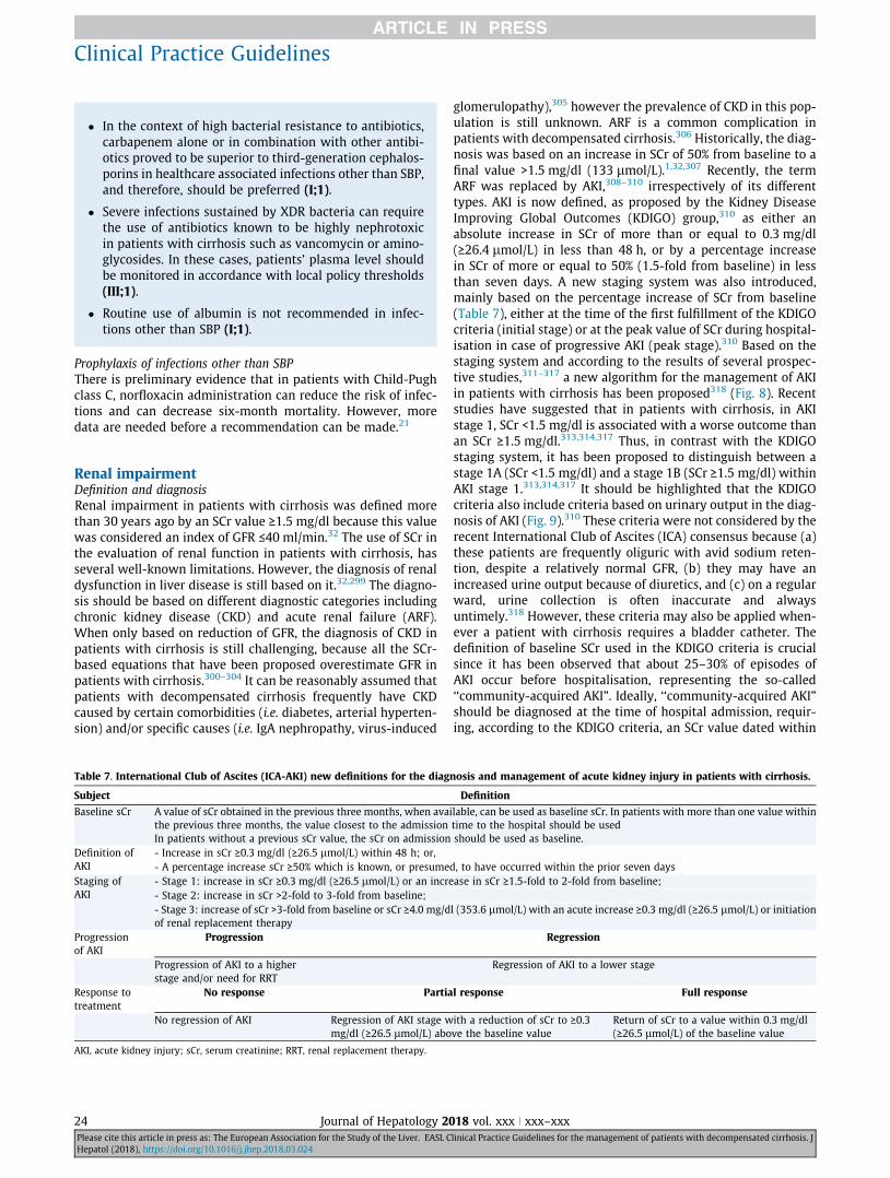
Ta
Su
Ba
DAKStAK
Prof
Retr
AK
Clinical Practice Guidelines
24PH
� In the context of high bacterial resistance to antibiotics,carbapenem alone or in combination with other antibi-otics proved to be superior to third-generation cephalos-porins in healthcare associated infections other than SBP,and therefore, should be preferred (I;1).
� Severe infections sustained by XDR bacteria can requirethe use of antibiotics known to be highly nephrotoxicin patients with cirrhosis such as vancomycin or amino-glycosides. In these cases, patients’ plasma level shouldbe monitored in accordance with local policy thresholds(III;1).
� Routine use of albumin is not recommended in infec-tions other than SBP (I;1).
Prophylaxis of infections other than SBPThere is preliminary evidence that in patients with Child-Pughclass C, norfloxacin administration can reduce the risk of infec-tions and can decrease six-month mortality. However, moredata are needed before a recommendation can be made.21
Renal impairmentDefinition and diagnosisRenal impairment in patients with cirrhosis was defined morethan 30 years ago by an SCr value ≥1.5 mg/dl because this valuewas considered an index of GFR ≤40 ml/min.32 The use of SCr inthe evaluation of renal function in patients with cirrhosis, hasseveral well-known limitations. However, the diagnosis of renaldysfunction in liver disease is still based on it.32,299 The diagno-sis should be based on different diagnostic categories includingchronic kidney disease (CKD) and acute renal failure (ARF).When only based on reduction of GFR, the diagnosis of CKD inpatients with cirrhosis is still challenging, because all the SCr-based equations that have been proposed overestimate GFR inpatients with cirrhosis.300–304 It can be reasonably assumed thatpatients with decompensated cirrhosis frequently have CKDcaused by certain comorbidities (i.e. diabetes, arterial hyperten-sion) and/or specific causes (i.e. IgA nephropathy, virus-induced
ble 7. International Club of Ascites (ICA-AKI) new definitions for the diag
bject
seline sCr A value of sCr obtained in the previous three months, when avaithe previous three months, the value closest to the admissionIn patients without a previous sCr value, the sCr on admission
efinition ofI
- Increase in sCr ≥0.3 mg/dl (≥26.5 lmol/L) within 48 h; or,- A percentage increase sCr ≥50% which is known, or presume
aging ofI
- Stage 1: increase in sCr ≥0.3 mg/dl (≥26.5 lmol/L) or an incre- Stage 2: increase in sCr >2-fold to 3-fold from baseline;- Stage 3: increase of sCr >3-fold from baseline or sCr ≥4.0 mg/dof renal replacement therapy
ogressionAKI
Progression
Progression of AKI to a higherstage and/or need for RRT
sponse toeatment
No response Partia
No regression of AKI Regression of AKI stage wmg/dl (≥26.5 lmol/L) abo
I, acute kidney injury; sCr, serum creatinine; RRT, renal replacement therapy.
Journal of Hepatology 20lease cite this article in press as: The European Association for the Study of the Liver. EASL Cepatol (2018), https://doi.org/10.1016/j.jhep.2018.03.024
glomerulopathy),305 however the prevalence of CKD in this pop-ulation is still unknown. ARF is a common complication inpatients with decompensated cirrhosis.306 Historically, the diag-nosis was based on an increase in SCr of 50% from baseline to afinal value >1.5 mg/dl (133 lmol/L).1,32,307 Recently, the termARF was replaced by AKI,308–310 irrespectively of its differenttypes. AKI is now defined, as proposed by the Kidney DiseaseImproving Global Outcomes (KDIGO) group,310 as either anabsolute increase in SCr of more than or equal to 0.3 mg/dl(≥26.4 lmol/L) in less than 48 h, or by a percentage increasein SCr of more or equal to 50% (1.5-fold from baseline) in lessthan seven days. A new staging system was also introduced,mainly based on the percentage increase of SCr from baseline(Table 7), either at the time of the first fulfillment of the KDIGOcriteria (initial stage) or at the peak value of SCr during hospital-isation in case of progressive AKI (peak stage).310 Based on thestaging system and according to the results of several prospec-tive studies,311–317 a new algorithm for the management of AKIin patients with cirrhosis has been proposed318 (Fig. 8). Recentstudies have suggested that in patients with cirrhosis, in AKIstage 1, SCr <1.5 mg/dl is associated with a worse outcome thanan SCr ≥1.5 mg/dl.313,314,317 Thus, in contrast with the KDIGOstaging system, it has been proposed to distinguish between astage 1A (SCr <1.5 mg/dl) and a stage 1B (SCr ≥1.5 mg/dl) withinAKI stage 1.313,314,317 It should be highlighted that the KDIGOcriteria also include criteria based on urinary output in the diag-nosis of AKI (Fig. 9).310 These criteria were not considered by therecent International Club of Ascites (ICA) consensus because (a)these patients are frequently oliguric with avid sodium reten-tion, despite a relatively normal GFR, (b) they may have anincreased urine output because of diuretics, and (c) on a regularward, urine collection is often inaccurate and alwaysuntimely.318 However, these criteria may also be applied when-ever a patient with cirrhosis requires a bladder catheter. Thedefinition of baseline SCr used in the KDIGO criteria is crucialsince it has been observed that about 25–30% of episodes ofAKI occur before hospitalisation, representing the so-called‘‘community-acquired AKI”. Ideally, ‘‘community-acquired AKI”should be diagnosed at the time of hospital admission, requir-ing, according to the KDIGO criteria, an SCr value dated within
nosis and management of acute kidney injury in patients with cirrhosis.
Definition
lable, can be used as baseline sCr. In patients with more than one value withintime to the hospital should be usedshould be used as baseline.
d, to have occurred within the prior seven daysase in sCr ≥1.5-fold to 2-fold from baseline;
l (353.6 lmol/L) with an acute increase ≥0.3 mg/dl (≥26.5 lmol/L) or initiation
Regression
Regression of AKI to a lower stage
l response Full response
ith a reduction of sCr to ≥0.3ve the baseline value
Return of sCr to a value within 0.3 mg/dl(≥26.5 lmol/L) of the baseline value
18 vol. xxx j xxx–xxxlinical Practice Guidelines for the management of patients with decompensated cirrhosis. J

Resolution
Close follow up
Response ?
NO
NO YES
Persistance Progression YES
Close monitoringRemove risk factors (withdrawal of nephrotoxic drugs, vasodilators and
NSAIDs, taper/withdraw diuretics and β-blockers, expand plasma volume, treat infections* when diagnosed)
Initial AKI# stage 1a°
Further treatment of AKI decided on a
case-by-case basis
Initial AKI# stage >1a°
Withdrawal of diuretics (if not yet applied) and volume expansion with albumin (1 g/kg) for 2 days
Does AKI meet criteria of HRS ?
Specific treatment for
other AKI phenotypes
Vasoconstrictors and albumin#AKI at the first fulfilling of KDIGO criteria
Fig. 8. Algorithm for the management of AKI in patients with cirrhosis (adapted from Ref. 318). AKI, acute kidney injury; HRS, hepatorenal syndrome;NSAID, non-steroidal anti-inflammatory.
KDIGO urine output criteria = an urinary output <0.5 ml/kg B.W./h/x 6-12 h
Stage Serum creatinine criteria
1° An urinary output <0.5 ml/kg B.W./h x 6-12 h
2° An urinary output <0.5 ml/kg B.W./h x 12 h
3° An urinary output <0.5 ml/kg B.W./h x 24 h or anuria per 12 h
Fig. 9. Criteria based on urinary output for the diagnosis of AKI (adaptedfrom Ref. 310). AKI, acute kidney injury; B.W., body weight.
Table 8. Definitions of kidney disease.
Definition Functional criteria Structuralcriteria
AKI Increase in sCr ≥50% within seven days,orincrease in sCr ≥0.3 mg/dl within two days
No criteria
AKD GFR <60ml/min per 1.73m2 for <3months,ordecrease in GFR ≥35% for < 3 months,orincrease in sCr ≥50 % for < 3 months
Kidney damagefor <3 months
CKD GFR <60 ml/min per 1.73 m2 for ≥3 months Kidney damagefor ≥3 months
AKD, acute kidney disease; AKI, acute kidney injury; CKD, chronic kidney disease;GFR, glomerular filtration rate; sCr, serum creatinine.
JOURNAL OF HEPATOLOGY
the last week before admission. This point is so crucial for theapplication of the KDIGO criteria that it has been suggested thatan SCr value be calculated when not available the seven daysprior to admission. The baseline SCr can be calculated by inver-sely applying the formulas that are used to calculate the esti-mated GFR, considering normal values of GFR of 75 ml/min.319
Whilst an imputed SCr is accepted in the general population,it can not be used in patients with cirrhosis.320 Indeed, allSCr-based formulas overestimate the true GFR in these patientsleading to an overestimation of the baseline SCr and thus under-estimating the prevalence of AKI on admission.320 Therefore, ithas been proposed that not only the value obtained in the lastseven days, but also that within the last three months be con-sidered as a baseline value of SCr in patients with cirrhosis(Table 7). In addition, an SCr value obtained within the last threemonths is the reference to define acute kidney disease (AKD), athird category of renal impairment, along with AKI and CKD,which has been recently proposed in KDIGO recommendations.AKD is clearly a distinct category with different outcome,whether or not it is associated with AKI. AKD is defined by aGFR <60 ml/min/1.73 m2 for less than three months, or adecrease in GFR ≥35% for less than three months, or an increasein SCr <50% within the last three months (Table 8). However, nodata exist about the prognostic impact of AKD, with or withoutAKI, in patients with cirrhosis. Thus, waiting for these data, itseems even more justified to make the diagnosis of AKI inpatients with cirrhosis on an increase in SCr ≥50% during thelast three months. This assumption may also facilitate the diag-nosis of AKI overlapping CKD.
Journal of Hepatology 201Please cite this article in press as: The European Association for the Study of the Liver. EASL ClinHepatol (2018), https://doi.org/10.1016/j.jhep.2018.03.024
Recommendations
� In patients with liver diseases, even a mild increase inSCr should be considered since it may underlie a markeddecrease of GFR (II-2;1).
� The first step to be addressed in the diagnostic process isto establish if the patient has a CKD, AKD or AKI as wellas an overlap between these diagnostic categories(II-2;1).
� The diagnosis of CKD should be based on a GFR <60 ml/min/1.73 m2 estimated by SCr-based formulas, with orwithout, signs of renal parenchymal damage (protein-uria/haeamturia/ultrasongraphy abnormalities) for atleast three months (II-2;1).
� The diagnostic process should be completed by stagingCKD, which relies on GFR levels, and by investigatingits cause. It should be highlighted that any SCr basedformula overestimates GFR in patients with cirrhosis(II-2, 1).
8 vol. xxx j xxx–xxx 25ical Practice Guidelines for the management of patients with decompensated cirrhosis. J
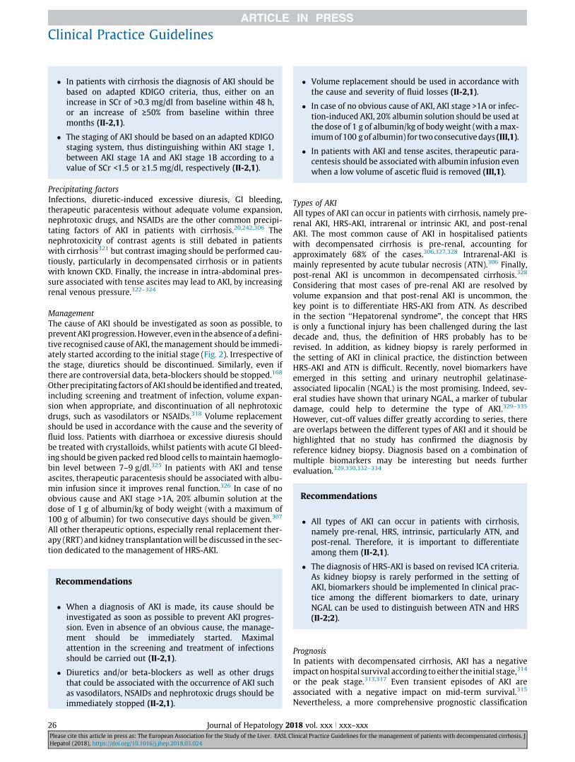
Clinical Practice Guidelines
26PH
� In patients with cirrhosis the diagnosis of AKI should bebased on adapted KDIGO criteria, thus, either on anincrease in SCr of >0.3 mg/dl from baseline within 48 h,or an increase of ≥50% from baseline within threemonths (II-2,1).
� The staging of AKI should be based on an adapted KDIGOstaging system, thus distinguishing within AKI stage 1,between AKI stage 1A and AKI stage 1B according to avalue of SCr <1.5 or ≥1.5 mg/dl, respectively (II-2,1).
Precipitating factorsInfections, diuretic-induced excessive diuresis, GI bleeding,therapeutic paracentesis without adequate volume expansion,nephrotoxic drugs, and NSAIDs are the other common precipi-tating factors of AKI in patients with cirrhosis.20,242,306 Thenephrotoxicity of contrast agents is still debated in patientswith cirrhosis321 but contrast imaging should be performed cau-tiously, particularly in decompensated cirrhosis or in patientswith known CKD. Finally, the increase in intra-abdominal pres-sure associated with tense ascites may lead to AKI, by increasingrenal venous pressure.322–324
ManagementThe cause of AKI should be investigated as soon as possible, topreventAKIprogression.However, even in theabsenceof adefini-tive recognised cause of AKI, themanagement should be immedi-ately started according to the initial stage (Fig. 2). Irrespective ofthe stage, diuretics should be discontinued. Similarly, even ifthere are controversial data, beta-blockers should be stopped.168
Otherprecipitating factors ofAKI shouldbe identifiedand treated,including screening and treatment of infection, volume expan-sion when appropriate, and discontinuation of all nephrotoxicdrugs, such as vasodilators or NSAIDs.318 Volume replacementshould be used in accordance with the cause and the severity offluid loss. Patients with diarrhoea or excessive diuresis shouldbe treated with crystalloids, whilst patients with acute GI bleed-ing should be given packed red blood cells tomaintain haemoglo-bin level between 7–9 g/dl.325 In patients with AKI and tenseascites, therapeutic paracentesis should be associated with albu-min infusion since it improves renal function.326 In case of noobvious cause and AKI stage >1A, 20% albumin solution at thedose of 1 g of albumin/kg of body weight (with a maximum of100 g of albumin) for two consecutive days should be given.307
All other therapeutic options, especially renal replacement ther-apy (RRT) and kidney transplantationwill be discussed in the sec-tion dedicated to the management of HRS-AKI.
Recommendations
� When a diagnosis of AKI is made, its cause should beinvestigated as soon as possible to prevent AKI progres-sion. Even in absence of an obvious cause, the manage-ment should be immediately started. Maximalattention in the screening and treatment of infectionsshould be carried out (II-2,1).
� Diuretics and/or beta-blockers as well as other drugsthat could be associated with the occurrence of AKI suchas vasodilators, NSAIDs and nephrotoxic drugs should beimmediately stopped (II-2,1).
Journal of Hepatology 201lease cite this article in press as: The European Association for the Study of the Liver. EASL Clinepatol (2018), https://doi.org/10.1016/j.jhep.2018.03.024
� Volume replacement should be used in accordance withthe cause and severity of fluid losses (II-2,1).
� In case of no obvious cause of AKI, AKI stage >1A or infec-tion-induced AKI, 20% albumin solution should be used atthe dose of 1 g of albumin/kg of bodyweight (with amax-imumof100 gof albumin) for twoconsecutive days (III,1).
� In patients with AKI and tense ascites, therapeutic para-centesis should be associated with albumin infusion evenwhen a low volume of ascetic fluid is removed (III,1).
Types of AKIAll types of AKI can occur in patients with cirrhosis, namely pre-renal AKI, HRS-AKI, intrarenal or intrinsic AKI, and post-renalAKI. The most common cause of AKI in hospitalised patientswith decompensated cirrhosis is pre-renal, accounting forapproximately 68% of the cases.306,327,328 Intrarenal-AKI ismainly represented by acute tubular necrosis (ATN).306 Finally,post-renal AKI is uncommon in decompensated cirrhosis.328
Considering that most cases of pre-renal AKI are resolved byvolume expansion and that post-renal AKI is uncommon, thekey point is to differentiate HRS-AKI from ATN. As describedin the section ‘‘Hepatorenal syndrome”, the concept that HRSis only a functional injury has been challenged during the lastdecade and, thus, the definition of HRS probably has to berevised. In addition, as kidney biopsy is rarely performed inthe setting of AKI in clinical practice, the distinction betweenHRS-AKI and ATN is difficult. Recently, novel biomarkers haveemerged in this setting and urinary neutrophil gelatinase-associated lipocalin (NGAL) is the most promising. Indeed, sev-eral studies have shown that urinary NGAL, a marker of tubulardamage, could help to determine the type of AKI.329–335
However, cut-off values differ greatly according to series, thereare overlaps between the different types of AKI and it should behighlighted that no study has confirmed the diagnosis byreference kidney biopsy. Diagnosis based on a combination ofmultiple biomarkers may be interesting but needs furtherevaluation.329,330,332–334
Recommendations
� All types of AKI can occur in patients with cirrhosis,namely pre-renal, HRS, intrinsic, particularly ATN, andpost-renal. Therefore, it is important to differentiateamong them (II-2,1).
� The diagnosis of HRS-AKI is based on revised ICA criteria.As kidney biopsy is rarely performed in the setting ofAKI, biomarkers should be implemented In clinical prac-tice among the different biomarkers to date, urinaryNGAL can be used to distinguish between ATN and HRS(II-2;2).
PrognosisIn patients with decompensated cirrhosis, AKI has a negativeimpact onhospital survival according to either the initial stage,314
or the peak stage.313,317 Even transient episodes of AKI areassociated with a negative impact on mid-term survival.315
Nevertheless, a more comprehensive prognostic classification
8 vol. xxx j xxx–xxxical Practice Guidelines for the management of patients with decompensated cirrhosis. J
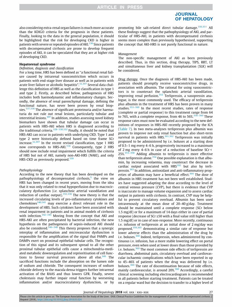
JOURNAL OF HEPATOLOGY
also considering extra-renal organ failures ismuchmore accuratethan the KDIGO criteria for the prognosis in these patients.Finally, looking to the data in the general population, it shouldbe highlighted that the risk for developing CKD is higher inpatientswith severe or repeatedepisodes ofAKI.336 Sincepatientswith decompensated cirrhosis are prone to develop frequentepisodes of AKI, it can be speculated that they are at higher riskof developing CKD.
Hepatorenal syndromeDefinition, diagnosis and classificationFor a long time, HRS has been defined as ‘‘a functional renal fail-ure caused by intrarenal vasoconstriction which occurs inpatients with end-stage liver disease as well as in patients withacute liver failure or alcoholic hepatitis”.32,307 Several data chal-lenge this definition of HRS as well as the classification in type 1and type 2. Firstly, as described below, pathogenesis of HRSincludes both haemodynamic and inflammatory changes. Sec-ondly, the absence of renal parenchymal damage, defining thefunctional nature, has never been proven by renal biop-sies.337,338 The absence of significant proteinuria and/or haema-turia do not rule out renal lesions, particularly tubular andinterstitial lesions.307 In addition, studies assessing novel kidneybiomarkers have shown that tubular damage can occur inpatients with HRS-AKI when HRS is diagnosed according tothe traditional criteria.328–330,332 Finally, it should be noted thatHRS-AKI can occur in patients with underlying CKD. Type 1 andtype 2 were historically defined based on time frame SCrincrease.32,307 In the recent revised classification, type 1 HRSnow corresponds to HRS-AKI.318 Consequently, type 2 HRSshould now include renal impairment which fulfills the criteriaof HRS but not of AKI, namely non-AKI-HRS (NAKI), and onlyHRS-CKD as previously proposed.339
PathophysiologyAccording to the new theory that has been developed on thepathophysiology of decompensated cirrhosis,5 the view onHRS has been changed in recent years, moving from the ideathat it was only related to renal hypoperfusion due to macrocir-culatory dysfunction (i.e. splanchnic arterial vasodilation andreduction of cardiac output).192,338 The new theory is that theincreased circulating levels of pro-inflammatory cytokines andchemokines340,341 may exercise a direct relevant role in thedevelopment of HRS. Such cytokines have been associated withrenal impairment in patients and in animal models of cirrhosiswith infection.342–345 Moving from the concept that AKI andHRS-AKI are often precipitated by bacterial infection, the newhypothesis on the pathogenesis of sepsis-induced AKI shouldalso be considered.346–348 This theory proposes that a synergicinterplay of inflammation and microvascular dysfunction isresponsible for the amplification of the signal that PAMPs andDAMPs exert on proximal epithelial tubular cells. The recogni-tion of this signal and its subsequent spread to all the otherproximal tubular epithelial cells cause a mitochondria-medi-ated metabolic downregulation and reprioritisation of cell func-tions to favour survival processes above all else.349 Thesacrificed functions include the absorption on the lumen sideof sodium and chloride. The consequent increases of sodiumchloride delivery to the macula densa triggers further intrarenalactivation of the RAAS and thus lowers GFR. Finally, severecholestasis may further impair renal function by worseninginflammation and/or macrocirculatory dysfunction, or by
Journal of Hepatology 20Please cite this article in press as: The European Association for the Study of the Liver. EASL CHepatol (2018), https://doi.org/10.1016/j.jhep.2018.03.024
promoting bile salt-related direct tubular damage.350,351 Allthese findings suggest that the pathophysiology of AKI, and par-ticular of HRS-AKI, in patients with decompensated cirrhosisseems more complex than previously hypothesised, supportingthe concept that AKI-HRS is not purely functional in nature.
ManagementThe non-specific management of AKI as been previouslydescribed. Thus, in this section, drug therapy, TIPS, RRT, LTand simultaneous liver and kidney transplantation (SLK) willbe considered.
Drug therapy. Once the diagnosis of HRS-AKI has been made,patients should promptly receive vasoconstrictive drugs, inassociation with albumin. The rational for using vasoconstric-tors is to counteract the splanchnic arterial vasodilation,improving renal perfusion.352 Terlipressin, a vasopressin ana-logue, is the most commonly used. The efficacy of terlipressinplus albumin in the treatment of HRS has been proven in manystudies.353–360 In the most recent studies, rates of response(complete or partial response) to this treatment range from 64to 76%, with a complete response, from 46 to 56%.358–360 Theseresponse rates must now be evaluated according to the new def-initions of responses in HRS-AKI recently proposed by the ICA(Table 7). In two meta-analyses terlipressin plus albumin wasproven to improve not only renal function but also short-termsurvival in patients with HRS.361,362 Terlipressin was initiallyproposed to be administered by i.v. boluses at a starting doseof 0.5–1 mg every 4–6 h, progressively increased to a maximumof 2 mg every 4–6 h in case of a reduction of baseline SCr <25%.353–358 Adding albumin to terlipressin is more effectivethan terlipressin alone.354 One possible explanation is that albu-min, by increasing volaemia, may counteract the decrease incardiac output associated with HRS192 but also by terli-pressin.363 In addition, antioxidant and anti-inflammatory prop-erties of albumin may have a beneficial effect.364 The dose ofalbumin in HRS treatment has not been well established. Stud-ies have suggested adapting the dose according to the level ofcentral venous pressure (CVP), but there is evidence that CVPis inaccurate to manage volume expansion and to assess cardiacoutput in patients with cirrhosis. In contrast, CVP may be help-ful to prevent circulatory overload. Albumin has been usedintravenously at the mean dose of 20–40 g/day. Treatmentshould be maintained until a complete response (SCr below1.5 mg/dl) or for a maximum of 14 days either in case of partialresponse (decrease of SCr ≥50 with a final value still higher than1.5 mg/dl) or in case of non-response. More recently, continuousi.v. infusion of terlipressin at an initial dose of 2 mg/day wasproposed,359,365 demonstrating a similar rate of response butlower adverse effects than the administration of the drug byi.v. boluses.360 Indeed, terlipressin, when administered by con-tinuous i.v. infusion, has a more stable lowering effect on portalpressure, even when used at lower doses than those provided byi.v. boluses.360 The most common side effects of terlipressin arediarrhoea, abdominal pain, circulatory overload and cardiovas-cular ischaemic complications which have been reported in upto 45–46% of patients when the drug was delivered by i.v.boluses.360 The rate of discontinuation because of side effects,mainly cardiovascular, is around 20%.360 Accordingly, a carefulclinical screening including electrocardiogram is recommendedin all patients before starting treatment. Patients can be treatedon a regular ward but the decision to transfer to a higher level of
18 vol. xxx j xxx–xxx 27linical Practice Guidelines for the management of patients with decompensated cirrhosis. J

� Albumin solution (20%) should be used at the dose20–40 g/day. Ideally, apart from routinely monitoringpatients with HRS-AKI, the serial measurement of CVPor other measures of assessing central blood volume,can help to prevent circulatory overload by optimisingthe fluid balance and helping to titrate the dose of albu-min (II-2;1).
� Noradrenaline can be an alternative to terlipressin. How-ever, limited information is available (I;2).
� In contrast to terlipressin, the use of noradrenalinealways requires a central venous line and, in severalcountries, the transfer of the patient to an ICU. Mido-drine plus octreotide can be an option only when terli-pressin or noradrenaline are unavailable, but itsefficacy is much lower than that of terlipressin (I;1).
� According to the new definition of HRS-AKI, completeresponse to the treatment should be defined by a finalSCr within 0.3 mg/dl (26.5 lmol/L) from the baselinevalue, while partial response should be defined by theregression of AKI stage to a final SCr ≥0.3 mg/dl (26.5lmol/L) from the baseline value (III;1).
� Adverse events related to terlipressin or noradrenalineinclude ischaemic and cardiovascular events. Thus, a care-ful clinical screening including electrocardiogram is rec-ommended before starting the treatment. Patients can betreated on a regular ward, but the decision to transfer tohigherdependency care shouldbecasebased. For thedura-tion of treatment, it is important to closely monitor thepatients. According to the type and severity of side effects,treatment should be modified or discontinued (I;1).
� In cases of recurrence of HRS-AKI upon treatment cessa-tion, a repeat course of therapy should be given (I;1).
� Terlipressin plus albumin is also effective in the treat-ment of HRS outside the criteria of AKI (HRS-NAKI), for-merly known as HRS type II. Unfortunately, recurrenceafter the withdrawal of the treatment is the norm, andcontroversial data exists on the impact of the treatmenton long-term clinical outcome, particularly from the per-
Clinical Practice Guidelines
care should be case based. Recurrent HRS in responders, afterthe end of the treatment, has been reported in up to 20% ofcases. Re-treatment is usually effective, however, in some cases,continuous recurrence occurs, thus a long-term treatment withterlipressin plus albumin and a long-term hospitalisation arerequired.366 The possibility of treating some of these patientsoutside the hospital has recently been proposed367 but even ifpromising, further studies are needed. Other vasoconstrictivedrugs include i.v. noradrenaline and oral midodrine plus subcu-taneous or i.v. octretide, both in combination with albumin.Noradrenaline, given by continuous i.v. infusion at the dose of0.5–3 mg/h, has been proven to be as effective as terlipressinregarding the increase in mean arterial pressure, the reversalof renal impairment and one-month survival.368–371 However,the number of patients treated with noradrenaline remainstoo small to definitively confirm its efficacy. In addition, in con-trast to terlipressin, the use of noradrenaline always requires acentral venous line and, in most countries, the transfer of thepatient to an intensive care unit (ICU). The combination mido-drine plus octreotide, used in countries where terlipressin isnot yet available,372 has been shown to be much less effectivethan terlipressin in the treatment of type 1 HRS in a recentRCT.359 Vasoconstrictors, in particular terlipressin, in associa-tion with albumin, have also been proposed in the treatmentof type 2 HRS. The treatment has been proven to be effectivein most cases but, unfortunately, recurrence after the with-drawal of treatment is the norm. In addition, there are contro-versial data about the impact of this treatment on outcomes,especially in candidates for LT.373,374 This may be, at least inpart, due to the suboptimal definition of type 2 HRS, as previ-ously discussed. The most relevant factors that may impairthe response to vasoconstrictors are: a) the baseline value ofSCr, b) the degree of inflammation and c) the degree of cholesta-sis.375–377 The finding that the higher baseline values of SCr, thelower rate of response to terlipressin plus albumin,375 probablyreflects the presence of renal parenchymal damage337 and rep-resents one of the main reasons behind the adoption of theKDIGO criteria for the definition of AKI in patients with cirrho-sis, and the introduction of the new algorithm for its manage-ment. Regarding inflammation, it has been recently shownthat, for the same value of baseline SCr, the rate of response isrelated to the number of extra-renal organ failures.376
Recommendations
� Vasoconstrictors and albumin are recommended in allpatients meeting the current definition of AKI-HRS stage>1A, should be expeditiously treated with vasoconstric-tors and albumin (III;1).
� Terlipressin plus albumin should be considered as thefirst-line therapeutic option for the treatment of HRS-AKI. Telipressin can be used by i.v. boluses at the initialdose of 1 mg every 4–6 h. However, giving terlipressinby continuous i.v. infusion at initial dose of 2 mg/daymakes it possible to reduce the global daily dose of thedrug and, thus, the rate of its adverse effects. In case ofnon-response (decrease in SCr <25% from the peak value),after twodays, thedose of terlipressin shouldbe increasedin a stepwise manner to a maximum of 12 mg/day (I;1).
spective of LT. As such, vasoconstrictors and albumin arenot recommended in this clinical scenario (I;1).
28 Journal of Hepatology 20Please cite this article in press as: The European Association for the Study of the Liver. EASL CHepatol (2018), https://doi.org/10.1016/j.jhep.2018.03.024
Transjugular intrahepatic portosystemic shunts. The use of TIPSmay improve renal function in patients with type 1 HRS.378,379
However, the applicability of TIPS in this clinical setting is usu-ally very limited because, in most patients, TIPS is contraindi-cated because of severe degree of liver failure. TIPS has beenstudied in patients with type 2 HRS380 and in the managementof refractory ascites, frequently associated with type 2 HRS. Inthese patients, TIPS has been shown to improve renal function.95,379
Renal replacement therapy. Renal replacement therapy should beconsidered in the management of AKI, whatever the type. As faras HRS-AKI, it should be considered in non-responders to vaso-constrictors. RRT should also be considered in patients withend-stage kidney disease. The indications for RRT are the samein patients with cirrhosis as in the general population including:severe and/or refractory electrolyte or acid-base imbalance,
18 vol. xxx j xxx–xxxlinical Practice Guidelines for the management of patients with decompensated cirrhosis. J

JOURNAL OF HEPATOLOGY
severe or refractory volume overload, and/or symptomaticazotaemia. However, published data on RRT in patients withcirrhosis are scant, with controversial effects on survival.381,382
It has been stated that indications for RRT depend on the per-spective of LT. It has been stated that RRT may be consideredin patients who are candidates for LT, while, in contrast, thedecision to initiate RRT in non-candidates should be individu-alised to avoid futility.20 However, it has recently been observedthat critically ill liver cirrhotic patients requiring RRT have veryhigh mortality independent of LT options. Thus, RRT and treat-ment at the ICU should not be limited to LT candidates butshould be based on the individual severity of illness.383 There-fore, repeated risk stratification is necessary during the courseof treatment, assisted by prognostic scores in addition to clinicaljudgment and patients0 wishes.383 The ideal timing for RRT ini-tiation has not been defined in patients with cirrhosis. However,data on AKI in patients with acute liver failure as well as in crit-ically ill patients without liver disease suggest that early RRTimproves survival.384–386 Both haemodialysis or continuousrenal replacement therapy (CRRT), have been used in patientswith cirrhosis. Despite the available evidence,387 CRRT is prob-ably better tolerated, providing greater cardiovascular stabilityand allowing a slower correction of severe or refractory hypona-traemia than haemodialysis.
Liver support systems. In two controlled studies, both the so-called artificial liver support systems, either the molecularadsorbents recirculating system (MARS�) or Prometheus�,showed promising beneficial effects in patients with type 1HRS, but should be further investigated.388,389
Liver transplantation and simultaneous liver-kidney transplanta-tion. The best therapeutic option in patients with HRS is LT.390
However, several studies have shown that SCr after LT is higherin patients transplanted with HRS, compared to those withoutHRS at the time of LT. In addition, the presence of HRS at thetime of LT has a negative impact on survival after LT.391 Thetreatment of type 2 HRS before LT has given conflicting resultson the clinical outcome after LT373,374 and thus, requires furtherinvestigation. SLK can be indicated in patients with cirrhosisand CKD in the following conditions: a) estimated GFR (usingMDRD6 equation) ≤40 ml/min or measured GFR using iothala-mate clearance ≤30 ml/min, b) proteinuria ≥2 g a day, c) kidneybiopsy showing >30% global glomerulosclerosis or >30% intersti-tial fibrosis, or d) inherited metabolic disease. SLK is also indi-cated in patients with cirrhosis and sustained AKI irrespectiveof its type, including HRS-AKI when refractory to drug therapy,in the following conditions: a) AKI on RRT for ≥4 weeks or b)estimated GFR ≤ 35 ml/min or measured GFR ≤25 ml/min ≥4weeks.392 Beyond these two conditions, in a candidate with highpriority for LT due to a high MELD score, the option of SLK maybe considered in the presence of risk factors for underlyingundiagnosed CKD (diabetes, hypertension, abnormal renalimaging and proteinuria >2 g/day).392 The development ofnew biomarkers of kidney fibrosis, a common and irreversiblefeature of CKD, is also promising in this context.393
Regarding the priority allocation of patients with HRS-AKI tothe waiting list, some rules should be applied in case ofresponse to drug therapy. In fact, by lowering SCr and increasingserum sodium concentration, the treatment can significantlylower the MELD and MELD-Na score, potentially delaying LT.Considering that the survival rate in responders at three months
Journal of Hepatology 20Please cite this article in press as: The European Association for the Study of the Liver. EASL CHepatol (2018), https://doi.org/10.1016/j.jhep.2018.03.024
is almost 50%, a specific policy of priority allocation is neededfor these patients. This can be made either by continuing to con-sider the baseline MELD and/or MELD-Na score394 rather thanthose during or after the end of the treatment, or by providingan exception to the MELD score.395
Recommendations
� There is insufficient data to advocate TIPS in HRS-AKI butit could be suggested in selected patients with HRS-NAKI(II-2;2).
� LT is the best therapeutic option for patients with HRSregardless of the response to drug therapy (I;1).
� The decision to initiate RRT should be based on the indi-vidual severity of illness (I;2).
� The indication for liver-kidney transplantation remainscontroversial. This procedure should be considered inpatients with significant CKD or with sustained AKIincluding HRS-AKI with no response to drug therapy(II-2;1).
1lin
Prevention of hepatorenal symdromeThe prevention of HRS-AKI, as for other causes of AKI, is basedon the use of albumin in patients who develop SBP272 and theprevention of SBP using norfloxacin,276 as discussed before. Inaddition, the use of pentoxyfilline may decrease the incidenceof renal failure in patients with cirrhosis27 and of type 1 HRSas well as mortality in patients with severe alcoholic hepati-tis.396 However, recent papers do not confirm theseresults397,398 and further studies are needed.
Recommendations
� Albumin (1.5 g/kg at diagnosis and 1 g/kg on day three)should be given in patients with SBP to prevent AKI (I;1).
� Norfloxacin (400mg/day) should be given as prophylaxisof SBP to prevent HRS-AKI (I;1).
Acute-on-chronic liver failureDefinitions and pathophysiologySince the CANONIC study, the first major international observa-tional study characterising the syndrome of ACLF,3 a large num-ber of publications have described the association of thissyndrome with different clinical, diagnostic and therapeuticapproaches. ACLF occurs in 30% of admitted patients3,399 andin 25% of outpatients,400 and is a major cause of death in cirrho-sis, with an approximately 50% mortality rate.400 Even thoughthere is an ongoing debate regarding the definition ofACLF,401–405 the concept of the development of ACLF is similaracross different continents and health systems. There is agree-ment that ACLF is not just decompensation of liver cirrhosis,but a distinct syndrome.406 The reason is that ACLF is definedby a multi-organ failure and has a higher short-term mortalitythan a ‘‘simple decompensation” of cirrhosis.3,401,406 The riskof developing ACLF is higher in outpatients with advanced liverdisease according to the presence of ascites, low mean arterial
8 vol. xxx j xxx–xxx 29ical Practice Guidelines for the management of patients with decompensated cirrhosis. J

Box 1. CLIF-C Acute Decompensation Score (Ref. 407).
10 x [0.03 x Age + 0.66 x Ln(Creatinine) + 1.71 x Ln(INR) + 0.88 x Ln(WBC) 0.05 x Sodium + 8]
CLIF-C Acute Decompensation score
Age in years; creatinine in mg/dl; WBC (white blood count) in 109 cells/L; sodium in mmol/L
Clinical Practice Guidelines
pressure or anaemia and with a high MELD score.400 ACLF devel-ops on the background of acute decompensation (AD) of cirrho-sis, but a remarkable number of patients (�40%) admitted tohospital developed ACLF on the first episode of AD of their liverdisease.3 Thus, the presence of AD is an important clinical fea-ture for the diagnosis of ACLF.3,401,406 The EASL-CLIF Consortiumhas proposed and validated a prognostic score (CLIF-C AD score)for patients with AD who do not develop ACLF407 The CLIF-C ADscore (Box 1) was proved to be more accurate for predicting out-come in these patients than the MELD or MELD-Na score.407
Once developed, ACLF is characterised by hepatic and extrahep-atic organ dysfunction and/or failure, highly activated systemicinflammation, and a high 28-day mortality.3,12 The overwhelm-ing and devasting inflammatory response is a key pathogenicmechanism in the development of ACLF, probably explainingwhy ACLF frequently happens in younger patients.3,401,406,408
The trigger of ACLF and this inflammatory response could notbe identified in 40–50% of the patients in CANONIC study,3
which might be associated with genetic predisposition, severeportal hypertension or other factors predisposing the patientsto development of AD and ACLF.409 However, identification ofthe precipitating events of AD are of great importance to pre-vent and manage ACLF.410,411
Precipitating eventsThe precipitating events vary between different populations,geographic areas and aetiologies. While in Western countries(Europe, North and Latin America) bacterial infection, followedby active alcohol intake or binge are major precipitatingevents,3,412,413 in Eastern countries (Asia, Pacific region) theexacerbation of hepatitis B, followed by alcohol or bacterialinfections are the major causes of AD and ACLF develop-ment.414–416 But there are a number of other insults, whichmight induce ACLF, such as superimposed infection with hepa-totropic viruses (especially HAV, HEV), DILI, GI bleeding, circula-tory dysfunction upon different situation (e.g. surgery, LVPwithout albumin). Therefore, in general the precipitating factorsmight be differentiated into three major categories, hepatotoxicinjury (active alcohol intake or binge, DILI), immunologicalinsults (flairs of viral or autoimmune hepatitis, bacterial, fungaland viral infections, common cold, subclinical infections, etc.)and haemodynamic derangement following procedures (haem-orrhage, surgery, LVP).
Bacterial infectionsOverall, the major precipitating factor for ACLF is bacterialinfections accounting for 30–57% of cases.409,410 The importanceof bacterial infections for the development of organ failures andACLF was also underlined by the studies of North American Con-sortium for End-stage Liver Disease (NACSELD), who havedefined ACLF by the development of two organ failures in pres-ence of bacterial infections.412 By contrast, bacterial infectionswere not considered to be precipitating events for ACLF accord-
30 Journal of Hepatology 20Please cite this article in press as: The European Association for the Study of the Liver. EASL CHepatol (2018), https://doi.org/10.1016/j.jhep.2018.03.024
ing to the definition of Asian Pacific Association for the Study ofthe Liver (APASL).402 However, nowadays there is evidence thatbacterial infections are mainly involved in the development oforgan failures and thereby of ACLF in Asia as well.415,416 In Wes-tern countries bacterial infections are the precipitating events inone-third of patients admitted with ACLF and in two-thirds ofpatients developing ACLF during follow-up.3,409,412,413 Basedon these data, preventive and early therapeutic interventionsfor the treatment of infections are of major importance to pre-vent the development of ACLF. The role of bacterial infectionsas triggers of AD and development of organ failures has alreadybeen discussed.
Active alcohol intake or bingeAlcoholic liver disease was the most prevalent in patients withAD and ACLF in the CANONIC study, as well as in recent reportsfrom India.415–417 Interestingly, active alcoholism and alcoholbinge were not only a major trigger in these patients, but ledto a more severe syndrome than other triggers in alcoholic cir-rhosis patients without heavy active alcoholism.3 The role andmechanisms of active alcoholism need further investigation,especially regarding prevention and treatment.
Reactivation and superimposed viral hepatitisReactivation of HBV in patients with cirrhosis is the main pre-cipitating event in the non-Caucasian Asian population,413,415
occurring mostly in genotypes B and D, and hepatitis B e antigenpositive patients. Interestingly, superimposed HAV and HEV canalso trigger ACLF in 14–18%.414,416 According to the Westernexperience, these are unusual causes.3,413 However, the role ofHEV might have been overlooked, and might gain more impor-tance now because of advances in diagnostics and increases inawareness.3,418 A timely recognition and treatment of the pre-cipitating event might prevent ACLF and improve outcome inthese patients.
Clinical and diagnostic features of ACLFAs discussed previously, organ failures in the presence of AD ofcirrhosis are the basis for the diagnosis of ACLF. However, in theCANONIC study the presence and grading of ACLF was based onmortality and the independent association of organ dysfunc-tion/failure with mortality, which was chosen to be ≥15% at28 days.3 Organ failures were defined based on a sequentialorgan failure assessment (SOFA) score, which was adapted topatients with cirrhosis, the CLIF SOFA score (Table 9). However,two organs received special attention, the kidney and the brain.3
In fact, it has been observed that even mild renal or brain dys-function in the presence of another organ failure, is associatedwith a significant short-term mortality and therefore definesthe presence of ACLF. Thus, patients with renal failure, definedas creatinine ≥2 mg/dl, were classified as ACLF grade Ia whilepatients with a non-renal and non-cerebral organ failurecombined either with mild renal dysfunction (creatininebetween 1.5 and 1.9 mg/dl) and/or grade I and II hepaticencephalopathy, as well as those with cerebral failure combinedwith mild renal dysfunction were classified as ACLF grade Ib(Table 10).3 Thereafter, patients with two organ failures areclassified as grade II ACLF, and have a 28-day mortality rate of32%. Patients with three or more organ failures are classifiedas grade III ACLF and have an average 28-day mortality of 78%(Table 9). According to this EASL-CLIF definition of ACLF,approximately one-quarter of patients admitted to the hospital
18 vol. xxx j xxx–xxxlinical Practice Guidelines for the management of patients with decompensated cirrhosis. J

Table 9. CLIF-Sequential Organ Failure Assessment (SOFA) score (adapted from Ref. n� 3).
The CLIF-Sequential Organ Failure Assessment (SOFA) score
Organ/system 0 1 2 3 4
Liver (bilirubin mg/dl) <1.2 ≥1.2–<2.0 ≥2.0–<6.0 ≥6.0–<12.0 ≥12.0Kidney (creatinine, mg/dl) <1.2 ≥1.2–<2.0 ≥2.0–<3.5 ≥3.5–<5.0 ≥5.0Cerebral (HE grade) No HE Grade I Grade II Grade III Grade IVCoagulation (INR and PLT count) <1.1 ≥1.1–<1.25 ≥1.25–<1.5 ≥1.5–<2.5 ≥2.5 or PLT ≤ 20.000/mm3
Circulation (MAP, mmHg and vasopressors) ≥70 <70 Dopamine ≤5* ordobutamine or
terlipressin
Dopamine >5* orE ≤0.1* or NE
≤0.1*
Dopamine >15*
or E >0.1* or NE>0.1*
LungsPaO2/FiO2, orSpO2/FiO2
>400>512
>300–≤400>357–≤512
>200–≤300>214–≤357
>100–≤200>89- ≤214
≤100≤89
E, epinephrine; FiO2, fraction of inspired oxygen; HE, hepatic encephalopathy; NE, norepinephrine; PaO2, partial pressure of arterial oxygen; SpO2, pulse oximetricsaturation. The bold text indicates the diagnostic criteria for organ failures.* lg/kg/min.
Table 10. Classification and grades of ACLF (adapted from Ref. 3).
Grades ofACLF
Clinical characteristics
No ACLF No organ failure, or single non-kidney organ failure, creatinine<1.5 mg/dl, no HE
ACLF Ia Single renal failureACLF Ib Single non-kidney organ failure, creatinine 1.5–1.9 mg/dl and/
or HE grade 1–2ACLF II Two organ failuresACLF III Three or more organ failures
ACLF, acute-on-chronic liver failure; HE, hepatic encephalopathy.
JOURNAL OF HEPATOLOGY
for AD of cirrhosis had ACLF at admission or develop it duringthe hospitalisation. After having simplified the CLIF SOFA scoreinto the CLIF Organ Failure score (Table 11), the EASL-CLIF Con-sortium formulated a new score, the CLIF-C ACLF score, whichenables the prediction of mortality in patients with ACLF.419
The CLIF-C ACLF score (Box 2) has been validated by differentindependent series of patients.417,420,421 Other scores wererecently proposed by the APASL ACLF Research Consortiumand by the NACSELD, but they were not compared specificallywith the CLIF-C-ACLF score.422,423
Table 11. Chronic Liver Failure – Organ Failure score system (adapted from Re
Organ/system 1 point
Liver Bilirubin <6 mg/dl 6 ≤BiKidney Creatinine <2 mg/dl 2 CreaBrain/HE (West Haven criteria) Grade 0Coagulation INR <2.0Circulation MAP ≥70 mmHgLungs PaO2/FiO2 >300,
orSpO2/FiO2 >357
PaO2
SpO2
Note: The bold text denotes criteria for diagnosing organ failures.FIO2, fraction of inspired oxygen; HE, hepatic encephalopathy; INR, international normaliSpO2, pulse oximetric saturation.a Patients submitted to mechanical ventilation due to HE and not to a respiratory failurb Other patients enrolled in the study with mechanical ventilation were considered as p
Box 2. CLIF-C Acute Liver Failure (ACLF) score (Ref. 419).
10 x [0.033 x Clif OFs + 0.04 x Age + 0.63 x Ln(WBC) 2]
Age in years; CLIF OF score as in Table 10; sodium in mmol/L
CLIF-C ACLF score
Journal of Hepatology 201Please cite this article in press as: The European Association for the Study of the Liver. EASL ClinHepatol (2018), https://doi.org/10.1016/j.jhep.2018.03.024
Recommendations
� The diagnosis of ACLF should be made in a patient withcirrhosis and AD (defined as the acute development orworsening of ascites, overt encephalopathy, GI-haemor-rhage, non-obstructive jaundice and/or bacterial infec-tions), when organ failure(s) involving high short-termmortality develop (II-2;1).
� The diagnosis and the grading of ACLF should be basedon the assessment of organ function as defined by theCLIF-C Organ Failure score (II-2,1).
� Potential precipitating factor(s), either hepatic (i.e. heavyalcohol intake, viral hepatitis, DILI, autoimmune hepati-tis) and/or extrahepatic (i.e. infections haemodynamicderangements following haemorrhage, surgery) shouldbe investigated. However, in a significant proportion ofpatients, a precipitant factor may not be identified(II-2,1).
Management of ACLFGeneral managementUnfortunately, there is no specific effective treatment forACLF.424 Therefore, treatment is currently based on organ sup-port and management of associated complications. The causeof liver injury can be specifically treated only in certain situa-tions such as in ACLF secondary to HBV infection, as described
f. 419).
2 points 3 points
lirubin <12 mg/dl Bilirubin ≥12 mg/dltinine <3.5 mg/dl Creatinine ≥3.5 mg/dl or renal replacement
Grades 1–2 Grades 3-4a
2.0 ≤INR <2.5 INR ≥2.5MAP <70 mmHg Use of vasopressors/FiO2 ≤300–>200,
or/FiO2 >214–≤357
PaO2/FiO2 ≤200b
orSpO2/FiO2 ≤214b
zed ratio; MAP, mean arterial pressure; PaO2, partial pressure of arterial oxygen;
e were considered as presenting a cerebral failure (cerebral score = 3).resenting a respiratory failure (respiratory score = 3).
8 vol. xxx j xxx–xxx 31ical Practice Guidelines for the management of patients with decompensated cirrhosis. J

Clinical Practice Guidelines
later. Patients with ACLF should ideally be admitted to intensivecare or intermediate care units, yet this decision should be indi-vidualised based on certain factors, particularly patients’ ageand associated comorbidities. Moreover, patients suitable forLT should be referred to a transplant centre early in the courseof ACLF. Late referral may make transplantation impossible dueto the rapid evolution of ACLF in most patients.425 In patients inwhom ACLF is associated with precipitating factors, such as bac-terial infections, GI bleeding, or drug toxicity, early identifica-tion and management of these conditions is crucial to patientsurvival. Nonetheless, it should be emphasised that this earlytreatment of triggering factors may not prevent the progressionof ACLF in all patients. Meanwhile, as already stated, in approx-imately half of patients with ACLF a precipitating factor cannotbe identified.3 Organ support is very important in the manage-ment of patients with ACLF.424 Haemodynamic function shouldbe monitored and vasopressor therapy administered in case ofmarked arterial hypotension. Hepatic encephalopathy shouldbe treated early with standard therapy. Special care should betaken to preserve airway patency to prevent aspiration pneu-monia. In patients with coagulation failure, either because ofimpairment of coagulation factors or low platelet count, substi-tutive therapy should be given only if there is clinically signifi-cant bleeding. If there is respiratory failure, patients should begiven oxygen therapy and ventilation, if required. Finally, ifthere is kidney failure its cause should be identified and man-aged accordingly. Volume expansion should be given to patientswith fluid loss or in the setting of SBP. Excessive volume expan-sion should be avoided. Patients meeting the criteria of AKI-HRSshould be treated with terlipressin and albumin or nore-pinephrine, if terlipressin is not available. Patients with sus-pected ATN should be treated with RRT if they meet criteriafor this treatment.392
Specific therapiesLiver support systems. Extracorporeal liver support systems, par-ticularly albumin dialysis (MARS system) and fractionatedplasma separation and adsorption (Prometheus system) havebeen evaluated as therapies for ACLF. These systems removealbumin-bound substances and other substances that accumu-late in the context of ACLF and may have deleterious effectson the function of different organs. Both methods have beenevaluated in large RCTs in patients with ACLF and no significanteffects on survival could be demonstrated.388,389 It should beemphasised however, that the definition of ACLF in both trialswas different than the current definition of ACLF based on theCANONIC study.3 Moreover, a sub-analysis of the Prometheusstudy showed a beneficial effect on survival in patients withMELD score higher than 30.389 This finding deserves furtherinvestigation. Nonetheless, based on the results of availableRCTs, extracorporeal liver support systems do not improve sur-vival of patients with ACLF and should not be recommended inthis indication.
Antiviral therapy for chronic hepatitis B. Reactivation of hepatitisB is a very common cause of ACLF in certain areas of the world,particularly in southwest Asia.414 A number of non-randomisedstudies and an RCT have shown that treatment with lamivudine,tenofovir or entecavir is associated with inhibition of HBV repli-cation, improvement of liver function, and higher survival inpatients with ACLF secondary to hepatitis B infection.426–429
The only RCT included 24 patients, 14 treated with tenofovir
32 Journal of Hepatology 20Please cite this article in press as: The European Association for the Study of the Liver. EASL CHepatol (2018), https://doi.org/10.1016/j.jhep.2018.03.024
and 13 treated with placebo, and showed significant differencesin three-month survival (57% vs. 15%, respectively).426 There-fore, it seems evident that the presence of HBV infection shouldbe investigated in all patients with ACLF and antiviral therapyshould be started as soon as possible.
Other therapies. A number of therapies have been assessed inpatients with ACLF, including dexamethasone, plasmaexchange, chinese herbs, caspase inhibitors, mesenchymal stemcells transplantation, and administration of granulocyte-colonystimulating factor (G-CSF).430–432 In most cases, the informationis still very preliminary and no recommendations can be maderegarding their potential use in clinical practice. However, anote on G-CSF seems pertinent because this approach has beenassessed in an RCT.432 The rationale behind this treatmentseems to be the mobilisation of stem cells from the bone mar-row and their engraftment within the liver, although other ben-eficial effects may also occur. The only RCT evaluating thistherapy included 47 patients with ACLF, as defined by the APASLcriteria, 23 treated with G-CSF (12 doses of 5 lg/kg subcuta-neously) and 24 treated with placebo in a double-blind manner.The main findings were an improvement in 60-day survival inthe G-CSF group vs. the placebo group (66% vs. 26%, respec-tively; p = 0.001) along with a reduction in Child-Pugh, SOFA,and MELD scores and a decrease in the occurrence of HRS, hep-atic encephalopathy and sepsis in G-CSF treated patients.Although these results are promising, additional studies in a lar-ger number of patients are needed.
Liver transplantation. Liver transplantation is theoretically thedefinitive treatment for ACLF because it allows the cure of ACLFsyndrome as well as the underlying liver disease.425 However,some important issues regarding LT for ACLF deserve a com-ment, particularly the accessibility of patients to LT, evaluationof candidate subjects, the outcomes of LT on survival, and futility.The accessibility of patients with ALCF to LT is probablydecreased compared to that of patients with other indicationsfor LT, because patients with ACLF have a high mortality rateafter diagnosis of the condition. Early referral to transplant cen-tres is therefore crucial. Then, because ACLF is a rapidly evolvingsyndrome, candidate patients need to be submitted to a ‘‘fast-track” clinical evaluation of organ function and potential comor-bidities that could contraindicate LT. Data on outcome ofpatients with ACLF treated with liver LT are scarce but nonethe-less, patient survival at three-months after LT is about 80%,much higher than what would be anticipated if patients werenot transplanted.425,433,434 Almost all patients with ACLF-3developed complications after LT, especially pulmonary, renaland infectious, compared to patients with no ACLF, or ACLF-1and -2. This emphasises the need for special management whentransplanting patients with ACLF-3, with repeated systematicscreening for infection and careful monitoring of renal and res-piratory parameters.434 Another point is that some patientswith ACLF are potentially too sick for LT. In the context of scar-city of donor livers, the potential benefit of LT for patients withACLF must be balanced with the rationing. Thus, more data isneeded to determine medical futility in patients withALCF.425,434 However, if LT is contraindicated or not availablefor patients with organ failures ≥4 or CLIF-C ACLFs >64 at days3–7 after diagnosis of ACLF-3, the intensive organ supportshould be discontinued owing to futility.425
18 vol. xxx j xxx–xxxlinical Practice Guidelines for the management of patients with decompensated cirrhosis. J

JOURNAL OF HEPATOLOGY
PH
Recommendations
� At present, there is no specific therapy for ACLF asidefrom antiviral therapy in patients with ACLF due to reac-tivation of HBV infection. Treatment of ACLF should bebased on organ support and management of precipitants(see point below) and associated complications. Patientsshould be treated in intermediate care or intensive caresettings. Organ function, particularly, liver, kidney, brain,lung, coagulation, and circulation should be monitoredfrequently and carefully throughout hospitalisation, asACLF is a dynamic condition. However, monitoring andmanagement should be individualised according tospecific circumstances, mainly patients’ age and comor-bidities (III, 1).
� Early identification and treatment of precipitating fac-tors of ACLF, particularly bacterial infections, are recom-mended. However, in some patients ACLF progressesdespite treatment of precipitating factors (III;1).
� Administration of nucleoside analogues (tenofovir, ente-cavir) should be instituted as early as possible in patientswith ACLF due to HBV infection (I;1).
� Early referral of patients with ACLF to liver transplantcentres for immediate evaluation is recommended (II-3;1).
� Withdrawal of ongoing intensive care support can besuggested in patients, who are not candidates for LT,with four or more organ failures after one week of ade-quate intensive treatment (II-2, 2).
� Despite promising results, the administration of G-CSFcan not be recommended at present (1;2).
Relative adrenal insufficiencyDefinition and pathophysiologyRelative adrenal insufficiency (RAI) is a condition of inadequatecortisol response to stress in the setting of critical illness,435 alsonamed as ‘‘Critical Illness Related Corticosteroid Insufficiency”(CIRCI).436 RAI has also been described in patients with cirrhosisand, although it is mainly present in critically ill patients withsepsis or septic shock (68.9%), it also affects non-critically ill cir-rhotic patients (41.8%), including those with compensated cir-rhosis.437–442 The pathophysiology of RAI in cirrhosis is notwell defined. Suppression of the hypothalamic-pituitaryadrenalaxis activity, reduced effective volemia, which may impair adre-nal perfusion, and both impaired cholesterol synthesis andenhanced pro-inflammatory cytokine production likely con-tribute to impair adrenal steroidogenesis.443,444 Adrenal dys-function blunts the vascular effect of angiotensin II,norepinephrine and vasopressin, leading to further sympatheticnervous system activity.445 These effects would worsen the car-dio-circulatory dysfunction of advanced cirrhosis, and favourgut bacterial overgrowth, and hence BT, by impairing intestinalmotility.445 This explains why RAI in decompensated cirrhosis isassociated with a higher probability of severe sepsis and type-1HRS, and higher short-term mortality.437,446
DiagnosisThe diagnosis of RAI is influenced by the method employed, asthe measurement of serum total cortisol, either at baseline or
Journal of Hepatology 20lease cite this article in press as: The European Association for the Study of the Liver. EASL Cepatol (2018), https://doi.org/10.1016/j.jhep.2018.03.024
after stimulation by the standard dose- or low dose-short Syn-acthen tests can be utilised.447 The consensus statements fromthe American College of Critical Care Medicine recommendreferring to a delta total serum cortisol <250 nmol/L (9 lg/dl)after adrenocorticotrophic hormone administration or a randomtotal cortisol <276 nmol/L (10 lg/dl) in critically ill patients.436
There is no reason for not employing these indications inpatients with cirrhosis. However, the diagnosis of RAI basedon serum total cortisol concentration, which is measured bystandard assays, may be flawed by the reduced serum levelsof cortisol binding globulin (CBG) and albumin frequently seenin patients with cirrhosis. This may lead to an overestimationof RAI, as more than 90% of circulating cortisol is bound to theseproteins.448 The assessment of serum-free cortisol concentra-tion would overcome this limitation. Serum-free cortisol levels<50 nmol/L at baseline, or <86 nmol/L (9 lg/dl) after adrenocor-ticotrophic hormone suggest the presence of RAI in critically illpatients.449 By comparing RAI diagnosis in clinically stablepatients with cirrhosis based on either total or free plasma cor-tisol, a clear discrepancy emerged, as the prevalence of RAI was58% using total cortisol criteria and 12% using free cortisol witha peak plasma level <33 nmol/ after stimulation.450 Unfortu-nately, the methods for determining free cortisol are complexand expensive, so they are not used in routine clinical practice.The surrogate methods that have been proposed for the calcula-tion of plasma free cortisol451,452 do not seem to be fully reliablein patients with cirrhosis.450 For these reasons, salivary cortisolhas received attention, as it correlates with free cortisol levelsirrespective of the concentration of binding proteins.453,454
Baseline salivary cortisol <1.8 ng/ml (<0.18 lg/dl) or an incre-ment <3 ng/ml (0.3 lg/dl)453 following a standard-dose shortSynacthen test are suggestive of RAI. However, even the evalu-ation of salivary cortisol is not without shortcomings.454
Recommendation
� Diagnosis of RAI should be based on a delta serum totalcortisol after 250 lg corticotropin injection of <248nmol/L (9 lg/dl) or a random total cortisol of <276nmol/L (<10 lg/dl) (II-2,1). As serum free cortisol con-centration can be influenced by the reduced serum levelsof CBG and albumin frequently seen in patients with cir-rhosis, salivary cortisol determination can be preferred(II-2;2).
1lin
Treatment of relative adrenal insufficiencyIt is not known whether cortisol supplementation in clinicallystable cirrhosis with RAI is of any value. Two studies have eval-uated the effects of treating RAI in critically ill patients with cir-rhosis. In one study 17 patients with cirrhosis and sepsis, inwhom RAI was diagnosed, received i.v. hydrocortisone (50 mg/6h), and were compared with 50 consecutive patients with cir-rhosis and septic shock who had previously been admitted tothe same ICU but did not receive steroids. A higher rate of shockresolution, survival in the ICU and hospital survival were seen inthe patients treated with hydrocortisone.455 In the secondstudy, 57 patients with cirrhosis, septic shock and RAI were ran-domised to receive either i.v. 50 mg of hydrocortisone or normalsaline every 6 h until haemodynamic stability was achieved, fol-lowed by steroid tapering over eight days. Lower vasopressor
8 vol. xxx j xxx–xxx 33ical Practice Guidelines for the management of patients with decompensated cirrhosis. J

Clinical Practice Guidelines
doses and higher rates of shock reversal were seen in patientswho received hydrocortisone. However, 28-day mortality didnot differ between the two groups. Moreover, shock relapseand GI bleeding occurred more often in the hydrocortisonegroup.456
Recommendation
� At present, hydrocortisone treatment (at a dose of 50mg/6h) of RAI cannot be recommended (I-2).
34PH
Cardiopulmonary complicationsCirrhotic cardiomyopathyDefinition and pathophysiologyCirrhotic cardiomyopathy (CCM) refers to chronic cardiac dys-function in a patient with established cirrhosis, characterisedby a blunted contractile response to stress (pharmacologi-cal/surgical or inflammatory) and an altered diastolic relaxation,often associated with electrophysiological abnormalities such asprolongation of the QTc interval. These phenomena occur in theabsence of any other cardiac disease.457 Systemic inflammationis thought to be key in inducing myocardial dysfunction associ-ated with impaired diastolic relaxation and decreased left ven-tricular ejection fraction, however, there are few controlledstudies.193,458,459 Shear stress generated by portal hypertensionexhibiting mechanical forces on myocardial fibres, may alsoplay a part.460 CCM is largely subclinical but its presence doesinfluence prognosis in advanced disease,461 and it certainlyimpacts on the course of interventions such as TIPS and LT.462
DiagnosisCharacterisation of systolic dysfunction in cirrhoticcardiomyopathySystolic dysfunction refers to impaired left ventricle contractileresponses to stress on echo, translating to a resting left ventric-ular ejection fraction (LVEF) <55%. For most patients with cirrho-sis, the resting systolic function is normal or even increased, dueto the hyperdynamic circulation and reduced afterload to main-tain cardiac output. To investigate systolic dysfunction in cirrho-sis, it is necessary to induce circulatory stress eitherpharmacologically or through exercise. Systolic dysfunction thenmanifests as a lack of an appropriate left ventricular contractileresponse to the applied stress. As disease advances, the progres-sive reduction in peripheral vascular resistance unmasks systolicdysfunction. Early studies used exercise stress testing to demon-strate a lack of increment in cardiac output or LVEF463,464 andthis was even shown when noradrenaline levels were increased,suggesting loss of sympathetic responsiveness.465 More recentstudies used pharmacological stress echo to show a bluntedresponse.466 However, other studies using cardiac MRI, haveshown normal chronotropic and inotropic responses suggestingthe techniques used may give rise to variability.467
Myocardial strain imaging for assessing systolic dysfunctionMyocardial strain imaging is a more recent echocardiographictechnique evaluating the degree of shortening of myocardialmuscle fibres (‘strain’) influencing cardiac wall motion. Themeasurement of left ventricular global longitudinal systolicstrain (GLS) is believed to be a sensitive marker of left ventric-
Journal of Hepatology 20lease cite this article in press as: The European Association for the Study of the Liver. EASL Cepatol (2018), https://doi.org/10.1016/j.jhep.2018.03.024
ular systolic function and facilitates the assessment of systolicdysfunction at rest,468,469 as well as having prognostic impor-tance in heart failure.470 Studies of strain imaging in cirrhosishave demonstrated variable results; some showing impairedsystolic strain in patients compared with healthy controls, albeitwith no correlation to Child-Pugh score.471,472 Others demon-strate systolic strain within normal range and not influencedby the presence of ascites.473,474 However, interestingly, whenpatients undergo LT, the systolic strain improves.471
Characterisation of diastolic dysfunction in cirrhoticcardiomyopathyNumerous echocardiographic criteria along with transmitralDoppler evaluation have been used to characterise diastolic dys-function including, early diastolic/atrial filling ratio (E/A), earlydiastolic filling/mitral annular velocity (E/e’) and tricuspid sys-tolic jet velocity. Such measurements are influenced by thepre- and afterload changes of portal hypertension. The latestAmerican Society of Echocardiography (ASE) and EuropeanAssociation of Cardiovascular Imaging guidelines for the evalu-ation of diastolic dysfunction recommend the following criteriabased on a normal LVEF (often the case in cirrhosis):475
i. Average E/e’>14ii. Septal e’ velocity <7 cm/s OR Lateral e’ velocity <10 cm/siii. Tricuspid velocity >2.8 m/siv. Left atrial volume index (LAVI) >34 ml/m2
Diastolic dysfunction translates to impaired relaxation of theleft ventricle, abnormal filling of the left atrium, and a higherleft atrial volume. Indeed, increased LAVI has been associatedwith greater risk of heart failure in ischaemic cardiac disease.476
Based on these guidelines, diastolic dysfunction is classified as:grade I if one of the three principle criteria (1,3 and 4 above) aremet; and grade II if two or more of the criteria are met.
However, there is heterogeneity in descriptions of the preva-lence of diastolic dysfunction in cirrhosis, in part reflecting thedifferent echo techniques and/or diagnostic criteria applied,and the influence of vasoactive agents such as beta-blockersand terlipressin.
Several studies using an E/A ratio of ≤1 criteria have demon-strated left atrial enlargement in patients with ascites andadvanced disease.464,477 Therapeutic paracentesis improves E/Aratio and importantly, in all studies, there is no relation to aetiol-ogy.478 In patients treatedwith TIPS therewas no relation to aeti-ology, but diastolic dysfunction does show a positive correlationwith higherMELD scores.462,479 A further study using E/e’ ratio todefine diastolic dysfunction in patients with ascites and elevatedplasma renin, demonstrated that an increased E/e’ was an inde-pendent predictor of development of HRS type 1 and one-yearmortality.480 By contrast, other studies fail to show a clear rela-tionship with disease severity or survival,460,472,481 albeit in twostudies echo criteria are not specified.460,472 Studies using theLAVI criteria suggest a closer association between left atrialenlargement and Child-Pugh C disease.482
Newer techniques such as cardiac MRI with ‘T1 mapping’ andLate Gadolinium enhancement are being deployed to assesswhether fibrosis or oedema modulates myocardial function inconditions such as amyloid and Fabry disease.483 Literature onusing such techniques in liver disease are very limited. A recentstudy in patients with chronic hepatitis C showed no significantdifferences in echo parameters to controls, but demonstrated
18 vol. xxx j xxx–xxxlinical Practice Guidelines for the management of patients with decompensated cirrhosis. J

JOURNAL OF HEPATOLOGY
lower post-contrast myocardial T1 time and higher partitioncoefficients, indicative of diffuse myocardial fibrosis.484
Natural historyImpact of disease related physiological stress: sepsis,decompensation and/or GI bleedingThere are limited studies of cardiac assessment during acute cir-rhosis decompensation and associated haemodynamic instabil-ity. In a seminal study of acute cirrhosis decompensation withSBP, a subgroup with HRS were shown to have lower cardiacoutputs at diagnosis and this correlated inversely with highinflammatory indices.193 A follow-on study by the same groupidentified that patients with HRS had cardiac outputs that werefurther reduced at follow-up, compared to patients who do notdevelop HRS after SBP, and these patients had higher plasmanoradrenaline and renin.485 Other studies recapitulate this witha demonstration of reduced kidney blood flow and importantly,suggest those with low cardiac index, also have increasedmortality.192,485
In relation to systemic inflammation and sepsis, one studyshowed lipopolysaccharide binding protein (LBP) levels (a sur-rogate for BT and lipopolysaccharide) in patients with ascitesto be associated with significant diastolic dysfunction and leftatrial enlargement. The E/e’ ratio in these patients correlatedwith LBP levels. This data supports findings from experimentalstudies, which have shown a role for inflammation, signallingthrough the inflammasome and macrophage activation, as keypathological processes related to myocardial dysfunction.486–488
Acute GI bleeding in cirrhosis is understandably associatedwith significant haemodynamic disturbances and has not beenstudied systematically in relation to cardiac function. Dataassessing chronotropic function suggest the QTc interval isincreased in cirrhotic patients during an acute bleeding episodecompared to non-cirrhotic patients, and that this is associatedwith higher MELD scores and independently predicts sur-vival.489 This contrasts with a more recent study that fails todemonstrate a clear link between QTc prolongation and mortal-ity.490 Possible reasons for this heterogeneity in outcomes arethe variable nature of vasoactive agents and their respectivedoses required for the control of bleeding in these studies. Forexample, one study showed terlirpessin decreased cardiac out-put by 17% and the reduction in wall motion after terlipressincorrelated with the Child-Pugh score.491
Impact of interventions on cirrhotic cardiomyopathyTIPS. Cardiac reserve is a major clinical consideration for electiveTIPS placement and a 2D echo to assess LVEF is standard prac-tice. Despite this, some patients do have cardiac decompensa-tion post TIPS insertion. Several studies show an associationbetween presence of diastolic dysfunction at the time of TIPSand poor survival.462,479 By contrast, others have not shownany difference in survival between patients with and withoutdiastolic dysfunction at the time of TIPS.492 However, uniformly,studies suggest an increase in left ventricular and atrial volumeover time, implying that such patients may be at greater risk offuture heart failure, based on literature for ischaemic heart dis-ease and dilated cardiomyopathy.476,493
Liver transplantation. Just as data on the effects of cirrhosis com-plications on cardiac function are variable, data on the impact of
Journal of Hepatology 20Please cite this article in press as: The European Association for the Study of the Liver. EASL CHepatol (2018), https://doi.org/10.1016/j.jhep.2018.03.024
the physiological stress of LT on patients with pre-existing car-diac dysfunction is heterogenous, largely because of the differ-ent echo criteria and thresholds applied.
One study in 173 transplant recipients assessed systolic(resting ejection fraction <55%) and diastolic (E/A ratio <1 or adeceleration time >200 ms) dysfunction and reported it occur-ring in 2% and 43% of patients, respectively. Whilst patients withdiastolic dysfunction were older, interestingly, outcomes werenot influenced by the presence of diastolic dysfunction.494 Bycontrast, another study used echo and brain natriuretic peptide(BNP) levels to grade severity of cardiac dysfunction. Thosepatients with higher BNP levels (>391) on day one tended tohave higher mortality and longer dialysis requirements posttransplantation. Of these, a subset with BNP levels >567 hadejection fractions <50%, and some of these died of cardiogenicshock within 72 h post-transplant. Autopsy in these patientsshowed diffuse myocardial fibrosis. In the main, BNP levelstended to decrease towards normal values over a week.495
A further study performed a detailed echo assessment,including myocardial strain assessment with speckle tracking,in patients undergoing LT compared to non-transplantedpatients over a median follow-up of 18 months. Whilst patientspre-transplant had increased left ventricular mass and diastolicdysfunction, following transplantation, there was a significantimprovement in systolic strain and reduced left ventricularmass. Conversely, cirrhosis patients who were not transplantedhad an increase in left ventricular mass, albeit systolic strain didnot change significantly.471 This implies that some of the patho-physiological changes in CCM, such as increased left ventricularmass and size, are reversible with resolution of the disease.However, studies with comprehensive characterisation of car-diac function post transplantation are limited.
Prognosis for cirrhotic cardiomyopathyData relating cardiac dysfunction (especially diastolic dysfunc-tion) with survival is variable. Some prospective studies, despitedetailed evaluation of patients, including those with ascites andusing speckle tracking, show no relation between cardiac dys-function and survival, even among more decompensatedpatients.473,496 Many of the patients in these studies have evi-dence of diastolic dysfunction and some with even advancedgrade II diastolic dysfunction albeit the GLS values in thesestudies are within the normal range.
Conversely, other studies suggest an association betweenpresence of diastolic dysfunction and higher two-year mortality,with diastolic dysfunction ranging from 38–67%, especially inpatients with severe ascites.461,497 Indeed, in one such study, amultivariate analysis showed left ventricular diastolic dysfunc-tion was an independent predictor of mortality.461 Anotherstudy followed 80 patients to assess one-year mortality, finding46% had diastolic dysfunction on echo criteria and about half ofthese had grade II dysfunction, in whom mean arterial bloodpressure was lower and MELD score higher than grade Ipatients. The presence of diastolic dysfunction was associatedwith a higher degree of ascites and plasma renin levels and38% of these patients developed criteria for HRS type I. Survivalwas 95% in those without diastolic dysfunction, compared to79% in those with grade I dysfunction and 39% with grade IIdiastolic dysfunction. E/e’ ratio was an independent predictorof survival.480
18 vol. xxx j xxx–xxx 35linical Practice Guidelines for the management of patients with decompensated cirrhosis. J
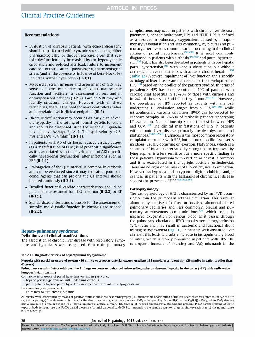
Ta
H65PuluCo--Le-
Alrigpavais
Clinical Practice Guidelines
36PH
Recommendations
� Evaluation of cirrhosis patients with echocardiographyshould be performed with dynamic stress testing eitherpharmacologically, or through exercise, given that sys-tolic dysfunction may be masked by the hyperdynamiccirculation and reduced afterload. Failure to incrementcardiac output after physiological/pharmacologicalstress (and in the absence of influence of beta-blockade)indicates systolic dysfunction (II-1;1).
� Myocardial strain imaging and assessment of GLS mayserve as a sensitive marker of left ventricular systolicfunction and facilitate its assessment at rest and indecompensated patients (II-2;2). Cardiac MRI may alsoidentify structural changes. However, with all thesetechniques, there is the need for more controlled studiesand correlation with clinical endpoints (III;2).
� Diastolic dysfunction may occur as an early sign of car-diomyopathy in the setting of normal systolic function,and should be diagnosed using the recent ASE guideli-nes, namely: Average E/e’>14; Tricuspid velocity >2.8m/s and LAVI >34 ml/m2 (II-1;1).
� In patients with AD of cirrhosis, reduced cardiac output(as a manifestation of CCM) is of prognostic significanceas it is associated with the development of AKI (specifi-cally hepatorenal dysfunction) after infections such asSBP (II-1;1).
� Prolongation of the QTc interval is common in cirrhosisand can be evaluated since it may indicate a poor out-come. Agents that can prolong the QT interval shouldbe used cautiously (II-2;2).
� Detailed functional cardiac characterisation should bepart of the assessment for TIPS insertion (II-2;2) or LT(II-1;1).
� Standardized criteria and protocols for the assessment ofsystolic and diastolic function in cirrhosis are needed(II-2;2).
Hepato-pulmonary syndromeDefinitions and clinical manifestationsThe association of chronic liver disease with respiratory symp-toms and hypoxia is well recognised. Four main pulmonary
ble 12. Diagnostic criteria of hepatopulmonary syndrome.
ypoxia with partial pressure of oxygen <80 mmHg or alveolar–arterial oxygyears).lmonary vascular defect with positive findings on contrast-enhanced echng-perfusion scanningmmonly in presence of portal hypertension, and in particular:hepatic portal hypertension with underlying cirrhosispre-hepatic or hepatic portal hypertension in patients without underlying c
ss commonly in presence of:acute liver failure, chronic hepatitis
l criteria were determined by means of positive contrast-enhanced echocardiographht atrial passage). The abbreviated formula for the alveolar–arterial gradient is as fortial pressure of alveolar oxygen, PaO2 partial pressure of arterial oxygen, FIO2 fractpor at body temperature, and PaCO2 partial pressure of arterial carbon dioxide (0.8 c4 to 8 mmHg.
Journal of Hepatology 20lease cite this article in press as: The European Association for the Study of the Liver. EASL Cepatol (2018), https://doi.org/10.1016/j.jhep.2018.03.024
complications may occur in patients with chronic liver disease:pneumonia, hepatic hydrotorax, HPS and PPHT. HPS is definedas a disorder in pulmonary oxygenation, caused by intrapul-monary vasodilatation and, less commonly, by pleural and pul-monary arteriovenous communications occurring in the clinicalsetting of portal hypertension.498,499 It is most commonlydiagnosed in patients with cirrhosis498,499 and portal hyperten-sion500 but, it has also been described in patients with pre-hepaticportal hypertension,501 with venous obstruction but withoutcirrhosis, and even in patients with acute or chronic hepatitis500
(Table 12). A severe impairment of liver function and a specificaetiology of liver disease are not needed for the development ofHPS,498 based on the profiles of the patients studied. In terms ofprevalence, HPS has been reported in 10% of patients withchronic viral hepatitis in 15–23% of those with cirrhosis andin 28% of those with Budd-Chiari syndrome.502–504 However,the prevalence of HPS reported in patients with cirrhosisundergoing LT evaluation ranges from 5–32%,504–508 whileintrapulmonary vascular dilatation (IPVD) can be detected byechocardiography in 50–60% of cirrhosis patients undergoingLT evaluation. No relationship seems to exist between HPSand CCM.504 The clinical manifestations of HPS in patientswith chronic liver disease primarily involve dyspnoea andplatypnoea.498,502,506 Dyspnoea is the most common respiratorycomplaint in patients with HPS, but it is non-specific. Its onset isinsidious, usually occurring on exertion. Platypnoea, which is ashortness of breath exacerbated by sitting up and improved bylying supine, is a less sensitive but a more specific finding inthese patients. Hypoxemia with exertion or at rest is commonand it is exacerbated in the upright position (orthodeoxia).There are no signs or hallmarks of HPS on physical examination.However, tachypnoea and polypnoea, digital clubbing and/orcyanosis in patients with the hallmarks of chronic liver diseasesuggest the presence of HPS.498,502,506
PathophysiologyThe pathophysiology of HPS is characterised by an IPVD occur-ring within the pulmonary arterial circulation. This vascularabnormality consists of diffuse or localised abnormal dilatedpulmonary capillaries and, less commonly, pleural and pul-monary arteriovenous communications,509 which result inimpaired oxygenation of venous blood as it passes throughthe pulmonary circulation. IPVD impairs ventilatory/perfusion(V/Q) ratio and may result in anatomic and functional shuntleading to hypoxaemia (Fig. 10). In patients with advanced livercirrhosis this leads to a subtle increase in intrapulmonary bloodshunting, which is more pronounced in patients with HPS. Theconsequent increase of shunting and V/Q mismatch in the
en gradient ≥15 mmHg in ambient air (≥20 mmHg in patients older than
ocardiography or abnormal uptake in the brain (>6%) with radioactive
irrhosis
y (i.e., microbubble opacification of the left heart chambers three to six cycles afterllows: PaO2 � PaO2 = (FIO2 [Patm–PH2O] � [PaCO2/0.8]) � PaO2, where PaO2 denotesion of inspired oxygen, Patm atmospheric pressure, PH2O partial pressure of waterorresponds to the standard gas-exchange respiratory ratio at rest); the normal range
18 vol. xxx j xxx–xxxlinical Practice Guidelines for the management of patients with decompensated cirrhosis. J

Normal HPS
Alveol
Capillary
8 µ
500 µ
O2 O2
Fig. 10. The pathophysiology of hepatopulmonary syndrome (adaptedfrom Ref. 498). HPS, hepatopulmonary syndrome.
JOURNAL OF HEPATOLOGY
upright position is the cause of the orthodeoxia.510 The patho-genesis of IPVD is probably multifactorial (Fig. 11). The releaseof nitric oxide, which is a potent vasodilator, plays a critical rolein the development of HPS. The increased release of nitric oxidein the pulmonary circulation is related to an increased expres-sion and activity of two isoforms of nitric oxide synthase(NOS), the endothelial NOS (eNOS) and the inducible NOS(iNOS).511–516 Meanwhile, BT and the BT-related endotoxaemiaand pro-inflammatory response also contribute to the accumu-lation of macrophages in the pulmonary microvasculature.517
Endothelial activation of fractalkine (CX3CL1), a chemokine, inthe lung may favour the adherence of monocytes in the pul-monary microcirculation.518 Monocytes express iNOS and pro-duce heme oxygenase-1, leading to increased carbonmonoxide production, further enhancing vasodilatation.519
• Hepatic injury/failure
• Portal hypertension
Increased release of ET-1
HEPATOPULMSYNDRO
ETBreceptor
Increased expression andactivity of eNOS
Increased release of NO
Endothelial cell Endothelia
Endothelial activation ofCX3CL1
Endothelia
Endothelialproliferati
VASODILATION
ANGIOGEN
Pulmonary c
Genetic factors
Fig. 11. The pathogenesis of hepatopulmonary syndrome. ET, endothelin; eNOnitric oxide; HO, Heme oxygenase-1; CO, carbon monoxide; CX3CL1, fractalkine
Journal of Hepatology 20Please cite this article in press as: The European Association for the Study of the Liver. EASL CHepatol (2018), https://doi.org/10.1016/j.jhep.2018.03.024
CX3CL1 and vascular endothelial growth factor (VEGF) A, pro-duced by circulating monocytes, also contribute to angiogene-sis, recently recognised as a further pathogenetic factor ofpulmonary IPVD in experimental HPS.520–522 A downregulationof miRNA-199 a-5p has recently been described as a contribu-tory mechanism of pulmonary microvascular endothelial cellproliferation and thus pathogenesis of HPS.523 Polymorphismsin genes involved in the regulation of angiogenesis have alsobeen associated with the risk of HPS in patients with cirrho-sis524 (Fig. 11). Finally, it has recently been observed that rosu-vastatin, by down-regulating protein expression of nuclearfactor kappa B and VEGF-1,2 and Rho-associated A kinase,may improve the intrapulmonary angiogenesis and thealveolar-arterial oxygen pressure gradient in common bile ductligation rats.525
DiagnosisIn patients with portal hypertension and the clinical suspicionof HPS partial pressure of oxygen (PaO2) in arterial blood gas(ABG) should be assessed. A PaO2 lower than 80 mmHg and oran alveolar-arterial oxygen gradient (P[A-a]O2) ≥15 mmHgwhile breathing ambient air at sea level should lead to furtherinvestigations (Table 12). For adults ≥65 years a P[A-a]O2 ≥20mmHg cut-off is used.526 However, it should be highlighted thatalthough these criteria are well established, enabling one tounify the diagnostic methods and thus to better understandthe disease, they are based on a consensus of experts. Pulseoximetry indirectly measures oxygen saturation (SpO2), it isnon-invasive and may be useful in the diagnosis of HPS in adultssince a SpO2 <96% was found to be highly sensitive (100%) andspecific (88%) for detecting HPS in patients with a PaO2 <70
• Portosystemic shunt
• Hyperdynamic circulation
• Bacterial translocation
Systemic inflammation
ONARYME
Recruitment ofmacrophagesin the lungs
Increased expression andactivity of iNOS and HO
Increased release of NO and CO
l cell
MacrophageIncreased adherence of
macrophages/monocytes toendothelial cells
VFG-A releasel cell
cellon
VASODILATION
ESIS
apillary
CX3CL1
S, endothelial nitric oxide synthase; iNOS, inducible nitric oxide synthase; NO,; VGF-A, vascular endothelial growth factor A.
18 vol. xxx j xxx–xxx 37linical Practice Guidelines for the management of patients with decompensated cirrhosis. J

Clinical Practice Guidelines
mmHg, limiting ABG testing to only 14% of patients.527 Thevalidity of this non-invasive approach was not confirmed,recently, in paediatric patients with HPS.528 Serial SpO2 mea-surements may be useful to monitor impaired oxygenation overtime in patients with HPS. The ABG is essential for the staging ofthe severity of HPS. HPS can be categorised as mild (PaO2 ≥80mmHg), moderate (PaO2 60–79 mmHg), severe (PaO2 50–59mmHg), and very severe (PaO2 <50 mmHg).498,500,501,503
Recently, it has been observed that HPS is associated with ele-vated von Willenbrand factor antigen (vWF-Ag) levels. Thus,vWF-Ag has been proposed as a potentially useful screening toolfor early detection of HPS, but further studies are needed to val-idate it.529 The chest X ray is usually non-specific, nevertheless,it can be used to effectively rule out other concomitant pul-monary diseases since only a mild interstitial pattern in thelower part of the lungs may be found, because of pulmonaryvasodilatation.498,500,501,503 A decrease in the single-breath dif-fusing capacity for carbon monoxide is the only alteration ofthe routine pulmonary function test that is frequently and con-sistently abnormal in patients with HPS. However, it is notspecific and it may not normalise after LT.498,500,501,503 All theother respiratory function tests are non-specific, showing nor-mal or reduced forced vital capacity or maximum forced expira-tory volume during the first second (FEV1). Thus, they can onlybe used to rule out other concomitant pulmonary diseases. Tho-racic CT scans have also been proposed as a complementarytechnique to rule out another underlying pulmonary pathol-ogy,498,499 although there is little information regarding theirspecific role in the diagnosis of HPS. It has been suggested thatthoracic CT scans can be useful to measure the calibre of theperipheral arteries and the bronchial/arterial relationship.530,531
Furthermore, CT scanning makes it possible to define the vascu-lar pattern of HPS in a similar manner to arteriography bydetecting pleural and pulmonary arteriovenous communica-tions. Contrast-enhanced transthoracic echocardiography withsaline (shaken to produce microbubbles >10 lm in diameter)is the most useful method to detect pulmonary vascular dilata-tion. After the administration of agitated saline in a peripheralvein, microbubble opacification of the left atrium within threeto six cardiac cycles after right-atrial opacification indicatesmicrobubble passage through an abnormally dilated vascularbed, since microbubbles do not pass through normal capillar-ies.532 The injection of technetium-99 m–labeled macro-aggre-gated albumin (MAA) in the peripheral vein for lung scanning(MAA scan) is a potential alternative diagnostic procedurealthough it is more invasive and less sensitive. Particles, witha 20–50 lm size, escape through the abnormal pulmonary cap-illaries and stay in downstream capillary beds supplied by sys-temic arteries, such as the brain, kidneys, and spleen.Quantitative imaging of the MAA scan in the brain and lungenables calculation of the degree of shunting.533,534 The mea-surement of shunting with MAA scans may be useful as a com-plementary diagnostic tool in patients with HPS in two clinicalsituations. Firstly, in patients with a severe hypoxaemia and acoexistent HPS and intrinsic lung disease since a shunting >6%at MAA scan proves the major contribution of HPS to hypox-aemia. Secondly, in patients with HPS and very severe hypox-aemia (PaO2 <50 mmHg), since the presence of shunting >20%is associated with a poor outcome after LT.535 Despite thepotential role of lung perfusion scintigraphy for prognostic usein patients with cirrhosis and IPVD, its diagnostic accuracy forHPS remains to be established.536 Finally, neither constrast eco-
38 Journal of Hepatology 20Please cite this article in press as: The European Association for the Study of the Liver. EASL CHepatol (2018), https://doi.org/10.1016/j.jhep.2018.03.024
cardiography nor MAA scan can differentiate discrete arteriove-nous communications from diffuse precapillary and capillarydilatations or intracardiac shunt. The former distinction can bemade by means of pulmonary angiography. The latter distinc-tion can be made by means of transoesophageal contrast-enhanced echocardiography that directly reveals the intra-atrialseptum. Pulmonary angiography should not be performed in allpatients with suspected HPS, but only in: a) patients with thesevere hypoxaemia (PaO2 <60 mmHg) poorly responsive toadministration of 100% oxygen, and b) patients strongly sus-pected (by means of a CT chest scan) of having arteriovenouscommunications that would be amenable to embolisation.
Recommendations
� In presence of tachypnoea and polypnoea, digital club-bing and/or cyanosis in a patient with the hallmarks ofchronic liver disease, HPS should be suspected andinvestigated (II-2,1).
� Pulse oximetry is the screening tool for HPS in adultpatients, but not in paediatric patients. For patients withSpO2 <96%, ABG analysis should be performed. A PaO2
lower than 80 mmHg and or an alveolar-arterial oxygengradient (P[A-a]O2) ≥15 mmHg while breathing ambientair, should lead to further investigations. For adults ≥65years aP[A-a]O2 ≥20 mmHgcut-off shouldbeused (II-2,1).
� The use of contrast (microbubble) echocardiography tocharacterise HPS is recommended (II-2;1).
� Trans-oesophageal contrast-enhanced echocardiographycan be performed to exclude definitively intra-cardiacshunts, albeit this technique is not devoid of risks(II-2;2).
� An MAA scan should be performed as a complementarytool to quantify the degree of shunting in patients withsevere hypoxaemia and coexistent intrinsic lung disease,or to assess the prognosis in patients with HPS and verysevere hypoxaemia (PaO2 <50 mmHg) (II-2;1).
� Neither contrast echocardiography nor MAA scan candefinitively differentiate discrete arteriovenous commu-nications from diffuse precapillary and capillary dilata-tions or cardiac shunts. Pulmonary angiography shouldbe performed only in patients with the severe hypox-aemia (PaO2 <60 mmHg), poorly responsive to adminis-tration of 100% oxygen, and in whom there is a strongsuspicion of arteriovenous communications that areamenable to embolisation (II-2;1).
1lin
Natural historyThe natural history of IPVD as well as of HPS is still unclear.
Most patients with IPVD maintain a normal gas exchange overtime, and it is not clear the reason why a subset of patients withIPVD develops HPS.537 A diagnosis of HPS is associated with apoor outcome in terms of both survival and quality oflife.505,507,508 Regarding survival, it should be highlighted thatin patients undergoing evaluation for LT, the mortality ratewas almost double in patients with HPS compared to patientswith cirrhosis without HPS, independent of other potential pre-dictors of mortality such as age, MELD score and comorbidi-ties.505 In patients with cirrhosis and HPS, who were not8 vol. xxx j xxx–xxxical Practice Guidelines for the management of patients with decompensated cirrhosis. J

JOURNAL OF HEPATOLOGY
evaluated for LT, the five-year survival rate was 23% while it was63% in patients with cirrhosis without HPS who were matchedfor the aetiology and severity of cirrhosis according to theChild-Pugh classification, age, and MELD score.503 Survival wassignificantly worse among patients with HPS and a PaO2 of lessthan 50 mmHg at the time of diagnosis.506,507
ManagementMedical treatmentSpontaneous resolution of HPS is uncommon. There is no estab-lished medical therapy currently available for HPS. Several drugshavebeenapplied for the treatmentofHPSwithconflicting results.However, no large randomised trial has been conducted, probablybecause of the low number of patients. Data from several uncon-trolled clinical studies and anecdotal evidence indicate that treat-ment with beta-blockers, cyclooxygenase inhibitors, systemicglucocorticoids and cyclophosphamide, almitrine bismesylate,inhaled nitric oxide, nitric oxide inhibitors, and antimicrobialagents has beenuniformlyunsuccessful.505 Pentoxifyllinehas alsobeen tried in the treatment of HPS in adults and children in twosmall pilot studieswith contradictory results in terms of improve-ments in oxygenation and frequent GI side effects.538,539 Adminis-tration of garlic was found to be associated with an improvementin the PaO2 in a small randomised study.540 However, a case ofmoderate hepatotoxicity associated with short-term, high-dosegarlicin therapy in an LT recipient with persistent HPS wasrecently reported.541 The use of TIPS has been proposed to reduceportal pressure in patients with HPS. However, data are insuffi-cient even when a systemic analysis review is considered.542 Inaddition, there is some concern that TIPS can enhance pulmonaryvasodilationbyexacerbating thehyperkinetic circulation. Thus,norecommendation for the use TIPS to treat HPS can be given.498,505
Finally, coil embolisation (embolotherapy) has been shown toimprove arterial oxygenation temporarily in the context of angio-graphic arteriovenous communications.531,543 Endothelin-1receptor antagonists or angiogenesis inhibitors have not beentested up to now in patients with HPS. Thus, long-term oxygentherapy remains the most frequently recommended therapy forsymptoms in patients with severe hypoxaemia. However, someaspects of this treatment such as efficacy, costs, and compliance,remain to be evaluated.
Recommendations
� Long-term oxygen therapy is recommended in patientswith HPS and severe hypoxaemia. Nevertheless, thereis no available data concerning effectiveness, tolerance,cost-effectiveness, compliance and effects on survivalrates of this therapy (II-2;1).
� No recommendation can be proposed regarding the useof drugs or the placement of TIPS for the treatment ofHPS (I;1).
PH
Liver transplantationThe most common and the only successful treatment for HPS isLT. LT results in a complete reversal or in a significant improve-ment of HPS in more than 85% of patients with severe hypox-aemia.544 In a prospective clinical study performed in the pre-MELD era, a pre-LT severe hypoxaemia, in particular when it
Journal of Hepatology 20lease cite this article in press as: The European Association for the Study of the Liver. EASL Cepatol (2018), https://doi.org/10.1016/j.jhep.2018.03.024
was associated with a large shunting at MAA scan, was foundto be a very strong predictor of mortality after LT.535 In 2007,five years after the introduction of MELD in US, the United Net-work for Organ Sharing (UNOS) recommended assigning anMELD score of 22 for the initial application of patients with sev-ere HPS (PaO2 <60 mmHg), with further increases every threemonths, to balance pre- and post-LT outcomes between HPSand non-HPS candidates.545 In the largest retrospective studycomparing the results of LT between the pre-MELD era andthe MELD era in patients with HPS, the five-year survival rateafter LT was found to improve from 67% during the pre-MELDera to 88% in the MELD era.546 Other data showed that inpost-MELD era, there was no association between pre-LT oxy-genation and waitlist survival in patients with HPS. These find-ings reflect not only the results of the introduction of HPS as aMELD exception, but also of an improved perioperative manage-ment in patients with HPS. The regular assessment of the sever-ity of hypoxaemia may facilitate LT prior to the occurrence ofvery severe hypoxaemia. In fact, hypoxaemia can worsen inpatients with HPS who are on the active transplantation list,with a median decrease in pO2 of 5.2 mmHg per year,508 andit has been recently confirmed that a pre-LT room-air PaO2
≤44.0 mmHg is still associated with increased post-LT mortal-ity.547 Thus, it has been suggested that an ABG analysis shouldbe carried out every six months, but no study has clarifiedwhich is the best method for conducting this (ABG analysis vs.pulse oximetry) nor how frequently it should be performed.Despite the increased survival rate in patients with HPS afterLT in the MELD era, it has recently been observed that HPSMELD exception patients had lower overall mortality comparedto others awaiting LT, suggesting that the appropriateness of theHPS MELD exception policy should be reassessed.548 There arevery few and small studies on the impact of HPS on anaestheticprocedures, as well as in the post-LT management in the ICU.Nevertheless, it seems that inhaled nitric oxide, methylene blue,extracorporeal membrane oxygenation and non-invasive venti-lation may improve oxygenation immediately post-LT.549–551
Recommendations
� Patients with HPS and PaO2 <60 mmHg should be evalu-ated for LT since it is the only treatment for HPS that hasbeen proven to be effective to date (II-2;1).
� Since a severe hypoxaemia (PaO2 <45–50 mmHg) isassociated with increased post-LT mortality, an ABGanalysis should be carried out every six months in orderto facilitate prioritisation to LT (II-2;1).
1lin
Portopulmonary hypertensionDefinition and diagnosisA diagnosis of PPHT should be considered in a patient withestablished portal hypertension in the absence of other causesof pulmonary artery or venous hypertension. namely: chronicthromboembolism, chronic lung disease/hypoxia; chronic leftheart disease.
Patients may be asymptomatic but often present with exer-tional dyspnoea and they may have clinical signs of right heartfailure when moderate to severe disease develops.552 Classifica-tion of severity is based on mean pulmonary arterial pressure
8 vol. xxx j xxx–xxx 39ical Practice Guidelines for the management of patients with decompensated cirrhosis. J

Clinical Practice Guidelines
(mPAP) and assumes there is high pulmonary vascular resis-tance (PVR). PPHT is graded as mild (mPAP ≥25 and <35mmHg); moderate (mPAP ≥35 and <45 mmHg), and severe(mPAP ≥45 mmHg).498 The diagnosis also requires there to benormal pulmonary occlusion pressures, to exclude elevation ofpulmonary pressure resulting from elevated left ventricular fill-ing pressure. Transthoracic Doppler echocardiography (TDE) isthe main screening tool for evaluating the presence of PPHTwhen screening high-risk patients, such as those being consid-ered for TIPS or LT.553–555 As a screening test, some studies sug-gest a pulmonary artery systolic pressure of >30 mmHg on TDEhas a negative predictive value of 100%, but a positive predictivevalue of only 59%.554 However, when assessing patients for LT,the threshold for right heart catheterisation is less clear, witha right ventricular systolic pressure >50 mmHg and/or signifi-cant right ventricular hypertrophy seen as the trigger for thisinvestigation to rule out significant PPHT.555
PathophysiologyIn patients with portal hypertension, PPHT is thought to arisefrom limited blood flow in the pulmonary arterial circulationbecause of vasoconstriction. Numerous factors are thought tobe responsible for this including: Changes in endogenous vaso-regulators; increased endothelin 1 and reduced prostacyclin syn-thase from pulmonary endothelial cells; proliferation of smoothmuscle cells/endothelial activation and platelet aggregation.
Natural history and prognosisFrom studies in patients evaluated for LT, the incidence isthought to be between 3–10% based on haemodynamic criteria.Furthermore, female sex and pre-existing autoimmune liver dis-ease are thought to be independent risk factors.556 Genetic vari-ation in oestradiol levels may increase the predisposition topulmonary artery vasoconstriction. Indeed, women are at threetimes greater risk than men.557 There is also an associationbetween patients who have moderate to severe PPHT and thepresence of large portosystemic shunts.558 However, there isno clear association between the severity of liver disease or por-tal hypertension and the development of severe PPHT.556,559
Studies quote survival rates at one year of between 35–46%without specific treatment.560,561 Mortality is often associatedwith other complications of liver disease such as hepatocellularcancer, sepsis and GI bleeding and right ventricular failure.Increased rates of mortality are related to higher right atrialpressure and lower cardiac index.559,562 In a multicentre reg-istry study, patients with PPHT were shown to have worse out-comes than patients with idiopathic pulmonary hypertension,with a five-year survival of 40% vs. 64%.563 However, a retro-spective French study challenges this, whilst reportingincreased mortality in those with a lower cardiac index, likelyreflecting failed compensation to increased right ventriculardysfunction, and patients with more advanced liver disease.564
Medical treatmentThe evidence base for pharmacological therapies in PPHT is lim-ited with most data extrapolated from studies in pulmonaryarterial hypertension not related to liver disease.565,566 Drugsto promote acute vasodilatation during right heart catheterisa-tion assessment, theoretically may be deleterious as they runthe risk of further reducing cardiac index. There is a lack of datato clarify this.567 Conversely, whilst patients with advanced
40 Journal of Hepatology 20Please cite this article in press as: The European Association for the Study of the Liver. EASL CHepatol (2018), https://doi.org/10.1016/j.jhep.2018.03.024
portal hypertension may be on treatment with beta-blockers,withdrawing beta-blocker therapy may help to increase cardiacoutput and thereby help exertional dyspnoea, in patients withadvanced PPHT.568
Endothelin receptor antagonists. Bosentan has been shown toimprove pulmonary artery haemodynamics and exercise toler-ance in patients with PPHT, independently of liver diseaseseverity.569–572 One retrospective study reports survival ratesof up to 89% at three years.573 Others have shown improve-ments in cardiac index up to 39%, albeit in a small number ofpatients, but an increase in aminotransferases, which respondedto dose reduction or discontinuation.571 The FDA places a cau-tion on this class of drug in patients with advanced liver dys-function. There is limited data on the use of other members inthis family of agent, including ambrisentan and macitentan,for PPHT.574,575
Phosphodiesterase subtype-5 inhibitors. Blockade of phosphodi-esterase-5 inhibitors facilitate the vasodilatory effects of nitricoxide, through reduced metabolism of cGMP. Small case seriessuggest that sildenafil improves functional capacity andincreases cardiac output.576–578 It should be noted that silde-nafil can precipitate variceal bleeding and as such, cautionshould be exercised.579
Prostacyclin analogues. Prostacyclin analogues have many poten-tial benefits including vasodilatory, reduced vascular smoothmuscle proliferation and anti-thrombotic. Case series suggestimproved pulmonary haemodynamics with i.v. epoprostenoland the potential for improved five-year survival compared toregistry data in pulmonary artery hypertension (70 vs.40%).580–583 However, lower doses than those used in idiopathicpulmonary hypertension are suggested to reduce the develop-ment of thrombocytopenia and splenomegaly. Other studieshave also looked at use of inhaled iloprost and reported short-term haemodynamic benefit.584
Impact of the management of other complications of cirrhosisCaution should be exercised when considering TIPS placementfor the treatment of other complications of cirrhosis in patientswith proven PPHT. The anticipated increase in right ventricularfilling pressures and cardiac output may precipitate markedincreases in PVR and right-sided pressure overload.585,586
Moderate PPHT (mPAP >35 and <45 mmHg) is a relative con-traindication for TIPS placement, and severe PPHT is an absolutecontraindication.586
Recommendations
� Screening for PPHT should be via TDE in patients deemedpotential recipients for TIPS or LT; in those with a posi-tive screening test, right heart catheterisation shouldbe performed (II-1;1).
� In patients with PPHT who are listed for transplantation,echocardiography should be repeated on the waitlist,albeit, the specific interval is unclear (III;1).
� Beta-blockers should be stopped and varices managedby endoscopic therapy in cases of proven PPHT (II-3;1).
18 vol. xxx j xxx–xxxlinical Practice Guidelines for the management of patients with decompensated cirrhosis. J

JOURNAL OF HEPATOLOGY
PH
� Therapies that have been approved for primary pul-monary arterial hypertension may have benefit in PPHTto improve exercise tolerance and haemodynamics. How-ever, endothelin antagonists should be used with cautionbecause of concerns over hepatic impairment (II-2;1).
� TIPS should not be used in patients with PPHT (II-3;1).
Liver transplantationHistorically, severe PPHT has been a relative contraindicationfor LT because of very poor outcomes. However, with the adventof improved haemodynamic control with agents such as i.v.prostacyclin, there are case series showing normal pulmonaryhaemodynamics almost two years post LT.587,588
Stratifying risk for LT. In patients with an mPAP ≥45–50 mmHg,most centres would deem this an absolute contraindication totransplantation irrespective of therapy applied.562,587,589
Patients with an mPAP >35 have increased risk post LT, associ-ated with increased hospital stay and longer ventilator require-ments.562,590,591 If LT is considered in such patients, it issuggested that their PPHT is treated aggressively to lower mPAPand improve right ventricular function.588,592,593
To facilitate access to LT before there is further progressionof PPHT to a point where transplantation risks are deemed toohigh, MELD exception (MELD 22 points) has been granted topatients with PPHT (mPAP >25 mmHg and PVR >240 dynes/sper cm�5) with at least moderate disease severity (baselinemPAP >35 mmHg).594 Patients are considered surgical candi-dates if, after targeted therapy to lower PAP, they haveimproved mPAP (<35 mmHg) and PVR (<400 dyne/s per cm�5)and/or normalise their PVR. Applying this exception has beennoted to reduce waitlist mortality.595
Per-operative considerations. All patients should be monitoredwith a pulmonary artery catheter. Therapy to lower mPAPshould be continued throughout the operative period, given thatthere is often a rise in cardiac output post re-perfusion and thismay add more stress on any pre-existing impaired right ventri-cle function.595–597 Indeed, in some cases, a severe acute rise inPAP may lead to graft failure because of hepatic congestionthrough a failing right ventricle. The management of suchadverse haemodynamics, in addition to i.v. prostacyclin orinhaled nitric oxide includes the use of extracorporeal mem-brane oxygen therapy (ECMO).598,599
Postoperative considerations. Monitoring PAP response to ther-apy is via serial transthoracic echo with tissue Doppler at 4–6-month intervals and consideration of tapering pulmonaryartery targeted therapy, though no controlled data exists toprovide guidance on this.581,600,601 Case reports and seriessuggest that 29–64% of patients with moderate to severePPHT under long-term follow-up post-transplant have beenable to discontinue therapy over time.599–602 Indeed, somesuggest a return to normal right ventricle function followingtherapy for PPHT in the pre-transplant period and then aftertransplant surgery.581,601 PPHT MELD exception patients haveworse one-year mortality or graft failure than patients with-out PPHT.603
Journal of Hepatology 201lease cite this article in press as: The European Association for the Study of the Liver. EASL Clinepatol (2018), https://doi.org/10.1016/j.jhep.2018.03.024
Recommendations
� If mPAP <35 mmHg and right ventricular function is pre-served, LT should be considered (II-2,1). A mPAP of ≥45mmHg should be considered an absolute contraindica-tion to LT irrespective of therapy applied (III,1).
� Therapy to lower mPAP and improve right ventricularfunction should be commenced in patients with mPAP≥35 mmHg. Right ventricular function should be period-ically evaluated (II-2,1).
� MELDexception can be considered inpatientswith provenPPHT in whom targeted therapy fails to decrease mPAP<35 mmHg but does facilitate normalisation of PVR to<240 dynes/s cm�5 and right ventricular function (II-3;2).
� MELD exception should be advocated in patients withproven PPHT of moderate severity (assessment mPAP≥35 mmHg) in whom targeted treatment lowers mPAP<35 mmHg and PVR <400 dynes/s cm�5 (II-2;1).
ConclusionsThese guidelines on the management of patients with decom-pensated cirrhosis were developed based on a new pathophys-iological background that offers the opportunity for morecomprehensive therapeutic or prophylactic approaches to man-age the disease. The knowledge of the key pathophysiologicmechanisms makes it possible nowadays to counteract the pro-gression of cirrhosis and so to prevent its complications. Thisrepresents a step forward, shifting our approach from treatingthe complications of decompensated cirrhosis to preventingtheir occurrence. However, to make this possible it is crucialto think about new models of specialist care for patients withcirrhosis. A care coordination programme, has been proven toimprove survival and to reduce emergent readmission to thehospital in these patients.604 Care coordinators can facilitatethe development of educational programmes for patients andcaregivers optimising their adherence to guideline recommen-dations. In addition, they can plan invasive procedures in aday hospital, allowing transfer of real-time information to pri-mary care physicians to improve quality and coordination ofcare. In so doing, it is possible to prevent unnecessary visits tothe emergency department and/or emergent readmission tothe hospital. These measures will progressively reduce the bur-den of cirrhosis.
Conflict of interestPaolo Angeli: Consultancy fee from Sequana Medical AG, GileadItaly and Biovie; Patent inventor from Biovie; Research grantfrom Gilead; Speaker’s fee from Bhering, Kedrion 2016. MauroBernardi: Consultancy fee from CLS Behring GmbH, BaxterHealthcare SA, Grifols SA; Speaker’s fee from CLS Behring GmbH,Baxter Healthcare SA, PPTA Europe, Octapharma AG, GileadSciences, AbbVie Italia. Wim Laleman: Speaker’s fee for Gore,Norgine, 4C, Abbvie, Sirtex; Consultancy fee for AbbVie, Gilead,MSD, Intercept; Research grant from Gilead. Jonel Trebicka:Speaker’s fee or Consultancy fee from Gore & associates (TIPS),Sequana medical (alpha-pump), Alexion (PNH), Versantis (lipo-somes). Aleksander Krag: None. Claire Francoz: None. PereGines: Advisory/Consultancy fee for Sequana Grifols, Mallinck-
8 vol. xxx j xxx–xxx 41ical Practice Guidelines for the management of patients with decompensated cirrhosis. J

Clinical Practice Guidelines
rodt, Ferring Pharmaceuticals; Research Funding from Sequana,Grifols, Ferring Pharmaceuticals.
Please refer to the accompanying ICMJE disclosure forms forfurther details.
AcknowledgmentsWe would like to thank Alessandra Brocca Dr, Marta Tonon MD,Husain-Syed Faeq MD for the great editorial work. We acknowledgeICREA for the ACADEMIA AWARD given to Pere Ginès. We would liketo thank the reviewers of this Clinical Practice Guideline for theirtime and critical reviewing: EASL Governing Board, AlexanderGerbes, Thierry Gustot, Guadalupe Garcia-Tsao.
References[1] EASL clinical practice guidelines on the management of ascites,
spontaneous bacterial peritonitis, and hepatorenal syndrome in cirrho-sis. J Hepatol 2010;53:397–417.
[2] D’Amico G. The clinical course of cirrhosis. Population based studies andthe need of personalized medicine. J Hepatol 2014;60:241–242.
[3] Moreau R, Jalan R, Gines P, Pavesi M, Angeli P, Cordoba J, et al. Acute-on-chronic liver failure is a distinct syndrome that develops in patientswith AD of cirrhosis. Gastroenterology 2013;144:1426–1437.
[4] D’Amico G, Garcia-Tsao G, Pagliaro L. Natural history and prognosticindicators of survival in cirrhosis: a systematic review of 118 studies. JHepatol 2006;44:217–231.
[5] Bernardi M, Moreau R, Angeli P, Schnabl B, Arroyo V. Mechanisms ofdecompensation and organ failure in cirrhosis: From peripheral arterialvasodilation to systemic inflammation hypothesis. J Hepatol2015;63:1272–1284.
[6] Jalan R, Fernandez J, Wiest R, Schnabl B, Moreau R, Angeli P, et al.Bacterial infections in cirrhosis: a position statement based on the EASLSpecial Conference. J Hepatol 2013;60:1310–1324.
[7] Arvaniti V, D’Amico G, Fede G, Manousou P, Tsochatzis E, PleguezueloM, et al. Infections in patients with cirrhosis increase mortality four-fold and should be used in determining prognosis. Gastroenterology2010;139:1246–1256.
[8] Schrier RW, Arroyo V, Bernardi M, Epstein M, Henriksen JH, Rodes J.Peripheral arterial vasodilation hypothesis: a proposal for the initiationof renal sodium and water retention in cirrhosis. Hepatology1988;8:1151–1157.
[9] Wiese S, Hove JD, Bendtsen F, Moller S. Cirrhotic cardiomyopathy:pathogenesis and clinical relevance. Nat Rev Gastroenterol Hepatol2014;11:177–186.
[10] Arroyo V, Terra C, Gines P. Advances in the pathogenesis and treatmentof type-1 and type-2 hepatorenal syndrome. J Hepatol 2007;46:935–946.
[11] Iwakiri Y, Groszmann RJ. The hyperdynamic circulation of chronic liverdiseases: from the patient to the molecule. Hepatology 2006;43:S121–S131.
[12] Claria J, Stauber RE, Coenraad MJ, Moreau R, Jalan R, Pavesi M, et al.Systemic inflammation in decompensated cirrhosis: Characterizationand role in acute-on-chronic liver failure. Hepatology 2016;64:1249–1264.
[13] Trautwein C, Friedman SL, Schuppan D, Pinzani M. Hepatic fibrosis:Concept to treatment. J Hepatol 2015;62:S15–S24.
[14] Alvarez MA, Cirera I, Sola R, Bargallo A, Morillas RM, Planas R.Long-term clinical course of decompensated alcoholic cirrhosis: aprospective study of 165 patients. J Clin Gastroenterol 2011;45:906–911.
[15] Powell Jr WJ, Klatskin G. Duration of survival in patients with Laennec’scirrhosis. Influence of alcohol withdrawal, and possible effects of recentchanges in general management of the disease. Am J Med1968;44:406–420.
[16] Shim JH, Lee HC, Kim KM, Lim YS, Chung YH, Lee YS, et al. Efficacy ofentecavir in treatment-naive patients with hepatitis B virus-relateddecompensated cirrhosis. J Hepatol 2010;52:176–182.
[17] Cheung MC, Walker AJ, Hudson BE, Verma S, McLauchlan J, Mutimer DJ,et al. Outcomes after successful direct-acting antiviral therapy forpatients with chronic hepatitis C and decompensated cirrhosis. JHepatol 2016;65:741–747.
[18] Lens S, Alvarado E, Mariño Z, et al. Effects of all-oral antiviraltherapy on HVPG and systemic hemodynamics in patients with
42 Journal of Hepatology 20Please cite this article in press as: The European Association for the Study of the Liver. EASL CHepatol (2018), https://doi.org/10.1016/j.jhep.2018.03.024
hepatitis C virus-associated cirrhosis. Gastroenterology 2017;153:1273–1283.
[19] Kang SH, Lee YB, Lee JH, Nam JY, Chang Y, Cho H, et al. Rifaximintreatment is associated with reduced risk of cirrhotic complications andprolonged overall survival in patients experiencing hepaticencephalopathy. Aliment Pharmacol Ther 2017;46:845–855.
[20] Ginès P, Schrier RW. Renal failure in cirrhosis. N Engl J Med2009;361:1279–1290.
[21] Moreau R, Elkrief L, Bureau C, Pararnau JM, Thavenot T, Saliba F, et al. Arandomized trial of 6-month norfloxacin therapy in patients withChild-Pugh class C cirrhosis. J Hepatol 2017;66:S1.
[22] Caraceni P, Riggio O, Angeli P, Alessandria C, Neri S, Foschi FG, et al.Long-term albumin administration in decompensated cirrhosis: anopen label randomized trial. Lancet 2018, [In press].
[23] Sola E, Sola C, Simon-Talero M, Martin-Llahi M, Castellote J, Garcia-Martinez R, et al. Midodrine and albumin for prevention of complica-tions of cirrhosis in patients in the waiting list for liver transplantation.A randomized, multicenter, double-blind, placebo-controlled trial. JHepatol 2017;66:S11.
[24] Abraldes JG, Albillos A, Banares R, Turnes J, Gonzalez R, Garcia-Pagan JC,et al. Simvastatin lowers portal pressure in patients with cirrhosis andportal hypertension: a randomized controlled trial. Gastroenterology2009;136:1651–1658.
[25] Abraldes JG, Villanueva C, Aracil C, Turnes J, Hernandez-Guerra M,Genesca J, et al. Addition of simvastatin to standard therapy for theprevention of variceal rebleeding does not reduce rebleeding butincreases survival in patients with cirrhosis. Gastroenterology2016;150:1160–1170.
[26] Villa E, Cammà C, Marietta M, Luongo M, Critelli R, Colopi S, et al.Enoxaparin prevents portal vein thrombosis and liver decompensationin patients with advanced cirrhosis. Gastroenterology 2012;143:1253–12609.
[27] Lebrec D, Thabut D, Oberti F, Perarnau JM, Condat B, Barraud H, et al.Pentoxifylline does not decrease short-term mortality but does reducecomplications in patients with advanced cirrhosis. Gastroenterology2010;138:1755–1762.
[28] Abraldes JG, Tarantino I, Turnes J, Garcia-Pagan JC, Rodes J, Bosch J.Hemodynamic response to pharmacological treatment of portal hyper-tension and long-term prognosis of cirrhosis. Hepatology 2003;37:902–908.
[29] Gines P, Quintero E, Arroyo V, Teres J, Bruguera M, Rimola A, et al.Compensated cirrhosis: natural history and prognostic factors. Hepa-tology 1987;7:122–128.
[30] Ripoll C, Groszmann R, Garcia-Tsao G, Grace N, Burroughs A, Planas R,et al. Hepatic venous pressure gradient predicts clinical decompensa-tion in patients with compensated cirrhosis. Gastroenterology2007;133:481–488.
[31] Moore KP, Wong F, Gines P, Bernardi M, Ochs A, Salerno F, et al. Themanagement of ascites in cirrhosis: report on the consensus conferenceof the International Ascites Club. Hepatology 2003;38:258–266.
[32] Arroyo V, Gines P, Gerbes AL, Dudley FJ, Gentilini P, Laffi G, et al.Definition and diagnostic criteria of refractory ascites and hepatorenalsyndrome in cirrhosis. International Ascites Club. Hepatology1996;23:164–176.
[33] Rimola A, Garcia-Tsao G, Navasa M, Piddock LJ, Planas R, Bernard B,et al. Diagnosis, treatment and prophylaxis of spontaneous bacterialperitonitis: a consensus document. International Ascites Club. J Hepatol2000;32:142–153.
[34] Bruns T, Lutz P, Stallmach A. Nischalke HD Low ascitic fluid protein doesnot indicate an increased risk for spontaneous bacterial peritonitis incurrent cohorts. J Hepatol 2015;63:527–528.
[35] Runyon BA, Canawati HN, Akriviadis EA. Optimization of ascitic fluidculture technique. Gastroenterology 1988;95:1351–1355.
[36] Runyon BA, Montano AA, Akriviadis EA, Antillon MR, Irving MA,McHutchison JG. The serum-ascites albumin gradient is superior tothe exudate-transudate concept in the differential diagnosis of ascites.Ann Intern Med 1992;117:215–220.
[37] Gerbes AL, Jüngst D, Xie YN, Permanetter W, Paumgartner G. Asciticfluid analysis for the differentiation of malignancy-related and nonma-lignant ascites. Proposal of a diagnostic sequence. Cancer 1991;68:1808–1814.
[38] Llach J, Gines P, Arroyo V, Rimola A, Tito L, Badalamenti S, et al.Prognostic value of arterial pressure, endogenous vasoactive systems,and renal function in cirrhotic patients admitted to the hospital for thetreatment of ascites. Gastroenterology 1988;94:482–487.
18 vol. xxx j xxx–xxxlinical Practice Guidelines for the management of patients with decompensated cirrhosis. J

JOURNAL OF HEPATOLOGY
[39] Caregaro L, Menon F, Angeli P, Amodio P, Merkel C, Bortoluzzi A, et al.Limitations of serum creatinine level and creatinine clearance asfiltration markers in cirrhosis. Arch Intern Med 1994;154:201–205.
[40] Bernardi M, Gitto S, Biselli M. The MELD score in patients awaiting livertransplant: strengths and weaknesses. J Hepatol 2011;54:1297–1306.
[41] Biselli M, Dall’Agata M, Gramenzi A, Gitto S, Liberati C, Brodosi L, et al. Anew prognostic model to predict dropout from the waiting list incirrhotic candidates for liver transplantation with MELD score <18.Liver Int 2015;35:184–191.
[42] Bernardi M, Santini C, Trevisani F, Baraldini M, Ligabue A, Gasbarrini G.Renal function impairment induced by change in posture in patientswith cirrhosis and ascites. Gut 1985;26:629–635.
[43] Ring-Larsen H, Henriksen JH, Wilken C, Clausen J, Pals H, Christensen NJ.Diuretic treatment in decompensated cirrhosis and congestive heartfailure: effect of posture. Br Med J 1986;292:1351–1353.
[44] Bernardi M, Laffi G, Salvagnini M, Azzena G, Bonato S, Marra F, et al.Efficacy and safety of the stepped care medical treatment of ascites inliver cirrhosis: a randomized controlled clinical trial comparing twodiets with different sodium content. Liver 1993;13:156–162.
[45] Gauthier A, Levy VG, Quinton A, Michel H, Rueff B, Descos L, et al. Salt orno salt in the treatment of cirrhotic ascites: a randomised study. Gut1986;27:705–709.
[46] Reynolds TB, Lieberman FL, Goodman AR. Advantages of treatment ofascites without sodium restriction and without complete removal ofexcess fluid. Gut 1978;19:549–553.
[47] Morando F, Rosi S, Gola E, Nardi M, Piano S, Fasolato S, et al. Adherenceto a moderate sodium restriction diet in outpatients with cirrhosis andascites: a real-life cross-sectional study. Liver Int 2015;35:1508–1515.
[48] Pockros PJ, Reynolds TB. Rapid diuresis in patients with ascites fromchronic liver disease: the importance of peripheral edema. Gastroen-terology 1986;90:1827–1833.
[49] Bernardi M, Trevisani F, Gasbarrini A, Gasbarrini G. Hepatorenaldisorders: role of the renin-angiotensin-aldosterone system. SeminLiver Dis 1994;14:23–34.
[50] Bernardi M, Servadei D, Trevisani F, Rusticali AG, Gasbarrini G.Importance of plasma aldosterone concentration on the natriureticeffect of spironolactone in patients with liver cirrhosis and ascites.Digestion 1985;31:189–193.
[51] Angeli P, Dalla Pria M, De Bei E, Albino G, Caregaro L, Merkel C, et al.Randomized clinical study of the efficacy of amiloride and potassiumcanrenoate in nonazotemic cirrhotic patients with ascites. Hepatology1994;19:72–79.
[52] Angeli P, Gatta A, Caregaro L, Menon F, Sacerdoti D, Merkel C, et al.Tubular site of renal sodium retention in ascitic liver cirrhosis evalu-ated by lithium clearance. Eur J Clin Invest 1990;20:111–117.
[53] Gatta A, Angeli P, Caregaro L, Menon F, Sacerdoti D, Merkel C. Apathophysiological interpretation of unresponsiveness to spironolac-tone in a stepped-care approach to the diuretic treatment of ascites innonazotemic cirrhotic patients. Hepatology 1991;14:231–236.
[54] Perez-Ayuso RM, Arroyo V, Planas R, Gaya J, Bory F, Rimola A, et al.Randomized comparative study of efficacy of furosemide vs. spirono-lactone in nonazotemic cirrhosis with ascites. Relationship between thediuretic response and the activity of the renin-aldosterone system.Gastroenterology 1983;84:961–968.
[55] Angeli P, Fasolato S, Mazza E, Okolicsanyi L, Maresio G, Velo E, et al.Combined vs. sequential diuretic treatment of ascites in non-azotaemicpatients with cirrhosis: results of an open randomised clinical trial. Gut2010;59:98–104.
[56] Santos J, Planas R, Pardo A, Durandez R, Cabre E, Morillas RM, et al.Spironolactone alone or in combination with furosemide in thetreatment of moderate ascites in nonazotemic cirrhosis. A randomizedcomparative study of efficacy and safety. J Hepatol 2003;39:187–192.
[57] Gerbes AL, Bertheau-Reitha U, Falkner C, Jüngst D, Paumgartner G.Advantages of the new loop diuretic torasemide over furosemide inpatients with cirrhosis and ascites. A randomized, double blind cross-over trial. J Hepatol 1993;17:353–358.
[58] Angeli P, Albino G, Carraro P, Dalla Pria M, Merkel C, Caregaro L, et al.Cirrhosis and muscle cramps: evidence of a causal relationship.Hepatology 1996;23:264–273.
[59] Elfert AA, Abo Ali L, Soliman S, Zakaria S, Shehab El-Din I, ElkhalawanyW, et al. Randomized placebo-controlled study of baclofen in thetreatment of muscle cramps in patients with liver cirrhosis. Eur JGastroenterol Hepatol 2016;28:1280–1284.
[60] Lee FY, Lee SD, Tsai YT, et al. A randomized controlled trial of quinidinein the treatment of cirrhotic patients with muscle cramps. J Hepatol1991;12:236–240.
Journal of Hepatology 20Please cite this article in press as: The European Association for the Study of the Liver. EASL CHepatol (2018), https://doi.org/10.1016/j.jhep.2018.03.024
[61] Lin CH, Shih FY, Ma MH, Chiang WC, Yang CW, Ko PC. Should bleedingtendency deter abdominal paracentesis? Dig Liver Dis 2005;37:946–951.
[62] Pache I, Bilodeau M. Severe haemorrhage following abdominal para-centesis for ascites in patients with liver disease. Aliment PharmacolTher 2005;21:525–529.
[63] Gines P, Tito L, Arroyo V, Planas R, Panes J, Viver J, et al. Randomizedcomparative study of therapeutic paracentesis with and withoutintravenous albumin in cirrhosis. Gastroenterology 1988;94:1493–1502.
[64] Gines A, Fernandez-Esparrach G, Monescillo A, Vila C, Domenech E,Abecasis R, et al. Randomized trial comparing albumin, dextran 70, andpolygeline in cirrhotic patients with ascites treated by paracentesis.Gastroenterology 1996;111:1002–1010.
[65] Sola-Vera J, Minana J, Ricart E, Planella M, Gonzalez B, Torras X, et al.Randomized trial comparing albumin and saline in the prevention ofparacentesis-induced circulatory dysfunction in cirrhotic patients withascites. Hepatology 2003;37:1147–1153.
[66] Bernardi M, Caraceni P, Navickis RJ, Wilkes MM. Albumin infusion inpatients undergoing large-volume paracentesis: a meta-analysis ofrandomized trials. Hepatology 2012;55:1172–1181.
[67] Moreau R, Valla DC, Durand-Zaleski I, Bronowicki JP, Durand F, ChaputJC, et al. Comparison of outcome in patients with cirrhosis and ascitesfollowing treatment with albumin or a synthetic colloid: a randomisedcontrolled pilot trail. Liver Int 2006;26:46–54.
[68] Gines P, Arroyo V, Quintero E, Planas R, Bory F, Cabrera J, et al.Comparison of paracentesis and diuretics in the treatment of cirrhoticswith tense ascites. Results of a randomized study. Gastroenterology1987;93:234–241.
[69] Salerno F, Badalamenti S, Incerti P, Tempini S, Restelli B, Bruno S, et al.Repeated paracentesis and i.v. albumin infusion to treat ’tense’ ascitesin cirrhotic patients. A safe alternative therapy. J Hepatol1987;5:102–108.
[70] Fernandez-Esparrach G, Guevara M, Sort P, Pardo A, Jimenez W, Gines P,et al. Diuretic requirements after therapeutic paracentesis in non-azotemic patients with cirrhosis. A randomized double-blind trial ofspironolactone vs. placebo. J Hepatol 1997;26:614–620.
[71] Elia C, Graupera I, Barreto R, Solà E, Moreira R, Huelin P, et al. Severeacute kidney injury associated with non-steroidal antiinflammatorydrugs in cirrhosis: a case-control study. J Hepatol 2015;63:593–600.
[72] Claria J, Kent JD, Lopez-Parra M, Escolar G, Ruiz-Del-Arbol L, Gines P,et al. Effects of celecoxib and naproxen on renal function in nona-zotemic patients with cirrhosis and ascites. Hepatology2005;41:579–587.
[73] Llach J, Gines P, Arroyo V, Salmeron JM, Gines A, Jimenez W, et al. Effectof dipyridamole on kidney function in cirrhosis. Hepatology1993;17:59–64.
[74] Pariente EA, Bataille C, Bercoff E, Lebrec D. Acute effects of captopril onsystemic and renal hemodynamics and on renal function in cirrhoticpatients with ascites. Gastroenterology 1985;88:1255–1259.
[75] Albillos A, Lledo JL, Rossi I, Perez-Paramo M, Tabuenca MJ, Banares R,et al. Continuous prazosin administration in cirrhotic patients: effectson portal hemodynamics and on liver and renal function. Gastroen-terology 1995;109:1257–1265.
[76] Cabrera J, Arroyo V, Ballesta AM, Rimola A, Gual J, Elena M, et al.Aminoglycoside nephrotoxicity in cirrhosis. Value of urinary beta 2-microglobulin to discriminate functional renal failure from acutetubular damage. Gastroenterology 1982;82:97–105.
[77] Guevara M, Fernández-Esparrach G, Alessandria C, Torre A, Terra C,Montañà X, et al. Effects of contrast media on renal function in patientswith cirrhosis: a prospective study. Hepatology 2004;40:646–651.
[78] Solomon R, Werner C, Mann D, D’Elia J, Silva P. Comparison of saline,mannitol, and furosemide to prevent acute decreases in renalfunction induced by radiocontrast agents. N Engl J Med 1994;331:1416–1420.
[79] Salerno F, Borroni G, Moser P, Badalamenti S, Cassara L, Maggi A, et al.Survival and prognostic factors of cirrhotic patients with ascites: astudy of 134 outpatients. Am J Gastroenterol 1993;88:514–519.
[80] Guardiola J, Baliellas C, Xiol X, Fernandez Esparrach G, Gines P, VenturaP, et al. External validation of a prognostic model for predicting survivalof cirrhotic patients with refractory ascites. Am J Gastroenterol2002;97:2374–2378.
[81] Huonker M, Schumacher YO, Ochs A, Sorichter S, Keul J, Rossle M.Cardiac function and haemodynamics in alcoholic cirrhosis and effectsof the transjugular intrahepatic portosystemic stent shunt. Gut1999;44:743–748.
18 vol. xxx j xxx–xxx 43linical Practice Guidelines for the management of patients with decompensated cirrhosis. J

Clinical Practice Guidelines
[82] Sanyal AJ, Freedman AM, Luketic VA, Purdum 3rd PP, Shiffman ML,DeMeo J, et al. The natural history of portal hypertension aftertransjugular intrahepatic portosystemic shunts. Gastroenterology1997;112:889–898.
[83] Wong F, Sniderman K, Liu P, Allidina Y, Sherman M, Blendis L.Transjugular intrahepatic portosystemic stent shunt: effects on hemo-dynamics and sodium homeostasis in cirrhosis and refractory ascites.Ann Intern Med 1995;122:816–822.
[84] Wong F, Sniderman K, Liu P, Blendis L. The mechanism of the initialnatriuresis after transjugular intrahepatic portosystemic shunt. Gas-troenterology 1997;112:899–907.
[85] Gerbes AL, Gülberg V, Waggershauser T, Holl J, Reiser M. Renal effects oftransjugular intrahepatic portosystemic shunt in cirrhosis: comparisonof patients with ascites, with refractory ascites, or without ascites.Hepatology 1998;28:683–688.
[86] Ochs A, Rossle M, Haag K, Hauenstein KH, Deibert P, Siegerstetter V,et al. The transjugular intrahepatic portosystemic stent-shunt proce-dure for refractory ascites. N Engl J Med 1995;332:1192–1197.
[87] Plauth M, Schutz T, Buckendahl DP, Kreymann G, Pirlich M, Grungreiff S,et al. Weight gain after transjugular intrahepatic portosystemic shunt isassociated with improvement in body composition in malnourishedpatients with cirrhosis and hypermetabolism. J Hepatol 2004;40:228–233.
[88] Gülberg V, Liss I, Bilzer M, Waggershauser T, Reiser M, Gerbes AL.Improved quality of life in patients with refractory or recidivant ascitesafter insertion of transjugular intrahepatic portosystemic shunts.Digestion 2002;66:127–130.
[89] Casado M, Bosch J, Garcia-Pagan JC, Bru C, Banares R, Bandi JC, et al.Clinical events after transjugular intrahepatic portosystemic shunt:correlation with hemodynamic findings. Gastroenterology1998;114:1296–1303.
[90] Riggio O, Angeloni S, Salvatori FM, De Santis A, Cerini F, Farcomeni A,et al. Incidence, natural history, and risk factors of hepatic encephalopa-thy after transjugular intrahepatic portosystemic shunt with polyte-trafluoroethylene-covered stent grafts. Am J Gastroenterol2008;103:2738–2746.
[91] Sauerbruch T, Mengel M, Dollinger M, Zipprich A, Rossle M, Panther E,et al. Prevention of rebleeding from esophageal varices in patients withcirrhosis receiving small-diameter stents vs. hemodynamically con-trolled medical therapy. Gastroenterology 2015;149:660–668.
[92] Wang Q, Lv Y, Bai M, Wang Z, Liu H, He C, et al. Eight millimetre coveredTIPS does not compromise shunt function but reduces hepaticencephalopathy in preventing variceal rebleeding. J Hepatol 2017;67:508–516.
[93] Pieper CC, Jansen C, Meyer C, Nadal J, Lehmann J, Schild HH, et al.Prospective evaluation of passive expansion of partially dilated tran-sjugular intrahepatic portosystemic shunt stent grafts-a three-dimen-sional sonography study. J Vasc Interv Radiol 2017;28:117–125.
[94] Bureau C, Garcia-Pagan JC, Otal P, Pomier-Layrargues G, Chabbert V,Cortez C, et al. Improved clinical outcome using polytetrafluo-roethylene-coated stents for TIPS: results of a randomized study.Gastroenterology 2004;126:469–475.
[95] Gines P, Uriz J, Calahorra B, Garcia-Tsao G, Kamath PS, Del Arbol LR,et al. Transjugular intrahepatic portosystemic shunting vs. paracentesisplus albumin for refractory ascites in cirrhosis. Gastroenterology2002;123:1839–1847.
[96] Lebrec D, Giuily N, Hadengue A, Vilgrain V, Moreau R, Poynard T, et al.Transjugular intrahepatic portosystemic shunts: comparison withparacentesis in patients with cirrhosis and refractory ascites: arandomized trial. French Group of Clinicians and a Group of Biologists.J Hepatol 1996;25:135–144.
[97] Narahara Y, Kanazawa H, Fukuda T, Matsushita Y, Harimoto H, KidokoroH, et al. Transjugular intrahepatic portosystemic shunt vs. paracentesisplus albumin in patients with refractory ascites who have good hepaticand renal function: a prospective randomized trial. J Gastroenterol2011;46:78–85.
[98] Rossle M, Ochs A, Gulberg V, Siegerstetter V, Holl J, Deibert P, et al. Acomparison of paracentesis and transjugular intrahepatic portosys-temic shunting in patients with ascites. N Engl J Med 2000;342:1701–1707.
[99] Salerno F, Merli M, Riggio O, Cazzaniga M, Valeriano V, Pozzi M, et al.Randomized controlled study of TIPS vs. paracentesis plus albumin incirrhosis with severe ascites. Hepatology 2004;40:629–635.
[100] Sanyal AJ, Genning C, Reddy KR, Wong F, Kowdley KV, Benner K, et al.The North American study for the treatment of refractory ascites.Gastroenterology 2003;124:634–641.
44 Journal of Hepatology 20Please cite this article in press as: The European Association for the Study of the Liver. EASL CHepatol (2018), https://doi.org/10.1016/j.jhep.2018.03.024
[101] Albillos A, Banares R, Gonzalez M, Catalina MV, Molinero LM. A meta-analysis of transjugular intrahepatic portosystemic shunt vs. paracen-tesis for refractory ascites. J Hepatol 2005;43:990–996.
[102] Bai M, Qi XS, Yang ZP, Yang M, Fan DM, Han GH. TIPS improves livertransplantation-free survival in cirrhotic patients with refractoryascites: an updated meta-analysis. World J Gastroenterol2014;20:2704–2714.
[103] Chen RP, Zhu Ge XJ, Huang ZM, Ye XH, Hu CY, Lu GR, et al. Prophylacticuse of transjugular intrahepatic portosystemic shunt aids in thetreatment of refractory ascites: metaregression and trial sequentialmeta-analysis. J Clin Gastroenterol 2014;48:290–299.
[104] D’Amico G, Luca A, Morabito A, Miraglia R, D’Amico M. Uncoveredtransjugular intrahepatic portosystemic shunt for refractory ascites: ameta-analysis. Gastroenterology 2005;129:1282–1293.
[105] Deltenre P, Mathurin P, Dharancy S, Moreau R, Bulois P, Henrion J, et al.Transjugular intrahepatic portosystemic shunt in refractory ascites: ameta-analysis. Liver Int 2005;25:349–356.
[106] Saab S, Nieto JM, Lewis SK, Runyon BA. TIPS vs. paracentesis forcirrhotic patients with refractory ascites. Cochrane Database Syst Rev2006:CD004889.
[107] Salerno F, Camma C, Enea M, Rossle M, Wong F. Transjugularintrahepatic portosystemic shunt for refractory ascites: a meta-analysisof individual patient data. Gastroenterology 2007;133:825–834.
[108] Maleux G, Perez-Gutierrez NA, Evrard S, Mroue A, Le Moine O, LalemanW, et al. Covered stents are better than uncovered stents fortransjugular intrahepatic portosystemic shunts in cirrhotic patientswith refractory ascites: a retrospective cohort study. Acta GastroenterolBelg 2010;73:336–341.
[109] Tan HK, James PD, Sniderman KW, Wong F. Long-term clinical outcomeof patients with cirrhosis and refractory ascites treated with tran-sjugular intrahepatic portosystemic shunt insertion. J GastroenterolHepatol 2015;30:389–395.
[110] Gaba RC, Parvinian A, Casadaban LC, Couture PM, Zivin SP, Lakhoo J,et al. Survival benefit of TIPS vs. serial paracentesis in patients withrefractory ascites: a single institution case-control propensity scoreanalysis. Clin Radiol 2015;70:e51–e57.
[111] Bureau C, Thabut D, Oberti F, Dharancy S, Carbonell N, Bouvier A, et al.Transjugular intrahepatic portosystemic shunts with covered stentsincrease transplant-free survival of patients with cirrhosis and recur-rent ascites. Gastroenterology 2017;152:157–163.
[112] Malinchoc M, Kamath PS, Gordon FD, Peine CJ, Rank J, ter Borg PC. Amodel to predict poor survival in patients undergoing transjugularintrahepatic portosystemic shunts. Hepatology 2000;31:864–871.
[113] Bureau C, Metivier S, D’Amico M, Peron JM, Otal P, Pagan JC, et al. Serumbilirubin and platelet count: a simple predictive model for survival inpatients with refractory ascites treated by TIPS. J Hepatol2011;54:901–907.
[114] Sarwar A, Zhou L, Novack V, Tapper EB, Curry M, Malik R, et al. Hospitalvolume and mortality after trans-jugular intrahepatic portosystemicshunt creation in the United States. Hepatology 2017.
[115] Angeli P, Volpin R, Piovan D, Bortoluzzi A, Craighero R, Bottaro S, et al.Acute effects of the oral administration of midodrine, an alpha-adrenergic agonist, on renal hemodynamics and renal function incirrhotic patients with ascites. Hepatology 1998;28:937–943.
[116] Singh V, Dhungana SP, Singh B, Vijayverghia R, Nain CK, Sharma N, et al.Midodrine in patients with cirrhosis and refractory or recurrent ascites:a randomized pilot study. J Hepatol 2012;56:348–354.
[117] Gadano A, Moreau R, Vachiery F, Soupison T, Yang S, Cailmail S, et al.Natriuretic response to the combination of atrial natriuretic peptideand terlipressin in patients with cirrhosis and refractory ascites. JHepatol 1997;26:1229–1234.
[118] Krag A, Moller S, Henriksen JH, Holstein-Rathlou NH, Larsen FS,Bendtsen F. Terlipressin improves renal function in patients withcirrhosis and ascites without hepatorenal syndrome. Hepatology2007;46:1863–1871.
[119] Lenaerts A, Codden T, Meunier JC, Henry JP, Ligny G. Effects of clonidineon diuretic response in ascitic patients with cirrhosis and activation ofsympathetic nervous system. Hepatology 2006;44:844–849.
[120] Singh V, Singh A, Singh B, Vijayvergiya R, Sharma N, Ghai A, et al.Midodrine and clonidine in patients with cirrhosis and refractory orrecurrent ascites: a randomized pilot study. Am J Gastroenterol2013;108:560–567.
[121] Rai N, Singh B, Singh A, Vijayvergiya R, Sharma N, Bhalla A, et al.Midodrine and tolvaptan in patients with cirrhosis and refractoryor recurrent ascites: a randomised pilot study. Liver Int 2017;37:406–414.
18 vol. xxx j xxx–xxxlinical Practice Guidelines for the management of patients with decompensated cirrhosis. J
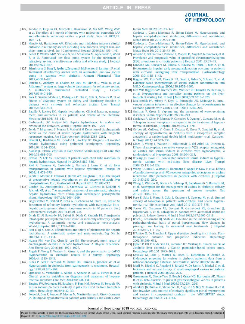
JOURNAL OF HEPATOLOGY
[122] Tandon P, Tsuyuki RT, Mitchell L, Hoskinson M, Ma MM, Wong WW,et al. The effect of 1 month of therapy with midodrine, octreotide-LARand albumin in refractory ascites: a pilot study. Liver Int 2009;29:169–174.
[123] Hanafy AS, Hassaneen AM. Rifaximin and midodrine improve clinicaloutcome in refractory ascites including renal function, weight loss, andshort-term survival. Eur J Gastroenterol Hepatol 2016;28:1455–1461.
[124] Bellot P, Welker MW, Soriano G, von Schaewen M, Appenrodt B, WiestR, et al. Automated low flow pump system for the treatment ofrefractory ascites: a multi-center safety and efficacy study. J Hepatol2013;58:922–927.
[125] Stirnimann G, Berg T, Spahr L, Zeuzem S, McPherson S, Lammert F, et al.Treatment of refractory ascites with an automated low-flow ascitespump in patients with cirrhosis. Aliment Pharmacol Ther2017;46:981–991.
[126] Bureau C, Adebayo D, Chalret de Rieu M, Elkrief L, Valla D, et al.Alfapump� system vs. large volume paracentesis for refractory ascites:A multicenter randomized controlled study. J Hepatol2017;67:940–949.
[127] Sola E, Sanchez-Cabus S, Rodriguez E, Elia C, Cela R, Moreira R, et al.Effects of alfapump system on kidney and circulatory function inpatients with cirrhosis and refractory ascites. Liver Transpl2017;23:583–593.
[128] Badillo R, Rockey DC. Hepatic hydrothorax. Clinical features, manage-ment, and outcomes in 77 patients and review of the literature.Medicine 2014;93:135–142.
[129] Garbuzenko DV, Arefyev NO. Hepatic hydrothorax: An update andreview of the literature. World J Hepatol 2017:1197–1204.
[130] Zenda T, Miyamoto S, Murata S, Mabuchi H. Detection of diaphragmaticdefect as the cause of severe hepatic hydrothorax with magneticresonance imaging. Am J Gastroenterol 1998;9:2288–2289.
[131] Hewett LJ, Bradshaw ML, Gordon LL, Rockey DC. Diagnosis of isolatedhepatic hydrothorax using peritoneal scintigraphy. Hepatology2016;64:1364–1366.
[132] Alonso JC. Pleural effusion in liver disease. Semin Respir Crit Care Med2010;31:698–705.
[133] Orman ES, Lok AS. Outcomes of patients with chest tube insertion forhepatic hydrothorax. Hepatol Int 2009;3:582–586.
[134] Xiol X, Tremosa G, Castellote J, Gornals J, Lama C, et al. Livertransplantation in patients with hepatic hydrothorax. Transpl Int2005;18:672–675.
[135] Sersté T, Moreno C, Francoz C, Razek WA, Paugham C, et al. The impactof preoperative hepatic hydrothorax on the outcome of adult livertransplantation. Eur J Gastroenterol Hepatol 2010;22:207–212.
[136] Gordon FD, Anastopoulos HT, Crenshaw W, Gilchrist B, McEniff N,Falchuk KR, et al. The successful treatment of symptomatic, refractoryhepatic hydrothorax with transjugular intrahepatic portosystemicshunt. Hepatology 1997;25:1366–1369.
[137] Siegerstetter V, Deibert P, Ochs A, Olschewski M, Blum HE, Rossle M.Treatment of refractory hepatic hydrothorax with transjugular intra-hepatic portosystemic shunt: long-term results in 40 patients. Eur JGastroenterol Hepatol 2001;13:529–534.
[138] Ditah IC, Al Bawardy BF, Saberi B, Ditah C, Kamath PS. Transjugularintrahepatic portosystemic stent shunt for medically refractory hepatichydrothorax: A systematic review and cumulative meta-analysis.World J Hepatol 2015;7:1797–1806.
[139] Hou F, Qi X, Guo X. Effectiveness and safety of pleurodesis for hepatichydrothorax: A systematic review and meta-analysis. Dig Dis Sci2016;61:3321–3334.
[140] Huang PM, Kuo SW, Chen JS, Lee JM. Thoracoscopic mesh repair ofdiaphragmatic defects in hepatic hydrothorax: A 10-year experience.Ann Thorac Surg 2016;101:1921–1927.
[141] Angeli P, Wong F, Watson H. Gines P, and the participants to CAPPS.Hyponatremia in cirrhosis: results of a survey. Hepatology2006;44:1535–1542.
[142] Gines P, Berl T, Bernardi M, Bichet DG, Hamon G, Jimenez W, et al.Hyponatremia in cirrhosis: from pathogenesis to treatment. Hepatol-ogy 1998;28:851–864.
[143] Spasovski G, Vanholder R, Allolio B, Annane D, Ball S, Bichet D, et al.Clinical practice guideline on diagnosis and treatment of hypona-traemia. Intensive Care Med 2014;40:320–331.
[144] Biggins SW, Rodriguez HJ, Bacchetti P, Bass NM, Roberts JP, Terrault NA.Serum sodium predicts mortality in patients listed for liver transplan-tation. Hepatology 2005;41:32–39.
[145] Porcel A, Diaz F, Rendon P, Macias M, Martin-Herrera L, Giron-GonzalezJA. Dilutional hyponatremia in patients with cirrhosis and ascites. Arch
Journal of Hepatology 20Please cite this article in press as: The European Association for the Study of the Liver. EASL CHepatol (2018), https://doi.org/10.1016/j.jhep.2018.03.024
Intern Med 2002;162:323–328;Cordoba J, Garcia-Martinez R, Simon-Talero M. Hyponatremic andhepatic encephalopathies: similarities, differences and coexistence.Metab Brain Dis 2010;25:73–80.
[146] Cordoba J, Garcia-Martinez R, Simon-Talero M. Hyponatremic andhepatic encephalopathies: similarities, differences and coexistence.Metab Brain Dis 2010;25:73–80.
[147] Amodio P, Del Piccolo F, Petteno E, Mapelli D, Angeli P, Iemmolo R, et al.Prevalence and prognostic value of quantified electroencephalogram(EEG) alterations in cirrhotic patients. J Hepatol 2001;35:37–45.
[148] Londono MC, Guevara M, Rimola A, Navasa M, Taura P, Mas A, et al.Hyponatremia impairs early posttransplantation outcome in patientswith cirrhosis undergoing liver transplantation. Gastroenterology2006;130:1135–1143.
[149] Biggins SW, Kim WR, Terrault NA, Saab S, Balan V, Schiano T, et al.Evidence-based incorporation of serum sodium concentration intoMELD. Gastroenterology 2006;130:1652–1660.
[150] Kim WR, Biggins SW, Kremers WK, Wiesner RH, Kamath PS, Benson JT,et al. Hyponatremia and mortality among patients on the liver-transplant waiting list. N Engl J Med 2008;359:1018–1026.
[151] McCormick PA, Mistry P, Kaye G, Burroughs AK, McIntyre N. Intra-venous albumin infusion is an effective therapy for hyponatraemia incirrhotic patients with ascites. Gut 1990;31:204–207.
[152] Quittnat F, Gross P. Vaptans and the treatment of water-retainingdisorders. Semin Nephrol 2006;26:234–243.
[153] Cardenas A, Gines P, Marotta P, Czerwiec F, Oyuang J, Guevara M, et al.Tolvaptan, an oral vasopressin antagonist, in the treatment of hypona-tremia in cirrhosis. J Hepatol 2012;56:571–578.
[154] Gerbes AL, Gulberg V, Gines P, Decaux G, Gross P, Gandjini H, et al.Therapy of hyponatremia in cirrhosis with a vasopressin receptorantagonist: a randomized double-blind multicenter trial. Gastroen-terology 2003;124:933–939.
[155] Gines P, Wong F, Watson H, Milutinovic S, del Arbol LR, Olteanu D.Effects of satavaptan, a selective vasopressin V(2) receptor antagonist,on ascites and serum sodium in cirrhosis with hyponatremia: arandomized trial. Hepatology 2008;48:204–213.
[156] O’Leary JG, Davis GL. Conivaptan increases serum sodium in hypona-tremic patients with end-stage liver disease. Liver Transpl2009;15:1325–1329.
[157] Wong F, Gines P, Watson H, Horsmans Y, Angeli P, Gow P, et al. Effectsof a selective vasopressin V2 receptor antagonist, satavaptan, on ascitesrecurrence after paracentesis in patients with cirrhosis. J Hepatol2010;53:283–290.
[158] Wong F, Watson H, Gerbes A, Vilstrup H, Badalamenti S, Bernardi M,et al. Satavaptan for the management of ascites in cirrhosis: efficacyand safety across the spectrum of ascites severity. Gut2012;61:108–116.
[159] Pose E, Sola E, Piano S, Gola E, Graupera I, Guevara M, et al. Limitedefficacy of tolvaptan in patients with cirrhosis and severe hypona-tremia: real-life experience. Am J Med 2017;130:372–375.
[160] Torres VE, Chapman AB, Devuyst O, Gansevoort RT, Grantham JJ,Higashihara E, et al. Tolvaptan in patients with autosomal dominantpolycystic kidney disease. N Engl J Med 2012;367:2407–2418.
[161] Bosch J, Groszmann RJ, Shah VH. Evolution in the understanding of thepathophysiological basis of portal hypertension: How changes inparadigm are leading to successful new treatments. J Hepatol2015;62:S121–S130.
[162] D’Amico G, De Franchis R. Upper digestive bleeding in cirrhosis. Post-therapeutic outcome and prognostic indicators. Hepatology2003;38:599–612.
[163] Jepsen P, Ott P, Andersen PK, Sorensen HT, Vilstrup H. Clinical course ofalcoholic liver cirrhosis: a Danish population-based cohort study.Hepatology 2010;51:1675–1682.
[164] Kovalak M, Lake J, Mattek N, Eisen G, Lieberman D, Zaman A.Endoscopic screening for varices in cirrhotic patients: data from anational endoscopic database. Gastrointest Endosc 2007;65:82–88.
[165] Merli M, Nicolini G, Angeloni S, Rinaldi V, De Santis A, Merkel C, et al.Incidence and natural history of small esophageal varices in cirrhoticpatients. J Hepatol 2003;38:266–272.
[166] Groszmann RJ, Garcia-Tsao G, Bosch J, Grace ND, Burroughs AK, PlanasR, et al. Beta-blockers to prevent gastroesophageal varices in patientswith cirrhosis. N Engl J Med 2005;353:2254–2261.
[167] Abraldes JG, Bureau C, Stefanescu H, Augustin S, Ney M, Blasco H, et al.Non invasive tools and risk of clinically significant portal hypertensionand varices in compensated cirrhosis : the ‘‘ANTICIPATE” study.Hepatology 2016;64:2173–2184.
18 vol. xxx j xxx–xxx 45linical Practice Guidelines for the management of patients with decompensated cirrhosis. J

Clinical Practice Guidelines
[168] De Franchis RBaveno VI faculty. Expanding consensus in portalhypertension: report of the BAVENO VI Consensus Workshop: Strati-fying risk and individualizing care for portal hypertension. J Hepatol2015;63:743–752.
[169] Garcia-Tsao G, Sanyal AJ, Grace ND, Carey WPractice GuidelinesCommittee of the American Association for the Study of LiverDiseasesPractice Parameters Committee of the American College ofGastroenterology. Prevention and management of gastroesophagealvarices and variceal hemorrhage in cirrhosis. Hepatology2007;46:922–938.
[170] North Italian Endoscopic Club for the Study and Treatment ofEsophageal Varices. Prediction of the first variceal hemorrhage inpatients with cirrhosis of the liver and esophageal varices. A prospec-tive multicenter study. N Engl J Med 1988;319:983–989.
[171] D’Amico G, Pasta L, Morabito A, et al. Competing risks and prognosticstages of cirrhosis: a 25-year inception cohort study of 494 patients.Aliment Pharmacol Ther 2014;39:1180–1193.
[172] Augustin S, Muntaner L, Altamirano JT, Gonzalez A, Saperas E, Dot J,et al. Predicting early mortality after acute variceal hemorrhage basedon classification and regression tree analysis. Clin GastroenterolHepatol 2009;7:1347–1354.
[173] Bosch J, Garcia-Pagan JC. Prevention of variceal rebleeding. Lancet2003;361:952–954.
[174] Garcia-Pagan JC, De Gottardi A, Bosch J. Review article: the modernmanagement of portal hypertension–primary and secondary prophy-laxis of variceal bleeding in cirrhotic patients. Aliment Pharmacol Ther2008;28:178–186.
[175] Hernandez-Gea V, Aracil C, Colomo A, Garupera I, Poca M, Torras X,et al. Development of ascites in compensated cirrhosis with severeportal hypertension treated with beta-blockers. Am J Gastroenterol2012;107:418–427.
[176] Puente A, Hernandez-Gea V, Graupera I, Roque M, Colomo A, Poca M,et al. Drugs plus ligation to prevent rebleeding in cirrhosis: an updatedsystematic review. Liver Int 2014;34:823–833.
[177] Villanueva C, Graupera I, Aracil C, Alvarado E, Minana J, Puente A, et al.A randomized trial to assess whether portal pressure guided therapy toprevent variceal rebleeding improves survival in cirrhosis. Hepatology2017;65:1693–1707.
[178] D’Amico G, Pagliaro L, Bosch J. Pharmacological treatment of portalhypertension: an evidence-based approach. Semin Liver Dis1999;19:475–505.
[179] Albillos A, Banares R, Gonzalez M, Ripoll C, Gonzalez R, Catalina MV,et al. Value of the hepatic venous pressure gradient to monitor drugtherapy for portal hypertension: a meta-analysis. Am J Gastroenterol2007;102:1116–1126.
[180] Li T, Ke W, Sun P, Chen X, Belgaumkar A, Huang Y, et al. Carvedilol forportal hypertension in cirrhosis: systematic review with meta-analysis.BMJ Open 2016;6:e010902.
[181] Serste T, Melot C, Francoz C, Durand F, Rautou PE, Valla D, et al.Deleterious effects of beta-blockers on survival in patients withcirrhosis and refractory ascites. Hepatology 2010;52:1017–1022.
[182] Wiest R, Albillos A, Gluud LL. The window hypothesis: haemodynamicand non-haemodynamic effects of beta-blockers improve survival ofpatients with cirrhosis during a window in the disease. Gut2012;61:967–969.
[183] Bossen L, Krag A, Vilstrup H, Watson H, Jepsen P. Non-selective beta-blockers do not affect mortality in cirrhosis patients with ascites: Posthoc analysis of three RCTs with 1198 patients. Hepatology2016;63:1968–1976.
[184] Bang UC, Benfield T, Hyldstrup L, Jensen JE, Bendtsen F. Effect ofpropranolol on survival in patients with decompensated cirrhosis: anationwide study based Danish patient registers. Liver Int2016;36:1304–1312.
[185] Reiberger T, Mandorfer M. Beta adrenergic blockade and decompen-sated cirrhosis. J Hepatol 2017;66:849–859.
[186] Leithead JA, Rajoriya N, Tehami N, Hodson J, Gunson BK, Tripathi D,et al. Non-selective b-blockers are associated with improved survival inpatients with ascites listed for liver transplantation. Gut 2015;64:1111–1119.
[187] Senzolo M, Cholongitas E, Burra P, Leandro G, Thalheimer U, Patch D,et al. Beta-blockers protect against spontaneous bacterial peritonitis incirrhotic patients: a meta-analysis. Liver Int 2009;29:1189–1193.
[188] Reiberger T, Ferlitsch A, Payer BA, Mandorfer M, Heinisch BB, Hayden H,et al. Non-selective beta-blocker therapy decreases intestinal perme-ability and serum levels of LBP and IL-6 in patients with cirrhosis. JHepatol 2013;58:911–921.
46 Journal of Hepatology 20Please cite this article in press as: The European Association for the Study of the Liver. EASL CHepatol (2018), https://doi.org/10.1016/j.jhep.2018.03.024
[189] Mookerjee RP, Pavesi M, Thomsen KL, Mehta G, Macnaughtan J,Bendtsen F, et al. Treatment with non-selective beta blockers isassociated with reduced severity of systemic inflammation andimproved survival of patients with acute-on-chronic liver failure. JHepatol 2016;64:574–582.
[190] Madsen BS, Nielsen KF, Fialla AD, Krag A. Keep the sick from harm inspontaneous bacterial peritonitis: Dose of beta blockers matters. JHepatol 2016;64:1455–1456.
[191] Serste T, Gustot T, Rautou PE, Francoz C, Njimi H, Durand F, et al. Severehyponatremia is a better predictor of mortality than MELDNa inpatients with cirrhosis and refractory ascites. J Hepatol 2012;57:274–280.
[192] Krag A, Bendtsen F, Henriksen JH, Moller S. Low cardiac output predictsdevelopment of hepatorenal syndrome and survival in patients withcirrhosis and ascites. Gut 2010;59:105–110.
[193] Ruiz-del-Arbol L, Urman J, Fernandez J, Gonzalez M, Navasa M,Monescillo A, et al. Systemic, renal, and hepatic hemodynamicderangement in cirrhotic patients with spontaneous bacterial peritoni-tis. Hepatology 2003;38:1210–1218.
[194] Payance A, Bissonnette J, Roux O, Elkrief L, Gault N, Francoz C, et al. Lackof clinical or haemodynamic rebound after abrupt interruption of beta-blockers in patients with cirrhosis. Aliment Pharmacol Ther2016;43:966–973.
[195] Garcia-Tsao G, Bosch J. Management of varices and variceal hemor-rhage in cirrhosis. N Engl J Med 2010;362:823–832.
[196] Villanueva C, Escorsell A. Optimizing general management of acutevariceal bleeding in cirrhosis. Curr Hepatol Rep 2014;13:198–207.
[197] Avgerinos A, Nevens F, Raptis S, Fevery J. Early administration ofsomatostatin and efficacy of sclerotherapy in acute oesophagealvariceal bleeds: the European Acute Bleeding OesophagealVariceal Episodes (ABOVE) randomised trial. Lancet 1997;350:1495–1499.
[198] Levacher S, Letoumelin P, Pateron D, Blaise M, Lapandry C, Pourriat JL.Early administration of terlipressin plus glyceryl trinitrate to controlactive upper gastrointestinal bleeding in cirrhotic patients. Lancet1995;346:865–868.
[199] Garcia-Tsao G, Abraldes JG, Berzigotti A, Bosch J. Portal hypertensivebleeding in cirrhosis: Risk stratification, diagnosis, and management:2016 practice guidance by the American Association for the study ofliver diseases. Hepatology 2017;65:310–335.
[200] Myburgh JA. Fluid resuscitation in acute illness–time to reappraise thebasics. N Engl J Med 2011;364:2543–2544.
[201] Villanueva C, Colomo A, Bosch A, Concepcion M, Hernandez-Gea V,Aracil C, et al. Transfusion strategies for acute upper gastrointestinalbleeding. N Engl J Med 2013;368:11–21.
[202] Seo YS, Park SY, KimMY, Kim JH, Park JY, Yim HJ, et al. Lack of differenceamong terlipressin, somatostatin, and octreotide in the control ofacute gastroesophageal variceal hemorrhage. Hepatology 2014;60:954–963.
[203] Azam Z, Hamid S, Jafri W, Salih M, Abbas Z, Abid S, et al. Short courseadjuvant terlipressin in acute variceal bleeding: a randomized doubleblind dummy controlled trial. J Hepatol 2012;56:819–824.
[204] Altraif I, Handoo FA, Aljumah A, Alalwan A, Dafalla M, Saeed AM, et al.Effect of erythromycin before endoscopy in patients presenting withvariceal bleeding: a prospective, randomized, double-blind, placebo-controlled trial. Gastrointest Endosc 2011;73:245–250.
[205] Banares R, Albillos A, Rincon D, Alonso S, Gonzalez M, Ruiz-del-Arbol L,et al. Endoscopic treatment vs. endoscopic plus pharmacologic treat-ment for acute variceal bleeding: a meta-analysis. Hepatology2002;35:609–615.
[206] Villanueva C, Piqueras M, Aracil C, Gomez C, Lopez-Balaguer JM,Gonzalez B, et al. A randomized controlled trial comparing ligation andsclerotherapy as emergency endoscopic treatment added to somato-statin in acute variceal bleeding. J Hepatol 2006;45:560–567.
[207] Rios Castellanos E, Seron P, Gisbert JP, Bonfill Cosp X. Endoscopicinjection of cyanoacrylate glue vs. other endoscopic procedures foracute bleeding gastric varices in people with portal hypertension.Cochrane Database Syst Rev 2015;5:CD010180.
[208] Bernard B, Grange JD, Khac EN, Amiot X, Opolon P, Poynard T. Antibioticprophylaxis for the prevention of bacterial infections in cirrhoticpatients with gastrointestinal bleeding: a meta-analysis. Hepatology1999;29:1655–1661.
[209] Fernandez J, Ruiz del Arbol L, Gomez C, Durandez R, Serradilla R,Guarner C, et al. Norfloxacin vs ceftriaxone in the prophylaxis ofinfections in patients with advanced cirrhosis and hemorrhage.Gastroenterology 2006;131:1049–1056.
18 vol. xxx j xxx–xxxlinical Practice Guidelines for the management of patients with decompensated cirrhosis. J

JOURNAL OF HEPATOLOGY
[210] Tandon P, Abraldes JG, Keough A, Bastiampillai R, Jayakumar S,Carbonneau M, et al. Risk of bacterial infection in patients withcirrhosis and acute variceal hemorrhage, based on child-pugh class, andeffects of antibiotics. Clin Gastroenterol Hepatol 2015;13:1189–1196.
[211] Cardenas A, Gines P, Uriz J, Bessa X, Salmeron JM, Mas A, et al. Renalfailure after upper gastrointestinal bleeding in cirrhosis: incidence,clinical course, predictive factors, and short-term prognosis. Hepatol-ogy 2001;34:671–676.
[212] Shaheen NJ, Stuart E, Schmitz SM, Mitchell KL, Fried MW, Zacks S, et al.Pantoprazole reduces the size of postbanding ulcers after variceal bandligation: a randomized, controlled trial. Hepatology 2005;41:588–594.
[213] Escorsell A, Pavel O, Cardenas A, Morillas R, Llop E, Villanueva C, et al.Esophageal balloon tamponade vs. esophageal stent in controlling acuterefractory variceal bleeding: A multicenter randomized, controlledtrial. Hepatology 2016;63:1957–1996.
[214] Monescillo A, Martínez-Lagares F, Ruiz-del-Arbol L, et al. Influence ofportal hypertension and its early decompression by TIPS placement onthe outcome of variceal bleeding. Hepatology 2004;40:793–801.
[215] García-Pagán JC, Caca K, Bureau C, et al. Early use of TIPS in patientswith cirrhosis and variceal bleeding. N Engl J Med 2010;362:2370–2379.
[216] Garcia-Pagan JC, Di Pascoli M, Caca K, Laleman W, Bureau C, AppenrodtB, et al. Use of early-TIPS for high-risk variceal bleeding: results of apost-RCT surveillance study. J Hepatol 2013;58:45–50.
[217] Rudler M, Cluzel P, Corvec TL, Benosman H, Rousseau G, Poynard T,et al. Early-TIPSS placement prevents rebleeding inhigh-risk patientswith variceal bleeding, without improving survival. Aliment PharmacolTher 2014;40:1074–1080.
[218] Augustin S, Altamirano J, Gonzalez A, Dot J, Abu-Suboh M, Armengol JR,et al. Effectiveness of combined pharmacologic and ligation therapy inhigh-risk patients with acute esophageal variceal bleeding. Am JGastroenterol 2011;106:1787–1795.
[219] Reverter E, Tandon P, Augustin S, Turon F, Casu S, Bastiampillai R, et al.A MELD-based model to determine risk of mortality among patientswith acute variceal bleeding. Gastroenterology 2014;146:412–419.
[220] Merli M, Nicolini G, Angeloni S, Gentili F, Attili AF, Riggio O. The naturalhistory of portal hypertensive gastropathy in patients with livercirrhosis and mild portal hypertension. Am J Gastroenterol2004;99:1959–1965.
[221] Thuluvath PJ, Yoo HY. Portal Hypertensive gastropathy. Am J Gastroen-terol 2002;97:2973–2978.
[222] Yoshikawa I, Murata I, Nakano S, Otsuki M. Effects of endoscopicvariceal ligation on portal hypertensive gastropathy and gastricmucosal blood flow. Am J Gastroenterol 1998;93:71–74.
[223] Urrunaga NH, Rockey DC. Portal hypertensive gastropathy and colopa-thy. Clin Liver Dis 2014;18:389–406.
[224] Hosking SW, Kennedy HJ, Seddon I, Triger DR. The role of propranolol incongestive gastropathy of portal hypertension. Hepatology1987;7:437–441.
[225] Perez-Ayuso RM, Pique JM, Bosch J, Panes J, Gonzalez A, Perez R, et al.Propranolol in prevention of recurrent bleeding from severe portalhypertensive gastropathy in cirrhosis. Lancet 1991;337:1431–1434.
[226] Ripoll C, Garcia-Tsao G. Treatment of gastropathy and gastric antralvascular ectasia in patients with portal hypertension. Curr TreatOptions Gastroenterol 2007;10:483–494.
[227] Kamath P, Lacerda M, Ahlquist D, McKusick MA, Andrews JC, NagorneyDA. Gastric Mucosal Responses to intrahepatic portosystemic shuntingin patients with cirrhosis. Gastroenterology 2000;118:905–911.
[228] Zhou Y, Qiao L, Wu J, Hu H, Xu C. Comparison of the efficacy ofoctreotide, vasopressin, and omeprazole in the control of acutebleeding in patients with portal hypertensive gastropathy: a controlledstudy. J Gastroenterol Hepatol 2002;17:973–979.
[229] Sarin SK, Lahoti D, Saxena SP, Murthy NS, Makwana UK. Prevalence,classification and natural history of gastric varices: a long-term follow-up study in 568 portal hypertension patients. Hepatology1992;16:1343–1349.
[230] Mishra SR, Sharma BC, Kumar A, Sarin SK. Primary prophylaxis ofgastric variceal bleeding comparing cyanoacrylate injection and beta-blockers: a randomized controlled trial. J Hepatol 2011;54:1161–1167.
[231] Rios CE, Seron P, Gisbert JP, Bonfill CX. Endoscopic injection ofcyanoacrylate glue vs. other endoscopic procedures for acute bleedinggastric varices in people with portal hypertension. Cochrane DatabaseSyst Rev 2015;5:CD010180.
[232] Mishra SR, Chander Sharma B, Kumar A, Sarin SK. Endoscopiccyanoacrylate injection vs. beta-blocker for secondary prophylaxis of
Journal of Hepatology 20Please cite this article in press as: The European Association for the Study of the Liver. EASL CHepatol (2018), https://doi.org/10.1016/j.jhep.2018.03.024
gastric variceal bleed: a randomised controlled trial. Gut2010;59:729–735.
[233] Chau TN, Patch D, Chan YW, Nagral A, Dick R, Burroughs AK. ‘‘Salvage”transjugular intrahepatic portosystemic shunts: gastric fundal com-pared with esophageal variceal bleeding. Gastroenterology1998;114:981–998.
[234] Hung HH, Chang CJ, Hou MC, Liao WC, Chan CC, Huang HC, et al.Efficacy of non-selective beta-blockers as adjunct to endoscopicprophylactic treatment for gastric variceal bleeding: a randomizedcontrolled trial. J Hepatol 2012;56:1025–1032.
[235] Lo GH, Liang HL, Chen WC, Chen MH, Lai KH, Hsu PI, et al. A prospective,randomized controlled trial of transjugular intrahepatic portosystemicshunt vs. cyanoacrylate injection in the prevention of gastric varicealrebleeding. Endoscopy 2007;39:679–685.
[236] Saad WE. Endovascular management of gastric varices. Clin Liver Dis2014;18:829–885.
[237] Mookerjee RP, Stadlbauer V, Lidder S, Wright GA, Hodges SJ, Davies NA,et al. Neutrophil dysfunction in alcoholic hepatitis superimposed oncirrhosis is reversible and predicts the outcome. Hepatology2007;46:831–840.
[238] Wasmuth HE, Kunz D, Yagmur E, Timmer-Stranghoner A, Vidacek D,Siewert E, et al. Patients with acute on chronic liver failure display‘‘sepsis-like” immune paralysis. J Hepatol 2005;42:195–201.
[239] Wiest R, Garcia-Tsao G. Bacterial translocation (BT) in cirrhosis.Hepatology 2005;41:422–433.
[240] Appenrodt B, Grunhage F, Gentemann MG, Thyssen L, Sauerbruch T,Lammert F. Nucleotide-binding oligomerization domain containing 2(NOD2) variants are genetic risk factors for death and spontaneousbacterial peritonitis in liver cirrhosis. Hepatology 2010;51:1327–1333.
[241] Fernandez J, Navasa M, Gomez J, et al. Bacterial infections in cirrhosis:epidemiological changes with invasive procedures and norfloxacinprophylaxis. Hepatology 2002;35:140–148.
[242] Fasolato S, Angeli P, Dallagnese L, et al. Renal failure and bacterialinfections in patients with cirrhosis: epidemiology and clinical features.Hepatology 2007;45:223–229.
[243] Piano S, Fasolato S, Salinas F, Romano A, Tonon M, Morando F, et al. Theempirical antibiotic treatment of nosocomial spontaneous bacterialperitonitis: Results of a randomized, controlled clinical trial. Hepatol-ogy 2016;63:1299–1309.
[244] Runyon BA. The evolution of ascitic fluid analysis in the diagnosis ofspontaneous bacterial peritonitis. Runyon BA. Am J Gastroenterol2003;98:1675–1677.
[245] Evans LT, Kim WR, Poterucha JJ, Kamath PS. Spontaneous bacterialperitonitis in asymptomatic outpatients with cirrhotic ascites. Hepa-tology 2003;37:897–901.
[246] Kim JJ, Tsukamoto MM, Mathur AK, Ghomri YM, Hou LA, Sheibani S,et al. Delayed paracentesis is associated with increased in-hospitalmortality in patients with spontaneous bacterial peritonitis. Am JGastroenterol 2014;109:1436–1442.
[247] Van de Geijn GM, van Gent M, van Pul-Bom N, Beunis MH, van TilburgAJ, Njo TL. A new flow cytometric method for differential cell countingin ascitic fluid. Cytometry B Clin Cytom 2016;90:506–511.
[248] Fleming C, Brouwer R, van Alphen A, Lindemans J, de Jonge R. UF-1000i:validation of the body fluid mode for counting cells in body fluids. ClinChem Lab Med 2014;52:1781–1790.
[249] Gülberg V, Gerbes AL, Sauerbruch T, Appenrodt B. Insufficient sensi-tivity of reagent strips for spontaneous bacterial peritonitis. Hepatology2007;46:1669.
[250] Bellot P, García-Pagán JC, Francés R, Abraldes JG, Navasa M, Pérez-Mateo M, et al. Bacterial DNA translocation is associated with systemiccirculatory abnormalities and intrahepatic endothelial dysfunction inpatients with cirrhosis. Hepatology 2010;52:2044–2052.
[251] Bruns T, Reuken PA, Stengel S, Gerber L, Appenrodt B, Schade JH, et al.The prognostic significance of bacterial DNA in patients with decom-pensated cirrhosis and suspected infection. Liver Int 2016;36:1133–1142.
[252] Runyon BA, Hoefs JC. Culture-negative neutrocytic ascites: a variant ofspontaneous bacterial peritonitis. Hepatology 1984;4:1209–1211.
[253] Gravito-Soares M, Gravito-Soares E, Lopes S, Ribeiro G, Figueiredo P.Spontaneous fungal peritonitis: a rare but severe complication of livercirrhosis. Eur J Gastroenterol Hepatol 2017;29:1010–1016.
[254] Chen CH, Shih CM, Chou JW, Liu YH, Hang LW, Hsia TC, et al. Outcomepredictors of cirrhotic patients with spontaneous bacterial empyema.Liver Int 2011;31:417–424.
18 vol. xxx j xxx–xxx 47linical Practice Guidelines for the management of patients with decompensated cirrhosis. J

Clinical Practice Guidelines
[255] Xiol X, Castellvi JM, Guardiola J, Sese E, Castellote J, Perello A, et al.Spontaneous bacterial empyema in cirrhotic patients: a prospectivestudy. Hepatology 1996;23:719–723.
[256] Soriano G, Castellote J, Alvarez C, Girbau A, Gordillo J, Baliellas C, et al.Secondary bacterial peritonitis in cirrhosis: a retrospective study ofclinical and analytical characteristics, diagnosis and management. JHepatol 2010;52:39–44.
[257] Rimola A, Salmeron JM, Clemente G, Rodrigo L, Obrador A, Miranda ML,et al. Two different dosages of cefotaxime in the treatment ofspontaneous bacterial peritonitis in cirrhosis: results of a prospective,randomized, multicenter study. Hepatology 1995;21:674–679.
[258] Runyon Runyon BA, McHutchison JG, Antillon MR, Akriviadis EA,Montano AA. Short-course vs. long-course antibiotic treatment ofspontaneous bacterial peritonitis. A randomized controlled study of100 patients. Gastroenterology 1991;100:1737–1742.
[259] Ricart E, Soriano G, Novella MT, Ortiz J, Sabat M, Kolle L, et al.Amoxicillin-clavulanic acid vs. cefotaxime in the therapy of bacterialinfections in cirrhotic patients. J Hepatol 2000;32:596–602.
[260] deLemos AS, Ghabril M, Rockey DC, Gu J, Barnhart HX, Fontana RJ, et al.Drug-induced liver injury network (DILIN). Amoxicillin-clavulanate-induced liver injury. Dig Dis Sci 2016;61:2406–2416.
[261] Terg R, Cobas S, Fassio E, Landeira G, Rios B, Vasen W, et al. Oralciprofloxacin after a short course of intravenous ciprofloxacin in thetreatment of spontaneous bacterial peritonitis: results of a multicenter,randomized study. J Hepatol 2000;33:564–569.
[262] Angeli P, Guarda S, Fasolato S, Miola E, Craighero R, Piccolo F, et al.Switch therapy with ciprofloxacin vs. intravenous ceftazidime in thetreatment of spontaneous bacterial peritonitis in patients with cirrho-sis: similar efficacy at lower cost. Aliment Pharmacol Ther 2006;23:75–84.
[263] Navasa M, Follo A, Llovet JM, Clemente G, Vargas V, Rimola A, et al.Randomized, comparative study of oral ofloxacin vs. intravenouscefotaxime in spontaneous bacterial peritonitis. Gastroenterology1996;111:1011–1017.
[264] Fernandez J, Bert F, Nicolas-Chanoine MH. The challenges of multi-drug-resistance in hepatology. J Hepatol 2016;65:1043–1054.
[265] Magiorakos AP, Srinivasan A, Carey RB, Carmeli Y, Falagas ME, Giske CG,et al. Multidrug-resistant, extensively drug-resistant and pandrug-resistant bacteria: an international expert proposal for interim stan-dard definitions for acquired resistance. Clin Microbiol Infect2012;18:268–281.
[266] Fernandez J, Acevedo J, Castro M, Garcia O, de Lope CR, Roca D, et al.Prevalence and risk factors of infections by multiresistant bacteria incirrhosis: a prospective study. Hepatology 2012;55:1551–1561.
[267] Wiest R, Krag A, Gerbes A. Spontaneous bacterial peritonitis: recentguidelines and beyond. Gut 2012;61:297–310.
[268] Piano S, Brocca A, Mareso S, Angeli P. Infections complicating cirrhosis.Liver Int 2018;38:126–133.
[269] Lutz P, Nischalke HD, Kramer B, Goeser F, Kaczmarek DJ, Schlabe S, et al.Antibiotic resistance in healthcare-related and nosocomial sponta-neous bacterial peritonitis. Eur J Clin Invest 2017;47:44–52.
[270] Singer M, Deutschman CS, Seymour CW, Shankar-Hari M, Annane D,Bauer M, et al. The third international consensus definitions for sepsisand septic shock (Sepsis-3). JAMA 2016;315:801–810.
[271] Piano S, Bartoletti M, Tonon M, Baldassarre M, Chies G, Romano A, et al.Assessment of Sepsis-3 criteria and quick SOFA in patients withcirrhosis and bacterial infections. Gut 2017.
[272] Sort P, Navasa M, Arroyo V, Aldeguer X, Planas R, Ruiz-del-Arbol L, et al.Effect of intravenous albumin on renal impairment and mortality inpatients with cirrhosis and spontaneous bacterial peritonitis. N EnglJMed 1999;341:403–409.
[273] Garioud A, Cadranel JF, Pauwels A, Nousbaum JB, Thévenot T, Dao T,et al. Association Nationale des Hépato-gastroentérologues des Hôpi-taux Généraux de France, Association Française pour l’Etude du Foie,Club de Réflexion des Cabinets et Groupes d’Hépato-Gastroentérologie.Albumin Use in Patients With Cirrhosis in France: Results of the ‘‘ALBU-LIVE” Survey: A Case for Better EASL Guidelines Diffusion and/orRevision. J Clin Gastroenterol 2017;51:831–838.
[274] Poca M, Concepcion M, Casas M, Alvarez-Urturi C, Gordillo J, Hernan-dez-Gea V, et al. Role of albumin treatment in patients with sponta-neous bacterial peritonitis. Clin Gastroenterol Hepatol 2012;10:309–315.
[275] Fernandez J, Tandon P, Mensa J, Garcia-Tsao G. Antibiotic prophylaxis incirrhosis: Good and bad. Hepatology 2016;63:2019–2031.
[276] Fernandez J, Navasa M, Planas R, Montoliu S, Monfort D, Soriano G, et al.Primary prophylaxis of spontaneous bacterial peritonitis delays hepa-
48 Journal of Hepatology 20Please cite this article in press as: The European Association for the Study of the Liver. EASL CHepatol (2018), https://doi.org/10.1016/j.jhep.2018.03.024
torenal syndrome and improves survival in cirrhosis. Gastroenterology2007;133:818–824.
[277] Terg R, Fassio E, Guevara M, Cartier M, Longo C, Lucero R, et al.Ciprofloxacin in primary prophylaxis of spontaneous bacterial peri-tonitis: A randomized, placebo-controlled study. J Hepatol2008;48:774–779.
[278] Loomba R, Wesley R, Bain A, et al. Role of fluoroquinolones in theprimary prophylaxis of spontaneous bacterial peritonitis: meta-analy-sis. Clin Gastroenterol Hepatol 2009;7:487–493.
[279] Saab S, Hernandez JC, Chi AC, et al. Oral antibiotic prophylaxis reducesspontaneous bacterial peritonitis occurrence and improves short-termsurvival in cirrhosis: a meta-analysis. Am J Gastroenterol 2009;104:993–1001.
[280] Krag A, Wiest R, Gluud LL. Fluoroquinolones in the primary prophylaxisof spontaneous bacterial peritonitis. Am J Gastroenterol2010;105:1444–1445.
[281] Gines P, Rimola A, Planas R, Vargas V, Marco F, Almela M, et al.Norfloxacin prevents spontaneous bacterial peritonitis recurrence incirrhosis: results of a double-blind, placebo-controlled trial. Hepatology1990;12:716–724.
[282] Bauer TM, Follo A, Navasa M, Vila J, Planas R, Clemente G, et al. Dailynorfloxacin is more effective than weekly rufloxacin in prevention ofspontaneous bacterial peritonitis recurrence. Dig Dis Sci2002;47:1356–1361.
[283] Terg R, Llano K, Cobas SM, Brotto C, Barrios A, Levi D, et al. Effects of oralciprofloxacin on aerobic gram-negative fecal flora in patients withcirrhosis: results of short- and long-term administration, with daily andweekly dosages. J Hepatol 1998;29:437–442.
[284] Bass NM, Mullen KD, Sanyal A, Poordad F, Neff G, Leevy CB, et al.Rifaximin treatment in hepatic encephalopathy. N Engl J Med2010;362:071–1081.
[285] Elfert A, Abo Ali L, Soliman S, Ibrahim S, Abd-Elsalam S. Randomized-controlled trial of rifaximin vs. norfloxacin for secondary prophylaxis ofspontaneous bacterial peritonitis. Eur J Gastroenterol Hepatol2016;28:1450–1454.
[286] Min YW, Lim KS, Min BH, Gwak GY, Paik YH, Choi MS, et al. Protonpump inhibitor use significantly increases the risk of spontaneousbacterial peritonitis in 1965 patients with cirrhosis and ascites: apropensity score matched cohort study. Aliment Pharmacol Ther2014;40:695–704.
[287] Dam G, Vilstrup H, Watson H, Jepsen P. Proton pump inhibitors as a riskfactor for hepatic encephalopathy and spontaneous bacterial peritonitisin patients with cirrhosis with ascites. Hepatology 2016;64:1265–1272.
[288] Mandorfer M, Bota S, Schwabl P, Bucsics T, Pfisterer N, Kruzik M, et al.Nonselective beta blockers increase risk for hepatorenal syndrome anddeath in patients with cirrhosis and spontaneous bacterial peritonitis.Gastroenterology 2014;146:1680–1690.
[289] Pande C, Kumar A, Sarin SK. Addition of probiotics to norfloxacin doesnot improve efficacy in the prevention of spontaneous bacterialperitonitis: a double-blind placebo-controlled randomized-controlledtrial. Eur J Gastroenterol Hepatol 2012;24:831–839.
[290] Fernandez J, Acevedo J, Arroyo V. Response to the clinical course andshort-term mortality of cirrhotic patients with non-spontaneous bac-terial peritonitis infections. Liver Int 2017;37:623.
[291] Arvaniti V, D’Amico G, Fede G, Manousou P, Tsochatzis E, PleguezueloM, et al. Infections in patients with cirrhosis increase mortality four-fold and should be used in determining prognosis. Gastroenterology2010;139:1256.
[292] Piano S, Morando F, Carretta G, Tonon M, Vettore E, Rosi S, et al.Predictors of early readmission in patients with cirrhosis after theresolution of bacterial infections. Am J Gastroenterol 2017;112:1575–1583.
[293] Bartoletti M, Giannella M, Lewis R, Caraceni P, Tedeschi S, Paul M, et al.A prospective multicentre study of the epidemiology and outcomes ofbloodstream infection in cirrhotic patients. Clin Microbiol Infect 2017.
[294] Merli M, Lucidi C, Di Gregorio V, Lattanzi B, Giannelli V, Giusto M, et al.An empirical broad spectrum antibiotic therapy in health-care-associ-ated infections improves survival in patients with cirrhosis: A ran-domized trial. Hepatology 2016;63:1632–1639.
[295] Fiore M, Chiodini P, Pota V, Sansone P, Passavanti MB, Leone S, et al.Risk of spontaneous fungal peritonitis in hospitalized cirrhotic patientswith ascites: a systematic review of observational studies and meta-analysis. Minerva Anestesiol 2017;83:1309–1316.
[296] Piano S, Angeli P. Reply letter. Hepatology 2016;64:998–999.
18 vol. xxx j xxx–xxxlinical Practice Guidelines for the management of patients with decompensated cirrhosis. J

JOURNAL OF HEPATOLOGY
[297] Guevara M, Terra C, Nazar A, Sola E, Fernandez J, Pavesi M, et al.Albumin for bacterial infections other than spontaneous bacterialperitonitis in cirrhosis. A randomized, controlled study. J Hepatol2012;57:759–765.
[298] Thevenot T, Bureau C, Oberti F, Anty R, Louvet A, Plessier A, et al. Effectof albumin in cirrhotic patients with infection other than spontaneousbacterial peritonitis. A randomized trial. J Hepatol 2015;62:822–830.
[299] Piano S, Romano A, Di Pascoli M, Angeli P. Why and how to measurerenal function in patients with liver disease. Liver Int 2017;37:116–122.
[300] Gonwa TA, Jennings L, Mai ML, Stark PC, Levey AS, Klintmalm GB.Estimation of glomerular filtration rates before and after orthotopicliver transplantation: evaluation of current equations. Liver Transpl2004;10:301–309.
[301] Hoek FJ, Kemperman FA, Krediet RT. A comparison between cystatin C,plasma creatinine and the Cockcroft and Gault formula for theestimation of glomerular filtration rate. Nephrol Dial Transplant2003;18:2024–2031.
[302] Inker LA, Schmid CH, Tighiouart H, Eckfeldt JH, Feldman HI, Greene T,et al. Estimating glomerular filtration rate from serum creatinine andcystatin C. N Engl J Med 2012;367:20–29.
[303] Roy L, Legault L, Pomier-Layrargues G. Glomerular filtration ratemeasurement in cirrhotic patients with renal failure. Clin Nephrol1998;50:342–346.
[304] Francoz C, Nadim MK, Baron A, Prie D, Antoine C, Belghiti J, et al.Glomerular filtration rate equations for liver-kidney transplantation inpatients with cirrhosis: validation of current recommendations. Hepa-tology 2014;59:1514–1521.
[305] Francoz C, Glotz D, Moreau R, Durand F. The evaluation of renalfunction and disease in patients with cirrhosis. J Hepatol2010;52:605–613.
[306] Garcia-Tsao G, Parikh CR, Viola A. Acute kidney injury in cirrhosis.Hepatology 2008;48:2064–2077.
[307] Salerno F, Gerbes A, Gines P, Wong F, Arroyo V. Diagnosis, preventionand treatment of hepatorenal syndrome in cirrhosis. Gut 2007;56:310–1318.
[308] Mehta RL, Kellum JA, Shah SV, Molitoris BA, Ronco C, Warnock DG, et al.Acute Kidney Injury Network: report of an initiative to improveoutcomes in acute kidney injury. Crit Care 2007;11:R31.
[309] Nadim MK, Kellum JA, Davenport A, Wong F, Davis C, Pannu N, et al.Hepatorenal syndrome: the 8th International Consensus Conference ofthe Acute Dialysis Quality Initiative (ADQI) Group. Crit Care 2012;16:R23.
[310] Khwaja A. KDIGO clinical practice guidelines for acute kidney injury.Nephron Clin Pract 2012;120:c179–c184.
[311] Belcher JM, Garcia-Tsao G, Sanyal AJ, Bhogal H, Lim JK, Ansari N, et al.Association of AKI with mortality and complications in hospitalizedpatients with cirrhosis. Hepatology 2013;57:753–762.
[312] de Carvalho JR, Villela-Nogueira CA, Luiz RR, Guzzo PL, da Silva Rosa JM,Rocha E, et al. Acute kidney injury network criteria as a predictor ofhospital mortality in cirrhotic patients with ascites. J Clin Gastroenterol2012;46:21–26.
[313] Fagundes C, Barreto R, Guevara M, Garcia E, Solà E, Rodríguez E, et al. Amodified acute kidney injury classification for diagnosis and riskstratification of impairment of kidney function in cirrhosis. J Hepatol2013;59:474–481.
[314] Piano S, Rosi S, Maresio G, Fasolato S, Cavallin M, Romano A, et al.Evaluation of the Acute Kidney Injury Network criteria in hospi-talized patients with cirrhosis and ascites. J Hepatol 2013;59:482–489.
[315] Tsien CD, Rabie R, Wong F. Acute kidney injury in decompensatedcirrhosis. Gut 2013;62:131–137.
[316] Wong F, O’Leary JG, Reddy KR, Patton H, Kamath PS, Fallon MB, et al.New consensus definition of acute kidney injury accurately predicts 30-day mortality in patients with cirrhosis and infection. Gastroenterology2013;145:1280–1288.
[317] Huelin P, Piano S, Solà E, Stanco M, Solé C, Moreira R, et al. Validation ofa staging system for acute kidney injury in patients with cirrhosis andassociation with acute-on-chronic liver failure. Clin GastroenterolHepatol 2017;15:438–445.
[318] Angeli P, Gines P, Wong F, Bernardi M, Boyer TD, Gerbes A, et al.Diagnosis and management of acute kidney injury in patients withcirrhosis: revised consensus recommendations of the InternationalClub of Ascites. J Hepatol 2015;62:968–974.
[319] Kidney Disease: Inproving Global Outcomes (KDIGO) CKD Work Group.KDIGO. Clinical practice guideline for the evaluation and managementof chronic kidney disease. Kidny Int 2012;2013:1–150.
Journal of Hepatology 20Please cite this article in press as: The European Association for the Study of the Liver. EASL CHepatol (2018), https://doi.org/10.1016/j.jhep.2018.03.024
[320] Rosi S, Piano S, Frigo AC, Morando F, Fasolato S, Cavallin M, et al. NewICA criteria for the diagnosis of acute kidney injury in cirrhotic patients:can we use an imputed value of serum creatinine? Liver Int2015;35:2108–2114.
[321] Guevara M, Fernandez-Esparrach G, Alessandria C, Torre A, Terra C,Montana X, et al. Effects of contrast media on renal function in patientswith cirrhosis: a prospective study. Hepatology 2004;40:646–651.
[322] Umgelter A, Reindl W, Franzen M, Lenhardt C, Huber W, Schmid RM.Renal resistive index and renal function before and after paracentesis inpatients with hepatorenal syndrome and tense ascites. Intensive CareMed 2009;35:152–156.
[323] Cabrera J, Falcón L, Gorriz E, Pardo MD, Granados R, Quinones A,Maynar M. Abdominal decompression plays a major role in earlypostparacentesis haemodynamic changes in cirrhotic patients withtense ascites. Gut 2001;48:384–389.
[324] de Cleva R, Silva FP, Zilberstein B, Machado DJ. Acute renal failure dueto abdominal compartment syndrome: report on four cases andliterature review. Rev Hospital das Clin 2001;56:123–130.
[325] Nadim MK, Durand F, Kellum JA, Levitsky J, O’Leary JG, Karvellas CJ,et al. Management of the critically ill patient with cirrhosis: Amultidisciplinary perspective. J Hepatol 2016;64:717–735.
[326] Umgelter A, Reindl W, Wagner KS, Franzen M, Stock K, Schmid RM,et al. Effects of plasma expansion with albumin and paracentesis onhaemodynamics and kidney function in critically ill cirrhotic patientswith tense ascites and hepatorenal syndrome: a prospective uncon-trolled trial. Critical care 2008;12:R4.
[327] Moreau R, Lebrec D. Diagnosis and treatment of acute renal failure inpatients with cirrhosis. Best Pract Res Clin Gastroenterol2007;21:111–123.
[328] Moreau R, Lebrec D. Acute renal failure in patients with cirrhosis:perspectives in the age of MELD. Hepatology 2003;37:233–243.
[329] Fagundes C, Pepin MN, Guevara M, Barreto R, Casals G, Sola E, et al.Urinary neutrophil gelatinase-associated lipocalin as biomarker in thedifferential diagnosis of impairment of kidney function in cirrhosis. JHepatol 2012;57:267–273.
[330] Belcher JM, Sanyal AJ, Peixoto AJ, Perazella MA, Lim J, Thiessen-Philbrook H, et al. Kidney biomarkers and differential diagnosis ofpatients with cirrhosis and acute kidney injury. Hepatology2014;60:622–632.
[331] Qasem AA, Farag SE, Hamed E, Emara M, Bihery A, Pasha H. Urinarybiomarkers of acute kidney injury in patients with liver cirrhosis. ISRNNephrol 2014;2014:376795.
[332] Barreto R, Elia C, Sola E, Moreira R, Ariza X, Rodriguez E, et al. Urinaryneutrophil gelatinase-associated lipocalin predicts kidney outcome anddeath in patients with cirrhosis and bacterial infections. J Hepatol2014;61:35–42.
[333] Ariza X, Sola E, Elia C, Barreto R, Moreira R, Morales-Ruiz M, et al.Analysis of a urinary biomarker panel for clinical outcomes assessmentin cirrhosis. PLoS One 2015;10:e0128145.
[334] Puthumana J, Ariza X, Belcher JM, Graupera I, Ginès P, Parikh CR. Urineinterleukin 18 and lipocalin 2 are biomarkers of acute tubular necrosisin patients with cirrhosis: a systematic review and meta-analysis. ClinGastroenterol Hepatol 2017;15:1003–1013.
[335] Huelin P, Elia C, Solà E, Solé C, Moreira R, Carol M. New diagnosticalgorithm of acute kidney injury in cirrhosis that includes categoriza-tion of stage 1 and assessment of urine NGAL. Relevance for thedifferential diagnosis and clinical outcomes. J Hepatol 2017;66:S11.
[336] Chawla LS, Eggers PW, Star RA, Kimmel PL. Acute kidney injury andchronic kidney disease as interconnected syndromes. N Engl J Med2014;371:58–66.
[337] Trawale JM, Paradis V, Rautou PE, Francoz C, Escolano S, Sallee M, et al.The spectrum of renal lesions in patients with cirrhosis: a clinico-pathological study. Liver Int 2010;30:725–732.
[338] Wadei HM, Geiger XJ, Cortese C, Mai ML, Kramer DJ, Rosser BG, et al.Kidney allocation to liver transplant candidates with renal failure ofundetermined etiology: role of percutaneous renal biopsy. Am JTransplant 2008;8:2618–2626.
[339] Wong F, Nadim MK, Kellum JA, Salerno F, Bellomo R, Gerbes A, et al.Working Party proposal for a revised classification system of renaldysfunction in patients with cirrhosis. Gut 2011;60:702–709.
[340] Stadlbauer V, Wright GA, Banaji M, Mukhopadhya A, Mookerjee RP,Moore K, et al. Relationship between activation of the sympatheticnervous system and renal blood flow autoregulation in cirrhosis.Gastroenterology 2008;134:111–119.
[341] Wiest R, Lawson M, Geuking M. Pathological bacterial translocation inliver cirrhosis. J Hepatol 2014;60:197–209.
18 vol. xxx j xxx–xxx 49linical Practice Guidelines for the management of patients with decompensated cirrhosis. J

Clinical Practice Guidelines
[342] Albillos A, de la Hera A, Gonzalez M, Moya JL, Calleja JL, Monserrat J,et al. Increased lipopolysaccharide binding protein in cirrhotic patientswith marked immune and hemodynamic derangement. Hepatology2003;37:208–217.
[343] Navasa M, Follo A, Filella X, Jimenez W, Francitorra A, Planas R, et al.Tumor necrosis factor and interleukin-6 in spontaneous bacterialperitonitis in cirrhosis: relationship with the development of renalimpairment and mortality. Hepatology 1998;27:1227–1232.
[344] Shah N, Dhar D, El Zahraa Mohammed F, Habtesion A, Davies NA,Jover-Cobos M, et al. Prevention of acute kidney injury in a rodentmodel of cirrhosis following selective gut decontamination is asso-ciated with reduced renal TLR4 expression. J Hepatol 2012;56:1047–1053.
[345] Shah N, Mohamed FE, Jover-Cobos M, Macnaughtan J, Davies N, MoreauR, et al. Increased renal expression and urinary excretion of TLR4 inacute kidney injury associated with cirrhosis. Liver Int 2013;33:398–409.
[346] Alobaidi R, Basu RK, Goldstein SL, Bagshaw SM. Sepsis-associated acutekidney injury. Semin Nephrol 2015;35:2–11.
[347] Emlet DR, Shaw AD, Kellum JA. Sepsis-associated AKI: epithelial celldysfunction. Semin Nephrol 2015;35:85–95.
[348] Prowle JR, Bellomo R. Sepsis-associated acute kidney injury: macrohe-modynamic and microhemodynamic alterations in the renal circula-tion. Semin Nephrol 2015;35:64–74.
[349] de Seigneux S, Martin PY. Preventing the progression of AKI to CKD: therole of mitochondria. J Am Soc Nephrol 2017;28:1327–1329.
[350] Bairaktari E, Liamis G, Tsolas O, Elisaf M. Partially reversible renaltubular damage in patients with obstructive jaundice. Hepatology2001;33:1365–1369.
[351] van Slambrouck CM, Salem F, Meehan SM, Chang A. Bile castnephropathy is a common pathologic finding fo kidney injury associ-ated with severe liver dysfunction. Kidney Int 2013;84:192–197.
[352] Durand F, Graupera I, Gines P, Olson JC, Nadim MK. Pathogenesis ofhepatorenal syndrome: implications for therapy. Am J Kidney Dis2016;67:318–328.
[353] Moreau R, Durand F, Poynard T, Duhamel C, Cervoni JP, Ichai P, et al.Terlipressin in patients with cirrhosis and type 1 hepatorenal syn-drome: a retrospective multicenter study. Gastroenterology 2002;122:923–930.
[354] Ortega R, Gines P, Uriz J, et al. Terlipressin therapy with and withoutalbumin for patients with hepatorenal syndrome: results of a prospec-tive, nonrandomized study. Hepatology 2002;36:941–948.
[355] Sanyal AJ, Boyer T, Garcia-Tsao G, Regenstein F, Rossaro L, Appenrodt B,et al. A randomized prospective double blind, placebo controlled studyof terlipressin for type 1 hepatorenal syndrome. Gastroenterology2008;134:1360–1368.
[356] Martin-Llahi M, Pepin MN, Guevara M, Diaz F, Torre A, Monescillo A,et al. Terlipressin and albumin vs albumin in patients with cirrhosisand hepatorenal syndrome: a randomized study. Gastroenterology2008;134:1352–1359.
[357] Boyer TD, Sanyal AJ, Wong F, Frederick RT, Lake JR, O’Leary JG, et al.REVERSE study investigators terlipressin plus albumin is more effectivethan albumin alone in improving renal function in patients withcirrhosis and hepatorenal syndrome type 1. Gastroenterology2016;150:1579–1589.
[358] Rodriguez E, Elia C, Sola E, Barreto R, Graupera I, Andrealli A, et al.Terlipressin and albumin for type-1 hepatorenal syndrome associatedwith sepsis. J Hepatol 2014;60:955–961.
[359] Cavallin M, Kamath PS, Merli M, Fasolato S, Toniutto P, Salerno F, et al.Terlipressin plus albumin vs. midodrine and octreotide plus albumin inthe treatment of hepatorenal syndrome: A randomized trial. Hepatol-ogy 2015;62:567–574.
[360] Cavallin M, Piano S, Romano A, Fasolato S, Frigo AC, Benetti G, et al.Terlipressin given by continuous intravenous infusion vs. intravenousboluses in the treatment of hepatorenal syndrome: A randomizedcontrolled study. Hepatology 2016;63:983–992.
[361] Gluud LL, Christensen K, Christensen E, Krag A. Terlipressin forhepatorenal syndrome. Cochrane Database Syst Rev 2012:CD005162.
[362] Facciorusso A, Chandar AK, Murad MH, Prokop LJ, Muscatiello N,Kamath PS. Singh SComparative efficacy of pharmacological strategiesfor management of type 1 hepatorenal syndrome: a systematic reviewand network meta-analysis. Lancet Gastroenterol Hepatol2017;2:94–102.
[363] Narahara Y, Kanazawa H, Taki Y, Kimura Y, Atsukawa M, Katakura T,et al. Effects of terlipressin on systemic, hepatic and renal hemody-
50 Journal of Hepatology 20Please cite this article in press as: The European Association for the Study of the Liver. EASL CHepatol (2018), https://doi.org/10.1016/j.jhep.2018.03.024
namics in patients with cirrhosis. J Gastroenterol Hepatol 2009;24:1791–1797.
[364] Garcia-Martinez R, Caraceni P, Bernardi M, Gines P, Arroyo V, Jalan R.Albumin: pathophysiologic basis of its role in the treatment of cirrhosisand its complications. Hepatology 2013;58:1836–1846.
[365] Gerbes AL, Huber E, Gulberg V. Terlipressin for hepatorenal syndrome:continuous infusion as an alternative to i.v. bolus administration.Gastroenterology 2009;137:1179–1181.
[366] Piano S, Morando F, Fasolato S, Cavallin M, Boscato N, Boccagni P, et al.Continuous recurrence of type 1 hepatorenal syndrome and long-termtreatment with terlipressin and albumin: a new exception to MELDscore in the allocation system to liver transplantation? J Hepatol2011;55:491–496.
[367] Gow PJ, Ardalan ZS, Vasudevan A, Testro AG, Ye B, Angus PW.Outpatient terlipressin infusion for the treatment of refractory ascites.Am J Gastroenterol 2016;111:1041–1042.
[368] Alessandria C, Ottobrelli A, Debernardi-Venon W, Todros L, CerenziaMT, Martini S, et al. Noradrenalin vs terlipressin in patients withhepatorenal syndrome: a prospective, randomized, unblinded, pilotstudy. J Hepatol 2007;47:499–505.
[369] Duvoux C, Zanditenas D, Hezode C, Chauvat A, Monin JL, Roudot-Thoraval F, et al. Effects of noradrenalin and albumin in patients withtype I hepatorenal syndrome: a pilot study. Hepatology 2002;36:374–380.
[370] Singh V, Ghosh S, Singh B, Kumar P, Sharma N, Bhalla A, et al.Noradrenaline vs. terlipressin in the treatment of hepatorenal syn-drome: a randomized study. J Hepatol 2012;56:1293–1298.
[371] Sharma P, Kumar A, Shrama BC, Sarin SK. An open label, pilot,randomized controlled trial of noradrenaline vs. terlipressin in thetreatment of type 1 hepatorenal syndrome and predictors of response.Am J Gastroenterol 2008;103:1689–1697.
[372] Esrailian E, Pantangco ER, Kyulo NL, Hu KQ, Runyon BA. Octreotide/Midodrine therapy significantly improves renal function and 30-daysurvival in patients with type 1 hepatorenal syndrome. Dig Dis Sci2007;52:742–748.
[373] Restuccia T, Ortega R, Guevara M, Gines P, Alessandria C, Ozdogan O,et al. Effects of treatment of hepatorenal syndrome before transplan-tation on posttransplantation outcome. A case-control study. J Hepatol2004;40:140–146.
[374] Rodriguez E, Henrique Pereira G, Sola E, Elia C, Barreto R, Pose E, et al.Treatment of type 2 hepatorenal syndrome in patients awaitingtransplantation: Effects on kidney function and transplantation out-comes. Liver Transpl 2015;21:1347–1354.
[375] Boyer TD, Sanyal AJ, Garcia-Tsao G, Blei A, Carl D, Bexon AS, et al.Predictors of response to terliupressin plus albumin in hepatorenalsyndrome (HRS) type 1: relationship of serum creatinine to hemody-namics. J Hepatol 2011;55:315–321.
[376] Piano S, Schmidt HH, Ariza X, Amoros A, Romano A, Solà E, et al. Impactof Acute-on-Chronic Liver Failure on response to treatment withterlipressin and albumin in patients with type 1 hepatorenal syndrome.J Hepatol 2017;66:S572.
[377] Nazar A, Pereira GH, Guevara M, Martín-Llahi M, Pepin MN, MarinelliM, et al. Predictors of response to therapy with terlipressin andalbumin in patients with cirrhosis and type 1 hepatorenal syndrome.Hepatology 2010;51:219–226.
[378] Brensing KA, Textor J, Perz J, Schiedermaier P, Raab P, Strunk H, et al.Long term outcome after transjugular intrahepatic portosystemic stent-shunt in non-transplant cirrhotics with hepatorenal syndrome: a phaseII study. Gut 2000;47:288–295.
[379] Guevara M, Gines P, Bandi JC, Gilabert R, Sort P, Jimenez W, et al.Transjugular intrahepatic portosystemic shunt in hepatorenal syn-drome: effects on renal function and vasoactive systems. Hepatology1998;28:416–422.
[380] Testino G, Ferro C, Sumberaz A, Messa P, Morelli N, Guadagni B, et al.Type-2 hepatorenal syndrome and refractory ascites: role of transjugu-lar intrahepatic portosystemic stent-shunt in eighteen patients withadvanced cirrhosis awaiting orthotopic liver transplantation. Hepato-gastroenterology 2003;50:1753–1755.
[381] Keller F, Heinze H, Jochimsen F, Passfall J, Schuppan D, Buttner P. Riskfactors and outcome of 107 patients with decompensated liver diseaseand acute renal failure (including 26 patients with hepatorenalsyndrome): the role of hemodialysis. Ren Fail 1995;17:135–146.
[382] Sourianarayanane A, Raina R, Garg G, McCullough AJ, O’Shea RS.Management and outcome in hepatorenal syndrome: need for renalreplacement therapy in non-transplanted patients. Int Urol Nephrol2014;46:793–800.
18 vol. xxx j xxx–xxxlinical Practice Guidelines for the management of patients with decompensated cirrhosis. J

JOURNAL OF HEPATOLOGY
[383] Staufer K, Roedl K, Kivaranovic D, Drolz A, Horvatits T, Rasoul-Rockenschaub S, et al. Renal replacement therapy in critically ill livercirrhotic patients-outcome and clinical implications. Liver Int2017;37:843–850.
[384] Wu VC, Ko WJ, Chang HW, Chen YS, Chen YW, Chen YM, et al. Earlyrenal replacement therapy in patients with postoperative acute liverfailure associated with acute renal failure: effect on postoperativeoutcomes. J Am Coll Surg 2007;205:266–276.
[385] Gaudry S, Hajage D, Schortgen F, Martin-Lefevre L, Pons B, Boulet E,et al. Initiation strategies for renal-replacement therapy in the inten-sive care unit. N Engl J Med 2016;375:122–133.
[386] Zarbock A, Kellum JA, Schmidt C, Van Aken H, Wempe C, PavenstadtH, et al. Effect of early vs delayed initiation of renal replacementtherapy on mortality in critically ill patients with acute kidneyinjury: the elain randomized clinical trial. JAMA 2016;315:2190–2199.
[387] Wong LP, Blackley MP, Andreoni KA, Chin H, Falk RJ, Klemmer PJ.Survival of liver transplant candidates with acute renal failure receivingrenal replacement therapy. Kidney Int 2005;68:362–370.
[388] Banares R, Nevens F, Larsen FS, Jalan R, Albillos A, Dollinger M, et al.Extracorporeal albumin dialysis with the molecular adsorbent recircu-lating system in acute-on-chronic liver failure: the RELIEF trial.Hepatology 2013;57:1153–1162.
[389] Kribben A, Gerken G, Haag S, Herget-Rosenthal S, Treichel U, Betz C,et al. Effects of fractionated plasma separation and adsorption onsurvival in patients with acute-on-chronic liver failure. Gastroenterol-ogy 2012;142:782–789.
[390] Boyer TD, Sanyal AJ, Garcia-Tsao G, Regenstein F, Rossaro L, AppenrodtB, et al. Impact of liver transplantation on the survival of patientstreated for hepatorenal syndrome type 1. Liver Transpl 2011;17:1328–1332.
[391] Gonwa TA, Klintmalm GB, Levy M, Jennings LS, Goldstein RM, HusbergBS. Impact of pretransplant renal function on survival after livertransplantation. Transplantation 1995;59:361–365.
[392] Nadim MK, Sung RS, Davis CL, Andreoni KA, Biggins SW, Danovitch GM,et al. Simultaneous liver-kidney transplantation summit: current stateand future directions. Am J Transplant 2012;12:2901–2908.
[393] Francoz C, Nadim MK, Durand F. Kidney biomarkers in cirrhosis. JHepatol 2016;65:809–824.
[394] Angeli P, Gines P. Hepatorenal syndrome, MELD score and livertransplantation: an evolving issue with relevant implications forclinical practice. J Hepatol 2012;57:1135–1140.
[395] Cillo U, Burra P, Mazzaferro V, Belli L, Pinna AD, Spada M, et al. Amultistep, consensus-based approach to organ allocation in livertransplantation: toward a ‘‘blended principle model”. Am J Transplant2015;1:2552–2561.
[396] Akriviadis E, Botla R, Briggs W, Han S, Reynolds T, Shakil O. Pentox-ifylline improves short-term survival in severe acute alcoholic hepati-tis: a double-blind, placebo-controlled trial. Gastroenterology2000;119:1637–1648.
[397] Mathurin P, Louvet A, Duhamel A, Nahon P, Carbonell N, Boursier J,et al. Prednisolone with vs without pentoxifylline and survival ofpatients with severe alcoholic hepatitis: a randomized clinical trial.JAMA 2013;310:1033–1041.
[398] Parker R, Armstrong MJ, Corbett C, Rowe IA, Houlihan DD. Systematicreview: pentoxifylline for the treatment of severe alcoholic hepatitis.Aliment Pharmacol Ther 2013;37:845–854.
[399] Allen AM, Kim WR. Epidemiology and healthcare burden of acute-on-chronic liver failure. Semin Liver Dis 2016;36:123–126.
[400] Piano S, Tonon M, Vettore E, Stanco M, Pilutti C, Romano A, et al.Incidence, predictors and outcomes of acute-on-chronic liver failure inoutpatients with cirrhosis. J Hepatol 2017;67:1177–1184.
[401] Arroyo V, Jalan R. Acute-on-chronic liver failure: definition, diagnosis,and clinical characteristics. Semin Liver Dis 2016;36:109–116.
[402] Sarin SK, Kedarisetty CK, Abbas Z, Amarapurkar D, Bihari C, Chan AC,et al. Acute-on-chronic liver failure: consensus recommendations of theAsian Pacific Association for the Study of the Liver (APASL) 2014.Hepatol Int 2014;8:453–471.
[403] Jalan R, Yurdaydin C, Bajaj JS, Acharya SK, Arroyo V, Lin HC, et al.Toward an improved definition of acute on chronic liver failure.Gastroenterology 2014;147:4–10.
[404] Bajaj JS. Defining acute-on-chronic liver failure: will East and West evermeet? Gastroenterology 2013;144:1337–1339.
[405] Kim TY, Song DS, Kim HY, Sinn DH, Yoon EL, Kim CW, et al.Characteristics and discrepancies in acute-on-chronic liver failure:need for a unified definition. PLoS One 2016;11:e0146745.
Journal of Hepatology 20Please cite this article in press as: The European Association for the Study of the Liver. EASL CHepatol (2018), https://doi.org/10.1016/j.jhep.2018.03.024
[406] Arroyo V, Moreau R, Jalan R, Gines P. Acute-on-chronic liver failure: Anew syndrome that will re-classify cirrhosis. J Hepatol 2015;62:S131–S143.
[407] Jalan R, Pavesi M, Saliba F, Amoros A, Fernandez J, Holland-Fischer P,et al. The CLIF Consortium Acute Decompensation score (CLIF-C ADs)for prognosis of hospitalised cirrhotic patients without acute-on-chronic liver failure. J Hepatol 2015;62:831–840.
[408] Arroyo V, Moreau R, Kamath PS, Jalan R, Gines P, Nevens F, et al. Acute-on-chronic liver failure in cirrhosis. Nat Rev Dis Primers 2016;2:16041.
[409] Trebicka J. Predisposing factors in acute-on-chronic liver failure. SeminLiver Dis 2016;36:167–173.
[410] Sarin SK, Choudhury A. Acute-on-chronic liver failure. Curr Gastroen-terol Rep 2016;18:61.
[411] Bajaj JS, O’Leary JG, Reddy KR, Wong F, Biggins SW, Patton H, et al.Survival in infection-related acute-on-chronic liver failure is defined byextrahepatic organ failures. Hepatology 2014;60:250–256.
[412] Silva PE, Fayad L, Lazzarotto C, Ronsoni MF, Bazzo ML, Colombo BS,et al. Single-centre validation of the EASL-CLIF consortium definition ofacute-on-chronic liver failure and CLIF-SOFA for prediction of mortalityin cirrhosis. Liver Int 2015;35:1516–1523.
[413] Li H, Chen LY, Zhang NN, Li ST, Zeng B, Pavesi M, et al. Characteristics,diagnosis and prognosis of acute-on-chronic liver failure in cirrhosisassociated to hepatitis B. Sci Rep 2016;6:25487.
[414] Shalimar, Saraswat V, Singh SP, Duseja A, Shukla A, Eapen CE, et al.Acute-on-chronic liver failure in India: The Indian National Associationfor Study of the Liver consortium experience. J Gastroenterol Hepatol2016;31:1742–1749.
[415] Shi Y, Yang Y, Hu Y, Wu W, Yang Q, Zheng M, et al. Acute-on-chronicliver failure precipitated by hepatic injury is distinct from thatprecipitated by extrahepatic insults. Hepatology 2015;62:232–242.
[416] Shalimar, Kumar D, Vadiraja PK, Nayak B, Thakur B, Das P, et al. Acuteon chronic liver failure because of acute hepatic insults: Etiologies,course, extrahepatic organ failure and predictors of mortality. JGastroenterol Hepatol 2016;31:856–864.
[417] Dhiman RK, Agrawal S, Gupta T, Duseja A, Chawla Y. Chronic liverfailure-sequential organ failure assessment is better than the asia-pacific association for the study of Liver criteria for defining acute-on-chronic liver failure and predicting outcome. World J Gastroenterol2014;20:14934–14941.
[418] Pischke S, Suneetha PV, Baechlein C, Barg-Hock H, Heim A, Kamar N,et al. Hepatitis E virus infection as a cause of graft hepatitis in livertransplant recipients. Liver Transpl 2010;16:74–82.
[419] Jalan R, Saliba F, Pavesi M, Amoros A, Moreau R, Gines P, et al.Development and validation of a prognostic score to predict mortalityin patients with acute-on-chronic liver failure. J Hepatol 2014;61:1038–1047.
[420] McPhail MJ, Shawcross DL, Abeles RD, Chang A, Patel V, Lee GH, et al.Increased survival for patients with cirrhosis and organ failure in liverintensive care and validation of the chronic liver failure-sequentialorgan failure scoring system. Clin Gastroenterol Hepatol 2015;13:1353–1360.
[421] Lee M, Lee JH, Oh S, Jang Y, Lee W, Lee HJ, et al. CLIF-SOFA scoringsystem accurately predicts short-term mortality in acutely decompen-sated patients with alcoholic cirrhosis: a retrospective analysis. LiverInt 2015;35:46–57.
[422] O’Leary JG, Reddy KR, Garcia-Tsao G, Biggins SW, Wong F, Fallon MB,et al. NACSELD acute-on-chronic liver failure (NACSELD-ACLF) scorepredicts 30-day survival in hospitalized patients with cirrhosis. Hepa-tology 2018.
[423] Choudhury A, Jindal A, Maiwall R, Sharma MK, Sharma BC, Pamecha V,et al. APASL ACLF Working PartyLiver failure determines the outcomein patients of acute-on-chronic liver failure (ACLF): comparison ofAPASL ACLF research consortium (AARC) and CLIF-SOFA models.Hepatol Int 2017;11:461–471.
[424] Hernaez R, Sola E, Moreau R, Gines P. Acute-on-chronic liver failure: anupdate. Gut 2017;66:541–553.
[425] Gustot T, Fernandez J, Garcia E, Morando F, Caraceni P, Alessandria C,et al. Clinical Course of acute-on-chronic liver failure syndrome andeffects on prognosis. Hepatology 2015;62:243–252.
[426] Garg H, Sarin SK, Kumar M, Garg V, Sharma BC, Kumar A. Tenofovirimproves the outcome in patients with spontaneous reactivation ofhepatitis B presenting as acute-on-chronic liver failure. Hepatology2011;53:774–780.
[427] Lin B, Pan CQ, Xie D, Xie J, Xie S, Zhang X, et al. Entecavir improves theoutcome of acute-on-chronic liver failure due to the acute exacerbationof chronic hepatitis B. Hepatol Int 2013;7:460–467.
18 vol. xxx j xxx–xxx 51linical Practice Guidelines for the management of patients with decompensated cirrhosis. J

Clinical Practice Guidelines
[428] Zhang Y, Hu XY, Zhong S, Yang F, Zhou TY, Chen G, et al. Entecavir vslamivudine therapy for naive patients with spontaneous reactivation ofhepatitis B presenting as acute-on-chronic liver failure. World JGastroenterol. 2014;20:4745–4752.
[429] Xiang-Hui Y, Lang X, Yan Z, Li Z, Xiao-Feng S, Hong R. Prediction ofprognosis to lamivudine in patients with spontaneous reactivation ofhepatitis B virus-related acute-on-chronic liver failure: using virologicresponse at week 4. Eur J Intern Med 2014;25:860–864.
[430] Chen JF, Wang KW, Zhang SQ, Lei ZY, Xie JQ, Zhu JY, et al. Dexametha-sone in outcome of patients with hepatitis B virus-related acute-on-chronic liver failure. J Gastroenterol Hepatol 2014;29:800–806.
[431] Guo YM, Li FY, Gong M, Zhang L, Wang JB, Xiao XH, et al. Short-termefficacy of treating hepatitis B virus-related acute-on-chronic liverfailure based on cold pattern differentiation with hot herbs: Arandomized controlled trial. Chin J Integr Med 2016;22:573–580.
[432] Garg V, Garg H, Khan A, Trehanpati N, Kumar A, Sharma BC, et al.Granulocyte colony-stimulating factor mobilizes CD34(+) cells andimproves survival of patients with acute-on-chronic liver failure.Gastroenterology 2012;142:505–512.
[433] Finkenstedt A, Nachbaur K, Zoller H, Joannidis M, Pratschke J, GraziadeiIW, et al. Acute-on-chronic liver failure: excellent outcomes after livertransplantation but high mortality on the wait list. Liver Transpl2013;19:879–886.
[434] Artru F, Louvet A, Ruiz I, Levesque E, Labreuche J, Ursic-Bedoya J, et al.Liver transplantation in the most severely ill cirrhotic patients: Amulticenter study in acute-on-chronic liver failure grade 3. J Hepatol2017;67:708–715.
[435] Bouachour G, Tirot P, Gouello JP, Mathieu E, Vincent JF, Alquier P.Adrenocortical function during septic shock. Intensive Care Med1995;21:57–62.
[436] Marik PE, Pastores SM, Annane D, Meduri GU, Sprung CL, Arlt W, et al.Recommendations for the diagnosis and management of corticosteroidinsufficiency in critically ill adult patients: consensus statements froman international task force by the American College of Critical CareMedicine. Crit Care Med 2008;36:1937–1949.
[437] Tsai MH, Peng YS, Chen YC, Liu NJ, Ho YP, Fang JT, et al. Adrenalinsufficiency in patients with cirrhosis, severe sepsis and septic shock.Hepatology 2006;43:673–681.
[438] Acevedo J, Fernandez J, Prado V, Silva A, Castro M, Pavesi M, et al.Relative adrenal insufficiency in decompensated cirrhosis: Relationshipto short-term risk of severe sepsis, hepatorenal syndrome, and death.Hepatology 2013;58:1757–1765.
[439] Jang JY, Kim TY, Sohn JH, Lee TH, Jeong SW, Park EJ, et al. Relativeadrenal insufficiency in chronic liver disease: its prevalence and effectson long-term mortality. Aliment Pharmacol Ther 2014;40:819–826.
[440] Tsai MH, Huang HC, Peng YS, Chen YC, Tian YC, Yang CW, et al. Criticalillness-related corticosteroid insufficiency in cirrhotic patients withacute gastroesophageal variceal bleeding: risk factors and associationwith outcome. Crit Care Med 2014;42:2546–2555.
[441] Fede G, Spadaro L, Tomaselli T, Privitera G, Scicali R, Vasianopoulou P,et al. Comparison of total cortisol, free cortisol, and surrogate markersof free cortisol in diagnosis of adrenal insufficiency in patients withstable cirrhosis. Clin Gastroenterol Hepatol 2014;12:504–512.
[442] Kim G, Huh JH, Lee KJ, Kim MY, Shim KY, Baik SK. Relative adrenalinsufficiency in patients with cirrhosis: a systematic review and meta-analysis. Dig Dis Sci 2017;62:1067–1079.
[443] Fede G, Spadaro L, Tomaselli T, Privitera G, Germani G, Tsochatzis E,et al. Adrenocortical dysfunction in liver disease: a systematic review.Hepatology 2012;55:1282–1291.
[444] McNeilly AD, Macfarlane DP, O’Flaherty E, Livingstone DE, Mitic T,McConnell KM, et al. Bile acids modulate glucocorticoid metabolismand the hypothalamic–pituitary–adrenal axis in obstructive jaundice. JHepatol 2010;52:705–711.
[445] Worlicek M, Knebel K, Linde HJ, Moleda L, Scholmerich J, Straub RH,et al. Splanchnic sympathectomy prevents translocation andspreading of E coli but not S aureus in liver cirrhosis. Gut 2010;59:1127–1134.
[446] Cholongitas E, Goulis I, Pagkalidou E, Haidich AB, Karagiannis AKA,Nakouti T, et al. Relative adrenal insufficiency is associated with theclinical outcome in patients with stable decompensated cirrhosis. AnnHepatol 2017;16:584–590.
[447] Trifan A, Chiriac S, Stanciu C. Update on adrenal insufficiency inpatients with liver cirrhosis. World J Gastroenterol 2013;19:445–456.
[448] Galbois A, Rudler M, Massard J, Fulla Y, Bennani A, Bonnefont-RousselotD, et al. Assessment of adrenal function in cirrhotic patients: salivarycortisol should be preferred. J Hepatol 2010;52:839–845.
52 Journal of Hepatology 20Please cite this article in press as: The European Association for the Study of the Liver. EASL CHepatol (2018), https://doi.org/10.1016/j.jhep.2018.03.024
[449] Hamrahian AH, Oseni TS, Arafah BM. Measurements of serum-freecortisol in critically ill patients. N Engl J Med 2004;350:1629–1638.
[450] Tan T, Chang L, Woodward A, McWhinney B, Galligan J, Macdonald GA,et al. Characterising adrenal function using directly measured plasmafree cortisol in stable severe liver disease. J Hepatol 2010;53:841–848.
[451] Coolens JL, Van Baelen H, Heyns W. Clinical use of unbound plasmacortisol as calculated from total cortisol and corticosteroid-bindingglobulin. J Steroid Biochem 1987;26:197–202.
[452] le Roux CW, Sivakumaran S, Alaghband-Zadeh J, Dhillo W, Kong WM,Wheeler MJ. Free cortisol index as a surrogate marker for serum freecortisol. Ann Clin Biochem 2002;39:406–408.
[453] Arafah BM, Nishiyama FJ, Tlaygeh H, Hejal R. Measurement of salivarycortisol concentration in the assessment of adrenal function in criticallyill subjects: a surrogate marker of the circulating free cortisol. J ClinEndocrinol Metab 2007;92:2965–2971.
[454] Thevenot T, Borot S, Remy-Martin A, Sapin R, Cervoni JP, Richou C, et al.Assessment of adrenal function in cirrhotic patients using concentra-tion of serum-free and salivary cortisol. Liver Int 2011;31:425–433.
[455] Fernandez J, Escorsell A, Zabalza M, Felipe V, Navasa M, Mas A, et al.Adrenal insufficiency in patients with cirrhosis and septic shock: Effectof treatment with hydrocortisone on survival. Hepatology 2006;44:1288–1295.
[456] Arabi YM, Aljumah A, Dabbagh O, Tamim HM, Rishu AH, Al-Abdulka-reem A, et al. Low-dose hydrocortisone in patients with cirrhosis andseptic shock: a randomized controlled trial. CMAJ 2010;182:1971–1977.
[457] Moller S, Henriksen JH. Cirrhotic cardiomyopathy: a pathophysiologicalreview of circulatory dysfunction in liver disease. Heart 2002;87:9–15.
[458] Hunter JD, Doddi M. Sepsis and the heart. Br J Anaesth 2010;104:3–11.[459] Mehta G, Gustot T, Mookerjee RP, Garcia-Pagan JC, Fallon MB, Shah VH,
et al. Inflammation and portal hypertension - the undiscoveredcountry. J Hepatol 2014;61:155–163.
[460] De BK, Majumdar D, Das D, Biswas PK, Mandal SK, Ray S, et al. Cardiacdysfunction in portal hypertension among patients with cirrhosis andnon-cirrhotic portal fibrosis. J Hepatol 2003;39:315–319.
[461] Karagiannakis DS, Vlachogiannakos J, Anastasiadis G, Vafiadis-Zoubou-lis I, Ladas SD. Diastolic cardiac dysfunction is a predictor of dismalprognosis in patients with liver cirrhosis. Hepatol Int 2014;8:588–594.
[462] Rabie RN, Cazzaniga M, Salerno F, Wong F. The use of E/A ratio as apredictor of outcome in cirrhotic patients treated with transjugularintrahepatic portosystemic shunt. Am J Gastroenterol 2009;104:2458–2466.
[463] Grose RD, Nolan J, Dillon JF, Errington M, Hannan WJ, Bouchier IA, et al.Exercise-induced left ventricular dysfunction in alcoholic and non-alcoholic cirrhosis. J Hepatol 1995;22:326–332.
[464] Wong F, Girgrah N, Graba J, Allidina Y, Liu P, Blendis L. The cardiacresponse to exercise in cirrhosis. Gut 2001;49:268–275.
[465] Bernardi M, Rubboli A, Trevisani F, Cancellieri C, Ligabue A, Baraldini M,et al. Reduced cardiovascular responsiveness to exercise-inducedsympathoadrenergic stimulation in patients with cirrhosis. J Hepatol1991;12:207–216.
[466] Kim MY, Baik SK, Won CS, Park HJ, Jeon HK, Hong HI, et al. Dobutaminestress echocardiography for evaluating cirrhotic cardiomyopathy inliver cirrhosis. Korean J Hepatol 2010;16:376–382.
[467] Krag A, Bendtsen F, Dahl EK, Kjaer A, Petersen CL, Moller S. Cardiacfunction in patients with early cirrhosis during maximal beta-adren-ergic drive: a dobutamine stress study. PLoS One 2014;9:e109179.
[468] Stanton T, Leano R, Marwick TH. Prediction of all-cause mortality fromglobal longitudinal speckle strain: comparison with ejection fractionand wall motion scoring. Circ Cardiovasc Imaging 2009;2:356–364.
[469] Lang RM, Bierig M, Devereux RB, Flachskampf FA, Foster E, Pellikka PA,et al. Recommendations for chamber quantification: a report from theAmerican Society of Echocardiography’s Guidelines and StandardsCommittee and the Chamber Quantification Writing Group, developedin conjunction with the European Association of Echocardiography, abranch of the European Society of Cardiology. J Am Soc Echocardiogr2005;18:1440–1463.
[470] Stampehl MR, Mann DL, Nguyen JS, Cota F, Colmenares C, Dokainish H.Speckle strain echocardiography predicts outcome in patients withheart failure with both depressed and preserved left ventricularejection fraction. Echocardiography 2015;32:71–78.
[471] Chen Y, Chan AC, Chan SC, Chok SH, Sharr W, Fung J, et al. A detailedevaluation of cardiac function in cirrhotic patients and its alterationwith or without liver transplantation. J Cardiol 2016;67:140–146.
[472] Sampaio F, Pimenta J, Bettencourt N, Fontes-Carvalho R, Silva AP,Valente J, et al. Systolic and diastolic dysfunction in cirrhosis: a tissue-
18 vol. xxx j xxx–xxxlinical Practice Guidelines for the management of patients with decompensated cirrhosis. J

JOURNAL OF HEPATOLOGY
Doppler and speckle tracking echocardiography study. Liver Int2013;33:1158–1165.
[473] Nazar A, Guevara M, Sitges M, Terra C, Sola E, Guigou C, et al. LEFTventricular function assessed by echocardiography in cirrhosis: rela-tionship to systemic hemodynamics and renal dysfunction. J Hepatol2013;58:51–57.
[474] Cesari M, Fasolato S, Rosi S, Angeli P. Cardiac dysfunction in patientswith cirrhosis: is the systolic component its main feature? Eur JGastroenterol Hepatol 2015;27:660–666.
[475] Nagueh SF, Smiseth OA, Appleton CP, Byrd 3rd BF, Dokainish H,Edvardsen T, et al. Recommendations for the evaluation of leftventricular diastolic function by echocardiography: an update fromthe American society of echocardiography and the european associa-tion of cardiovascular imaging. Eur Heart J Cardiovasc Imaging2016;17:1321–1360.
[476] Takemoto Y, Barnes ME, Seward JB, Lester SJ, Appleton CA, Gersh BJ,et al. Usefulness of left atrial volume in predicting first congestive heartfailure in patients > or = 65 years of age with well-preserved leftventricular systolic function. Am J Cardiol 2005;96:832–836.
[477] Valeriano V, Funaro S, Lionetti R, Riggio O, Pulcinelli G, Fiore P, et al.Modification of cardiac function in cirrhotic patients with and withoutascites. Am J Gastroenterol 2000;95:3200–3205.
[478] Pozzi M, Carugo S, Boari G, Pecci V, de Ceglia S, Maggiolini S, et al.Evidence of functional and structural cardiac abnormalities incirrhotic patients with and without ascites. Hepatology 1997;26:1131–1137.
[479] Cazzaniga M, Salerno F, Pagnozzi G, Dionigi E, Visentin S, Cirello I, et al.Diastolic dysfunction is associated with poor survival in patients withcirrhosis with transjugular intrahepatic portosystemic shunt. Gut2007;56:869–875.
[480] Ruiz-del-Arbol L, Achecar L, Serradilla R, Rodriguez-Gandia MA, RiveroM, Garrido E, et al. Diastolic dysfunction is a predictor of poor outcomesin patients with cirrhosis, portal hypertension, and a normal creatinine.Hepatology 2013;58:1732–1741.
[481] Cesari M, Frigo AC, Tonon M, Angeli P. Cardiovascular predictors ofdeath in patients with cirrhosis. Hepatology 2017.
[482] Merli M, Torromeo C, Giusto M, Iacovone G, Riggio O, Puddu PE.Survival at 2 years among liver cirrhotic patients is influenced by leftatrial volume and left ventricular mass. Liver Int 2017;37:700–706.
[483] Kozor R, Nordin S, Treibel TA, Rosmini S, Castelletti S, Fontana M, et al.Insight into hypertrophied hearts: a cardiovascular magnetic resonancestudy of papillary muscle mass and T1 mapping. Eur Heart J CardiovascImaging 2016;18:1034–1040.
[484] Ngu PJ, Butler M, Pham A, Roberts SK, Taylor AJ. Cardiac remodellingidentified by cardiovascular magnetic resonance in patients withhepatitis C infection and liver disease. Int J Cardiovasc Imaging2016;32:629–636.
[485] Ruiz-del-Arbol L, Monescillo A, Arocena C, Valer P, Gines P, Moreira V,et al. Circulatory function and hepatorenal syndrome in cirrhosis.Hepatology 2005;42:439–447.
[486] Zhang W, Xu X, Kao R, Mele T, Kvietys P, Martin CM, et al. Cardiacfibroblasts contribute to myocardial dysfunction in mice with sepsis:the role of NLRP3 inflammasome activation. PLoS One 2014;9:e107639.
[487] Gaskari SA, Liu H, D’Mello C, Kunos G, Lee SS. Blunted cardiac responseto hemorrhage in cirrhotic rats is mediated by local macrophage-released endocannabinoids. J Hepatol 2015;62:1272–1277.
[488] Liu H, Lee SS. Nuclear factor-kappaB inhibition improves myocardialcontractility in rats with cirrhotic cardiomyopathy. Liver Int 2008;28:640–648.
[489] Trevisani F, Di Micoli A, Zambruni A, Biselli M, Santi V, Erroi V, et al. QTinterval prolongation by acute gastrointestinal bleeding in patientswith cirrhosis. Liver Int 2012;32:1510–1515.
[490] Zhao J, Qi X, Hou F, Ning Z, Zhang X, Deng H, et al. Prevalence, riskfactors and in-hospital outcomes of QTc interval prolongation in livercirrhosis. Am J Med Sci 2016;352:285–295.
[491] Krag A, Bendtsen F, Mortensen C, Henriksen JH, Moller S. Effects of asingle terlipressin administration on cardiac function and perfusion incirrhosis. Eur J Gastroenterol Hepatol 2010;22:1085–1092.
[492] Wannhoff A, Hippchen T, Weiss CS, Friedrich K, Rupp C, Neumann-Haefelin C, et al. Cardiac volume overload and pulmonary hypertensionin long-term follow-up of patients with a transjugular intrahepaticportosystemic shunt. Aliment Pharmacol Ther 2016;43:955–965.
[493] Busk TM, Bendtsen F, Henriksen JH, Fuglsang S, Clemmesen JO, LarsenFS, et al. Effects of transjugular intrahepatic portosystemic shunt (TIPS)on blood volume distribution in patients with cirrhosis. Dig Liver Dis2017;49:1353–1359.
Journal of Hepatology 20Please cite this article in press as: The European Association for the Study of the Liver. EASL CHepatol (2018), https://doi.org/10.1016/j.jhep.2018.03.024
[494] Raevens S, De Pauw M, Geerts A, Berrevoet F, Rogiers X, Troisi RI, et al.Prevalence and outcome of diastolic dysfunction in liver transplanta-tion recipients. Acta Cardiol 2014;69:273–280.
[495] Saner FH, Neumann T, Canbay A, Treckmann JW, Hartmann M,Goerlinger K, et al. High brain-natriuretic peptide level predictscirrhotic cardiomyopathy in liver transplant patients. Transpl Int2011;24:425–432.
[496] Sampaio F, Pimenta J, Bettencourt N, Fontes-Carvalho R, Silva AP,Valente J, et al. Systolic dysfunction and diastolic dysfunction do notinfluence medium-term prognosis in patients with cirrhosis. Eur JIntern Med 2014;25:241–246.
[497] Alexopoulou A, Papatheodoridis G, Pouriki S, Chrysohoou C, Raftopou-los L, Stefanadis C, et al. Diastolic myocardial dysfunction does notaffect survival in patients with cirrhosis. Transpl Int 2012;25:1174–1181.
[498] Rodriguez-Roisin R, Krowka MJ, Herve P, Fallon MB. ERS task forcepulmonary-hepatic vascular disorders (PHD) scientific committee.Pulmonary-hepatic vascular disorders (PHD). Eur Respir J 2004;24:861–880.
[499] Machicao VI, Balakrishnan M, Fallon MB. Pulmonary complications inchronic liver disease. Hepatology 2014;59:1627–1637.
[500] Kaymakoglu S, Kahraman T, Kudat H, Demir K, Cakaloglu Y, Adalet I,et al. Hepatopulmonary syndrome in noncirrhotic portal hypertensivepatients. Dig Dis Sci 2003;48:556–560.
[501] Fuhrmann V, Madl C, Mueller C, Holzinger U, Kitzberger R, Funk GC,et al. Hepatopulmonary syndrome in patients with hypoxic hepatitis.Gastroenterology 2006;131:69–75.
[502] Rodríguez-Roisin R, Agustí AG, Roca J. The hepatopulmonary syndrome:new name, old complexities. Thorax 1992;47:897–902.
[503] Fallon MB, Abrams GA. Pulmonary dysfunction in chronic liver disease.Hepatology 2000;32:859–865.
[504] Voiosu AM, Daha IC, Voiosu TA, Mateescu BR, Dan GA, Baicus� CR, et al.Prevalence and impact on survival of hepatopulmonary syndrome andcirrhotic cardiomyopathy in a cohort of cirrhotic patients. Liver Int2015;35:2547–2555.
[505] Rodríguez-Roisin R, Krowka MJ. Hepatopulmonary syndrome; a liver-induced lung vascular disorder. N Engl J Med 2008;358:2378–2387.
[506] Schenk P, Schoniger-Hekele M, Fuhrmann V, Madl C, Silberhumer G,Muller C. Prognostic significance of the hepatopulmonary syndrome inpatients with cirrhosis. Gastroenterology 2003;125:1042–1052.
[507] Swanson KL, Wiesner RH, Krowka MJ. Natural history of hepatopul-monary syndrome: impact of liver transplantation. Hepatology2005;41:1122–1129.
[508] Fallon MB, Krowka MJ, Brown RS, Trotter JF, Zacks S, Roberts KE, et al.Impact of hepatopulmonary syndrome on quality of life and survival inliver transplant candidates. Gastroenterology 2008;135:1168–1175.
[509] Schraufnagel DE, Kay JM. Structural and pathologic changes in the lungvasculature in chronic liver disease. Clin Chest Med 2006;17:1–15.
[510] Boryczka G, Hartleb M, Rudzki K, Janik MA. Influence of an upright bodyposition on the size of intrapulmonary blood shunts in patients withadvanced liver cirrhosis. J Physiol Pharmacol 2015;66:855–861.
[511] Fallon MB, Abrams GA, Luo B, Hou Z, Dai J, Ku DD. The role ofendothelial nitric oxide synthase in the pathogenesis of a rat model ofhepatopulmonary syndrome. Gastroenterology 1997;113:606–614.
[512] Luo B, Liu L, Tang L, Zhang J, Ling Y, Fallon MB. ET-1 and TNF- alpha inHPS: analysis in prehepatic portal hypertension and biliary andnonbiliary cirrhosis in rats. Am J Physiol Gastrointest Liver Physiol2004;286:G294–G303.
[513] Wiest R, Groszmann RJ. The paradox of nitric oxide in cirrhosis andportal hypertension: too much, not enough. Hepatology 2002;35:478–491.
[514] Ling Y, Zhang J, Luo B, Song D, Liu L, Tang L, et al. The role of endothelin-1 and the endothelin B receptor in the pathogenesis of hepatopul-monary syndrome in the rat. Hepatology 2004;39:1593–1602.
[515] Tang L, Luo B, Patel RP, Ling Y, Zhang J, Fallon MB. Modulation ofpulmonary endothelial endothelin B receptor expression and signaling:implications for experimental hepatopulmonary syndrome. Am JPhysiol Lung Cell Mol Physiol 2007;292:L1467–L1472.
[516] Frossard JL, Schiffer E, Cikirikcioglu B, Bourquin BJ, Morel DR, PastorMC. Opposite regulation of endothelial NO synthase by HSP90 andcaveolin in liver and lungs of rats with hepatopulmonary syndrome.Am J Physiol Gastrointest Liver Physiol 2007;293:G864–G870.
[517] Rabiller A, Nunes H, Lebrec D, Tazi KA, Wartski M, Dulmet E, et al.Prevention of gram-negative translocation reduces the severity ofhepatopulmonary syndrome. Am J Respir Crit Care Med 2002;166:514–517.
18 vol. xxx j xxx–xxx 53linical Practice Guidelines for the management of patients with decompensated cirrhosis. J

Clinical Practice Guidelines
[518] Thenappan T, Goel A, Marsboom G, Fang YH, Toth PT, Zhang HJ, et al. Acentral role for CD68(1) macrophages in hepatopulmonary syndrome:reversal by macrophage depletion. Am J Respir Crit Care Med2011;183:1080–1091.
[519] Zhang J, Yang W, Luo B, Hu B, Maheshwari A, Fallon MB. The role of CX(3)CL1/CX(3)CR1 in pulmonary angiogenesis and intravascular mono-cyte accumulation in rat experimental hepatopulmonary syndrome. JHepatol 2012;57:752–758.
[520] Carter EP, Hartsfield CL, Miyazono M, Jakkula M, Morris Jr KG,McMurtry IF. Regulation of heme oxygenase-1 by nitric oxide duringhepatopulmonary syndrome. Am J Physiol Lung Cell Mol Physiol2002;283:L346–L353.
[521] Zhang J, Luo B, Tang L, Wang Y, Stockard CR, Kadish I, et al. Pulmonaryangiogenesis in a rat model of hepatopulmonary syndrome. Gastroen-terology 2009;136:1070–1080.
[522] Zhang J, Yang W, Hu B, WuW, Fallon MB. Endothelin-1 activation of theendothelin B receptor modulates pulmonary endothelial CX3CL1 andcontributes to pulmonary angiogenesis in experimental hepatopul-monary syndrome. Am J Pathol 2014;184:1706–1714.
[523] Zeng J, Chen L, Chen B, Lu K, Belguise K, Wang X, et al. MicroRNA-199 a-5p Regulates the Proliferation of Pulmonary Microvascular EndothelialCells in Hepatopulmonary Syndrome. Cell Physiol Biochem2015;37:1289–1300.
[524] Roberts KE, Kawut SM, Krowka MJ, Brown Jr RS, Trotter JF, Shah V, et al.Genetic risk factors for hepatopulmonary syndrome in patients withadvanced liver disease. Gastroenterology 2010;39:130–139.
[525] Chang CC, Wang SS, Hsieh HG, Lee WS, Chuang CL, Hc Lin, et al.Rosuvastatin improves hepatopulmonary syndrome through inhibitionof inflammatory angiogenesis of lung. Clin Sci 2015;129:449–460.
[526] Rodriquez-Roisin R, Krowka MJ, Herve P, Fallon MB. ERS (EuropeanRespiratory Society) Task ForcePHD Scientific Committee. Highlights ofthe ERS task force on pulmonary-hepatic vascular disorders (PHD). JHepatol 2005;42:924–927.
[527] Arguedas MR, Singh H, Faulk DK, Fallon MB. Utility of pulse oximetryscreening for hepatopulmonary syndrome. Clin Gastroenterol Hepatol2007;5:749–754.
[528] Hoerning A, Raub S, Neudorf U, Muntjes C, Kathemann S, Lainka E, et al.Pulse oximetry is insufficient for timely diagnosis of hepatopulmonarysyndrome in children with liver cirrhosis. J Pediatr 2014;164:546–552.
[529] Horvatits T, Drolz A, Roedl K, Herkner H, Ferlitsch A, Perkmann T, et al.Von Willebrand factor antigen for detection of hepatopulmonarysyndrome in patients with cirrhosis. J Hepatol 2014;61:544–549.
[530] Köksal D, Kaçar S, Köksal AS, Tüfekçioglu O, Küçükay F, Okten S, et al.Evaluation of intrapulmonary vascular dilatations with high-resolutioncomputed thorax tomography in patients with hepatopulmonarysyndrome. J Clin Gastroenterol 2006;40:77–83.
[531] Lee KN, Lee HJ, Shin WW, Webb WR. Hypoxemia and liver cirrhosis(hepatopulmonary syndrome) in eight patients: comparison of thecentral and peripheral pulmonary vasculature. Radiology 1999;211:549–553.
[532] Abrams GA, Jaffe CC, Hoffer PB, Binder HJ, Fallon MB. Diagnostic utilityof contrast echocardiography and lung perfusion scan in patients withhepatopulmonary syndrome. Gastroenterology 1995;109:1283–1288.
[533] Wolfe JD, Tashkin DP, Holly FE, Brachman MB, Genovesi MG. Hypox-emia of cirrhosis: detection of abnormal small pulmonary vascularchannels by a quantitative radionuclide method. Am J Med1977;63:746–754.
[534] Abrams GA, Nanda NC, Dubovsky EV, Krowka MJ, Fallon MB. Use ofmacroaggregated albumin lung perfusion scan to diagnose hepatopul-monary syndrome: a new approach. Gastroenterology 1998;114:305–310.
[535] Arguedas MR, Abrams GA, Krowka MJ, Fallon MB. Prospective evalu-ation of outcomes and predictors of mortality in patients with hepato-pulmonary syndrome undergoing liver transplantation. Hepatology2003;37:192–197.
[536] Kalambokis G, Tsianos EV. Pitfalls in the assessment of intrapulmonaryshunt using lung perfusion scintigraphy in patients with cirrhosis. LiverInt 2010;31:138–139.
[537] Kochar R, Tanikella R, Fallon MB. Serial pulse oximetry in hepatopul-monary syndrome. Dig Dis Sci 2011;56:1862–1868.
[538] Tanikella R, Philips GM, Faulk DK, Kawut SM, Fallon MB. Pilot study ofpentoxifylline in hepatopulmonary syndrome. Liver Transpl 2008;14:1199–1203.
[539] Gupta LB, Kumar A, Jaiswal AK, Yusuf J, Metha V, Tyagi S, et al.Pentoxyfylline therapy for hepatopulmonary syndrome: a piloto study.Arch Intern Med 2008;168:1820–1823.
54 Journal of Hepatology 20Please cite this article in press as: The European Association for the Study of the Liver. EASL CHepatol (2018), https://doi.org/10.1016/j.jhep.2018.03.024
[540] De BK, Dutta D, Pal SK, Gangopadhyay S, Das Baksi S, Pani A. The role ofgarlic in hepatopulmonary syndrome: a randomized controlled trial.Can J Gastroenterol 2010;24:183–188.
[541] Shaikh SA, Tischer S, Choi EK, Fontana RJ. Good for the lung but bad forthe liver? Garlic-induced hepatotoxicity following liver transplanta-tion. J Clin Pharm Ther 2017;42:646–648.
[542] Tsauo J, Weng N, Ma H, Jiang M, Zhao H, Li X. Role of transjugularintrahepatic portosystemic shunts in the management of hepatopul-monary syndrome: a systemic literature review. J Vasc Interv Radiol2015;26:1266–1271.
[543] Poterucha JJ, Krowka MJ, Dickson ER, Cortese DA, Stanson AW, KromRA. Failure of hepatopulmonary syndrome to resolve after livertransplantation and successful treatment with embolotherapy. Hepa-tology 1995;21:96–100.
[544] Krowka MJ, Mandell MS, Ramsay MA, Kawut SM, Fallon MB, Man-zarbeitia C, et al. Hepatopulmonary syndrome and portopulmonaryhypertension: a report of the multicenter liver transplant database.Liver Transpl 2004;10:174–182.
[545] Gupta S, Castel H, Rao RV, Picard M, Lilly L, Faughnan ME, et al.Improved survival after liver transplantation in patients with hep-atopulmonary syndrome. Am J Transplant 2010;10:354–363.
[546] Fallon MB, Mulligan DC, Gish RG, Krowka MJ. Model for end-stage liverdisease (MELD) exception for hepatopulmonary syndrome. LiverTranspl 2006;12:S105–S107.
[547] Iyer VN, Swanson KL, Cartin-Ceba R, Dierkhising RA, Rosen CB,Heimbach JK, et al. Hepatopulmonary syndrome: favorable outcomesin the MELD exception era. Hepatology 2013;57:427–2435.
[548] Goldberg DS, Krok K, Batra S, Trotter JF, Kawut SM, Fallon MB. Impact ofthe hepatopulmonary syndrome MELD exception policy on outcomesof patients after liver transplantation: an analysis of the UNOSdatabase. Gastroenterology 2014;146:1256–1265.
[549] Fleming GM, Cornell TT, Welling TH, Magee JC, Annich GM. Hepatopul-monary syndrome use of extracorporeal life support for life-threateninghypoxia following liver transplantation. Liver Transpl 2008;14:966–970.
[550] Monsel A, Mal H, Brisson H, Luo R, Eyraud D, Vénizet C, et al.Extracorporeal membrane oxygenation as a brifge to liver trnsplanta-tion for acute respiratory distress syndrome-induced life-threateninghypoxemia aggravated by hepatopulmonary syndrome. Crit Care2011;15:R234.
[551] Chihara Y, Egawa H, Tsuboi T, Oga T, Handa T, Yamamoto K, et al.Immediate nonivasive ventilation may improve mortality in patientswith hepatopulmonary syndrome after liver transplantation. LiverTranspl 2011;17:44–148.
[552] McLaughlin VV, Archer SL, Badesch DB, Barst RJ, Farber HW, Lindner JR,et al. ACCF/AHA 2009 expert consensus document on pulmonaryhypertension a report of the American College of Cardiology Founda-tion Task Force on Expert Consensus Documents and the AmericanHeart Association developed in collaboration with the AmericanCollege of Chest Physicians; American Thoracic Society, Inc.; and thePulmonary Hypertension Association. J Am Coll Cardiol 2009;53:1573–1619.
[553] Torregrosa M, Genesca J, Gonzalez A, Evangelista A, Mora A, Margarit C,et al. Role of Doppler echocardiography in the assessment of portopul-monary hypertension in liver transplantation candidates. Transplanta-tion 2001;71:572–574.
[554] Raevens S, Colle I, Reyntjens K, Geerts A, Berrevoet F, Rogiers X, et al.Echocardiography for the detection of portopulmonary hypertension inliver transplant candidates: an analysis of cutoff values. Liver Transpl2013;19:602–610.
[555] Cotton CL, Gandhi S, Vaitkus PT, Massad MG, Benedetti E, Mrtek RG,et al. Role of echocardiography in detecting portopulmonary hyper-tension in liver transplant candidates. Liver Transpl 2002;8:1051–1054.
[556] Kawut SM, Krowka MJ, Trotter JF, Roberts KE, Benza RL, Badesch DB,et al. Clinical risk factors for portopulmonary hypertension. Hepatology2008;48:196–203.
[557] Paulus JK, Roberts KE. Oestrogen and sexual Dimorphism of pulmonaryarterial hypertension; a transitional challenge. Eur Respir J 2013;41:1014–1016.
[558] Talwalkar JA, Swanson KL, Krowka MJ, Andrews JC, Kamath PS.Prevalence of spontaneous portosystemic shunts in patients withportopulmonary hypertension and effect on treatment. Gastroenterol-ogy 2011;141:1673–1679.
[559] Hadengue A, Benhayoun MK, Lebrec D, Benhamou JP. Pulmonaryhypertension complicating portal hypertension: prevalence and rela-tion to splanchnic hemodynamics. Gastroenterology 1991;100:520–528.
18 vol. xxx j xxx–xxxlinical Practice Guidelines for the management of patients with decompensated cirrhosis. J

JOURNAL OF HEPATOLOGY
[560] Robalino BD, Moodie DS. Association between primary pulmonaryhypertension and portal hypertension: analysis of its pathophysiologyand clinical, laboratory and hemodynamic manifestations. J Am CollCardiol 1991;17:492–498.
[561] Swanson KL, Wiesner RH, Nyberg SL, Rosen CB, Krowka MJ. Survival inportopulmonary hypertension: Mayo Clinic experience categorized bytreatment subgroups. Am J Transplant 2008;8:2445–2453.
[562] Cartin-Ceba R, Krowka MJ. Portopulmonary hypertension. Clin Liver Dis2014;18:421–438.
[563] Condino AA, Ivy DD, O’Connor JA, Narkewicz MR, Mengshol S,Whitworth JR, et al. Portopulmonary hypertension in pediatric patients.J Pediatr 2005;147:20–26.
[564] Le Pavec J, Souza R, Herve P, Lebrec D, Savale L, Tcherakian C, et al.Portopulmonary hypertension: survival and prognostic factors. Am JRespir Crit Care Med 2008;178:637–643.
[565] Krowka MJ, Cartin-Ceba R. Portopulmonary hypertension: formidabledual threat vs. hopeful dual therapy. Liver Transpl 2014;20:635–636.
[566] Kawut SM, Horn EM, Berekashvili KK, Garofano RP, Goldsmith RL,Widlitz AC, et al. New predictors of outcome in idiopathic pulmonaryarterial hypertension. Am J Cardiol 2005;95:199–203.
[567] Ricci GL, Melgosa MT, Burgos F, Valera JL, Pizarro S, Roca J, et al.Assessment of acute pulmonary vascular reactivity in portopulmonaryhypertension. Liver Transpl 2007;13:1506–1514.
[568] Provencher S, Herve P, Jais X, Lebrec D, Humbert M, Simonneau G, et al.Deleterious effects of beta-blockers on exercise capacity and hemody-namics in patients with portopulmonary hypertension. Gastroenterol-ogy 2006;130:120–126.
[569] Halank M, Miehlke S, Hoeffken G, Schmeisser A, Schulze M, Strasser RH.Use of oral endothelin-receptor antagonist bosentan in the treatment ofportopulmonary hypertension. Transplantation 2004;77:1775–1776.
[570] Hoeper MM, Halank M, Marx C, Hoeffken G, Seyfarth HJ, Schauer J, et al.Bosentan therapy for portopulmonary hypertension. Eur Respir J2005;25:502–508.
[571] Savale L, Magnier R, Le Pavec J, Jais X, Montani D, O’Callaghan DS, et al.Efficacy, safety and pharmacokinetics of bosentan in portopulmonaryhypertension. Eur Respir J 2013;41:96–103.
[572] Barth F, Gerber PJ, Reichen J, Dufour JF, Nicod LP. Efficiency andsafety of bosentan in child C cirrhosis with portopulmonaryhypertension and renal insufficiency. Eur J Gastroenterol Hepatol2006;18:1117–1119.
[573] Hoeper MM, Seyfarth HJ, Hoeffken G, Wirtz H, Spiekerkoetter E, PletzMW, et al. Experience with inhaled iloprost and bosentan in portopul-monary hypertension. Eur Respir J 2007;30:1096–1102.
[574] Cartin-Ceba R, Swanson K, Iyer V, Wiesner RH, Krowka MJ. Safety andefficacy of ambrisentan for the treatment of portopulmonary hyper-tension. Chest 2011;139:109–114.
[575] DuBrock HM, Channick RN, Krowka MJ. What’s new in the treatment ofportopulmonary hypertension? Expert Rev Gastroenterol Hepatol2015;9:983–992.
[576] Reichenberger F, Voswinckel R, Steveling E, Enke B, Kreckel A,Olschewski H, et al. Sildenafil treatment for portopulmonary hyper-tension. Eur Respir J 2006;28:563–567.
[577] Cadden IS, Greanya ED, Erb SR, Scudamore CH, Yoshida EM. The use ofsildenafil to treat portopulmonary hypertension prior to liver trans-plantation. Ann Hepatol 2009;8:158–161.
[578] Fisher JH, Johnson SR, Chau C, Kron AT, Granton JT. Effectiveness ofphosphodiesterase-5 inhibitor therapy for portopulmonary hyperten-sion. Can Respir J 2015;22:42–46.
[579] Tzathas C, Christidou A, Ladas SD. Sildenafil (viagra) is a risk factor foracute variceal bleeding. Am J Gastroenterol 2002;97:1856.
[580] Kuo PC, Johnson LB, Plotkin JS, Howell CD, Bartlett ST, Rubin LJ.Continuous intravenous infusion of epoprostenol for the treat-ment of portopulmonary hypertension. Transplantation 1997;63:604–606.
[581] Sussman N, Kaza V, Barshes N, Stribling R, Goss J, O’Mahony C, et al.Successful liver transplantation following medical management ofportopulmonary hypertension: a single-center series. Am J Transplant2006;6:2177–2182.
[582] Fix OK, Bass NM, De Marco T, Merriman RB. Long-term follow-up ofportopulmonary hypertension: effect of treatment with epoprostenol.Liver Transpl 2007;13:875–885.
[583] Awdish RL, Cajigas HR. Early initiation of prostacyclin in portopul-monary hypertension: 10 years of a transplant center’s experience.Lung 2013;191:593–600.
Journal of Hepatology 20Please cite this article in press as: The European Association for the Study of the Liver. EASL CHepatol (2018), https://doi.org/10.1016/j.jhep.2018.03.024
[584] Melgosa MT, Ricci GL, Garcia-Pagan JC, Blanco I, Escribano P, AbraldesJG, et al. Acute and long-term effects of inhaled iloprost in portopul-monary hypertension. Liver Transpl 2010;16:348–356.
[585] Colombato LA, Spahr L, Martinet JP, Dufresne MP, Lafortune M, FenyvesD, et al. Haemodynamic adaptation two months after transjugularintrahepatic portosystemic shunt (TIPS) in cirrhotic patients. Gut1996;39:600–604.
[586] Boyer TD, Haskal ZJ. American Association for the Study of Liver D. TheRole of Transjugular Intrahepatic Portosystemic Shunt (TIPS) in theManagement of Portal Hypertension: update 2009. Hepatology2010;51:306.
[587] Yoshida EM, Erb SR, Pflugfelder PW, Ostrow DN, Ricci DR, Ghent CN,et al. Single-lung vs. liver transplantation for the treatment ofportopulmonary hypertension–a comparison of two patients. Trans-plantation 1993;55:688–690.
[588] Krowka MJ, Wiesner RH, Heimbach JK. Pulmonary contraindications,indications and MELD exceptions for liver transplantation: a contem-porary view and look forward. J Hepatol 2013;59:367–374.
[589] Kuo PC, Plotkin JS, Gaine S, Schroeder RA, Rustgi VK, Rubin LJ, et al.Portopulmonary hypertension and the liver transplant candidate.Transplantation 1999;67:1087–1093.
[590] Krowka MJ, Plevak DJ, Findlay JY, Rosen CB, Wiesner RH, Krom RA.Pulmonary hemodynamics and perioperative cardiopulmonary-relatedmortality in patients with portopulmonary hypertension undergoingliver transplantation. Liver Transpl 2000;6:443–450.
[591] Mangus RS, Kinsella SB, Marshall GR, Fridell JA, Wilkes KR, Tector AJ.Mild to moderate pulmonary hypertension in liver transplantation. JSurg Res 2013;184:1150–1156.
[592] Raevens S, De Pauw M, Reyntjens K, Geerts A, Verhelst X, Berrevoet F,et al. Oral vasodilator therapy in patients with moderate to severeportopulmonary hypertension as a bridge to liver transplantation. Eur JGastroenterol Hepatol 2013;25:495–502.
[593] Ramsay MA, Simpson BR, Nguyen AT, Ramsay KJ, East C, Klintmalm GB.Severe pulmonary hypertension in liver transplant candidates. LiverTranspl Surg 1997;3:494–500.
[594] Krowka MJ, Fallon MB, Mulligan DC, Gish RG. Model for end-stage liverdisease (MELD) exception for portopulmonary hypertension. LiverTranspl 2006;12:S114–S116.
[595] DuBrock HM, Goldberg DS, Sussman NL, Bartolome SD, Kadry Z, SalgiaRJ, et al. Predictors of waitlist mortality in portopulmonary hyperten-sion. Transplantation 2017;101:1609–1615.
[596] Ramsay M. Portopulmonary hypertension and right heart failure inpatients with cirrhosis. Curr Opin Anaesthesiol 2010;23:145–150.
[597] Paulsen AW, Whitten CW, Ramsay MA, Klintmalm GB. Considerationsfor anesthetic management during veno-venous bypass in adulthepatic transplantation. Anesth Analg 1989;68:489–496.
[598] Ramsay MA, Spikes C, East CA, Lynch K, Hein HA, Ramsay KJ, et al. Theperioperative management of portopulmonary hypertension withnitric oxide and epoprostenol. Anesthesiology 1999;90:299–301.
[599] Stratta C, Lavezzo B, Ballaris MA, Panio A, Crucitti M, Andruetto P, et al.Extracorporeal membrane oxygenation rescue therapy in a case ofportopulmonary hypertension during liver transplantation: a casereport. Transplant Proc 2013;45:2774–2775.
[600] Ashfaq M, Chinnakotla S, Rogers L, Ausloos K, Saadeh S, Klintmalm GB,et al. The impact of treatment of portopulmonary hypertension onsurvival following liver transplantation. Am J Transplant2007;7:1258–1264.
[601] Hollatz TJ, Musat A, Westphal S, Decker C, D’Alessandro AM, Keevil J,et al. Treatment with sildenafil and treprostinil allows successful livertransplantation of patients with moderate to severe portopulmonaryhypertension. Liver Transpl 2012;18:686–695.
[602] Khaderi S, Khan R, Safdar Z, Stribling R, Vierling JM, Goss JA, et al. Long-term follow-up of portopulmonary hypertension patients after livertransplantation. Liver Transpl 2014;20:724–727.
[603] Goldberg DS, Batra S, Sahay S, Kawut SM, Fallon MB. MELD exceptionsfor portopulmonary hypertension: current policy and future imple-mentation. Am J Transplant 2014;14:2081–2087.
[604] Morando F, Maresio G, Piano S, Fasolato S, Cavallin M, Romano A, et al.How to improve care in outpatients with cirrhosis and ascites: a newmodel of care coordination by consultant hepatologists. J Hepatol2013;59:257–264.
18 vol. xxx j xxx–xxx 55linical Practice Guidelines for the management of patients with decompensated cirrhosis. J