early detection of leukemia by counting of white blood cells
TRANSCRIPT
Early detection of Leukemia by counting of white blood cells
1Eslam Tavakoli
Eslam Tavkoli1
Department of Electrical and Computer Engineering, Khaje Nasir Toosi University of Technology Email: [email protected] © 2020 K. N. Toosi University of Technology
hite blood cells(WBCs) play an important role in immune system. So that WBCs are used to detect
different types of disease such as anemia, leukemia, malaria, etc. in this paper, we are going to address leukemia and its types, cuases, symptoms, diagnosis, and treatments. Finally, we will discuss on methods that can help to early detection of leukemia by counting of WBCs in microscopic images.
Leukemia
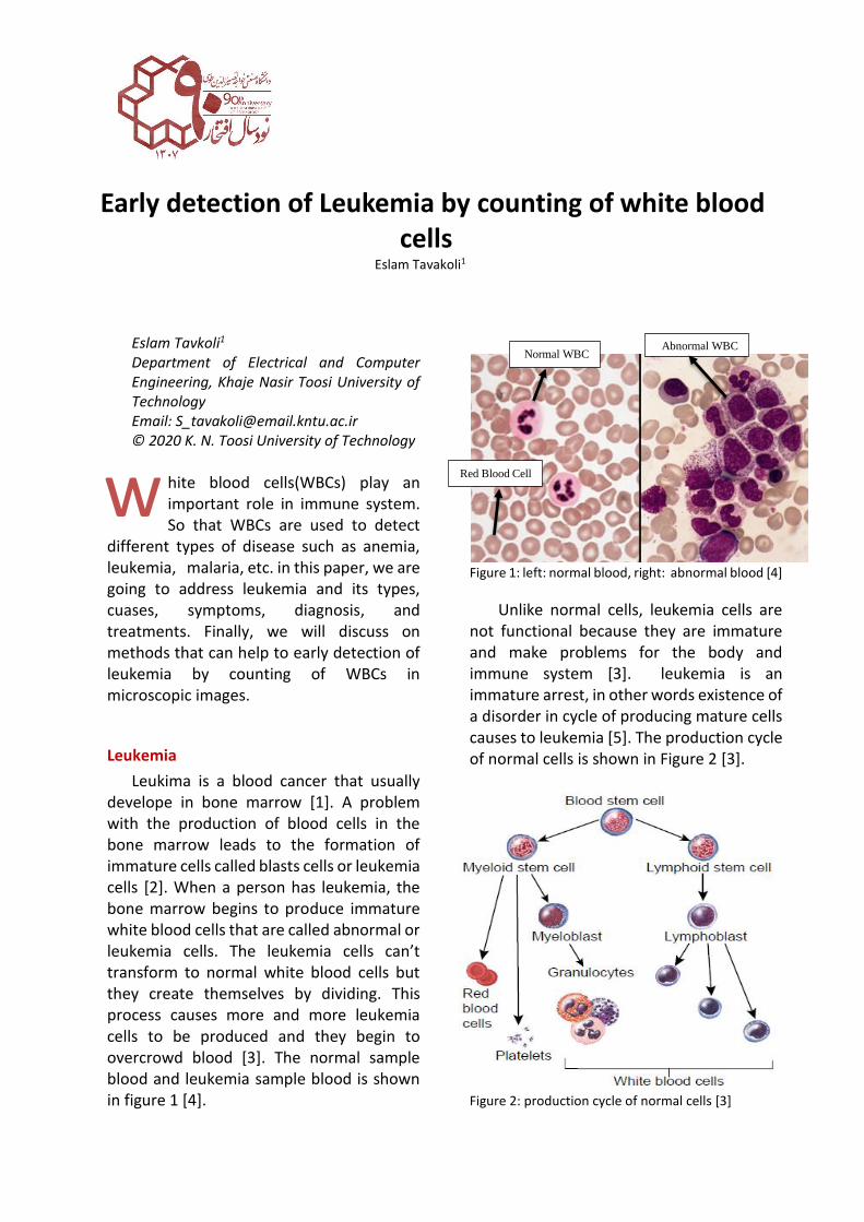
Leukima is a blood cancer that usually develope in bone marrow [1]. A problem with the production of blood cells in the bone marrow leads to the formation of immature cells called blasts cells or leukemia cells [2]. When a person has leukemia, the bone marrow begins to produce immature white blood cells that are called abnormal or leukemia cells. The leukemia cells can’t transform to normal white blood cells but they create themselves by dividing. This process causes more and more leukemia cells to be produced and they begin to overcrowd blood [3]. The normal sample blood and leukemia sample blood is shown in figure 1 [4].
Figure 1: left: normal blood, right: abnormal blood [4]
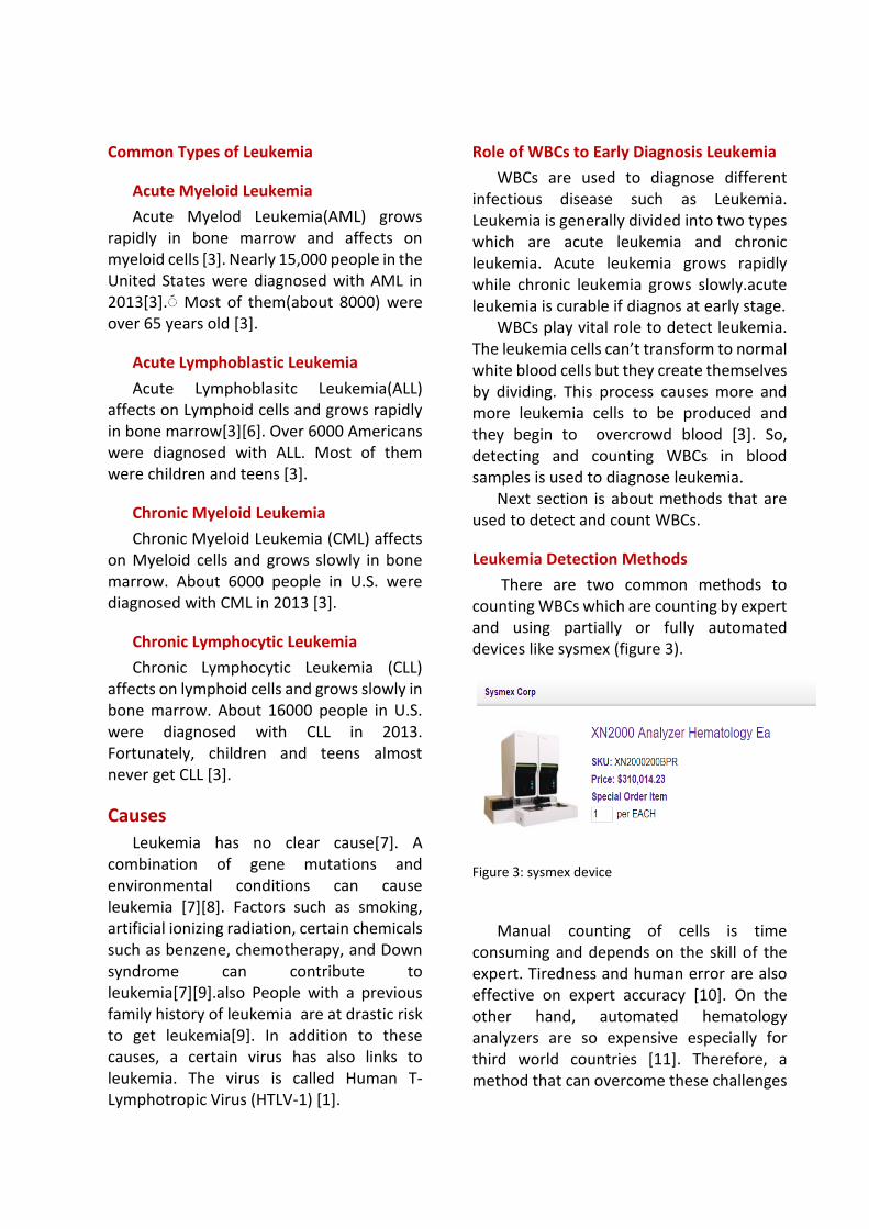
Unlike normal cells, leukemia cells are not functional because they are immature and make problems for the body and immune system [3]. leukemia is an immature arrest, in other words existence of a disorder in cycle of producing mature cells causes to leukemia [5]. The production cycle of normal cells is shown in Figure 2 [3].
Figure 2: production cycle of normal cells [3]
w
Normal WBC Abnormal WBC
Red Blood Cell
Common Types of Leukemia
Acute Myeloid Leukemia
Acute Myelod Leukemia(AML) grows rapidly in bone marrow and affects on myeloid cells [3]. Nearly 15,000 people in the United States were diagnosed with AML in 2013[3].
Most of them(about 8000) were
over 65 years old [3].
Acute Lymphoblastic Leukemia
Acute Lymphoblasitc Leukemia(ALL) affects on Lymphoid cells and grows rapidly in bone marrow[3][6]. Over 6000 Americans were diagnosed with ALL. Most of them were children and teens [3].
Chronic Myeloid Leukemia
Chronic Myeloid Leukemia (CML) affects on Myeloid cells and grows slowly in bone marrow. About 6000 people in U.S. were diagnosed with CML in 2013 [3].
Chronic Lymphocytic Leukemia
Chronic Lymphocytic Leukemia (CLL) affects on lymphoid cells and grows slowly in bone marrow. About 16000 people in U.S. were diagnosed with CLL in 2013. Fortunately, children and teens almost never get CLL [3].
Causes
Leukemia has no clear cause[7]. A combination of gene mutations and environmental conditions can cause leukemia [7][8]. Factors such as smoking, artificial ionizing radiation, certain chemicals such as benzene, chemotherapy, and Down syndrome can contribute to leukemia[7][9].also People with a previous family history of leukemia are at drastic risk to get leukemia[9]. In addition to these causes, a certain virus has also links to leukemia. The virus is called Human T-Lymphotropic Virus (HTLV-1) [1].
Role of WBCs to Early Diagnosis Leukemia
WBCs are used to diagnose different infectious disease such as Leukemia. Leukemia is generally divided into two types which are acute leukemia and chronic leukemia. Acute leukemia grows rapidly while chronic leukemia grows slowly.acute leukemia is curable if diagnos at early stage.
WBCs play vital role to detect leukemia. The leukemia cells can’t transform to normal white blood cells but they create themselves by dividing. This process causes more and more leukemia cells to be produced and they begin to overcrowd blood [3]. So, detecting and counting WBCs in blood samples is used to diagnose leukemia.
Next section is about methods that are used to detect and count WBCs.
Leukemia Detection Methods
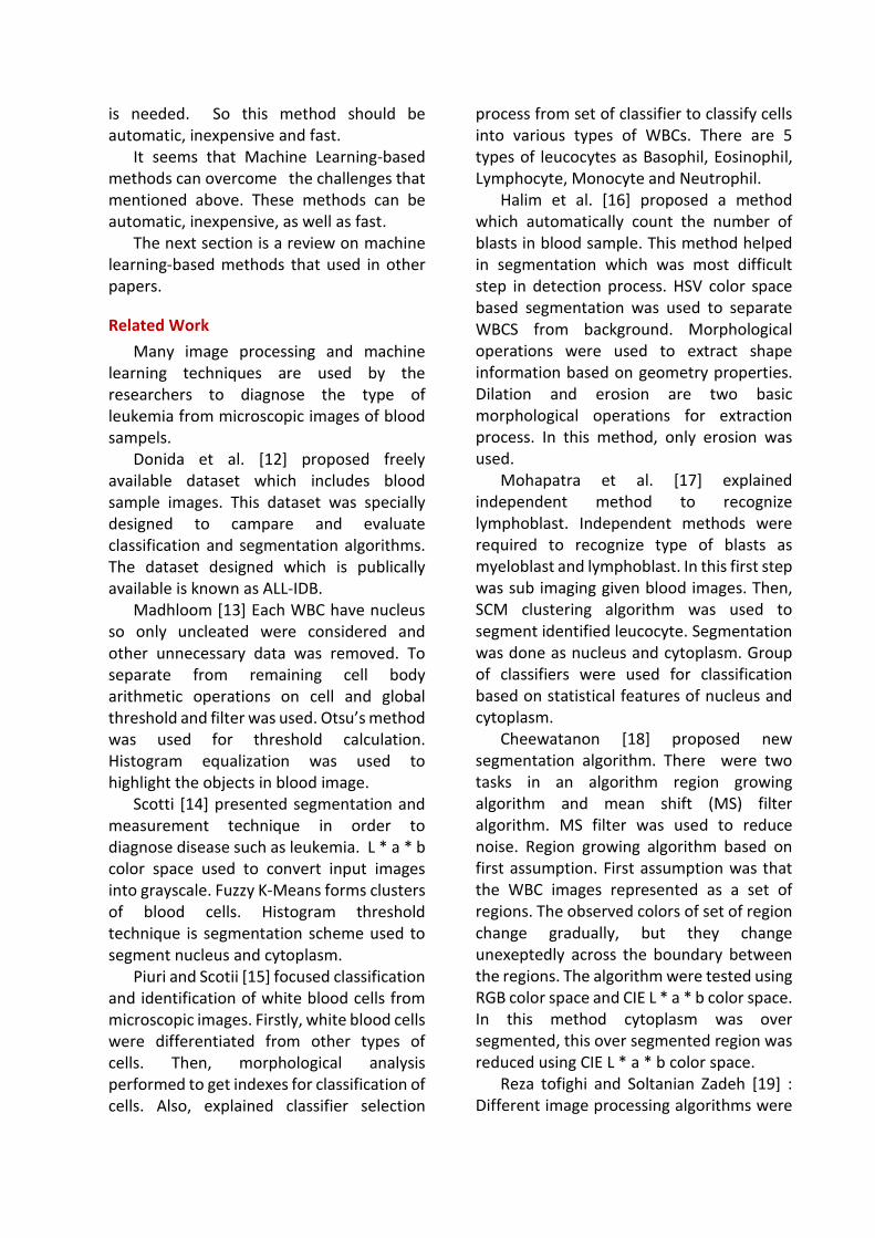
There are two common methods to counting WBCs which are counting by expert and using partially or fully automated devices like sysmex (figure 3).
Figure 3: sysmex device
Manual counting of cells is time
consuming and depends on the skill of the expert. Tiredness and human error are also effective on expert accuracy [10]. On the other hand, automated hematology analyzers are so expensive especially for third world countries [11]. Therefore, a method that can overcome these challenges
is needed. So this method should be automatic, inexpensive and fast.
It seems that Machine Learning-based methods can overcome the challenges that mentioned above. These methods can be automatic, inexpensive, as well as fast.
The next section is a review on machine learning-based methods that used in other papers.
Related Work
Many image processing and machine learning techniques are used by the researchers to diagnose the type of leukemia from microscopic images of blood sampels.
Donida et al. [12] proposed freely available dataset which includes blood sample images. This dataset was specially designed to campare and evaluate classification and segmentation algorithms. The dataset designed which is publically available is known as ALL-IDB.
Madhloom [13] Each WBC have nucleus so only uncleated were considered and other unnecessary data was removed. To separate from remaining cell body arithmetic operations on cell and global threshold and filter was used. Otsu’s method was used for threshold calculation. Histogram equalization was used to highlight the objects in blood image.
Scotti [14] presented segmentation and measurement technique in order to diagnose disease such as leukemia. L * a * b color space used to convert input images into grayscale. Fuzzy K-Means forms clusters of blood cells. Histogram threshold technique is segmentation scheme used to segment nucleus and cytoplasm.
Piuri and Scotii [15] focused classification and identification of white blood cells from microscopic images. Firstly, white blood cells were differentiated from other types of cells. Then, morphological analysis performed to get indexes for classification of cells. Also, explained classifier selection
process from set of classifier to classify cells into various types of WBCs. There are 5 types of leucocytes as Basophil, Eosinophil, Lymphocyte, Monocyte and Neutrophil.
Halim et al. [16] proposed a method which automatically count the number of blasts in blood sample. This method helped in segmentation which was most difficult step in detection process. HSV color space based segmentation was used to separate WBCS from background. Morphological operations were used to extract shape information based on geometry properties. Dilation and erosion are two basic morphological operations for extraction process. In this method, only erosion was used.
Mohapatra et al. [17] explained independent method to recognize lymphoblast. Independent methods were required to recognize type of blasts as myeloblast and lymphoblast. In this first step was sub imaging given blood images. Then, SCM clustering algorithm was used to segment identified leucocyte. Segmentation was done as nucleus and cytoplasm. Group of classifiers were used for classification based on statistical features of nucleus and cytoplasm.
Cheewatanon [18] proposed new segmentation algorithm. There were two tasks in an algorithm region growing algorithm and mean shift (MS) filter algorithm. MS filter was used to reduce noise. Region growing algorithm based on first assumption. First assumption was that the WBC images represented as a set of regions. The observed colors of set of region change gradually, but they change unexeptedly across the boundary between the regions. The algorithm were tested using RGB color space and CIE L * a * b color space. In this method cytoplasm was over segmented, this over segmented region was reduced using CIE L * a * b color space.
Reza tofighi and Soltanian Zadeh [19] : Different image processing algorithms were

proposed to distinguish different types of white blood cells. A method was used for segmentation of cytoplasm and nuceus of cells. Then segmented parts were used for number of feature extraction. Sequentiial Forward Selection algorithm was used to select distinct features from segmented region. Features were extracted based on LBP. Classification algorithms Artificial Neural Networks (ANN) and Support Vector Machine (SVM) are compared which shows SVM was more accurate classification algorithm.
R Muhammad Iman Razzak et al. [20] suggest an automated system to diagnose blood disease. Segmentation process becomes difficult with increasing number of overlapping of cells. Analaysis is done on ALL-IDB dataset [12] which contain 64000 blood cells. Contours aware segmentation is applied on images. It helps segmenting overlapped cells and touching cells. Next step is classification into blood constituents. CNN based extreme machine learning is applied to classify the iamges into different subtypes. Shape features and texture features are used to identity defected RBCs. There is an overall accuracy of 98.14% .
Fatemeh Kazemi et al. [21] presents a techniques to identify subtype of AML. Preprocessing includes selective median filtering and unsharp masking. Input image is converted from RGB color space to CIEL*a*b color space. Then segmentation is done using K-Means clustering algorithm. Cluster with minimum value is considered as the cluster of nuclei. Morphological filtering is applied on segmented images in order to improve perceptibilityand visibility of area of interest. Processes like edge enhancement, canny edge detection, dilation and hole filling is done to get desire output. Classification is done based on Hausdorff dimension, irregularity, elongation, form factor, compactness, N:C ratio (nucleus to cytoplasm), color features, energy entropy, contrast, correlation, and homogeneity.
SVM classifier is used for classification. Variance of Euclidean distances was found to be 462.812 for blast nucleus and 38.87 for normal cells.
Reymond Joseph A. et al. [22] depend on CBC and NC ratio. The complete blood count is the count of WBC in blood, which is estimated to be about 4000 to 10000. If it increase it is an indication of leukemia. Neuclear cytoplasmic ration is also calculated. Preprocessing involves three steps. First, the region of interest can be extracted by multiplying the RGB color planes. Second, binarization is done using Otsu’s method and finally mathematical morphology through dilation is applied. Using watershed algorithm overlapping cells are determined and separated. It calculated the CBC and NC ratio. Next step is applying a genetic algorithm. It includes selection which chooses a value from databes, cross over which combines an image chosen from the input image and from nth generation and mutation which choosen a random value from the available range of characteristics values. This is applied to a Naïve Bayes classifier. It classifies the input images in to leukemic or normal by calculating the likelihood of the neew object. Accuracy is found to be 90.33%
Thi Phuong et al. [23] proposed new article in 2019 to solve problem of counting blood cells. They declared : “ This paper proposes a novel blood cell counting procedure to address this challenge. The proposed method adopts SegNet - a deep learning semantic segmentation to simultaneously segment RBCs and WBCs. The global accuracy of the segmentation of WBCs, RBCs, and the background of peripheral blood smear images obtains 89% when segment WBCs and RBCs from the background of blood smear images. Moreover, an effective solution to separate grouped or overlapping cells and cell count is presented using Euclidean distance transform, local maxima, and connected
component labeling. The counting result of the proposed procedure achieves an accuracy of 93.3% for red blood cell count using dataset 1 and 97.38% for white blood cell count using dataset 2 “ .
Generalizability
In the previous section, we looked at some works that were performed to count cells. All these works led to good result, but there is an important gap. These works just evaluated their methods on one dataset such as ALL-IDB [12]. In fact, none of these works have examined the generalizability of their methods and didn’t evaluate their methods on various datasets.
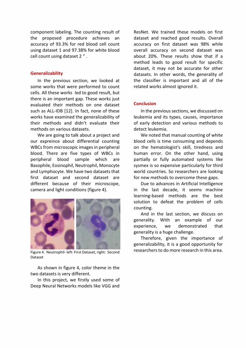
We are going to talk about a project and our expreince about differential counting WBCs from microscopic images in peripheral blood. There are five types of WBCs in peripheral blood sample which are Basophile, Eosinophil, Neutrophil, Monocyte and Lymphocyte. We have two datasets that first dataset and second dataset are different because of their microscope, camera and light conditions (figure 4).
Figure 4. Neutrophil- left: First Dataset, right: Second Dataset
As shown in figure 4, color theme in the two datasets is very different.
In this project, we firstly used some of Deep Neural Networks models like VGG and
ResNet. We trained these models on first dataset and reached good results. Overall accuracy on first dataset was 98% while overall accuracy on second dataset was about 20%. These results show that if a method leads to good result for specific dataset, it may not be accurate for other datasets. In other words, the generality of the classifier is important and all of the related works almost ignored it.
Conclusion
In the previous sections, we discussed on leukemia and its types, causes, importance of early detection and various methods to detect leukemia.
We noted that manual counting of white blood cells is time consuming and depends on the hematologist's skill, tiredness and human error. On the other hand, using partially or fully automated systems like sysmex is so expensive particularly for third world countries. So researchers are looking for new methods to overcome these gaps.
Due to advances in Artificial Intelligence in the last decade, it seems machine learning-based methods are the best solution to defeat the problem of cells counting.
And in the last section, we discuss on generality. With an example of our experience, we demonstrated that generality is a huge challenge.
Therefore, given the importance of generalizability, it is a good opportunity for researchers to do more research in this area.
References
[1] Weblink: https://www.medicalnewstoday.com/articles/142595#treatment
[2] Weblink: https://en.wikipedia.org/wiki/Leukemia#cite_note-NCIBook2013-2
[3] Textbook: “ what you need to know about Leukemia ”, National Cancer Institute [4] Weblink: http://schroedergen677s11.weebly.com/ [5] Li, Peng; Yan, Shuxin; Dong, Xin; Li, Zhao; Qiu, Yu; Ji, Chunyan; Zhang, Jingru; Ji, Min; Li, Wei; Wang, Hongchun; Liu, Zhi; Wang, Xing L.; Ye, Jingjing; Ma, Daoxin , “ cell cycle arrest and apoptosis induction activity of nitidine chloride on acute myloid leukiemia cells“ , Medicinal Chemistry, Volume 14, Number 1, pp. 60-66(7), 2018 [6] T. Terwilliger, M. Abdul-Hay, “ Acute lymphoblastic leukemia: a comprehensive review and 2017 update“, Blood Cancer Journal, 2017 [7] Blackadar CB. , “Historical review of the causes of cancer”, world journal of clinical oncology; 7(1): 54-86, 2016 [8] H. M. Idris, A. Y. Elderdery, H. B. Khalil, J. J. Mills, “ Genetic Polymorphism of GSTp1,GSTM1 Genes and Susceptibility to Chronic Myeloid Luekemia”, Asian Pacific journal of cancer prevention, 2020 [9] Report: “ A Snapshot of Leukemia “ , National Cancer Institute, 2014 [10] R. T Raphael, K. R Joy, “ Segmentation and classification techniqes of leukemia using image processing: an overview “ , international conference on intelligent sustainable systems(ICISS),IEEE Xplore,2019 [11] N. H. Harun, N. A. AbuBakar, U. A. Mohan, M. M. Nadzir, M. G. Hassan, R. Adollah, “ Automated Cell Counting System For Chronic Leukemia”, IEEE Jordan International Joint Conference on Electrical and Information Technology(JEEIT), 2019 [12] Labati RD, Piuri V, Scotti F , “ ALL-IDB: the acute lymphoblastic leukemia image data base for image processing ”, In: Benot M, Schelkens P (eds) Proceedings of the 18th IEEE ICIP international conference on image processing, 11–14 Sept. IEEE Publisher, Brussels, Belgium, pp 2045–2048, 2011 [13] Madhloom HT, Kareem SA, Ariffin H, Zaidan AA, Alanazi HO, Zaidan BB,”Anautomated white blood cell nucleus localization and segmentation using image arithmetic and automated threshold ”, J Appl Sci 10(11):959–966 , 2010 [14] Scotti F, “ Robust segmentation and measurements techniques of white cells in blood microscope images”, In: Daponte P, Linnenbrink T, editors. In: Proceedings of the IEEE instrumentation and measurement technology conference, 24–27 Apr. IEEE Publisher,
Sorrento, Italy, pp 43–48 , 2006 [15] Piuri V, Scotti F , “ Morphological classification of blood leucocytes by microscope Images “, In: Proceedings of the IEEE international conference on computational intelligence for measurement systems and applications, 14–16 July. IEEE Publisher, Boston, MA, USA, pp 103–108 , 2004 [16] Halim NHA, Mashor MY, Hassan R, “ Automatic blasts counting for acute leukemia based on blood samples ”, Int J Res Rev Comput Sci 2(4) , 2011 [17] Mohapatra S, Patra D, Satpathy S, “ An ensemble classifier system for early diagnosis of acute lymphoblastic leukemia in blood microscopic images”, J Neural Comput Appl 24:1887–1904 , 2014 [18] Cheewatanon J, Leauhatong T, Airpaiboon S, Sangwarasilp M, “ A new white blood cell segmentation using mean shift filter and region growing algorithm”, Int J Appl Biomed Eng 4:30–35, 2011 [19] Rezatofighia SH, Soltanian-Zadeh H, “ Automatic recognition of five types of white blood cells in peripheral blood” , Comput Med Imag Graph 35:333–343 , 2011 [20] M. I. Razzak,S. Naz, “Microscopic Blood Smear Segmentation and Classification using Deep Contour Aware CNN and Extreme Machine Learning”, IEEE Conference on Computer Vision and Pattern Recognition Workshops, 2017
[21] F. Kazmi, T. A. Najafabadi,B. N. Araabi ," Automatic recognition of Acute Myelogenous Leukemia in blood microscopic images using K-means clustering and support vector machine" , Journal of Medical signals and sensors, 2016 [22] Reymond Joseph A. Cabrera, Criselle Amor P. Legaspi, Erika Jasmine G. Papa, Reden D. Samonte, Donata D. Acula " HeMatic: An Automated Leukemia Detector With Separation of Overlapping Blood Cells through Image Processing and Genetic Algorithm" , IEEE International Conference on Applied System Innovation IEEE-ICASI 2017 - Meen, Prior & Lam (Eds). 2017 [23] Tran, T.; Binh Minh, L.; Lee, S.; Kwon, K. Blood Cell Count Using Deep Learning Semantic Segmentation. Preprints, 2019090075 (doi: 10.20944/preprints201909.0075.v1), 2019