drain in eubacteria
TRANSCRIPT
Vol. 168, No. 3JOURNAL OF BACTERIOLOGY, Dec. 1986, p. 1147-11540021-9193/86/121147-08$02.00/0Copyright © 1986, American Society for Microbiology
Comparative Action of Glyphosate as a Trigger of EnergyDrain in Eubacteria
RANDY S. FISCHER, ALAN BERRY, C. GREG GAINES, AND ROY A. JENSENt*Center for Somatic-cell Genetics and Biochemistry, State University ofNew York, Binghamton, New York 13901
Received 1 May 1986/Accepted 10 September 1986
Escherichia coli, Bacillus subtilis, and Pseudomonas aeruginosa, each possessing a 5-enolpyruvylshikimate3-phosphate synthase that is sensitive to inhibition by glyphosate [N-(phosphonomethyl)glycitie], provide a goodcross-section of organisms exemplifying the biochemical diversity of the aromatic pathway targeted by thispotent antimicrobial compound. The pattern of growth inhibition, the alteration in levels of aromatic-pathwayenzynies, and the accumulation of early-pathway metabolites after the addition of glyphosate were distinctivefor each organism. Substantial intracellular shikimate-3-phosphate accumulated in response to glyphosatetreatment in all three organisms. Both E. coli and P. aeruginosa, but not B. subtilis, accumulatednear-millimolar levels of shikimate-3-phosphate in the culture medium. Intracellular backup of common-pathway precursors of shikimate-3-phosphate was substantial in B. subtilis, moderate in P. aeruginosa, and notdetectable in E. coli. The full complement of aromatic amino acids prevented growth inhibition and metaboliteaccumulation in E. coli and P. aeruginosa where amino acid end products directly control early-pathwayenzyme activity. In contrast, the initial prevention of growth inhibition in the presence of aromatic amino acidsin B. subtilis was succeeded by progressively greater growth inhibition that correlated with rapid metaboliteaccumulation. In B. subtilis glyphosate can decrease prephenate concentrations sufficiently to uncouple thesequentially acting loops of feedback inhibition that ordinarily link end product excess to feedback inhibitionof 3-deoxy-D-arabino-heptulosonate 7-phosphate synthase by prephenate. The consequential unrestrained entryof energy-rich substrates into the aromatic pathway, even in the presence of aromatic amino acid end products,is an energy drain that potentially accounts for the inability of end products to fully reverse glyphosateinhibition in B. subtilis. Even in E. coi, after glyphosate inhibition and metabolite accumulation were allowedto become fully established, a transient period where end products were capable of only partial reversal ofgrowth inhibition occurred. The distinctive metabolism produced by dissimilation of different carbon sourcesalso produced profound effects upon glyphosate sensitivity.
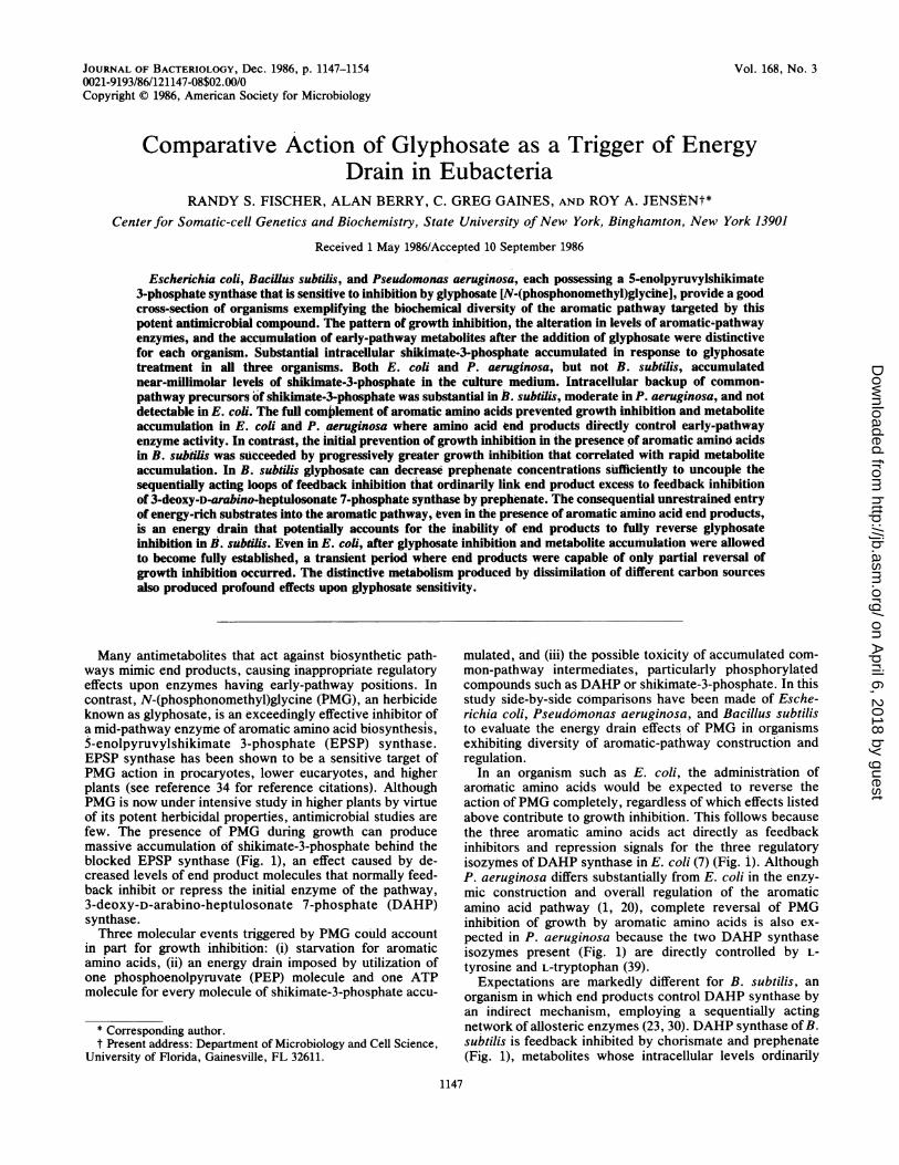
Many antimetabolites that act against biosynthetic path-ways mimic end products, causing inappropriate regulatoryeffects upon enzymes having early-pathway positions. Incontrast, N-(phosphonomethyl)glycine (PMG), an herbicideknown as glyphosate, is an exceedingly effective inhibitor ofa mid-pathway enzyme of aromatic amino acid biosynthesis,5-enolpyruvylshikimate 3-phosphate (EPSP) synthase.EPSP synthase has been shown to be a sensitive target ofPMG action in procaryotes, lower eucaryotes, and higherplants (see reference 34 for reference citations). AlthoughPMG is now under intensive study in higher plants by virtueof its potent herbicidal properties, antimicrobial studies arefew. The presence of PMG during growth can producemassive accumulation of shikimate-3-phosphate behind theblocked EPSP synthase (Fig. 1), an effect caused by de-creased levels of end product molecules that normally feed-back inhibit or repress the initial enzyme of the pathway,3-deoxy-D-arabino-heptulosonate 7-phosphate (DAHP)synthase.Three molecular events triggered by PMG could account
in part for growth inhibition: (i) starvation for aromaticamino acids, (ii) an energy drain imposed by utilization ofone phosphoenolpyruvate (PEP) molecule and one ATPmolecule for every molecule of shikimate-3-phosphate accu-
* Corresponding author.t Present address: Department of Microbiology and Cell Science,
University of Florida, Gainesville, FL 32611.
mulated, and (iii) the possible toxicity of accumulated com-mon-pathway intermediates, particularly phosphorylatedcompounds such as DAHP or shikimate-3-phosphate. In thisstudy side-by-side comparisons have been made of Esche-richia coli, Pseudomonas aeruginosa, and Bacillus subtilisto evaluate the energy drain effects of PMG in organismsexhibiting diversity of aromatic-pathway construction andregulation.
In an organism such as E. coli, the administration ofaromatic amino acids would be expected to reverse theaction of PMG completely, regardless of which effects listedabove contribute to growth inhibition. This follows becausethe three aromatic amino acids act directly as feedbackinhibitors and repression signals for the three regulatoryisozymes of DAHP synthase in E. coli (7) (Fig. 1). AlthoughP. aeruginosa differs substantially from E. coli in the enzy-mic construction and overall regulation of the aromaticamino acid pathway (1, 20), complete reversal of PMGinhibition of growth by aromatic amino acids is also ex-pected in P. aeruginosa because the two DAHP synthaseisozymes present (Fig. 1) are directly controlled by L-tyrosine and L-tryptophan (39).
Expectations are markedly different for B. subtilis, anorganism in which end products control DAHP synthase byan indirect mechanism, employing a sequentially actingnetwork of allosteric enzymes (23, 30). DAHP synthase of B.subtilis is feedback inhibited by chorismate and prephenate(Fig. 1), metabolites whose intracellular levels ordinarily
1147
on April 6, 2018 by guest
http://jb.asm.org/
Dow
nloaded from
1148 FISCHER ET AL.
4 steps
ANT
;.E4P ie 1 X 3steps 2 3 4 6DAHP -.S K-~3 PSP- CHA- PPA---0-.AGN
8HPP ---*T YR
PMG
Escherich'a co/i Psuooa euioaBacillus subtilis
: T YRO SI N E -20; 00i SNE000
PHNYLALANINE'f~ET77777 TYPTPHAN
'-TRYPTOPHAN.
FIG. 1. Allosteric control of DAHP synthase and PMG inhibition of EPSP synthase in E. coli, B. suhtilis, and P. aeruginosa. Thewedge-shaped symbol indicates EPSP synthase as the target of PMG action, and the shaded area portrays the comparative allostery of DAHPsynthase in the three microbes. E. coli possesses three isozymes of DAHP synthase, each differentially controlled by one of the aromaticamino acids (6). P. aeruginosa possesses two isozymes of DAHP synthase, a major species that is feedback inhibited by L-tyrosine and aminor species that is feedback inhibited by L-tryptophan or by chorismate (39). B. subtilis possesses a single DAHP synthase enzyme, subjectto feedback inhibition by prephenate (23). Enzymes are numbered as follows: 1, DAHP synthase; 2, shikimate kinase; 3, EPSP synthase; 4,chorisniate synthase; 5, anthranilate synthase; 6, chorismate mutase; 7, prephenate dehydratase; 8, prephenate dehydrogenase; 9, arogenatedehydratase; 10, arogenate dehydrogenase. Abbreviations: E4P, D-erythrose-4-phosphate; SHK, shikimate; S3P, shikimate-3-phosphate;CHA, chorismate; ANT, anthranilate; TRP, L-tryptophan; PPA, prephenate; PPY, phenylpyruvate; PHE, L-phenylalanine; AGN,L-arogenate; HPP, 4-hydroxyphenylpyruvate; TYR, L-tyrosine.
reflect overall end product levels. In this study we pursue thequestion whether PMG (through prevention of chorismateand prephenate formation in B. subtilis) has the potential touncouple the normal control of DAHP synthase by aromaticamino acid end products.
MATERIALS AND METHODS
Microorganisms. All bacterial strains used were wild-typeprototrophs. E. coli K-12 EMG2 (3, 13) was obtained fromB. Bachmann of the E. coli Genetic Stock Center, Depart-ment of Human Genetics, Yale University School of Medi-cine, New Haven, Conn. P. aeruginosa PAO1 (19) and B.subtilis 168 strain NP1 (27) were from the culture collectionof our laboratory.Growth of bacteria and culture conditions. E. coli was
grown in M9 minimal medium (29) containing 0.2% (wt/vol)glucose. B. subtilis was grown on the minimal medium ofSpizizen (36), containing glucose (0.2%, wt/vol) as the car-bon source. In addition, both media were supplemented withZnCl2, MnCl2 4H20, And CoCl2 6H20 at final concentra-tions of 1, 2, and 7 pug/ml, respectively. P. aeruginosa wasgrown in a tninimal salts medium containing (per liter) 7 g ofK2HPO4, 3 g of KH2PO4, 0.1 g of MgSO4- 7H20, 1 g of(NH4)2504, and 0.5% (wt/vol) fructose as the carbon source.Agar (Difco Laboratories, Detroit, Mich.) was added to givea final concentration of 1.5% (wt/vol) for solid media.
Culture turbidities for growth studies were determinedwith a Klett-Summerson colorimeter (Klett ManufacturingCo., Inc., New York, N.Y.) equipped with a no. 54 greenfilter for P. aeruginosa or a no. 66 red filter for E. coli and B.subtilis. Exponentially growing cultures were used to inoc-ulate either 20 ml (for E. coli and B. subtilis) or 10 ml (for P.aeruginosa) of fresh mnedium containing supplements asindicated. These cultures were grown in 125-ml sidearmflasks with vigorous shaking at 37°C.
Extracts were prepared from 1,000-mnl cultures containedin 2,800-ml Fernbach flasks which were shaken at 300 rpm at37°C. Cultures in the late-exponential phase of growth were
harvested by centrifugation, washed twice with cold 50 mMpotassium phosphate buffer (pH 7.0), and stored as wholecell pellets at -20°C.
Preparation of crude extracts. Extracts for DAHP synthasedeterminations were prepared in 50 mM potassium phos-phate buffer (pH 7.0) containing either 1 mM MgCl2 and 100mM KCl for B. subtilis or 1 mM dithiothreitol for E. coli. AllEPSP synthase activities were determined from extractsprepared in 25 mM HEPES (N-2-hydroxyethylpiperazine-N'-2-ethanesulfonic acid) buffer (pH 7.5 at 37°C). The pH ofthe HEPES buffer was adjusted with tetramethylammoniumhydroxide to avoid the introduction of cations. The extractfrom E. coli to be used for anthranilate synthase determina-tions was prepared in 10 mM Tris hydrochloride buffer (pH7.8 at 25°C) containing 10 mM MgCl2 and 10 mM 2-mercaptoethanol. Extracts from B. subtilis to be used foranthranilate synthase assays were prepared in the bufferdescribed by Kane et al. (25), except that 20 mM L-glutaminewas added to the extract buffer. The extracts for determiningprephenate dehydrogenase activities were prepared in 100mM Tris hydrochloride buffer (pH 8.5 at 25°C) containing 50mM KCl plus 0.5 mM dithiothreitol.
Cell pellets, suspended in 3 to 5 ml of the required buffer,were disrupted by either ultrasound or lysozyme. For E. coliand P. aeruginosa, cells were disrupted at 4°C with three 20-and 30-s bursts, respectively, of ultrasound with an intensityof 100 W from a Lab-Line Ultratip Labsonic System (Lab-Line Instruments, Inc., Melrose Park, Ill.). Sonicationlargely inactivated EPSP synthase, anthranilate synthase,and prephenate dehydrogenase in B. subtilis crude extracts,and only extracts of B. subtilis to be used for DAHPsynthase assays could be prepared by sonication. With B.subtilis two 20-s bursts at 100 W were sufficient to achieve adegree of disruption comparable to that obtained with E. coliand P. aeruginosa. B. subtilis extracts to be used for EPSPsynthase, anthranilate synthase, and prephenate dehydroge-nase assays were prepared by gentle lysis with lysozyme(100 ,ugIml) at 37°C for 15 min. DNase at a final concentra-tion of 25 ,ug/ml was added to degrade DNA. The disrupted
J. BACTERIOL.
on April 6, 2018 by guest
http://jb.asm.org/
Dow
nloaded from

ENERGY DRAIN IN BACTERIA 1149
cell suspensions were ultracentrifuged at 145,000 x g for 60min at 4°C to remove cell debris. With the exception of theextracts used for assay of EPSP synthase from B. subtilisand P. aeruginosa, the resulting supernatants were thendesalted by passage over a Sephadex G-25 column (PD-10;Pharmacia Fine Chemicals, Uppsala, Sweden). The B. sub-tilis and P. aeruginosa extracts used for EPSP synthaseassays were extensively dialyzed against 25 mM HEPESbuffer (adjusted to pH 7.5 as described above) to ensureremoval of potential activating cations.
Analytical procedures. Protein concentrations were esti-mated by the method of Bradford (5) as described in Bio-Radtechnical bulletin 1051 (Bio-Rad Laboratories, Richmond,Calif.). DAHP synthase was assayed by the method ofSrinivasan and Sprinson (37) as modified by Jensen andNester (24). Reaction mixtures for the B. subtilis enzymewere prepared as described by Jensen and Nester (24), butwere modified for E. coli to contain 50 mM potassiumphosphate buffer (pH 7.0) containing 1 mM dithiothreitol, 1mM PEP, 1 mM D-erythrose-4-phosphate, and 1 mMMgSO4. Anthranilate synthase from B. subtilis was assayedby the method of Kane and Jensen (26), whereas anthranilatesynthase from E. coli was assayed as described by Calhounet al. (11). EPSP synthase was assayed by following thedisappearance of PEP (17) as modified by Rubin et al. (34),except that standard reaction mixtures contained 3 mM PEP,3 mM shikimate-3-phosphate, 25 mM HEPES buffer (pH 7.5at 37°C), and the necessary activating cation as designatedbelow. Prephenate dehydrogenase was assayed as describedby Champney and Jensen (12) for B. subtilis, whereas themethod of Byng et al. (9) was used for E. coli.
Determination of metabolite levels. For quantitation ofintracellular metabolites, whole cell pellets were washedtwice with 50 mM potassium phosphate buffer (pH 7.0),resuspended in the buffer, and disrupted by sonication (four30-s bursts at 100 W). Cell debris was removed from theextract by ultracentrifugation at 145,000 x g for 60 min at4°C. The protein concentration of each extract was thendetermined, and accumulated DAHP was quantitated by themethod of Jensen and Nester (23). To determine intracellularlevels of shikimate and shikimate-3-phosphate, samples ofeach extract were heated in a 100°C water bath for 2 min,centrifuged at 12,000 x g for 10 min, and passed through a0.2-p,m polycarbonate membrane (Nuclepore Corp.,Pleasanton, Calif.) to remove most of the protein, nucleicacid, and particulates. Shikimate and shikimate-3-phosphatelevels were determined in these preparations by using high-performance liquid chromatography. Samples were injectedinto a 20-,ul loop and eluted from a Baker Amino column (4.6by 250 mm, 5-jxm particle size) (J.T. Baker Chemical Co.,Phillipsburg, N.J.) with a mobile phase consisting of acetoni-trile-H20-phosphoric acid (95:4:1) at a flow rate of 1.0ml/min. Shikimate and shikimate-3-phosphate were detectedat 215 nm and quantitated using a standard curve generatedfrom the recorded peak heights obtained with authenticshikimate and shikimate-3-phosphate.
Extracellular shikimate and shikimate-3-phosphate levelswere determined directly from the cell-free media afterharvesting. A sample of each medium was passed through a0.4-ram polycarbonate membrane (Nuclepore) before proc-essing as described above for measuring intracellular accu-mulation.
Biochemicals and chemicals. Unless indicated otherwise,all biochemicals and commercially prepared enzymes wereobtained from Sigma Chemical Co., St. Louis, Mo. Analyt-ical-grade PMG (99.9% pure) was a gift from Monsanto Co.,
z
-LJ
50-
mM PMG
10-~~~~~0
TIME (HRS)
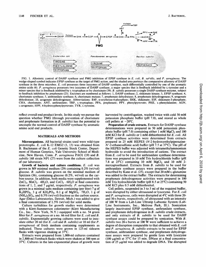
FIG. 2. PMG inhibition of growth of E. coli. Growth conditionswere outlined in Materials and Methods. PMG concentrations: 0,none;O0,0.5SmM;-A,2.5mM;A, 5.0mM;U,7.5 mM;LI, 10.0OmM;@, 15.0 mM. The inset shows the doubling times plotted as afunction of PMG concentration.
St. Louis, Mo. Shikimate-3-phosphate was prepared aspreviously described (28), except that it was converted to atetramethylammonium salt instead of the conventional so-dium or potassium salt. Barium prephenate was preparedfrom culture supernatants of a tyrosine auxotroph of Salmo-nella typhimurium (14) and was converted to the potassiumsalt with a twofold excess of K204 before use. Chorismatewas prepared from culture supernatants of Klebsiella pneu-moniae by the method of Gibson (16). Acetonitrile andphosphoric acid for high-performance liquid chromatogra-phy and the standard reagent-grade chemicals were obtainedfrom Fisher Scientific Co., Springfield, N.J.
RESULTS
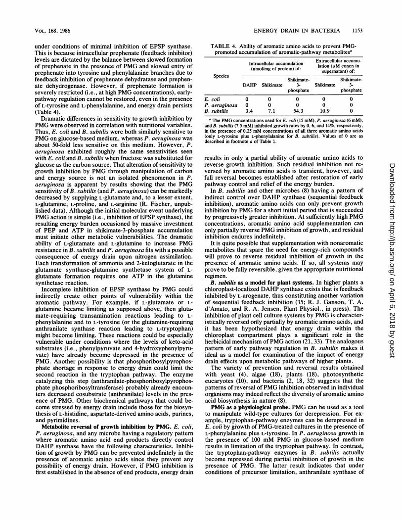
Inhibition of growth by PMG. PMG was capable of exert-ing effective antimetabolite action against all three microor-ganisms under study. Figure 2 illustrates the family ofgrowth curves generated in E. ccli in the presence of PMGconcentrations ranging between 0.5 and 15 mM. Care wastaken to ensure that experiments were started with exponen-tially dividing cell populations. Reports of initial growth lagsafter the addition of PMG (2, 32) probably resulted from the
VOL. 168, 1986
on April 6, 2018 by guest
http://jb.asm.org/
Dow
nloaded from
1150 FISCHER ET AL.
25-
1-10.~~~~~~~~~1
1 2 3 4 5 6 7 8 9 10
TIME (HRS)
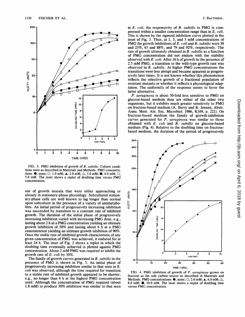
FIG. 3. PMG inhibition of growth of B. subtilis. Culture condi-
tions were as described in Materials and Methods. PMG concentra-
tions: 0, none; 0, 1.0 mM; A, 2.0 mM; A, 3.0 mM; *, 4.0 mM; O,5.0 mM. The inset shows a replot of doubling time versus PMG
concentration.
use of growth inocula that were either approaching or
already in stationary-phase physiology. Subcultured station-
ary-phase cells are well known to lag longer than normal
upon subculture in the presence of a variety of antimetabo-
lites. An initial period of progressively increasing inhibition
was succeeded by transition to a constant rate of inhibited
growth. The duration of the initial phase of progressively
increasing inhibition varied with increasing PMG dose, e.g.,
lasting about 2 h at a PMG concentration yielding an ultimate
growth inhibition of 50% and lasting about 6 h at a PMG
concentration yielding an ultimate growth inhibition of 90%.
Once the stable rate of inhibited growth characteristic of any
given concentration of PMG was achieved, it endured for at
least 24 h. The inset of Fig. 2 shows a replot in which the
doubling time eventually achieved is plotted against PMG
concentration. About 2 mM PMG was required to inhibit the
growth rate of E. ccli by 50%.
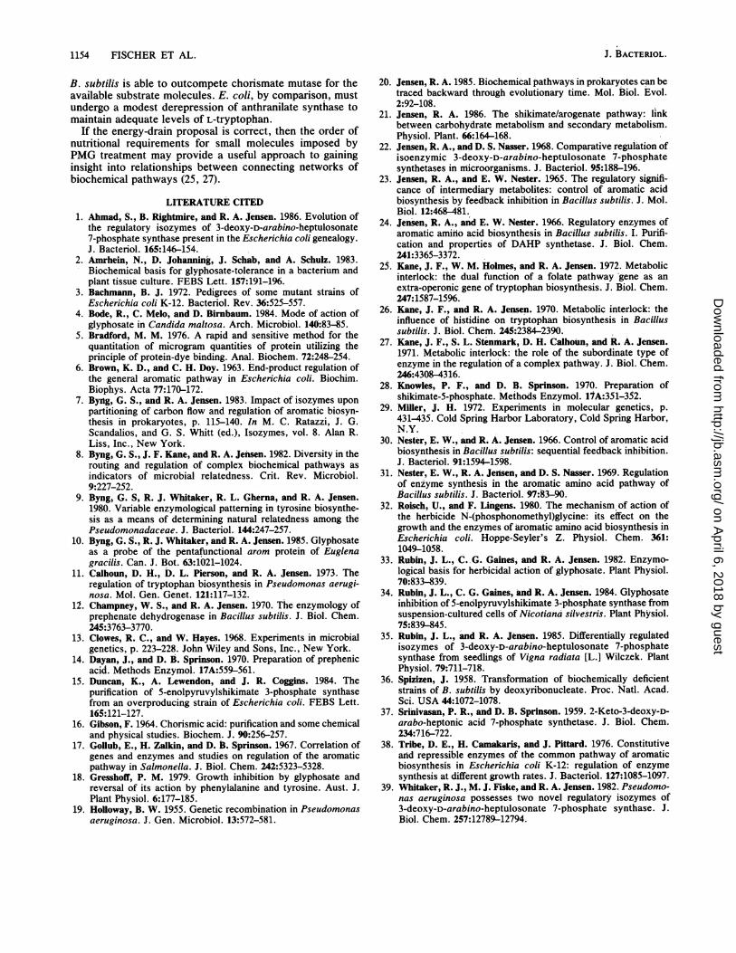
The family of growth curves generated in B. subtilis in the
presence of PMG is shown in Fig. 3. An initial phase of
progressively increasing inhibition similar to that seen in E.
coli was observed, although the time required for transition
to a stable rate of inhibited growth appeared to be shorter,
e.g., no longer than 4 h at the highest PMG concentration
used. Although the concentration of PMG required (about
1.8 mM) to produce 50% inhibition was similar to that seen
in E. coli, the responsivity of B. subtilis to PMG is com-pressed within a smaller concentration range than in E. coli.This is shown by the sigmoid inhibition curve plotted in theinset of Fig. 3. Thus, at 1, 3, and 5 mM concentrations ofPMG the growth inhibitions of E. coli and B. subtilis were 30and 25%, 65 and 88%, and 79 and 92%, respectively. Therate of growth ultimately obtained in B. subtilis as a functionof PMG concentration did not endure with the stabilityobserved with E. coli. After 16 h of growth in the presence of2.5 mM PMG, a transition to the wild-type growth rate wasobserved in B. subtilis. At higher PMG concentrations thetransitions were less abrupt and became apparent at progres-sively later times. It is not known whether this phenomenonreflects the selective growth of a fractional population ofresistant mutants or whether it reflects a physiological adap-tation. The uniformity of the response seems to favor thelatter alternative.
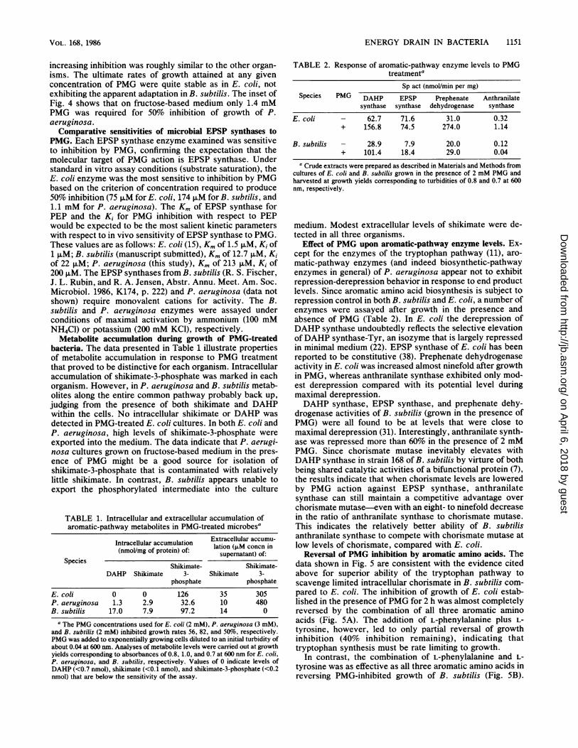
P. aeruginosa is about 50-fold less sensitive to PMG onglucose-based medium than are either of the other twoorganisms, but it exhibits much greater sensitivity to PMGon fructose-based medium (A. Berry and R. Jensen, Abstr.Annu. Meet. Am. Soc. Microbiol. 1986, K169, p. 221). Onfructose-based medium the family of growth-inhibitioncurves generated for P. aeruginosa was similar to thoseobtained with E. coli and B. subtilis on glucose-basedmedium (Fig. 4). Relative to the doubling time on fructose-based medium, the duration of the period of progressively
500-
400 C
300-
200-
100-
0 2
versusPMG con~~~cnrton
H~~~~~z~~~~~~~5
H~~~~~0 2' 4 6 1
010 20 30 40 50 ~~~~~160
TIME (HRS)FIG. 4. PMG inhibition of growth of P. aeruginosa grown on
fructose as the sole carbon source as described in Materials andMethods. PMG concentrations: @, none; 0, 2.0 mM; A, 6.0 mM; A,8.0 mM;E0, 10.0 mM. The inset shows a replot of doubling timeversus PMG concentration.
J. BACTERIOL.
on April 6, 2018 by guest
http://jb.asm.org/
Dow
nloaded from
ENERGY DRAIN IN BACTERIA 1151
increasing inhibition was roughly similar to the other organ-
isms. The ultimate rates of growth attained at any givenconcentration of PMG were quite stable as in E. coli, notexhibiting the apparent adaptation in B. subtilis. The inset ofFig. 4 shows that on fructose-based medium only 1.4 mMPMG was required for 50% inhibition of growth of P.aeruginosa.Comparative sensitivities of microbial EPSP synthases to
PMG. Each EPSP synthase enzyme examined was sensitiveto inhibition by PMG, confirming the expectation that themolecular target of PMG action is EPSP synthase. Understandard in vitro assay conditions (substrate saturation), theE. coli enzyme was the most sensitive to inhibition by PMGbased on the criterion of concentration required to produce50% inhibition (75 ,uM for E. coli, 174 ,uM for B. subtilis, and1.1 mM for P. aeruginosa). The Km of EPSP synthase forPEP and the Ki for PMG inhibition with respect to PEPwould be expected to be the most salient kinetic parameterswith respect to in vivo sensitivity of EPSP synthase to PMG.These values are as follows: E. coli (15), Km of 1.5 FxM, Ki of1 ,uM; B. subtilis (manuscript submitted), Km of 12.7 ,uM, Kiof 22 ,uM; P. aeruginosa (this study), Km of 213 ,uM, Ki of200 ,iM. The EPSP synthases from B. subtilis (R. S. Fischer,J. L. Rubin, and R. A. Jensen, Abstr. Annu. Meet. Am. Soc.Microbiol. 1986, K174, p. 222) and P. aeruginosa (data notshown) require monovalent cations for activity. The B.subtilis and P. aeruginosa enzymes were assayed underconditions of maximal activation by ammonium (100 mMNH4Cl) or potassium (200 mM KCl), respectively.
Metabolite accumulation during growth of PMG-treatedbacteria. The data presented in Table 1 illustrate propertiesof metabolite accumulation in response to PMG treatmentthat proved to be distinctive for each organism. Intracellularaccumulation of shikimate-3-phosphate was marked in eachorganism. However, in P. aeruginosa and B. subtilis metab-olites along the entire common pathway probably back up,judging from the presence of both shikimate and DAHPwithin the cells. No intracellular shikimate or DAHP was
detected in PMG-treated E. coli cultures. In both E. coli andP. aeruginosa, high levels of shikimate-3-phosphate were
exported into the medium. The data indicate that P. aerugi-nosa cultures grown on fructose-based medium in the pres-ence of PMG might be a good source for isolation ofshikimate-3-phosphate that is contaminated with relativelylittle shikimate. In contrast, B. subtilis appears unable toexport the phosphorylated intermediate into the culture
TABLE 1. Intracellular and extracellular accumulation ofaromatic-pathway metabolites in PMG-treated microbesa
Extracellular accumu-Intracellular accumulation Eationular ccnmin(nmol/mg of protein) of: supernatant) of:
SpeciesShikimate- Shikimate-
DAHP Shikimate 3- Shikimate 3-phosphate phosphate
E. coli 0 0 126 35 305P. aeruginosa 1.3 2.9 32.6 10 480B. subtilis 17.0 7.9 97.2 14 0
a The PMG concentrations used for E. coli (2 mM), P. aeruginosa (3 mM),and B. subtilis (2 mM) inhibited growth rates 56, 82, and 50%o, respectively.PMG was added to exponentially growing cells diluted to an initial turbidity ofabout 0.04 at 600 nm. Analyses of metabolite levels were carried out at growthyields corresponding to absorbances of 0.8, 1.0, and 0.7 at 600 nm for E. coli,P. aeruginosa, and B. subtilis, respectively. Values of 0 indicate levels ofDAHP (<0.7 nmol), shikimate (<0.1 nmol), and shikimate-3-phosphate (<0.2nmol) that are below the sensitivity of the assay.
TABLE 2. Response of aromatic-pathway enzyme levels to PMGtreatmenta
Sp act (nmol/min per mg)Species PMG DAHP EPSP Prephenate Anthranilate
synthase synthase dehydrogenase synthase
E. coli - 62.7 71.6 31.0 0.32+ 156.8 74.5 274.0 1.14
B. subtilis - 28.9 7.9 20.0 0.12+ 101.4 18.4 29.0 0.04
a Crude extracts were prepared as described in Materials and Methods fromcultures of E. coli and B. subtilis grown in the presence of 2 mM PMG andharvested at growth yields corresponding to turbidities of 0.8 and 0.7 at 600nm, respectively.
medium. Modest extracellular levels of shikimate were de-tected in all three organisms.
Effect of PMG upon aromatic-pathway enzyme levels. Ex-cept for the enzymes of the tryptophan pathway (11), aro-matic-pathway enzymes (and indeed biosynthetic-pathwayenzymes in general) of P. aeruginosa appear not to exhibitrepression-derepression behavior in response to end productlevels. Since aromatic amino acid biosynthesis is subject torepression control in both B. subtilis and E. coli, a number ofenzymes were assayed after growth in the presence andabsence of PMG (Table 2). In E. coli the derepression ofDAHP synthase undoubtedly reflects the selective elevationofDAHP synthase-Tyr, an isozyme that is largely repressedin minimal medium (22). EPSP synthase of E. coli has beenreported to be constitutive (38). Prephenate dehydrogenaseactivity in E. coli was increased almost ninefold after growthin PMG, whereas anthranilate synthase exhibited only mod-est derepression compared with its potential level duringmaximal derepression.DAHP synthase, EPSP synthase, and prephenate dehy-
drogenase activities of B. subtilis (grown in the presence ofPMG) were all found to be at levels that were close tomaximal derepression (31). Interestingly, anthranilate synth-ase was repressed more than 60% in the presence of 2 mMPMG. Since chorismate mutase inevitably elevates withDAHP synthase in strain 168 of B. subtilis by virture of bothbeing shared catalytic activities of a bifunctional protein (7),the results indicate that when chorismate levels are loweredby PMG action against EPSP synthase, anthranilatesynthase can still maintain a competitive advantage overchorismate mutase-even with an eight- to ninefold decreasein the ratio of anthranilate synthase to chorismate mutase.This indicates the relatively better ability of B. subtilisanthranilate synthase to compete with chorismate mutase atlow levels of chorismate, compared with E. coli.
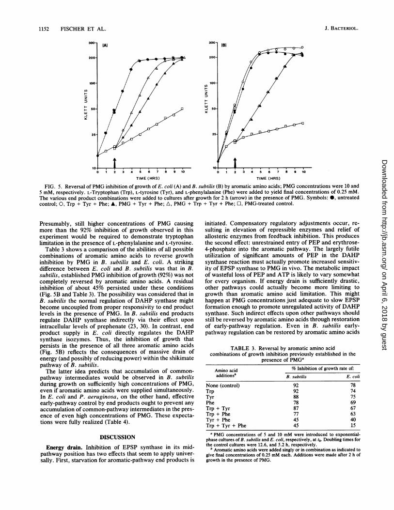
Reversal of PMG inhibition by aromatic amino acids. Thedata shown in Fig. 5 are consistent with the evidence citedabove for superior ability of the tryptophan pathway toscavenge limited intracellular chorismate in B. subtilis com-pared to E. coli. The inhibition of growth of E. coli estab-lished in the presence of PMG for 2 h was almost completelyreversed by the combination of all three aromatic aminoacids (Fig. SA). The addition of L-phenylalanine plus L-tyrosine, however, led to only partial reversal of growthinhibition (40% inhibition remaining), indicating thattryptophan synthesis must be rate limiting to growth.
In contrast, the combination of L-phenylalanine and L-tyrosine was as effective as all three aromatic amino acids inreversing PMG-inhibited growth of B. subtilis (Fig. 5B).
VOL. 168, 1986
on April 6, 2018 by guest
http://jb.asm.org/
Dow
nloaded from
1152 FISCHER ET AL.
C')
z
wJiye
3 4 T 6 7 8 9 10
TIME (HRS)
0 1 2 3 4 5 6 7 8 9 10
TIME (HRS)
FIG. 5. Reversal of PMG inhibition of growth of E. coli (A) and B. subtilis (B) by aromatic amino acids; PMG concentrations were 10 and5 mM, respectively. L-Tryptophan (Trp), L-tyrosine (Tyr), and L-phenylalanine (Phe) were added to yield final concentrations of 0.25 mM.The various end product combinations were added to cultures after growth for 2 h (arrow) in the presence of PMG. Symbols: *, untreatedcontrol; 0, Trp + Tyr + Phe; A, PMG + Tyr + Phe; A, PMG + Trp + Tyr + Phe; O, PMG-treated control.
Presumably, still higher concentrations of PMG causingmore than the 92% inhibition of growth observed in thisexperiment would be required to demonstrate tryptophanlimitation in the presence of L-phenylalanine and L-tyrosine.
Table 3 shows a comparison of the abilities of all possiblecombinations of aromatic amino acids to reverse growthinhibition by PMG in B. subtilis and E. coli. A strikingdifference between E. coli and B. subtilis was that in B.subtilis, established PMG inhibition of growth (92%) was notcompletely reversed by aromatic amino acids. A residualinhibition of about 45% persisted under these conditions(Fig. SB and Table 3). The possibility was considered that inB. subtilis the normal regulation of DAHP synthase mightbecome uncoupled from proper responsivity to end productlevels in the presence of PMG. In B. subtilis end productsregulate DAHP synthase indirectly via their effect uponintracellular levels of prephenate (23, 30). In contrast, endproduct supply in E. coli directly regulates the DAHPsynthase isozymes. Thus, the inhibition of growth thatpersists in the presence of all three aromatic amino acids(Fig. SB) reflects the consequences of massive drain ofenergy (and possibly of reducing power) within the shikimatepathway of B. subtilis.The latter idea predicts that accumulation of common-
pathway intermediates would be observed in B. subtilisduring growth on sufficiently high concentrations of PMG,even if aromatic amino acids were supplied simultaneously.In E. coli and P. aeruginosa, on the other hand, effectiveearly-pathway control by end products ought to prevent anyaccumulation of common-pathway intermediates in the pres-ence of even high concentrations of PMG. These expecta-tions were fully realized (Table 4).
DISCUSSION
Energy drain. Inhibition of EPSP synthase in its mid-pathway position has two effects that seem to apply univer-sally. First, starvation for aromatic-pathway end products is
initiated. Compensatory regulatory adjustments occur, re-
sulting in elevation of repressible enzymes and relief ofallosteric enzymes from feedback inhibition. This producesthe second effect: unrestrained entry of PEP and erythrose-4-phosphate into the aromatic pathway. The largely futileutilization of significant amounts of PEP in the DAHPsynthase reaction must actually promote increased sensitiv-ity of EPSP synthase to PMG in vivo. The metabolic impactof wasteful loss of PEP and ATP is likely to vary somewhatfor every organism. If energy drain is sufficiently drastic,other pathways could actually become more limiting togrowth than aromatic amino acid limitation. This mighthappen at PMG concentrations just adequate to slow EPSPformation enough to promote unregulated activity of DAHPsynthase. Such indirect effects upon other pathways shouldstill be reversed by aromatic amino acids through restorationof early-pathway regulation. Even in B. subtilis early-pathway regulation can be restored by aromatic amino acids
TABLE 3. Reversal by aromatic amino acidcombinations of growth inhibition previously established in the
presence of PMG'
Amino acid % Inhibition of growth rate of:additionsb B. subtilis E. coli
None (control) 92 78Trp 92 74Tyr 88 75Phe 78 69Trp + Tyr 87 67Trp + Phe 77 63Tyr + Phe 45 40Trp + Tyr + Phe 45 15
a PMG concentrations of 5 and 10 mM were introduced to exponential-phase cultures of B. subtilis and E. coli, respectively, at to. Doubling times forthe control cultures were 12.6, and 5.2 h, respectively.
b Aromatic amino acids were added singly or in combination as indicated togive final concentrations of 0.25 mM each. Additions were made after 2 h ofgrowth in the presence of PMG.
z
ww
J. BACTERIOL.
on April 6, 2018 by guest
http://jb.asm.org/
Dow
nloaded from
ENERGY DRAIN IN BACTERIA 1153
under conditions of minimal inhibition of EPSP synthase.This is because intracellular prephenate (feedback inhibitor)levels are dictated by the balance between slowed formationof prephenate in the presence of PMG and slowed entry ofprephenate into tyrosine and phenylalanine branches due tofeedback inhibition of prephenate dehydratase and prephen-ate dehydrogenase. However, if prephenate formation isseverely restricted (i.e., at high PMG concentrations), early-pathway regulation cannot be restored, even in the presence
of L-tyrosine and L-phenylalanine, and energy drain persists(Table 4).Dramatic differences in sensitivity to growth inhibition by
PMG were observed in correlation with nutritional variables.Thus, E. coli and B. subtilis were both similarly sensitive toPMG on glucose-based medium, whereas P. aeruginosa was
about 50-fold less sensitive on this medium. However, P.aeruginosa exhibited roughly the same sensitivities seen
with E. coli and B. subtilis when fructose was substituted forglucose as the carbon source. That alteration of sensitivity togrowth inhibition by PMG through manipulation of carbonand energy source is not an isolated phenomenon in P.aeruginosa is apparent by results showing that the PMGsensitivity of B. subtilis (and P. aeruginosa) can be markedlydecreased by supplying L-glutamate and, to a lesser extent,L-glutamine, L-proline, and L-arginine (R. Fischer, unpub-lished data). Although the initial molecular event underlyingPMG action is simple (i.e., inhibition of EPSP synthase), theresulting energy burden occasioned by massive investrmentof PEP and ATP in shikimate-3-phosphate accumulationmust initiate other metabolic vulnerabilities. The dramaticability of L-glutamate and L-glutamine to increase PMGresistance in B. subtilis and P. aeruginosa fits with a possibleconsequence of energy drain upon nitrogen assimilation.Each transformation of ammonia and 2-ketoglutarate in theglutamate synthase-glutamine synthetase system of L-glutamate formation requires one ATP in the glutaminesynthetase reaction.Incomplete inhibition of EPSP synthase by PMG could
indirectly create other points of vulnerability within thearomatic pathway. For example, if L-glutamate or L-glutamine became limiting as supposed above, then gluta-mate-requiring transamination reactions leading to L-phenylalanine and to L-tyrosine (or the glutamine-requiringanthranilate synthase reaction leading to L-tryptophan)might become limiting. These reactions could be especiallyvulnerable under conditions where the levels of keto-acidsubstrates (i.e., phenylpyruvate and 4-hydroxyphenylpyru-vate) have already become depressed in the presence ofPMG. Another possibility is that phosphoribosylpyrophos-phate shortage in response to energy drain could limit thesecond reaction in the tryptophan pathway. The enzymecatalyzing this step (anthranilate-phosphoribosylpyrophos-phate phosphoribosyltransferase) probably already encoun-
ters decreased cosubstrate (anthranilate) levels in the pres-
ence of PMG. Other biochemical pathways that could be-come stressed by energy drain include those for the biosyn-thesis of L-histidine, aspartate-derived amino acids, purines,and pyrimidines.
Metabolite reversal of growth inhibition by PMG. E. coli,P. aeruginosa, and any microbe having a regulatory patternwhere aromatic amino acid end products directly controlDAHP synthase have the following characteristics. Inhibi-tion of growth by PMG can be prevented indefinitely in thepresence of aromatic amino acids since they prevent any
possibility of energy drain. However, if PMG inhibition isfirst established in the absence of end products, energy drain
TABLE 4. Ability of aromatic amino acids to prevent PMG-promoted accumulation of aromatic-pathway metabolitesa
Intracellular accumulation Extracellular accumu-
(nmol/mg of protein)ofa lation (>.M concn insupernatant) of:
Species Shikimate Shikimate-DAHP Shikimate 3- Shikimate 3-
phosphate phosphate
E. coli 0 0 0 0 0P. aeruginosa 0 0 0 0 0B. subtilis 3.4 7.1 54.3 10.9 0
a The PMG concentrations used for E. coli (15 mM), P. aeruginosa (6 mM),and B. subtilis (7.5 mM) inhibited growth rates by 0, 6, and 14%t, respectively,in the presence of 0.25 mM concentrations of all three aromatic amino acids(only L-tyrosine plus L-phenylalanline for B. subtilis). Values of 0 are asdescribed in footnote a of Table 1.
results in only a partial ability of aromatic amino acids toreverse growth inhibition. Such residual inhibition not re-versed by aromatic amino acids is transient, however, andfull reversal becomes established after restoration of earlypathway control and relief of the energy burden.
In B. subtilis and other microbes (8) having a pattern ofindirect control over DAHP synthase (sequential feedbackinhibition), aromatic amino acids can only prevent growthinhibition by PMG for a short initial period that is succeededby progressively greater inhibition. At sufficiently high PMGconcentrations, aromatic amino acid supplementation canonly partially reverse PMG inhibition of growth, and residualinhibition endures indefinitely.
It is quite possible that supplementation with nonaromaticmetabolites that spare the need for energy-rich compoundswill prove to reverse residual inhibition of growth in thepresence of aromatic amino acids. If so, all systems mayprove to be fully reversible, given the appropriate nutritionalregimen.
B. subtilis as a model for plant systems. In higher plants achloroplast-localized DAtiP synthase exists that is feedbackinhibited by L-arogenate, thtis constituting another variationof sequential feedback inhibition (35; R. J. Ganson, T. A.d'Amato, and R. A. Jensen, Plant Physiol., in press). Theinhibition of plant cell culture systems by PMG is character-istically reversed orily partially by aromatic amino acids, andit has been hypothesized that energy drain within thechloroplast compartment plays a significant role in theherbicidal mechanism ofPMG action (21, 33). The analogouspattern of early pathway regulation in B. subtilis makes itideal as a model for examination of the impact of energydrain effects upon metabolic pathways of higher plants.The variety of prevention and reversal results obtained
with yeast (4), algae (18), plants (18), photosyntheticeucaryotes (10), and bacteria (2, 18, 32) suggests that thepatterns of reversal ofPMG inhibition observed in individualorganisms may indeed reflect the diversity of aromatic aminoacid biosynthesis in nature (8).PMG as a physiological probe. PMG can be used as a tool
to manipulate wild-type cultures for derepression. For ex-ample, tryptophan-pathway enzymes can be derepressed inE. coli by growth of PMG-treated cultures in the presence ofL-phenylalanine plus L-tyrosine. In P. aeruginosa growth inthe presence of 100 mM PMG in glucose-based mediumresults in limitation of the tryptophan pathway. In contrast,the tryptophan-pathway enzymes in B. subtilis actuallybecome repressed during partial inhibition of growth in thepresence of PMG. The latter result indicates that underconditions of precursor limitation, anthranilate synthase of
VOL. 168, 1986
on April 6, 2018 by guest
http://jb.asm.org/
Dow
nloaded from
1154 FISCHER ET AL.
B. subtilis is able to outcompete chorismate mutase for theavailable substrate molecules. E. coli, by comparison, mustundergo a modest derepression of anthranilate synthase tomaintain adequate levels Of L-tryptophan.
If the energy-drain proposal is correct, then the order ofnutritional requirements for small molecules imposed byPMG treatment may provide a useful approach to gaininginsight into relationships between connecting networks ofbiochemical pathways (25, 27).
LITERATURE CITED
1. Ahmad, S., B. Rightmire, and R. A. Jensen. 1986. Evolution ofthe regulatory isozymes of 3-deoxy-D-arabino-heptulosonate7-phosphate synthase present in the Escherichia coli genealogy.J. Bacteriol. 165:146-154.
2. Amrhein, N., D. Johanning, J. Schab, and A. Schulz. 1983.Biochemical basis for glyphosate-tolerance in a bacterium andplant tissue culture. FEBS Lett. 157:191-196.
3. Bachmann, B. J. 1972. Pedigrees of some mutant strains ofEscherichia coli K-12. Bacteriol. Rev. 36:525-557.
4. Bode, R., C. Melo, and D. Birnbaum. 1984. Mode of action ofglyphosate in Candida maltosa. Arch. Microbiol. 140:83-85.
5. Bradford, M. M. 1976. A rapid and sensitive method for thequantitation of microgram quantities of protein utilizing theprinciple of protein-dye binding. Anal. Biochem. 72:248-254.
6. Brown, K. D., and C. H. Doy. 1963. End-product regulation ofthe general aromatic pathway in Escherichia coli. Biochim.Biophys. Acta 77:170-172.
7. Byng, G. S., and R. A. Jensen. 1983. Impact of isozymes upon
partitioning of carbon flow and regulation of aromatic biosyn-thesis in prokaryotes, p. 115-140. In M. C. Ratazzi, J. G.Scandalios, and G. S. Whitt (ed.), Isozymes, vol. 8. Alan R.Liss, Inc., New York.
8. Byng, G. S., J. F. Kane, and R. A. Jensen. 1982. Diversity in therouting and regulation of complex biochemical pathways as
indicators of microbial relatedness. Crit. Rev. Microbiol.9:227-252.
9. Byng, G. S, R. J. Whitaker, R. L. Gherna, and R. A. Jensen.1980. Variable enzymological patterning in tyrosine biosynthe-sis as a means of determining natural relatedness among thePseudomonadaceae. J. Bacteriol. 144:247-257.
10. Byng, G. S., R. J. Whitaker, and R. A. Jensen. 1985. Glyphosateas a probe of the pentafunctional arom protein of Euglenagracilis. Can. J. Bot. 63:1021-1024.
11. Calhoun, D. H., D. L. Pierson, and R. A. Jensen. 1973. Theregulation of tryptophan biosynthesis in Pseudomonas aerugi-nosa. Mol. Gen. Genet. 121:117-132.
12. Champney, W. S., and R. A. Jensen. 1970. The enzymology ofprephenate dehydrogenase in Bacillus subtilis. J. Biol. Chem.245:3763-3770.
13. Clowes, R. C., and W. Hayes. 1968. Experiments in microbialgenetics, p. 223-228. John Wiley and Sons, Inc., New York.
14. Dayan, J., and D. B. Sprinson. 1970. Preparation of prephenicacid. Methods Enzymol. 17A:559-561.
15. Duncan, K., A. Lewendon, and J. R. Coggins. 1984. Thepurification of 5-enolpyruvylshikimate 3-phosphate synthasefrom an overproducing strain of Escherichia coli. FEBS Lett.165:121-127.
16. Gibson, F. 1964. Chorismic acid: purification and some chemicaland physical studies. Biochem. J. 90:256-257.
17. Gollub, E., H. Zalkin, and D. 13. Sprinson. 1967. Correlation ofgenes and enzymes and studies on regulation of the aromaticpathway in Salmonella. J. Biol. Chem. 242:5323-5328.
18. Gresshoff, P. M. 1979. Growth inhibition by glyphosate andreversal of its action by phenylalanine and tyrosine. Aust. J.Plant Physiol. 6:177-185.
19. Holloway, B. W. 1955. Genetic recombination in Pseudomonasaeruginosa. J. Gen. Microbiol. 13:572-581.
20. Jensen, R. A. 1985. Biochemical pathways in prokaryotes can betraced backward through evolutionary time. Mol. Biol. Evol.2:92-108.
21. Jensen, R. A. 1986. The shikimate/arogenate pathway: linkbetween carbohydrate metabolism and secondary metabolism.Physiol. Plant. 66:164-168.
22. Jensen 1R. A., and D. S. Nasser. 1968. Comparative regulation ofisoenzymic 3-deoxy-D-arabino-heptulosonate 7-phosphatesynthetases in mricroorganisms. J. Bacteriol. 95:188-196.
23. Jensen, R. A., and E. W. Nester. 1965. The regulatory signifi-cance of intermediary metabolites: control of aromatic acidbiosynthesis by feedback inhibition in Bacillus subtilis. J. Mol.Biol. 12:468-481.
24. Jensen, R. A., and E. W. Nester. 1966. Regulatory enzymes ofaromatic amino acid biosynthesis in Bacillus subtilis. I. Purifi-cation and properties of DAHP synthetase. J. Biol. Chem.241:3365-3372.
25. Kane, J. F., W. M. Holmes, and R. A. Jensen. 1972. Metabolicinterlock: the dual function of a folate pathway gene as anextra-operonic gene of tryptophan biosynthesis. J. Biol. Chem.247:1587-1596.
26. Kane, J. F., and R. A. Jensen. 1970. Metabolic interlock: theinfluence of histidine on tryptophan biosynthesis in Bacillussubtilis. J. Biol. Chem. 245:2384-2390.
27. Kane, J. F., S. L. Stenmark, D. H. Calhoun, and R. A. Jensen.1971. Metabolic interlock: the role of the subordinate type ofenzyme in the regulation of a complex pathway. J. Biol. Chem.246:4308-4316.
28. Knowles, P. F., and D. B. Sprinson. 1970. Preparation ofshikimate-5-phosphate. Methods Enzymol. 1A:351-352.
29. Miller, J. H. 1972. Experiments in molecular genetics, p.431-435. Cold Spring Harbor Laboratory, Cold Spring Harbor,N.Y.
30. Nester, E. W., and R. A. Jensen. 1966. Control of aromatic acidbiosynthesis in Bacillus subtilis: sequential feedback inhibition.J. Bacteriol. 91:1594-1598.
31. Nester, E. W., R. A. Jensen, and D. S. Nasser. 1969. Regulationof enzyme synthesis in the aromatic amino acid pathway ofBacillus subtilis. J. Bacteriol. 97:83-90.
32. Roisch, U., and F. Liilgens. 1980. The mechanism of action ofthe herbicide N-(phosphonomethyl)glycine: its effect on thegrowth and the enzymes of aromatic amino acid biosynthesis inEscherichia coli. Hoppe-Seyler's Z. Physiol. Chem. 361:1049-1058.
33. Rubin, J. L., C. G. Gaines, and R. A. Jensen. 1982. Enzymo-logical basis for herbicidal action of glyphosate. Plant Physiol.70:833-839.
34. Rubin, J. L., C. G. Gaines, and R. A. Jensen. 1984. Glyphosateinhibition of 5-enolpyruvylshikimate 3-phosphate synthase fromsuspension-cultured cells of Nicotiana silvestris. Plant Physiol.75:839-845.
35. Rubin, J. L., and R. A. Jensen. 1985. Differentially regulatedisozymes of 3-deoxy-D-arabino-heptulosonate 7-phosphatesynthase from seedlings of Vigna radiata [L.] Wilczek. PlantPhysiol. 79:711-718.
36. Spizizen, J. 1958. Transformation of biochemically deficientstrains of B. subtilis by deoxyribonucleate. Proc. Natl. Acad.Sci. USA 44:1072-1078.
37. Srinivasan, P. R., and D. B. Sprinson. 1959. 2-Keto-3-deoxy-D-arabo-heptonic acid 7-phosphate synthetase. J. Biol. Chem.234:716-722.
38. Tribe, D. E., H. Camakaris, and J. Pittard. 1976. Constitutivearid repressible enzymes of the common pathway of aromaticbiosynthesis in Escherichia coli K-12: regulation of enzymesynthesis at different growth rates. J. Bacteriol. 127:1085-1097.
39. Vhitaker, R. J., M. J. Fiske, and R. A. Jensen. 1982. Pseudomo-nas aeruginosa possesses two novel regulatory isozymes of3-deoxy-D-arabino-heptulosonate 7-phosphate synthase. J.Biol. Chem. 257:12789-12794.
J. BACTERIOL.
on April 6, 2018 by guest
http://jb.asm.org/
Dow
nloaded from