dr. vibha hegde - 18
TRANSCRIPT
71People’s Journal of Scientific Research Vol. 4(1), Jan. 2011
Pediatric Endodontics- Endodontist’s viewVibha Hegde
Department of Pediatric Endodontics, Yerala Medical Trust Dental College & Hospital, Kharghar, Navi-Mumbai-410210
Abstract:The dental diseases affecting the pulp and periapical tissues in the primary and permanent dentitions pose
treatment challenges for the endodontists because of the vast variations in these dentitions basically due to factors likelongetivity of primary teeth , coronal structure and root canal morphology and anatomy of the teeth which needs to becritically analysed before rendering treatment. In recent years, new materials, equipments and instruments have evolvedto a great extent and simplified the endodontic treatment procedures for the clinicians.
The aim of this article is to highlight the clinical techniques and treatment considerations in treating the vital andnon-vital as well as emphasize on surgical management of cases.
Key Words: Pediatric Endodontics, Partial Pulpectomy, pulpotomy.
Informative Article
Table I: Anatomic differences between primary and permanent teeth and its significance from pediatric endodontics point of view.
----------------------------------------------------------------------------Corresponding Author: Dr. Vibha Hegde, Senior Professor &Post-Graduate Teacher, 1101/B, Rajkamal Height, Rajkaml Lane,Parel, Mumbai-400012Phone No .: +91 9820765066E-mail : [email protected]
Introduction: A number of factors are involved in the
development of pulp and periapical disease in primaryand permanent teeth, with dental caries being the mainfactor. Although these factors are similar, the clinicalmanagement of a primary or permanent tooth with pulpor periapical disease may be quite different. This isbased mainly on the differences between the two typesof teeth, with primary tooth longevity, coronal structuralintegrity, root canal morphology, and root anatomy(Hibbard & Ireland, 1957) being important features tobe taken into account when planning the treatment(Table I & II).
The diagnosis of pulp disease is especiallydifficult in young patients because they are usuallyunable to give an accurate account of their symptoms.The diagnosis is dependent on the combination of agood history, clinical and radiological examination andspecial tests. According to Camp (2008), primary teethwith history of spontaneous pain should not receivevital pulpal treatment and are candidate forpulpectomy or extraction. Electric pulp tests andthermal tests are not reliable on primary tooth. Dopplerflowmetry might be of great help in determining vitality(Evans et al, 1999). Interpretation of radiographs ofprimary teeth is always complicated by the presence
of the succedaneous tooth and surrounding follicle,resulting into misdiagnosis.
The treatment of primary and permanent teethhas changed dramatically in recent years as new
Tooth Anatomic Primary teeth Permanent teeth Significance Features
Overall size Smaller larger ---
Pulp chamber Larger as compared Smaller as compared. Ease of access openingto crown to crown
Cervical constriction Marked constriction Less constricted in Lateral perforationcervical region
Root trunk Short with thin floor Large with thicker floor Easy furcation involvement.of pulp chamber of pulp chamber
Root anatomy Thin, slender (ribbon shaped), Thicker, not flared Limitation in canal enlargement,flared Instrument breakage, Perforation
Accessory canals Present frequently in furcation Comparatively less Incomplete pulp extirpationarea and roots in number

72People’s Journal of Scientific Research Vol. 4(1), Jan. 2011
Table II: Histological differences between primary and permanent teeth and its significance
materials have been developed. Therapy for childrenhas a high rate of success with less post-operativediscomfort. Because of the formative state of the pulp,vital procedures heal nicely with good dentin bridging.On the other hand, internal resorption commonly occursfrom pulpal inflammation in a primary tooth.
Vital Pulp Therapy:Indirect pulp capping:-
Indirect pulp treatment is recommended as themost appropriate procedure for treating primary teethwith deep caries and reversible pulp inflammationprovided that the tooth has been sealed with a leakagefree restoration (Fuks, 2002). There is insufficientevidence to support the use of any one specific liningmaterial for indirect pulp treatment. However, newerresearch appears to be directed towards the use ofglass ionomer cements (Massara et al, 2002). Pulpcapping with resin composites in monkeys producedthe lowest incidence of bacterial microleakage, pulpalinflammation and incidence of pulpal necrosis whencompared with calcium hydroxide and glass ionomercement (Cox & Suzuki, 1994).
Direct pulp capping-Direct pulp capping of a carious pulp exposure
in a primary tooth is not recommended as treatmentfailure might result in internal resorption or acutedentoalveolar abscess. In case of inadvertently exposedpulp, free of oral contamination, calcium hydroxide
medicament can be used as it maintains the pulp vitality(Fuks, 2005). Presently, direct pulp capping should stillbe looked on with some reservations in primary teeth.Caicedo et al (2006) demonstrated good pulp responsein primary teeth after direct pulp capping or pulpotomywith MTA (Mineral Trioxide Aggregate) and concludedthat MTA might be a favourable material for pulpcapping and pulpotomy in primary teeth.
Pulpotomy:Pulpotomy and partial pulpectomy techniques
for devitalized primary teeth have been developed topreclude an almost impossible obturation problem.Pulpotomy is still the most common treatment forcariously exposed pulp in symptom free primary molars.Formocresol has been a popular pulpotomy medicamentin the primary dentition for the past 70 years since it isintroduction by Sweet in 1932 (Vij et al, 2004).Nevertheless several studies have reported that theclinical success of FC pulpotomies decreases with time,and the histologic response of the primary pulp is“capricious” ranging from chronic inflammation tonecrosis (Rolling & Thylstrup, 1975). Presently, thereare several pulp dressing medicaments that have beenproposed that are equal to if not better than, Formocresoland can be used as alternatives to pulpotomies inprimary teeth. These include: electrosurgery (Fishmanet al, 1996), laser (Elliot et al, 1999), glutaraldehyde(Araujo et al, 1995), calcium hydroxide (Huth et al,2005), freeze dried bone (Fadavi & Anderson, 1996),
Pediatric Endodontics- Endodontist’s view ------------------------------------------ V. Hegde
Tooth Anatomic Primary teeth Permanent teeth SignificanceFeatures
Apical foramen Enlarged Constricted Abundant blood supply - exaggeratedinflammatory response
Pulp function Formative, Nerve supply, Formative, Nerve supply, Degeneration of neural elements-lessNutritive, protective, and Nutritive, and protective sensitive to operative proceduresResorptive
Cellular response to injury More extensive Lesser Incidence of reparative dentin formationis more
Localization of infection Poorer Better More chances of spread of infection –and inflammation space involvement (Cellulitis)Density of innervation Less More Less sensitive to operative procedures
Pulp nerve fibres End at the odontoblastic Terminate among the Less sensitive to painlayer odontoblasts and even
beyond the predentin
Inflammatory response to Severe Not demonstrated Subsequent metaplasia with resultantCalcium hydroxide internal primary root resorption

73People’s Journal of Scientific Research Vol. 4(1), Jan. 2011
bone-morphogenic protein (Nakashima, 1994),osteogenic protein (Rutherford et al, 1993), ferric sulfate(Ibricevic & al-Jame, 2000), mineral trioxide aggregate(MTA) (Fuks, 2008) and sodium hypochlorite (Vargaset al, 2006).
Non-Vital Pulptherapy:Pulpectomy-Non-vital primary teeth may be retained successfullywhen pulpectomy procedure is employed. A single visitor two- visit pulpectomy may be undertaken. Primarymolar roots are severely curved and the pulps are flatand tortuous with numerous branches andinterconnections. This necessitates modifications inbiomechanical procedures. The root canals are cleanedand shaped and subsequently filled with resorbablepaste (Zinc oxide eugenol, or calcium hydroxide oriodoform base). Recently investigators have found thatVitapex (a mixture of calcium hydroxide and iodoform)has superior success rate to that of traditionally usedzinc oxide eugenol (100% versus 78.5% at 16 months)and is removed more readily if extruded through anapex (Mortazavi & Mesbahi, 2004).
Treatment Modalities for Young PermanentTeeth:Apexification and apexogenesis-
When providing treatment for patients withmixed and young permanent dentitions, certain clinicalscenarios may require interdisciplinary consultation andintervention such as following traumatic injuries andwhenever permanent teeth require endodontictherapy.Young pulps in immature permanent teeth arelarger than at a more mature stage. Immaturepermanent teeth have funnel shaped apical foraminawhich are commonly called “blunderbuss”. Walls ofroot canal are very thin in newly erupted immatureteeth which are further weakened during the cleaning
Fig. I: A Case of Non- vital 45 with open apices.
and shaping procedures (Fig. I). The relatively thindentin walls of the large obturated canals place thetooth at greater risk for root fracture over time. In theseinstances, the treatment objective is to maximize theopportunity for apical development and closure, knownas apexogenesis or apexification, and enhancecontinued root dentin formation. Figure 1 shows a caseof non-vital 45 with open apices of an old female childwho reported to clinics with a complaint of decayedteeth. In the following case root canal was cleanedand calcium hydroxide was placed within 1-2mm ofroot apex to encourage either root growth or apicalrepair (Fig. II). Recent studies suggest 3 monthlychange of Calcium hydroxide (Mackie, 1998). HenceCalcium hydroxide dressing was changed every 3months and radiographic follow up was done at 1,3,6,9months. After the barrier was evident radiographically(Fig. III) and clinically, the tooth was reisolated andopened for final obturation of canal with gutta-percha(Fig. IV).
Fig. II: Apexification with Calcium hydroxide.
Fig.III: Radiographically and clinically evident calcific barrier.
The current data available on the use of MTAin vital pulp therapy indicate that it is the optimummaterial and better than the traditionally used materialcalcium hydroxide. It has a better long term sealingability and stimulates a high quality and a greateramount of reparative dentin and has also demonstrateda high success rate (Witherspoon, 2008).
Pediatric Endodontics- Endodontist’s view ------------------------------------------ V. Hegde
74People’s Journal of Scientific Research Vol. 4(1), Jan. 2011
If attempts to induce root end closure areunsuccessful with persistent sinus tract, it calls forsurgical intervention. With mutual consent of parentsthe procedure can be carried out under sedation orgeneral anesthesia. Root canal filling procedure iscompleted prior to surgical opening and removal ofapical filling, followed by root end closure with MTA.
Recent Advances:Pulp revascularization and Stem cell research
holds great hope for the future and can be consideredas novel treatment modalities for the management ofprimary and young permanent teeth (Banchs & Trope,2004).
Conclusion:A successful pediatric endodontic outcome
should be based on (1) re-establishment of healthyperiodontal tissues; (2) freedom from pathologic rootresorption; (3) maintenance of the primary tooth in aninfection-free state to hold space for the eruption of itspermanent successor; (4) in the case of youngpermanent teeth, maintenance of the maximum amountof noninflamed portions of pulp tissue to enhanceapexogenesis and root dentin formation. Withadherence to sound principles in case selection andtechniques, pediatric pulp therapy is a major healthbenefit to the child. The treatment modalities andmedicaments that have been discussed, highlighting themost substantiated and qualifying those that needsfurther confirmation by additional research. Theclinician must realize that these recommendations arenot absolute and will continue to be modified.
Bibliography:
1. Araujo FB, Ely LB, Pergo AM, Pesce HF: A clinicalevaluation of 2% buffered glutaraldehyde inpulpotomies of human deciduous teeth: A 24- month
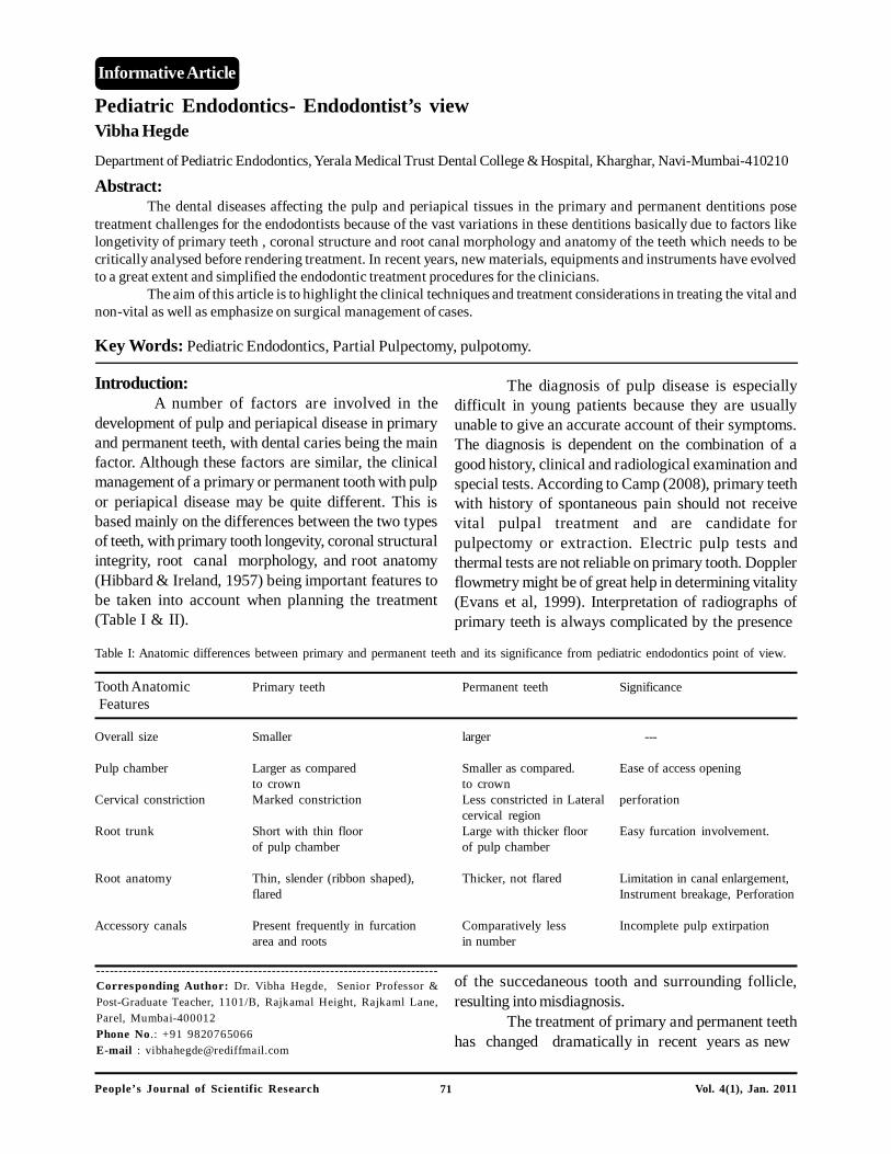
Fig. IV: Post Operative radiograph (12 months follow up).
study. Brazilian Dental Journal, 1995;6(1):41-44.2. Banchs F, Trope M: Revascularisation of immature
permanent teeth with apical periodontitis: newtechnique protocol? Journal of Endodontic, 2004;30(4):196-200.
3. Cox CF, Suzuki S: Re-evaluating pulp protection:calcium hydroxide liners vs cohesive hybridization.Journal of American Dental Association, 1994;125(7):823-831.
4. Camp JH: Diagnosis dilemmas in vital pulp therapy:treatment for the toothache is changing especially inyoung immature teeth. Pediatric Dentistry, 2008; 30(3):197-205.
5. Caicedo R, Abbott PV, Alongi DJ, Alarcon MY: Clinical,radigraphical and histological analysis of the effectsof mineral trioxide aggregate used in direct pulp cappingand pulpotomies in primary teeth. Australian DentalJournal, 2006;51(4):297-305.
6. Elliot RD, Roberts MW, Burkes J, Phillips C: Evaluationof carbon dioxide laser on vital human primary pulptissue. Pediatric Dentistry, 1999;21(6):327-331.
7. Evans D, Reid J, Strang R, Stirrups D: A comparison ofLaser Doppler flowmetry with other methods ofassessing the vitality of traumatized anterior teeth.Endodontics & Dental Traumatology, 1999; 15(6): 284-290.
8. Fadavi S, Anderson AW: A comparison of the pulpalresponse to freeze-dried bone, calcium hydroxide, andzinc-oxide-eugenol in primary teeth in two cynomolgusmonkeys. Pediatric Dentistry, 1996;18(1):52-56.
9. Fishman SA, Udin RD, Good DL, Rodef F: Success ofelectrofulguration pulpotomies covered by zinc oxideeugenol or calcium hydroxide: A clinical study.Pediatric Dentistry, 1996;18(5):385-390.
10. Fuks AB: Current concepts in vital primary pulp therapy.Europian Journal of Pediatrics Dentistry, 2002;3(3):115-120.
11. Fuks AB: Pulp therapy for the primary dentition. In:Pediatric Dentistry: infancy through adolescence. JRPinkham, PS Casamassimo, HW Fields (Jr.), DGMcTigue, AJ Nawak. (Eds.); 4thEdn.; Elsevier: A divisionof Reed Elsevier India, New Delhi, 2005:pp375-393.
12. Fuks AB: Vital pulp therapy with newer materials forprimary teeth: New directions and treatmentperspectives. Journal of Endodontics, 2008;34(7 suppl):S18-S24.
13. Hibbard ED, Ireland RL: Morphology of the root canalsof primary molar teeth. Journal of Dentistry forChildren, 1957; 24: 250-257.
14. Huth KC, Paschos E, Hajek-Al-Khatar N, Hollweck R,Crispin A, Hickel R, Folwaczny M: Effectiveness of 4pulpotomy techniques: Randomized controlled trial.Journal of Dental Research, 2005;84(12):1144-1148.
Pediatric Endodontics- Endodontist’s view ------------------------------------------ V. Hegde
75People’s Journal of Scientific Research Vol. 4(1), Jan. 2011
15. Ibricevic H, al-Jame Q: Ferric Sulphate as pulpotomyagent in primary teeth: twenty- month clinical followup. Journal of Clinical Pediatric Dentistry ,2000;24:269-272.
16. Mackie IC: (UK National Clinical Guidelines in PediatricDentistry) Management and Root canal treatment ofnon-vital immature permanent incisor teethInternational Journal of Pediatric Dentistry,1998;8:289-293
17. Massara MLA, Alves JB, Brandao PRG: Atraumaticrestorative treatment: clinical, ultrastructural andchemical analysis. Caries Research, 2002; 36(6): 430-436.
18. Mortazavi M, Mesbahi M: Comparison of Zinc oxideeugenol and vitapex for root canal treatment of necroticprimary teeth. International Journal of PediatricDentistry, 2004;14(6):417-424.
19. Nakashima M: Induction of dentine formation on canineamputated pulp by recombinant human bone morpho-genetic proteins (BMP) 2 & 4. Journal of DentalResearch, 1994;73(9): 1515-1522.
20. Rolling I, Thylstrup A: A 3 year follow up study ofpulpotomised primary molars treated with theFormocresol technique. European Journal of OralSciences, 1975;83(2):47-53.
21. Rutherford RB, Wahle J, Tucker M, Rueger D, CharetteM: Induction of reparative dentine formation in monkeysby recombinant human osteogenetic protein-1. Archivesof Oral Biology, 1993;38(7):571-576.
22. Vij R, Coll JA, Shetlron P, Farooq NS: Caries control andother variables associated with success of primary molarvital pulp therapy. Pediatric Dentistry 2004;26(3):214-220.
23. Vargas KG, Packham B, Lowman D: Preliminaryevaluation of sodium hypochlorite for pulpotomies inprimary molars. Pediatric Dentistry, 2006; 28(6):511-517
24. Witherspoon DE: Vital pulp therapy with newermaterials: new directions and treatment perspective –Permanent teeth. Pedeatrics Dentistry, 2008;30(3):220-224.
Pediatric Endodontics- Endodontist’s view ------------------------------------------ V. Hegde